


Abstract
As chronic lead (Pb) exposure is a serious threat to public health in many countries, we intended to provide a review of the effective antidote for Pb intoxication. In this context, we explored the recent literature focusing on animal studies that provide clear evidence of the efficacy of supplementation with certain antioxidants, such as spirulina, curcumin, garlic, vitamin C, and vitamin E, for different periods of time, in the conditions of Pb exposure, drawing attention to the mechanisms of their beneficial action. Data collected clearly indicate that spirulina, curcumin, garlic, vitamin C, and vitamin E may play an important role in alleviating some of the toxic effects caused by Pb. Their use may represent an interesting direction in the development of new therapies to combat the toxicity of this toxic, non-essential metal. However, it seems essential to identify a safe dose range for these antioxidants in order to derive benefits from their properties during Pb intoxication. Clinical trials seem to be necessary to recognize the long-term effects of supplementation with these antioxidants and to establish the minimum effective dose with minimal side effects. Future studies are also required to elucidate the molecular mechanisms of action of spirulina, curcumin, garlic, vitamin C, and vitamin E against Pb toxicity and the mechanisms of their interactions with chelating agents to enhance potential beneficial effects.
1. Introduction
1.1. Lead—Background
Lead (chemical symbol Pb) is a soft and malleable metal with an atomic number of 82. Freshly cut, it is whitish-blue, and tarnishes to a dull gray in the air. It is located in period 6 and group 14 of the periodic table. Lead’s high density, low melting point, conductivity, and relatively low susceptibility to oxidation account for its usefulness. Considering its non-biodegradable nature and continuous use, its concentration and accumulation in the environment have been an ongoing issue for years, until now. This condition is caused by continuous use of Pb in many industries [1], as listed further in this article. Depending on the level of country development, Pb sources and Pb use in general vary from dietary exposure (food, soil, drinking water, contaminated spices included) [2,3,4,5,6], industrial fumes, improper battery recycling [7] in low and middle-income countries to gasoline, pipes, and paints in highly developed regions of the world [8].
Although the incidence of Pb poisoning has declined in recent years, especially in high-income countries, the problem still persists in some regions of low- and middle-income countries. It may be associated with the introduction of more efficient and precise regulations—for example, in the U.S., Pb-free paints and Pb-free gasoline were introduced by the 1980s [9]. For instance, paints containing this metal are still in use in Nigeria and other developing countries [10]. According to Zhou et al. [11], China, India, and Bangladesh were the top three countries with the largest number of deaths and DALYs (disability-adjusted life years) due to Pb exposure in 2019. In 2021, the Centers for Disease Control and Prevention changed the blood Pb reference value in children to 3.5 μg/dL, whereas in the 1960s, the same organization proposed 60 μg/dL as the threshold [12].
1.2. Absorption, Distribution, and Excretion of Pb—The Most Important Aspects
Pb enters the human body in a few ways, i.e., through the gastrointestinal tract, respiratory system, and skin [13,14]. Absorption of this metal primarily occurs in the duodenum and depends on a variety of factors, including the particle size, route of exposure, nutritional status, health, and age of individuals [14]. Pb, which utilizes channels or transporters of other elements such as calcium (Ca), iron (Fe), and zinc (Zn) [15], is absorbed into the body, with children generally absorbing a higher percentage than adults [14]. Children can absorb 40–50% of an oral dose of water-soluble Pb compared to 3–10% for adults [14]. The average intake of Pb by adults is estimated to be between 320 and 440 µg/day in different countries. The estimated amounts of Pb intake with food in Poland are in the range of 50–520 µg/day for adults and 40–350 µg/day for children aged 1–3 years [16]. Inhalation represents a significant exposure pathway, particularly for workers in Pb industries. Approximately 30–50% of inhaled Pb is deposited in the lungs [14], leading to gradual destruction of this tissue. From pathologies such as oxidative stress generating inflammation, increased levels of tumor necrosis factor alpha (TNF-α), nuclear factor kappa-light-chain-enhancer of activated B cells (NF-κB), and interleukin 1 (IL-1) [17] observed at the cellular level to incidence of asthma, allergies [18], and lung cancer [19].
After absorption, Pb accumulates in soft tissues such as liver, kidney, heart, spleen, brain, lung, and muscles, and mineralized tissues, including bone and teeth, where it may be stored in the form of complexes with phosphate for a long period [14,20]. Approximately 95% of Pb accumulates in mineralized tissues [13]. The remaining 5% is stored in soft tissues [21]. In the blood, approximately 94–99% of Pb is incorporated into red blood cells (RBC), while the remaining 1–6% resides in the plasma [14,20,21,22]. In RBC, Pb binds to several intracellular proteins, mainly hemoglobin (Hb) and delta-aminolevulinic acid dehydratase (ALAD) [14,20]. In the plasma, 40–75% of Pb is protein-bound, of which 90% binds to albumin [14,20,23], one of the most abundant proteins in the human circulation, responsible for transport functions [24]. In the plasma, Pb may also interact with other proteins, e.g., transferrin (Tf), gamma-globulin, and metalloprotein (MT) [14,25]. Moreover, Pb may bind to citrate, cysteamine, glutathione, histidine, metalloproteins, and oxylate in the plasma [14]. It may also occur in the free ionized (Pb2+) form, although Pb2+ represents a small percentage of total Pb in this compartment. Notably, no level of Pb is considered completely safe, even at its low concentrations. In adults, a blood Pb level of 5 µg/dL (0.24 µmol/L) or above is considered elevated, including adults in the workplace [26] and pregnant women [27]; however, it should be considered that during gestation, there is no safe level of Pb exposure. In children, a blood Pb level of 3.5 µg/dL (0.17 µmol/L) or greater requires monitoring [28]. Blood Pb levels of 10 µg/dL (0.48 µmol/L) or higher are considered toxic [29].
Regarding the excretion of Pb, absorbed Pb is filtered through the glomeruli and can accumulate in the renal tubular epithelium [30]. It is primarily excreted in the urine (~76%) [31], while fecal excretion accounts for approximately one-third of total excretion of absorbed Pb (~33%) [14,31]. Small amounts of this metal are excreted through sweat (~8%) [14,31]. A summary of Pb exposure routes, distribution, and excretion is provided in Figure 1.
Figure 1.
Schematic diagram summarizing absorption, distribution, and excretion of Pb—based on the available literature cited in Section 1.2. ALB: albumin; ALAD: delta-aminolevulinic acid dehydratase; Hb: hemoglobin; MT: metalloprotein; RBC: red blood cells; Tf: transferrin.
1.3. Environmental Exposure to Pb—A Brief Outline
Although some industrial sources of Pb have been eliminated, excessive environmental exposure to this toxic metal continues to be a public health problem. Many inhabitants of modern cities are chronically exposed to environmental Pb levels. In the general population, which is not occupationally exposed to this xenobiotic, food and beverages are the major sources of exposure [31]. Additionally, airborne Pb particles from burning Pb-containing materials may be inhaled by individuals and markedly raise their blood Pb levels, consequently leading to poisoning with this toxic metal. Depending on the level of exposure, the time required to remove Pb from the body varies. The greater the exposure, the longer the time needed for Pb to leave the body and for health improvement to occur. The amount of time necessary for health improvement may range from months to years, depending on each specific case and on the level of previous Pb exposure [32].
The development of industrialization has had an impact on constant Pb pollution. The standards of this metal in the air changed over time and differ depending on ambient outdoor air or workplace exposure. The latter are much higher as they apply to occupational settings. In 1978, OSHA (Occupational Safety and Health Administration) workplace standard lowered the permissible exposure limit (PEL) from 200 µg/m3 to 50 µg/m3 [33]. Historically established Pb limits for ambient outdoor air ranged from 1.5 µg/m3 in 1978, and were then reduced to 0.15 µg/m3 in 2008 [34]. The current threshold for this element in the air—0.15 µg/m3—was set by the U.S. EPA (U.S. Environmental Protection Agency) in 2016, averaged over a 3-month period [35]. In comparison, in the EU, the actual limit value is 0.5 µg/m3, averaging over a 1-year period [36]. According to Patocka and Cerny [37], in industrial regions, the amount of this metal in the atmosphere ranges from 0.2 to 5.0 μg/m3. Air, water, and soil are mainly polluted by industrial emissions from mining, fossil fuel burning, and manufacturing. Furthermore, Pb can still be found in human households, especially in wall paints, toys, cosmetics, and pipes [31,38,39]. The most important environmental sources of exposure to Pb involve industrial activities, factory chimneys, vehicle emissions, waste disposal, mining and processing of Pb ores, fertilizers and pesticides, Pb-based products, renovation work, contaminated food and water, and tobacco smoking [1,40,41]. The latter is a significant public health concern due to its association with various health risks [42]. It has been reported that a single cigarette contains 0.6–2.0 µg of Pb [43]. Environmental Pb sources are summarized in Figure 2.
Figure 2.
Environmental exposure to Pb—based on the available literature cited in Section 1.3.
It should be highlighted that children are especially susceptible to Pb exposure, even to its small doses through ingestion, particularly during the first two years of life, when they develop mouthing behaviors and increased mobility. Children’s tissues are also more prone to greater accumulation and absorption of this metal (especially bones and blood). Iron deficiency, which is a common condition in childhood, may even promote Pb absorption [44]. Blood Pb concentrations in children living in Pb-contaminated environments typically increase rapidly between 6 and 12 months of age, peak between 18 and 36 months of age, and then gradually decrease [45]. Based on current knowledge, the WHO has targeted blood Pb concentrations of 5 μg/dL or less for children; however, adverse effects of Pb exposure may occur at levels below this threshold [46]. In 2012, the US National Toxicology Program of the National Institutes of Health reported that, after accounting for other risk factors, blood Pb concentrations < 5 µg/dL (<50 ppb) are strongly linked to intellectual deficits, diminished academic abilities, attention deficits, and behavioral problems (ADHD, irritability, antisocial behaviors) [47]. Concentrations of <10 µg/dL have been proven to cause delayed puberty, impaired postnatal growth, and decreased hearing. As reported by some authors, a higher incidence of delinquency and aggression was found in adolescents with higher bone Pb concentrations [48].
Pb toxicity still remains one of the most frequently studied topics in environmental health. Environmental pollution with this metal and consequent health effects, particularly in children, are still a global problem. Many authors observed unfavorable changes in hematological and biochemical parameters in children with elevated blood Pb (PbB) levels. For example, Li et al. [49] revealed that PbB levels are associated with decreased erythrocyte (RBC) count, hemoglobin (Hb) level, mean corpuscular hemoglobin (MCH), and platelet (PLT) count in children aged 5–8 years in China. They also found that children with PbB ≥ 100 µg/L were 2.72, 2.51, and 3.76 times more likely to present with decreased RBC count, Hb level, and PLT count, respectively, compared to those with PbB ≤ 100 µg/L. Other authors, who examined the relationship between PbB ≥ 10 μg/dL and anemia in children aged 1–7 years from Lucknow (northern India), demonstrated that elevated PbB levels (≥10 μg/dL) in children are significantly associated with an increased risk of anemia [50]. They found that, in children with PbB ≥ 10 μg/dL, the Hb level and hematocrit (Ht) index were lower than in those with PbB ≤ 10 μg/dL [50]. Rawat et al. [51] also showed a reduced Hb level, Ht index, RBC count, mean corpuscular volume (MCV), MCH, and mean corpuscular hemoglobin concentration (MCHC) as well as an elevated white blood cell (WBC) count in children with PbB > 10 μg/dL aged 4–12 years from an area with Pb exposure due to the presence of an informal battery recycling unit (India), compared to those with PbB < 10 μg/dL. The level of PbB ≥ 10 μg/dL has also been reported by Hegazy et al. [52] to be associated with anemia. Similarly, a decreased Hb level and Ht index, as well as reduced MCV, MCH, and MCHC values, were found by Alvarez-Ortega et al. [53] in children with PbB 1.7 μg/dL aged 5–16 years from the Paseco Bolivar neighborhood (melting activities) in Cartagena (Colombia). Lowered MCH and MCHC values were also observed by Dai et al. [54] in preschool children with PbB > 5 μg/dL living in a waste-exposed area in China. Another study, which aimed to assess the association between PbB and Ht in one to five year-old children living near a primary Pb smelter, showed that the increased risk of decreased Ht in one year-old children was 2% at levels between 0.97 and 1.93 μmol/L (20 and 39 μg/dL), 18% at levels between 1.39 and 2.90 μmol/L (40 and 60 μg/dL), and 40% at PbB > 2.90 μmol/L (60 μg/dL) [55]. In turn, a decreasing trend in the Hb content with increasing PbB levels was observed in children aged 3–6 years living in Mumbai (a large industrialized city in India) by Tripathi and co-workers [56]. Other researchers found negative correlations between PbB levels and Hb content [57] and between PbB levels and MCV or MCH [58] in children aged 5–9 years living in a tile-glazing area in Ecuador and in children aged 4–15 years old living in Cartagena (Colombia), respectively. A negative association between PbB levels and Hb, Ht, MCV, and MCH was also noted by Kuang et al. [59] in children aged 7–11 years from Nanjing, an industrial city in eastern China. Jacob et al. [60] reported that an increase in PbB levels by 10 μg/dL was associated with reduced MCV and MCH in girls aged 5–14 years with PbB levels in the range of 34–61 μg/dL and >60 μg/dL.
Regarding biochemical parameters, Jin and co-workers [61] demonstrated reduced activity of ALAD in RBC and increased plasma malondialdehyde (MDA) levels, one of the well-known secondary products of lipid peroxidation (LPO) [62], in children aged 3–6 years with PbB ≥ 100 μg/dL living in Anshan city, a northeast Chinese city famous for its steel refinery. Elevated LPO levels in the blood were also found by Cabral et al. [63] in children with PbB 0.7 μmol/L (14.5 μg/dL) living near the Mbeubeuss landfill located near Dakar (Senegal) and by Ahamed et al. [64] in children with aplastic anemia, who exhibited significantly higher PbB levels (9.86 μg/dL) than the control group (4.23 μg/dL). Some components of the antioxidative system have also been observed to be changed. For example, Diouf et al. [65] demonstrated a decrease in the activity of glutathione peroxidase (GPx), accompanied by an increase in the activity of glutathione reductase (GR) and in the glutathione status (GSSG/GSH) in the blood of children living in the urban area of Dakar (Senegal). The mean level of PbB in these children was significantly higher, i.e., 9.97 μg/dL within a range of 3.10–22 μg/dL, compared to that noted in children from rural regions (5.21 μg/dL). Similarly, Ahamed et al. [64] reported a decreased level of reduced glutathione (GSH) and elevated catalase (CAT) activity in children with aplastic anemia, who exhibited a significantly higher PbB level (9.86 μg/dL) than the control group (4.23 μg/dL). In turn, Nascimento et al. [66] investigated biomarkers of exposure to ChE inhibitor insecticides and toxic metals/metalloids in children aged five to 16 years living in a rural area of southern Brazil, at two different time points, i.e., period 1, when pesticides were not used in the region, and period 2, when pesticides were extensively used in agriculture. Their research demonstrated elevated activity of GPx and glutathione S-transferase (GST) in period 2, which was probably a compensatory mechanism in response to the increasing exposure to xenobiotics, including Pb.
Hepatic enzymes, which are commonly used clinical biomarkers to assess liver injury and disease, have also been reported to be altered. For example, Rawat et al. [51] reported elevated activity of aspartate aminotransferase (AST) and alkaline phosphatase (ALP) in the serum of Pb-exposed children aged 4–12 years with PbB < 10 μg/dL/PbB > 10 μg/dL from an area with informal battery recycling in India. Betanzos-Robledo et al. [67], who conducted a study to estimate the association between cumulative Pb exposure during early life from 1 to 4 years of age and biomarkers of hepatic steatosis in young adulthood in a Mexico City birth cohort, demonstrated associations between cumulative blood Pb levels in early childhood and elevated levels of AST and alanine aminotransferase (ALT) in the blood in young adulthood. The authors concluded that chronic exposure to Pb during childhood is associated with multiple biomarkers of hepatic steatosis in young adulthood in a general-risk Mexican population. Some unfavorable changes in hepatotoxicity biomarkers were also observed in adults chronically exposed to Pb. For example, Chen et al. [68], who investigated blood Pb levels and clinical characteristics of subjects in exposed and reference groups to evaluate the environmental Pb exposure risk to human liver health, found a 2.9-fold increase in the activity of gamma-glutamyl transferase (GGT) in the serum of the exposed individuals, compared to those from the reference group. The concentration of Pb in the exposed group was almost two times higher than in the control group. PbB levels (27.71 μg/L) and enhanced activity of AST, ALT, ALP, and GGT were also observed by Yan et al. [69] in residents near a mining and smelting area in northwestern China.
Some renal biomarkers essential in assessing kidney function have also been investigated. For example, Bernard et al. [70], who studied renal tubular function in children aged 12–15 years recruited from two schools in the vicinity of a Pb smelter and children of similar age from a school in a rural area, found that children from the polluted areas had higher urinary excretion of retinol binding protein (RBP), the most sensitive biomarker for loss of proximal renal tubule function [71], which paralleled the level of Pb in the blood (girls: 94.4 and 129 μg/L; boys: 109 and 149 μg/L). Verberk et al. [72], who evaluated children residing at various distances from a Pb smelter, demonstrated a relationship between PbB levels (342 μg/L) and the activity of N-acetyl-beta-D-glucosaminidase (NAG) in urine, suggesting renal tubular damage due to Pb exposure. Increased lactate dehydrogenase (LDH) activity in the urine and proteinuria were noted by Cabral et al. [63] in children with PbB of 0.7 μmol/L (14.5 μg/dL) living near the Mbeubeuss landfill located near Dakar (Senegal). Finally, Fels et al. [73], who investigated the adverse effects of chronic low-level Pb exposure on kidney function in children from unexposed areas and from Pb-contaminated regions, showed elevated urinary excretion of two indicators of proximal tubular function, i.e., beta-2 microglobulin (β-2 M) and Clara cell protein, which positively correlated with the PbB level (133 μg/L).
Moreover, environmental Pb exposure has been reported to affect some inflammatory biomarkers. For example, Sirivarasai et al. [74] revealed significantly elevated levels of C-reactive protein (hs-CRP), one of the important biomarkers of inflammation exhibiting elevated expression during inflammatory conditions [75], in the study population with PbB of 9.21 μg/dL. Wang et al. [76] found a positive association between the plasma Pb level (8.67 μg/L) and the pro-inflammatory cytokine tumor necrosis factor alpha (TNF-α) in children. Zhang et al. [77] also demonstrated a positive association between the PbB level (37.23 μg/L) and other pro-inflammatory cytokines, such as interleukin 1 beta (IL-1β) and interleukin 6 (IL-6), in children from Guiyu, a waste dismantling center in Guangdong Province, China. In turn, Kataba et al. [78], who examined the association between chronic environmental Pb exposure and pro-inflammatory cytokines, including TNF-α, in adult males and females living in Kabwe (Zambia), showed that PbB at the level < 10 μg/dL was associated with increased TNF-α levels in female subjects.
The lipid profile, also known as a lipid panel, is one of the basic tests used to assess the body’s lipid metabolism and has been included in studies on environmental Pb exposure. For example, He et al. [79], who determined Pb levels in permanent residents aged 3–79 years in Jangxi Province (China) and Pb effects on routine hematological and biochemical indices, found that the PbB level is positively correlated with cholesterol. They showed that, for each 1 μg/L increase in PbB, the risk of elevated cholesterol increases by 2.3%. A positive correlation between the levels of PbB and cholesterol was also noted by Park and Han [80], who investigated the impact of PbB levels on cardiovascular disease in adults. The same authors also observed a negative correlation between PbB levels and high-density lipoprotein (HDL). In turn, Xu et al. [81] found a positive association between the PbB level and low-density lipoprotein (LDL) in adolescents aged 12–19 years. In another study, Qiao et al. [82] investigated the impact of Pb exposure on serum metabolic profiles in preschool children. The researchers showed elevated levels of cholesterol and LDL in the serum of these children. The changes observed in the lipid profile clearly suggest that exposure to Pb affects lipid metabolism, which in turn may increase the risk of cardiovascular problems. An increase in the cholesterol and LDL levels may be a significant indicator of children’s health in the context of long-term environmental exposure to Pb.
1.4. Occupational Exposure to Pb—A Brief Outline
Occupational exposure to Pb, a widely used industrial metal that may adversely impact human health even in small amounts, occurs mainly as a result of inhalation of airborne particles [83]. Workers in numerous different sectors are exposed to this xenobiotic. Among them are battery workers and recyclers, construction workers, bridge reconstruction workers, glass and ceramic manufacturers, Pb manufacturing industry employees, Pb mining workers, plumbers, pipe fitters, firing range instructors, workers in recycling plants for electronics, steel welders and cutters, shipbuilders, rubber product manufacturers, automobile industry workers, paint manufacturers, as well as Pb smelter and refining workers [84,85,86] (Figure 3).
Figure 3.
Subjects at risk of exposure to Pb in the workplace—based on the available literature cited in Section 1.4.
The adverse health effects of chronic occupational exposure to Pb have been widely studied. Some unfavorable alterations in hematological and biochemical parameters measured in workers occupationally exposed to this metal are summarized in Figure 4 for the reader’s convenience. Each of these indices was investigated in animal models to assess the protective effects of such antioxidants as spirulina, curcumin, garlic, vitamin C, and vitamin E (reviewed in this work) under conditions of Pb exposure. The results from these studies are collected in five tables presented in the subsequent sections of this review.
Figure 4.
Summary of studies on unfavorable effects in hematological and biochemical indices measured in workers occupationally exposed to Pb—based on the available literature cited in Section 1.4. CATB: blood catalase; CDB: blood conjugated dienes; GpxB: blood glutathione peroxidase; GRB: blood glutathione reducatse; GSTB: blood glutathione S-transferase; PbB: blood lead level; LPHB: blood lipid hydroperoxides; MDAB: blood malondialdehye; ProtCB: blood protein carbonyl groups; ROSB: blood reactive oxygen species; GSHB: blood reduced glutathione; SODB: blood superoxide dismutase; TASB: blood total antioxidant status; TOSB: blood total oxidant status; BUN: blood urea nitrogen; PbBone: bone lead level; Ht: hematocrit; Hb: hemoglobin; PbK: kidney lead level; MCH: mean corpuscular hemoglobin; MCHC: mean corpuscular hemoglobin concentration; MCV: mean corpuscular volume; PbL: liver lead level; ALTS: serum alanine aminotransferase; ALBS: serum albumin; ALPS: serum alkaline phosphatase; ASTS: serum aspartate aminotransferase; CholS: serum cholesterol; CreS: serum creatinine; GGTS: serum gamma-glutamyltransferase; HDLS: serum high density lipoprotein; IL-6S: serum interleukin 6; IL-1βS: serum interleukin 1 beta; LDHS: serum lactate dehydrogenase; LDLS: serum low-density lipoprotein; TPS: serum total protein; TGS: serum trigliceryde; TNF-alphaS: serum tumor necrosis factor-alpha; UreaS: serum urea; UAS: serum uric acid; OSI: oxidative stress index; PbLG: pulmonal lead level; RBC: red blood cell count; WBC: white blood cell count; ALBU: urinary albumin; PbU: urinary lead level; 8-OHdGU: urinary 8-hydroksy-2′-deoxyguanosine. ↑: increase, ↓: decrease, ↙: trend towards a decrease. Gray: Pb in the blood and soft/mineralized tissues; Red: hematological indices; Pink: liver tests; Yellow: biomarkers of nephrotoxicity and DNA damage; Green: biomarkers of inflammation and lipidogram; Turquoise and blue: biomarkers of oxidative stress.
It has been noted that the occupational exposure to Pb leads to an increase in the concentration of this metal in the blood [86,87,88,89,90,91,92,93,94,95,96,97,98,99,100,101,102], urine [93,94,103], and in such internal organs as liver, kidney, bone, and lung [104]. It has also been shown that occupational exposure to Pb leads to unfavoruable changes in blood indices [89,90,93,98,105] and many biochemical parameters in the blood/urine including biomarkers of hepatotoxicity [86,87,89,97,98], biomarkers of renal function [98,100,101,106,107], biomarkers of DNA damage [108], biomarkers of inflammation [109], and biomarkers of oxidative stress [86,91,110] including those associated with the oxidation of lipids [89,92,94,95,96,97,98,103,111,112] and proteins [97]. Additionally, it has been shown that occupational exposure to Pb causes unfavorable alterations in lipid profile [86] and reduces the level/activities of non-enzymatic [91,110,113] and enzymatic antioxidants [89,91,92,94,103,111,113]. A downward trend in the total antioxidant capacity was also observed [91]. All the above-mentioned changes noted in workers occupationally exposed to Pb were detected at the specific blood Pb level summarized in Table 1.
Table 1.
Summary of the blood Pb concentrations in workers occupationally exposed to this xenobiotic, in whom side effects presented in Figure 4 were detected.
1.5. Mechanisms of the Toxic Action of Pb in a Nutshell
Pb is known as a highly toxic metal with no known biological function. It can induce oxidative stress through depletion of antioxidants and enhanced formation of ROS, which are detrimental to the cell and capable of reacting with membrane lipids, especially polyunsaturated fatty acids (PUFAs), resulting in cellular damage [114]. Excessive lipid peroxidation, i.e., a complex oxidative process in which free radicals attack lipids [115], leads to alteration in the properties of membranes and membrane enzymes and further damage to proteins and nucleic acids, which is implicated in various human pathologies [116]. Pb is also capable of producing ROS by increasing the activity of xanthine oxidase [117], which catalyzes the oxidation of hypoxanthine to xanthine and then to uric acid, generating ROS [118]. Due to its high affinity for sulfhydryl (-SH) groups, Pb can also cause inhibition of thiol (-SH)-containing antioxidants such as SOD and CAT and glutathione-related enzymes, including GPx, GR, and GST [119,120]. This leads to weakening of antioxidant defense and, consequently, triggers oxidative stress, characterized by an imbalance between the production and accumulation of ROS in cells and tissues and the ability of a biological system to detoxify them [121], which, in turn, generates inflammation and cell signaling alterations in various organs and systems [15]. The above-mentioned enzymatic antioxidants neutralize the adverse impact of free radicals (SOD) [114], reduce hydrogen peroxide (H2O2) and lipid peroxides (CAT, GPx) [122], regenerate reduced glutathione by catalyzing the NADPH-dependent reduction in glutathione disulfide (GSSG) to two molecules of GSH (GR) [123], and catalyze conjugation of GSH with electrophilic substrates in response to various stress conditions to facilitate detoxification (GST) [124], thereby playing a vital role in cellular defense mechanisms. Pb also depletes GSH, which plays a key role in maintaining cellular redox homeostasis and control of ROS [125,126]. Moreover, Pb affects the activity of ALAD, a zinc-dependent enzyme, which is essential in the early stages of haem biosynthesis, as haem is the main component of Hb. The inhibition of ALAD by this toxic metal causes accumulation of delta-aminolevulinic acid (ALA) in the organism and is a crucial mechanism underlying its toxic effects, leading to the development of anemia [21,22]. Pb exhibits a high affinity for the sulphydryl groups (-SH) in the enzyme’s active site and displaces the zinc ions there, which are essential for the enzyme to function properly [127].
Additionally, due to their similar biophysicochemical properties, Pb is able to compete with essential divalent cations, such as calcium (Ca2+), magnesium (Mg2+), iron (Fe2+), and zinc (Zn2+), or replace them at critical binding sites in proteins and enzymes, thereby disrupting their normal function [128]. This substitution can affect ionic transportation, signaling pathways, and cell proliferation. It has been established that a deficiency of such elements as Ca, Zn, or Fe can elevate Pb absorption [15]. Pb can also replace some monovalent ions, including sodium (Na+) [41], whose appropriate level is not only essential for survival but also important for hydromineral homeostasis [129]. Moreover, Pb can increase inositol trisphosphate (IP3) [130], which acts on receptors located in the endoplasmic reticulum (ER), leading to the release of Ca2+ into the cytosol. The released Ca2+ binds to calmodulin (Ca2+-binding protein), which phosphorylates protein kinase C (PKC), regulating enzyme activity and gene expression [131,132]. Pb is also capable of inducing the accumulation of ROS in the mitochondria, leading to damage to mitochondrial membranes. This metal can open the mitochondrial transition pore, stimulate the release of Ca2+ from mitochondria, and lead to deterioration of mitochondrial morphology and mitochondrial function [133], which may result in cell apoptosis [128] through the release of cytochrome-c (CytC) from mitochondria into the cytosol, where it interacts with apoptotic protease activating factor 1 (Apaf-1), which in turn initiates the caspase cascade executing this form of programmed cell death [134]. ROS generated by Pb exposure can also activate various inflammatory pathways, including the transcription nuclear factor kappa B (NF-κB) signaling pathway, which is involved in inflammation and stress response and regulates the expression of pro-inflammatory cytokines, such as TNF-α, IL-6, and IL-1β, thereby amplifying the inflammatory response [135]. A summary of the mechanisms of the toxic action of Pb is shown in Figure 5.
Figure 5.
Schematic diagram summarizing the mechanisms of Pb toxicity—based on the available literature cited in Section 1.5. Apaf1: apoptotic protease activating factor 1; Bcl-2: B-cell lymphoma 2 protein; Ca: calcium; CAT: catalase; CytC: cytochrome-c; Gpx: glutathione peroxidase; GR: glutathione reductase; GST: glutathione S-transferase; IP3: inositol trisphosphate; IL-6: interleukin 6; IL-1beta: interleukin 1 beta; Fe: iron; Pb: lead; LPO: lipid peroxidation; Mg: magnesium; NFκB: nuclear factor kappa-light-chain-enhancer of activated B cells; Bax: pro-apoptotic protein; p53: p53 protein; PKC: protein kinase C; ROS: reactive oxygen species; R: receptor; GSH: reduced glutathione; Na: sodium; SOD: superoxide dismutase; TNF-alpha: tumor necrosis factor alpha; XO: xantine oxidase; Zn: zinc. ↑: increase; ↓: decrease.
This work is an attempt to provide a thorough review of a potential antidote that may be effective in alleviating Pb toxicity, which does not perform any biological functions in the body. Bearing in mind that exposure to this toxic element remains a public health concern, we collected data from many animal-based studies to recognize whether and to what extent such antioxidants as spirulina, curcumin, and garlic, as well as vitamin C and vitamin E, are able to counteract Pb-induced toxicity. Our particular attention was focused on the effect of these antioxidants on hematological indices, biomarkers of oxidative stress, liver and kidney function, inflammation, lipid profile, apoptotic biomarkers, and histopathological changes in internal organs under conditions of Pb intoxication.
2. Methodology—Literature Search Strategy on the Effects of Spirulina, Curcumin, Garlic, Vitamin C, and Vitamin E Against Toxicity Induced by Pb in Animals
The literature search in English-language databases (i.e., PubMed, Scopus, and Web of Science) was conducted from September 2025 until February 2026 to identify studies on the potential benefits of spirulina, curcumin, garlic, vitamin C, and vitamin E with respect to Pb exposure. Only research articles and abstracts written in English were reviewed. Additionally, the reference lists of selected papers collected from the above-mentioned databases were manually reviewed to identify additional records (i.e., full-text papers or abstracts) that were potentially relevant to the topic. The search was focused on the ‘Title’ and ‘Abstract’ and such keywords as “lead compounds”, “lead toxicity”, “lead exposure”, “metal complexes”, “metal toxicity”, “animals”, “rats”, “mice”, “spirulina”, “curcumin”, “garlic”, “vitamin C”, “vitamin E”, “tissues”, “blood”, “urine”, “hematological indices”, “inflammantion”, “renal biomarkers”, “hepatic biomarkers”, “lipid profile” “oxidative stress”, “antioxidants”, “apoptosis” linked with “AND”.
3. Adverse Effects of Pb Intoxication—Animal Studies
It has been shown that intoxication with Pb causes changes in certain hematological indices [136,137,138,139,140,141,142,143,144,145,146] and biochemical parameters in the blood/urine including biomarkers of hepatotoxicity [136,138,139,140,141,142,144,145,146,147,148,149,150,151,152,153,154] and nephrotoxicity [138,139,140,142,146,155,156,157,158], biomarkers of DNA oxidation [159], pro-inflammatory cytokins [157,160,161], biomarkers of oxidative stress [138,146,149,152,160,161,162,163,164,165,166,167,168,169], indices of lipid panel [144,149,150,152,156,164,168], and biomarkers of the antioxidative system [138,143,144,146,147,149,152,159,160,162,165,167,168,169,170]. Moreover, it has been found that exposure to Pb leads to an increase in the activity of the enzyme that helps in the oxidation of hypoxanthine to xanthine and catalyzes further the oxidation of xanthine to uric acid, generating reactive oxygen species [147]. The intoxication with this metal has also been observed to increase its concentration in the blood [137,138,159,162,171,172,173] and urine [174].
The changes in various biochemical parameters have also been found in different internal organs of animals intoxicated with Pb. For example, it has been demonstrated that the exposure to this metal elevates the activity of such enzymes as LDH in the liver [145]; AST and ALT in the liver [175,176], kidney [137,175], and brain [137,175]; and ALP in the same organs [137,176,177]. It has also been found that intoxication with Pb elevates the ROS level in the liver [162,178], kidney [140,157,162,178], brain [178], and testes [157]; intensifies LPO in the liver [138,144,150,154,168,176,178,179,180], kidney [137,138,142,144,155,157,168,178,179,181], brain [137,153,165,178,182,183,184,185,186], heart [144,168,187,188], lung [179,187], and testes [157,185,189,190,191]; enhances the levels of CD and LPH in the liver, kidney, and lung [179] as well as the level of LPH in the testes [192]. Exposure to this xenobiotic also enhances the level of ProtC in the liver [178], kidney [140,142,178], and brain [178,186,193], and elevates the activity of XO in the liver [145] and brain [194]. Additionally, Pb has been noted to reduce the activity of other enzymes associated with oxidative stress such as SOD in the liver [141,145,150,154,168,175,176,177], kidney [137,140,142,155,157,158,168,175], brain [137,153,182,183,186,194], heart [168,187], lung [187], and testes [157,180,189,190,191,192]; CAT in the liver [150,168,176,177], kidney [137,140,142,155,157,158,168], brain [137,153,182,183,186,194], heart [157,168], lung [187], and testes [157,180,189,192]; GPx in the liver [168], kidney [140,142,155,158,168,181], brain [183,194], heart [168], and testes [191,192]; GR in the liver [168], kidney [155,168], and heart [168]; and GST in the kidney [155]. Moreover, this metal has also been found to reduce the level of GSH in the liver [143,148,150,151,154,162,168,175,176,177], kidney [137,140,148,157,168,175], brain [137,153,182,184,185,193], heart [168,187], lung [187], and testes [157,185,189]; as well as the level of TAS in the liver [138], kidney [138], and testes [195]. Biomarkers of apoptosis have been changed as well. For example, Abdrabou et al. [180] found the elevated level of expression of caspase-3 in testicular tissue. In turn, Nasr et al. [190] noted the lowered level of expression of Bcl-2 and the elevated level of expression of p53 in the same tissue. An increase in the testicular caspase-3 level was also noted by Ibrahim et al. [195]. Moreover, an increase in the caspase-3 level was found by El-Tanawy [196] and Alhusaini et al. [151] in the liver and by Galal et al. [197] in the brain. Furthermore, Han et al. [198] demonstrated the elevated level of expression of the Bax protein in the brain. Further, Abdelhamid et al. [141] showed the elevated level of such inflammatory response indices as TNF-α, IL6, and IL-1β in the liver. An increase in the level of TNF-α in the liver was also found by El-Tanawy [196]. In turn, Khalil et al. [186] and Ibrahim et al. [195] revealed the elevated level of TNF-α in the brain and testes, respectively. The latter authors also demonstrated the enhanced TOS level in the testes. The concentrations of Pb in different internal organs, such as the liver [138,145,164,171,175,176,178,199], kidney [137,138,157,164,171,175,178,181,199], brain [137,171,173,175,178,187,199], testes [157,190], bone [171,172,199], heart [187], and muscle [152,199] have been noted to be increased. The above-mentioned changes are summarized in Figure 6. In turn, unfavorable alterations noted in different organs of animals exposed to Pb are collected in Table 2.
Figure 6.
Summary of studies on unfavorable effects in hematological and biochemical indices measured in animals exposed to Pb—based on the available literature cited in Section 3. Bcl-2: B-cell lymphoma 2 protein; CATB: blood catalase; GpxB: blood glutathione peroxidase; GRB: blood glutathione reducatse; PbB: blood lead level, LPHB: blood lipid hydroperoxides; MDAB: blood malondialdehye; ProtCB: blood protein carbonyl groups; ROSB: blood reactive oxygen species; GSHB: blood reduced glutathione; SODB: blood superoxide dismutase; TASB: blood total antioxidant status; TOSB: blood total oxidant status; BUN: blood urea nitrogen; XOB: blood xanthine oxidase; CD: conjugated diene; Ht: hematocrit; Hb: hemoglobin; LPO: lipid peroxidation; MCV: mean corpuscular volume; MCH: mean corpuscular hemoglobin; MCHC: mean corpuscular hemoglobin concentration; PLT: platelets count; Bax: pro-apoptotic protein; p53: p53 protein; RBC: red blood cell count; ALTS: serum alanine aminotransferase; ALBS: serum albumin; ALPS: serum alkaline phosphatase; ASTS: serum aspartate aminotransferase; CholS: serum cholesterol; CreS: serum creatinine; GGTS: serum gamma-glutamyltransferase; HDLS: serum high density lipoprotein; IL-6S: serum interleukin 6; IL-1βS: serum interleukin 1 beta; LDHS: serum lactate dehydrogenase; LDLS: serum low-density lipoprotein; TPS: serum total protein; TGS: serum trigliceryde; TNF-αS: serum tumor necrosis factor-alpha; UreaS: serum urea; UAS: serum uric acid; 8-OHdGU: urinary 8-hydroksy-2′-deoxyguanosine; WBC: white blood cell count. ↑: increase, ↓: decrease. Gray: Pb in the blood and urine; Red: hematological indices; Pink: liver tests; Yellow: biomarkers of nephrotoxicity and DNA damage; Green: biomarkers of inflammation and lipidogram; Turquoise and blue: biomarkers of oxidative stress.
Table 2.
Summary of unfavorable changes in Pb exposure in different organs—Animal Studies.
4. Beneficial Effects of Spirulina, Curcumin, Garlic, Vitamin C, and Vitamin E in Pb Intoxication—Animal Studies
Many studies have been conducted on animals to determine the beneficial effects of supplementation with spirulina, curcumin, garlic, vitamin C, or vitamin E under conditions of Pb exposure. Data obtained in these studies show that administration of these antioxidants during Pb poisoning leads to a reduction in Pb levels in the blood and internal organs, e.g., liver, kidney, brain, spleen, testes, heart, placenta, and bone; prevents Pb effects on hematological parameters; reverses elevated levels of hepatic, renal, and inflammatory biomarkers; and reduces the levels of ROS and biomarkers of LPO. Other beneficial effects of administration of the above-mentioned antioxidants in Pb-exposed animals were associated with the prevention of Pb effects on enzymatic and non-enzymatic biomarkers of the antioxidative system; reduction in protein oxidation; normalization of biomarkers of apoptosis; prevention of Pb-induced DNA damage; and normalization of some biomarkers of hormonal health. All these beneficial effects of spirulina, curcumin, garlic, vitamin C, and vitamin E in the conditions of Pb exposure are collected in Table 3, Table 4, Table 5, Table 6 and Table 7 and briefly summarized in Figure 7A,B.
Table 3.
Summary of the beneficial effects of spirulina on Pb toxicity: an animal model.
Table 4.
Summary of the beneficial effects of curcumin on Pb toxicity: an animal model.
Table 5.
Summary of the beneficial effects of garlic on Pb toxicity: an animal model.
Table 6.
Summary of the beneficial effects of vitamin C on Pb toxicity: an animal model.
Table 7.
Summary of the beneficial effects of vitamin E on Pb toxicity: an animal model.
Figure 7.
(A). Summary of the beneficial effects of spirulina, curcumin, and garlic in Pb poisoning—based on the results obtained from the animal-based studies reviewed in this work. The information presented in this figure derives from data provided in Table 3, Table 4 and Table 5. (B). Summary of the beneficial effects of vitamin C and vitamin E in Pb poisoning—based on the results obtained from the animal-based studies reviewed in this work. The information presented in this figure derives from data provided in Table 6 and Table 7.
5. Mechanisms of the Protective Action of Spirulina, Curcumin, Garlic, Vitamin C, and Vitamin E—A Brief Outline
Spirulina, curcumin, garlic, vitamin C, and vitamin E are important antioxidants that contribute significantly to overall health. They have been widely investigated for their diverse health benefits [259,260,261,262,263,264]. In this part of the review, the protective mechanisms of spirulina, curcumin, garlic, vitamin C, and vitamin E in the Pb-induced toxicity are summarized.
5.1. Microalgae—Spirulina
Spirulina (Arthrospira platensis), a valuable source of food with health-promoting effects, is rich in multiple bioactive compounds with potent antioxidant properties. These include phycocyanin, polysaccharides, phenolic compounds, vitamins, carotenoids, and minerals. Thanks to the presence of these components, spirulina effectively combats ROS and improves a variety of oxidative stress-induced symptoms by enhancing the body’s antioxidation capacity [259]. Kim et al. [265], who evaluated the effect of spirulina supplementation on antioxidant status and oxidative DNA damage in hypercholesterolemia-induced rabbits, showed that supplementation with this blue-green alga significantly reduced the increased LPO level in high-cholesterol-fed rabbits. Oxidative stress biomarkers such as GSH, GPx, GR, and GST were also significantly improved in the liver and red blood cells of these animals. Moreover, the level of DNA damage in lymphocytes was markedly reduced [265]. Based on these findings, it can be concluded that dietary supplementation with spirulina is a promising strategy to protect cells from LPO and oxidative DNA damage. In turn, Bermejo et al. [266], who examined the in vitro scavenger activities of different ROS, the effects on LPO, and the iron-chelating ability of Spirulina platensis protean extract and phycocyanin, reported that the extract inhibited the generation of hydroxyl radical (HO•) and peroxyl radical (ROO•) and suppressed the LPO process. Phycocyanin (the major component of this microalga) also inhibited the production of HO•, showing behavior similar to ion hydroxyl radical-scavenger activity. Moreover, phycocyanin inhibited the generation of ROO• and showed inhibitory activity against the LPO process [266]. Spirulina has also been reported to exhibit anti-inflammatory properties. It was demonstrated that this microalgae significantly reduces the levels of pro-inflammatory cytokines, such as TNF-α, IL-6, and IL-1β [267], and regulates interleukin 2 (IL-2), interleukin 4 (IL-4), interleukin 10 (IL-10), and interferon gamma (IFN-γ) [268]. As highlighted by some authors, the anti-inflammatory properties of spirulina may be attributable to the inhibition of cyclooxygenase-2 (COX-2) activity [269], which is known to be involved in inflammation [270,271]. It has also been reported that the up- or down-regulation of MAPK, ERK, JNK, and p38 significantly contributes to the antioxidant and anti-inflammatory activities of spirulina [268]. Phycocyanin has also been reported to reduce the levels of inflammatory factors by blocking the TLR2/NF-κB and TLR4/NF-κB pathways [272]. It can also act on the PI3K/Akt/mTOR pathway, thus directly inhibiting inflammation [272]. Several studies have also demonstrated the anti-apoptotic potential of spirulina. It has been shown that this microalga reduces cytochrome-c release from mitochondria, increases the mitochondrial membrane potential, decreases the expression of the pro-apoptotic protein Bax, increases the expression of the anti-apoptotic protein Bcl-2, suppresses the Bax/Bcl2 ratio [273], and down-regulates caspase-3 gene expression [197]. Additionally, it has metal-binding properties. It can bind metal ions from solutions and adsorb heavy metals [274,275] due to the presence of functional groups such as -OH, -C=O, -CH, and CO, which serve as active sites for metal ion binding [274]. It has been reported that spirulina can bind and excrete metals, including Pb, mercury, and cadmium, from the body [276], thereby reducing metal-induced toxicity. Additionally, Spirulina platensis has been demonstrated to have iron-chelating properties [266]. It affects iron ions catalyzing oxidation processes, in which highly reactive radicals, e.g., HO•, are formed from hydrogen peroxide (H2O2) [277]. The ability of spirulina to bind metals makes it a potential therapeutic agent for detoxifying heavy metals in humans.
5.2. Polyphenols—Curcumin
Curcumin is a lipid-soluble antioxidant of a polyphenolic nature, which exhibits a wide range of biological activities and significant pharmacological effects [260]. It exerts antioxidant activity that can interrupt the chain reactions involved in free radical generation, thereby significantly reducing oxidative stress levels [278]. This phenolic compound is able to directly neutralize ROS, as it efficiently scavenges superoxide anion (O2•−), singlet oxygen (1O2), HO•, ROO•, and H2O2. It also prevents LPO and exhibits metal chelating properties [279,280,281,282,283,284,285]. Mishra et al. [286] demonstrated that, at low superoxide concentrations, curcumin effectively causes superoxide dismutation without undergoing any chemical change. In contrast, at higher superoxide concentrations, curcumin inhibits superoxide activity by reacting with this radical species. The antioxidant potential of this bioactive polyphenol is primarily attributed to the presence of key functional moieties, including -OCH3, -OH, and C=C [287]. Curcumin acts as a donor of H-atoms and electrons due to its unique structure, which includes β-diketone and phenolic groups. Studies conducted by Barzegar and Moosavi-Movahedi [283] demonstrated that curcumin can undergo either electron transfer and/or H-atom donation to react with free radicals. Three different functionally dissociable groups and a β-diketone site are important factors responsible for the higher antioxidant potency of this phenolic antioxidant [283]. Moreover, curcumin has strong anti-inflammatory potential. It exerts potent anti-inflammatory effects by inhibiting the activation of the nuclear factor-κB (NF-κB) signaling pathway, regulating the mitogen-activated protein kinase extracellular signal-regulated kinase (ERK) phosphorylation cascade, and modulating the Janus kinase/signal transducer and activator of transcription (JAK/STAT) pathway [288]. In addition, curcumin can function as an anti-apoptotic agent. It regulates apoptosis-related proteins, specifically by blocking important pro-apoptotic molecules (caspase cascades), down-regulating Bax and p53 (pro-apoptotic proteins), and up-regulating Bcl-2 (anti-apoptotic protein). It enhances survival pathways through extracellular signal-regulated kinase (ERK1/2) and Bax proteins [289].
5.3. Spices—Garlic
Garlic (Allium sativum), well-known for its numerous health benefits [261], can also act as a powerful antioxidant [262]. Its antioxidant potential has been attributed to the presence of several bioactive organosulfur compounds. Among them, S-allylcysteine and S-allylmercaptocysteine have been identified as the major organosulfur components that play an important role in radical scavenging activity [290]. It has been reported that alliin (S-allyl-cysteine sulfoxide) and allicin can effectively scavenge O2•− [291]. Moreover, alliin, S-allylcysteine, and allyl disulfide are able to scavenge HO• [292,293]. Additionally, allyl disulfide and allicin prevented LPO [292,293]. It has also been found that garlic mitigates Pb toxicity by improving antioxidant defense mechanisms and chelating ability [294]. It has also been shown to inhibit inflammatory mediators such as TNF-α and IL-1β by targeting the transcription factor nuclear factor-kappa B (NF-κB) signaling pathway. This inhibitory effect was observed in lipopolysaccharide-stimulated macrophages [295]. Garlic has also been reported to reduce apoptosis by attenuating oxidative stress, inhibiting the release of cytochrome-c from mitochondria, reducing the activity of caspase-3 (the most critical executor of apoptosis), up-regulating Bcl-2 expression, and down-regulating Bax expression [296,297].
5.4. Vitamins—Vitamin C and E
L-ascorbic acid (vitamin C), the most abundant water-soluble compound [298], and vitamin E (α-tocopherol), a lipid-soluble substance [263], are well-known for their antioxidant properties. L-ascorbic acid is involved in the first line of antioxidant defense and can work both inside and outside cells and neutralize free radicals, thereby preventing free radical damage [264]. It is the most important antioxidant protecting extracellular fluids and the hydrophilic interior of cells [299,300]. It functions as a redox buffer and neutralizes harmful oxygen-derived species, e.g., H2O2 and 1O2 [264]. In addition, it can react with such free radicals as O2•− and HO•, thereby participating in the initiation and propagation phases of free radical reactions. In these reactions, L-ascorbic acid is converted into the semidehydroascorbyl radical, a relatively long-lived radical, which is subsequently converted into dehydroascorbic acid. Both semidehydroascorbyl radical and dehydroascorbate are reduced back to L-ascorbic acid. This process is carried out by NADH-dependent semidehydroascorbate reductase and GSH-dependent dehydroascorbate reductase, respectively [299,300]. L-ascorbic acid, semidehydroascorbyl radical, and dehydroascorbic acid constitute a reversible oxidation-reduction system [299]. L-ascorbic acid also has the ability to protect against LPO by scavenging ROS and by one-electron reduction in lipid hydroperoxyl radicals (LOO•) via the vitamin E redox cycle [264]. It should be mentioned that L-ascorbic acid interacts with vitamin E [263] and protects this compound. The protective effect of vitamin C is associated with the regeneration of α-tocopherol, which is oxidized to tocopheryl radicals (TocO•) in reactions with free radicals. TocO• can subsequently participate in radical-radical termination reactions, giving rise to nonradical products [301]. α-Tocopherol prevents the toxic effects of O2•− and organic radicals in the lipid environment of the cell. It can react with LOO•, which are highly reactive intermediates formed when lipid radicals react with oxygen during the propagation phase of LPO [264]. The role of vitamin E as an antioxidant against free radical-mediated LPO is its most important physiological function [263]. Vitamin C and vitamin E are also capable of chelating metal ions, thus reducing their catalytic activity to form ROS [29]. It has been demonstrated that vitamin C administered orally to Pb-exposed rats exhibited chelating properties [250]. Therefore, it may play a useful role in the removal of Pb from the organism in individuals with excessive Pb exposure. The importance of metal chelating agents should be emphasized, as they decrease redox potential and thus stabilize oxidized metal ions. Vitamin C also exerts potential effects in alleviating inflammatory status by reducing inflammatory biomarkers such as C-reactive protein (hs-CRP) and pro-inflammatory cytokines like TNF-α, IL-1β, and IL-6. It also increases the production of the anti-inflammatory cytokine interleukin 10 (IL-10) and enhances the activity of anti-inflammatory enzymes such as heme oxygenase-1 (HO-1) [302,303]. It has been reported that vitamin C provides anti-inflammatory benefits through various mechanisms and complex signaling pathways, including attenuation of the activation of key pathways such as NF-κB, MAPK, and TLR4/MyD88/PI3K [303]. Similar to vitamin C, vitamin E reduces pro-inflammatory biomarkers IL-6, IL-1β, TNF-α, and CRP, suppresses the NF-κB pathway, and reduces COX-2 activity. Moreover, both vitamins suppress apoptosis by reducing the release of cytochrome-c from mitochondria and inhibiting caspase activity [304]. A summary of the protective mechanisms of spirulina, curcumin, garlic, vitamin C, and vitamin E against Pb toxicity is presented in Figure 8.
Figure 8.
Summary of the protective mechanisms of spirulina, curcumin, garlic, vitamin C, and vitamin E against Pb toxicity—based on the available literature cited in Section 5. Apaf1: apoptotic protease activating factor 1; Bcl-2: B-cell lymphoma 2 protein; Ca: calcium; CAT: catalase; CytC: cytochrome-c; Gpx: glutathione peroxidase; GR: glutathione reductase; GST: glutathione S-transferase; IL-6: interleukin 6; IL-1beta: interleukin 1 beta; NFkB: nuclear factor kappa-light-chain-enhancer of activated B cells; Bax: pro-apoptotic protein; ROS: reactive oxygen species; GSH: reduced glutathione; SOD: superoxide dismutase; TNF-α: tumor necrosis factor alpha. ↓: decrease; ↑: increase; ⊥: inhibition; →: metal-binding properties; →: ROS scavenging activity; →: antioxidant effect; →: anti-inflammatory effect; →: anti-apoptotic effect.
6. Limitations of Using Spirulina, Curcumin, Garlic, Vitamin C, and Vitamin E—A Summarizing Note
Although spirulina, curcumin, garlic, vitamin C, and vitamin E are well recognized for their potential health benefits, largely due to their antioxidant and anti-inflammatory properties, they can cause various side effects. For example, spirulina, which has high nutritional value and is generally considered safe [305], may cause some negative effects and accumulate metals and other contaminants. A recent study conducted by Sochacka et al. [306], important in the context of consumer safety, investigated commercially available spirulina dietary supplements in Poland. The researchers demonstrated that, among the analyzed elements (Al, Ba, Cd, Co, Cr, Cu, Ga, Mo, Mn, Ni, Pb, Rb, Sr, Tl, V, Zn), such metals as aluminum (Al), manganese (Mn), strontium (Sr), and zinc (Zn) were predominant. Moreover, in the spirulina dietary supplements, which were tested in terms of the content of pharmaceutical residues, including cardiovascular drugs, antidepressants, antibiotics, and sulfonamides, caffeine was the most frequently detected compound. It was also observed that the detection patterns varied depending on the type of supplement, with higher rates of thiabendazole and metronidazole. Notably, the presence of contaminants in spirulina may originate from its cultivation. Manufacturing practices and growth environments can influence the quality and safety of spirulina supplements. When spirulina grows in uncontrolled conditions, it can accumulate heavy metals and other contaminants and thus pose a real threat to the consumer. Based on the results of their study, the authors highlighted the need for harmonized regulations, transparent labeling, and standardized monitoring protocols to ensure the safety of these products. The study conducted by Jiang and co-workers [307] also revealed the presence of cyanotoxins, including hepatotoxic microcystins produced by cyanobacterial contaminants, in 36 spirulina food products in China [307], which draws attention to the potential risks arising from exposure through the consumption of contaminated dietary supplements made from spirulina. The adverse effects of spirulina include diarrhea, nausea, bloating, upset stomach, edema, headache, and abdominal and muscle pain [308,309]. Such symptoms as headache, muscle pain, facial flushing, sweating, and difficulty concentrating have been described in individuals taking 1 g of spirulina per os daily [310]. Spirulina can also cause allergic reactions [311,312]. Spirulina has also been found to lead to rhabdomyolysis [310]. This condition was diagnosed in a 28-year-old man who was admitted to the hospital with weakness and myalgias localized in his chest and loin. He had been taking 100% Hawaiian spirulina (A. platensis) tablets as a dietary supplement (3 g per day) for one month. The tests have shown an elevated level of creatine kinase (CK), myoglobin, ALT, AST, LDH, and aldolase. Urinalysis revealed myoglobinuria [310]. Spirulina can also cause liver damage. Hepatotoxic effects of this microalga have been reported in a 52-year-old Japanese man who was taking spirulina. He had elevated AST and ALT levels [313]. Additionally, when administered with conventional antidiabetic drugs, spirulina can lead to a more pronounced reduction in blood glucose levels, which might increase the risk of hypoglycemia. The hypoglycemic effect of spirulina has been demonstrated in diabetic rats receiving this microalga [314]. Another study aimed to elucidate the effects of an ethanol extract and a butanol fraction of Spirulina platensis on insulin release and glucose homeostasis in type 2 diabetic rats showed that both the ethanol extract and the butanol fraction stimulated insulin release from mouse islets and pancreatic β-cells in a concentration-dependent manner. The butanol fraction also stimulated insulin secretion from perfused rat pancreas, reduced glucose absorption, and promoted gut motility [315]. Spirulina can also interact with some immunosuppressive, anticoagulant, and antiplatelet drugs [311]. Therefore, it should be used with caution when taking immunosuppressants, blood thinners, or diabetes medications. Moreover, individuals with autoimmune diseases should avoid spirulina due to its immune-boosting properties [316]. It should also be mentioned that spirulina contains phenylalanine [317]. Therefore, people with phenylketonuria, a rare metabolic disease, who cannot metabolize this amino acid, should avoid this microalgae.
Regarding curcumin, despite its well-established safety, some negative effects have also been observed. For example, in the study conducted by Lao et al. [318], some subjects receiving 500–12,000 mg of standardized curcumin powder extract experienced minimal toxicity, i.e., they developed diarrhea, headache, rash, and yellow stool. In another study, individuals receiving 0.45 g and 3.5 g curcumin daily for one to four months reported diarrhea and nausea and a rise in the serum ALP and LDH levels [319]. Moreover, curcumin has been reported to cause allergic reactions [320] and hepatotoxicity [321,322]. Unfavorable changes after curcumin administration were also noted in animals. For example, in an animal model on acute and chronic toxicities of oral curcumin-loaded nanocomplexes (CNCs), the liver function biomarkers, i.e., ALT and AST, were significantly increased in both mice and hamsters treated with high doses of CNCs (i.e., 0.8 g/kg body weight: mice, 1.61 g/kg body weight: hamsters, daily for 6 months), compared to the control group, in the chronic toxicity study. It was also demonstrated that the RBC count and Ht index were significantly increased in female mice from the high-dose CNC group. The WBC count in the female mice was also significantly higher. This increase was noted in the low-dose (0.09 g/kg body weight) and medium-dose (0.27 g/kg body weight) CNC groups. Similarly, RDW was higher in the female medium-dose and high-dose CNC groups. In turn, the MCH value was significantly lower in female mice from the medium-dose group. Moreover, animals undergoing high-dose CNC treatment exhibited slight changes in tissues, with the presence of some precipitates indicating mild inflammation [323]. It has also been reported that curcumin can induce DNA damage and chromosomal alterations in vivo at concentrations similar to those reported to exert beneficial effects [324]. These negative properties of curcumin may be mediated by its pro-oxidant potential, which may be related to the conjugated β-diketone structure of this compound [325]. In addition, curcumin was found to induce a state of overt iron deficiency anemia in mice fed with diets poor in this element. Mice fed a low-iron diet (5 mg iron/kg diet) supplemented with curcumin at 2% had a significantly reduced serum iron level, transferrin saturation, Hb level, Ht index, hepatic hepcidin content, and iron concentration in the liver and spleen. Curcumin also induced changes in liver proteins consistent with iron depletion: TfR1 turned out to be increased, and ferritin decreased [326]. These findings clearly demonstrate that curcumin affects systemic iron metabolism, particularly in conditions of subclinical iron deficiency, which may limit its use in patients with marginal or depleted iron stores [326]. Curcumin has also been revealed to inhibit the activity of drug-metabolizing enzymes, i.e., cytochrome P450 and glutathione-S-transferase [327,328]. As highlighted by some authors, the inhibition of these enzymes may result in an undesired increase in plasma concentrations of some drugs and cause toxicity in individuals supplemented with curcumin [324]. This is a well-known phytochemical that can interact with several classes of conventional drugs, including chemotherapeutic agents, antihistamines, analgesics, antidepressants, cardiovascular agents, antibiotics, immunomodulators, and anticoagulants [329].
Garlic, a polyphenolic and organosulfur-rich nutraceutical spice [330], can also cause adverse effects. These include nausea, vomiting, diarrhea, bloating, flatulence, small intestinal obstruction, epigastric and esophageal pain, tachycardia, insomnia, hematemesis, and hematochezia [329,331]. Allergic reactions have also been observed [331,332], e.g., allergic contact dermatitis, generalized urticaria, angioedema, pemphigus, anaphylaxis, and photoallergy [333]. Garlic may also alter platelet function and coagulation (with a possible risk of bleeding) and cause burns when applied to the skin [333]. Some of the side effects of garlic have also been demonstrated in rodent models. For example, the animal study conducted by Ruffin and Hunter [334], which aimed to observe possible side effects of the use of garlic as an antihypertensive agent, demonstrated that doses of garlic administered to rats at 6 h intervals daily for 28 days resulted in erratic pulse rates, abnormal electrocardiograms, weight loss, lethargy, weak soft feces, dehydration, and increased skin sensitivity on the hindlimbs and forelimbs. Another study, performed on a rat model, in which the effects of chronic garlic intake on various endogenous antioxidant enzymes and LPO in the liver and kidneys were investigated, showed that a 1000 mg/kg/day dose of garlic administered for 30 days induced marked histopathological and ultrastructural changes in both organs [335]. Changes in liver and lung tissues of rats receiving a high oral dose of garlic, i.e., 500 mg/kg/day (via gavage, daily for 4 weeks), were also reported by Alnaqeeb et al. [336]. The dose of 500 mg/kg/day has been reported to be approximately equivalent to 10 cloves/day. In another study, in which female and male rats were given 300 and 600 mg/kg per day of a garlic aqueous extract for 21 days, toxic effects affecting weight growth, biological parameters, and histological structures were found [337]. Additionally, Joseph et al. [338] observed a significant rise in AST activity and urea levels in the serum of rats fed garlic extract (2 mL/100 g body wt, intragastrically) for 10 days. An increase in serum urea, which may suggest renal function impairment, was also noted by Kamal and co-workers [339] in rats that received garlic aqueous extract orally (1 mL/rat daily) from gestational day 6 to 19. These animals had also significantly elevated serum levels of cholesterol, TG, and LDL. Moreover, fetal and maternal kidney and lung tissues exhibited a varying degree of histopathological manifestations characterized by renal necrosis, pulmonary hemorrhage, severe congestion, and vascular thickening. In turn, Chan et al. [340] examined the effect and possible modes of action of diallyl trisulfide (DAT)-rich garlic oil upon functional blood coagulation and anticoagulation factors. The results showed that administration of 50 mg garlic oil/kg body weight for 6 weeks to rats decreased RBC count, Hb level, Ht index, and PLT count, and increased WBC count and plasma fibrinogen concentration. Garlic has also been found to suppress the expression and activity of CYP2C9 in immortalized human hepatocytes (Fa2N-4 cells) [341], which clearly indicates that this popular food ingredient may reduce the serum concentrations of drugs metabolized by this enzyme. Garlic can also increase bleeding risk when combined with anticoagulant or antiplatelet medications [342,343]. Moreover, it may potentiate the blood pressure-lowering effects of certain drugs, potentially leading to hypotension [344]. Additionally, as garlic may help lower blood sugar, combining this popular cooking spice with insulin or antidiabetic agents could increase the risk of hypoglycemia [345].
Vitamin C, an essential nutrient involved in many biological functions [346], can also cause side effects. At doses of 5–10 g, it may induce nausea, diarrhea, abdominal bloating with pain, and stomach cramps [347]. Other adverse effects have also been reported. For example, Podmore et al. [348], who assessed the levels of oxidative damage to peripheral blood lymphocytes in terms of modified DNA bases in 30 healthy volunteers (16 females and 14 males aged between 17 and 49), reported that supplementation with vitamin C at the dose of 500 mg per day for 6 weeks resulted in a significant increase in the level of 8-oxoadenine, a well-known biomarker of ROS-mediated DNA damage able to induce mutagenesis [349]. Thus, ascorbic acid can act as a strong pro-oxidant in certain conditions [348]. In addition, vitamin C can serve as a source of toxic free radicals in iron- and copper-catalyzed reactions [350]. It may exert a pro-oxidant effect at high doses (1000 mg/kg body weight) [351]. Vitamin C has also been reported to increase urinary oxalate levels in a dose-dependent manner, elevating the risk of urinary stone formation [347]. Daily administration of 1 g to 2 g of ascorbic acid to both normal subjects and calcium oxalate stone-formers has been found to result in increased oxalate excretion, which plays a critical role in calcium oxalate stone formation [352]. Therefore, doses of vitamin C exceeding 1 g daily may have adverse consequences [353], and supplementation with this vitamin should be used with caution. The estimated upper limit intake of this vitamin for adults is 2 g/day [354]. It should also be mentioned that, in subjects with excessive iron accumulation, supplementation with vitamin C, which enhances the absorption of non-heme iron [355], requires special attention and careful balancing of benefits and potential risks. Vitamin C also increases the absorption of aluminum (Al) from the digestive tract [356], which can be dangerous for people with kidney diseases who take Al-containing compounds. Interaction of vitamin C with such medications, such as blood thinners, certain cancer treatments, and statins, has also been reported [357,358,359]. It is recommended to limit the intake of vitamin C in supplements to 500 mg/day [360].
Like vitamin C, vitamin E, the major lipid-soluble antioxidant essential for human health [361], can cause adverse effects as well. Transient nausea and gastric distress have been observed in some patients taking high doses of this vitamin, i.e., 2000–2500 mg/day. Diarrhea and intestinal cramps have been reported at a dose of 3200 mg/day. Non-specific side effects, which have been reported rarely, also include fatigue and muscle weakness [362]. It is estimated that the upper limit intake of this vitamin for adults is 1000 mg per day [354]. It should also have been mentioned that α-tocopherol exhibits pro-oxidant activity. Its pro-oxidant potential has been demonstrated in both in vitro [301,363] and in vivo [364] conditions. In the latter case, the authors found a 27% increase in plasma oxidation activity in patients receiving two capsules of d-α-tocopherol (250 mg; equivalent 372 IU vitamin E per capsule) daily for 6 weeks. They emphasized that this observation is important in light of a meta-analysis of the dose–response relationship between supplementation with vitamin E and total mortality. The study conducted by Miller et al. [365] demonstrated that high-dose vitamin E (i.e., ≥400 IU/day) supplementation may increase all-cause mortality. As vitamin E has pro-oxidant effects, Pearson et al. [364] suggested that the pro-oxidant properties of high-dose vitamin E may explain the increased mortality observed in adults taking such high doses (i.e., ≥400 IU/day) of this vitamin for more than one year. In light of these findings, it seems justified to review the current recommendations for the maximum vitamin E dosage of 1000 mg/day (1500 IU/day). It should also be mentioned that vitamin E may act antagonistically towards vitamin K and increase the risk of bleeding, particularly when combined with anticoagulant or antiplatelet drugs [366,367,368]. Vitamin E, due to its antioxidant potential, may also protect cancer cells from oxidative stress caused by treatment with chemotherapeutic medications [369], which could potentially reduce the effectiveness of therapy. Additionally, combining this natural antioxidant with cholesterol-lowering drugs may reduce their heart-protective effects [370].
Based on the findings presented above, it can be concluded that we are not completely free from the risk and that excessive consumption of spirulina, curcumin, garlic, vitamin C, or vitamin E may lead to unfavorable effects. Therefore, this should be taken into account when employing supplementation with these substances.
7. Summary and Future Research Directions
Exposure to Pb can affect all body systems, resulting in severe health problems. As intoxication with this xenobiotic remains a growing health concern worldwide, there is a high demand for developing more effective prevention and treatment strategies for Pb poisoning. The results from many animal studies included in this review clearly show that spirulina, curcumin, garlic, vitamin C, and vitamin E are able to reverse some adverse health effects of exposure to Pb, which suggests that these antioxidants can represent a natural intervention to improve health outcomes in individuals affected by exposure to this metal. However, supplements based on spirulina, curcumin, garlic, vitamin C, and vitamin E taken at inappropriate doses may have unwanted health consequences, as summarized in the previous chapter of this review. Bearing in mind their unfavorable effects, the concentrations of these antioxidants that could potentially be used during supplementation in individuals environmentally and/or occupationally exposed to Pb must be carefully considered to avoid adverse outcomes. It seems that there is a need to identify a safe dose range for these antioxidants in order to derive the benefits from their properties during Pb poisoning. To fully take advantage of their benefits, human studies with a sufficiently large sample size and a proper control group are needed to examine the long-term effects of supplementation with these antioxidants. It is important to assess their safety, efficacy, and potential risks. During longitudinal research, monitoring health outcomes will be necessary in order to assess the long-term impacts. There is also the need to conduct further animal studies to determine appropriate supplementation doses for these substances and then develop clinical trials in humans. It should also be mentioned that the quality and composition of supplements administered to individuals chronically exposed to Pb should be taken into account, as some of them, especially those based on spirulina, tend to accumulate specific metals and other contaminants [306]. In this context, it seems reasonable to monitor the concentrations of some metals in the blood of individuals chronically exposed to Pb during prolonged supplementation with such dietary supplements as spirulina. Although they can be a valuable source of nutrients offering a wide range of health benefits [371], their potential contamination may significantly influence product quality, thereby posing a health risk and affecting therapeutic efficacy.
Additionally, human clinical trials on a wide range of populations, including individuals environmentally and/or occupationally exposed to this xenobiotic and children who are vulnerable to Pb poisoning, are also vital, as they could help optimize the dosage level and time of administration. Many hematological and biochemical parameters examined in animal-based models, as presented in this review, improved after administration of spirulina, curcumin, garlic, vitamin C, or vitamin E under Pb exposure. Therefore, future trials should focus on a wide range of indices, i.e., in addition to the measurement of Pb levels in blood and urine, they could include, e.g., hematological indices, biomarkers of oxidative stress, inflammatory cytokines, liver and renal function tests, lipidogram, and hormonal panels to provide a comprehensive picture of the effects. In these studies, the administration of the above-mentioned antioxidants in combination with other natural supplements or chelating agents, which help to remove or neutralize toxic metals, should also be considered in order to identify possible interactions that may enhance therapeutic efficacy. It should not be ruled out that combined administration of these antioxidants with natural supplements or natural/synthetic chelators could ensure better protection against chronic Pb accumulation. The combined treatment could enhance antioxidant defense, improve detoxification, reduce organ damage caused by Pb intoxication, and facilitate excretion of Pb, thus offering a comprehensive approach to dealing with the toxicity of this toxic metal. Some animal-based studies showed the efficacy of combined treatment with antioxidants in conditions of Pb exposure. For example, combined treatment with vitamin C and vitamin E has been reported to enhance biological recovery from Pb-induced toxicity [372] and more effectively inhibit LPO in rats with Pb poisoning [189,373]. The combination treatment with both vitamins also more effectively enhanced the antioxidant system in Pb-intoxicated rats [189]. It has also been shown that supplementation with vitamin C may provide synergistic support to chelation therapy in managing Pb-induced oxidative stress [374]. One of the studies demonstrated that the combined administration of vitamin C with thiol chelators to Pb-exposed rats was most effective in increasing hepatic GSH levels and CAT activity [258]. Moreover, the study showed that the combined treatments with vitamin C, E, and thiol chelators were able to effectively reduce the Pb-induced decrease in the renal CAT activity and the increase in the renal LPO level. Additionally, the study demonstrated a beneficial role of vitamin E administered together with thiol chelators in reducing body Pb burden [258]. The reduced hepatic and renal Pb burden and enhanced urinary elimination of Pb in rats exposed to Pb and supplemented with vitamin C, together with thiol chelators, have also been revealed by other authors [375]. In turn, the combined administration of vitamin E with EDTA more effectively reduced the LPO levels in the liver, kidney, and brain of Pb-intoxicated rats [243]. Finally, vitamin C and curcumin administered in conjunction prevented Pb-induced brain damage [376] and exerted a protective effect against Pb-induced nephrotoxicity [377]. Thus, the studies presented above clearly show the beneficial synergistic effects of combined treatments with vitamins, vitamins with chelators, or vitamins with curcumin in the mitigation of Pb intoxication.
8. Conclusions
This comprehensive review shows the promising potential of spirulina, curcumin, garlic, vitamin C, and vitamin E for future applications and their potential use in novel therapies aimed at the mitigation of the toxicity of Pb, which is one of the most toxic metals to all living organisms, and for which no safe level of exposure has been established. Concurrently, it underscores the need for clinical trials to verify whether the beneficial effects observed in animal models of Pb intoxication are reproducible in humans with Pb poisoning. Such trials could help to establish the minimum effective dose with minimal side effects but maximum therapeutic benefits. Future studies are also needed to elucidate both the molecular mechanisms of action of curcumin, spirulina, garlic, vitamin C, and vitamin E against Pb toxicity, which have not yet been well recognized, and the mechanisms by which some of these antioxidants interact with chelating agents to enhance therapeutic outcomes.
Author Contributions
Review, conception, and design: A.Ś.; analysis of literature: A.Ś., and Z.R.; methodology: A.Ś., and Z.R.; manuscript preparation: A.Ś., Z.R., L.R., and T.M.-K.; visualization: A.Ś.; reviewing the final version of the manuscript: A.Ś., Z.R., and T.M.-K. All authors have read and agreed to the published version of the manuscript.
Funding
This research received no external funding.
Institutional Review Board Statement
Not applicable.
Informed Consent Statement
Not applicable.
Data Availability Statement
No new data were created or analyzed in this study. Data sharing is not applicable to this article.
Conflicts of Interest
The authors declare no conflicts of interest.
References
- Poręba, R.; Gać, P.; Poręba, M.; Andrzejak, R. Environmental and occupational exposure to lead as a potential risk factor for cardiovascular disease. Environ. Toxicol. Pharmacol. 2011, 31, 267–277. [Google Scholar] [CrossRef] [Scilit]
- Dong, Z.; Hu, J. Development of lead source-specific exposure standards based on aggregate exposure assessment: Bayesian inversion from biomonitoring information to multipathway exposure. Environ. Sci. Technol. 2012, 46, 1144–1152. [Google Scholar] [CrossRef] [Scilit] [PubMed]
- Duan, X.L. Highlights of the Chinese Exposure Factors Handbook (Children); China Environmental Science Press: Beijing, China, 2016. [Google Scholar]
- Gyasi, E.O.; Roostaei, J.; Gibson, J.M. Lead distribution in urban soil in a medium-sized city: Household scale analysis. Environ. Sci. Technol. 2021, 55, 3696–3705. [Google Scholar] [CrossRef] [Scilit] [PubMed]
- Triantafyllidou, S.; Burkhardt, J.; Tully, J.; Cahalan, K.; DeSantis, M.; Lytle, D.; Schock, M. Variability and sampling of lead (Pb) in drinking water: Assessing potential human exposure depends on the sampling protocol. Environ. Int. 2021, 146, 106259. [Google Scholar] [CrossRef] [Scilit] [PubMed]
- Zhang, X.; Wang, Z.; Liu, L.; Zhan, N.; Qin, J.; Lu, X.; Cheng, M. Assessment of the risks from dietary lead exposure in China. J. Hazard Mater. 2021, 418, 126134. [Google Scholar] [CrossRef] [Scilit] [PubMed]
- Dignam, T.; Kaufmann, R.B.; LeStourgeon, L.; Brown, M.J. Control of lead sources in the United States, 1970-2017: Public Health Progress and Current Challenges to Eliminating Lead Exposure. J. Public Health Manag. Pract. 2019, 25, S13–S22. [Google Scholar] [CrossRef] [Scilit] [PubMed]
- Olufemi, A.C.; Mji, A.; Mukhola, M.S. Potential health risks of lead exposure from early life through later life: Implications for public health education. Int. J. Environ. Res. Public Health 2022, 19, 16006. [Google Scholar] [CrossRef] [Scilit] [PubMed]
- Fabolude, G.; Knoble, C.; Vu, A.; Yu, D. Beyond mining: A pioneer attempt to assessing lead exposure risks in Nigeria. Environ. Impact Assess. Rev. 2025, 114, 107923. [Google Scholar] [CrossRef] [Scilit]
- Kessler, R. Lead-based decorative paints: Where are they still sold-and why? Environ. Health Perspect. 2014, 122, A96–A103. [Google Scholar] [CrossRef] [Scilit]
- Zhou, N.; Huang, Y.; Li, M.; Zhou, L.; Jin, H. Trends in global burden of diseases attributable to lead exposure in 204 countries and territories from 1990 to 2019. Front. Public Health 2022, 10, 1036398. [Google Scholar] [CrossRef] [Scilit]
- Allwood, P.B.; Falk, H.; Svendsen, E.R. A historical perspective on the CDC childhood lead poisoning prevention program. Am. J. Public Health 2022, 112, S635–S639. [Google Scholar] [CrossRef] [Scilit]
- Rădulescu, A.; Lundgren, S. A pharmacokinetic model of lead absorption and calcium competitive dynamics. Sci. Rep. 2019, 9, 14225. [Google Scholar] [CrossRef] [Scilit] [PubMed]
- ATSDR, Agency for Toxic Substances and Disease Registry. Toxicokinetics, susceptible populations, biomarkers, chemical interactions, Chapter 3. In Toxicological Profile for Perfluoroalkyls; Atlanta, G.A., Ed.; U.S. Department of Health and Human Services, Public Health Service: Washington, DC, USA, 2021; pp. 277–344. [Google Scholar] [CrossRef] [Scilit]
- Gonzalez-Villalva, A.; Rojas-Lemus, M.; López-Valdez, N.; Bizarro-Nevares, P.; Morales-Ricardes, G.; Casarrubias-Tabarez, B.; Cervantes-Valencia, M.E.; Ustarroz-Cano, M.; García-Peláez, I.; Fortoul, T.I. Lead systemic toxicity: A persistent problem for health. Toxicology 2025, 515, 154163. [Google Scholar] [CrossRef] [Scilit]
- Kabata-Pendias, A.; Pendias, H. Biogeochemia Pierwiastków Śladowych; Wydawnictwo naukowe PWN: Warszawa, Poland, 1993; pp. 13–316. [Google Scholar]
- Akaras, N.; Kucukler, S.; Gur, C.; Ileriturk, M.; Kandemir, F.M. Sinapic acid protects against lead acetate-induced lung toxicity by reducing oxidative stress, apoptosis, inflammation, and endoplasmic reticulum stress damage. Environ. Toxicol. 2024, 39, 3820–3832. [Google Scholar] [CrossRef] [Scilit] [PubMed]
- Wang, I.J.; Karmaus, W.J.J.; Yang, C.C. Lead exposure, IgE, and the risk of asthma in children. J. Expo. Sci. Environ. Epidemiol. 2017, 27, 478–483. [Google Scholar] [CrossRef] [Scilit] [PubMed]
- Steenland, K.; Barry, V.; Anttila, A.; Sallmen, M.; Mueller, W.; Ritchie, P.; McElvenny, D.M.; Straif, K. Cancer incidence among workers with blood lead measurements in two countries. Occup. Environ. Med. 2019, 76, 603–610. [Google Scholar] [CrossRef] [Scilit] [PubMed]
- Gonick, H.C. Lead-binding proteins: A review. J. Toxicol. 2011, 2011, 686050. [Google Scholar] [CrossRef] [Scilit] [PubMed]
- Collin, M.S.; Venkatraman, S.K.; Vijayakumar, N.; Kanimozhi, V.; Arbaaz, S.M.; Stacey, R.G.S.; Anusha, J.; Choudhary, R.; Lvov, V.; Tovar, G.I.; et al. Bioaccumulation of lead (Pb) and its effects on human: A review. J. Hazard. Mater. Adv. 2022, 7, 100094. [Google Scholar] [CrossRef] [Scilit]
- Sangeetha, K.S.S.; Umamaheswari, S. Human exposure to lead, mechanism of toxicity and treatment strategy—A review. J. Clin. Diagn. Res. 2020, 14, LE01–LE05. [Google Scholar] [CrossRef] [Scilit]
- Saha, A.; Yakovlev, V.V. Structural changes of human serum albumin in response to a low concentration of heavy ions. J. Biophotonics 2010, 3, 670–677. [Google Scholar] [CrossRef] [Scilit]
- van de Wouw, J.; Joles, J.A. Albumin is an interface between blood plasma and cell membrane, and not just a sponge. Clin. Kidney J. 2021, 15, 624–634. [Google Scholar] [CrossRef] [Scilit] [PubMed]
- Guo, M.; He, L.; Strong, P.J.; Wang, H. Binding between lead ions and the high-abundance serum proteins. Chemosphere 2014, 112, 472–480. [Google Scholar] [CrossRef] [Scilit]
- Batra, J.; Thakur, A.; Meena, S.K.; Singh, L.; Kumar, J.; Juyal, D. Blood lead levels among the occupationally exposed workers and its effect on calcium and vitamin D metabolism: A case-control study. J. Fam. Med. Prim. Care 2020, 9, 2388–2393. [Google Scholar] [CrossRef] [Scilit]
- Cantor, A.G.; McDonagh, M.S.; Blazina, I.; Griffin, J.; Grusing, S.; Hendrickson, R. Screening for elevated blood lead levels in pregnant women: A systematic review for the U.S. In Preventive Services Task Force [Internet]; Report No.: 18-05245-EF-2; Agency for Healthcare Research and Quality (US): Rockville, MD, USA, 2019. [Google Scholar]
- MedlinePlus [Internet]; [updated 24 June 2020 by Jacob Berman]; Lead levels—Blood [Reviewed 5/19/2025 by David C. Dugdale]; National Library of Medicine (US): Bethesda, MD, USA, 2020. Available online: https://medlineplus.gov/ (accessed on 9 April 2026).
- Jomova, K.; Valko, M. Advances in metal-induced oxidative stress and human disease. Toxicology 2011, 283, 65–87. [Google Scholar] [CrossRef] [Scilit] [PubMed]
- Wismer, T. Lead. In Small Animal Toxicology, 3rd ed.; Peterson, M.E., Talcott, A.P., Eds.; W.B. Saunders: Philadelphia, PA, USA, 2013; Chapter 53; pp. 609–615. [Google Scholar] [CrossRef] [Scilit]
- Flora, S.J.S.; Flora, G.; Saxena, G. Environmental occurrence, health effects and management of lead poisoning. In Lead—Chemistry, Analytical Aspects, Environmental Impact and Health Effects; Casas, J.S., Sordo, J., Eds.; Elsevier Science B.V.: Amsterdam, The Netherlands, 2006; Chapter 4; pp. 158–228. [Google Scholar] [CrossRef] [Scilit]
- Balza, J. Hidden toxins: Bathtubs as a potential source of lead exposure in children. J. Toxicol. Environ. Health Part A 2022, 85, 376–380. [Google Scholar] [CrossRef] [Scilit]
- OSHA, Occupational Safety and Health Administration, Occupational Safety and Health Standards, Toxic and Hazardous Substances, Lead (29 CFR 1910.1025). Available online: https://www.osha.gov/laws-regs/regulations/standardnumber/1910/1910.1025 (accessed on 15 May 2026).
- EPA, United States Environmental Protection Agency. Timeline of Lead (Pb) National Ambient Air Quality Standards (NAAQS); updated on 31 July 2025; United States Environmental Protection Agency: Washington, DC, USA, 2025.
- EPA, United States Environmental Protection Agency. National Ambient Air Quality Standards (NAAQS) for Lead (Pb); updated on 5 May 2026; United States Environmental Protection Agency: Washington, DC, USA, 2025.
- EU, European Union Air Quality Standards. Directive 2008/50/EC of the European Parliament and of the Council of 21 May 2008 on Ambient Air Quality and Cleaner Air for Europe; European Union: Brussels, Belgium, 2008.
- Patocka, J.; Cerny, K. Inorganic lead toxicology. Acta Med. 2003, 46, 65–72. [Google Scholar] [CrossRef] [Scilit]
- Ali, M.U.; Gulzar, M.Z.; Sattar, B.; Sehar, S.; Abbas, Q.; Adnan, M.; Sun, J.; Luo, Z.; Hu, G.; Yu, R.; et al. Silent threats of lead-based paints in toys and households to children’s health and development. J. Hazard. Mater. 2025, 486, 136984. [Google Scholar] [CrossRef] [Scilit] [PubMed]
- Al-Saleh, I.; Al-Enazi, S.; Shinwari, N. Assessment of lead in cosmetic products. Regul. Toxicol. Pharmacol. 2009, 54, 105–113. [Google Scholar] [CrossRef] [Scilit]
- Braley, J. Environmental and health implications of lead pollution: A call for sustainable solutions. Environ. Pollut. Clim. Change 2024, 8, 417. [Google Scholar]
- Madkour, L.H. Heavy metals and free radical-induced cell death mechanisms. In Reactive Oxygen Species (ROS), Nanoparticles, and Endoplasmic Reticulum (RE) Stress-Induced Cell Death Mechanisms; Madkour, L.H., Ed.; Springer: Berlin/Heidelberg, Germany, 2020; Chapter 6; pp. 131–157. [Google Scholar] [CrossRef] [Scilit]
- Sandal, S.; Verghese, P.S.; Taneja, A.; Massey, D.D.; Habi, M. Cigarettes as a source of heavy metal toxicity: Evaluating human health risks. Discov. Public Health 2025, 22, 311. [Google Scholar] [CrossRef] [Scilit]
- Chiba, M.; Masironi, R. Toxic and Trace Elements in Tobacco; World Health Organization Report: Geneva, Switzerland, 1991. [Google Scholar]
- Wright, R.O.; Shannon, M.W.; Wright, R.J.; Hu, H. Association between iron deficiency and low-level lead poisoning in an urban primary care clinic. Am. J. Public. Health 1999, 89, 1049–1053. [Google Scholar] [CrossRef] [Scilit]
- Lanphear, B.P.; Hornung, R.; Ho, M.; Howard, C.R.; Eberly, S.; Knauf, K. Environmental lead exposure during early childhood. J. Pediatr. 2002, 140, 40–47. [Google Scholar] [CrossRef] [Scilit] [PubMed]
- Wani, A.L.; Ara, A.; Usmani, J.A. Lead toxicity: A review. Interdiscip. Toxicol. 2015, 8, 55–64. [Google Scholar] [CrossRef] [Scilit] [PubMed]
- National Toxicology Program. Monograph on Health Effects of Low-Level Lead; National Institute of Environmental Health Sciences: Research Triangle Park, NC, USA, 2012; Volume 13, pp. pp. 15–148.
- Needleman, H.L.; Riess, J.A.; Tobin, M.J.; Biesecker, G.E.; Greenhouse, J.B. Bone lead levels and delinquent behavior. JAMA 1996, 275, 363–369. [Google Scholar] [CrossRef] [Scilit]
- Li, C.; Ni, Z.M.; Ye, L.X.; Chen, J.W.; Wang, Q.; Zhou, Y.K. Dose-response relationship between blood lead levels and hematological parameters in children from central China. Environ. Res. 2018, 164, 501–506. [Google Scholar] [CrossRef] [Scilit] [PubMed]
- Ahamed, M.; Singh, S.; Behari, J.R.; Kumar, A.; Siddiqui, M.K.J. Interaction of lead with some essential trace metals in the blood of anemic children from Lucknow, India. Clin. Chim. Acta 2007, 377, 92–97. [Google Scholar] [CrossRef] [Scilit] [PubMed]
- Rawat, P.S.; Singh, S.; Zahid, M.; Mehrotra, S. An integrated assessment of lead exposure in children: Correlation with biochemical and haematological indices. J. Trace Elem. Med. Biol. 2021, 68, 126835. [Google Scholar] [CrossRef] [Scilit]
- Hegazy, A.A.; Zaher, M.M.; Abd El-Hafez, M.A.; Morsy, A.A.; Saleh, R.A. Relation between anemia and blood levels of lead, copper, zinc and iron among children. BMC Res. Notes. 2010, 3, 133. [Google Scholar] [CrossRef] [Scilit]
- Alvarez-Ortega, N.; Caballero-Gallardo, K.; Olivero-Verbel, J. Low blood lead levels impair intellectual and hematological function in children from Cartagena, Caribbean coast of Colombia. J. Trace Elem. Med. Biol. 2017, 44, 233–240. [Google Scholar] [CrossRef] [Scilit]
- Dai, Y.; Huo, X.; Zhang, Y.; Yang, T.; Li, M.; Xu, X. Elevated lead levels and changes in blood morphology and erythrocyte CR1 in preschool children from an e-waste area. Sci. Total Environ. 2017, 592, 51–59. [Google Scholar] [CrossRef] [Scilit]
- Schwartz, J.; Landrigan, P.J.; Baker, E.L.; Orenstein, W.A.; von Lindern, I.H. Lead-induced anemia: Dose-response relationships and evidence for a threshold. Am. J. Public Health 1990, 80, 165–168. [Google Scholar] [CrossRef] [Scilit]
- Tripathi, R.M.; Raghunath, R.; Mahapatra, S.; Sadasivan, S. Blood lead and its effect on Cd, Cu, Zn, Fe and hemoglobin levels of children. Sci. Total Environ. 2001, 277, 161–168. [Google Scholar] [CrossRef] [Scilit] [PubMed]
- Bergdahl, I.A.; Vahter, M.; Counter, S.A.; Schütz, A.; Buchanan, L.H.; Ortega, F.; Laurell, G.; Skerfving, S. Lead in plasma and whole blood from lead-exposed children. Environ. Res. 1999, 80, 25–33. [Google Scholar] [CrossRef] [Scilit] [PubMed]
- Olivero-Verbel, J.; Duarte, D.; Echenique, M.; Guette, J.; Johnson-Restrepo, B.; Parsons, P.J. Blood lead levels in children aged 5–9 years living in Cartagena, Colombia. Sci. Total Environ. 2007, 372, 707–716. [Google Scholar] [CrossRef] [Scilit] [PubMed]
- Kuang, W.; Chen, Z.; Shi, K.; Sun, H.; Li, H.; Huang, L.; Bi, J. Adverse health effects of lead exposure on physical growth, erythrocyte parameters and school performances for school-aged children in eastern China. Environ. Int. 2020, 145, 106130. [Google Scholar] [CrossRef] [Scilit] [PubMed]
- Jacob, B.; Ritz, B.; Heinrich, J.; Hoelscher, B.; Wichmann, H.E. The effect of low-level blood lead on hematologic parameters in children. Environ. Res. 2000, 82, 150–159. [Google Scholar] [CrossRef] [Scilit]
- Jin, Y.; Liao, Y.; Lu, C.; Li, G.; Yu, F.; Zhi, X.; Xu, J.; Liu, S.; Liu, M.; Yang, J. Health effects in children aged 3-6 years induced by environmental lead exposure. Ecotoxicol. Environ. Saf. 2006, 63, 313–317. [Google Scholar] [CrossRef] [Scilit]
- Tsikas, D. Assessment of lipid peroxidation by measuring malondialdehyde (MDA) and relatives in biological samples: Analytical and biological challenges. Anal. Biochem. 2017, 524, 13–30. [Google Scholar] [CrossRef] [Scilit]
- Cabral, M.; Dieme, D.; Verdin, A.; Garçon, G.; Fall, M.; Bouhsina, S.; Dewaele, D.; Cazier, F.; Tall-Dia, A.; Diouf, A.; et al. Low-level environmental exposure to lead and renal adverse effects: A cross-sectional study in the population of children bordering the Mbeubeuss landfill near Dakar, Senegal. Hum. Exp. Toxicol. 2012, 31, 1280–1291. [Google Scholar] [CrossRef] [Scilit]
- Ahamed, M.; Akhtar, M.J.; Verma, S.; Kumar, A.; Siddiqui, M.K. Environmental lead exposure as a risk for childhood aplastic anemia. Biosci. Trends 2011, 5, 38–43. [Google Scholar] [CrossRef] [Scilit]
- Diouf, A.; Garçon, G.; Diop, Y.; Ndiaye, B.; Thiaw, C.; Fall, M.; Kane-Barry, O.; Ba, D.; Haguenoer, J.M.; Shirali, P. Environmental lead exposure and its relationship to traffic density among Senegalese children: A cross-sectional study. Hum. Exp. Toxicol. 2006, 25, 637–644. [Google Scholar] [CrossRef] [Scilit] [PubMed]
- Nascimento, S.; Brucker, N.; Göethel, G.; Sauer, E.; Peruzzi, C.; Gauer, B.; Tureta, E.; Flesch, I.; Cestonaro, L.; Saint’Pierre, T.; et al. Children environmentaly exposed to agrochemicals in rural areas present changes in oxidative status and DNA damage. Biol. Trace Elem. Res. 2022, 200, 3511–3518. [Google Scholar] [CrossRef] [Scilit] [PubMed]
- Betanzos-Robledo, L.; Cantoral, A.; Peterson, K.E.; Hu, H.; Hernández-Ávila, M.; Perng, W.; Jansen, E.; Ettinger, A.S.; Mercado-García, A.; Solano-González, M.; et al. Association between cumulative childhood blood lead exposure and hepatic steatosis in young Mexican adults. Environ. Res. 2021, 196, 110980. [Google Scholar] [CrossRef] [Scilit]
- Chen, Y.; Xu, X.; Zeng, Z.; Lin, X.; Qin, Q.; Huo, X. Blood lead and cadmium levels associated with hematological and hepatic functions in patients from an e-waste-polluted area. Chemosphere 2019, 220, 531–538. [Google Scholar] [CrossRef] [Scilit] [PubMed]
- Yan, J.; Zhang, H.; Niu, J.; Luo, B.; Wang, H.; Tian, M.; Li, X. Effects of lead and cadmium co-exposure on liver function in residents near a mining and smelting area in northwestern China. Environ. Geochem. Health 2022, 44, 4173–4189. [Google Scholar] [CrossRef] [Scilit]
- Bernard, A.M.; Vyskocil, A.; Roels, H.; Kriz, J.; Kodl, M.; Lauwerys, R. Renal effects in children living in the vicinity of a lead smelter. Environ. Res. 1995, 68, 91–95. [Google Scholar] [CrossRef] [Scilit] [PubMed]
- Norden, A.G.; Lapsley, M.; Unwin, R.J. Urine retinol-binding protein 4: A functional biomarker of the proximal renal tubule. Adv. Clin. Chem. 2014, 63, 85–122. [Google Scholar] [CrossRef] [Scilit]
- Verberk, M.M.; Willems, T.E.; Verplanke, A.J.; De Wolff, F.A. Environmental lead and renal effects in children. Arch. Environ. Health 1996, 51, 83–87. [Google Scholar] [CrossRef] [Scilit] [PubMed]
- Fels, L.M.; Wünsch, M.; Baranowski, J.; Norska-Borówka, I.; Price, R.G.; Taylor, S.A.; Patel, S.; De Broe, M.; Elsevier, M.M.; Lauwerys, R.; et al. Adverse effects of chronic low level lead exposure on kidney function—A risk group study in children. Nephrol. Dial. Transplant. 1998, 13, 2248–2256. [Google Scholar] [CrossRef] [Scilit]
- Sirivarasai, J.; Wananukul, W.; Kaojarern, S.; Chanprasertyothin, S.; Thongmung, N.; Ratanachaiwong, W.; Sura, T.; Sritara, P. Association between inflammatory marker, environmental lead exposure, and glutathione S-transferase gene. Biomed. Res. Int. 2013, 2013, 474963. [Google Scholar] [CrossRef] [Scilit]
- Sproston, N.R.; Ashworth, J.J. Role of C-reactive protein at sites of inflammation and infection. Front Immunol. 2018, 9, 754. [Google Scholar] [CrossRef] [Scilit]
- Wang, M.; Xia, W.; Zeng, Q.; Zhang, W.; Qian, X.; Bao, S.; Zhou, A.; Li, Y.; Xu, S. Associations between prenatal and postnatal lead exposure and preschool children humoral and cellular immune responses. Ecotoxicol. Environ. Saf. 2021, 207, 111536. [Google Scholar] [CrossRef] [Scilit] [PubMed]
- Zhang, Y.; Huo, X.; Lu, X.; Zeng, Z.; Faas, M.M.; Xu, X. Exposure to multiple heavy metals associate with aberrant immune homeostasis and inflammatory activation in preschool children. Chemosphere 2020, 257, 127257. [Google Scholar] [CrossRef] [Scilit] [PubMed]
- Kataba, A.; Yohannes, Y.B.; Nakata, H.; Yabe, J.; Toyomaki, H.; Muzandu, K.; Zyambo, G.; Ikenaka, Y.; Choongo, K.; Ishizuka, M.; et al. Association between chronic environmental lead (Pb) exposure and cytokines in males and females of reproductive age from Kabwe, Zambia. Int. J. Environ. Res. Public Health 2023, 20, 5596. [Google Scholar] [CrossRef] [Scilit]
- He, W.; Fu, J.; Fu, R.; Song, X.; Huang, S.; Wang, Y.; Lu, K.; Wu, H. Internal blood lead exposure levels in permanent residents of Jiangxi Province and its effects on routine hematological and biochemical indices. Front Public Health 2024, 12, 1357588. [Google Scholar] [CrossRef] [Scilit]
- Park, Y.; Han, J. Blood lead levels and cardiovascular disease risk: Results from the Korean National Health and nutrition examination survey. Int. J. Environ. Res. Public Health 2021, 18, 10315. [Google Scholar] [CrossRef] [Scilit]
- Xu, C.; Shu, Y.; Fu, Z.; Hu, Y.; Mo, X. Associations between lead concentrations and cardiovascular risk factors in U.S. adolescents. Sci. Rep. 2017, 7, 9121. [Google Scholar] [CrossRef] [Scilit] [PubMed]
- Qiao, L.; Wang, L.; Bao, M.; Li, M.; Shi, S.; Wan, P.; Deng, H.; Han, J. Low-level environmental lead exposure alters serum metabolic profiles in preschool children. Int. J. Environ. Health Res. 2025, 29, 1–12. [Google Scholar] [CrossRef] [Scilit] [PubMed]
- WHO, World Health Organzation. Exposure to lead: A major public health concern. In Preventing Disease Through Healthy Environments, 4th ed.; World Health Organzation: Geneva, Switzerland, 2025. [Google Scholar]
- Lam, Y.; Sze, C.W.; Tong, Y.; Ng, T.B.; Shaw, P.C.; Zhang, Y. A review of the potential issues of pollution caused by the mineral elements, mercury, lead, arsenic, its possible impacts on the human beings and the suggested solutions. Chin. Med. 2012, 3, 167–178. [Google Scholar] [CrossRef]
- ATSDR, Agency for Toxic Substances and Disease Registry. Case Studies in Environmental Medicine. In Lead Toxicity; Agency for Toxic Substances and Disease Registry: Atlanta, GA, USA, 2017. [Google Scholar]
- Batool, Z.; Yousafazi, N.A.; Murad, M.S.; Shahid, S.; Iqbal, A. Lead toxicity and evaluation of oxidative stress in humans. PSM Biol. Res. 2017, 2, 79–82. [Google Scholar]
- Al-Neamy, F.R.M.; Almehdi, A.M.; Alwash, R.; Pasha, M.A.H.; Ibrahim, A.; Bener, A. Occupational lead exposure and amino acid profiles and liver function tests in industrial workers. Int. J. Environ. Health Res. 2001, 11, 181–188. [Google Scholar] [CrossRef] [Scilit]
- Can, S.; Bağci, C.; Ozaslan, M.; Bozkurt, A.İ.; Cengiz, B.; Çakmak, E.A.; Kocabaş, R.; Karadağ, E.; Tarakçioğlu, M. Occupational lead exposure effect on liver functions and biochemical parameters. Acta Physiol. Hung. 2008, 95, 395–403. [Google Scholar] [CrossRef] [Scilit] [PubMed]
- Chinde, S.; Kumari, M.; Devi, K.R.; Murty, U.S.; Rahman, M.F.; Kumari, S.I.; Mahboob, M.; Grover, P. Assessment of genotoxic effects of lead in occupationally exposed workers. Environ. Sci. Pollut. Res. 2014, 21, 11469–11480. [Google Scholar] [CrossRef] [Scilit] [PubMed]
- Chwalba, A.; Maksym, B.; Dobrakowski, M.; Kasperczyk, S.; Pawlas, N.; Birkner, E.; Kasperczyk, A. The effect of occupational chronic lead exposure on the complete blood count and the levels of selected hematopoietic cytokines. Toxicol. Appl. Pharmacol. 2018, 355, 174–179. [Google Scholar] [CrossRef] [Scilit] [PubMed]
- Dobrakowski, M.; Pawlas, N.; Hudziec, E.; Kozłowska, A.; Mikołajczyk, A.; Birkner, E.; Kasperczyk, S. Glutathione, glutathione-related enzymes, and oxidative stress in individuals with subacute occpuational exposure to lead. Enriron. Toxicol. Pharmacol. 2016, 45, 235–240. [Google Scholar] [CrossRef] [Scilit] [PubMed]
- Dobrakowski, M.; Pawlas, N.; Kasperczyk, A.; Kozłowska, A.; Olewińska, E.; Machoń-Grecka, A.; Kasperczyk, S. Oxidative DNA damage and oxidative stress in lead-exposed workers. Hum. Exp. Toxicol. 2017, 36, 744–754. [Google Scholar] [CrossRef] [Scilit]
- Dongre, N.N.; Suryakar, A.N.; Patil, A.J.; Ambekar, J.G.; Rathi, D.B. Biochemical effects of lead exposure on systolic and diastolic blood pressure, heme biosynthesis and hematological parameters in automobile workers of North Karnataka (India). Indian J. Clin. Biochem. 2011, 26, 400–406. [Google Scholar] [CrossRef] [Scilit]
- Grover, P.; Rekhadevi, P.V.; Danadevi, K.; Vuyyuri, S.B.; Mahboob, M.; Rahman, M.F. Genotoxicity evaluation in workers occupationally exposed to lead. Int. J. Hyg. Environ. Health 2010, 213, 99–106. [Google Scholar] [CrossRef] [Scilit]
- Jiun, Y.S.; Hsien, L.T. Lipid peroxidation in workers exposed to lead. Arch. Environ. Health 1994, 49, 256–259. [Google Scholar] [CrossRef] [Scilit]
- Kasperczyk, S.; Birkner, E.; Kasperczyk, A.; Kasperczyk, J. Lipids, lipid peroxidation and 7-ketocholesterol in workers exposed to lead. Hum. Exp. Toxicol. 2005, 24, 287–295. [Google Scholar] [CrossRef] [Scilit]
- Kasperczyk, S.; Słowińska-Łożyńska, L.; Kasperczyk, A.; Wielkoszyński, T.; Birkner, E. The effect of occupational lead exposure on lipid peroxidation, protein carbonylation, and plasma viscosity. Toxicol. Ind. Health 2015, 31, 1165–1171. [Google Scholar] [CrossRef] [Scilit] [PubMed]
- Khan, D.A.; Qayyum, S.; Saleem, S.; Khan, F.A. Lead-induced oxidative stress adversely affects health of the occupational workers. Toxicol. Ind. Health 2008, 24, 611–618. [Google Scholar] [CrossRef] [Scilit]
- Kim, Y.; Harada, K.; Ohmori, S.; Lee, B.K.; Miura, H.; Ueda, A. Evaluation of lead exposure in workers at a lead-acid battery factory in Korea: With focus on activity of erythrocyte pyrimidine 5′-nucleotidase (P5N). Occup. Environ. Med. 1995, 52, 484–488. [Google Scholar] [CrossRef] [Scilit]
- Lilis, R.; Fischbein, A.; Valciukas, J.A.; Blumberg, W.; Selikoff, I.J. Kindey function and lead: Relationships in several occupational groups with different levels of exposure. Am. J. Ind. Med. 1980, 1, 405–412. [Google Scholar] [CrossRef] [Scilit]
- Pinto de Almeida, A.R.; Carvalho, F.M.; Spinola, A.G.; Rocha, H. Renal dysfunction in Brazilian lead workers. Am. J. Nephrol. 1987, 7, 455–458. [Google Scholar] [CrossRef] [Scilit]
- Singh, B.; Dhawan, D.; Nehru, B.; Garg, M.L.; Mangal, P.C.; Chand, B.; Trehan, P.N. Impact of lead pollution on the status of other trace metals in blood and alterations in hepatic functions. Biol. Trace Elem. Res. 1994, 40, 21–29. [Google Scholar] [CrossRef] [Scilit]
- Ye, X.B.; Fu, H.; Zhu, J.L.; Ni, W.M.; Lu, Y.W.; Kuang, X.Y.; Yang, S.L.; Shu, B.X. A study on oxidative stress in lead-exposed workers. J. Toxicol. Environ. Health Part A 1999, 57, 161–172. [Google Scholar] [CrossRef] [Scilit]
- Gerhardsson, L.; Englyst, V.; Lundström, N.G.; Nordberg, G.; Sandberg, S.; Steinvall, F. Lead in tissues of deseased lead smelter workers. J. Trace Elem. Med. Biol. 1995, 9, 136–143. [Google Scholar] [CrossRef] [Scilit] [PubMed]
- Pumallanqui-Ramirez, B.; Li, J.; Rueda-Torres, L.; Rosales-Rimache, J. Relationship between blood lead levels and anemia: A cross-sectional study on mining workers from Peru. Adv. Public Health 2024, 2024, 5517405. [Google Scholar] [CrossRef] [Scilit]
- Wang, V.S.; Lee, M.T.; Chiou, J.Y.; Gu, C.F.; Wu, C.C.; Wu, T.N.; Lai, J.S. Relationship between blood lead levels and renal function in lead battery workers. Int. Arch. Occup. Environ. Health 2002, 75, 569–575. [Google Scholar] [CrossRef] [Scilit]
- Vyskočil, A.; Fiala, Z.; Šalandová, J.; Popler, A.; Ettlerová, E.; Emminger, S. The urinary excretion of specific proteins in workers exposed to lead. Arch. Toxicol. 1991, 14, 218–221. [Google Scholar] [CrossRef] [Scilit]
- Pawlas, N.; Olewińska, E.; Markiewicz-Górka, I.; Kozłowska, A.; Januszewska, L.; Lundh, T.; Januszewska, E.; Pawlas, K. Oxidative damage of DNA in subjects occupationally exposed to lead. Adv. Clin. Exp. Med. 2017, 26, 939–945. [Google Scholar] [CrossRef] [Scilit]
- Machoń-Grecka, A.; Dobrakowski, M.; Boroń, M.; Lisowska, G.; Kasperczyk, A.; Kasperczyk, S. The influence of occupational chronic lead exposure on the levels of selected pro-inflamatory cytokines and angiogenic factors. Hum. Exp. Toxicol. 2017, 36, 467–473. [Google Scholar] [CrossRef] [Scilit]
- Fracasso, M.E.; Perbellini, L.; Solda, S.; Talamini, G.; Franceschetti, P. Lead induced DNA strand breaks in lymphocytes of exposed workers: Role of reactive oxygen species and protein kinase C. Mutat. Res. 2002, 515, 159–169. [Google Scholar] [CrossRef] [Scilit] [PubMed]
- Ito, Y.; Niiya, Y.; Kurita, H.; Shima, S.; Sarai, S. Serum lipid peroxide level and blood superoxide dismutase activity in workers with occupationel exposure to lead. Int. Arch. Occup. Environ. Health 1985, 56, 119–127. [Google Scholar] [CrossRef] [Scilit]
- Singh, Z.; Chadha, P.; Sharma, S. Evaluation of oxidative stress and genotoxicity in battery manufacturing workers occupationally exposed to lead. Toxicol. Int. 2013, 20, 95–100. [Google Scholar] [CrossRef] [Scilit]
- Sugawara, E.; Nakamura, K.; Miyake, T.; Fukumura, A.; Seki, Y. Lipid peroxidation and concentration of glutathione in erythrocytes from workers exposed to lead. Br. J. Ind. Med. 1991, 48, 239–242. [Google Scholar] [CrossRef] [Scilit]
- Krishnamurthy, P.; Wadhwani, A. Antioxidant enzyme and human health. In Antioxidant Enzyme; El-Missiry, M.A., Ed.; OpenTech: London, UK, 2012; p. 412. [Google Scholar] [CrossRef] [Scilit]
- Ayala, A.; Muñoz, M.F.; Argüelles, S. Lipid peroxidation: Production, metabolism, and signaling mechanisms of malondialdehyde and 4-hydroxy-2-nonenal. Oxid. Med. Cell. Longev. 2014, 2014, 360438. [Google Scholar] [CrossRef] [Scilit]
- Zheng, Y.; Sun, J.; Luo, Z.; Li, Y.; Huang, Y. Emerging mechanisms of lipid peroxidation in regulated cell death and its physiological implications. Cell Death Dis. 2024, 15, 859. [Google Scholar] [CrossRef] [Scilit] [PubMed]
- Besong, E.E.; Ashonibare, P.J.; Obembe, O.O.; Folawiyo, M.A.; Adeyemi, D.H.; Hamed, M.A.; Akhigbe, T.M.; Akhigbe, R.E. Zinc protects against lead-induced testicular damage via modulation of steroidogenic and xanthine oxidase/uric acid/caspase 3-mediated apoptotic signaling in male Wistar rats. Aging Male 2023, 26, 2224428. [Google Scholar] [CrossRef] [Scilit] [PubMed]
- Kuppusamy, P.; Zweier, J.L. Characterization of free radical generation by xanthine oxidase: Evidence for hydroxyl radical generation. J. Biol. Chem. 1989, 264, 9880–9884. [Google Scholar] [CrossRef] [Scilit]
- Nemsadze, K.; Sanikidze, T.; Ratiani, L.; Gabunia, L.; Sharashenidze, T. Mechanisms of lead-induced poisoning. Georgian Med. News 2009, 172–173, 92–96. [Google Scholar]
- Vašková, J.; Kočan, L.; Vaško, L.; Perjési, P. Glutathione-related enzymes and proteins: A review. Molecules 2023, 28, 1447. [Google Scholar] [CrossRef] [Scilit]
- Pizzino, G.; Irrera, N.; Cucinotta, M.; Pallio, G.; Mannino, F.; Arcoraci, V.; Squadrito, F.; Altavilla, D.; Bitto, A. Oxidative stress: Harms and benefits for human health. Oxid. Med. Cell. Longev. 2017, 2017, 8416763. [Google Scholar] [CrossRef] [Scilit]
- Flohé, L.; Toppo, S.; Orian, L. The glutathione peroxidase family: Discoveries and mechanism. Free Rad. Biol. Med. 2022, 187, 113–122. [Google Scholar] [CrossRef] [Scilit]
- Čolak, E.; Žorić, L. Antioxidants and age-related maculard. In Handbook of Nutrition, Diet, and the Eye, 2nd ed.; Preedy, V.R., Watson, R.R., Eds.; Academic Press: Cambridge, MA, USA, 2019; Chapter 6; pp. 85–106. [Google Scholar] [CrossRef] [Scilit]
- Aloke, C.; Onisuru, O.O.; Achilonu, I. Glutathione S-transferase: A versatile and dynamic enzyme. Biochem. Biophys. Res. Commun. 2024, 734, 150774. [Google Scholar] [CrossRef] [Scilit] [PubMed]
- Couto, N.; Wood, J.; Barber, J. The role of glutathione reductase and related enzymes on cellular redox homeostasis network. Free Rad. Biol. Med. 2016, 95, 27–42. [Google Scholar] [CrossRef] [Scilit] [PubMed]
- Urlich, K.; Jakob, U. The role of thiols in antioxidant systems. Free Rad. Biol. Med. 2019, 140, 14–27. [Google Scholar] [CrossRef] [Scilit]
- Jangid, A.P.; John, P.J.; Yadav, D.; Mishra, S.; Sharma, P. Impact of chronic lead exposure on selected biological markers. Indian J. Clin. Biochem. 2012, 27, 83–89. [Google Scholar] [CrossRef] [Scilit]
- Witkowska, D.; Słowik, J.; Chilicka, K. Heavy metals and human health: Possible exposure pathways and the competition for protein binding sites. Molecules 2021, 26, 6060. [Google Scholar] [CrossRef] [Scilit]
- Bernal, A.; Zafra, M.A.; Simón, M.J.; Mahía, J. Sodium homeostasis, a balance necessary for life. Nutrients 2023, 15, 395. [Google Scholar] [CrossRef] [Scilit] [PubMed]
- Dave, V.; Vitarella, D.; Aschner, J.L.; Fletcher, P.; Kimelberg, H.K.; Aschner, M. Lead increases inositol 1,4,5-triphosphate levels but does not interfere with calcium transients in primary rat astrocytes. Brain Res. 1993, 618, 9–18. [Google Scholar] [CrossRef] [Scilit] [PubMed]
- Hermes-Lima, M. How do Ca2+ and 5-aminolevulinic acid-derived oxyradicals promote injury to isolated mitochondria? Free Radic. Biol. Med. 1995, 19, 381–390. [Google Scholar] [CrossRef] [Scilit] [PubMed]
- Singh, N.; Kumar, A.; Gupta, V.K.; Sharma, B. Biochemical and molecular bases of lead-induced toxicity in mammalian systems and possible mitigations. Chem. Res. Toxicol. 2018, 31, 1009–1021. [Google Scholar] [CrossRef] [Scilit]
- Lidsky, T.I.; Schneider, J.S. Lead neurotoxicity in children: Basic mechanisms and clinical correlates. Brain 2003, 126, 5–19. [Google Scholar] [CrossRef] [Scilit]
- Yadav, N.; Gogada, R.; O’Malley, J.; Gundampati, R.K.; Jayanthi, S.; Hashmi, S.; Lella, R.; Zhang, D.; Wang, J.; Kumar, R.; et al. Molecular insights on cytochrome c and nucleotide regulation of apoptosome function and its implication in cancer. Biochim. Biophys. Acta Moll. Cell Res. 2020, 1867, 118573. [Google Scholar] [CrossRef] [Scilit]
- Altanam, S.Y.; Darwish, N.; Bakillah, A. Exploring the interplay of antioxidants, inflammation, and oxidative stress: Mechanisms, therapeutic potential, and clinical implications. Diseases 2025, 13, 309. [Google Scholar] [CrossRef] [Scilit]
- Khan, M.S.H.; Mostafa, M.; Jahan, M.S.; Sayed, M.A.; Hossain, M.A. Effect of garlic and vitamin B-complex in lead acetate induced toxicities in mice. Bangl. J. Vet. Med. 2008, 6, 203–210. [Google Scholar] [CrossRef] [Scilit]
- Sharma, V.; Sharma, A.; Kansal, L. The effect of oral administration of Allium sativum extracts on lead nitrate induced toxicity in male mice. Food Chem. Toxicol. 2010, 48, 928–936. [Google Scholar] [CrossRef] [Scilit]
- Abdel-Moneim, A.M.; El-Toweissy, M.Y.; Ali, A.M.; Awad Allah, A.A.; Darwish, H.S.; Sadek, I.A. Curcumin ameliorates lead (Pb(2+))-induced hemato-biochemical alterations and renal oxidative damage in a rat model. Biol. Trace Elem. Res. 2015, 168, 206–220. [Google Scholar] [CrossRef] [Scilit] [PubMed]
- Haridy, M.; Al-Amgad, Z.; Sakai, H.; Mohi-Eldin, M. Ameliorating effects of garlic, calcium, and vitamin C on chronic lead toxicity in albino rats. Comp. Clin. Pathol. 2013, 23, 1215–1223. [Google Scholar] [CrossRef] [Scilit]
- Gargouri, M.; Soussi, A.; Akrouti, A.; Magné, C.; El Feki, A. Ameliorative effects of Spirulina platensis against lead-induced nephrotoxicity in newborn rats: Modulation of oxidative stress and histopathological changes. EXCLI J. 2018, 17, 215–232. [Google Scholar] [CrossRef] [Scilit]
- Abdelhamid, F.M.; Mahgoub, H.A.; Ateya, A.I. Ameliorative effect of curcumin against lead acetate-induced hemato-biochemical alterations, hepatotoxicity, and testicular oxidative damage in rats. Environ. Sci. Pollut. Res. 2020, 27, 10950–10965. [Google Scholar] [CrossRef] [Scilit]
- Gargouri, M.; Akrouti, A.; Magné, C.; El Feki, A.; Soussi, A. Protective effects of spirulina against hemato-biochemical alterations, nephrotoxicity, and DNA damage upon lead exposition. Hum. Exp. Toxicol. 2020, 39, 855–869. [Google Scholar] [CrossRef] [Scilit]
- Wang, C.; Liang, J.; Zhang, C.; Bi, Y.; Shi, X.; Shi, Q. Effect of ascorbic acid and thiamine supplementation at different concentrations on lead toxicity in liver. Ann. Occup. Hyg. 2007, 51, 563–569. [Google Scholar] [CrossRef] [Scilit]
- Sajitha, G.R.; Jose, R.; Andrews, A.; Ajantha, K.G.; Augustine, P.; Augusti, K.T. Garlic oil and vitamin E prevent the adverse effects of lead acetate and ethanol separately as well as in combination in the drinking water of rats. Indian J. Clin. Biochem. 2010, 25, 280–288. [Google Scholar] [CrossRef] [Scilit]
- Kilikdar, D.; Mukherjee, D.; Mitra, E.; Ghosh, A.K.; Basu, A.; Chandra, A.M.; Bandyoapdhyay, D. Protective effect of aqueous garlic extract against lead-induced hepatic injury in rats. Indian J. Exp. Biol. 2011, 49, 498–510. [Google Scholar]
- Abubakar, K.; Mailafiya, M.M.; Chiroma, S.M.; Danmaigoro, A.; Zyoud, T.Y.T.; Abdul Rahim, E.; Abu Bakar Zakaria, M.Z. Ameliorative effect of curcumin on lead-induced hematological and hepatorenal toxicity in a rat model. J. Biochem. Mol. Toxicol. 2020, 34, e22483. [Google Scholar] [CrossRef] [Scilit]
- Kilikdar, D.; Mukherjee, D.; Dutta, M.; Ghosh, A.K.; Rudra, S.; Chandra, A.M.; Bandyopadhyay, D. Protective effect of aqueous garlic extract against lead-induced cardiac injury in rats. J. Cell Tissue Res. 2013, 13, 3817–3828. [Google Scholar]
- Tandon, S.; Singh, S.; Prasad, S. Influence of garlic on the disposition and toxicity of lead and cadmium in the rat. Pharm. Biol. 2001, 39, 450–454. [Google Scholar] [CrossRef] [Scilit]
- Bashandy, S.A.E. Beneficial effect of combined administration of vitamin C and vitamin E in amelioration of chronic lead hepatotoxicity. Egypt. J. Hosp. Med. 2006, 23, 371–384. [Google Scholar] [CrossRef] [Scilit]
- Ponce-Canchihuamán, J.C.; Pérez-Méndez, O.; Hernández-Muñoz, R.; Torres-Durán, P.V.; Juárez-Oropeza, M.A. Protective effects of spirulina maxima on hyperlipidemia and oxidative-stress induced by lead acetate in the liver and kidney. Lipids Health Dis. 2010, 9, 35. [Google Scholar] [CrossRef] [Scilit]
- Alhusaini, A.; Fadda, L.; Hasan, I.H.; Zakaria, E.; Alenazi, A.M.; Mahmoud, A.M. Curcumin ameliorates lead-induced hepatotoxicity by suppressing oxidative stress and inflammation, and modulating Akt/GSK-3β signaling pathway. Biomolecules 2019, 9, 703. [Google Scholar] [CrossRef] [Scilit]
- Aladaileh, S.H.; Khafaga, A.F.; Abd El-Hack, M.E.; Al-Gabri, N.A.; Abukhalil, M.H.; Alfwuaires, M.A.; Bin-Jumah, M.; Alkahtani, S.; Abdel-Daim, M.M.; Aleya, L.; et al. Spirulina platensis ameliorates the subchronic toxicities of lead in rabbits via anti-oxidative, anti-inflammatory, and immune stimulatory properties. Sci. Total Environ. 2020, 701, 134879. [Google Scholar] [CrossRef] [Scilit]
- Ebuehi, O.A.; Ogedegbe, R.A.; Ebuehi, O.M. Oral administration of vitamin C and vitamin E ameliorates lead-induced hepatotoxicity and oxidative stress in the rat brain. Niger. Q. J. Hosp. Med. 2012, 22, 85–90. [Google Scholar]
- Baxla, S.L.; Gora, R.H.; Kerketta, P.; Kumnar, N.; Roy, B.K.; Patra, P.H. Hepatoprotective effect of Curcuma longa against lead induced toxicity in Wistar rats. Vet. World 2013, 6, 664–667. [Google Scholar] [CrossRef] [Scilit]
- Ghoniem, M.H.; El-Sharkawy, N.I.; Hussein, M.M.A.; Moustafa, G.G. Efficacy of curcumin on lead induced nephrotoxicity in female albinio rats. J. Am. Sci. 2012, 8, 502–510. [Google Scholar]
- Hossain, M.A.; Akanda, M.R.; Mostafa, M.; Awal, M.A. Therapeutic competence of dried garlic powder (Allium sativum) on biochemical parameters in lead (Pb) exposed broiler chickens. A. Adv. Vet. Anim. Res. 2014, 1, 189–195. [Google Scholar] [CrossRef] [Scilit]
- Salem, N.A.; Salem, E.A. Protective antioxidant efficiency of garlic against lead-induced renal and testicular toxicity in adult male rats. J. Heavy Met. Toxic. Dis. 2016, 1, 1–15. [Google Scholar] [CrossRef] [Scilit]
- Ansar, S.; Farhat, S.; Albati, A.A.M.; Abudawood, M.; Hamed, S. Effect of curcumin and curcumin nanoparticles against lead induced nephrotoxicity. Biomed. Res. 2019, 30, 57–60. [Google Scholar] [CrossRef] [Scilit]
- Attri, J.; Dhawan, V.; Mahmood, S.; Pandhi, P.; Parwana, H.K.; Nath, R. Effect of vitamin C supplementation on oxidative DNA damage in an experimental model of lead-induced hypertension. Ann. Nutr. Metab. 2003, 47, 294–301. [Google Scholar] [CrossRef] [Scilit]
- El-Sherbiny, M.; Araffa, A.; Mantawy, M.M.; Hassan, H.M. Protective role of curcumin against oxidative stress, immunosuppressive and cytotoxic effects of lead exposure. World Appl. Sci. J. 2010, 11, 1557–1562. [Google Scholar]
- Changlek, S.; Rana, M.N.; Phyu, M.P.; Karim, N.; Majima, H.J.; Tangpong, J. Curcumin suppresses lead-induced inflammation and memory loss in mouse model and in silico molecular docking. Foods 2022, 11, 856. [Google Scholar] [CrossRef] [Scilit]
- Flora, G.; Gupta, D.; Tiwari, A. Preventive efficacy of bulk and nanocurcumin against lead-induced oxidative stress in mice. Biol. Trace Elem. Res. 2013, 152, 31–40. [Google Scholar] [CrossRef] [Scilit]
- Sarkar, A.; Sengupta, D.; Mandal, S.; Sen, G.; Dutta Chowdhury, K.; Chandra Sadhukhan, G. Treatment with garlic restores membrane thiol content and ameliorates lead induced early death of erythrocytes in mice. Environ. Toxicol. 2015, 30, 396–410. [Google Scholar] [CrossRef] [Scilit]
- Erdoğan, Z.; Erdoğan, S.; Aksu, T.; Baytok, E. The effects of dietary lead exposure and ascorbic acid on performance, lipid peroxidation status and biochemical parameters of broilers. Turk. J. Vet. Anim. Sci. 2005, 29, 1053–1059. [Google Scholar]
- Hosseinzadeh, S.; Roshan, V.D.; Mahjoub, S. Continuous exercise training and curcumin attenuate changes in brain-derived neurotrophic factor and oxidative stress induced by lead acetate in the hippocampus of male rats. Pharm. Biol. 2013, 51, 240–245. [Google Scholar] [CrossRef] [Scilit]
- Hamed, E.A.; Sayyed, H.G.; Abo-El Gait, A.T.; Galal, H.M. The effect of vitamin E on lead induced gonadal dysfunctions in adult Wistar albino rats. Bull. Egypt Soc. Physiol. Sci. 2014, 34, 220–236. [Google Scholar] [CrossRef] [Scilit]
- Salehi, I.; Karamian, R.; Komaki, A.; Tahmasebi, L.; Taheri, M.; Nazari, M.; Shahidi, S.; Sarihi, A. Effects of vitamin E on lead-induced impairments in hippocampal synaptic plasticity. Brain Res. 2015, 1629, 270–281. [Google Scholar] [CrossRef] [Scilit]
- Sajitha, G.R.; Augusti, K.T.; Jose, R. Prophylactic effects of garlic oil and onion oil fractions as compared to vitamin E on rats orally fed with lead acetate solution. Indian J. Clin. Biochem. 2016, 31, 260–269. [Google Scholar] [CrossRef] [Scilit]
- Zahid, M.; Rawat, P.S.; Singh, S.; Gupta, A.K.; Ahmad, R.; Mahdi, A.A.; Ahmad, M.K.; Mehrotra, S. Assessment of role and efficacy of curcumin and quercetin in preventing lead-induced oxidative stress in rats. Indian J. Clin. Biochem. 2022, 37, 303–310. [Google Scholar] [CrossRef] [Scilit]
- Eshginia, S.; Marjani, A. The effect of vitamin C on the erythrocyte antioxidant enzymes in intoxicated-lead rat offsprings. J. Clin. Diagn. Res. 2013, 7, 1078–1081. [Google Scholar] [CrossRef] [Scilit]
- Senapati, S.K.; Dey, S.; Dwivedi, S.K.; Swarup, D. Effect of garlic (Allium sativum L.) extract on tissue lead level in rats. J. Ethnopharmacol. 2001, 76, 229–232. [Google Scholar] [CrossRef] [Scilit]
- Ebrahimzadeh-Bideskan, A.; Sadeghi, A.; Alipour, F.; Kinmehr, M. The effects of ascorbic acid and garlic on bone mineralization in lead exposed pregnant rats. Zahedan J. Res. Med. Sci. 2015, 17, e24477. [Google Scholar] [CrossRef] [Scilit]
- Saleh, H.A.; Abd El-Aziz, G.S.; Mustafa, H.N.; Saleh, A.H.A.; Mal, A.O.; Deifalla, A.H.S.; Aburas, M. Protective effect of garlic extract against maternal and foetal cerebellar damage induced by lead administration during pregnancy in rats. Folia Morphol. 2018, 77, 1–15. [Google Scholar] [CrossRef] [Scilit]
- Badiei, K.; Mostaghni, K.; Nowrooziasl, A.; Naeini, T. Ameliorated effects of Allium sativum on subclinical lead toxicity in goats. Pak. Vet. J. 2006, 26, 184–186. [Google Scholar]
- Sharma, A.; Sharma, V.; Kansal, L. Therapeutic effects of Allium sativum on lead-induced biochemical changes in soft tissues of Swiss albino mice. Pharmacog. Mag. 2009, 5, 364–371. [Google Scholar] [CrossRef] [Scilit]
- Sharma, A.; Sharma, V.; Kansal, L. Amelioration of lead-induced hepatotoxicity by Allium sativum extracts in Swiss albino mice. Libyan J. Med. 2010, 5, 4621. [Google Scholar] [CrossRef] [Scilit]
- Ait Hamadouche, N.; Slimani, M.; Aoues, A. Beneficial effect administration of vitamin C in amelioration of lead hepatotoxicity. Not. Sci. Biol. 2012, 4, 7–13. [Google Scholar] [CrossRef] [Scilit]
- Manoj Kumar, V.; Henley, A.K.; Nelson, C.J.; Indumati, O.; Prabhakara Rao, Y.; Rajanna, S.; Rajanna, B. Protective effect of Allium sativum (garlic) aqueous extract against lead-induced oxidative stress in the rat brain, liver, and kidney. Environ. Sci. Pollut. Res. Int. 2017, 24, 1544–1552. [Google Scholar] [CrossRef] [Scilit]
- Upasani, C.D.; Khera, A.; Balaraman, R. Effect of lead with vitamin E, C, or spirulina on malondialdehyde, conjugated dienes and hydroperoxides in rats. Indian J. Exp. Biol. 2001, 39, 70–74. [Google Scholar]
- Abdrabou, M.I.; Elleithy, E.M.M.; Yasin, N.A.E.; Shaheen, Y.M.; Galal, M. Ameliorative effects of Spirulina maxima and Allium sativum on lead acetate-induced testicular injury in male albino rats with respect to caspase-3 gene expression. Acta Histochem. 2019, 121, 198–206. [Google Scholar] [CrossRef] [Scilit]
- El-Khishin, I.A.; El-Fakharany, Y.M.M.; Abdel Hamid, O.I. Role of garlic extract and silymarin compared to dimercaptosuccinic acid (DMSA) in treatment of lead induced nephropathy in adult male albino rats. Toxicol. Rep. 2015, 2, 824–832. [Google Scholar] [CrossRef] [Scilit] [PubMed]
- Shukla, P.K.; Khanna, V.K.; Khan, M.Y.; Srimal, R.C. Protective effect of curcumin against lead neurotoxicity in rat. Hum. Exp. Toxicol. 2003, 22, 653–658. [Google Scholar] [CrossRef] [Scilit] [PubMed]
- Gargouri, M.; Ghorbel-Koubaa, F.; Bonenfant-Magné, M.; Magné, C.; Dauvergne, X.; Ksouri, R.; Krichen, Y.; Abdelly, C.; El Feki, A. Spirulina or dandelion-enriched diet of mothers alleviates lead-induced damages in brain and cerebellum of newborn rats. Food Chem. Toxicol. 2012, 50, 2303–2310. [Google Scholar] [CrossRef] [Scilit] [PubMed]
- Tüzmen, M.N.; Yücel, N.C.; Kalburcu, T.; Demiryas, N. Effects of curcumin and tannic acid on the aluminum- and lead-induced oxidative neurotoxicity and alterations in NMDA receptors. Toxicol. Mech. Methods 2015, 25, 120–127. [Google Scholar] [CrossRef] [Scilit] [PubMed]
- Ayoka, A.O.; Ademoye, A.K.; Imafidon, C.E.; Ojo, E.O.; Oladele, A.A. Aqueous extract of Allium sativum (Linn.) bulbs ameliorated pituitary-testicular injury and dysfunction in Wistar rats with Pb-induced reproductive disturbances. Maced. J. Med. Sci. 2016, 4, 200–212. [Google Scholar] [CrossRef] [Scilit]
- Khalil, S.R.; Khalifa, H.A.; Abdel-Motal, S.M.; Mohammed, H.H.; Elewa, Y.H.A.; Mahmoud, H.A. Spirulina platensis attenuates the associated neurobehavioral and inflammatory response impairments in rats exposed to lead acetate. Ecotoxicol. Environ. Saf. 2018, 157, 255–265. [Google Scholar] [CrossRef] [Scilit]
- Upasani, C.D.; Balaraman, R. Protective effect of spirulina on lead induced deleterious changes in the lipid peroxidation and endogenous antioxidants in rats. Phytother. Res. 2003, 17, 330–334. [Google Scholar] [CrossRef] [Scilit]
- Patra, R.C.; Swarup, D. Effect of antioxidant ascorbic acid, l-methionine or α-tocopherol alone or along with chelator on cardiac tissue of lead-treated rats. Vet. Arh. 2004, 74, 235–244. [Google Scholar]
- Ayinde, O.C.; Ogunnowo, S.; Ogedegbe, R.A. Influence of vitamin C and vitamin E on testicular zinc content and testicular toxicity in lead exposed albino rats. BMC Pharmacol. Toxicol. 2012, 13, 17. [Google Scholar] [CrossRef] [Scilit] [PubMed]
- Nasr, N.E.; Elmadawy, M.A.; Almadaly, E.A.; Abdo, W.; Zamel, M.M. Garlic powder attenuated apoptosis associated with lead acetate-induced testicular damage in adult male rats. Alex. J. Vet. Sci. 2017, 54, 70–78. [Google Scholar] [CrossRef] [Scilit]
- Sudjarwo, S.A.; Sudjarwo, G.W.; Koerniasari. Protective effect of curcumin on lead acetate-induced testicular toxicity in Wistar rats. Res. Pharm. Sci. 2017, 12, 381–390. [Google Scholar] [CrossRef] [Scilit] [PubMed]
- Al-Masri, S.A. Effect of pumpkin oli and vitamin E on lead induced testicular toxicity in male rats. J. Anim. Plant Sci. 2015, 25, 72–77. [Google Scholar]
- Dairam, A.; Limson, J.L.; Watkins, G.M.; Antunes, E.; Daya, S. Curcuminoids, curcumin, and demethoxycurcumin reduce lead-induced memory deficits in male Wistar rats. J. Agric. Food Chem. 2007, 55, 1039–1044. [Google Scholar] [CrossRef] [Scilit] [PubMed]
- Okesola, M.A.; Ajiboye, B.O.; Oyinloye, B.E.; Ojo, O.A. Neuromodulatory effects of ethyl acetate fraction of Zingiber officinale Roscoe extract in rats with lead-induced oxidative stress. J. Integr. Med. 2019, 17, 125–131. [Google Scholar] [CrossRef] [Scilit]
- Ibrahim, I.A.; Shalaby, A.A.; Abd Elaziz, R.T.; Bahr, H.I. Chlorella vulgaris or Spirulina platensis mitigate lead acetate-induced testicular oxidative stress and apoptosis with regard to androgen receptor expression in rats. Environ. Sci. Pollut. Res. Int. 2021, 28, 39126–39138. [Google Scholar] [CrossRef] [Scilit]
- El-Tantawy, W.H. Antioxidant effects of spirulina supplement against lead acetate-induced hepatic injury in rats. J. Tradit. Complement. Med. 2016, 6, 327–331. [Google Scholar] [CrossRef] [Scilit]
- Galal, M.K.; Elleithy, E.M.M.; Abdrabou, M.I.; Yasin, N.A.E.; Shaheen, Y.M. Modulation of caspase-3 gene expression and protective effects of garlic and spirulina against CNS neurotoxicity induced by lead exposure in male rats. Neurotoxicology 2019, 72, 15–28. [Google Scholar] [CrossRef] [Scilit]
- Han, J.M.; Chang, B.J.; Li, T.Z.; Choe, N.H.; Quan, F.S.; Jang, B.J.; Cho, I.H.; Hong, H.N.; Lee, J.H. Protective effects of ascorbic acid against lead-induced apoptotic neurodegeneration in the developing rat hippocampus in vivo. Brain Res. 2007, 1185, 68–74. [Google Scholar] [CrossRef] [Scilit]
- Adeniyi, T.T.; Ajayi, G.O.; Akinloye, O.A. Effect of ascorbic acid and Allium sativum on tissue lead level in female Rattus Navigicus. Niger. J. Health Biomed. Sci. 2008, 7, 38–41. [Google Scholar] [CrossRef] [Scilit]
- Aldahmash, B.A.; El-Nager, D.M. The protective effect of vitamin C against toxicity induced by lead-acetate on liver and spleen in Swiss albino mice. Pak. J. Zool. 2014, 46, 1425–1431. [Google Scholar]
- Aziz, F.M.; Maulood, I.M.; Chawsheen, M.A.H. Effects of melatonin, vitamin C and E alone or in acombination on lead-induced injury in liver and kidney organs of rats. J. Pharm. 2012, 2, 13–18. [Google Scholar]
- Shalan, M.G.; Mostafa, M.S.; Hassouna, M.M.; El-Nabi, S.E.; El-Refaie, A. Amelioration of lead toxicity on rat liver with vitamin C and silymarin supplements. Toxicology 2005, 206, 1–15. [Google Scholar] [CrossRef] [Scilit] [PubMed]
- Soliman, M.M.; Baiomy, A.A.; Yassin, M.H. Molecular and histopathological study on the ameliorative effects of curcumin against lead acetate-induced hepatotoxicity and nephrototoxicity in Wistar rats. Biol. Trace Elem. Res. 2015, 167, 91–102. [Google Scholar] [CrossRef] [Scilit] [PubMed]
- Tugbobo, O.S.; Oloyede, O.I.; Daramola, A.O. Protection by garlic extract against lead induced tissue atrophy in albino rats. Arch. Appl. Sci. Res. 2012, 4, 65–71. [Google Scholar]
- Ismail, S.A.A. Amelioratibe potential of Spirulina platensis against lead acetate induced immuno-suppression and kidney apoptosis in rats. Ann. Clin. Pathol. 2017, 5, 1120. [Google Scholar]
- Shaban El-Neweshy, M.; Said El-Sayed, Y. Influence of vitamin C supplementation on lead-induced histopathological alterations in male rats. Exp. Toxicol. Pathol. 2011, 63, 221–227. [Google Scholar] [CrossRef] [Scilit]
- Abubakar, K.; Muhammad Mailafiya, M.; Danmaigoro, A.; Musa Chiroma, S.; Abdul Rahim, E.B.; Abu Bakar Zakaria, M.Z. Curcumin attenuates lead-induced cerebellar toxicity in rats via chelating activity and inhibition of oxidative stress. Biomolecules 2019, 9, 453. [Google Scholar] [CrossRef] [Scilit]
- Agu, A.U.; Chime, S.C.; Duru, K.; Abah, S.I.; Ogonnadi, K.C.; Ugwu, A.U. Protective effects of aqueous extract of Allium sativum (garlic) on the histomorphology and biochemical parameters of lead-induced cerebellar injury on the adult male Wistar rats. Int. J. Membr. Sci. Technol. 2023, 10, 2383–2390. [Google Scholar] [CrossRef] [Scilit]
- Chang, B.J.; Jang, B.J.; Son, T.G.; Cho, I.H.; Quan, F.S.; Choe, N.H.; Nahm, S.S.; Lee, J.H. Ascorbic acid ameliorates oxidative damage induced by maternal low-level lead exposure in the hippocampus of rat pups during gestation and lactation. Food Chem. Toxicol. 2012, 50, 104–108. [Google Scholar] [CrossRef] [Scilit]
- Daniel, S.; Limson, J.L.; Dairam, A.; Watkins, G.M.; Daya, S. Through metal binding, curcumin protects against lead- and cadmium-induced lipid peroxidation in rat brain homogenates and against lead-induced tissue damage in rat brain. J. Inorg. Biochem. 2004, 98, 266–275. [Google Scholar] [CrossRef] [Scilit]
- Mailafiya, M.M.; Abubakar, K.; Chiroma, S.M.; Danmaigoro, A.; Zyoud, T.Y.T.; Rahim, E.B.A.; Moklas, M.A.M.; Zakaria, Z.A.B. Curcumin-loaded cockle shell-derived calcium carbonate nanoparticles ameliorates lead-induced neurotoxicity in rats via attenuation of oxidative stress. Food Sci. Nutr. 2022, 11, 2211–2231. [Google Scholar] [CrossRef] [Scilit]
- Nam, S.M.; Seo, J.S.; Go, T.H.; Nahm, S.S.; Chang, B.J. Ascorbic acid supplementation prevents the detrimental effects of prenatal and postnatal lead exposure on the Purkinje cell and related proteins in the cerebellum of developing rats. Biol. Trace Elem. Res. 2019, 190, 446–456. [Google Scholar] [CrossRef] [Scilit]
- Aggarwal, T.; Sharma, V. Exploring the protective effects of garlic essential oil against lead nitrate-induced reproductive toxicity in Swiss albino mice. J. Nat. Remedies 2025, 25, 887–900. [Google Scholar] [CrossRef] [Scilit]
- Ait Hamadouche, N.; Sadi, N.; Kharoubi, O.; Slimani, M.; Aoues, A. The protective effect of vitamin E against genotoxicity of lead acetate intraperitoneal administration in male rat. Not. Sci. Biol. 2013, 5, 412–419. [Google Scholar] [CrossRef] [Scilit]
- Asadpour, R.; Shahbazfar, A.A.; Kianifard, D.; Azari, M.; Zaboli, N. Comparison of the protective effects of garlic (Allium sativum L) extract, vitamin E and N-acetyl cystein on testis structure and sperm quality in rats treated with lead acetate. Rev. Med. Vet. 2013, 164, 27–33. [Google Scholar]
- Makhlouf, M.M.M.; Eldien, H.M.S.; Zagloul, D.A.M.; Abu Dief, E.E.; ElHaliem, N.G.A. The effect of lead acetate on testicular structure and protective effect of vitamin E in adult albino rat. Egypt. J. Histol. 2008, 31, 406–418. [Google Scholar]
- Obidike, I.R.; Ezema, W.S.; Aka, L.O.; Omoja, V.U.; Odo, R.I.; Onuoha, E.O.; Obodoechi, L.O. Effects of aqueous garlic (Allium sativum) extract on testicular morphology and function in lead nitrate (Pb(NO3)2)-treated albino rats. Comp. Clin. Pathol. 2013, 22, 685–690. [Google Scholar] [CrossRef] [Scilit]
- Hsu, P.C.; Liu, M.Y.; Hsu, C.C.; Chen, L.Y.; Guo, Y.L. Effects of vitamin E and/or C on reactive oxygen species-related lead toxicity in the rat sperm. Toxicology 1998, 128, 169–179. [Google Scholar] [CrossRef] [Scilit]
- Ramah, A.; El-Shwarby, R.M.; Nabila, M.A.; El-Shewey, E.A. The effect of lead toxicity on male albino rats reproduction with ameliorate by vitamin E and pumpkin seeds oil. Bharati Vidyapeeth Med. J. 2015, 28, 43052. [Google Scholar] [CrossRef] [Scilit]
- Gargouri, M.; Ben Saad, H.; Ben Amara, I.; Magné, C.; El Feki, A. Spirulina exhibits hepatoprotective effects against lead induced oxidative injury in newborn rats. Cell Mol. Biol. 2016, 62, 85–93. [Google Scholar]
- Simsek, N.; Karadeniz, A.; Kalkan, Y.; Keles, O.N.; Unal, B. Spirulina platensis feeding inhibited the anemia- and leucopenia-induced lead and cadmium in rats. J. Hazard Mater. 2009, 164, 1304–1309. [Google Scholar] [CrossRef] [Scilit]
- Asadpour, R.; Azari, M.; Hejazi, M.; Tayefi, H.; Zaboli, N. Protective effects of garlic aquous extract (Allium sativum), vitamin E, and N-acetylcysteine on reproductive quality of male rats exposed to lead. Vet. Res. Forum 2013, 4, 251–257. [Google Scholar]
- Massadeh, A.M.; Al-Safi, S.A.; Momani, I.F.; Alomary, A.A.; Jaradat, Q.M.; AlKofahi, A.S. Garlic (Allium sativum L.) as a potential antidote for cadmium and lead intoxication: Cadmium and lead distribution and analysis in different mice organs. Biol. Trace Elem. Res. 2007, 120, 227–234. [Google Scholar] [CrossRef] [Scilit]
- Ajayi, G.O.; Adeniyi, T.T.; Babayemi, D.O. Hepatoprotective and some haematological effects of Allium sativum and vitamin C in lead-exposed Wistar rats. Int. J. Med. Med. Sci. 2009, 1, 64–67. [Google Scholar]
- Waseem, N.; Burr, S.A.; Hamid, S. Amelioration of lead induced changes in ovary of mice by garlic extract. J. Pal. Med. Assoc. 2014, 64, 798–801. [Google Scholar]
- Pourjafar, M.; Aghbolaghi, P.A.; Shakhse-Niaie, M. Effect of garlic along with lead acetate administration on lead burden of some tissues in mice. Pak. J. Biol. Sci. 2007, 10, 2772–2774. [Google Scholar] [CrossRef] [Scilit]
- Tugbobo, O.S.; Oloyede, O.I.; Adewale, O.B. Dietary protection by garlic extract against lead induced oxidative stress and genetic birth defects. Oxid. Antioxid. Med. Sci. 2014, 3, 79–82. [Google Scholar] [CrossRef] [Scilit]
- Hanafy, M.S.; Shalaby, S.M.; el-Fouly, M.A.; Abd el-Aziz, M.I.; Soliman, F.A. Effect of garlic on lead contents in chicken tissues. Dtsch. Tierarztl. Wochenschr. 1994, 101, 157–158. [Google Scholar]
- Hossain, M.A.; Akanda, M.R.; Hasa, M.M.I.; Awal, M.A. Effects of dried garlic powder (Allium sativum) against the subclinical lead (Pb) poisoning in broiler chickens. Wayamba J. Anim. Sci. 2014, 6, P981–P991. [Google Scholar]
- Yassin, M.M. Prophylactic efficacy of crushed garlic lobes, black seed or olive oils on cholinesterase activity in central nervous system parts and serum of lead intoxicated rabbits. Turk. J. Biol. 2005, 29, 173–180. [Google Scholar]
- Alodeani, E.A.; Makhlouf, M.M.M. Protective effect of garlic against lead acetate toxicity in lung alveoli of rabbit: Ultrastructural stuydies. Life Sci. J. 2014, 11, 997–1005. [Google Scholar]
- Badiei, K.; Pourjaafar, M.; Noweooziasi, A. Effect of dried garlic powder (Allium sativum) on lead content of different tissue following subclinical lead poisoning in goats. Iran. J. Vet. Res. 2005, 6, 12–16. [Google Scholar]
- Attia, K.M.; Assar, M.H.; Farouk, Z.M.; Basuney, H.A. Possible protective effects of black seed (Nigella sativa) or garlic (Allium sativum) against lead-induced toxicity in growing rabbits. Alex. J. Vet. Sci. 2020, 64, 52–65. [Google Scholar] [CrossRef] [Scilit]
- Gupta, S.; Sharma, V. Administration of garlic essential oil restored the altered enzymatic and non-enzymatic parameters and pulmonary histoarchitecture in mice subjected to lead nitrate. J. Nat. Remedies 2024, 24, 1695–1707. [Google Scholar] [CrossRef] [Scilit]
- Gupta, S.; Aggarwal, T.; Sharma, V. Unravelling protective efficacy of Allium sativum essential oil against lead nitrate-induced toxicity in Swiss albino male mice lungs: A study on biochemical parameters. Biochem. Cell. Arch. 2024, 24, 1587–1592. [Google Scholar] [CrossRef] [Scilit]
- Sharma, S.; Sharma, K.; Sharma, V. Garlic essential oil confers shielding against nephrotoxicity elicited by lead nitrate in Swiss albino mice. Appl. Biol. Chem. J. 2023, 4, 102–112. [Google Scholar] [CrossRef] [Scilit]
- Sharma, K.; Sharma, V. Prophylactic efficacy of Allium sativum essential oil on hepatic tissues of mice model exposed to inorganic lead salt. Toxicol. Int. 2023, 30, 455–468. [Google Scholar] [CrossRef] [Scilit]
- Gupta, S.; Sharma, V. Amelioration of lead nitrate induced pulmonary toxicity by garlic essential oil in inflammatory markers of Swiss albino male mice. J. Appl. Nat. Sci. 2025, 17, 302–308. [Google Scholar] [CrossRef] [Scilit]
- Aggarwal, T.; Rajpoot, A.; Sharma, V. Garlic bulb essential oil (GBEO) as a potential mitigator of lead nitrate-induced oxidative stress in Swiss albino male mice. Toxicol. Int. 2024, 31, 677–686. [Google Scholar] [CrossRef] [Scilit]
- Sharma, S.; Sharma, V. Alleviation of lead nitrate-induced nephrotoxicity by garlic essential oil in albino mice: A comprehensive study on inflammation and genotoxicity. Toxicol. Int. 2024, 31, 283–293. [Google Scholar] [CrossRef] [Scilit]
- Rajpoot, A.; Sharma, V. Protective effects of Allium sativum essential oil against lead nitrate-induced cardiotoxicity: Modulation of lipid metabolism, nitric oxide dynamics, inflammatory mediators, and histological profiles in Swiss albino mice. Toxicol. Rep. 2025, 14, 101950. [Google Scholar] [CrossRef] [Scilit] [PubMed]
- Sharma, K.; Sharma, V. Allium sativum essential oil supplementation reverses the hepatic inflammation, genotoxicity and apoptotic effects in Swiss albino mice intoxicated with the lead nitrate. Biol. Trace Elem. Res. 2024, 202, 3258–3277. [Google Scholar] [CrossRef] [Scilit]
- Patra, R.C.; Swarup, D.; Dwivedi, S.K. Antioxidant effects of α-tocopherol, ascorbic acid and L-methionine on lead induced oxidative stress to the liver, kidney and brain in rats. Toxicology 2001, 162, 81–88. [Google Scholar] [CrossRef] [Scilit]
- Abdel-Kader, M.M.; Afify, A.A.; Hegazy, A.M. Roles of N-acetylcysteine, methionine, vitamin C and vitamin E as antioxidants against lead toxicity in rats. Aust. J. Basic Appl. Sci. 2011, 5, 1178–1183. [Google Scholar]
- Upadhyay, A.K.; Mathur, R.; Bhadauria, M.; Nirala, S.K. Therapeutic influence of zinc and ascorbic acid against lead induced biochemical alterations. Therapie 2009, 64, 383–388. [Google Scholar] [CrossRef] [Scilit]
- Dalley, J.W.; Gupta, P.K.; Lam, F.C.; Hung, C.T. Interaction of L-ascorbic acid on the disposition of lead in rats. Pharmacol. Toxicol. 1989, 64, 360–364. [Google Scholar] [CrossRef] [Scilit]
- Mishra, M.; Acharya, U.R. Protective action of vitamins on the spermatogenesis in lead-treated Swiss mice. J. Trace Elem. Med. Biol. 2004, 18, 173–178. [Google Scholar] [CrossRef] [Scilit] [PubMed]
- Wang, C.; Zhang, Y.; Liang, J.; Shan, G.; Wang, Y.; Shi, Q. Impacts of ascorbic acid and thiamine supplementation at different concentrations on lead toxicity in testis. Clin. Chim. Acta 2006, 370, 82–88. [Google Scholar] [CrossRef] [Scilit]
- Kandeil, M.A.; Hassanin, K.M.A.; Abd El Tawab, M.A.; Safwa, G.M. Antioxidant role of vitamin C in alleviating the reproductive toxicity of lead acetate in male rats. J. Adv. Vet. Res. 2020, 10, 9–12. [Google Scholar]
- Goyer, R.A.; Cherian, G. Ascorbic acid and EDTA treatment of lead toxicity in rats. Life Sci. 1979, 24, 433–438. [Google Scholar] [CrossRef] [Scilit]
- Suzuki, T.; Yoshida, A. Effect of dietary supplementation of iron and ascorbic acid on lead toxicity in rats. J. Nutr. 1979, 109, 983–988. [Google Scholar] [CrossRef] [Scilit]
- Suzuki, T.; Yoshida, A. Effectiveness of dietary iron and ascorbic acid in the prevention and cure of moderately long-term lead toxicity in rats. J. Nutr. 1979, 109, 1974–1978. [Google Scholar] [CrossRef] [Scilit]
- Flora, S.J.; Tandon, S.K. Preventive and therapeutic effects of thiamine, ascorbic acid and their combination in lead intoxication. Acta Pharmacol. Toxicol. 1986, 58, 374–378. [Google Scholar] [CrossRef] [Scilit] [PubMed]
- Rendón-Ramirez, A.; Cerbón-Solórzano, J.; Maldonado-Vega, M.; Quintanar-Escorza, M.A.; Calderón-Salinas, J.V. Vitamin-E reduces the oxidative damage on delta-aminolevulinic dehydratase induced by lead intoxication in rat erythrocytes. Toxicol. In Vitro 2007, 21, 1121–1126. [Google Scholar] [CrossRef] [Scilit]
- Khodamoradi, N.; Komaki, A.; Salehi, I.; Shahidi, S.; Sarihi, A. Effect of vitamin E on lead exposure-induced learning and memory impairment in rats. Physiol. Behav. 2015, 144, 90–94. [Google Scholar] [CrossRef] [Scilit] [PubMed]
- Chaurasia, S.S.; Kar, A. Protective effects of vitamin E against lead-induced deterioration of membrane associated type-I iodothyronine 5′-monodeiodinase (5′D-I) activity in male mice. Toxicology 1997, 124, 203–209. [Google Scholar] [CrossRef] [Scilit] [PubMed]
- Fahim, M.A.; Tariq, S.; Adeghate, E. Vitamin E modifies the ultrastructure of testis and epididymis in mice exposed to lead intoxication. Ann. Anat. 2013, 195, 272–277. [Google Scholar] [CrossRef] [Scilit]
- Flora, S.J.S.; Pande, M.; Mehta, A. Beneficial effect of combined administration of some naturally occurring antioxidants (vitamins) and thiol chelators in the treatment of chronic lead intoxication. Chem.-Biol. Interact. 2003, 145, 267–280. [Google Scholar] [CrossRef] [Scilit]
- Han, P.; Li, J.; Zhong, H.; Xie, J.; Zhang, P.; Lu, Q.; Li, J.; Xu, P.; Chen, P.; Leng, L.; et al. Anti-oxidation properties and therapeutic potentials of spirulina. Algal Res. 2021, 55, 102240. [Google Scholar] [CrossRef] [Scilit]
- Urošević, M.; Nikolić, L.; Gajić, I.; Nikolić, V.; Dinić, A.; Miljković, V. Curcumin: Biological activities and modern pharmaceutical forms. Antibiotics 2022, 11, 135. [Google Scholar] [CrossRef] [Scilit]
- Szychowski, K.A.; Rybczyńska-Tkaczyk, K.; Gaweł-Bęben, K.; Świeca, M.; Karaś, M.; Jakubczyk, A.; Matysiak, M.; Binduga, U.E.; Gmiński, J. Characterization of active compounds of different garlic (Allium sativum L.) cultivars. Pol. J. Food Nutr. Sci. 2018, 68, 73–81. [Google Scholar] [CrossRef] [Scilit]
- Banerjee, S.K.; Mukherjee, P.K.; Maulik, S.K. Garlic as an antioxidant: The good, the bad and the ugly. Phytother. Res. 2003, 17, 97–106. [Google Scholar] [CrossRef] [Scilit] [PubMed]
- Niki, E.; Abe, K. Vitamin E: Structure, properties and functions. In Food Chemistry, Function and Analysis; Niki, E., Ed.; The Royal Society of Chemistry: London, UK, 2019; Chapter 1; pp. 1–11. [Google Scholar] [CrossRef] [Scilit]
- Pehlivan, F.E. Vitamin C: An antioxidant agent. In Vitamin C; Hamza, A.H., Ed.; IntechOpen: London, UK, 2017; Chapter 2; pp. 23–35. [Google Scholar] [CrossRef] [Scilit]
- Kim, M.Y.; Cheong, S.H.; Lee, J.H.; Kim, M.J.; Sok, D.E.; Kim, M.R. Spirulina improves antioxidant status by reducing oxidative stress in rabbits fed a high-cholesterol diet. J. Med. Food. 2010, 13, 420–426. [Google Scholar] [CrossRef] [Scilit]
- Bermejo, P.; Piñero, E.; Villar, Á.M. Iron-chelating ability and antioxidant properties of phycocyanin isolated from a protean extract of Spirulina platensis. Food Chem. 2008, 110, 436–445. [Google Scholar] [CrossRef] [Scilit]
- Abdel-Daim, M.M.; Farouk, S.M.; Madkour, F.F.; Azab, S.S. Anti-inflammatory and immunomodulatory effects of Spirulina platensis in comparison to dunaliella salina in acetic acid-induced rat experimental colitis. Immunopharmacol. Immunotoxicol. 2015, 37, 126–139. [Google Scholar] [CrossRef] [Scilit]
- Wu, Q.; Liu, L.; Miron, A.; Klímová, B.; Wan, D.; Kuča, K. The antioxidant, immunomodulatory, and anti-inflammatory activities of Spirulina: An overview. Arch. Toxicol. 2016, 90, 1817–1840. [Google Scholar] [CrossRef] [Scilit] [PubMed]
- Reddy, C.M.; Bhat, V.B.; Kiranmai, G.; Reddy, M.N.; Reddanna, P.; Madyastha, K.M. Selective inhibition of cyclooxygenase-2 by C-phycocyanin, a biliprotein from Spirulina platensis. Biochem. Biophys. Res. Commun. 2000, 277, 599–603. [Google Scholar] [CrossRef] [Scilit]
- Wallert, M.; Börmel, L.; Lorkowski, S. Inflammatory diseases and vitamin E—What do we know and where do we go? Moll. Nutr. Food Res. 2020, 65, e2000097. [Google Scholar] [CrossRef] [Scilit]
- Costa, M.L.; Sales, C.H.; Neto, J.V.; Sarti, F.M.; Rogero, M.M.; Fisberg, R.M. Vitamin E is associated with inflammatory biomarkers and metabolic syndrome: Insights from a population study. Nutr. Res. 2025, 140, 161–172. [Google Scholar] [CrossRef] [Scilit]
- Liu, R.; Qin, S.; Li, W. Phycocyanin: Anti-inflammatory effect and mechanism. Biomed. Pharmacother. 2022, 153, 113362. [Google Scholar] [CrossRef] [Scilit]
- Jadaun, P.; Yadav, D.; Bisen, P.S. Spirulina platensis prevents high glucose-induced oxidative stress mitochondrial damage mediated apoptosis in cardiomyoblasts. Cytotechnology 2018, 70, 523–536. [Google Scholar] [CrossRef] [Scilit]
- Rezaei, H. Biosorption of chromium by using Spirulina sp. Arab. J. Chem. 2013, 9, 846–885. [Google Scholar] [CrossRef] [Scilit]
- Al-Homaidan, A.A.; Alabdullatif, J.A.; Al-Hazzani, A.A.; Al-Ghanayem, A.A.; Alabbad, A.F. Adsorptive removal of cadmium ions by Spirulina platensis dry biomass. Saudi J. Biol. Sci. 2015, 22, 795–800. [Google Scholar] [CrossRef] [Scilit]
- Thitame, S.N.; Aher, A.A.; Mopagar, V. The detoxifying effects of spirulina in promoting liver health and heavy metal elimination—A review. J. Pharm. Bioall. Sci. 2025, 17, S43–S45. [Google Scholar] [CrossRef] [Scilit]
- Schafer, F.Q.; Qian, S.Y.; Buettner, G.R. Iron and free radical oxidations in cell membranes. Cell Moll. Biol. 2000, 46, 657–662. [Google Scholar]
- Avendaño-Briseño, K.A.; Escutia-Martínez, J.; Hernández-Cruz, E.Y.; Pedraza-Chaverri, J. Antioxidant Effect of curcumin and its impact on mitochondria: Evidence from biological models. J. Xenobiot. 2025, 15, 139. [Google Scholar] [CrossRef] [Scilit] [PubMed]
- Priyadarsini, K.I. Free radical reactions of curcumin in membrane models. Free Rad. Biol. Med. 1997, 23, 838–843. [Google Scholar] [CrossRef] [Scilit] [PubMed]
- Shen, L.; Zhang, H.Y.; Ji, H.F. A theoretical study on Cu(II)-chelating properties of curcumin and its implications for curcumin as a multipotent agent to combat Alzheimer’s disease. J. Moll. Struct. Theochem. 2005, 757, 199–202. [Google Scholar] [CrossRef] [Scilit]
- Ak, T.; Gülçin, I. Antioxidant and radical scavenging properties of curcumin. Chem. Biol. Interact. 2008, 174, 27–37. [Google Scholar] [CrossRef] [Scilit]
- Agnihotri, N.; Mishra, P.C. Scavenging mechanism of curcumin toward the hydroxyl radical: A theoretical study of reactions producing ferulic acid and vanillin. J. Phys. Chem. A 2011, 115, 14221–14232. [Google Scholar] [CrossRef] [Scilit]
- Barzegar, A.; Moosavi-Movahedi, A.A. Intracellular ROS protection efficiency and free radical-scavenging activity of curcumin. PLoS ONE 2011, 6, e26012. [Google Scholar] [CrossRef] [Scilit]
- Liu, Y.H.; Lin, Y.S.; Huang, Y.W.; Fang, S.U.; Lin, S.Y.; Hou, W.C. Protective effects of minor components of curcuminoids on hydrogen peroxide-treated human HaCaT keratinocytes. J. Agric. Food Chem. 2016, 64, 3598–3608. [Google Scholar] [CrossRef] [Scilit]
- Mary, P.V.; Vijayakumar, S.; Shankar, R. Metal chelating ability and antioxidant properties of curcumin-metal complexes—A DFT approach. J. Mol. Graph. Model. 2018, 79, 1–14. [Google Scholar] [CrossRef] [Scilit]
- Mishra, B.; Priyadarsini, K.I.; Bhide, M.K.; Kadam, R.M.; Mohan, H. Reactions of superoxide radicals with curcumin: Probable mechanisms by optical spectroscopy and EPR. Free Radic. Res. 2004, 38, 355–362. [Google Scholar] [CrossRef] [Scilit]
- Virk, T.L.; Liu, Q.; Yuan, Y.; Xu, X.; Chen, F. Curcumin as therapeutic modulator of impaired antioxidant defense system: Implications for oxidative stress-associated reproductive dysfunction. Biology 2025, 14, 750. [Google Scholar] [CrossRef] [Scilit]
- Liu, M.; Wang, J.; Song, Z.; Pei, Y. Regulation mechanism of curcumin mediated inflammatory pathway and its clinical application: A review. Front Pharmacol. 2025, 16, 1642248. [Google Scholar] [CrossRef] [Scilit]
- Kandezi, N.; Mohammadi, M.; Ghaffari, M.; Gholami, M.; Motaghinejad, M.; Safari, S. Novel insight to neuroprotective potential of curcumin: A mechanistic review of possible involvement of mitochondrial biogenesis and PI3/Akt/GSK3 or PI3/Akt/CREB/BDNF signaling pathways. Int. J. Mol. Cell. Med. 2020, 9, 1–32. [Google Scholar] [CrossRef] [Scilit]
- Imai, J.; Ide, N.; Nagae, S.; Moriguchi, T.; Matsuura, H.; Itakura, Y. Antioxidant and radical scavenging effects of aged garlic extract and its constituents. Planta Med. 1994, 60, 417–420. [Google Scholar] [CrossRef] [Scilit]
- Chung, L.Y. The antioxidant properties of garlic compounds: Allyl cysteine, alliin, allicin, and allyl disulfide. J. Med. Food 2006, 9, 205–213. [Google Scholar] [CrossRef] [Scilit]
- Prasad, K.; Laxdal, V.A.; Yu, M.; Raney, B.L. Evaluation of hydroxyl radical-scavenging property of garlic. Moll. Cell. Biochem. 1996, 154, 55–63. [Google Scholar] [CrossRef] [Scilit]
- Prasad, K.; Laxdal, V.A.; Yu, M.; Raney, B.L. Antioxidant activity of allicin, an active principle in garlic. Moll. Cell. Biochem. 1995, 148, 183–189. [Google Scholar] [CrossRef] [Scilit]
- Liu, H.; Zhang, S.; Qiu, M.; Wang, A.; Ye, J.; Fu, S. Garlic (Allium sativum) and Fu-ling (Poria cocos) mitigate lead toxicity by improving antioxidant defense mechanisms and chelating ability in the liver of grass carp (Ctenopharyngodon idella). Ecotoxicology 2021, 30, 885–898. [Google Scholar] [CrossRef] [Scilit]
- Rabe, S.Z.; Ghazanfari, T.; Siadat, Z.; Rastin, M.; Rabe, S.Z.; Mahmoudi, M. Anti-inflammatory effect of garlic 14-kDa protein on LPS-stimulated-J774A.1 macrophages. Immunopharmacol. Immunotoxicol. 2015, 37, 158–164. [Google Scholar] [CrossRef] [Scilit]
- Cho, S.J.; Rhee, D.K.; Pyo, S. Allicin, a major component of garlic, inhibits apoptosis of macrophage in a depleted nutritional state. Nutrition 2006, 22, 1177–1184. [Google Scholar] [CrossRef] [Scilit]
- Ma, L.; Chen, S.; Li, S.; Deng, L.; Li, Y.; Li, H. Effect of allicin against ischemia/hypoxia-induced H9c2 myoblast apoptosis via eNOS/NO pathway-mediated antioxidant activity. Evid. Based Complement. Altern. Med. 2018, 2018, 3207973. [Google Scholar] [CrossRef] [Scilit]
- Paciolla, C.; Fortunato, S.; Dipierro, N.; Paradiso, A.; De Leonardis, S.; Mastropasqua, L.; de Pinto, M.C. Vitamin C in plants: From functions to biofortification. Antioxidants 2019, 8, 519. [Google Scholar] [CrossRef] [Scilit]
- Bartosz, G. Druga Twarz Tlenu. Wolne Rodniki w Przyrodzie, 2nd ed.; Wydawnictwo naukowe PWN: Warszawa, Poland, 2013; pp. 15–436. [Google Scholar]
- Ścibior, A. Wybrane Parametry Układu Antyoksydacyjnego Szczurów w Interakcji Wanad-Selen oraz Wanad-Cynk. Ph.D. Dissertation, Maria Curie-Skłodowska University, Lublin, Poland, 1999. [Google Scholar]
- Witting, P.K.; Mohr, D.; Stocker, R. Assessment of prooxidant activity of vitamin E in human low-density lipoprotein and plasma. Methods Enzymol. 1999, 299, 362–375. [Google Scholar] [CrossRef] [Scilit]
- Ellulu, M.S.; Rahmat, A.; Patimah, I.; Khaza’ai, H.; Abed, Y. Effect of vitamin C on inflammation and metabolic markers in hypertensive and/or diabetic obese adults: A randomized controlled trial. Drug Des. Dev. Ther. 2015, 9, 3405–3412. [Google Scholar] [CrossRef] [Scilit]
- Chan, S.; Xiong, P.; Zhao, M.; Zhang, S.; Zheng, R.; Ye, J.; Chan, I.K.; Li, C.; Zhong, Z. Anti-inflammatory effects of natural products from vitamin C-rich fruits. Food Front. 2024, 5, 2383–2422. [Google Scholar] [CrossRef] [Scilit]
- Bhardwaj, J.K.; Kumari, P.; Saraf, P.; Yadav, A.S. Antiapoptotic effects of vitamins C and E against cypermethrin-induced oxidative stress and spermatogonial germ cell apoptosis. J. Biochem. Mol. Toxicol. 2018, 32, e22174. [Google Scholar] [CrossRef] [Scilit]
- Gutiérrez-Salmeán, G.; Fabila-Castillo, L.; Chamorro-Cevallos, G. Nutritional and toxicological aspects of spirulina (Arthrospira). Nutr. Hosp. 2015, 32, 34–40. [Google Scholar] [CrossRef] [Scilit]
- Sochacka, M.; Kózka, B.; Kurek, E.; Giebułtowicz, J. Spirulina and Chlorella dietary supplements—Are they a source solely of valuable nutrients. Int. J. Mol. Sci. 2025, 26, 10468. [Google Scholar] [CrossRef] [Scilit]
- Jiang, Y.; Xie, P.; Chen, J.; Liang, G. Detection of the hepatotoxic microcystins in 36 kinds of cyanobacteria spirulina food products in China. Food Addit. Contam. Part A Chem. Anal. Control Expo. Risk Assess. 2008, 25, 885–894. [Google Scholar] [CrossRef] [Scilit]
- Drugs and Lactation Database (LactMed®) [Internet]; 2006–Spirulina, 21 June 2021; National Institute of Child Health and Human Development: Bethesda, MD, USA, 2006.
- Rzymski, P.; Jaśkiewicz, M. Microalgal food supplements from the perspective of Polish consumers: Patterns of use, adverse events, and beneficial effects. J. Appl. Phycol. 2017, 29, 1841–1850. [Google Scholar] [CrossRef] [Scilit]
- Mazokopakis, E.E.; Karefilakis, C.M.; Tsartsalis, A.N.; Milkas, A.N.; Ganotakis, E.S. Acute rhabdomyolysis caused by spirulina (Arthrospira platensis). Phytomedicine 2008, 15, 525–527. [Google Scholar] [CrossRef] [Scilit]
- Kerna, N.A.; Nwokorie, U.; Ortigas, M.A.C.; Chawla, S.; Pruitt, K.D.; Flores, J.V.; Holets, H.M.; Carsrud, N.D.V.; Waugh, S.; Anderson, I.I.J. Spirulina consumption: Concerns regarding contaminants and uncommon but possible adverse reactions and interactions. EC Pharmacol. Toxicol. 2022, 10, 69–78. [Google Scholar]
- Gromek, W.; Kołdej, N.; Kurowski, M.; Majsiak, E. Spirulina (Arthrospira platensis): Antiallergic agent or hidden allergen? A literature review. Foods 2024, 13, 1052. [Google Scholar] [CrossRef] [Scilit]
- Iwasa, M.; Yamamoto, M.; Tanaka, Y.; Kaito, M.; Adachi, Y. Spirulina-associated hepatotoxicity. Am. J. Gastroenterol. 2002, 97, 3212–3213. [Google Scholar] [CrossRef]
- El-Sayed, S.M.; Hikal, M.; El-Khair, B.A.; El-Ghobashy, R.; El-Assar, A. Hypoglycemic and hypolipidemic effects of spirulina platensis, phycocyanin, phycocyanopeptide and phycocyanobilin on male diabetic rats. Arab. Univ. J. Agric. Sci. 2018, 26, 1121–1134. [Google Scholar] [CrossRef] [Scilit]
- Hannan, J.M.A.; Ansari, P.; Azam, S.; Flatt, P.R.; Abdel Wahab, Y.H.A. Effects of Spirulina platensis on insulin secretion, dipeptidyl peptidase IV activity and both carbohydrate digestion and absorption indicate potential as an adjunctive therapy for diabetes. Br. J. Nutr. 2020, 124, 1021–1034. [Google Scholar] [CrossRef] [Scilit]
- De Carvalho, J.F.; Martinez, A.T.A. Spirulina ingestion and autoimmune disease onset or flare. Adv. Rheumatol. 2025, 65, 15. [Google Scholar] [CrossRef] [Scilit]
- Podgórska-Kryszczuk, I. Spirulina—An invaluable source of macro- and micronutrients with broad biological activity and application potential. Molecules 2024, 29, 5387. [Google Scholar] [CrossRef] [Scilit]
- Lao, C.D.; Ruffin, M.T.; Normolle, D.; Heath, D.D.; Murray, S.I.; Bailey, J.M.; Boggs, M.E.; Crowell, J.; Rock, C.L.; Brenner, D.E. Dose escalation of a curcuminoid formulation. BMC Complement. Altern. Med. 2006, 6, 10. [Google Scholar] [CrossRef] [Scilit] [PubMed]
- Sharma, R.A.; Euden, S.A.; Platton, S.L.; Cooke, D.N.; Shafayat, A.; Hewitt, H.R.; Marczylo, T.H.; Morgan, B.; Hemingway, D.; Plummer, S.M.; et al. Phase I clinical trial of oral curcumin: Biomarkers of systemic activity and compliance. Clin. Cancer Res. 2004, 10, 6847–6854. [Google Scholar] [CrossRef] [Scilit] [PubMed]
- Palaniappan, V.; Karthikeyan, K. Turmeric: The yellow allergen. Indian Dermatol. Online J. 2022, 14, 459–464. [Google Scholar] [CrossRef] [Scilit]
- Smith, D.N.; Pungwe, P.; Comer, L.L.; Ajayi, T.A.; Suarez, M.G. Turmeric-associated liver injury: A rare case of drug-induced liver injury. Cureus 2023, 15, e36978. [Google Scholar] [CrossRef] [Scilit]
- Pugnale, S.; Schilter, L.; Galofaro, L. Drug-induced liver injury associated with turmeric and black pepper based dietary supplements consumption: A case report. Clin. Case Rep. 2025, 13, e71489. [Google Scholar] [CrossRef] [Scilit] [PubMed]
- Jantawong, C.; Priprem, A.; Intuyod, K.; Pairojkul, C.; Pinlaor, P.; Waraasawapati, S.; Mongkon, I.; Chamgramol, Y.; Pinlaor, S. Curcumin-loaded nanocomplexes: Acute and chronic toxicity studies in mice and hamsters. Toxicol. Rep. 2021, 8, 1346–1357, Erratum in Toxicol. Rep. 2023, 11, 35. https://doi.org/10.1016/j.toxrep.2023.06.010. [Google Scholar] [CrossRef] [Scilit]
- Burgos-Morón, E.; Calderón-Montaño, J.M.; Salvador, J.; Robles, A.; López-Lázaro, M. The dark side of curcumin. Int. J. Cancer 2010, 126, 1771–1775. [Google Scholar] [CrossRef] [Scilit]
- Yoshino, M.; Haneda, M.; Naruse, M.; Htay, H.H.; Tsubouchi, R.; Qiao, S.L.; Li, W.H.; Murakami, K.; Yokochi, T. Prooxidant activity of curcumin: Copper-dependent formation of 8-hydroxy-2′-deoxyguanosine in DNA and induction of apoptotic cell death. Toxicol. In Vitro 2004, 18, 783–789. [Google Scholar] [CrossRef] [Scilit]
- Jiao, Y.; Wilkinson, J.; Di, X.; Wang, W.; Hatcher, H.; Kock, N.D.; D’Agostino, R., Jr.; Knovich, M.A.; Torti, F.M.; Torti, S.V. Curcumin, a cancer chemopreventive and chemotherapeutic agent, is a biologically active iron chelator. Blood 2009, 113, 462–469. [Google Scholar] [CrossRef] [Scilit] [PubMed]
- Oetari, S.; Sudibyo, M.; Commandeur, J.N.; Samhoedi, R.; Vermeulen, N.P. Effects of curcumin on cytochrome P450 and glutathione S-transferase activities in rat liver. Biochem. Pharmacol. 1996, 51, 39–45. [Google Scholar] [CrossRef] [Scilit] [PubMed]
- Thapliyal, R.; Maru, G.B. Inhibition of cytochrome P450 isozymes by curcumins in vitro and in vivo. Food Chem. Toxicol. 2001, 39, 541–547. [Google Scholar] [CrossRef] [Scilit]
- Bahramsoltani, R.; Rahimi, R.; Farzaei, M.H. Pharmacokinetic interactions of curcuminoids with conventional drugs: A review. J. Ethnopharmacol. 2017, 209, 1–12. [Google Scholar] [CrossRef] [Scilit]
- El-Saadony, M.T.; Saad, A.M.; Korma, S.A.; Salem, H.M.; El-Mageed, T.A.A.; Alkafaas, S.S.; Elsalahaty, M.I.; Elkafas, S.S.; Mosa, W.F.A.; Ahmed, A.E.; et al. Garlic bioactive substances and their therapeutic applications for improving human health: A comprehensive review. Front Immunol. 2024, 15, 1277074. [Google Scholar] [CrossRef] [Scilit] [PubMed]
- Aviello, G.; Abenavoli, L.; Borrelli, F.; Capasso, R.; Izzo, A.A.; Lembo, F.; Romano, B.; Capasso, F. Garlic: Empiricism or science? Nat. Prod. Commun. 2009, 4, 1785–1796. [Google Scholar] [CrossRef] [Scilit]
- Hellu, T.S.; Schuldt, M.M.; Gomez, R.A.; Adams, K.E. Garlic: A potential food allergen? J. Food Allergy 2023, 5, 65–67. [Google Scholar] [CrossRef] [Scilit]
- Borrelli, F.; Capasso, R.; Izzo, A.A. Garlic (Allium sativum L.): Adverse effects and drug interactions in humans. Mol. Nutr. Food Res. 2007, 51, 1386–1397. [Google Scholar] [CrossRef] [Scilit]
- Ruffin, J.; Hunter, S.A. An evaluation of the side effects of garlic as an antihypertensive agent. Cytobios 1983, 37, 85–89. [Google Scholar]
- Banerjee, S.K.; Maulik, M.; Manchanda, S.C.; Dinda, A.K.; Das, T.K.; Maulik, S.K. Garlic-induced alteration in rat liver and kidney morphology and associated changes in endogenous antioxidant status. Food Chem. Toxicol. 2001, 39, 793–797. [Google Scholar] [CrossRef] [Scilit]
- Alnaqeeb, M.A.; Thomson, M.; Bordia, T.; Ali, M. Histopathological effects of garlic on liver and lung of rats. Toxicol. Lett. 1996, 85, 157–164. [Google Scholar] [CrossRef] [Scilit]
- Fehri, B.; Aiache, J.M.; Korbi, S.; Monkni, M.; Ben Said, M.; Memmi, A.; Hizaoui, B.; Boukef, K. Effets toxiques engendrés par une administration réitérée d’Allium sativum L [Toxic effects induced by the repeat administration of Allium sativum L]. J. Pharm. Belg. 1991, 46, 363–374. [Google Scholar] [PubMed]
- Joseph, P.K.; Rao, K.R.; Sundaresh, C.S. Toxic effects of garlic extract and garlic oil in rats. Indian J. Exp. Biol. 1989, 27, 977–979. [Google Scholar]
- Kamal, Z.; Al-Amgad, Z.; Zigo, F.; Farkašová, Z.; Zigová, M.; Ahmad, A.M.; Said, A.H.; Rehan, I.F. Heavy consumption of garlic (Allium sativum) exert nephro- and pulmonary toxicity at maternal and embryonic level in the Albino rats. J. Appl. Anim. Res. 2023, 51, 776–788. [Google Scholar] [CrossRef] [Scilit]
- Chan, K.C.; Yin, M.C.; Chao, W.J. Effect of diallyl trisulfide-rich garlic oil on blood coagulation and plasma activity of anticoagulation factors in rats. Food Chem. Toxicol. 2007, 45, 502–507. [Google Scholar] [CrossRef] [Scilit] [PubMed]
- Ho, B.E.; Shen, D.D.; McCune, J.S.; Bui, T.; Risler, L.; Yang, Z.; Ho, R.J. Effects of garlic on cytochromes P450 2C9- and 3A4-mediated drug metabolism in human hepatocytes. Sci. Pharm. 2010, 78, 473–481. [Google Scholar] [CrossRef] [Scilit] [PubMed]
- Tattelman, E. Health effects of garlic. Am. Fam. Physician. 2005, 72, 103–106. [Google Scholar]
- Mousa, S.A. Antithrombotic effects of naturally derived products on coagulation and platelet function. Methods Mol. Biol. 2010, 663, 229–240. [Google Scholar] [CrossRef] [Scilit]
- Sleiman, C.; Daou, R.M.; Al Hazzouri, A.; Hamdan, Z.; Ghadieh, H.E.; Harbieh, B.; Romani, M. Garlic and hypertension: Efficacy, mechanism of action, and clinical implications. Nutrients 2024, 16, 2895. [Google Scholar] [CrossRef] [Scilit]
- Saikat, A.S.M.; Hossain, R.; Mina, F.B.; Das, S.; Khan, I.N.; Mubarak, M.S.; Islam, M.T. Antidiabetic effect of garlic. Rev. Bras. Farmacogn. 2022, 32, 1–11. [Google Scholar] [CrossRef] [Scilit]
- Janda, K.; Kasprzak, M.; Wolska, J. Vitamin C– structure, properties, occurrence and functions. Pomeranian J. Life Sci. 2015, 61, 419–425. [Google Scholar]
- Doseděl, M.; Jirkovský, E.; Macáková, K.; Krčmová, L.K.; Javorská, L.; Pourová, J.; Mercolini, L.; Remião, F.; Nováková, L.; Mladěnka, P. Vitamin C—Sources, physiological role, kinetics, deficiency, use, toxicity, and determination. Nutrients 2021, 13, 615. [Google Scholar] [CrossRef] [Scilit]
- Podmore, I.D.; Griffiths, H.R.; Herbert, K.E.; Mistry, N.; Mistry, P.; Lunec, J. Vitamin C exhibits pro-oxidant properties. Nature 1998, 392, 559. [Google Scholar] [CrossRef] [Scilit]
- Kruchinin, A.A.; Kamzeeva, P.N.; Zharkov, D.O.; Aralov, A.V.; Makarov, A.V. 8-Oxoadenine: A new player of the oxidative stress in mammals? Int. J. Mol. Sci. 2024, 25, 1342. [Google Scholar] [CrossRef] [Scilit] [PubMed]
- Timoshnikov, V.A.; Kobzeva, T.V.; Polyakov, N.E.; Kontoghiorghes, G.J. Redox interactions of vitamin C and iron: Inhibition of the pro-oxidant activity by deferiprone. Int. J. Moll. Sci. 2020, 21, 3967. [Google Scholar] [CrossRef] [Scilit]
- Sotler, R.; Poljšak, B.; Dahmane, R.; Jukić, T.; Pavan Jukić, D.; Rotim, C.; Trebše, P.; Starc, A. Prooxidant activities of antioxidants and their impact on health. Acta Clin. Croat. 2019, 58, 726–736. [Google Scholar] [CrossRef] [Scilit] [PubMed]
- Assimos, D.G. Vitamin C supplementation and urinary oxalate excretion. Rev. Urol. 2004, 6, 167. [Google Scholar]
- Levine, M.; Rumsey, S.C.; Daruwala, R.; Park, J.B.; Wang, Y. Criteria and recommendations for vitamin C intake. JAMA 1999, 281, 1415–1423. [Google Scholar] [CrossRef] [Scilit] [PubMed]
- Institute of Medicine. Food and Nutrition Board. Dietary Reference Intakes for Vitamin C, Vitamin E, Selenium and Carotenoids; National Academy Press: Washington, DC, USA, 2000. [Google Scholar]
- Teucher, B.; Olivares, M.; Cori, H. Enhancers of iron absorption: Ascorbic acid and other organic acids. Int. J. Vitam. Nutr. Res. 2004, 74, 403–419. [Google Scholar] [CrossRef] [Scilit]
- Domingo, J.L.; Gomez, M.; Llobet, J.M.; Corbella, J. Influence of some dietary constituents on aluminum absorption and retention in rats. Kidney Int. 1991, 39, 598–601. [Google Scholar] [CrossRef] [Scilit]
- Tousoulis, D.; Antoniades, C.; Stefanadis, C. Statins and antioxidant vitamins: Should co-administration be avoided? JACC 2006, 47, 200. [Google Scholar] [CrossRef] [Scilit]
- Subramani, T.; Yeap, S.K.; Ho, W.Y.; Ho, C.L.; Omar, A.R.; Aziz, S.A.; Rahman, N.M.; Alitheen, N.B. Vitamin C suppresses cell death in MCF-7 human breast cancer cells induced by tamoxifen. J. Cell. Mol. Med. 2014, 18, 305–313. [Google Scholar] [CrossRef] [Scilit] [PubMed]
- Kim, K.; Bae, O.N.; Koh, S.H.; Kang, S.; Lim, K.M.; Noh, J.Y.; Shin, S.; Kim, I.; Chung, J.H. High-dose vitamin C injection to cancer patients may promote thrombosis through procoagulant activation of erythrocytes. Toxicol. Sci. 2015, 147, 350–359. [Google Scholar] [CrossRef] [Scilit]
- Herbert, V. Hemochromatosis and vitamin C. Ann. Intern. Med. 1999, 131, 475–476. [Google Scholar] [CrossRef] [Scilit] [PubMed]
- Rizvi, S.; Raza, S.T.; Ahmed, F.; Ahmad, A.; Abbas, S.; Mahdi, F. The role of vitamin e in human health and some diseases. Sultan Qaboos Univ. Med. J. 2014, 14, e157–e165. [Google Scholar] [CrossRef] [Scilit]
- Azzi, A.; Negis, Y. Vitamin E excess. In Encyclopedia of Molecular Mechanisms of Disease; Lang, F., Ed.; Springer: Berlin/Heidelberg, Germany, 2009; pp. 2223–2224. [Google Scholar]
- Bowry, V.W.; Stocker, R. Tocopherol-mediated peroxidation. The prooxidant effect of vitamin E on the radical-initiated oxidation of human low-density lipoprotein. J. Am. Chem. Soc. 1993, 115, 6029–6044. [Google Scholar] [CrossRef] [Scilit]
- Pearson, P.; Lewis, S.A.; Britton, J.; Young, I.S.; Fogarty, A. The pro-oxidant activity of high-dose vitamin E supplements in vivo. Biodrugs 2006, 20, 271–273. [Google Scholar] [CrossRef] [Scilit]
- Miller, E.R., III; Pastor-Barriuso, R.; Dalal, D.; Riemersma, R.A.; Appel, L.J.; Guallar, E. Meta-analysis: High-dosage vitamin E supplementation may increase all-cause mortality. Ann. Intern. Med. 2005, 142, 37–46. [Google Scholar] [CrossRef] [Scilit]
- Freedman, J.E.; Keaney, J.F. Vitamin E inhibition of platelet aggregation is independent of antioxidant activity. J. Nutr. 2001, 131, 374S–377S. [Google Scholar] [CrossRef] [Scilit]
- Pastori, D.; Carnevale, R.; Cangemi, R.; Saliola, M.; Nocella, C.; Bartimoccia, S.; Vicario, T.; Farcomeni, A.; Violi, F.; Pignatelli, P. Vitamin E serum levels and bleeding risk in patients receiving oral anticoagulant therapy: A retrospective cohort study. J. Am. Heart Assoc. 2013, 2, e000364. [Google Scholar] [CrossRef] [Scilit]
- Podszun, M.; Frank, J. Vitamin E-drug interactions: Molecular basis and clinical relevance. Nutr. Res. Rev. 2014, 27, 215–231. [Google Scholar] [CrossRef] [Scilit]
- Talib, W.H.; Ahmed Jum’AH, D.A.; Attallah, Z.S.; Jallad, M.S.; Al Kury, L.T.; Hadi, R.W.; Mahmod, A.I. Role of vitamins A, C, D, E in cancer prevention and therapy: Therapeutic potentials and mechanisms of action. Front. Nutr. 2024, 10, 1281879. [Google Scholar] [CrossRef] [Scilit]
- Owen, K.N.; Dewald, O. Vitamin E Toxicity, [Updated13 February 2023]. In StatPearls [Internet]; StatPearls Publishing: Treasure Island, FL, USA, 2026. Available online: https://www.ncbi.nlm.nih.gov/books/NBK564373/ (accessed on 9 April 2026).
- Wu, J.Y.; Tso, R.; Teo, H.S.; Haldar, S. The utility of algae as sources of high value nutritional ingredients, particularly for alternative/complementary proteins to improve human health. Front. Nutr. 2023, 10, 1277343. [Google Scholar] [CrossRef] [Scilit]
- Mumtaz, S.; Ali, S.; Khan, R.; Shakir, H.A.; Tahir, H.M.; Mumtaz, S.; Andleeb, S. Therapeutic role of garlic and vitamins C and E against toxicity induced by lead on various organs. Environ. Sci. Pollut. Res. Int. 2020, 27, 8953–8964. [Google Scholar] [CrossRef] [Scilit]
- Li, X.R.; Long, Y.H.; Fang, X.; Liu, X.G. Effect of vitamin C and E on antioxidative enzyme, NOS activity and NO contents in hippocampus of rats with lead poisoning. Zhejiang Da Xue Xue Bao Yi Xue Ban. 2008, 37, 189–192. [Google Scholar] [CrossRef]
- Waidande, S.S.; Kshirsagar, M.; Thorat, V.M.; Tiwari, D.D. Role of antioxidant supplementation in enhancing chelation therapy for lead-induced oxidative stress in rats. Cureus 2025, 17, e7969. [Google Scholar] [CrossRef] [Scilit]
- Dhawan, M.; Kachru, D.N.; Tandon, S.K. Influence of thiamine and ascorbic acid supplementation on the antidotal efficacy of thiol chelators in experimental lead intoxication. Arch. Toxicol. 1988, 62, 301–330. [Google Scholar] [CrossRef] [Scilit]
- Alhusaini, A.M.; Fadda, L.M.; Alsharafi, H.; Alshamary, A.F.; Hasan, I.H. L-ascorbic acid and curcumin prevents brain damage induced via lead acetate in rats: Possible mechanisms. Dev. Neurosci. 2022, 44, 59–66. [Google Scholar] [CrossRef] [Scilit]
- Alhusaini, A.M.; Fadda, L.M.; Hasan, I.H.; Ali, H.M.; Badr, A.; Elorabi, N.; Alomar, H.; Alqahtani, Q.; Zakaria, E.; Alanazi, A. Role of some natural anti-oxidants in the down regulation of Kim, VCAM1, cystatin C protein expression in lead acetate-induced acute kidney injury. Pharmacol. Rep. 2020, 72, 360–367. [Google Scholar] [CrossRef] [Scilit]
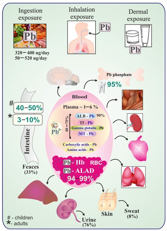
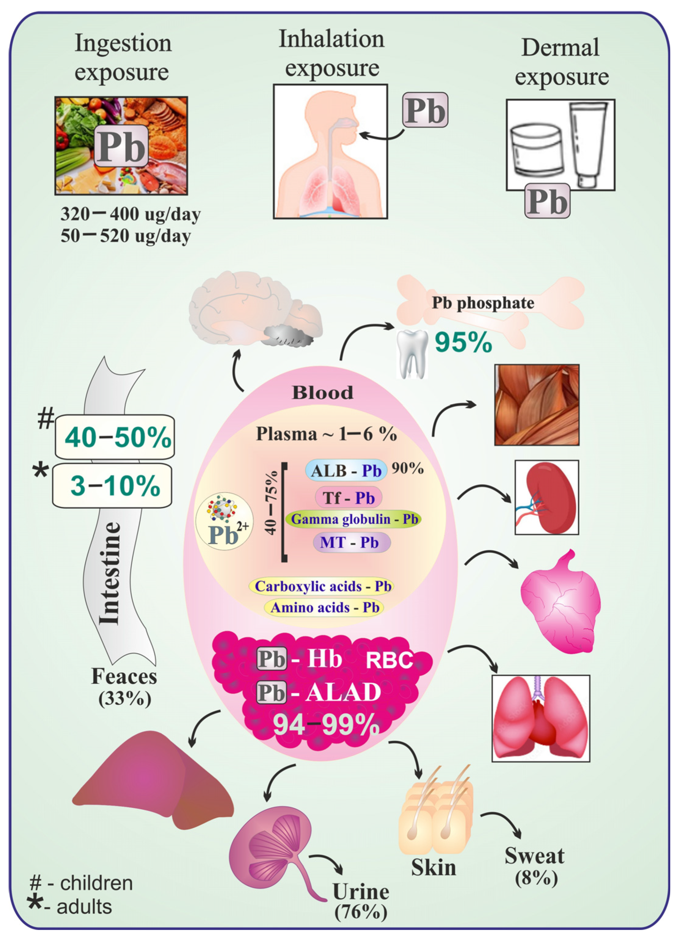
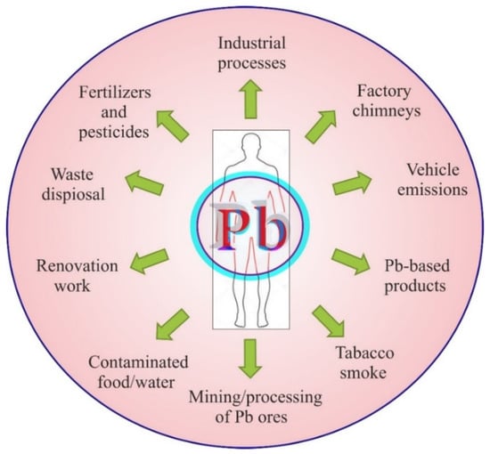
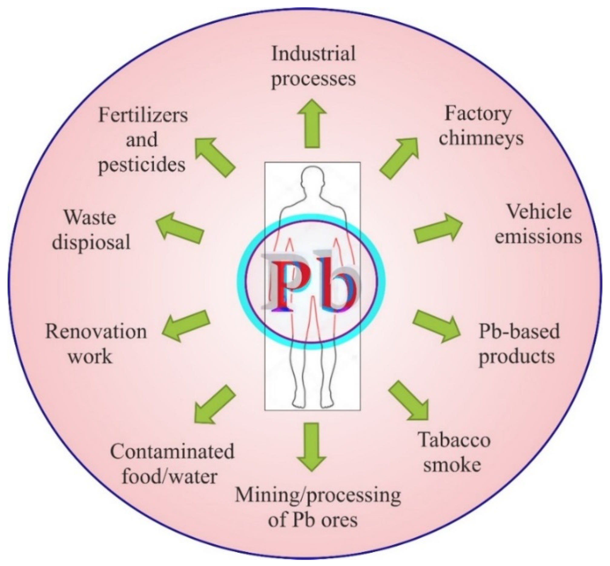
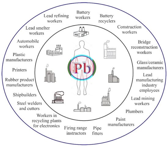
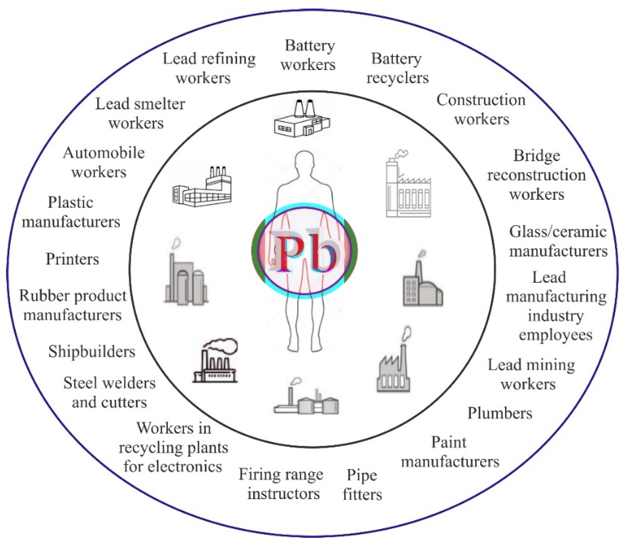
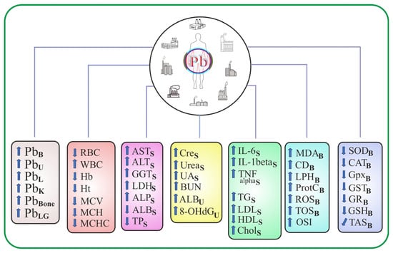
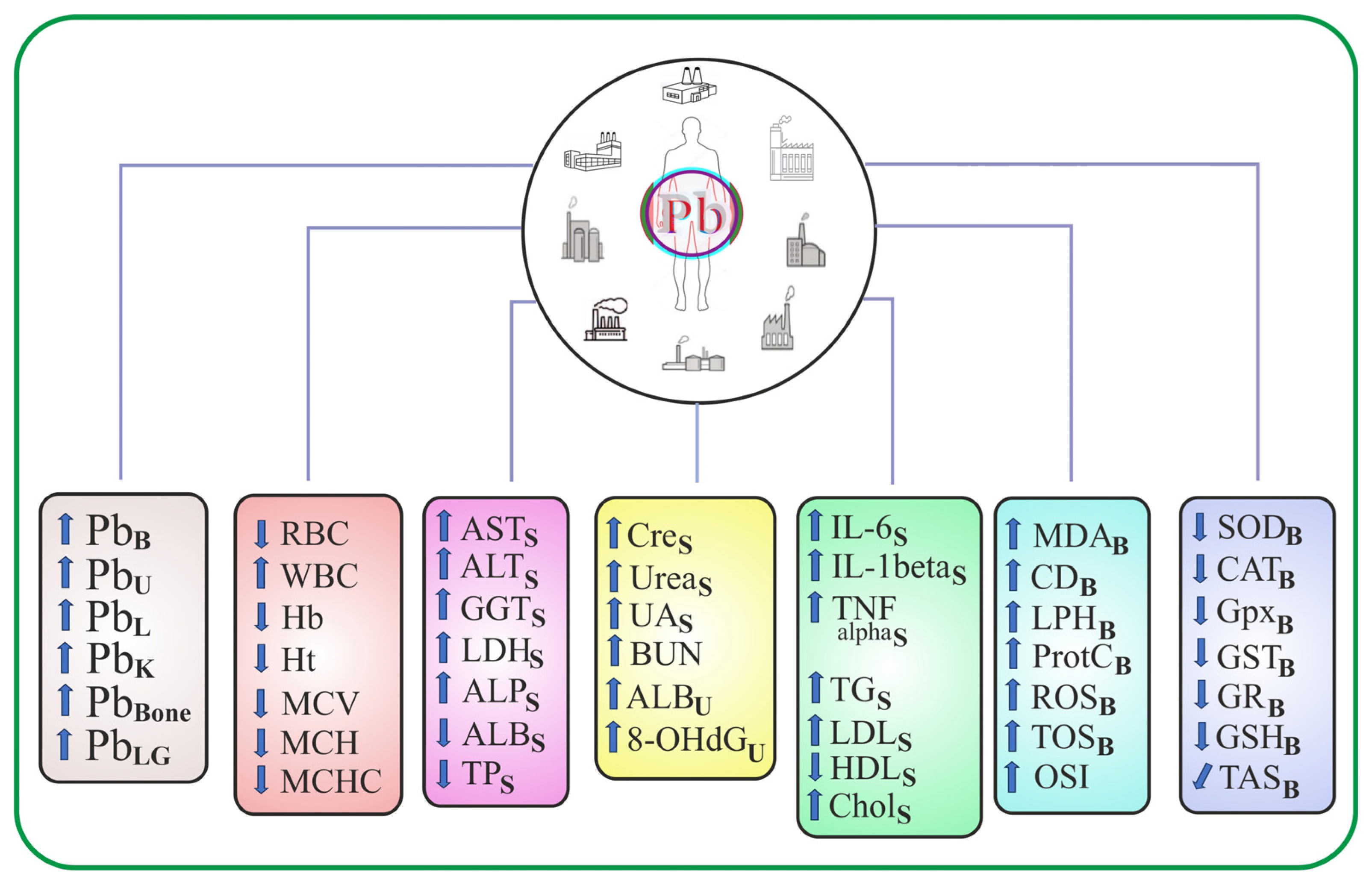
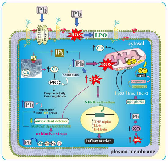
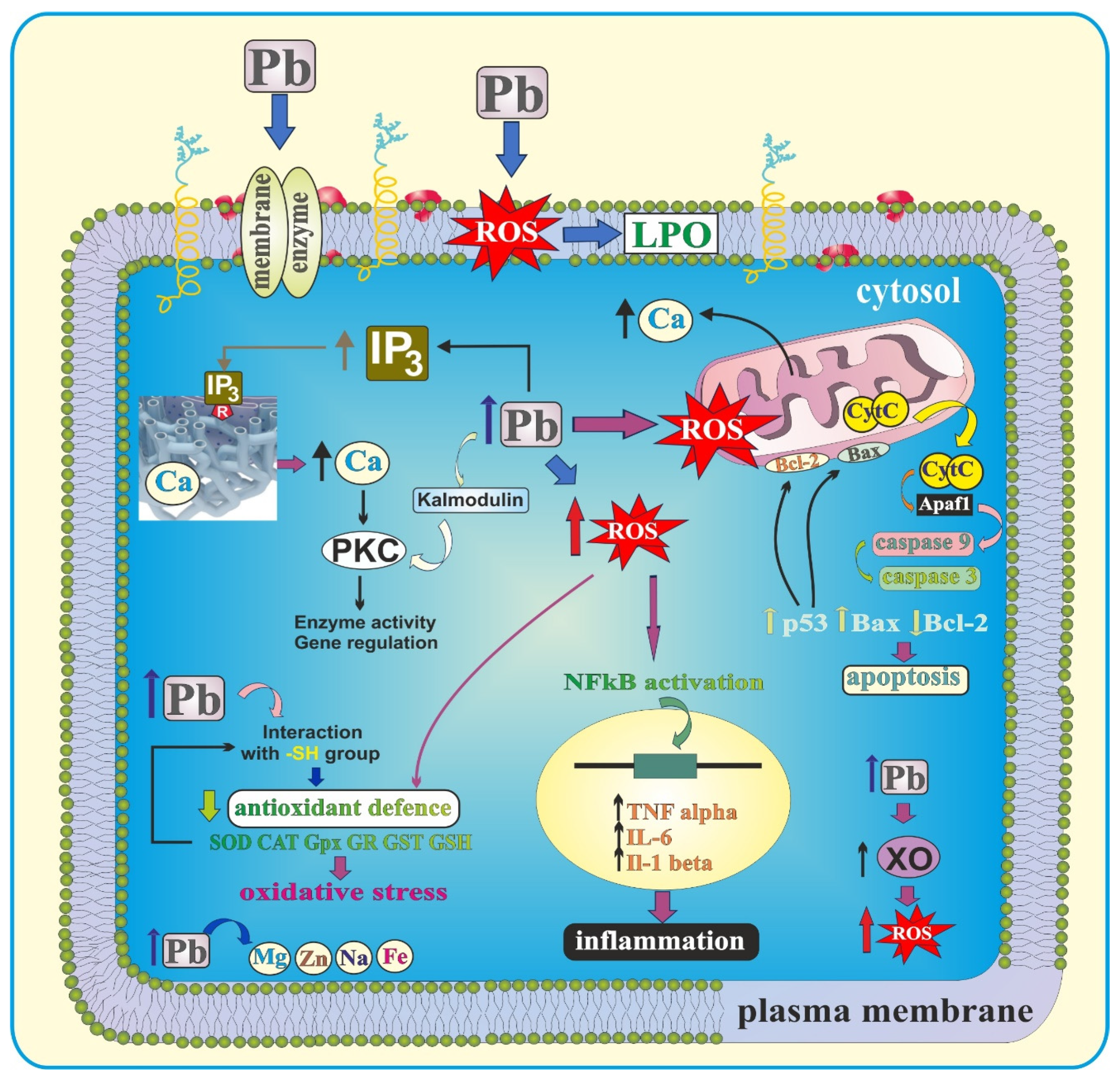
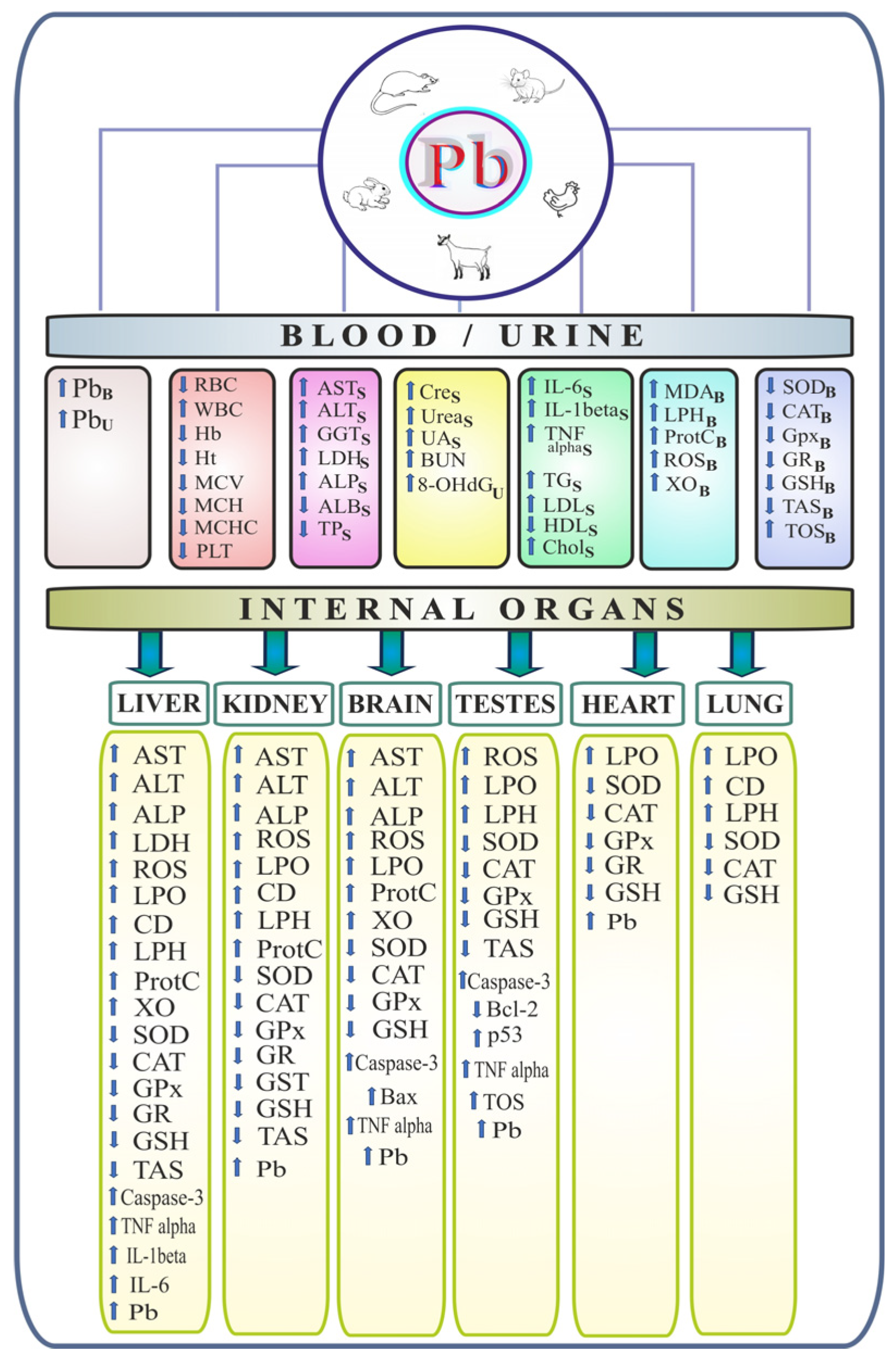
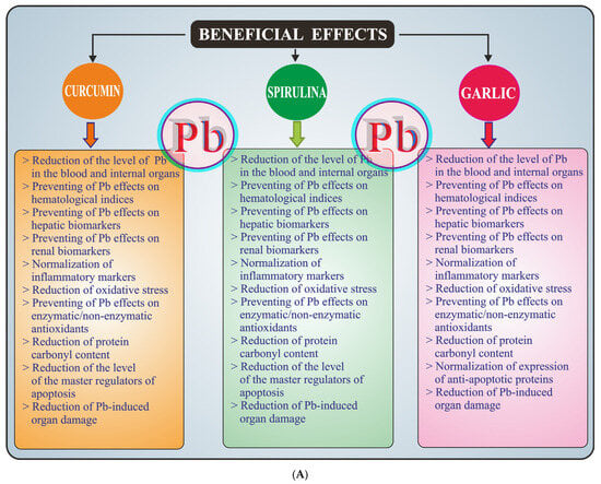
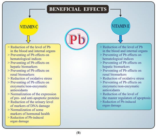
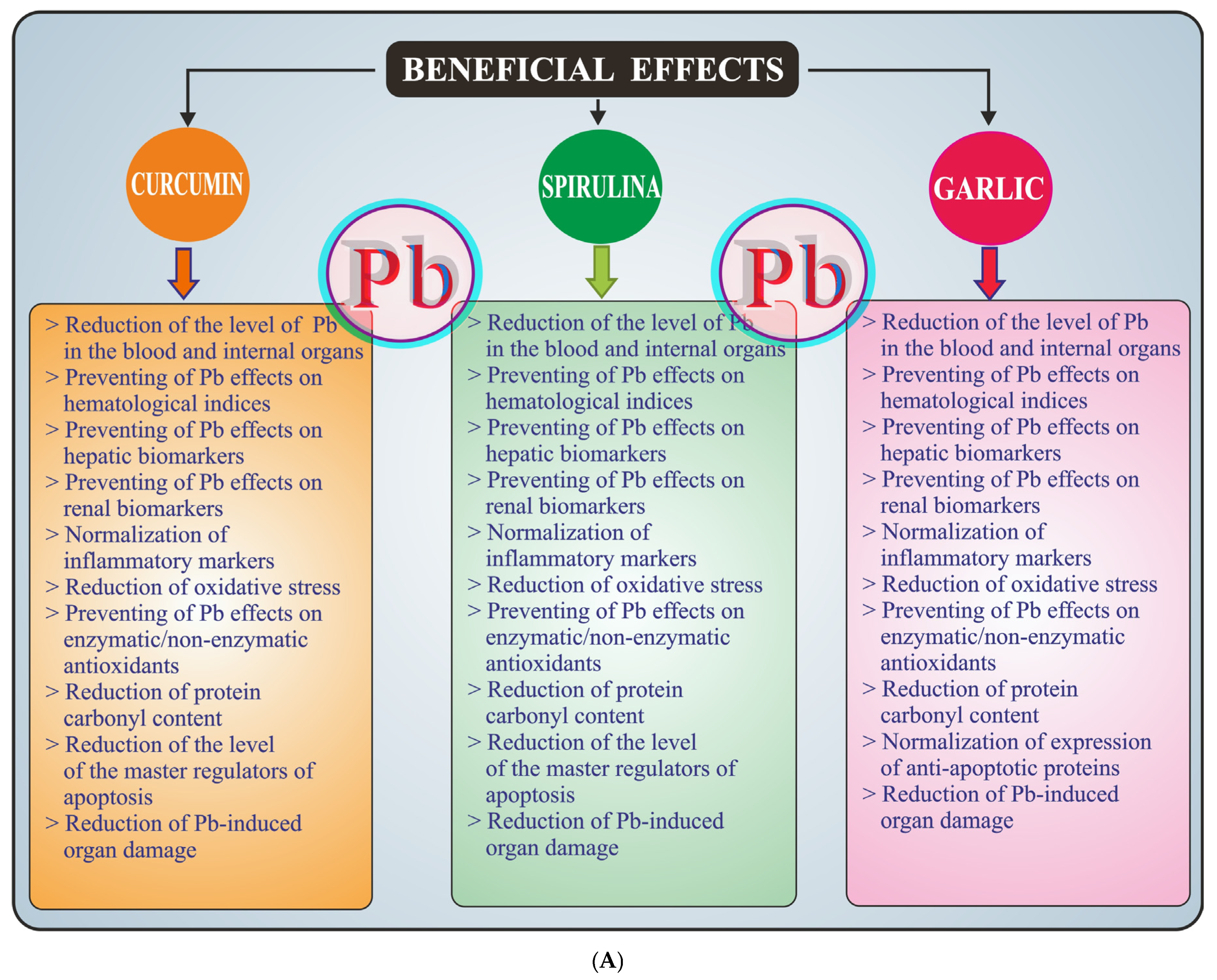
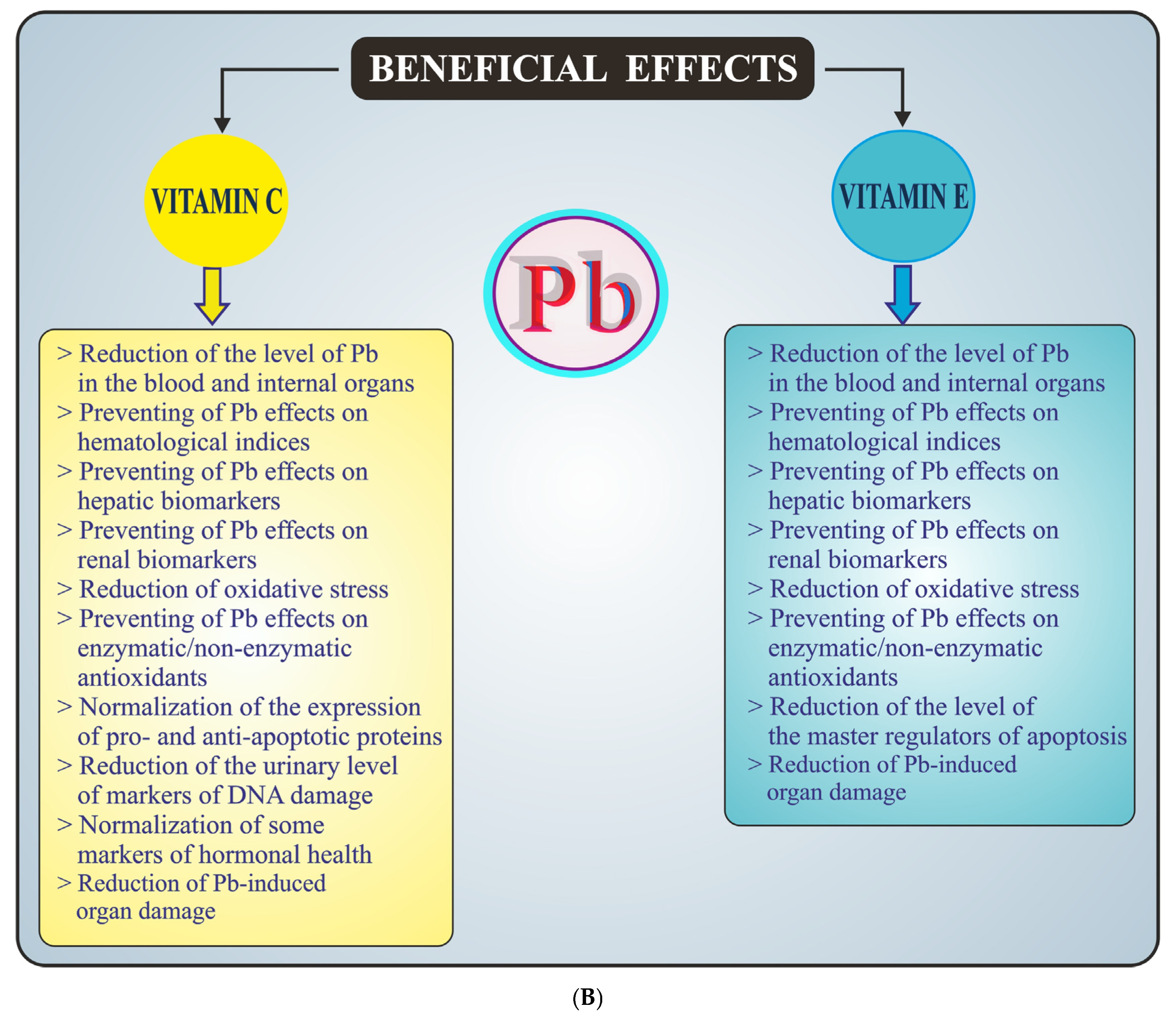
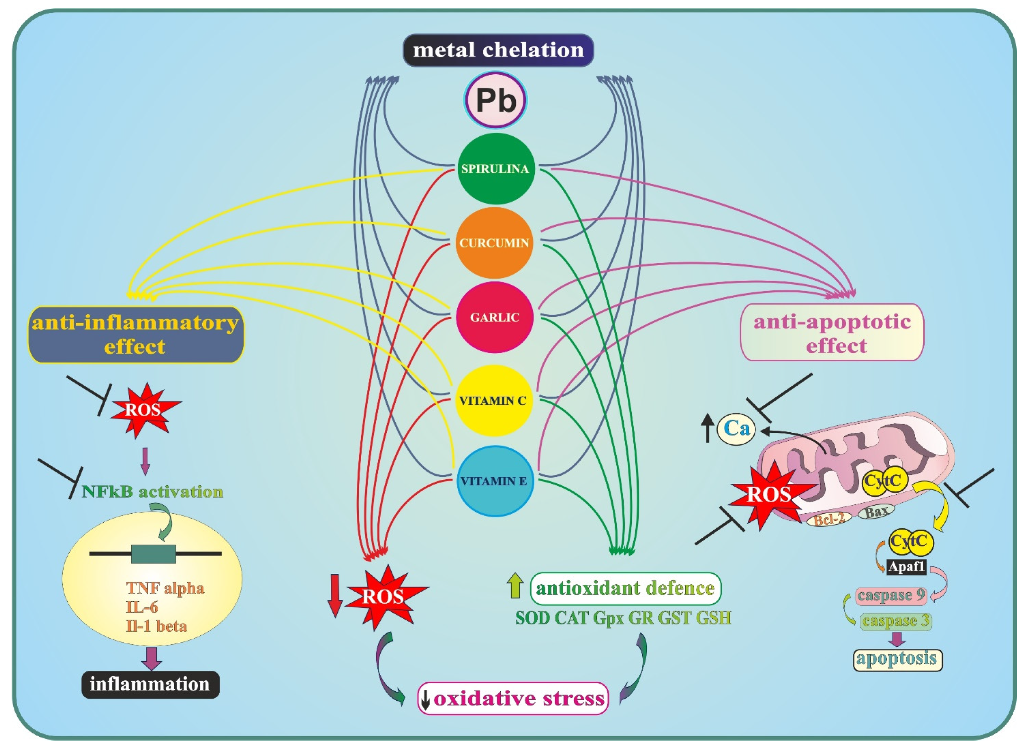

Disclaimer/Publisher’s Note: The statements, opinions and data contained in all publications are solely those of the individual author(s) and contributor(s) and not of MDPI and/or the editor(s). MDPI and/or the editor(s) disclaim responsibility for any injury to people or property resulting from any ideas, methods, instructions or products referred to in the content. |
© 2026 by the authors. Licensee MDPI, Basel, Switzerland. This article is an open access article distributed under the terms and conditions of the Creative Commons Attribution (CC BY) license.