


Abstract
This systematic review aimed to summarize the available evidence on the associations between the female reproductive tract microbiota and endometrial cancer (EC). While gut microbiota has been studied extensively, microbial communities within the endometrium, cervix, and vagina remain relatively understudied. A systematic literature search was conducted in PubMed, Scopus, Web of Science, EMBASE, and the Cochrane Library for studies published up to January 2025. Predefined PECO-based criteria included studies on women or human cell models assessing genital tract microbiota in EC versus non-EC controls, focusing on composition, diversity, or function; reviews and non-microbiota studies were excluded. A total of 21 studies were included. Overall, there was a consistent depletion of protective Lactobacillus species and enrichment of anaerobic, pro-inflammatory bacteria like Prevotella, Atopobium, and Porphyromonas in EC tissues. Beta-diversity was significantly different between EC and controls across studies, indicating distinct microbial profiles. Some studies also identified fungal and viral taxa associated with EC, and functional assays demonstrated that certain species could modulate host immune responses or promote tumor growth. Despite methodological heterogeneity and the lack of evidence supporting causality due to the observational design, the findings support an association between altered genital tract microbiota and EC.
1. Introduction
The microbiota is the collection of all microbes, including bacteria, archaea, fungi and viruses, that naturally live in and on our bodies. Technically, the term microbiota refers to the community of microorganisms themselves, whereas the microbiome refers to the collective genomes of these microorganisms. These communities colonize various anatomic regions, such as the gastrointestinal, oral, and genitourinary tracts, and can be classified as symbionts, commensals, or pathogens. Microbial communities vary depending on the anatomical site and are influenced by factors including health status, genetics, diet, and hygiene [1].
Several studies suggest that the microbiota is essential in the immune and endocrine systems [2,3]. Furthermore, microbiota alterations have been associated with the development of diseases, including cancer [4]. Dysbiosis, or imbalances in the intestinal and vaginal microbiota, appears to be associated with the onset of gynecological malignancies, including uterine, cervical, vulvar, and vaginal cancers [5]. Specifically, recent evidence suggests that the gut microbiota plays a significant role in the development and progression of endometrial cancer (EC) through several mechanisms, including immune modulation, metabolite production, and alteration of the inflammatory environment [6].
EC is the most common gynecological cancer in high-income countries and is the fourth most common cancer among women, following breast, lung, and colorectal cancers [7]. Major risk factors include genetic predisposition, hormonal imbalances involving hypoestrogenism, prolonged exposure to estrogen, obesity, nulliparity, and advanced age. Variations in the gut microbiota have been closely linked to the onset of EC [8]. Notably, diet is one of the primary factors influencing the microbiota composition. Diets rich in fiber and probiotics appear to exert a protective effect by reducing systemic inflammation, enhancing immune function, and maintaining normal estrogen levels [9]. In contrast, a high-fat, high-calorie diet may cause dysbiosis, promote systemic inflammation, and alter estrogen metabolism. This concept aligns with the emerging notion of the estrobolome, the collection of gut microbial genes capable of metabolizing estrogens, whereby increased microbial β-glucuronidase activity may enhance enterohepatic recirculation of estrogens and, in turn, hypothetically contribute to endometrial carcinogenesis [10].
Based on this evidence, the gut microbiota may play a significant role in initiating and progressing EC through multiple mechanisms, including immune modulation, metabolite production, and inflammation regulation [9,10]. However, despite growing attention to the intestinal microbiota, other microbial ecosystems, particularly those anatomically and patho-physiologically closer to the endometrium, have received considerably less focus. For a long time, the uterus was believed to be a sterile environment. However, advancements in detection technologies have since revealed the presence of a resident microbial community within the endometrium [11]. In this context, the lower and upper genital tract microbiota, directly adjacent to the endometrial environment, represent promising but understudied areas in EC pathogenesis. Therefore, this systematic review aims to summarize the available evidence on the female reproductive tract microbiota and its association with the pathogenesis of EC.
2. Materials and Methods
2.1. Study Design
This systematic review was designed before the online search, outlining the study population, exposure, comparator, outcome measures, and study eligibility criteria. The study was exempt from institutional review board approval. The methodology followed the Cochrane Handbook for Systematic Reviews of Diagnostic Test Accuracy. The review was reported following the Preferred Reporting Items for Systematic Reviews and Meta-Analyses of Diagnostic Test Accuracy (PRISMA-DTA) guidelines [12,13]. The protocol was registered on PROSPERO (CRD 420251024643).
2.2. Search Strategy and Eligibility Criteria
A comprehensive literature search was conducted across EMBASE, Scopus, PubMed, Web of Science, and the Cochrane Library from inception until January 2025 by a certified professional librarian (Biblioteca Meneghetti—University of Verona). The search strategy was performed with and without MeSH terms to include non-indexed more recent records and to make the search query replicable in all electronic databases: (Endometrial Neoplasm OR Neoplasm, Endometrial OR Endometrial Carcinoma OR Carcinoma, Endometrial OR Cancer of Endometrium OR Endometrium Cancer OR Cancer, Endometrium OR Cancer of the Endometrium OR Carcinoma of Endometrium OR Endometrium Carcinoma OR Endometrial Cancer OR Cancer, Endometrial) AND (Microbiota OR Microbial Community OR Community, Microbial OR Microbial Community Composition OR Community Composition, Microbial OR Composition, Microbial Community OR Microbiome OR Human Microbiome OR Microbiome, Human OR Microbial Community Structure OR Community Structure, Microbial). Additionally, references from the selected studies were manually reviewed.
Inclusion and exclusion criteria were pre-defined by all contributors using the Population, Exposure, Comparison, and Outcome (PECO) framework. Population: Women or models based on human cells in vitro or ex vivo. Exposure: Diagnosis of EC or EC tissue, including all subtypes and stages and endometrial hyperplasia (EH). Comparison: Women or tissues without EC or EH, healthy or with non-neoplastic or pre-neoplastic pathologies (e.g., benign conditions). Outcome: Evaluation of the microbiota of the female genital tract (endometrium, cervix, vagina) conducted primarily through 16S rRNA gene sequencing, although other microbiological analysis methods were also considered. Any of the following microbiota evaluations were eligible: microbiota composition (taxonomy), microbial diversity (alpha/beta diversity), and functional activity of the microbiota (metabolic pathways, gene expression). Studies were excluded if they were review articles or did not include a microbiota analysis of the female genital tract. We harmonized diversity terminology across studies by defining α-diversity as within-sample richness/evenness (e.g., Shannon, Simpson, Chao1, Pielou’s evenness) and β-diversity as between-sample community dissimilarity (e.g., Bray–Curtis, Jaccard, weighted/unweighted UniFrac, Aitchison). For synthesis, we recorded the index/metric and classified results as ‘higher’, ‘lower’, or ‘no difference’ versus controls.
2.3. Study Selection and Data Extraction
Two independent reviewers (RV, FB) screened titles and abstracts for relevance and assessed the full texts of potentially eligible studies. Disagreements were resolved through discussions with a second reviewer (SG). A standardized data extraction form was developed to collect key study details, including study characteristics (first author, publication year, study design, research setting), sample data (population studied, sampling technique, sample size), and outcomes (taxonomy, alpha/beta diversity, metabolic pathways, gene expression, and clinical correlation). To minimize bias inherent to low-biomass microbiome studies, we prioritized reports using aseptic sampling (e.g., sterile, lubricant-free speculum with cervical cleansing) and laboratory contamination controls (negative controls/blank extractions), and we interpreted findings from studies that did not report these measures cautiously in the synthesis.
3. Results
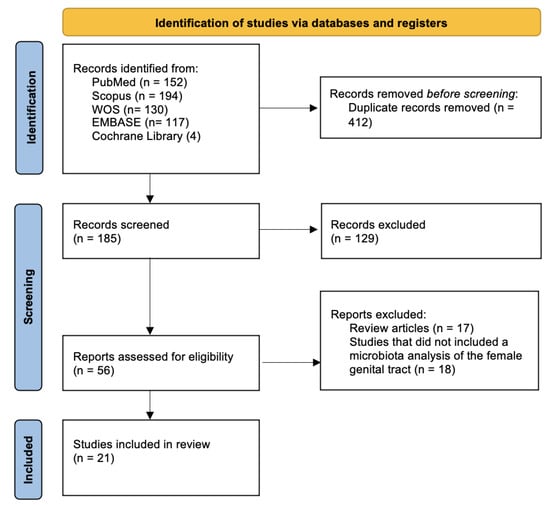
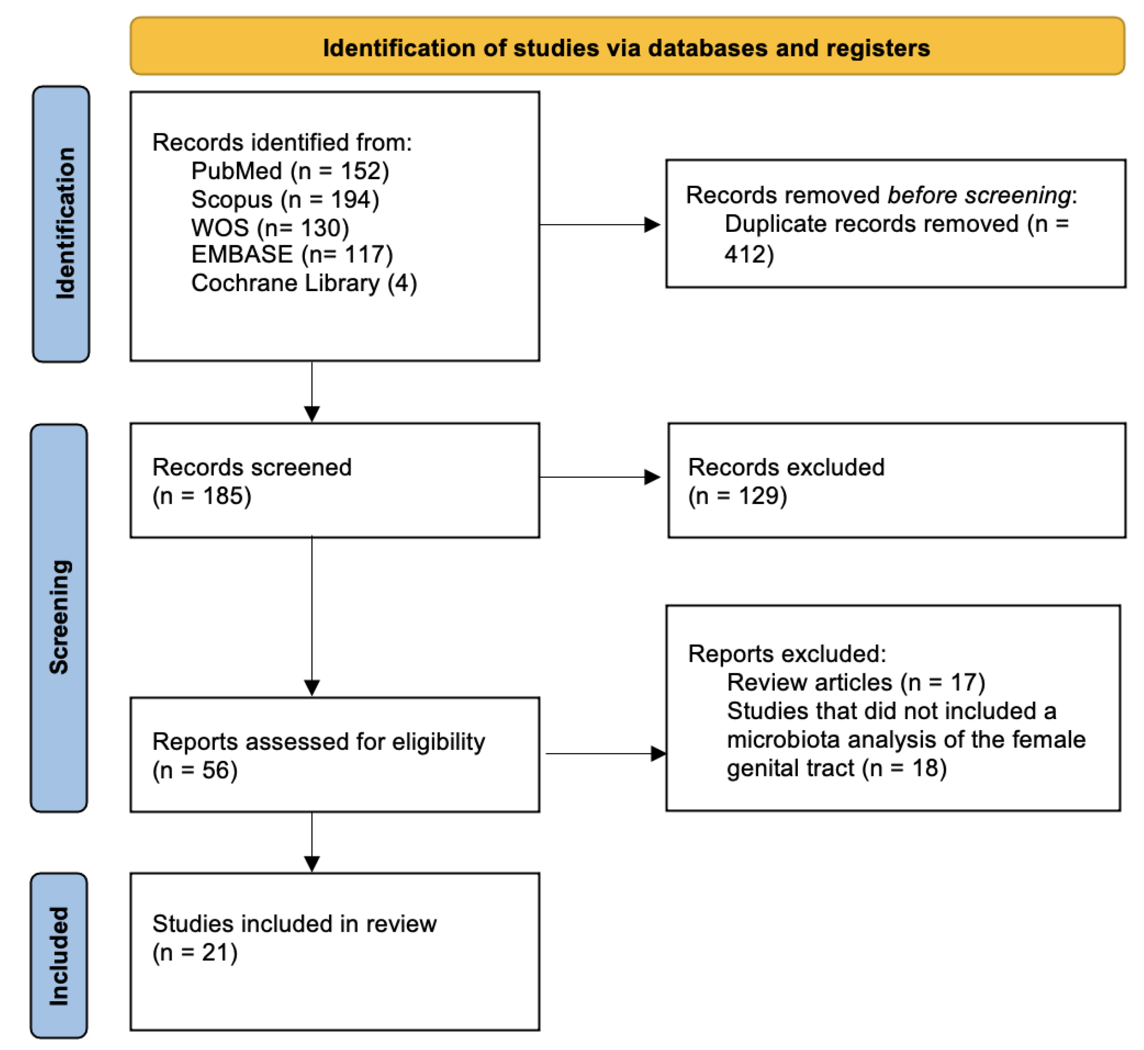
A total of 597 records were initially retrieved through database searches. After removing 412 duplicates, 185 unique records were screened based on titles and abstracts. Of these, 129 studies were excluded due to irrelevance to the topic, non-original content, or lack of specific microbiota analysis. We then assessed 56 full-text articles for eligibility, excluding 35 that did not meet the inclusion criteria: 18 investigated the microbiota of anatomical sites other than the female genital tract (most commonly the intestinal microbiota), while the remaining 17 were excluded because they were not original articles.
Ultimately, 21 studies, published between 2016 and 2025, were included in the final synthesis (Figure 1). To facilitate a structured synthesis of evidence, we categorized them into three groups based on the type of analyzed biological sample. In the “Endometrial Microbiota in Endometrial Cancer” chapter, we included all studies that analyzed endometrial tissue samples. In “Cervicovaginal Microbiota in Endometrial Cancer” we included studies that analyzed vaginal or cervical tissue samples. In “Functional and in vitro studies,” we included studies that conducted in vitro experiments to assess microbial effects on host cells. Comprehensive summary table outlining the parameters used to identify the microbiota and JBI Analytical Cross-Sectional appraisal matrix are present in Supplementary Materials.
Figure 1.
PRISMA flow diagram.
3.1. Endometrial Microbiota in Endometrial Cancer
3.1.1. Bacteria
We included 12 studies (Table 1) that compared the endometrial microbiota of patients with EC or endometrial hyperplasia to that of healthy individuals or patients with benign uterine pathologies [14,15,16,17,18,19,20,21,22,23,24]. Only one study stands out as an exception: Wang et al. compared the microbiota present in EC tissue with that found in the healthy peri-tumor tissue of the same patients [23].
These studies used α-diversity and β-diversity to assess the microbial diversity between groups: α-diversity refers to within-sample diversity, accounting for species richness and evenness. β-diversity quantifies the compositional dissimilarity between microbial communities from different patients.
All except two studies reported a significantly increased α-diversity in EC compared to controls, indicating a richer and more evenly distributed microbial community in malignant tissue [15,17,19,23,24]. In contrast, reduced α-diversity was observed in EC versus healthy controls in Li et al. [18] and in EH compared to benign controls in Ying [22], while Leoni et al. found no significant differences in α-diversity between EC and controls [20].
All studies but one reported significant differences in β-diversity between EC and controls, indicating distinct microbial community structures associated with malignancy [15,17,18,19,23,24]. Only Leoni et al. reported no significant group-level separation based on β-diversity metrics [20].
Regarding taxonomic signatures, EC tissues were consistently enriched in anaerobic and pro-inflammatory bacteria, with frequent detection of Prevotella, Atopobium, Porphyromonas, Peptoniphilus, and Dialister [15,18,19,23]. Han et al. and Wang et al. showed an increase in Fusobacterium and Sneathia [19,23], while Kuźmycz et al. specifically highlighted Anaerococcus vaginalis as a potential functional cofactor in EC, capable of inducing ROS in endometrial cells [24]. Streptococcus, Gardnerella, and Acinetobacter were found more often in EC than controls across multiple studies [17,18,20,23]. The most consistent finding across identified studies is the depletion of Lactobacillus spp., especially L. crispatus and L. iners, in EC compared to controls. These studies suggest that Lactobacilli are considered protective commensals, maintaining pH balance, epithelial barrier integrity, and suppressing pathogens [16,17,20,23]. Gonzalez-Bosquet et al. showed a reduction in EC of Desulfobacter, Desulfomicrobium, Parabacteroides, and Proteus compared to control samples [14].
Concerning EH, Bacillus pseudofirmus, Stenotrophomonas, and Delftia were identified as enriched genera with potential biomarker value [16,22].
A recent large-scale study by Liu et al. further expanded on these findings by analyzing the intratumoral microbiota of 239 EC patients and 116 controls using data from the TCGA-UCEC [25]. The authors identified 26 microbial taxa significantly associated with patient prognosis and used Cox and LASSO regression models to develop a microbiota-based risk score, suggesting a relationship between the prognosis of patients with EC and the composition of the intratumoral microbiota [25]. The low-risk group exhibited a microbial profile linked to higher immune activity and a better-predicted response to immune checkpoint inhibitors (PD1/PD-L1 and CTLA-4). In contrast, high-risk patients had elevated immune dysfunction and exclusion scores [25].
3.1.2. Fungi and Viruses
Two studies investigated the role of fungi and viruses of endometrium in EC. Han et al. showed enrichment of Penicillium spp. in EC tissues, along with estradiol-like metabolites, suggesting fungal involvement in hormonal mimicry and tumor promotion [19]. Deligdisch et al. identified human mammary tumor virus (HMTV) sequences in 23% of EC samples but not in controls, proposing a possible viral contribution to endometrial carcinogenesis [26].
Table 1.
Methodological characteristics and main results of studies on the endometrial microbiome.
3.2. Cervicovaginal Microbiota in Endometrial Cancer
We included six studies published between 2019 and 2024 that investigated the cervicovaginal microbiota in EC patients (Table 2). Samples were aseptically collected from the vagina or cervix during surgery or preoperatively using swabs or from tissue biopsies. All these studies compared the cervicovaginal microbiota of patients with cancer or EH to that of healthy individuals or patients with benign uterine conditions. EC patients did not exhibit significant alterations in α-diversity compared to controls. Only Semertzidou et al. reported an increase in α-diversity in the EC group [27], while Hakimjavadi et al. reported an increase only in patients with high-grade EC [28].
Table 2.
Methodological characteristics and main results of studies on the cervicovaginal microbiota.
Across all these studies, β-diversity was significantly increased between cases and controls, indicating a significant shift in microbial composition associated with malignancy. Cervical and vaginal environments in EC were less frequently dominated by Lactobacillus spp. and instead colonized by complex, polymicrobial anaerobic communities [27,28,29,30,31,32]. Specifically, Barczyński et al. and Walther-António et al. reported reductions in Lactobacillus iners and L. crispatus, respectively [30,32], accompanied by increases in Mobiluncus curtisii, Dialister pneumosintes, Atopobium vaginae, and Porphyromonas spp. Similarly, Semertzidou et al. demonstrated consistent depletion of L. crispatus in EC patients across multiple anatomical sites, coupled with overrepresentation of Prevotella, Peptoniphilus, and Porphyromonas [27].
The presence and abundance of specific bacterial taxa were frequently associated with tumor type, grade, and host factors. Walsh et al. identified Porphyromonas somerae in 100% of women with type II EC, suggesting a subtype-specific microbial signature [31]. Hakimjavadi et al. showed that microbial community state types (CSTs) varied significantly across tumor grades and histological subtypes, with Fusobacterium nucleatum and Prevotella bivia enriched in high-grade and serous tumors (CST IV) and L. gasseri (CST II) reduced across the EC cohort. These CSTs also correlated with inflammatory gene expression and predicted tumor phenotype with high accuracy (AUC = 0.88) [28].
3.3. Functional and In Vitro Studies
While the observational studies we included provided crucial insights into the microbial composition in EC, functional and in vitro studies are essential to understand the mechanistic roles of specific microbial taxa in tumor initiation and progression. This group of studies goes beyond association, exploring how selected microbes interact with host tissues and influence cellular behavior. We included only three relevant papers (Semertzidou et al., Caselli et al. and Crooks et al.).
Semertzidou et al. demonstrated a potential protective role for Lactobacillus crispatus, showing that its conditioned media significantly inhibited the proliferation of EC organoids in vitro. This finding suggests that beyond association, specific members of the commensal flora may exert direct anti-tumor effects, potentially via modulation of epithelial immunity, pH, or metabolite secretion [27].
Caselli et al. and Crooks et al. provided mechanistic insights into how Porphyromonas somerae may influence the development or progression of endometrial cancer: through direct invasion of epithelial cells, P. somerae may evade immune surveillance and modulate host intracellular metabolism, including the production of succinate, a metabolite known to stabilize HIF-1α, a driver of cancer progression. Furthermore, they showed that P. somerae induces pro-inflammatory cytokines, potentially creating a tumor-promoting environment within the endometrium [33,34].
4. Discussion
Across most of the included studies, the α-diversity of endometrial microbiota was significantly increased in EC; these findings suggest a shift toward a more polymicrobial environment potentially driven by inflammation, immune modulation, or epithelial barrier disruption within the endometrial niche. Some studies reported conflicting results; there could be several reasons underlying these discrepancies. One possible explanation is the inclusion of patients with different characteristics regarding menopause status and BMI. In fact, Walsh et al. demonstrated that these factors significantly influence the diversity of the vaginal microbiota [31]. Another reason could be using different indices to measure α-diversity (such as the Shannon index, Chao index, and Pielou’s evenness). Finally, we believe that tumor grading could also be one of the contributing factors. Hakimjavadi suggests that early lesions might maintain Lactobacillus dominance (lower α-diversity), while advanced tumors may exhibit microbial infiltration and immune-mediated breakdown, increasing α-diversity. These reasons also apply to cervicovaginal α-diversity, as no consistent trend in α-diversity was observed across the studies analyzed in relation to endometrial cancer. In contrast to the heterogeneity observed in α-diversity, endometrial and cervicovaginal β-diversity consistently distinguished EC-associated microbial communities from benign controls. Nearly all reviewed studies reported statistically significant differences in microbial community structure based on Bray–Curtis dissimilarity, UniFrac metrics, or Aitchison distances. The observed increase in β-diversity reflects the taxonomic signatures reported in the studies we reviewed: Both the endometrial and cervicovaginal environments consistently show a depletion of protective commensals such as Lactobacillus crispatus and L. iners and an environment of pro-inflammatory and potentially pathogenic taxa. In most of the studies we reviewed, these pathogenic taxa identified in the female reproductive tract were primarily composed of Prevotella, Atopobium, Peptoniphilus, Dialister, and Porphyromonas. Between these genera, the most studied is Porphyromonas, specifically P. somerae, which is capable of invading endometrial cells in vitro and inducing the expression of pro-inflammatory cytokines (IL1α, IL1β, IL17α, TNFα), which are associated with inflammatory processes and possibly with endometrial tumor progression.
These findings are further enriched by the emerging literature on the gut microbiota’s role in EC pathogenesis. Under normal conditions, gut microbial communities maintain a dynamic equilibrium; however, disruptions due to factors like obesity, diabetes, and hormonal imbalance, common in EC patients, lead to dysbiosis characterized by a decline in beneficial microbes and an expansion of opportunistic or pro-inflammatory taxa like Firmicutes and Bacteroides. These bacteria producing β-glucuronidase enzymes can deconjugate estrogens in the intestine, promoting reabsorption and elevating systemic estrogen levels, a known risk factor of EC [6,35]. Together, these results suggest that this shift from beneficial to pro-inflammatory bacteria, whether within the gut or the female reproductive tract, may not be isolated phenomena but rather interconnected reflections of broader systemic dysregulation. Accordingly, clarifying the determinants of microbiome composition across body sites is essential; beyond the factors noted above, three domains are especially influential: (1) Diet, which consistently remodels gut taxa and functions across Mediterranean, high-fiber/plant-based, ketogenic, high-protein, and Western patterns, is associated with corresponding shifts in SCFAs, bile acids, and other metabolites [36]. (2) Host genetic background, host genetic variants are associated with structural and functional traits of the gut microbiota [37,38]. (3) Geographic/environmental context, with harmonized global datasets showing robust, region-specific community signatures that can even predict a sample’s world region of origin [39]. Therefore, these elements must be explicitly considered when designing and interpreting studies.
This systematic review employed a rigorous selection process, improving result reliability and providing updated insights on the relation between female reproductive tract microbiota and endometrial cancer; furthermore, to our knowledge, this is the first review in the literature that studies this relationship in depth, also because most of the studies we included were published in the last four years. However, several limitations must be acknowledged: (1) The study is a systematic review based on observational studies. This type of design does not allow for establishing a causal link between microbiota and endometrial cancer. It is difficult to determine whether microbial alterations are a cause or a consequence of the disease. (2) Heterogeneity of the included studies. Sample collection, storage, and preparation methods varied widely across studies, as did the different α- and β-diversity indices and distance metrics used to quantify community structure. These factors may impact the microbial composition detected. (3) Two studies analyzed only DNA (16S or shotgun). This is the standard practice for compositional studies. This limitation arises when trying to understand real-time metabolic activity: in that case, it would be better to use RNA (metatranscriptomics). DNA, on the other hand, may include traces of non-viable microorganisms, potentially overestimating certain species. (4) The pooling of EC with EH in a subset of primary studies. EH represents a biologic continuum that may differ meaningfully from invasive EC, and EC outcomes may vary by grade and histotype. Incomplete and heterogeneous reporting in several studies precluded consistent stratification; pooled EC + EH findings should therefore be interpreted with caution. (5) We note that opportunistic infections (e.g., bacterial vaginosis or subclinical endometritis) can transiently inflate α-diversity and shift community structure, confounding associations with endometrial cancer. To limit confounding from opportunistic infections and transient perturbations of the genital-tract microbiota, most primary studies screened and excluded women with active genital infections and recent antimicrobial/hormonal exposure, standardized pre-operative sterile swab collection (often before routine douching), and reported aseptic handling; for example, Wang et al. [23]. excluded genital infections, recent antibiotics, and intercourse/douching within 48 h, while Barczyński et al. [32] sampled intraoperatively before the betadine douche and excluded recent intravaginal infections; pre-operative sterile sampling was also used by Walsh et al. [31].
5. Conclusions
This systematic review highlights a significant association between alterations in the female genital tract microbiota and endometrial carcinoma. Endometrial cancer appears to be linked to the depletion of Lactobacillus spp. and an increased abundance of pro-inflammatory bacterial species. Establishing a causal relationship is of fundamental importance, as modulation of the uterine and vaginal microbiota may represent a promising target for the development of conservative therapeutic strategies in endometrial hyperplasia.
Supplementary Materials
The following supporting information can be downloaded at: https://www.mdpi.com/article/10.3390/ijms26188877/s1.
Author Contributions
R.V. Conceptualization, writing—review and editing. F.B. Formal analysis. P.F. Conceptualization. V.S. supervision. C.C. formal analysis. G.C. data curation. A.S.L. methodology. P.C.Z. data curation. M.B. data curation. I.P. methodology. S.U. supervision. S.G. data curation. All authors. All authors have read and agreed to the published version of the manuscript.
Funding
This research received no external funding.
Institutional Review Board Statement
Not applicable.
Informed Consent Statement
Not applicable.
Acknowledgments
This study was performed with HIF (Health Innovation Factory) Department Research Center, University of Verona.
Conflicts of Interest
The authors declare no conflict of interest. The funders had no role in the design of the study; in the collection, analyses, or interpretation of data; in the writing of the manuscript, or in the decision to publish the results.
Abbreviations
The following abbreviations are used in this manuscript:
| CST | Community State Type (vaginal microbial community type) |
| EC | Endometrial Cancer |
| EH | Endometrial Hyperplasia |
| AEH | Atypical Endometrial Hyperplasia |
| EMT | Epithelial–Mesenchymal Transition |
| FFPE | Formalin-Fixed Paraffin-Embedded |
| HGSC/HSOC | High-Grade Serous Ovarian Cancer |
| ITS1 | Internal Transcribed Spacer 1 |
| ROS | Reactive Oxygen Species |
| USC | Uterine Serous Carcinoma |
References
- Dekaboruah, E.; Suryavanshi, M.V.; Chettri, D.; Verma, A.K. Human Microbiome: An Academic Update on Human Body Site Specific Surveillance and Its Possible Role. Arch. Microbiol. 2020, 202, 2147–2167. [Google Scholar] [CrossRef] [Scilit]
- O’Riordan, K.J.; Moloney, G.M.; Keane, L.; Clarke, G.; Cryan, J.F. The Gut Microbiota-Immune-Brain Axis: Therapeutic Implications. Cell Rep. Med. 2025, 6, 101982. [Google Scholar] [CrossRef] [Scilit]
- Clemente-Suárez, V.J.; Redondo-Flórez, L.; Rubio-Zarapuz, A.; Martín-Rodríguez, A.; Tornero-Aguilera, J.F. Microbiota Implications in Endocrine-Related Diseases: From Development to Novel Therapeutic Approaches. Biomedicines 2024, 12, 221. [Google Scholar] [CrossRef] [Scilit]
- Cullin, N.; Azevedo Antunes, C.; Straussman, R.; Stein-Thoeringer, C.K.; Elinav, E. Microbiome and Cancer. Cancer Cell 2021, 39, 1317–1341. [Google Scholar] [CrossRef] [Scilit]
- Chase, D.; Goulder, A.; Zenhausern, F.; Monk, B.; Herbst-Kralovetz, M. The Vaginal and Gastrointestinal Microbiomes in Gynecologic Cancers: A Review of Applications in Etiology, Symptoms and Treatment. Gynecol. Oncol. 2015, 138, 190–200. [Google Scholar] [CrossRef] [Scilit]
- Zhang, B.; Mohd Sahardi, N.F.N.; Di, W.; Long, X.; Shafiee, M.N. The Gut-Endometrium Axis: Exploring the Role of Microbiome in the Pathogenesis and Treatment of Endometrial Cancer-A Narrative Review. Cancers 2025, 17, 1044. [Google Scholar] [CrossRef] [Scilit]
- Siegel, R.L.; Giaquinto, A.N.; Jemal, A. Cancer Statistics, 2024. CA Cancer J. Clin. 2024, 74, 12–49. [Google Scholar] [CrossRef] [Scilit] [PubMed]
- Raglan, O.; Kalliala, I.; Markozannes, G.; Cividini, S.; Gunter, M.J.; Nautiyal, J.; Gabra, H.; Paraskevaidis, E.; Martin-Hirsch, P.; Tsilidis, K.K.; et al. Risk Factors for Endometrial Cancer: An Umbrella Review of the Literature. Int. J. Cancer 2019, 145, 1719–1730. [Google Scholar] [CrossRef] [Scilit] [PubMed]
- Wilson, A.S.; Koller, K.R.; Ramaboli, M.C.; Nesengani, L.T.; Ocvirk, S.; Chen, C.; Flanagan, C.A.; Sapp, F.R.; Merritt, Z.T.; Bhatti, F.; et al. Diet and the Human Gut Microbiome: An International Review. Dig. Dis. Sci. 2020, 65, 723–740. [Google Scholar] [CrossRef] [Scilit]
- Pollet, R.M.; D’Agostino, E.H.; Walton, W.G.; Xu, Y.; Little, M.S.; Biernat, K.A.; Pellock, S.J.; Patterson, L.M.; Creekmore, B.C.; Isenberg, H.N.; et al. An Atlas of β-Glucuronidases in the Human Intestinal Microbiome. Struct. Lond. Engl. 1993 2017, 25, 967–977.e5. [Google Scholar] [CrossRef] [Scilit] [PubMed]
- Chen, C.; Song, X.; Wei, W.; Zhong, H.; Dai, J.; Lan, Z.; Li, F.; Yu, X.; Feng, Q.; Wang, Z.; et al. The Microbiota Continuum along the Female Reproductive Tract and Its Relation to Uterine-Related Diseases. Nat. Commun. 2017, 8, 875. [Google Scholar] [CrossRef] [Scilit]
- Stroup, D.F. Meta-Analysis of Observational Studies in EpidemiologyA Proposal for Reporting. JAMA 2000, 283, 2008. [Google Scholar] [CrossRef] [Scilit]
- Liberati, A.; Altman, D.G.; Tetzlaff, J.; Mulrow, C.; Gøtzsche, P.C.; Ioannidis, J.P.A.; Clarke, M.; Devereaux, P.J.; Kleijnen, J.; Moher, D. The PRISMA Statement for Reporting Systematic Reviews and Meta-Analyses of Studies That Evaluate Health Care Interventions: Explanation and Elaboration. PLoS Med. 2009, 6, e1000100. [Google Scholar] [CrossRef] [Scilit] [PubMed]
- Gonzalez-Bosquet, J.; McDonald, M.E.; Bender, D.P.; Smith, B.J.; Leslie, K.K.; Goodheart, M.J.; Devor, E.J. Microbial Communities in Gynecological Cancers and Their Association with Tumor Somatic Variation. Cancers 2023, 15, 3316. [Google Scholar] [CrossRef] [Scilit]
- Chen, P.; Guo, Y.; Jia, L.; Wan, J.; He, T.; Fang, C.; Li, T. Interaction Between Functionally Activate Endometrial Microbiota and Host Gene Regulation in Endometrial Cancer. Front. Cell Dev. Biol. 2021, 9, 727286. [Google Scholar] [CrossRef] [Scilit]
- Chao, A.; Chao, A.-S.; Lin, C.-Y.; Weng, C.H.; Wu, R.-C.; Yeh, Y.-M.; Huang, S.-S.; Lee, Y.-S.; Lai, C.-H.; Huang, H.-J.; et al. Analysis of Endometrial Lavage Microbiota Reveals an Increased Relative Abundance of the Plastic-Degrading Bacteria Bacillus Pseudofirmus and Stenotrophomonas Rhizophila in Women with Endometrial Cancer/Endometrial Hyperplasia. Front. Cell. Infect. Microbiol. 2022, 12, 1031967. [Google Scholar] [CrossRef] [Scilit]
- Kaakoush, N.O.; Olzomer, E.M.; Kosasih, M.; Martin, A.R.; Fargah, F.; Lambie, N.; Susic, D.; Hoehn, K.L.; Farrell, R.; Byrne, F.L. Differences in the Active Endometrial Microbiota across Body Weight and Cancer in Humans and Mice. Cancers 2022, 14, 2141. [Google Scholar] [CrossRef] [Scilit] [PubMed]
- Li, C.; Gu, Y.; He, Q.; Huang, J.; Song, Y.; Wan, X.; Li, Y. Integrated Analysis of Microbiome and Transcriptome Data Reveals the Interplay Between Commensal Bacteria and Fibrin Degradation in Endometrial Cancer. Front. Cell. Infect. Microbiol. 2021, 11, 748558. [Google Scholar] [CrossRef] [Scilit]
- Han, X.; Zheng, J.; Zhang, L.; Zhao, Z.; Cheng, G.; Zhang, W.; Qu, P. Endometrial Microbial Dysbiosis and Metabolic Alteration Promote the Development of Endometrial Cancer. Int. J. Gynaecol. Obstet. Off. Organ Int. Fed. Gynaecol. Obstet. 2024, 167, 810–822. [Google Scholar] [CrossRef] [Scilit] [PubMed]
- Leoni, C.; Vinci, L.; Marzano, M.; D’Erchia, A.M.; Dellino, M.; Cox, S.N.; Vitagliano, A.; Visci, G.; Notario, E.; Filomena, E.; et al. Endometrial Cancer: A Pilot Study of the Tissue Microbiota. Microorganisms 2024, 12, 1090. [Google Scholar] [CrossRef] [Scilit]
- Hawkins, G.M.; Burkett, W.C.; McCoy, A.N.; Nichols, H.B.; Olshan, A.F.; Broaddus, R.; Merker, J.D.; Weissman, B.; Brewster, W.R.; Roach, J.; et al. Differences in the Microbial Profiles of Early Stage Endometrial Cancers between Black and White Women. Gynecol. Oncol. 2022, 165, 248–256. [Google Scholar] [CrossRef] [Scilit] [PubMed]
- Ying, X.; Xu, G.; Wang, H.; Wang, Y. An Altered Uterine Microbiota with Endometrial Hyperplasia. BMC Microbiol. 2024, 24, 258. [Google Scholar] [CrossRef] [Scilit]
- Wang, L.; Yang, J.; Su, H.; Shi, L.; Chen, B.; Zhang, S. Endometrial Microbiota from Endometrial Cancer and Paired Pericancer Tissues in Postmenopausal Women: Differences and Clinical Relevance. Menopause 2022, 29, 1168–1175. [Google Scholar] [CrossRef] [Scilit]
- Kuźmycz, O.; Kowalczyk, A.; Bolanowska, A.; Drozdzowska, A.; Lach, J.; Wierzbińska, W.; Kluz, T.; Stączek, P. A Comprehensive Analysis of the Uterine Microbiome in Endometrial Cancer Patients—Identification of Anaerococcus as a Potential Biomarker and Carcinogenic Cofactor. Front. Cell. Infect. Microbiol. 2025, 15, 1511625. [Google Scholar] [CrossRef] [Scilit]
- Liu, J.; Qu, Y.; Li, Y.-Y.; Xu, Y.-L.; Yan, Y.-F.; Qin, H. Exploring Prognostic Microbiota Markers in Patients with Endometrial Carcinoma: Intratumoral Insights. Heliyon 2024, 10, e27879. [Google Scholar] [CrossRef] [Scilit] [PubMed]
- Deligdisch, L.; Marin, T.; Lee, A.T.; Etkind, P.; Holland, J.F.; Melana, S.; Pogo, B.G.T. Human Mammary Tumor Virus (HMTV) in Endometrial Carcinoma. Int. J. Gynecol. Cancer 2013, 23, 1423–1428. [Google Scholar] [CrossRef] [Scilit] [PubMed]
- Semertzidou, A.; Whelan, E.; Smith, A.; Ng, S.; Roberts, L.; Brosens, J.J.; Marchesi, J.R.; Bennett, P.R.; MacIntyre, D.A.; Kyrgiou, M. Microbial Signatures and Continuum in Endometrial Cancer and Benign Patients. Microbiome 2024, 12, 118. [Google Scholar] [CrossRef] [Scilit]
- Hakimjavadi, H.; George, S.H.; Taub, M.; Dodds, L.V.; Sanchez-Covarrubias, A.P.; Huang, M.; Pearson, J.M.; Slomovitz, B.M.; Kobetz, E.N.; Gharaibeh, R.; et al. The Vaginal Microbiome Is Associated with Endometrial Cancer Grade and Histology. Cancer Res. Commun. 2022, 2, 447–455. [Google Scholar] [CrossRef] [Scilit]
- Gressel, G.M.; Usyk, M.; Frimer, M.; Kuo, D.Y.S.; Burk, R.D. Characterization of the Endometrial, Cervicovaginal and Anorectal Microbiota in Post-Menopausal Women with Endometrioid and Serous Endometrial Cancers. PLoS ONE 2021, 16, e0259188. [Google Scholar] [CrossRef] [Scilit]
- Walther-António, M.R.S.; Chen, J.; Multinu, F.; Hokenstad, A.; Distad, T.J.; Cheek, E.H.; Keeney, G.L.; Creedon, D.J.; Nelson, H.; Mariani, A.; et al. Potential Contribution of the Uterine Microbiome in the Development of Endometrial Cancer. Genome Med. 2016, 8, 122. [Google Scholar] [CrossRef] [Scilit]
- Walsh, D.M.; Hokenstad, A.N.; Chen, J.; Sung, J.; Jenkins, G.D.; Chia, N.; Nelson, H.; Mariani, A.; Walther-Antonio, M.R.S. Postmenopause as a Key Factor in the Composition of the Endometrial Cancer Microbiome (ECbiome). Sci. Rep. 2019, 9, 19213. [Google Scholar] [CrossRef] [Scilit]
- Barczyński, B.; Frąszczak, K.; Grywalska, E.; Kotarski, J.; Korona-Głowniak, I. Vaginal and Cervical Microbiota Composition in Patients with Endometrial Cancer. Int. J. Mol. Sci. 2023, 24, 8266. [Google Scholar] [CrossRef] [Scilit]
- Caselli, E.; Soffritti, I.; D’Accolti, M.; Piva, I.; Greco, P.; Bonaccorsi, G. Atopobium Vaginae And Porphyromonas Somerae Induce Proinflammatory Cytokines Expression In Endometrial Cells: A Possible Implication For Endometrial Cancer? Cancer Manag. Res. 2019, 11, 8571–8575. [Google Scholar] [CrossRef] [Scilit] [PubMed]
- Crooks, T.A.; Madison, J.D.; Walsh, D.M.; Herbert, W.G.; Jeraldo, P.R.; Chia, N.; Cliby, W.A.; Kaufmann, S.H.; Walther-Antonio, M.R.S. Porphyromonas Somerae Invasion of Endometrial Cancer Cells. Front. Microbiol. 2021, 12, 674835. [Google Scholar] [CrossRef] [Scilit] [PubMed]
- Yang, C.; Qin, L.-H.; Li, L.; Wei, Q.-Y.; Long, L.; Liao, J.-Y. The Causal Relationship between the Gut Microbiota and Endometrial Cancer: A Mendelian Randomization Study. BMC Cancer 2025, 25, 248. [Google Scholar] [CrossRef] [Scilit]
- Ross, F.C.; Patangia, D.; Grimaud, G.; Lavelle, A.; Dempsey, E.M.; Ross, R.P.; Stanton, C. The interplay between diet and the gut microbiome: Implications for health and disease. Nat. Rev. Microbiol. 2024, 22, 671–686. [Google Scholar] [CrossRef] [Scilit] [PubMed]
- Zhernakova, D.V.; Wang, D.; Liu, L.; Andreu-Sánchez, S.; Zhang, Y.; Ruiz-Moreno, A.J.; Peng, H.; Plomp, N.; Del Castillo-Izquierdo, Á.; Gacesa, R.; et al. Host genetic regulation of human gut microbial structural variation. Nature 2024, 625, 813–821. [Google Scholar] [CrossRef] [Scilit]
- Qin, Y.; Havulinna, A.S.; Liu, Y.; Jousilahti, P.; Ritchie, S.C.; Tokolyi, A.; Sanders, J.G.; Valsta, L.; Brożyńska, M.; Zhu, Q.; et al. Combined effects of host genetics and diet on human gut microbiota and incident disease in a single population cohort. Nat. Genet. 2022, 54, 134–142. [Google Scholar] [CrossRef] [Scilit]
- Abdill, R.J.; Graham, S.P.; Rubinetti, V.; Ahmadian, M.; Hicks, P.; Chetty, A.; McDonald, D.; Ferretti, P.; Gibbons, E.; Rossi, M.; et al. Integration of 168,000 samples reveals global patterns of the human gut microbiome. Cell 2025, 188, 1100–1118.e17. [Google Scholar] [CrossRef] [Scilit]

Disclaimer/Publisher’s Note: The statements, opinions and data contained in all publications are solely those of the individual author(s) and contributor(s) and not of MDPI and/or the editor(s). MDPI and/or the editor(s) disclaim responsibility for any injury to people or property resulting from any ideas, methods, instructions or products referred to in the content. |
© 2025 by the authors. Licensee MDPI, Basel, Switzerland. This article is an open access article distributed under the terms and conditions of the Creative Commons Attribution (CC BY) license (https://creativecommons.org/licenses/by/4.0/).