


Abstract
Pseudorabies virus (PRV) establishes persistent latent infections by effectively evading the host’s antiviral innate immune response. PRV has developed sophisticated strategies to bypass immune surveillance through coevolution with its host. Currently, no effective vaccine exists to prevent or treat infections caused by emerging PRV variants, and the interactions between PRV and the host’s innate immune defenses remain incompletely understood. Nevertheless, ongoing research is uncovering insights that may lead to novel treatments and preventive approaches for herpesvirus-related diseases. This review summarizes recent advances in understanding how PRV disrupts key adaptors in immune signaling pathways to evade antiviral immunity. Additionally, we explored the intrinsic cellular defenses that play crucial roles in combating viral invasion. A deeper understanding of the immune evasion strategies of PRV could inform the development of new therapeutic targets and vaccines.
1. Introduction
Pseudorabies (PR), also known as Aujeszky’s disease, is a highly contagious disease caused by the pseudorabies virus (PRV). PRV belongs to the alpha-herpesvirus subfamily and is an enveloped virus with a large linear double-stranded DNA genome encoding more than 70 proteins [1]. While pigs are natural hosts, PRV can infect a wide range of mammals [2]. Although vaccination strategies have been well performed in North America and Europe, PRV remains a significant swine disease in many regions, including China. The emergence of PRV variants in China in 2011 caused significant economic losses in the pig industry, complicating eradication efforts [3,4]. Long-term immune pressure may contribute to PRV variation, enabling immune evasion and creating new challenges for future control efforts. Recently, reports on human cases of PRV infection further revealed the potential risk of PRV transmission from pigs to humans, which poses an increasing threat to public health [5,6,7].
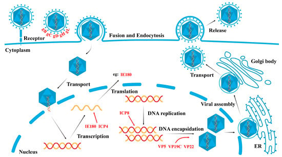
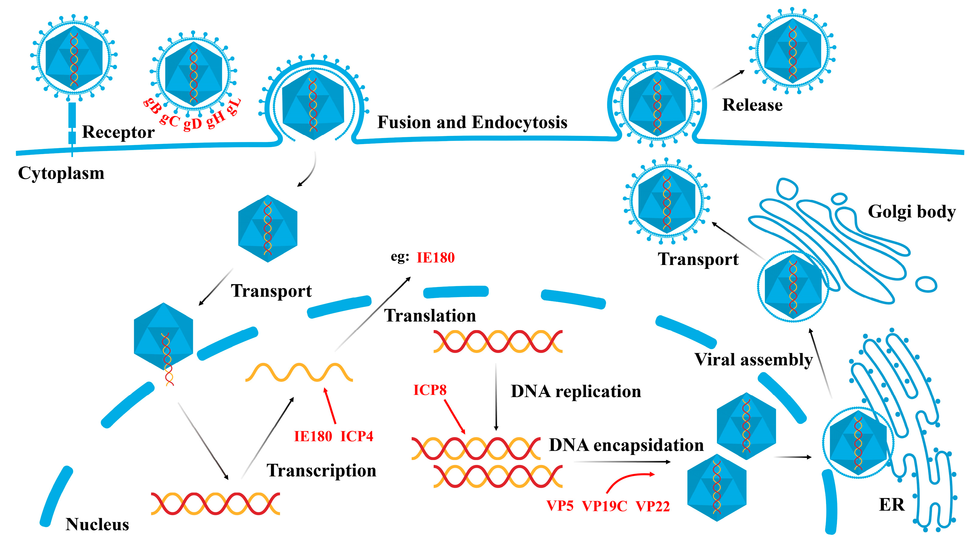
Like other alpha-herpesviruses, PRV can establish latent infections in its natural hosts and is often used as a model virus to study the biology of alpha-herpesviruses and host innate immune responses. The PRV genome is complex and highly structured, consisting of unique long (UL) and unique short (US) regions flanked by inverted repeat sequences. These regions encode proteins responsible for viral replication, virulence, and immune evasion. The PRV life cycle begins with the virus attaching to host cells through the viral glycoproteins gD, gC, gB, gH, and gL. Following attachment, the virus fuses with the host’s plasma membrane, allowing entry into the cell. The viral capsid and associated proteins are then transported to the nucleus, where the viral DNA is released. Inside the nucleus, the virus undergoes sequential gene expression, starting with the immediate-early gene IE180, followed by early genes involved in DNA replication and, finally, late genes encoding structural proteins. Newly synthesized viral DNA is packaged into capsids, which are assembled in the nucleus. The mature virions are enveloped and released from the cell to infect new cells (Figure 1). PRV can also establish latency in sensory neurons, remaining dormant until sensory neurons reactivate under certain conditions [1,8].
Figure 1.
Life cycle of a PRV. The life cycle of PRV begins with the interaction of PRV glycoproteins with specific receptors on the host cell surface, triggering endocytosis and fusion with the cell membrane. Once inside the cell, the viral nucleocapsid is transported to the nucleus, where the viral DNA is released. Inside the nucleus, viral genes undergo transcription, and the viral genome is subsequently replicated. The viral proteins are translated in the cytoplasm and transported back to the nucleus to participate in the assembly of the nucleocapsid. The nucleocapsid then exits the nucleus and is transported to the endoplasmic reticulum (ER) and Golgi apparatus, where it associates with membrane proteins and glycoproteins to form mature viral particles. These mature viral particles are ultimately released into the extracellular space through the process of budding.
Viral infections trigger rapid immune responses from the host to combat the infection. Among these responses, the type I interferon (IFN) pathway plays a pivotal role in viral suppression, immunomodulation, and adaptive immune response regulation [9,10]. Upon viral infection, host cells activate multiple signaling cascades that recognize specific vial components, disrupting viral replication. The recognition of viral DNA and the activation of downstream signaling pathways are critical in antiviral innate immunity. Cytosolic DNA sensors, such as Toll-like receptors (TLRs), RIG-I-like receptors (RLRs), IFI16 (interferon-gamma-inducible protein 16), AIM2 (absent in melanoma 2), and cGAS (cyclic GMP-AMP synthase), are essential for detecting DNA viruses such as PRV [11,12,13,14,15]. Once detected, these sensors initiate cellular signaling cascades that lead to the production of IFNs, the expression of IFN-stimulated genes (ISGs), and the establishment of an antiviral state [16,17].
As a DNA virus, PRV can establish persistent infections by evading IFN-I-mediated innate immune responses. This review examines recent findings on how PRV circumvents IFN-I-mediated antiviral immunity and the associated downstream signaling pathways [18].
3. The IFNAR–JAK–STAT Signaling Pathway and Its Downstream ISGs
The IFNAR–JAK–STAT signaling pathway plays a central role in antiviral innate immunity [88]. Upon binding to IFNAR1 and IFNAR2, IFN initiates a cascade that recruits and phosphorylates tyrosine kinase 2 (TYK2) and JAK1, leading to the activation of STAT1 and STAT2 [89,90]. The phosphorylated STAT1/STAT2 heterodimer binds to IRF9 in the cytoplasm, forming the IFN-stimulated gene factor 3 (ISGF3) complex, which translocates to the nucleus. There, it binds to ISG promoters, driving the transcription of hundreds of ISGs [91]. Viral inhibition of the JAK–STAT pathway is a common immune evasion strategy, as it prevents ISG production and IFN amplification. PRV inhibits IFN-induced STAT phosphorylation and ISG transcription [92]. Multiple studies have demonstrated that PRV disrupts the JAK–STAT pathway through mechanisms such as the degradation of key signaling molecules (such as IFNAR1 and JAKs), leading to the blockade of ISGF3 from binding to ISG promoters [93,94,95,96].
3.1. ISGs and the Antiviral Response
During PRV infection, ISGs can be induced through both interferon-dependent and interferon-independent mechanisms, helping the host establish an antiviral state. In response, PRV has evolved mechanisms to target ISGs and evade immune detection.
3.1.1. OASL
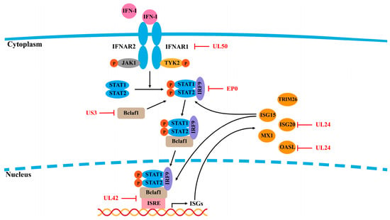
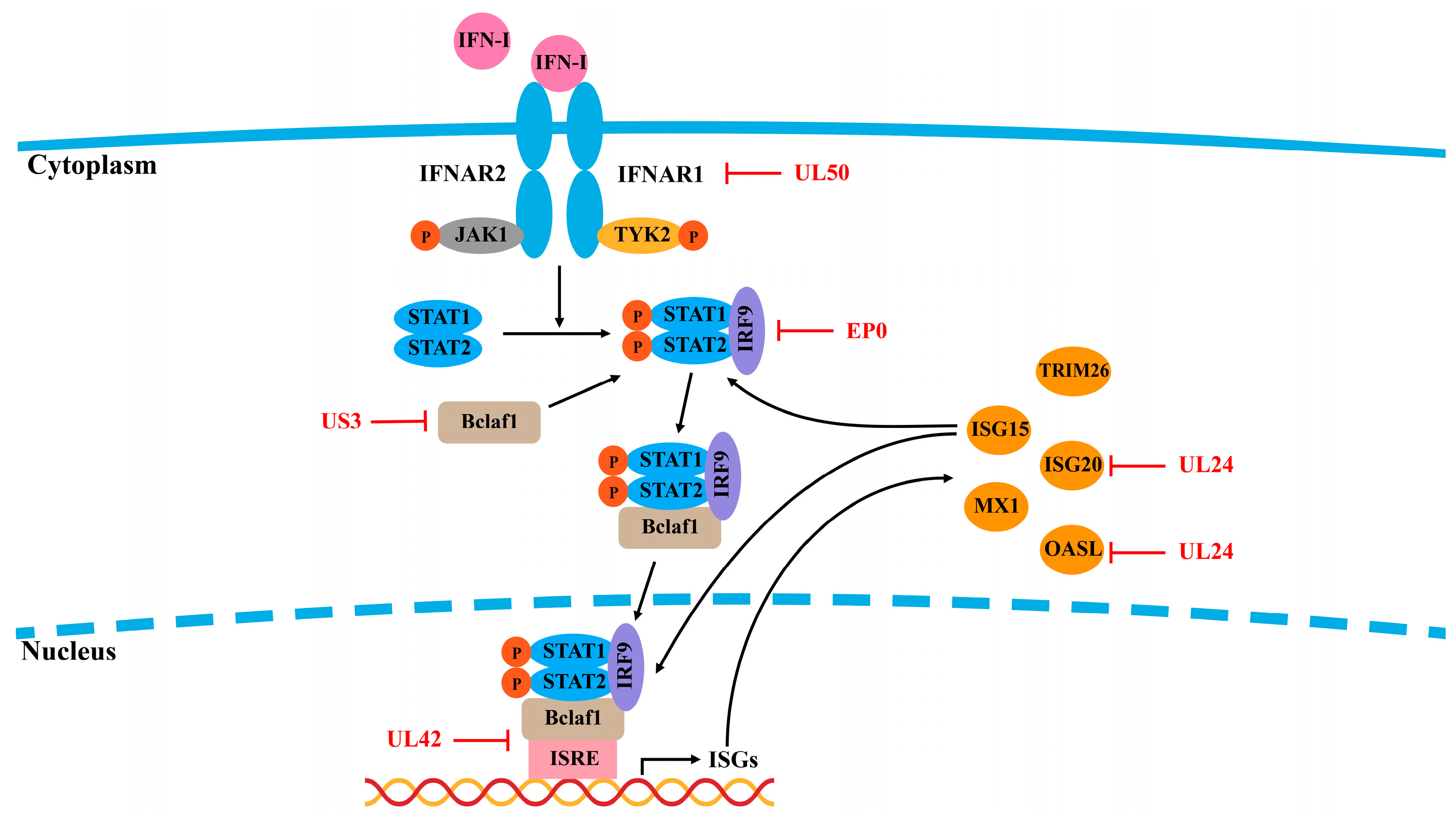
Oligoadenylate synthase (OAS) is one of the key IFN effectors that act in the early stages of viral infection by degrading viral RNA, thereby inhibiting viral replication [97]. The OAS family consists of four distinct OAS isoforms, one of which is oligoadenylate synthase-like (OASL). Research by Chen et al. demonstrated that OASL restricts PRV replication by enhancing RIG-I signaling and increasing RIG-I-mediated IFN expression. Notably, PRV counters this antiviral response via the UL24 protein to suppress OASL transcription, thereby impairing the RIG-I signaling pathway and counteracting OASL antiviral activity [45] (Figure 4).
Figure 4.
PRV evades the IFNAR–JAK–STAT signaling pathway. Type I IFNs exert their antiviral effects by binding to their heterodimeric receptors, IFNAR1 and IFNAR2, thereby activating the downstream JAK–STAT signaling pathway, which promotes the production of ISGs. Some ISGs, such as ISG15, can also enhance the JAK–STAT signaling pathway to upregulate the production of interferon-stimulated factors and increase antiviral activity. PRV-encoded proteins target components of the IFNAR–JAK–STAT signaling pathway to inhibit signal transduction or directly interact with ISGs, facilitating immune evasion.
3.1.2. ISG15 and ISG20
ISG15 and ISG20 are also critical in antiviral defense against PRV. ISG20 enhances IFN-β expression, but PRV UL24 suppresses ISG20 transcription, diminishing its antiviral effects [98]. Our studies indicate that ISG15 expression is upregulated during the early stages of PRV infection, where it impedes viral replication by increasing IFN-β expression and activating ISRE promoters [99]. Furthermore, ISG15 enhances IFN-α-mediated antiviral activity by facilitating STAT1/STAT2 nuclear translocation and STAT1/STAT2/IRF9 (ISGF3) complex formation [100]. Mice deficient in ISG15 presented increased susceptibility to PRV infection, although the mechanisms by which PRV evades ISG15-related antiviral immune responses remain to be elucidated [101]. Zhang et al. reported that PRV UL42 competitively binds to ISRE, thereby interfering with the binding of ISGF3 to ISRE, which results in decreased production of ISGs [94]. Additionally, the proteasomal degradation pathway is responsible for the degradation of Bclaf1 mediated by PRV US3, further impeding the binding of ISGF3 to ISRE [95] (Figure 4).
3.2. IFITM1 and IFITM2
IFN-inducible transmembrane proteins (IFITMs) are key host restriction factors with broad antiviral activity. IFITM2 plays a crucial role in controlling PRV by interfering with viral binding and entry [102]. Similarly, IFITM1 has significant antiviral functions, as PRV infection increases viral titers in IFITM1-knockdown cells and reduces PRV entry in IFITM1-overexpressing cells [103]. However, it is still unknown whether PRV antagonizes the IFITM function. A recently identified ISG protein, TMEM41B, promotes PRV replication by regulating lipid homeostasis [104].
3.3. PKR
Protein kinase R (PKR), which is activated by double-stranded RNA, is another ISG that plays a critical role in viral defense by phosphorylating eIF2α, reducing viral protein translation [105]. PRV has evolved mechanisms to counteract PKR activation and eIF2α phosphorylation [106,107]. For example, PRV suppresses eIF2α phosphorylation in vitro and in vivo, aiding in viral replication [108]. The PRV immediate-early protein IE180 also inhibits eIF2 phosphorylation during the early stages of viral replication [109]. While PKR is activated during PRV infection, the virus dephosphorylates eIF2α early on to increase virus replication [110].
3.4. TRIM26
Tripartite motif (TRIM) family proteins are characterized by a RING domain, B-box domains, a coiled-coil domain, and a variable C-terminal region that is responsible for interacting with diverse targets. These proteins play crucial roles in innate immunity, cell proliferation, autophagy, antiviral therapy, and tumor development. Among them, TRIM26 has been implicated in various biological processes, especially in regulating viral infections [111,112,113]. Wu et al. explored the interaction between TRIM26 and PRV and revealed that TRIM26 promotes PRV infection by facilitating NDP52-mediated autophagic degradation of MAVS. This discovery reveals a novel mechanism through which PRV evades host antiviral innate immunity, offering valuable insights into the interplay among viral infection, autophagy, and the innate immune response [114].
4. PRV Evades Apoptosis/Necrosis, Autophagy, and ER Stress
Upon PRV infection, viral proteins or nucleic acids trigger cellular stress responses, including apoptosis, necrosis, autophagy, and ER stress. These responses are critical for clearing the virus and maintaining cellular homeostasis. However, PRV, which can cause lifelong infections and severe diseases in various hosts, has developed multiple strategies to counteract these defenses.
4.1. Apoptosis
Apoptosis, also known as programmed cell death, is a vital defense mechanism against invading pathogens, including viruses. To evade this process, viruses have evolved sophisticated strategies to subvert apoptosis for immune evasion and enhanced viral replication. Several studies have shown that PRV infection induces apoptosis by upregulating the expression of pro-apoptotic Bcl family proteins [115], causing oxidative stress, free radical damage, and DNA damage [116]. Yeh et al. reported that PRV induces apoptosis via activation of the p38 MAPK and JNK/SAPK signaling pathways [117]. Additionally, low doses of Houttuynia cordata Thunb or emodin have been shown to inhibit PRV-induced apoptosis, reduce viral replication, and improve survival rates in mice [118,119].
Apoptosis is an important process regulating the pathogenesis of virus infection. Targeting apoptotic signaling during the late stages of infection represents a therapeutic strategy for herpesvirus-related diseases. Recent research revealed that caspases 3, 7, and 9 are activated in PRV-infected HeLa cells and that treatment with caspase inhibitors partially reduces apoptosis. PRV also modestly activates caspase-8 in PAMs and THP-1 cells, suggesting that caspase-8 may play a role in herpesvirus-induced apoptosis [120]. In addition to MAPK activation, PRV-induced oxidative stress and free radical formation are important contributors to apoptosis [117,121]. Interestingly, Aleman et al. reported that while PRV-infected neurons showed pathological changes, they presented no morphological or histochemical signs of apoptosis. Instead, apoptosis occurs primarily in the infiltrating immune cells surrounding PRV-infected neurons, suggesting that PRV may inhibit neuronal apoptosis to evade immune responses during acute infection [120].
PRV has developed several strategies to inhibit apoptotic signaling pathways. Notably, the PRV US3 protein has been shown to inhibit apoptosis in swine-testicle (ST) cells during the late stage of infection, depending on its enzyme activity [121]. Additionally, PRV activates the Akt and NF–κB pathways to protect infected cells from virus-induced apoptosis [121]. PRV US3 localizes to mitochondria and plays a crucial role in preventing the apoptosis induced by PRV [122]. Consistent with these findings, a recombinant virus lacking the US3 gene (PRV-∆US3) induces greater levels of apoptosis and has lower viral titers than wild-type PRV [123]. Broad-spectrum caspase inhibitors effectively inhibit apoptosis in ST and Hep-2 cells infected with either PRV-∆US3 or WT PRV [123]. A recent study elucidated the molecular mechanism by which the PRV glycoprotein M (gM), a late envelope glycoprotein, induces apoptosis during the late stage of infection. PRV-gM enhances PRV replication and pathogenicity by competitively interacting with BCL-XL to promote BAK oligomerization, ultimately activating caspase-3/7 [124].
4.2. Autophagy
Autophagy is an evolutionarily conserved catabolic process that plays a critical role in antiviral defense by degrading viral components and limiting viral replication. It is now recognized as an essential component of both innate and adaptive immunity. However, several herpesviruses, including PRV, have developed strategies to evade or exploit the autophagic machinery to increase their survival and replication [125,126]. Upon viral infection, host cells activate autophagy to degrade viral particles and components while simultaneously triggering an antiviral interferon response to antagonize viral replication.
Sun et al. explored the relationship between PRV replication and autophagy and reported that PRV induces autophagy in the early stages of infection [127]. However, as PRV infection progresses, PRV proteins suppress autophagy, increasing viral titers. Mechanistically, the tegument protein US3 may decrease autophagy levels by activating the AKT/mTOR pathway in infected cells [127]. These findings suggest that while autophagy initially contributes to PRV clearance, the virus has evolved mechanisms to antagonize this response. Xu et al. reported that PRV-induced autophagy enhances replication through the classical Beclin1–ATG7–ATG5 signaling pathway in neurons and that PRV infection promotes light chain 3 (LC3-I) autophagy in N2a cells [128]. Thus, PRV-induced autophagy both supports viral replication and antagonizes the host immune response.
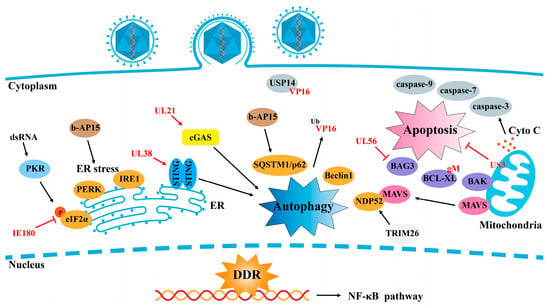
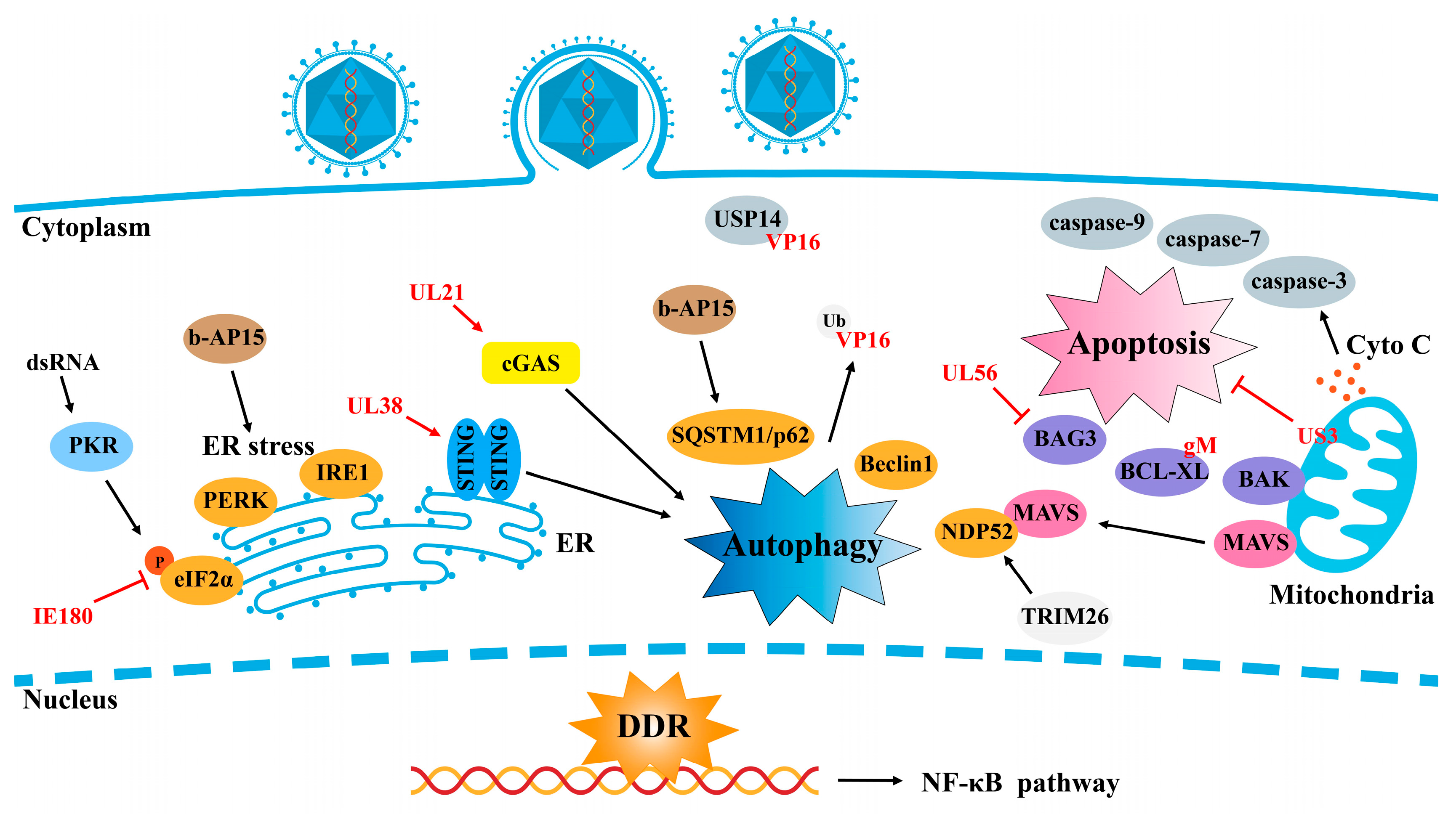
Lyu et al. identified a regulatory role for Bcl2-associated athanogene 3 (BAG3), a chaperone-mediated selective autophagy protein that downregulates PRV lytic infection [129]. In light of the complex relationship between autophagy and PRV, several therapeutic targets have been identified. Xing et al. reported that Platycodon grandiflorus polysaccharide (PGPS) could inhibit PRV replication by upregulating the AKT/mTOR pathway, thus reducing PRV-induced autophagy [130]. Ming et al. demonstrated that inhibiting deubiquitinase 14 (USP14) promotes the ubiquitination of PRV VP16, facilitating its degradation via SQSTM1/p62-mediated selective autophagy. Pretreatment with the USP14 inhibitor b-AP15 activates ER stress and autophagy, inhibiting PRV infection in vivo [131] (Figure 5), suggesting that USP14 is a potential therapeutic target for treating alpha-herpesvirus infections.
Figure 5.
PRV evades apoptosis/necrosis, autophagy, and ER stress. During PRV infection, cells can undergo apoptosis, necroptosis, autophagy, and ER stress in response to viral nucleic acids and proteins, which are aimed at limiting viral infection and exerting antiviral effects. However, PRV-encoded proteins can counteract these responses or target host proteins for autophagic degradation, thereby facilitating the evasion of the host immune response by PRV.
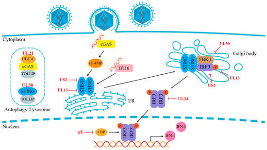
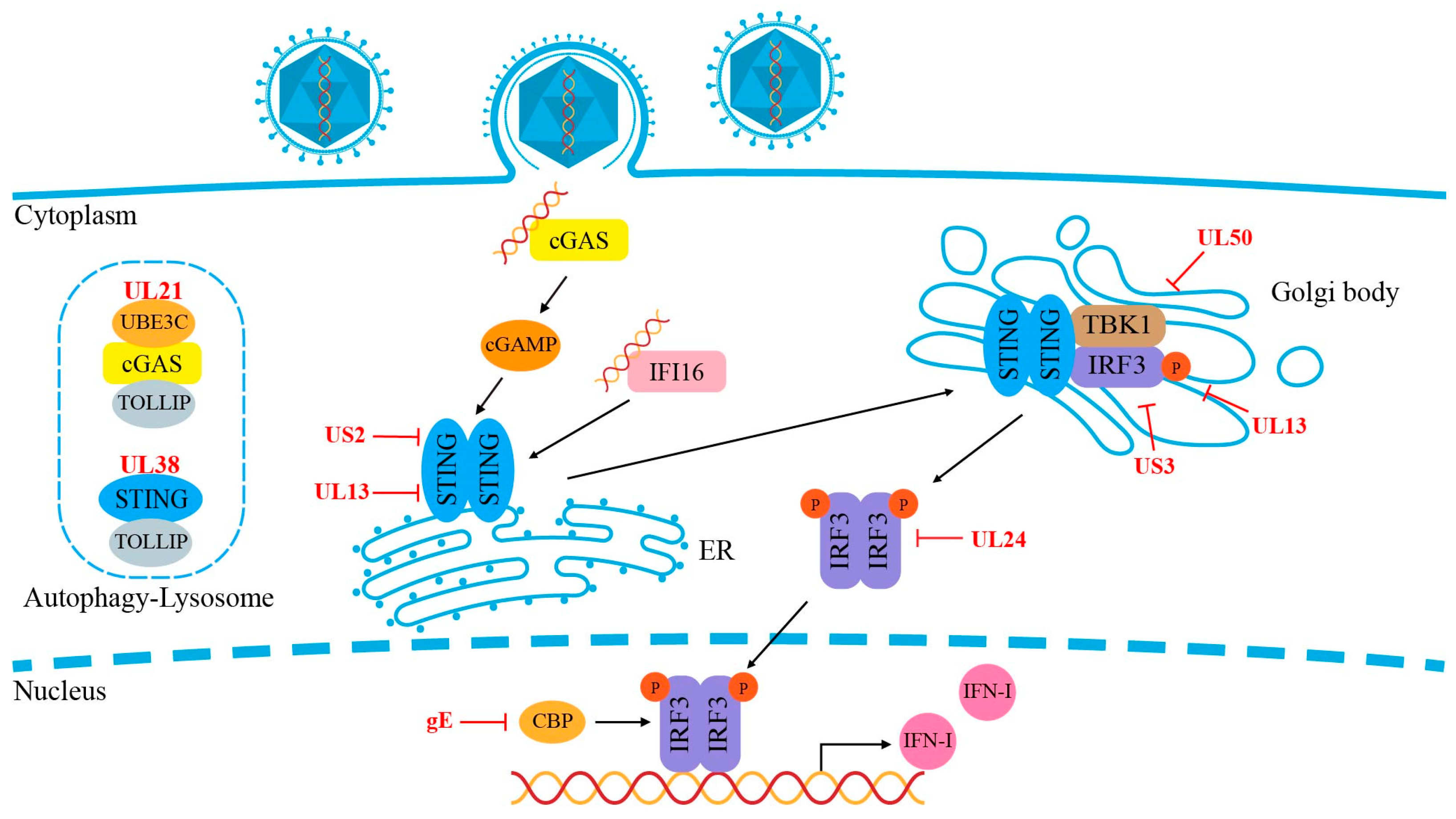
Ma et al. described a distinct mechanism by which PRV exploits TOLLIP-mediated selective autophagy to circumvent host immunity [64]. Their study revealed that the PRV tegument protein UL21 inhibits type I IFN signaling by degrading cGAS through the autophagic degradation of STING by recruiting TOLLIP, thereby inhibiting cGAS–STING antiviral signaling [66] (Figure 5), representing a significant immune evasion strategy employed by the PRV. Wu et al. also demonstrated that PRV-induced TRIM26 degrades MAVS through NDP52-mediated selective autophagy, providing further insight into the virus’s ability to escape antiviral innate immunity and the complex interactions among PRV, autophagy, and the host immune response [114].
4.3. ER Stress
The endoplasmic reticulum (ER) is a vital organelle responsible for key cellular processes, including protein synthesis, folding, transport, and secretion. The ER can detect various external stimuli, such as pathogen invasion, heat shock, hypoxia, and the accumulation of unfolded or misfolded proteins. In response, the ER activates the unfolded protein response (UPR) to restore cellular homeostasis [132]. Viral infections can also trigger autophagy via the UPR pathway [133,134,135].
Several studies have shown that viral infections induce ER stress, and some viruses exploit host cell membranes to build membrane components for their progeny virions during this process [136]. Alpha-herpesviruses, including PRV, have been found to suppress ER stress to increase viral replication. Yang et al. demonstrated that glucose-regulated protein 78 (GRP78), a marker of ER stress, is upregulated in the early stages of PRV infection, suggesting that PRV induces ER stress and activates UPR pathways [137]. Recent research has provided new insights into PRV replication and pathogenesis, showing that the activation of protein kinase RNA-like ER kinase (PERK) and inositol-requiring protein-1 (IRE1) pathways supports PRV replication in BHK21 cells, with glycoprotein B playing a pivotal role in inducing ER stress [138]. These findings expand our understanding of ER stress and the role of UPR in viral replication.
5. Conclusions
PRV can establish lifelong infections in hosts, leading to severe complications, especially in immunocompromised individuals. It is also a common cause of congenital viral infections. PRV triggers several host defense mechanisms, with the innate immune response being the first line of defense, primarily limiting viral infection by producing type I IFN and ISGs. Throughout the ongoing battle between viruses and hosts, PRV has developed various strategies to evade immune defenses, facilitating its survival and replication. Multiple viral proteins, particularly envelope and IE proteins, target key adapters in antiviral signaling pathways such as TLR2/4, cGAS, STING, NF–kB, TBK1, and IRF3 to inhibit host immune processes, including apoptosis, necrosis, autophagy, and ER stress, contributing to immune evasion and persistent infection.
Emerging evidence highlights the potential of TLR agonists as antibacterial, anticancer, and vaccine adjuvants, as well as their importance in vaccine development and immunotherapy against viruses such as HSV, HBV, and HIV. TLR antagonists have also been explored as therapeutic agents to suppress excessive immune responses. This review of the immune evasion mechanisms of PRV, particularly its ability to target PRRs and cellular processes, may offer valuable insights for the development of antiviral drugs and vaccines.
6. Future Perspective
As a member of the herpesvirus family, PRV serves as a valuable model for investigating the interactions between viral and host cellular mechanisms, particularly in the context of modulating innate immune signaling pathways and altering the antiviral state of host cells. Given its broad host range and the functional similarities of its encoded proteins to those of HSV, PRV offers significant potential for elucidating the roles of alpha-herpesvirus proteins in various physiological processes within host cells. This understanding may provide critical insights for the development of novel antiviral strategies. Based on the study of virus–host interactions, novel drug targets can be identified, facilitating the development of innovative antiviral therapeutics. Furthermore, our observations have highlighted the significant effects of PRV on cell death, including apoptosis, necrosis, and autophagy, as well as the interplay between viral infection and ER stress-related responses. This review elucidates the diversity of cellular antiviral mechanisms and broadens the scope for future research in antiviral immunity.
Author Contributions
Conceptualization and design: H.L. and H.C.; writing of the first draft: C.W. and H.L.; figures: L.L. and X.Z.; revision: H.L. and H.C.; final editing of text: H.L. and H.C. All authors have read and agreed to the published version of the manuscript.
Funding
This research was funded by the Science and Technology Program of Henan Province (grant no. 242102111027) and the Program for young backbone teachers in Universities of Henan Province (grant no. 2021GGJS034).
Conflicts of Interest
The authors declare no conflicts of interest.
References
- Pomeranz, L.E.; Reynolds, A.E.; Hengartner, C.J. Molecular Biology of Pseudorabies Virus: Impact on Neurovirology and Veterinary Medicine. Microbiol. Mol. Biol. Rev. 2005, 69, 462–500. [Google Scholar] [CrossRef] [Scilit] [PubMed]
- Cramer, S.D.; Campbell, G.A.; Njaa, B.L.; Morgan, S.E.; Smith, S.K.; McLin, W.R.; Brodersen, B.W.; Wise, A.G.; Scherba, G.; Langohr, I.M.; et al. Pseudorabies virus infection in Oklahoma hunting dogs. J. Vet. Diagn. Investig. 2011, 23, 915–923. [Google Scholar] [CrossRef] [Scilit] [PubMed]
- Bo, Z.; Li, X. A Review of Pseudorabies Virus Variants: Genomics, Vaccination, Transmission, and Zoonotic Potential. Viruses 2022, 14, 1003. [Google Scholar] [CrossRef] [Scilit] [PubMed]
- Tan, L.; Yao, J.; Yang, Y.; Luo, W.; Yuan, X.; Yang, L.; Wang, A. Current Status and Challenge of Pseudorabies Virus Infection in China. Virol. Sin. 2021, 36, 588–607. [Google Scholar] [CrossRef] [Scilit] [PubMed]
- Liu, Q.; Kuang, Y.; Li, Y.; Guo, H.; Zhou, C.; Guo, S.; Tan, C.; Wu, B.; Chen, H.; Wang, X. The Epidemiology and Variation in Pseudorabies Virus: A Continuing Challenge to Pigs and Humans. Viruses 2022, 14, 1463. [Google Scholar] [CrossRef] [Scilit]
- Guo, Z.; Chen, X.X.; Zhang, G. Human PRV Infection in China: An Alarm to Accelerate Eradication of PRV in Domestic Pigs. Virol. Sin. 2021, 36, 823–828. [Google Scholar] [CrossRef] [Scilit]
- Wong, G.; Lu, J.; Zhang, W.; Gao, G.F. Pseudorabies virus: A neglected zoonotic pathogen in humans? Emerg. Microbes Infect. 2019, 8, 150–154. [Google Scholar] [CrossRef] [Scilit]
- Nauwynck, H.; Glorieux, S.; Favoreel, H.; Pensaert, M. Cell biological and molecular characteristics of pseudorabies virus infections in cell cultures and in pigs with emphasis on the respiratory tract. Vet. Res. 2007, 38, 229–241. [Google Scholar] [CrossRef] [Scilit]
- Samuel, C.E. Antiviral Actions of Interferons. Clin. Microbiol. Rev. 2001, 14, 778–809. [Google Scholar] [CrossRef] [Scilit]
- Katze, M.G.; He, Y.; Gale, M. Viruses and interferon: A fight for supremacy. Nat. Rev. Immunol. 2002, 2, 675–687. [Google Scholar] [CrossRef] [Scilit]
- Hemmi, H.; Takeuchi, O.; Kawai, T.; Kaisho, T.; Sato, S.; Sanjo, H.; Matsumoto, M.; Hoshino, K.; Wagner, H.; Takeda, K.; et al. A Toll-like receptor recognizes bacterial DNA. Nature 2000, 408, 740–745. [Google Scholar] [CrossRef] [Scilit] [PubMed]
- Unterholzner, L.; Keating, S.E.; Baran, M.; Horan, K.A.; Jensen, S.B.; Sharma, S.; Sirois, C.M.; Jin, T.; Latz, E.; Xiao, T.S.; et al. IFI16 is an innate immune sensor for intracellular DNA. Nat. Immunol. 2010, 11, 997–1004. [Google Scholar] [CrossRef] [Scilit] [PubMed]
- Hornung, V.; Ablasser, A.; Charrel-Dennis, M.; Bauernfeind, F.; Horvath, G.; Caffrey, D.R.; Latz, E.; Fitzgerald, K.A. AIM2 recognizes cytosolic dsDNA and forms a caspase-1-activating inflammasome with ASC. Nature 2009, 458, 514–518. [Google Scholar] [CrossRef] [Scilit]
- Takaoka, A.; Wang, Z.; Choi, M.K.; Yanai, H.; Negishi, H.; Ban, T.; Lu, Y.; Miyagishi, M.; Kodama, T.; Honda, K.; et al. DAI (DLM-1/ZBP1) is a cytosolic DNA sensor and an activator of innate immune response. Nature 2007, 448, 501–505. [Google Scholar] [CrossRef] [Scilit]
- Sun, L.; Wu, J.; Du, F.; Chen, X.; Chen, Z.J. Cyclic GMP-AMP synthase is a cytosolic DNA sensor that activates the type I interferon pathway. Science 2013, 339, 786–791. [Google Scholar] [CrossRef] [Scilit]
- Ma, Z.; Damania, B. The cGAS-STING Defense Pathway and Its Counteraction by Viruses. Cell Host Microbe 2016, 19, 150–158. [Google Scholar] [CrossRef] [Scilit]
- Lee, H.C.; Chathuranga, K.; Lee, J.S. Intracellular sensing of viral genomes and viral evasion. Exp. Mol. Med. 2019, 51, 1–13. [Google Scholar] [CrossRef] [Scilit]
- Zhang, R.; Tang, J. Evasion of I Interferon-Mediated Innate Immunity by Pseudorabies Virus. Front. Microbiol. 2021, 12, 801257. [Google Scholar] [CrossRef] [Scilit]
- Fernandes-Santos, C.; de Azeredo, E.L. Innate Immune Response to Dengue Virus: Toll-like Receptors and Antiviral Response. Viruses 2022, 14, 992. [Google Scholar] [CrossRef] [Scilit]
- Liu, Z.-M.; Yang, M.-H.; Yu, K.; Lian, Z.-X.; Deng, S.-L. Toll-like receptor (TLRs) agonists and antagonists for COVID-19 treatments. Front. Pharmacol. 2022, 13, 989664. [Google Scholar] [CrossRef] [Scilit]
- Heil, F.; Hemmi, H.; Hochrein, H.; Ampenberger, F.; Kirschning, C.; Akira, S.; Lipford, G.; Wagner, H.; Bauer, S. Species-Specific Recognition of Single-Stranded RNA via Toll-like Receptor 7 and 8. Science 2004, 303, 1526–1529. [Google Scholar] [CrossRef] [Scilit] [PubMed]
- Barton, G.M. Viral recognition by Toll-like receptors. Semin. Immunol. 2007, 19, 33–40. [Google Scholar] [CrossRef] [Scilit] [PubMed]
- Préhaud, C.; Mégret, F.; Lafage, M.; Lafon, M. Virus Infection Switches TLR-3-Positive Human Neurons to Become Strong Producers of Beta Interferon. J. Virol. 2005, 79, 12893–12904. [Google Scholar] [CrossRef] [Scilit] [PubMed]
- Kim, C.-U.; Jeong, Y.-J.; Lee, P.; Lee, M.-S.; Park, J.-H.; Kim, Y.-S.; Kim, D.-J. Extracellular nucleoprotein exacerbates influenza virus pathogenesis by activating Toll-like receptor 4 and the NLRP3 inflammasome. Cell. Mol. Immunol. 2022, 19, 715–725. [Google Scholar] [CrossRef] [Scilit]
- Gay, N.J.; Symmons, M.F.; Gangloff, M.; Bryant, C.E. Assembly and localization of Toll-like receptor signalling complexes. Nat. Rev. Immunol. 2014, 14, 546–558. [Google Scholar] [CrossRef] [Scilit]
- Xagorari, A.; Chlichlia, K. Toll-like receptors and viruses: Induction of innate antiviral immune responses. Open Microbiol. J. 2008, 2, 49–59. [Google Scholar] [CrossRef] [Scilit]
- Kurt-Jones, E.A.; Chan, M.; Zhou, S.; Wang, J.; Reed, G.; Bronson, R.; Arnold, M.M.; Knipe, D.M.; Finberg, R.W. Herpes simplex virus 1 interaction with Toll-like receptor 2 contributes to lethal encephalitis. Proc. Natl. Acad. Sci. USA 2004, 101, 1315–1320. [Google Scholar] [CrossRef] [Scilit]
- Delale, T.; Paquin, A.; Asselin-Paturel, C.; Dalod, M.; Brizard, G.; Bates, E.E.M.; Kastner, P.; Chan, S.; Akira, S.; Vicari, A.; et al. MyD88-Dependent and -Independent Murine Cytomegalovirus Sensing for IFN-α Release and Initiation of Immune Responses In Vivo1. J. Immunol. 2005, 175, 6723–6732. [Google Scholar] [CrossRef] [Scilit]
- Compton, T.; Kurt-Jones, E.A.; Boehme, K.W.; Belko, J.; Latz, E.; Golenbock, D.T.; Finberg, R.W. Human Cytomegalovirus Activates Inflammatory Cytokine Responses via CD14 and Toll-Like Receptor 2. J. Virol. 2003, 77, 4588–4596. [Google Scholar] [CrossRef] [Scilit]
- Boehme, K.W.; Guerrero, M.; Compton, T. Human Cytomegalovirus Envelope Glycoproteins B and H Are Necessary for TLR2 Activation in Permissive Cells1. J. Immunol. 2006, 177, 7094–7102. [Google Scholar] [CrossRef] [Scilit]
- Arav-Boger, R.; Wojcik, G.L.; Duggal, P.; Ingersoll, R.G.; Beaty, T.; Pass, R.F.; Yolken, R.H. Polymorphisms in Toll-like receptor genes influence antibody responses to cytomegalovirus glycoprotein B vaccine. BMC Res. Notes 2012, 5, 140. [Google Scholar] [CrossRef] [Scilit] [PubMed]
- Xie, F.; von Dadelszen, P.; Nadeau, J. CMV Infection, TLR-2 and -4 Expression, and Cytokine Profiles in Early-Onset Preeclampsia with HELLP Syndrome. Am. J. Reprod. Immunol. 2014, 71, 379–386. [Google Scholar] [CrossRef] [Scilit] [PubMed]
- Redondo, N.; Rodríguez-Goncer, I.; Parra, P.; Ruiz-Merlo, T.; López-Medrano, F.; González, E.; Polanco, N.; Trujillo, H.; Hernández, A.; San Juan, R.; et al. Influence of single-nucleotide polymorphisms in TLR3 (rs3775291) and TLR9 (rs352139) on the risk of CMV infection in kidney transplant recipients. Front. Immunol. 2022, 13, 929995. [Google Scholar] [CrossRef] [Scilit] [PubMed]
- Zhou, Q.; Zhang, L.; Lin, Q.; Liu, H.; Ye, G.; Liu, X.; Jiao, S.; Li, J.; Tang, Y.; Shi, D.; et al. Pseudorabies Virus Infection Activates the TLR-NF-κB Axis and AIM2 Inflammasome to Enhance Inflammatory Responses in Mice. J. Virol. 2023, 97, e0000323. [Google Scholar] [CrossRef] [Scilit]
- Ye, C.; Wan, C.; Chen, J.; Li, G.; Li, Y.; Wang, Y.; Tao, Q.; Peng, L.; Fang, R. Cathelicidin CATH-B1 Inhibits Pseudorabies Virus Infection via Direct Interaction and TLR4/JNK/IRF3-Mediated Interferon Activation. J. Virol. 2023, 97, e0070623. [Google Scholar] [CrossRef] [Scilit]
- Stok, J.E.; Vega Quiroz, M.E.; van der Veen, A.G. Self RNA Sensing by RIG-I-like Receptors in Viral Infection and Sterile Inflammation. J. Immunol. 2020, 205, 883–891. [Google Scholar] [CrossRef] [Scilit]
- Onomoto, K.; Onoguchi, K.; Yoneyama, M. Regulation of RIG-I-like receptor-mediated signaling: Interaction between host and viral factors. Cell. Mol. Immunol. 2021, 18, 539–555. [Google Scholar] [CrossRef] [Scilit]
- Crill, E.K.; Furr-Rogers, S.R.; Marriott, I. RIG-I is required for VSV-induced cytokine production by murine glia and acts in combination with DAI to initiate responses to HSV-1. Glia 2015, 63, 2168–2180. [Google Scholar] [CrossRef] [Scilit]
- Zhu, H.; Zheng, C. The Race between Host Antiviral Innate Immunity and the Immune Evasion Strategies of Herpes Simplex Virus 1. Microbiol. Mol. Biol. Rev. 2020, 84, 1110–1128. [Google Scholar] [CrossRef] [Scilit]
- Zhao, J.; Qin, C.; Liu, Y.; Rao, Y.; Feng, P. Herpes Simplex Virus and Pattern Recognition Receptors: An Arms Race. Front. Immunol. 2021, 11, 613799. [Google Scholar] [CrossRef] [Scilit]
- Yu, H.; Bruneau, R.C.; Brennan, G.; Rothenburg, S. Battle Royale: Innate Recognition of Poxviruses and Viral Immune Evasion. Biomedicines 2021, 9, 765. [Google Scholar] [CrossRef] [Scilit] [PubMed]
- Chiang, J.J.; Sparrer, K.M.J.; van Gent, M.; Lässig, C.; Huang, T.; Osterrieder, N.; Hopfner, K.-P.; Gack, M.U. Viral unmasking of cellular 5S rRNA pseudogene transcripts induces RIG-I-mediated immunity. Nat. Immunol. 2018, 19, 53–62. [Google Scholar] [CrossRef] [Scilit] [PubMed]
- Xing, J.; Wang, S.; Lin, R.; Mossman, K.L.; Zheng, C. Herpes Simplex Virus 1 Tegument Protein US11 Downmodulates the RLR Signaling Pathway via Direct Interaction with RIG-I and MDA-5. J. Virol. 2012, 86, 3528–3540. [Google Scholar] [CrossRef] [Scilit]
- Zhao, J.; Zeng, Y.; Xu, S.; Chen, J.; Shen, G.; Yu, C.; Knipe, D.; Yuan, W.; Peng, J.; Xu, W.; et al. A Viral Deamidase Targets the Helicase Domain of RIG-I to Block RNA-Induced Activation. Cell Host Microbe 2016, 20, 770–784. [Google Scholar] [CrossRef] [Scilit]
- Chen, X.; Kong, N.; Xu, J.; Wang, J.; Zhang, M.; Ruan, K.; Li, L.; Zhang, Y.; Zheng, H.; Tong, W.; et al. Pseudorabies virus UL24 antagonizes OASL-mediated antiviral effect. Virus Res. 2021, 295, 198276. [Google Scholar] [CrossRef] [Scilit]
- Zhao, N.; Wang, F.; Kong, Z.; Shang, Y. Pseudorabies Virus Tegument Protein UL13 Suppresses RLR-Mediated Antiviral Innate Immunity through Regulating Receptor Transcription. Viruses 2022, 14, 1465. [Google Scholar] [CrossRef] [Scilit]
- Tuladhar, S.; Kanneganti, T.D. NLRP12 in innate immunity and inflammation. Mol. Asp. Med. 2020, 76, 100887. [Google Scholar] [CrossRef] [Scilit]
- Elliott, E.I.; Sutterwala, F.S. Initiation and perpetuation of NLRP3 inflammasome activation and assembly. Immunol. Rev. 2015, 265, 35–52. [Google Scholar] [CrossRef] [Scilit]
- Laval, K.; Vernejoul, J.B.; Cleemput, J.V.; Koyuncu, O.O.; Enquist, L.W. Virulent Pseudorabies Virus Infection Induces a Specific and Lethal Systemic Inflammatory Response in Mice. J. Virol. 2018, 92, e01614-18. [Google Scholar] [CrossRef] [Scilit]
- Ye, C.; Huang, Q.; Jiang, J.; Li, G.; Xu, D.; Zeng, Z.; Peng, L.; Peng, Y.; Fang, R. ATP-dependent activation of NLRP3 inflammasome in primary murine macrophages infected by pseudorabies virus. Vet. Microbiol. 2021, 259, 109130. [Google Scholar] [CrossRef] [Scilit]
- Sun, W.; Liu, S.; Huang, X.; Yuan, R.; Yu, J. Cytokine storms and pyroptosis are primarily responsible for the rapid death of mice infected with pseudorabies virus. R. Soc. Open Sci. 2021, 8, 210296. [Google Scholar] [CrossRef] [Scilit] [PubMed]
- Yu, D.; Zheng, S.; Sui, L.; Xi, Y.; He, T.; Liu, Z. The role of AIM2 in inflammation and tumors. Front Immunol. 2024, 15, 1466440. [Google Scholar] [CrossRef] [Scilit]
- Briard, B.; Place, D.E.; Kanneganti, T.-D. DNA Sensing in the Innate Immune Response. Physiology 2020, 35, 112–124. [Google Scholar] [CrossRef] [Scilit]
- Huérfano, S.; Šroller, V.; Bruštíková, K.; Horníková, L.; Forstová, J. The Interplay between Viruses and Host DNA Sensors. Viruses 2022, 14, 666. [Google Scholar] [CrossRef] [Scilit]
- Kwon, J.; Bakhoum, S.F. The Cytosolic DNA-Sensing cGAS–STING Pathway in Cancer. Cancer Discov. 2020, 10, 26–39. [Google Scholar] [CrossRef] [Scilit]
- Decout, A.; Katz, J.D.; Venkatraman, S.; Ablasser, A. The cGAS–STING pathway as a therapeutic target in inflammatory diseases. Nat. Rev. Immunol. 2021, 21, 548–569. [Google Scholar] [CrossRef] [Scilit]
- Erttmann, S.F.; Swacha, P.; Aung, K.M.; Brindefalk, B.; Jiang, H.; Härtlova, A.; Uhlin, B.E.; Wai, S.N.; Gekara, N.O. The gut microbiota prime systemic antiviral immunity via the cGAS-STING-IFN-I axis. Immunity 2022, 55, 847–861.e10. [Google Scholar] [CrossRef] [Scilit]
- Wang, J.; Li, G.L.; Ming, S.L.; Wang, C.F.; Shi, L.J.; Su, B.Q.; Wu, H.T.; Zeng, L.; Han, Y.Q.; Liu, Z.H.; et al. BRD4 inhibition exerts anti-viral activity through DNA damage-dependent innate immune responses. PLoS Pathog. 2020, 16, e1008429. [Google Scholar] [CrossRef] [Scilit]
- Wang, J.; Lu, S.F.; Wan, B.; Ming, S.L.; Li, G.L.; Su, B.Q.; Liu, J.Y.; Wei, Y.S.; Yang, G.Y.; Chu, B.B. Maintenance of cyclic GMP-AMP homeostasis by ENPP1 is involved in pseudorabies virus infection. Mol. Immunol. 2018, 95, 56–63. [Google Scholar] [CrossRef] [Scilit]
- Eaglesham, J.B.; Kranzusch, P.J. Conserved strategies for pathogen evasion of cGAS-STING immunity. Curr. Opin. Immunol. 2020, 66, 27–34. [Google Scholar] [CrossRef] [Scilit]
- Bo, Z.; Miao, Y.; Xi, R.; Zhong, Q.; Bao, C.; Chen, H.; Sun, L.; Qian, Y.; Jung, Y.S.; Dai, J. PRV UL13 inhibits cGAS-STING-mediated IFN-β production by phosphorylating IRF3. Vet. Res. 2020, 51, 118. [Google Scholar] [CrossRef] [Scilit] [PubMed]
- Lv, L.; Cao, M.; Bai, J.; Jin, L.; Wang, X.; Gao, Y.; Liu, X.; Jiang, P. PRV-encoded UL13 protein kinase acts as an antagonist of innate immunity by targeting IRF3-signaling pathways. Vet. Microbiol. 2020, 250, 108860. [Google Scholar] [CrossRef] [Scilit] [PubMed]
- Liu, X.; Zhang, M.; Ye, C.; Ruan, K.; Xu, A.; Gao, F.; Tong, G.; Zheng, H. Inhibition of the DNA-Sensing pathway by pseudorabies virus UL24 protein via degradation of interferon regulatory factor 7. Vet. Microbiol. 2021, 255, 109023. [Google Scholar] [CrossRef] [Scilit]
- Ma, Z.; Bai, J.; Jiang, C.; Zhu, H.; Liu, D.; Pan, M.; Wang, X.; Pi, J.; Jiang, P.; Liu, X. Tegument protein UL21 of alpha-herpesvirus inhibits the innate immunity by triggering CGAS degradation through TOLLIP-mediated selective autophagy. Autophagy 2023, 19, 1512–1532. [Google Scholar] [CrossRef] [Scilit]
- Xie, J.; Zhang, X.; Chen, L.; Bi, Y.; Idris, A.; Xu, S.; Li, X.; Zhang, Y.; Feng, R. Pseudorabies Virus US3 Protein Inhibits IFN-β Production by Interacting with IRF3 to Block Its Activation. Front. Microbiol. 2021, 12, 761282. [Google Scholar] [CrossRef] [Scilit] [PubMed]
- Yan, Z.; Xie, J.; Hou, Z.; Zhang, Y.; Yue, J.; Zhang, X.; Chen, L.; Yang, Y.; Li, X.; Li, H.; et al. Pseudorabies virus UL38 attenuates the cGAS-STING signaling pathway by recruiting Tollip to promote STING for autophagy degradation. Virol. J. 2024, 21, 107. [Google Scholar] [CrossRef] [Scilit] [PubMed]
- Lu, M.; Qiu, S.; Zhang, L.; Sun, Y.; Bao, E.; Lv, Y. Pseudorabies virus glycoprotein gE suppresses interferon-β production via CREB-binding protein degradation. Virus Res. 2021, 291, 198220. [Google Scholar] [CrossRef] [Scilit]
- Burdette, D.L.; Vance, R.E. STING and the innate immune response to nucleic acids in the cytosol. Nat. Immunol. 2013, 14, 19–26. [Google Scholar] [CrossRef] [Scilit]
- Zhang, Z.-D.; Zhong, B. Regulation and function of the cGAS-MITA/STING axis in health and disease. Cell Insight 2022, 1, 100001. [Google Scholar] [CrossRef] [Scilit]
- Kong, Z.; Yin, H.; Wang, F.; Liu, Z.; Luan, X.; Sun, L.; Liu, W.; Shang, Y. Pseudorabies virus tegument protein UL13 recruits RNF5 to inhibit STING-mediated antiviral immunity. PLoS Pathog. 2022, 18, e1010544. [Google Scholar] [CrossRef] [Scilit]
- Kong, Z.; Chen, X.; Gong, L.; Wang, L.; Zhang, Y.; Guan, K.; Yao, W.; Kang, Y.; Lu, X.; Zhang, Y.; et al. Pseudorabies virus tegument protein US2 antagonizes antiviral innate immunity by targeting cGAS-STING signaling pathway. Front. Immunol. 2024, 15, 1403070. [Google Scholar] [CrossRef] [Scilit] [PubMed]
- Li, T.; Chen, J.; Cristea, I.M. Human cytomegalovirus tegument protein pUL83 inhibits IFI16-mediated DNA sensing for immune evasion. Cell Host Microbe 2013, 14, 591–599. [Google Scholar] [CrossRef] [Scilit] [PubMed]
- Jiang, Z.; Wei, F.; Zhang, Y.; Wang, T.; Gao, W.; Yu, S.; Sun, H.; Pu, J.; Sun, Y.; Wang, M.; et al. IFI16 directly senses viral RNA and enhances RIG-I transcription and activation to restrict influenza virus infection. Nat. Microbiol. 2021, 6, 932–945. [Google Scholar] [CrossRef] [Scilit] [PubMed]
- Jønsson, K.L.; Laustsen, A.; Krapp, C.; Skipper, K.A.; Thavachelvam, K.; Hotter, D.; Egedal, J.H.; Kjolby, M.; Mohammadi, P.; Prabakaran, T.; et al. IFI16 is required for DNA sensing in human macrophages by promoting production and function of cGAMP. Nat. Commun. 2017, 8, 14391. [Google Scholar] [CrossRef] [Scilit]
- Orzalli, M.H.; DeLuca, N.A.; Knipe, D.M. Nuclear IFI16 induction of IRF-3 signaling during herpesviral infection and degradation of IFI16 by the viral ICP0 protein. Proc. Natl. Acad. Sci. USA 2012, 109, E3008–E3017. [Google Scholar] [CrossRef] [Scilit]
- Diner, B.A.; Lum, K.K.; Toettcher, J.E.; Cristea, I.M. Viral DNA Sensors IFI16 and Cyclic GMP-AMP Synthase Possess Distinct Functions in Regulating Viral Gene Expression, Immune Defenses, and Apoptotic Responses during Herpesvirus Infection. mBio 2016, 7, e01553-16. [Google Scholar] [CrossRef] [Scilit]
- Lee, H.-R.; Choi, U.Y.; Hwang, S.-W.; Kim, S.; Jung, J.U. Viral Inhibition of PRR-Mediated Innate Immune Response: Learning from KSHV Evasion Strategies. Mol. Cells 2016, 39, 777–782. [Google Scholar] [CrossRef] [Scilit]
- Jakobsen, M.R.; Bak, R.O.; Andersen, A.; Berg, R.K.; Jensen, S.B.; Jin, T.; Laustsen, A.; Hansen, K.; Østergaard, L.; Fitzgerald, K.A.; et al. IFI16 senses DNA forms of the lentiviral replication cycle and controls HIV-1 replication. Proc. Natl. Acad. Sci. USA 2013, 110, E4571–E4580. [Google Scholar] [CrossRef] [Scilit]
- Zhang, X.; Chen, G.; Yin, J.; Li, L.; Huang, K.; Du, Q.; Tong, D.; Huang, Y. Pseudorabies virus infection activates the NLRP3 and IFI16 inflammasomes to trigger pyroptosis. Vet. Microbiol. 2023, 284, 109826. [Google Scholar] [CrossRef] [Scilit]
- Yum, S.; Li, M.; Fang, Y.; Chen, Z.J. TBK1 recruitment to STING activates both IRF3 and NF-κB that mediate immune defense against tumors and viral infections. Proc. Natl. Acad. Sci. USA 2021, 118, e2100225118. [Google Scholar] [CrossRef] [Scilit]
- Lv, L.; Bai, J.; Gao, Y.; Jin, L.; Wang, X.; Cao, M.; Liu, X.; Jiang, P. Peroxiredoxin 1 Interacts with TBK1/IKKε and Negatively Regulates Pseudorabies Virus Propagation by Promoting Innate Immunity. J. Virol. 2021, 95, e00923-21. [Google Scholar] [CrossRef] [Scilit] [PubMed]
- Bui-Nguyen, T.M.; Pakala, S.B.; Sirigiri, R.D.; Xia, W.; Hung, M.C.; Sarin, S.K.; Kumar, V.; Slagle, B.L.; Kumar, R. NF-kappaB signaling mediates the induction of MTA1 by hepatitis B virus transactivator protein HBx. Oncogene 2010, 29, 1179–1189. [Google Scholar] [CrossRef] [Scilit] [PubMed]
- Rodríguez, C.I.; Nogal, M.L.; Carrascosa, A.L.; Salas, M.L.; Fresno, M.; Revilla, Y. African swine fever virus IAP-like protein induces the activation of nuclear factor kappa B. J. Virol. 2002, 76, 3936–3942. [Google Scholar] [CrossRef] [Scilit] [PubMed]
- de Oliveira, D.E.; Ballon, G.; Cesarman, E. NF-kappaB signaling modulation by EBV and KSHV. Trends Microbiol. 2010, 18, 248–257. [Google Scholar] [CrossRef] [Scilit] [PubMed]
- Zhang, X.; Xie, J.; Gao, M.; Yan, Z.; Chen, L.; Wei, S.; Feng, R. Pseudorabies Virus ICP0 Abolishes Tumor Necrosis Factor Alpha-Induced NF-κB Activation by Degrading P65. Viruses 2022, 14, 954. [Google Scholar] [CrossRef] [Scilit]
- Wang, T.Y.; Yang, Y.L.; Feng, C.; Sun, M.X.; Peng, J.M.; Tian, Z.J.; Tang, Y.D.; Cai, X.H. Pseudorabies Virus UL24 Abrogates Tumor Necrosis Factor Alpha-Induced NF-κB Activation by Degrading P65. Viruses 2020, 12, 51. [Google Scholar] [CrossRef] [Scilit]
- Romero, N.; Favoreel, H.W. Pseudorabies Virus Infection Triggers NF-κB Activation via the DNA Damage Response but Actively Inhibits NF-κB-Dependent Gene Expression. J. Virol. 2021, 95, e0166621. [Google Scholar] [CrossRef] [Scilit]
- de Weerd, N.A.; Samarajiwa, S.A.; Hertzog, P.J. Type I Interferon Receptors: Biochemistry and Biological Functions *. J. Biol. Chem. 2007, 282, 20053–20057. [Google Scholar] [CrossRef] [Scilit]
- Platanias, L.C. Mechanisms of type-I- and type-II-interferon-mediated signalling. Nat. Rev. Immunol. 2005, 5, 375–386. [Google Scholar] [CrossRef] [Scilit]
- Stark, G.R.; Darnell, J.E., Jr. The JAK-STAT Pathway at Twenty. Immunity 2012, 36, 503–514. [Google Scholar] [CrossRef] [Scilit]
- McCormack, R.; Hunte, R.; Podack, E.R.; Plano, G.V.; Shembade, N. An Essential Role for Perforin-2 in Type I IFN Signaling. J. Immunol. 2020, 204, 2242–2256. [Google Scholar] [CrossRef] [Scilit] [PubMed]
- Brukman, A.; Enquist, L.W. Suppression of the interferon-mediated innate immune response by pseudorabies virus. J. Virol. 2006, 80, 6345–6356. [Google Scholar] [CrossRef] [Scilit]
- Zhang, R.; Xu, A.; Qin, C.; Zhang, Q.; Chen, S.; Lang, Y.; Wang, M.; Li, C.; Feng, W.; Zhang, R.; et al. Pseudorabies Virus dUTPase UL50 Induces Lysosomal Degradation of Type I Interferon Receptor 1 and Antagonizes the Alpha Interferon Response. J. Virol. 2017, 91, e01148-17. [Google Scholar] [CrossRef] [Scilit] [PubMed]
- Zhang, R.; Chen, S.; Zhang, Y.; Wang, M.; Qin, C.; Yu, C.; Zhang, Y.; Li, Y.; Chen, L.; Zhang, X.; et al. Pseudorabies Virus DNA Polymerase Processivity Factor UL42 Inhibits Type I IFN Response by Preventing ISGF3-ISRE Interaction. J. Immunol. 2021, 207, 613–625. [Google Scholar] [CrossRef] [Scilit]
- Qin, C.; Zhang, R.; Lang, Y.; Shao, A.; Xu, A.; Feng, W.; Han, J.; Wang, M.; He, W.; Yu, C.; et al. Bclaf1 critically regulates the type I interferon response and is degraded by alphaherpesvirus US3. PLoS Pathog. 2019, 15, e1007559. [Google Scholar] [CrossRef] [Scilit]
- Yin, Y.; Romero, N.; Favoreel, H.W. Pseudorabies Virus Inhibits Type I and Type III Interferon-Induced Signaling via Proteasomal Degradation of Janus Kinases. J. Virol. 2021, 95, e0079321. [Google Scholar] [CrossRef] [Scilit]
- Choi, U.Y.; Kang, J.S.; Hwang, Y.S.; Kim, Y.J. Oligoadenylate synthase-like (OASL) proteins: Dual functions and associations with diseases. Exp. Mol. Med. 2015, 47, e144. [Google Scholar] [CrossRef] [Scilit]
- Chen, X.; Sun, D.; Dong, S.; Zhai, H.; Kong, N.; Zheng, H.; Tong, W.; Li, G.; Shan, T.; Tong, G. Host Interferon-Stimulated Gene 20 Inhibits Pseudorabies Virus Proliferation. Virol. Sin. 2021, 36, 1027–1035. [Google Scholar] [CrossRef] [Scilit]
- Liu, H.; Li, S.; Yang, X.; Wang, X.; Li, Y.; Wang, C.; Chen, L.; Chang, H. Porcine ISG15 modulates the antiviral response during pseudorabies virus replication. Gene 2018, 679, 212–218. [Google Scholar] [CrossRef] [Scilit]
- Liu, H.; Li, C.; He, W.; Chen, J.; Yang, G.; Chen, L.; Chang, H. Free ISG15 inhibits Pseudorabies virus infection by positively regulating type I IFN signaling. PLoS Pathog. 2022, 18, e1010921. [Google Scholar] [CrossRef] [Scilit]
- Li, C.; He, W.F.; Li, L.X.; Chen, J.; Yang, G.Q.; Chang, H.T.; Liu, H.M. Interferon-Stimulated Gene 15 Knockout in Mice Impairs IFNα-Mediated Antiviral Activity. Viruses 2022, 14, 1862. [Google Scholar] [CrossRef] [Scilit] [PubMed]
- Xie, J.; Bi, Y.; Xu, S.; Han, Y.; Idris, A.; Zhang, H.; Li, X.; Bai, J.; Zhang, Y.; Feng, R. Host antiviral protein IFITM2 restricts pseudorabies virus replication. Virus Res. 2020, 287, 198105. [Google Scholar] [CrossRef] [Scilit]
- Wang, J.; Wang, C.F.; Ming, S.L.; Li, G.L.; Zeng, L.; Wang, M.D.; Su, B.Q.; Wang, Q.; Yang, G.Y.; Chu, B.B. Porcine IFITM1 is a host restriction factor that inhibits pseudorabies virus infection. Int. J. Biol. Macromol. 2020, 151, 1181–1193. [Google Scholar] [CrossRef] [Scilit]
- Li, X.Q.; Zeng, L.; Liang, D.G.; Qi, Y.L.; Yang, G.Y.; Zhong, K.; Chu, B.B.; Wang, J. TMEM41B Is an Interferon-Stimulated Gene That Promotes Pseudorabies Virus Replication. J. Virol. 2023, 97, e0041223. [Google Scholar] [CrossRef] [Scilit]
- Daher, A.; Labbé, R.P.; Radetskyy, R.; Gatignol, A. The interferon-induced protein kinase R: The base of a riboprotein scaffolding regulating the human immunodeficiency virus. Virologie 2017, 21, 31–44. [Google Scholar]
- Fusade-Boyer, M.; Dupré, G.; Bessière, P.; Khiar, S.; Quentin-Froignant, C.; Beck, C.; Lecollinet, S.; Rameix-Welti, M.A.; Eléouët, J.F.; Tangy, F.; et al. Evaluation of the Antiviral Activity of Sephin1 Treatment and Its Consequences on eIF2α Phosphorylation in Response to Viral Infections. Front. Immunol. 2019, 10, 134. [Google Scholar] [CrossRef] [Scilit]
- Dauber, B.; Wolff, T. Activation of the Antiviral Kinase PKR and Viral Countermeasures. Viruses 2009, 1, 523–544. [Google Scholar] [CrossRef] [Scilit]
- Zhu, T.; Jiang, X.; Xin, H.; Zheng, X.; Xue, X.; Chen, J.L.; Qi, B. GADD34-mediated dephosphorylation of eIF2α facilitates pseudorabies virus replication by maintaining de novo protein synthesis. Vet. Res. 2021, 52, 148. [Google Scholar] [CrossRef] [Scilit]
- Van Opdenbosch, N.; Van den Broeke, C.; De Regge, N.; Tabarés, E.; Favoreel, H.W. The IE180 protein of pseudorabies virus suppresses phosphorylation of translation initiation factor eIF2α. J. Virol. 2012, 86, 7235–7240. [Google Scholar] [CrossRef] [Scilit]
- Xu, S.; Chen, D.; Chen, D.; Hu, Q.; Zhou, L.; Ge, X.; Han, J.; Guo, X.; Yang, H. Pseudorabies virus infection inhibits stress granules formation via dephosphorylating eIF2α. Vet. Microbiol. 2020, 247, 108786. [Google Scholar] [CrossRef] [Scilit]
- Ran, Y.; Zhang, J.; Liu, L.L.; Pan, Z.Y.; Nie, Y.; Zhang, H.Y.; Wang, Y.Y. Autoubiquitination of TRIM26 links TBK1 to NEMO in RLR-mediated innate antiviral immune response. J. Mol. Cell Biol. 2016, 8, 31–43. [Google Scholar] [CrossRef] [Scilit]
- Uchil, P.D.; Quinlan, B.D.; Chan, W.T.; Luna, J.M.; Mothes, W. TRIM E3 ligases interfere with early and late stages of the retroviral life cycle. PLoS Pathog. 2008, 4, e16. [Google Scholar] [CrossRef] [Scilit] [PubMed]
- Zhao, P.; Jing, H.; Dong, W.; Duan, E.; Ke, W.; Tao, R.; Li, Y.; Cao, S.; Wang, H.; Zhang, Y.; et al. TRIM26-mediated degradation of nucleocapsid protein limits porcine reproductive and respiratory syndrome virus-2 infection. Virus Res. 2022, 311, 198690. [Google Scholar] [CrossRef] [Scilit]
- Chengyue, W.; Mengdong, W.; Xiaoquan, W.; Yeping, C.; Hao, L.; Liumei, S.; Jianle, R.; Zhendong, Z. TRIM26 facilitates PRV infection through NDP52-mediated autophagic degradation of MAVS. Vet. Res. 2024, 55, 84. [Google Scholar] [CrossRef] [Scilit]
- Cheung, A.K.; Chen, Z.; Sun, Z.; McCullough, D. Pseudorabies virus induces apoptosis in tissue culture cells. Arch. Virol. 2000, 145, 2193–2200. [Google Scholar] [CrossRef] [Scilit]
- Lai, I.H.; Chang, C.D.; Shih, W.L. Apoptosis Induction by Pseudorabies Virus via Oxidative Stress and Subsequent DNA Damage Signaling. Intervirology 2019, 62, 116–123. [Google Scholar] [CrossRef] [Scilit]
- Yeh, C.J.; Lin, P.Y.; Liao, M.H.; Liu, H.J.; Lee, J.W.; Chiu, S.J.; Hsu, H.Y.; Shih, W.L. TNF-alpha mediates pseudorabies virus-induced apoptosis via the activation of p38 MAPK and JNK/SAPK signaling. Virology 2008, 381, 55–66. [Google Scholar] [CrossRef] [Scilit]
- Ren, X.; Sui, X.; Yin, J. The effect of Houttuynia cordata injection on pseudorabies herpesvirus (PrV) infection in vitro. Pharm. Biol. 2011, 49, 161–166. [Google Scholar] [CrossRef] [Scilit]
- Cai, X.; Wang, Z.; Li, X.; Zhang, J.; Ren, Z.; Shao, Y.; Xu, Y.; Zhu, Y. Emodin as an Inhibitor of PRV Infection In Vitro and In Vivo. Molecules 2023, 28, 6567. [Google Scholar] [CrossRef] [Scilit]
- Alemañ, N.; Quiroga, M.I.; López-Peña, M.; Vázquez, S.; Guerrero, F.H.; Nieto, J.M. Induction and Inhibition of Apoptosis by Pseudorabies Virus in the Trigeminal Ganglion during Acute Infection of Swine. J. Virol. 2001, 75, 469–479. [Google Scholar] [CrossRef] [Scilit]
- Chang, C.D.; Lin, P.Y.; Liao, M.H.; Chang, C.I.; Hsu, J.L.; Yu, F.L.; Wu, H.Y.; Shih, W.L. Suppression of apoptosis by pseudorabies virus Us3 protein kinase through the activation of PI3-K/Akt and NF-κB pathways. Res. Vet. Sci. 2013, 95, 764–774. [Google Scholar] [CrossRef] [Scilit] [PubMed]
- Deruelle, M.; Geenen, K.; Nauwynck, H.J.; Favoreel, H.W. A point mutation in the putative ATP binding site of the pseudorabies virus US3 protein kinase prevents Bad phosphorylation and cell survival following apoptosis induction. Virus Res. 2007, 128, 65–70. [Google Scholar] [CrossRef] [Scilit] [PubMed]
- Deruelle, M.J.; De Corte, N.; Englebienne, J.; Nauwynck, H.J.; Favoreel, H.W. Pseudorabies virus US3-mediated inhibition of apoptosis does not affect infectious virus production. J. Gen. Virol. 2010, 91, 1127–1132. [Google Scholar] [CrossRef] [Scilit]
- Zhou, Q.; Shi, D.; Tang, Y.D.; Zhang, L.; Hu, B.; Zheng, C.; Huang, L.; Weng, C. Pseudorabies virus gM and its homologous proteins in herpesviruses induce mitochondria-related apoptosis involved in viral pathogenicity. PLoS Pathog. 2024, 20, e1012146. [Google Scholar] [CrossRef] [Scilit]
- Mao, J.; Lin, E.; He, L.; Yu, J.; Tan, P.; Zhou, Y. Autophagy and Viral Infection. In Autophagy Regulation of Innate Immunity; Cui, J., Ed.; Springer: Singapore, 2019; pp. 55–78. [Google Scholar]
- Gerada, C.; Ryan, K.M. Autophagy, the innate immune response and cancer. Mol. Oncol. 2020, 14, 1913–1929. [Google Scholar] [CrossRef] [Scilit] [PubMed]
- Sun, M.; Hou, L.; Tang, Y.-d.; Liu, Y.; Wang, S.; Wang, J.; Shen, N.; An, T.; Tian, Z.; Cai, X. Pseudorabies virus infection inhibits autophagy in permissive cells in vitro. Sci. Rep. 2017, 7, 39964. [Google Scholar] [CrossRef] [Scilit]
- Xu, C.; Wang, M.; Song, Z.; Wang, Z.; Liu, Q.; Jiang, P.; Bai, J.; Li, Y.; Wang, X. Pseudorabies virus induces autophagy to enhance viral replication in mouse neuro-2a cells in vitro. Virus Res. 2018, 248, 44–52. [Google Scholar] [CrossRef] [Scilit]
- Lyu, C.; Li, W.-D.; Wang, S.-W.; Peng, J.-M.; Yang, Y.-B.; Tian, Z.-J.; Cai, X.-H. Host BAG3 Is Degraded by Pseudorabies Virus pUL56 C-Terminal 181L-185L and Plays a Negative Regulation Role during Viral Lytic Infection. Int. J. Mol. Sci. 2020, 21, 3148. [Google Scholar] [CrossRef] [Scilit]
- Xing, Y.; Wang, L.; Xu, G.; Guo, S.; Zhang, M.; Cheng, G.; Liu, Y.; Liu, J. Platycodon grandiflorus polysaccharides inhibit Pseudorabies virus replication via downregulating virus-induced autophagy. Res. Vet. Sci. 2021, 140, 18–25. [Google Scholar] [CrossRef] [Scilit]
- Ming, S.L.; Zhang, S.; Wang, Q.; Zeng, L.; Zhou, L.Y.; Wang, M.D.; Ma, Y.X.; Han, L.Q.; Zhong, K.; Zhu, H.S.; et al. Inhibition of USP14 influences alphaherpesvirus proliferation by degrading viral VP16 protein via ER stress-triggered selective autophagy. Autophagy 2022, 18, 1801–1821. [Google Scholar] [CrossRef] [Scilit]
- Verchot, J. How does the stressed out ER find relief during virus infection? Curr. Opin. Virol. 2016, 17, 74–79. [Google Scholar] [CrossRef] [Scilit] [PubMed]
- Zhang, P.; Su, C.; Jiang, Z.; Zheng, C. Herpes Simplex Virus 1 UL41 Protein Suppresses the IRE1/XBP1 Signal Pathway of the Unfolded Protein Response via Its RNase Activity. J. Virol. 2017, 91, e02056-16. [Google Scholar] [CrossRef] [Scilit] [PubMed]
- Sharma, M.; Bhattacharyya, S.; Sharma, K.B.; Chauhan, S.; Asthana, S.; Abdin, M.Z.; Vrati, S.; Kalia, M. Japanese encephalitis virus activates autophagy through XBP1 and ATF6 ER stress sensors in neuronal cells. J. Gen. Virol. 2017, 98, 1027–1039. [Google Scholar] [CrossRef] [Scilit]
- Cai, Y.; Arikkath, J.; Yang, L.; Guo, M.-L.; Periyasamy, P.; Buch, S. Interplay of endoplasmic reticulum stress and autophagy in neurodegenerative disorders. Autophagy 2016, 12, 225–244. [Google Scholar] [CrossRef] [Scilit]
- Mukhopadhyay, T.K.; Halliwell, D.; O’Dwyer, C.; Shamlou, P.A.; Levy, M.S.; Allison, N.; Gorringe, A.; Reddin, K.M. Rapid characterization of outer-membrane proteins in Neisseria lactamica by SELDI-TOF-MS (surface-enhanced laser desorption ionization-time-of-flight MS) for use in a meningococcal vaccine. Biotechnol. Appl. Biochem. 2005, 41 Pt 2, 175–182. [Google Scholar] [CrossRef] [Scilit]
- Yang, S.; Zhu, J.; Zhou, X.; Wang, H.; Li, X.; Zhao, A. Induction of the unfolded protein response (UPR) during pseudorabies virus infection. Vet. Microbiol. 2019, 239, 108485. [Google Scholar] [CrossRef] [Scilit]
- Chen, L.; Ni, M.; Ahmed, W.; Xu, Y.; Bao, X.; Zhuang, T.; Feng, L.; Guo, M. Pseudorabies virus infection induces endoplasmic reticulum stress and unfolded protein response in suspension-cultured BHK-21 cells. J. Gen. Virol. 2022, 103, 001818. [Google Scholar] [CrossRef] [Scilit]
Disclaimer/Publisher’s Note: The statements, opinions and data contained in all publications are solely those of the individual author(s) and contributor(s) and not of MDPI and/or the editor(s). MDPI and/or the editor(s) disclaim responsibility for any injury to people or property resulting from any ideas, methods, instructions or products referred to in the content. |
© 2024 by the authors. Licensee MDPI, Basel, Switzerland. This article is an open access article distributed under the terms and conditions of the Creative Commons Attribution (CC BY) license (https://creativecommons.org/licenses/by/4.0/).