


Abstract
Despite therapeutic advances, colorectal cancer (CRC) is still one of the deadliest cancers, partly due to local recurrence and metastatic disease. Tumour cells that spread by gaining access to peripheral blood are called circulating tumour cells (CTCs). These may be present before there are any clinical signs, but can be detected within blood samples. CTCs from patients with CRC may be isolated in a laboratory for characterization and multiple analyses. In this review, we focus on the prognostic potential of CTCs detection, by evaluating the reported progress and applications of such analyses. Our search found 77 relevant studies that reported CTC detection in CRC. Both cell count and features were reported as promising prognosis biomarkers. Since CTCs are rare and can lose their differentiation, new tools are being developed to improve detection. CTCs may have potential as prognostic biomarkers for CRC in terms of survival prediction, anticipating chemotherapy resistance, and surgical planning. CTCs are not yet used in clinical practice, and further investigations are required in order to better frame their practical value.
1. Introduction
Colorectal cancer (CRC) is the third most common cause of cancer-related death worldwide [1]. This high mortality may be explained by the relatively high incidence and high rates of local regional and metastatic disease in CRC. The incidence is increasing in the context of organised screening programmes and an ageing population. The preponderance of advanced and metastatic disease is still a reality despite research advances in treatment. Quality of life is a vital aspect of our care for patients with CRC, and individualised treatments may help to limit side effects. Since prognostic biomarkers may allow for more tailored treatments for our patients, circulating tumour cells (CTCs) have potential to be helpful in this context.
The aim of this systematic review is to examine how CTCs in patients with CRC can be used as prognostic biomarkers. We will summarise the progress reported in the literature so far, and will discuss future research.
2. Circulating Tumour Cells (CTCs): Isolation Enrichment and Detection
CTCs are epithelial cancer cells from the primary tumour or metastases that gained access to the circulatory system, and are detectable in sampled peripheral blood. They are believed to be directly involved in the biology of the metastatic process [2]. From this perspective, CTCs may then be defined as surrogate tumour material.
Different techniques have been developed to isolate CTCs from the bloodstream, based on biophysical methods (deformability, size, density, and surface charge), and/or immunoaffinity status.
CellSearch is the only FDA-approved (Food Drug Administration) technology for CTC isolation. It identifies Epithelial Cell Adhesion Molecule (EpCAM) expressing cells thanks to antibody-labelled magnetic nanoparticles and the use of fluorescent microscopy to detect cytokeratin()+ CK(+), DAPI(+), and CD45(−) cells [EpCAM(+)/CK(+)/CD45(−)/DAPI(+)] [3].
Once CTCs are isolated, they are available for multiple analyses such as genetic, epigenetic, transcriptomic, proteomic, and identification of surface cell markers or living cell properties. All these analyses have the potential to be used in the clinical setting for either diagnosis, prognosis, prediction of recurrences, or to adapt therapeutics for different types of solid cancers [4].
3. Review Methodology
We searched for published studies that reported the use of CTCs as a prognostic marker for CRC patients. Our search was performed with PubMed from 1975 to 1 February, 2021. As search terms, we used “colorectal cancer (CRC)”, “circulating tumour cells (CTC)”, and “prognosis biomarker”. All articles in English or French reporting information about CTCs, CRC and prognosis were included in our initial search. We excluded all articles where the study population was not patients with CRC. Case reports were excluded. Any articles about CTCs that did not integrate prognostic factors were also excluded.
Articles were eligible for inclusion if they reported either quantitative data (presence/absence or number of CTCs) or qualitative data (specific CTC elements).
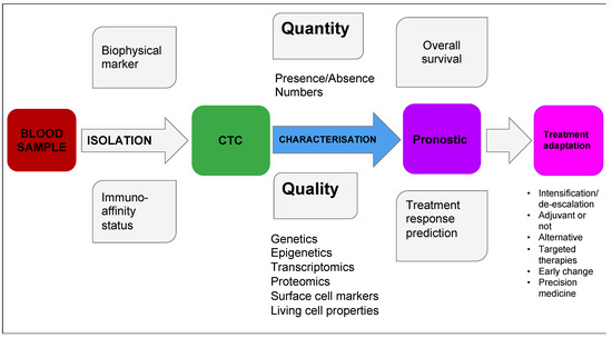
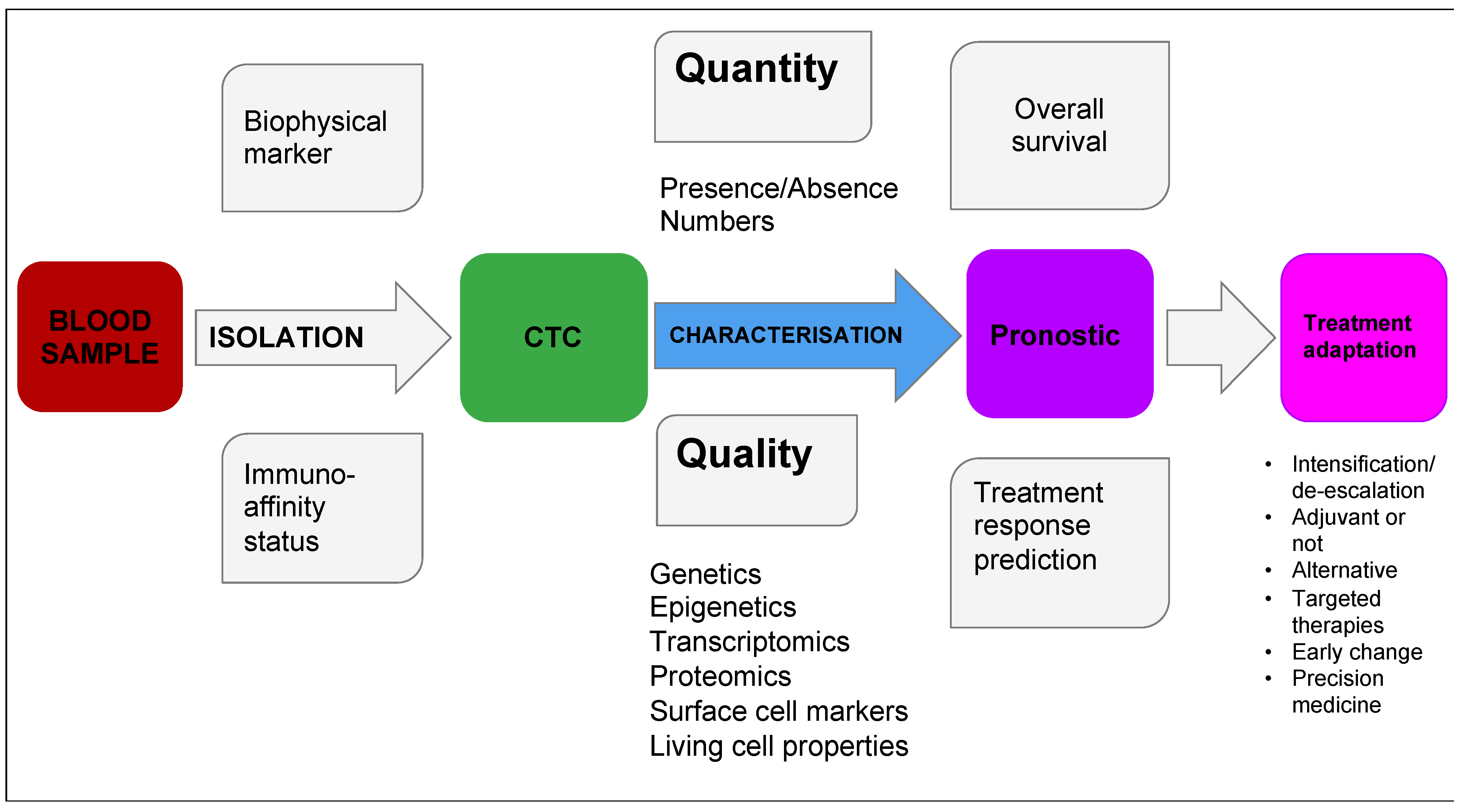
For the definition of “prognosis”, we included articles that discussed overall survival regardless of the treatment, and those that discussed the sensitivity or resistance to a specific treatment. We were also interested in discussions regarding treatment modulation, such as adding or cancelling treatment, drug intensification or de-escalation, possibility of targeted therapy, alternative treatment including early change or interruption of active care (Figure 1). These were of interest because they facilitate precision medicine that enables us to give the best care with the least side effect as possible.
Figure 1.
Colorectal cancer (CRC) circulating tumour cells (CTCs), from blood sample to clinical application.
4. Results
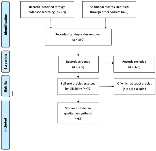
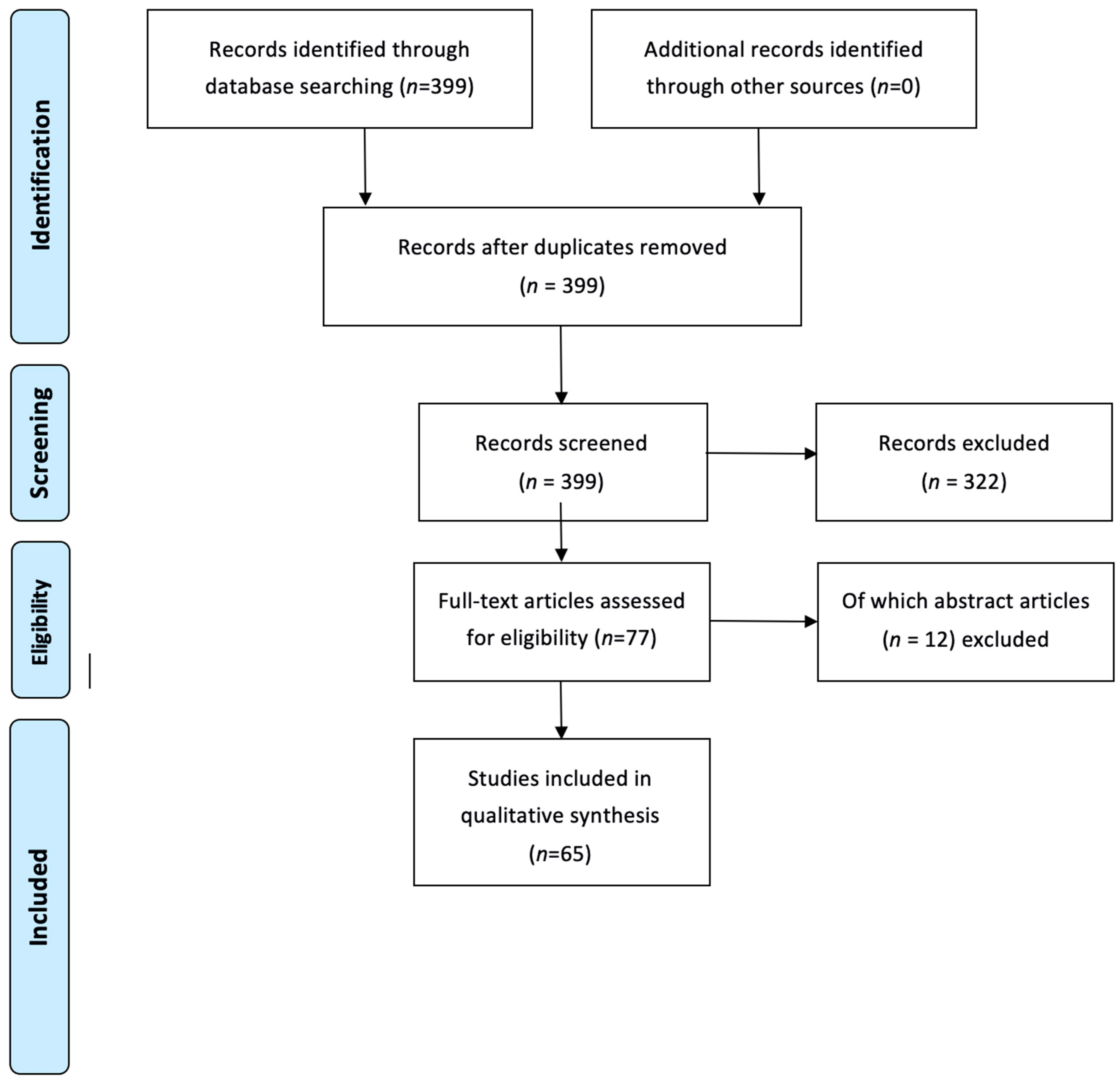
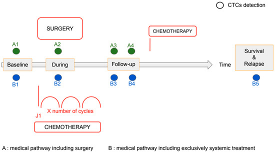
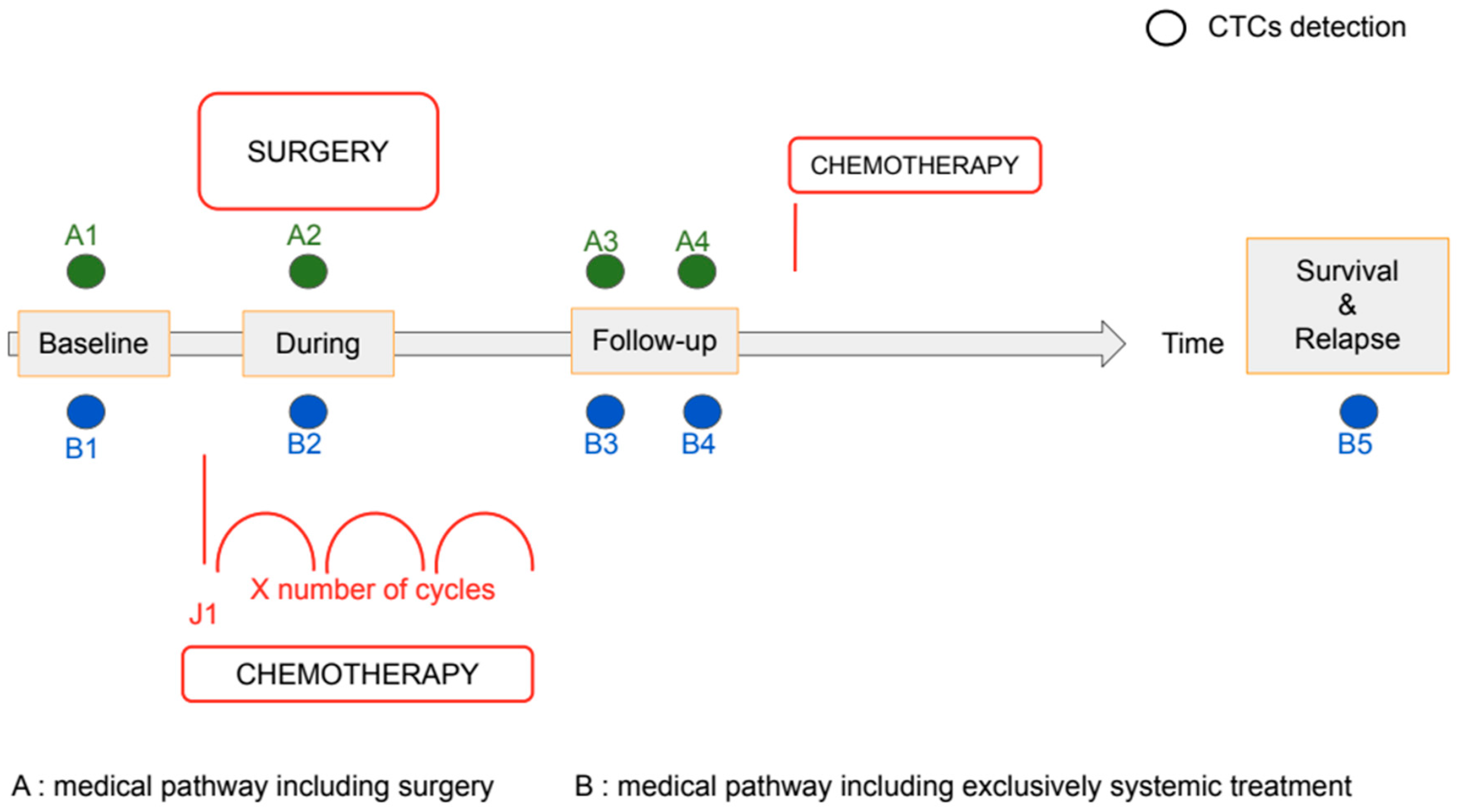
We found 399 articles in our initial search. After excluding reports (Table 1) not meeting the selection criteria (Table 1), there were 65 relevant studies (Figure 2). We classified studies according to type of marker used (quantitative, qualitative or both), techniques (Table 2), and their CTC detection time points (Figure 3). They are summarised in Table 3, Table 4 and Table 5.
Table 1.
Exclusion criteria.
Figure 2.
Flowchart.
Table 2.
Number of studies according to their marker type and results.
Figure 3.
Time point for CTCs detection for colon cancer without neo-adjuvant treatment.
Table 3.
Summary of quantitative included studies, separated into categories according to technique.
Table 4.
Summary of qualitative included studies.
Table 5.
Summary of included studies that used both quantitative and qualitative markers.
4.1. Types of Markers
The majority of articles use CTCs as a quantitative prognostic biomarker (Table 2). This is likely to be due to the only FDA-approved method (CellSearch) being a CTC count method. CellSearch, an EpCAM based technique, was used in 14 of the 77 included studies.
EpCAM is part of the cell adhesion molecule (CAM) family. It was first identified in colon cancer in 1979 [70]. Epithelial Cell Adhesion Molecule (EpCAM) is expressed by normal epithelial cells and is highly expressed in a majority (70%) of primary tumour.
Concerning colon adenocarcinomas, EpCAM is overexpressed in 81% [71] and it is known to promote tumour expansion. EpCAM is involved in numerous independent pathways [72]. It plays a part in the activation of the Wnt signalling pathway that is known for its role in carcinogenesis. On one hand, EpCAM activates this pathway through its interaction and stabilization of Lrp6. On the other hand, it downregulates the expression of some this pathway inhibitors (fgf3). EpCAM is also known to promote oncogenesis through its interaction with -catenin, by activating cell proliferation via proto-oncogenes, such as c-Myc or Cyclin A [73].
Thus, EpCAM is often use to characterise CTCs. However, during metastatic spread, epithelial–mesenchymal transition (EMT) occurs and tumour cells lose their epithelial marker, one of them being EpCAM. This cancer expansion modality highlights a clear limit of anti-EpCAM based detection system [74].
In addition to this loss of specific markers, CTCs are also difficult to separate because of their low concentration in the blood. To tackle these difficulties, newer strategies have been proposed, such as the using blood samples close to the tumour, such as mesenteric and portal vein blood samples, and detection methods such as specific gene reverse transcriptase quantitative polymerase chain reaction (RT-qPCR). This quantitative RNA-based method for CTC detection is more sensitive but has limitations. Target genes need to be carefully selected as they can be weakly expressed in normal blood cells leading to false-positives. Moreover if chosen target genes are expressed differently from one CTC to another, this may lead to an inaccurate tumour cell count [75].
After total mononuclear cell RNA extraction, RT-PCR is conducted on mRNA that are supposed to be specific to CTCs. More and more genes are being explored, focusing on different CTCs characteristics, such as epithelial (CEA, CK20, and CK19) and stem cells–like potential.
4.2. Prognostic Thresholds
For studies that utilised the CellSearch system, there were threshold changes according to CRC stage. For metastatic CRC, CTC count above or equal to 3 CTC per 7.5 mL of blood was used by Matsusaka et al., 2011 [5], Aggarwal et al., 2013 [7], and Sastre et al., 2013 [9] as a positive marker to determine “high CTC” patients. These studies all reported a significant correlation between baseline “high CTC” status and reduced survival. Camera et al., 2020 [17], similarly reported a prognostic difference in the presence of disease refractory to standard treatment or unresectable CRC. Coumans et al., 2012 [6], reported that survival for patients with metastatic CRC patients with CTCs is reduced by 6.6 months for each 10-fold CTC increase.
Krebs et al., 2015 [11] and Aranda et al., 2020 [16] showed benefits of escalating therapy from a three-drug regimen to a four-drug chemotherapy regimen for patients above their CTC prognostic threshold.
For patients undergoing surgery, CTC count above or equal to 1 CTC per 7.5 mL of blood was used by Bork et al., 2015 [10] and van Dalum et al., 2015 [13] to determine survival. These finding suggest a reduced surgical utility for patients above the prognostic threshold.
4.3. Techniques
There were multiple quantitative techniques other than CellSearch used in the included studies. The epithelial markers CK20 and CEA were the most commonly used among included studies. Taniguchi et al., 2000 [76] used CEA whereas Katsumata et al., 2006 [24], and Hinz et al., 2017 [44], used CK2O, and reported a significant association between CTC at time of surgery and survival. Allen-Mersh et al., 2007 [25] and Ito et al., 2002 [21] used either one or both of these markers to investigate the correlation between CTC detection after surgery and survival. For rectal cancer patients, CK20 was chosen by Hinz et al., 2015 [39], to predict non-response among rectal cancer undergoing neo-adjuvant chemoradiation.
Other newer techniques are being developed such as combined markers detection [31] or new molecule presence, like survivin [29].
4.4. Qualitative Data from CTCs
There was some overlap in the reporting of quantitative and qualitative markers. When CTC are detected without being destroyed, many complementary qualitative analyses are available. Study investigators have focused on markers of the aggressiveness of disease, expressed as either proteins (surface cell marker) or genes.
New marker development is focusing on discovering specific CTC subpopulations that may have particular effects on prognosis. Zhao et al., 2017 [67] combined CTC count and EMT state profile to predict disease aggressiveness. Tseng et al., 2015 [58] correlated the presence of CD133+ CD44+ CD54+ CTC subpopulation with lower survival in metastatic patients that did not undergo metastasis resection. Abdallah et al., 2016 [59] used MRP1 positive CTC as a prognosis marker in patient that were going to initiate a new chemotherapy agent.
Since markers are not always easy to determine [64], some clinicians try to associate their detection with other techniques. One of them is to collect blood closer from the tumour. Tseng et al., 2015 [58], used CTC analysis from mesenteric venous blood collected while the patient was on the operating table.
5. Discussion
5.1. Main Findings
The main finding from our search of the literature regarding the prognostic utility of CTCs for patients with CRC is that there are multiple quantitative and qualitative tests that have been used, with some success, in predicting overall survival and success of treatment, such as surgery and chemotherapy.
The presence of CTCs is a key risk factor for disease progression and severity. In breast cancer, they are considered as efficient prognostic biomarkers and included in the 7th AJCC Cancer Staging Manual (new category M0(i) introduced, and defined “by the presence of circulating or disseminated tumour cells not exceeding 0.2 mm detectable in bone marrow, circulating blood or other non-regional tissues of non-metastatic patients”. For patients with CRC, this type of staging is not yet undertaken. Indeed, there is ongoing debate regarding the utility of CTC detection due to the variations in techniques and conflicting results [77,78]. Our review confirmed that there are a range of heterogeneous techniques and results.
Most of our selected studies integrate metastatic patients (46/77). Additional prospective studies that investigate early CRC are needed in order to determine whether CTCs have any prognostic utility in this context. Rectal cancer was the subject for only 3 of the 77 included studies, and requires further evaluation in prognostic studies.
Isolation and examination of CTCs allows different analyses focusing on a variety of cell elements. The studies included in our review show that whatever quantitative or qualitative techniques (or both) are used, CTCs have potential as biomarkers to predict differences in outcomes such as survival and disease prognosis. To improve CTCs detection, new biomarkers are being studied and selected in order to help distinguish CTCs from other cells such as hematopoietic cells.
5.2. Detecting Circulating Tumour Cells
Since CTCs are epithelial cells that are found in the circulation, one option to detect their presence would be to use epithelial markers, such as CEA and cytokeratins. CEA is part of the immunoglobulin superfamily and is the product of the CEACAM5 gene [79], and is involved in cellular adhesion. It is commonly found on the surface of small and large bowels, rectum, pancreas, lung, and kidney cells. Being at a low rate for healthy people, it is already being used in routine clinical practice as a biomarker for CRC. However, its specificity is not the best as it can be increased in heavy smokers, inflammatory bowel disease, chronic obstructive pulmonary disease, pancreatitis or other adenocarcinomas than colorectal (such as ovaries, lung, kidney). On the RNA side, CEA mRNA can be detected in almost all epithelial cells, including CTCs, and is not found in non-epithelial cells [80]. Cytokeratins are part of the intermediate filaments of epithelial cells cytoskeleton. They are expressed in a tissue-specific manner. CK20 expression is limited to the gastric and intestinal epithelium, urothelium, and Merkel cells, and from malignancies that originate from these sites [30,81,82]. CK20 is not expressed in hematopoietic cells [83]. The main limitation of these biomarkers is that cancer cells undergo epithelial–mesenchymal transition (EMT) in the circulation, which can cause down regulation of epithelial markers. Therefore, these markers will only partially detect CTCs.
Yokobori et al., 2013 [37] chose to detect CTCs by using a marker that is not lost during the EMT process: plastin3 (PLS3). It codes for a ubiquitous protein that inhibits depolymerization of actin fibres. PLS3 is actually an EMT inducer and is therefore over-expressed by CTCs [84]. While working on EMT, Armstrong et al., 2011 [85] found that CTCs expressed both epithelial, and stem cell markers. Some of the included studies utilised stemness markers such as survivin, CD44 variants 6 and 9, and CD133. A good strategy seems to combine both categories of markers (epithelial and stemness) [42]. Survivin is an inhibitor of apoptosis and is highly conserved in CTCs cells. By limiting this programmed cell death, tumour cells develop aggressiveness [86]. Survivin is found in many cancer tissues, including CRC [29] and is not expressed in normal ones [87].
CD44 is a gene involved in adhesion cells and growth and invasion in tumour cells. CD44 is expressed by metastasis initiating cells [88] also known as cancer stem cells in CRC, but its exact function in these particular cells is still unknown, especially the exon that plays the central role [89]. CD133 (also known as prominin-1), is a transmembrane protein, initially described on the surface of hematopoietic stem cells; it is now known as a stemness marker for normal and cancer cells. It is not specific for CRC [90], but is considered as a key marker of tumoural stem cells in CRC [31]. Metastatic colon tissues are formed of CD133 (+) and (−) cells that can both initiate tumour cells. Since CD133 is also known to be expressed by endothelial cells, its expression by RT-PCR might be caused by bone marrow-derived circulating endothelial cells [35].
Other biomarkers with a key role in cancer biology have also been investigated in order to identify subpopulations of CTCs with a higher potential for aggressiveness [35]. Epithelial growth factor receptor (EGFR) is expressed in different cell types except hematopoietic cells and is commonly used as a therapeutic target in CRC. As more and more anti-EGFR resistance is identified, it may become important to detect CTC modification in terms of its expression of EGFR during anti-EGFR treatment [41]. Similarly, as TYMS polymorphisms seem predict response to 5FU–based chemotherapy [91], which is the main chemotherapy used as a neoadjuvant setting before rectal surgery, and also the ultraviolet excision repair protein, RAD23 homolog B (RAD23B), which potentially be “induced by the genetic damage introduced by radiotherapy” [92] before rectal surgery, both monitoring of thymidylate synthase (TYMS) and excision repair protein, RAD23 homolog B (RAD23B), can be used to predict resistance to chemotherapy/radiotherapy used in rectal cancers [69].
5.3. The Future
Our updated review confirms the potential of CTC biomarker detection as a prognostic tool, in keeping with the findings of a previous meta-analysis [93]. Molecular characterization at both cellular and genomic levels may be of utility in the prognosis for patients with CRC, whatever the initial stage (metastatic or not). Furthermore, where CTCs remain present despite chemotherapy treatment, this may be used to indicate failure of therapy and predict survival. CTCs remain of great interest to clinicians who look after patients with CRC due to their potential for utilization in clinical practice. They might give a “snapshot” of the disease within time and space, and enable the individualised therapy for patients. This may be possible by identifying patients at risk, facilitate more accurate cancer surveillance and potentially the adaptation of treatments. One focus for the future application of such a test would be the search for the most sensitive and specific (and cost-effective) method for use in routine daily clinical practice.
There were a number of articles excluded because they focused on other liquid biopsy techniques. However, they demonstrate the breadth of interest in the search for applicable circulating prognostic biomarkers for CRC. Some include the “OMICS” (circulating tumoural DNA, miRNA, circulating protein), the microenvironment (tumour-derived exosomes, circulating immune cells with tumour associated macrophage) and neo-vascularisation (circulating endothelial cell, VEGF [Vascular Endothelial Growth Factor], EPC [endothelial progenitor cells], CEC [Circulating endothelial cells]) [94]. Interestingly, some of these other biomarkers made CTCs less prevalent. For example, DNA mutation analysis is now often assessed on circulating tumour DNA rather than on CTC [95].
Several interesting avenues require further evaluation concerning the detection and analysis of CTCs. There may be some role in early diagnosis through screening [96]. CTCs may also be helpful in identifying cancer primary site [97] in cases where there is diagnostic uncertainty. Further investigations regarding tumour biology and the metastatic process are in development, focusing on tumour microenvironment, the epithelial to mesenchymal transition, circulating cancer cell stemness, and others [98,99]. Furthermore, when CTC are isolated without being destroyed, CTC-derived xenografts could lead to patient specific drug screening [100,101,102].
6. Conclusions
We identified 77 studies that reported data regarding the prognostic utility of CTCs for patients with CRC. CTCs seem to have a high potential for use as a prognostic biomarker in CRC in both quantitative and qualitative terms. Ongoing investigations are required to evaluate the role of CTC analysis in routine clinical practice.
Funding
This research received no external funding.
Conflicts of Interest
The authors declare no conflict of interest.
References
- Globocan 2018. Available online: https://gco.iarc.fr/today/data/factsheets/populations/900-world-fact-sheets.pdf (accessed on 6 April 2021).
- Pantel, K.; Alix-Panabières, C.; Riethdorf, S. Cancer micrometastases. Nat. Rev. Clin. Oncol. 2009, 6, 339–351. [Google Scholar] [CrossRef] [Scilit]
- Vasseur, A.; Kiavue, N.; Bidard, F.-C.; Pierga, J.-Y.; Cabel, L. Clinical utility of circulating tumor cells: An update. Mol. Oncol. 2020. [Google Scholar] [CrossRef] [Scilit]
- Cabel, L.; Proudhon, C.; Gortais, H.; Loirat, D.; Coussy, F.; Pierga, J.-Y.; Bidard, F.-C. Circulating tumor cells: Clinical validity and utility. Int. J. Clin. Oncol. 2017, 22, 421–430. [Google Scholar] [CrossRef] [Scilit] [PubMed]
- Matsusaka, S.; Suenaga, M.; Mishima, Y.; Kuniyoshi, R.; Takagi, K.; Terui, Y.; Mizunuma, N.; Hatake, K. Circulating tumor cells as a surrogate marker for determining response to chemotherapy in Japanese patients with metastatic colorectal cancer. Cancer Sci. 2011, 102, 1188–1192. [Google Scholar] [CrossRef] [Scilit] [PubMed]
- Coumans, F.A.W.; Ligthart, S.T.; Uhr, J.W.; Terstappen, L.W.M.M. Challenges in the enumeration and phenotyping of CTC. Clin. Cancer Res. Off. J. Am. Assoc. Cancer Res. 2012, 18, 5711–5718. [Google Scholar] [CrossRef] [Scilit] [PubMed]
- Aggarwal, C.; Meropol, N.J.; Punt, C.J.; Iannotti, N.; Saidman, B.H.; Sabbath, K.D.; Gabrail, N.Y.; Picus, J.; Morse, M.A.; Mitchell, E.; et al. Relationship among circulating tumor cells, CEA and overall survival in patients with metastatic colorectal cancer. Ann. Oncol. Off. J. Eur. Soc. Med. Oncol. 2013, 24, 420–428. [Google Scholar] [CrossRef] [Scilit]
- Otsuka, K.; Imai, H.; Soeda, H.; Komine, K.; Ishioka, C.; Shibata, H. Practical utility of circulating tumour cells as biomarkers in cancer chemotherapy for advanced colorectal cancer. Anticancer Res. 2013, 33, 625–629. [Google Scholar] [PubMed]
- Sastre, J.; Vidaurreta, M.; Gómez, A.; Rivera, F.; Massutí, B.; López, M.R.; Abad, A.; Gallen, M.; Benavides, M.; Aranda, E.; et al. Prognostic value of the combination of circulating tumor cells plus KRAS in patients with metastatic colorectal cancer treated with chemotherapy plus bevacizumab. Clin. Colorectal Cancer 2013, 12, 280–286. [Google Scholar] [CrossRef] [Scilit] [PubMed]
- Bork, U.; Rahbari, N.N.; Schölch, S.; Reissfelder, C.; Kahlert, C.; Büchler, M.W.; Weitz, J.; Koch, M. Circulating tumour cells and outcome in non-metastatic colorectal cancer: A prospective study. Br. J. Cancer 2015, 112, 1306–1313. [Google Scholar] [CrossRef] [Scilit] [PubMed]
- Krebs, M.G.; Renehan, A.G.; Backen, A.; Gollins, S.; Chau, I.; Hasan, J.; Valle, J.W.; Morris, K.; Beech, J.; Ashcroft, L.; et al. Circulating Tumor Cell Enumeration in a Phase II Trial of a Four-Drug Regimen in Advanced Colorectal Cancer. Clin. Colorectal Cancer 2015, 14, 115–122.e2. [Google Scholar] [CrossRef] [Scilit]
- Sotelo, M.J.; Sastre, J.; Maestro, M.L.; Veganzones, S.; Viéitez, J.M.; Alonso, V.; Grávalos, C.; Escudero, P.; Vera, R.; Aranda, E.; et al. Role of circulating tumor cells as prognostic marker in resected stage III colorectal cancer. Ann. Oncol. 2015, 26, 535–541. [Google Scholar] [CrossRef] [Scilit]
- van Dalum, G.; Stam, G.-J.; Scholten, L.F.A.; Mastboom, W.J.B.; Vermes, I.; Tibbe, A.G.J.; De Groot, M.R.; Terstappen, L.W.M.M. Importance of circulating tumor cells in newly diagnosed colorectal cancer. Int. J. Oncol. 2015, 46, 1361–1368. [Google Scholar] [CrossRef] [Scilit]
- Connor, A.A.; McNamara, K.; Al-Sukhni, E.; Diskin, J.; Chan, D.; Ash, C.; Lowes, L.E.; Allan, A.L.; Zogopoulos, G.; Moulton, C.-A.; et al. Central, But Not Peripheral, Circulating Tumor Cells are Prognostic in Patients Undergoing Resection of Colorectal Cancer Liver Metastases. Ann. Surg. Oncol. 2016, 23, 2168–2175. [Google Scholar] [CrossRef] [Scilit]
- Rothé, F.; Maetens, M.; Rouas, G.; Paesmans, M.; Van den Eynde, M.; Van Laethem, J.-L.; Vergauwe, P.; Deboever, G.; Bareche, Y.; Vandeputte, C.; et al. CTCs as a prognostic and predictive biomarker for stage II/III Colon Cancer: A companion study to the PePiTA trial. BMC Cancer 2019, 19, 304. [Google Scholar] [CrossRef] [Scilit]
- Aranda, E.; Viéitez, J.M.; Gómez-España, A.; Gil Calle, S.; Salud-Salvia, A.; Graña, B.; Garcia-Alfonso, P.; Rivera, F.; Quintero-Aldana, G.A.; Reina-Zoilo, J.J.; et al. FOLFOXIRI plus bevacizumab versus FOLFOX plus bevacizumab for patients with metastatic colorectal cancer and ≥3 circulating tumour cells: The randomised phase III VISNÚ-1 trial. ESMO Open 2020, 5. [Google Scholar] [CrossRef] [Scilit]
- Camera, S.; Akin Telli, T.; Woff, E.; Vandeputte, C.; Kehagias, P.; Guiot, T.; Critchi, G.; Wissam, Y.; Bregni, G.; Trevisi, E.; et al. Prognostic Value of the Pace of Tumor Progression as Assessed by Serial 18F-FDG PET/CT Scan and Liquid Biopsy in Refractory Colorectal Cancer: The CORIOLAN Trial. Cancers 2020, 12, 2752. [Google Scholar] [CrossRef] [Scilit] [PubMed]
- Bessa, X.; Elizalde, J.I.; Boix, L.; Piñol, V.; Lacy, A.M.; Saló, J.; Piqué, J.M.; Castells, A. Lack of prognostic influence of circulating tumor cells in peripheral blood of patients with colorectal cancer. Gastroenterology 2001, 120, 1084–1092. [Google Scholar] [CrossRef] [Scilit] [PubMed]
- Sadahiro, S.; Suzuki, T.; Tokunaga, N.; Yurimoto, S.; Yasuda, S.; Tajima, T.; Makuuchi, H.; Murayama, C.; Matsuda, K. Detection of tumor cells in the portal and peripheral blood of patients with colorectal carcinoma using competitive reverse transcriptase-polymerase chain reaction. Cancer 2001, 92, 1251–1258. [Google Scholar] [CrossRef] [Scilit]
- Guller, U.; Zajac, P.; Schnider, A.; Bösch, B.; Vorburger, S.; Zuber, M.; Spagnoli, G.C.; Oertli, D.; Maurer, R.; Metzger, U.; et al. Disseminated single tumor cells as detected by real-time quantitative polymerase chain reaction represent a prognostic factor in patients undergoing surgery for colorectal cancer. Ann. Surg. 2002, 236, 768–775; discussion 775–776. [Google Scholar] [CrossRef] [Scilit]
- Ito, S.; Nakanishi, H.; Hirai, T.; Kato, T.; Kodera, Y.; Feng, Z.; Kasai, Y.; Ito, K.; Akiyama, S.; Nakao, A.; et al. Quantitative detection of CEA expressing free tumor cells in the peripheral blood of colorectal cancer patients during surgery with real-time RT-PCR on a LightCycler. Cancer Lett. 2002, 183, 195–203. [Google Scholar] [CrossRef] [Scilit]
- Bessa, X.; Piñol, V.; Castellví-Bel, S.; Piazuelo, E.; Lacy, A.M.; Elizalde, J.I.; Piqué, J.M.; Castells, A. Prognostic value of postoperative detection of blood circulating tumor cells in patients with colorectal cancer operated on for cure. Ann. Surg. 2003, 237, 368–375. [Google Scholar] [CrossRef] [Scilit] [PubMed]
- Sadahiro, S.; Suzuki, T.; Ishikawa, K.; Saguchi, T.; Maeda, Y.; Yasuda, S.; Makuuchi, H.; Yurimoto, S.; Murayama, C. Detection of carcinoembryonic antigen messenger RNA-expressing cells in portal and peripheral blood during surgery does not influence relapse in colorectal cancer. Ann. Surg. Oncol. 2005, 12, 988–994. [Google Scholar] [CrossRef] [Scilit]
- Katsumata, K.; Sumi, T.; Mori, Y.; Hisada, M.; Tsuchida, A.; Aoki, T. Detection and evaluation of epithelial cells in the blood of colon cancer patients using RT-PCR. Int. J. Clin. Oncol. 2006, 11, 385–389. [Google Scholar] [CrossRef] [Scilit] [PubMed]
- Allen-Mersh, T.G.; McCullough, T.K.; Patel, H.; Wharton, R.Q.; Glover, C.; Jonas, S.K. Role of circulating tumour cells in predicting recurrence after excision of primary colorectal carcinoma. Br. J. Surg. 2007, 94, 96–105. [Google Scholar] [CrossRef] [Scilit] [PubMed]
- Friederichs, J.; Gertler, R.; Rosenberg, R.; Dahm, M.; Nekarda, H.; Holzmann, B.; Siewert, J.R. Correlation of CK-20-positive cells in peripheral venous blood with serum CEA levels in patients with colorectal carcinoma. World J. Surg. 2007, 31, 2329–2334. [Google Scholar] [CrossRef] [Scilit]
- Sadahiro, S.; Suzuki, T.; Maeda, Y.; Yurimoto, S.; Yasuda, S.; Makuuchi, H.; Kamijo, A.; Murayama, C. Detection of carcinoembryonic antigen messenger RNA-expressing cells in peripheral blood 7 days after curative surgery is a novel prognostic factor in colorectal cancer. Ann. Surg. Oncol. 2007, 14, 1092–1098. [Google Scholar] [CrossRef] [Scilit]
- Koyanagi, K.; Bilchik, A.J.; Saha, S.; Turner, R.R.; Wiese, D.; McCarter, M.; Shen, P.; Deacon, L.; Elashoff, D.; Hoon, D.S.B. Prognostic relevance of occult nodal micrometastases and circulating tumor cells in colorectal cancer in a prospective multicenter trial. Clin. Cancer Res. 2008, 14, 7391–7396. [Google Scholar] [CrossRef] [Scilit]
- Yie, S.; Lou, B.; Ye, S.; Cao, M.; He, X.; Li, P.; Hu, K.; Rao, L.; Wu, S.; Xiao, H.-B.; et al. Detection of survivin-expressing circulating cancer cells (CCCs) in peripheral blood of patients with gastric and colorectal cancer reveals high risks of relapse. Ann. Surg. Oncol. 2008, 15, 3073–3082. [Google Scholar] [CrossRef] [Scilit]
- Tsouma, A.; Aggeli, C.; Lembessis, P.; Zografos, G.-N.; Korkolis, D.-P.; Pectasides, D.; Skondra, M.; Pissimissis, N.; Tzonou, A.; Koutsilieris, M. Multiplex RT-PCR-based detections of CEA, CK20 and EGFR in colorectal cancer patients. World J. Gastroenterol. 2010, 16, 5965–5974. [Google Scholar] [CrossRef] [Scilit]
- Iinuma, H.; Watanabe, T.; Mimori, K.; Adachi, M.; Hayashi, N.; Tamura, J.; Matsuda, K.; Fukushima, R.; Okinaga, K.; Sasako, M.; et al. Clinical significance of circulating tumor cells, including cancer stem-like cells, in peripheral blood for recurrence and prognosis in patients with Dukes’ stage B and C colorectal cancer. J. Clin. Oncol. 2011, 29, 1547–1555. [Google Scholar] [CrossRef] [Scilit] [PubMed]
- Vardakis, N.; Messaritakis, I.; Papadaki, C.; Agoglossakis, G.; Sfakianaki, M.; Saridaki, Z.; Apostolaki, S.; Koutroubakis, I.; Perraki, M.; Hatzidaki, D.; et al. Prognostic significance of the detection of peripheral blood CEACAM5mRNA-positive cells by real-time polymerase chain reaction in operable colorectal cancer. Clin. Cancer Res. 2011, 17, 165–173. [Google Scholar] [CrossRef] [Scilit]
- de Albuquerque, A.; Kubisch, I.; Stölzel, U.; Ernst, D.; Boese-Landgraf, J.; Breier, G.; Stamminger, G.; Fersis, N.; Kaul, S. Prognostic and predictive value of circulating tumor cell analysis in colorectal cancer patients. J. Transl. Med. 2012, 10, 222. [Google Scholar] [CrossRef] [Scilit] [PubMed]
- Liu, Z.-P.; Li, L.-M.; Liu, X.-L.; Zhang, D.-X. Comparative analysis of tumor markers and evaluation of their predictive value in patients with colorectal cancer. Onkologie 2012, 35, 108–113. [Google Scholar] [CrossRef] [Scilit]
- Pilati, P.; Mocellin, S.; Bertazza, L.; Galdi, F.; Briarava, M.; Mammano, E.; Tessari, E.; Zavagno, G.; Nitti, D. Prognostic value of putative circulating cancer stem cells in patients undergoing hepatic resection for colorectal liver metastasis. Ann. Surg. Oncol. 2012, 19, 402–408. [Google Scholar] [CrossRef] [Scilit]
- Shimada, R.; Iinuma, H.; Akahane, T.; Horiuchi, A.; Watanabe, T. Prognostic significance of CTCs and CSCs of tumor drainage vein blood in Dukes’ stage B and C colorectal cancer patients. Oncol. Rep. 2012, 27, 947–953. [Google Scholar] [CrossRef] [Scilit] [PubMed]
- Yokobori, T.; Iinuma, H.; Shimamura, T.; Imoto, S.; Sugimachi, K.; Ishii, H.; Iwatsuki, M.; Ota, D.; Ohkuma, M.; Iwaya, T.; et al. Plastin3 is a novel marker for circulating tumor cells undergoing the epithelial-mesenchymal transition and is associated with colorectal cancer prognosis. Cancer Res. 2013, 73, 2059–2069. [Google Scholar] [CrossRef] [Scilit] [PubMed]
- Barbazán, J.; Muinelo-Romay, L.; Vieito, M.; Candamio, S.; Díaz-López, A.; Cano, A.; Gómez-Tato, A.; Casares de Cal, M. de los Á.; Abal, M.; López-López, R. A multimarker panel for circulating tumor cells detection predicts patient outcome and therapy response in metastatic colorectal cancer. Int. J. Cancer 2014, 135, 2633–2643. [Google Scholar] [CrossRef] [Scilit] [PubMed]
- Hinz, S.; Röder, C.; Tepel, J.; Hendricks, A.; Schafmayer, C.; Becker, T.; Kalthoff, H. Cytokeratin 20 positive circulating tumor cells are a marker for response after neoadjuvant chemoradiation but not for prognosis in patients with rectal cancer. BMC Cancer 2015, 15, 953. [Google Scholar] [CrossRef] [Scilit] [PubMed]
- Katoh, S.; Goi, T.; Naruse, T.; Ueda, Y.; Kurebayashi, H.; Nakazawa, T.; Kimura, Y.; Hirono, Y.; Yamaguchi, A. Cancer stem cell marker in circulating tumor cells: Expression of CD44 variant exon 9 is strongly correlated to treatment refractoriness, recurrence and prognosis of human colorectal cancer. Anticancer Res. 2015, 35, 239–244. [Google Scholar] [PubMed]
- Musella, V.; Pietrantonio, F.; Di Buduo, E.; Iacovelli, R.; Martinetti, A.; Sottotetti, E.; Bossi, I.; Maggi, C.; Di Bartolomeo, M.; de Braud, F.; et al. Circulating tumor cells as a longitudinal biomarker in patients with advanced chemorefractory, RAS-BRAF wild-type colorectal cancer receiving cetuximab or panitumumab. Int. J. Cancer 2015, 137, 1467–1474. [Google Scholar] [CrossRef] [Scilit]
- Ning, Y.; Hanna, D.L.; Zhang, W.; Mendez, A.; Yang, D.; El-Khoueiry, R.; Matsusaka, S.; Sunakawa, Y.; Stremitzer, S.; Parekh, A.; et al. Cytokeratin-20 and Survivin-Expressing Circulating Tumor Cells Predict Survival in Metastatic Colorectal Cancer Patients by a Combined Immunomagnetic qRT-PCR Approach. Mol. Cancer Ther. 2015, 14, 2401–2408. [Google Scholar] [CrossRef] [Scilit]
- Kust, D.; Šamija, I.; Kirac, I.; Radić, J.; Kovačević, D.; Kusić, Z. Cytokeratin 20 positive cells in blood of colorectal cancer patients as an unfavorable prognostic marker. Acta Clin. Belg. 2016, 71, 235–243. [Google Scholar] [CrossRef] [Scilit]
- Hinz, S.; Hendricks, A.; Wittig, A.; Schafmayer, C.; Tepel, J.; Kalthoff, H.; Becker, T.; Röder, C. Detection of circulating tumor cells with CK20 RT-PCR is an independent negative prognostic marker in colon cancer patients—A prospective study. BMC Cancer 2017, 17, 53. [Google Scholar] [CrossRef] [Scilit] [PubMed]
- Insua, Y.V.; de la Cámara, J.; Vázquez, E.B.; Fernández, A.; Rivera, F.V.; Silva, M.J.V.V.; Barbazán, J.; Muinelo-Romay, L.; Folgar, S.C.; Abalo, A.; et al. Predicting Outcome and Therapy Response in mCRC Patients Using an Indirect Method for CTCs Detection by a Multigene Expression Panel: A Multicentric Prospective Validation Study. Int. J. Mol. Sci. 2017, 18, 1265. [Google Scholar] [CrossRef] [Scilit] [PubMed]
- Ning, Y.; Zhang, W.; Hanna, D.L.; Yang, D.; Okazaki, S.; Berger, M.D.; Miyamoto, Y.; Suenaga, M.; Schirripa, M.; El-Khoueiry, A.; et al. Clinical relevance of EMT and stem-like gene expression in circulating tumor cells of metastatic colorectal cancer patients. Pharmacogenomics J. 2018, 18, 29–34. [Google Scholar] [CrossRef] [Scilit] [PubMed]
- Shou, X.; Li, Y.; Hu, W.; Ye, T.; Wang, G.; Xu, F.; Sui, M.; Xu, Y. Six-gene Assay as a new biomarker in the blood of patients with colorectal cancer: Establishment and clinical validation. Mol. Oncol. 2019, 13, 781–791. [Google Scholar] [CrossRef] [Scilit]
- Uen, Y.-H.; Lin, S.-R.; Wu, D.-C.; Su, Y.-C.; Wu, J.-Y.; Cheng, T.-L.; Chi, C.-W.; Wang, J.-Y. Prognostic significance of multiple molecular markers for patients with stage II colorectal cancer undergoing curative resection. Ann. Surg. 2007, 246, 1040–1046. [Google Scholar] [CrossRef] [Scilit]
- Lu, C.-Y.; Uen, Y.-H.; Tsai, H.-L.; Chuang, S.-C.; Hou, M.-F.; Wu, D.-C.; Juo, S.-H.H.; Lin, S.-R.; Wang, J.-Y. Molecular detection of persistent postoperative circulating tumour cells in stages II and III colon cancer patients via multiple blood sampling: Prognostic significance of detection for early relapse. Br. J. Cancer 2011, 104, 1178–1184. [Google Scholar] [CrossRef] [Scilit]
- Lu, C.-Y.; Tsai, H.-L.; Uen, Y.-H.; Hu, H.-M.; Chen, C.-W.; Cheng, T.-L.; Lin, S.-R.; Wang, J.-Y. Circulating tumor cells as a surrogate marker for determining clinical outcome to mFOLFOX chemotherapy in patients with stage III colon cancer. Br. J. Cancer 2013, 108, 791–797. [Google Scholar] [CrossRef] [Scilit] [PubMed]
- Divella, R.; Daniele, A.; Abbate, I.; Bellizzi, A.; Savino, E.; Simone, G.; Giannone, G.; Giuliani, F.; Fazio, V.; Gadaleta-Caldarola, G.; et al. The presence of clustered circulating tumor cells (CTCs) and circulating cytokines define an aggressive phenotype in metastatic colorectal cancer. Cancer Causes Control CCC 2014, 25, 1531–1541. [Google Scholar] [CrossRef] [Scilit] [PubMed]
- Chang, Y.-T.; Huang, M.-Y.; Yeh, Y.-S.; Huang, C.-W.; Tsai, H.-L.; Cheng, T.-L.; Wang, J.-Y. A Prospective Study of Comparing Multi-Gene Biomarker Chip and Serum Carcinoembryonic Antigen in the Postoperative Surveillance for Patients with Stage I-III Colorectal Cancer. PLoS ONE 2016, 11, e0163264. [Google Scholar] [CrossRef] [Scilit] [PubMed]
- Wu, W.; Zhang, Z.; Gao, X.H.; Shen, Z.; Jing, Y.; Lu, H.; Li, H.; Yang, X.; Cui, X.; Li, Y.; et al. Clinical significance of detecting circulating tumor cells in colorectal cancer using subtraction enrichment and immunostaining-fluorescence in situ hybridization (SE-iFISH). Oncotarget 2017, 8, 21639–21649. [Google Scholar] [CrossRef] [Scilit]
- Chou, W.-C.; Wu, M.-H.; Chang, P.-H.; Hsu, H.-C.; Chang, G.-J.; Huang, W.-K.; Wu, C.-E.; Hsieh, J.C.-H. A Prognostic Model Based on Circulating Tumour Cells is Useful for Identifying the Poorest Survival Outcome in Patients with Metastatic Colorectal Cancer. Int. J. Biol. Sci. 2018, 14, 137–146. [Google Scholar] [CrossRef] [Scilit]
- Yang, C.; Shi, D.; Wang, S.; Wei, C.; Zhang, C.; Xiong, B. Prognostic value of pre- and post-operative circulating tumor cells detection in colorectal cancer patients treated with curative resection: A prospective cohort study based on ISET device. Cancer Manag. Res. 2018, 10, 4135–4144. [Google Scholar] [CrossRef] [Scilit]
- Iinuma, H.; Okinaga, K.; Adachi, M.; Suda, K.; Sekine, T.; Sakagawa, K.; Baba, Y.; Tamura, J.; Kumagai, H.; Ida, A. Detection of tumor cells in blood using CD45 magnetic cell separation followed by nested mutant allele-specific amplification of p53 and K-ras genes in patients with colorectal cancer. Int. J. Cancer 2000, 89, 337–344. [Google Scholar] [CrossRef] [Scilit]
- Gazzaniga, P.; Gradilone, A.; Petracca, A.; Nicolazzo, C.; Raimondi, C.; Iacovelli, R.; Naso, G.; Cortesi, E. Molecular markers in circulating tumour cells from metastatic colorectal cancer patients. J. Cell. Mol. Med. 2010, 14, 2073–2077. [Google Scholar] [CrossRef] [Scilit] [PubMed]
- Tseng, J.-Y.; Yang, C.-Y.; Yang, S.-H.; Lin, J.-K.; Lin, C.-H.; Jiang, J.-K. Circulating CD133(+)/ESA(+) cells in colorectal cancer patients. J. Surg. Res. 2015, 199, 362–370. [Google Scholar] [CrossRef] [Scilit] [PubMed]
- Abdallah, E.A.; Fanelli, M.F.; Souza E Silva, V.; Machado Netto, M.C.; Gasparini, J.L., Jr.; Araújo, D.V.; Ocea, L.M.M.; Buim, M.E.C.; Tariki, M.S.; da Silva Alves, V.; et al. MRP1 expression in CTCs confers resistance to irinotecan-based chemotherapy in metastatic colorectal cancer. Int. J. Cancer 2016, 139, 890–898. [Google Scholar] [CrossRef] [Scilit]
- Barbazan, J.; Dunkel, Y.; Li, H.; Nitsche, U.; Janssen, K.-P.; Messer, K.; Ghosh, P. Prognostic Impact of Modulators of G proteins in Circulating Tumor Cells from Patients with Metastatic Colorectal Cancer. Sci. Rep. 2016, 6, 22112. [Google Scholar] [CrossRef] [Scilit] [PubMed]
- Satelli, A.; Batth, I.S.; Brownlee, Z.; Rojas, C.; Meng, Q.H.; Kopetz, S.; Li, S. Potential role of nuclear PD-L1 expression in cell-surface vimentin positive circulating tumor cells as a prognostic marker in cancer patients. Sci. Rep. 2016, 6, 28910. [Google Scholar] [CrossRef] [Scilit] [PubMed]
- Fang, C.; Fan, C.; Wang, C.; Huang, Q.; Meng, W.; Yu, Y.; Yang, L.; Hu, J.; Li, Y.; Mo, X.; et al. Prognostic value of CD133+ CD54+ CD44+ circulating tumor cells in colorectal cancer with liver metastasis. Cancer Med. 2017, 6, 2850–2857. [Google Scholar] [CrossRef] [Scilit]
- Wong, S.C.C.; Chan, C.M.L.; Ma, B.B.Y.; Hui, E.P.; Ng, S.S.M.; Lai, P.B.S.; Cheung, M.T.; Lo, E.S.F.; Chan, A.K.C.; Lam, M.Y.Y.; et al. Clinical significance of cytokeratin 20-positive circulating tumor cells detected by a refined immunomagnetic enrichment assay in colorectal cancer patients. Clin. Cancer Res. 2009, 15, 1005–1012. [Google Scholar] [CrossRef] [Scilit]
- Kuboki, Y.; Matsusaka, S.; Minowa, S.; Shibata, H.; Suenaga, M.; Shinozaki, E.; Mizunuma, N.; Ueno, M.; Yamaguchi, T.; Hatake, K. Circulating tumor cell (CTC) count and epithelial growth factor receptor expression on CTCs as biomarkers for cetuximab efficacy in advanced colorectal cancer. Anticancer Res. 2013, 33, 3905–3910. [Google Scholar] [PubMed]
- Abdallah, E.A.; Fanelli, M.F.; Buim, M.E.C.; Machado Netto, M.C.; Gasparini, J.L., Jr.; Souza E Silva, V.; Dettino, A.L.A.; Mingues, N.B.; Romero, J.V.; Ocea, L.M.M.; et al. Thymidylate synthase expression in circulating tumor cells: A new tool to predict 5-fluorouracil resistance in metastatic colorectal cancer patients. Int. J. Cancer 2015, 137, 1397–1405. [Google Scholar] [CrossRef] [Scilit]
- Melero, J.T.; Ortega, F.G.; Gonzalez, A.M.; Carmona-Saez, P.; Garcia Puche, J.L.; Sugarbaker, P.H.; Delgado, M.; Lorente, J.A.; Serrano, M.J. Prognostic factor analysis of circulating tumor cells in peripheral blood of patients with peritoneal carcinomatosis of colon cancer origin treated with cytoreductive surgery plus an intraoperative hyperthermic intraperitoneal chemotherapy procedure (CRS + HIPEC). Surgery 2016, 159, 728–735. [Google Scholar] [CrossRef] [Scilit] [PubMed]
- Zhao, R.; Cai, Z.; Li, S.; Cheng, Y.; Gao, H.; Liu, F.; Wu, S.; Liu, S.; Dong, Y.; Zheng, L.; et al. Expression and clinical relevance of epithelial and mesenchymal markers in circulating tumor cells from colorectal cancer. Oncotarget 2017, 8, 9293–9302. [Google Scholar] [CrossRef] [Scilit]
- Bahnassy, A.A.; Salem, S.E.; Mohanad, M.; Abulezz, N.Z.; Abdellateif, M.S.; Hussein, M.; Zekri, C.A.N.; Zekri, A.-R.N.; Allahloubi, N.M.A. Prognostic significance of circulating tumor cells (CTCs) in Egyptian non-metastatic colorectal cancer patients: A comparative study for four different techniques of detection (Flowcytometry, CellSearch, Quantitative Real-time PCR and Cytomorphology). Exp. Mol. Pathol. 2019, 106, 90–101. [Google Scholar] [CrossRef] [Scilit] [PubMed]
- Troncarelli Flores, B.C.; Souza E Silva, V.; Ali Abdallah, E.; Mello, C.A.L.; Gobo Silva, M.L.; Gomes Mendes, G.; Camila Braun, A.; Aguiar, S., Jr.; Thomé Domingos Chinen, L. Molecular and Kinetic Analyses of Circulating Tumor Cells as Predictive Markers of Treatment Response in Locally Advanced Rectal Cancer Patients. Cells 2019, 8, 641. [Google Scholar] [CrossRef] [Scilit] [PubMed]
- Herlyn, M.; Steplewski, Z.; Herlyn, D.; Koprowski, H. Colorectal carcinoma-specific antigen: Detection by means of monoclonal antibodies. Proc. Natl. Acad. Sci. USA 1979, 76, 1438–1442. [Google Scholar] [CrossRef] [Scilit]
- Went, P.T.; Lugli, A.; Meier, S.; Bundi, M.; Mirlacher, M.; Sauter, G.; Dirnhofer, S. Frequent EpCam protein expression in human carcinomas. Hum. Pathol. 2004, 35, 122–128. [Google Scholar] [CrossRef] [Scilit]
- Huang, L.; Yang, Y.; Yang, F.; Liu, S.; Zhu, Z.; Lei, Z.; Guo, J. Functions of EpCAM in physiological processes and diseases (Review). Int. J. Mol. Med. 2018, 42, 1771–1785. [Google Scholar] [CrossRef] [Scilit] [PubMed]
- Maetzel, D.; Denzel, S.; Mack, B.; Canis, M.; Went, P.; Benk, M.; Kieu, C.; Papior, P.; Baeuerle, P.A.; Munz, M.; et al. Nuclear signalling by tumour-associated antigen EpCAM. Nat. Cell Biol. 2009, 11, 162–171. [Google Scholar] [CrossRef] [Scilit] [PubMed]
- Gorges, T.M.; Tinhofer, I.; Drosch, M.; Röse, L.; Zollner, T.M.; Krahn, T.; von Ahsen, O. Circulating tumour cells escape from EpCAM-based detection due to epithelial-to-mesenchymal transition. BMC Cancer 2012, 12, 178. [Google Scholar] [CrossRef] [Scilit] [PubMed]
- Andergassen, U.; Kölbl, A.C.; Mahner, S.; Jeschke, U. Real-time RT-PCR systems for CTC detection from blood samples of breast cancer and gynaecological tumour patients (Review). Oncol. Rep. 2016, 35, 1905–1915. [Google Scholar] [CrossRef] [Scilit] [PubMed]
- Taniguchi, T.; Makino, M.; Suzuki, K.; Kaibara, N. Prognostic significance of reverse transcriptase-polymerase chain reaction measurement of carcinoembryonic antigen mRNA levels in tumor drainage blood and peripheral blood of patients with colorectal carcinoma. Cancer 2000, 89, 970–976. [Google Scholar] [CrossRef]
- Yang, C.; Zou, K.; Zheng, L.; Xiong, B. Prognostic and clinicopathological significance of circulating tumor cells detected by RT-PCR in non-metastatic colorectal cancer: A meta-analysis and systematic review. BMC Cancer 2017, 17, 725. [Google Scholar] [CrossRef] [Scilit]
- Vojtechova, G.; Benesova, L.; Belsanova, B.; Minarikova, P.; Levy, M.; Lipska, L.; Suchanek, S.; Zavoral, M.; Minarik, M. Monitoring of Circulating Tumor Cells by a Combination of Immunomagnetic Enrichment and RT-PCR in Colorectal Cancer Patients Undergoing Surgery. Adv. Clin. Exp. Med. Off. Organ Wroclaw Med. Univ. 2016, 25, 1273–1279. [Google Scholar] [CrossRef] [Scilit]
- Kuespert, K.; Pils, S.; Hauck, C.R. CEACAMs: Their role in physiology and pathophysiology. Curr. Opin. Cell Biol. 2006, 18, 565–571. [Google Scholar] [CrossRef] [Scilit]
- Mori, M.; Mimori, K.; Ueo, H.; Karimine, N.; Barnard, G.F.; Sugimachi, K.; Akiyoshi, T. Molecular detection of circulating solid carcinoma cells in the peripheral blood: The concept of early systemic disease. Int. J. Cancer 1996, 68, 739–743. [Google Scholar] [CrossRef]
- Ribal, M.J.; Mengual, L.; Marín, M.; Algaba, F.; Ars, E.; Fernández, P.L.; Oliva, R.; Villavicencio, H.; Alcaraz, A. Molecular staging of bladder cancer with RT-PCR assay for CK20 in peripheral blood, bone marrow and lymph nodes: Comparison with standard histological staging. Anticancer Res. 2006, 26, 411–419. [Google Scholar] [PubMed]
- Moll, R.; Löwe, A.; Laufer, J.; Franke, W.W. Cytokeratin 20 in human carcinomas. A new histodiagnostic marker detected by monoclonal antibodies. Am. J. Pathol. 1992, 140, 427–447. [Google Scholar] [PubMed]
- Burchill, S.A.; Bradbury, M.F.; Pittman, K.; Southgate, J.; Smith, B.; Selby, P. Detection of epithelial cancer cells in peripheral blood by reverse transcriptase-polymerase chain reaction. Br. J. Cancer 1995, 71, 278–281. [Google Scholar] [CrossRef] [Scilit] [PubMed][Green Version]
- Sugimachi, K.; Yokobori, T.; Iinuma, H.; Ueda, M.; Ueo, H.; Shinden, Y.; Eguchi, H.; Sudo, T.; Suzuki, A.; Maehara, Y.; et al. Aberrant expression of plastin-3 via copy number gain induces the epithelial-mesenchymal transition in circulating colorectal cancer cells. Ann. Surg. Oncol. 2014, 21, 3680–3690. [Google Scholar] [CrossRef] [Scilit] [PubMed]
- Armstrong, A.J.; Marengo, M.S.; Oltean, S.; Kemeny, G.; Bitting, R.L.; Turnbull, J.D.; Herold, C.I.; Marcom, P.K.; George, D.J.; Garcia-Blanco, M.A. Circulating tumor cells from patients with advanced prostate and breast cancer display both epithelial and mesenchymal markers. Mol. Cancer Res. MCR 2011, 9, 997–1007. [Google Scholar] [CrossRef] [Scilit]
- Sarela, A.I.; Macadam, R.C.; Farmery, S.M.; Markham, A.F.; Guillou, P.J. Expression of the antiapoptosis gene, survivin, predicts death from recurrent colorectal carcinoma. Gut 2000, 46, 645–650. [Google Scholar] [CrossRef] [Scilit]
- Ambrosini, G.; Adida, C.; Altieri, D.C. A novel anti-apoptosis gene, survivin, expressed in cancer and lymphoma. Nat. Med. 1997, 3, 917–921. [Google Scholar] [CrossRef] [Scilit]
- Todaro, M.; Gaggianesi, M.; Catalano, V.; Benfante, A.; Iovino, F.; Biffoni, M.; Apuzzo, T.; Sperduti, I.; Volpe, S.; Cocorullo, G.; et al. CD44v6 is a marker of constitutive and reprogrammed cancer stem cells driving colon cancer metastasis. Cell Stem Cell 2014, 14, 342–356. [Google Scholar] [CrossRef] [Scilit]
- Kimura, Y.; Goi, T.; Nakazawa, T.; Hirono, Y.; Katayama, K.; Urano, T.; Yamaguchi, A. CD44variant exon 9 plays an important role in colon cancer initiating cells. Oncotarget 2013, 4, 785–791. [Google Scholar] [CrossRef] [Scilit][Green Version]
- Shmelkov, S.V.; Butler, J.M.; Hooper, A.T.; Hormigo, A.; Kushner, J.; Milde, T.; St Clair, R.; Baljevic, M.; White, I.; Jin, D.K.; et al. CD133 expression is not restricted to stem cells, and both CD133+ and CD133- metastatic colon cancer cells initiate tumors. J. Clin. Investig. 2008, 118, 2111–2120. [Google Scholar] [CrossRef] [Scilit]
- Tsuji, T.; Hidaka, S.; Sawai, T.; Nakagoe, T.; Yano, H.; Haseba, M.; Komatsu, H.; Shindou, H.; Fukuoka, H.; Yoshinaga, M.; et al. Polymorphism in the thymidylate synthase promoter enhancer region is not an efficacious marker for tumor sensitivity to 5-fluorouracil-based oral adjuvant chemotherapy in colorectal cancer. Clin. Cancer Res. Off. J. Am. Assoc. Cancer Res. 2003, 9, 3700–3704. [Google Scholar]
- Schauber, C.; Chen, L.; Tongaonkar, P.; Vega, I.; Lambertson, D.; Potts, W.; Madura, K. Rad23 links DNA repair to the ubiquitin/proteasome pathway. Nature 1998, 391, 715–718. [Google Scholar] [CrossRef] [Scilit] [PubMed]
- Tan, Y.; Wu, H. The significant prognostic value of circulating tumor cells in colorectal cancer: A systematic review and meta-analysis. Curr. Probl. Cancer 2018, 42, 95–106. [Google Scholar] [CrossRef] [Scilit]
- Ramcharan, S.K.; Lip, G.Y.H.; Stonelake, P.S.; Blann, A.D. Angiogenin outperforms VEGF, EPCs and CECs in predicting Dukes’ and AJCC stage in colorectal cancer. Eur. J. Clin. Invest. 2013, 43, 801–808. [Google Scholar] [CrossRef] [Scilit]
- Germano, G.; Mauri, G.; Siravegna, G.; Dive, C.; Pierce, J.; Di Nicolantonio, F.; D’Incalci, M.; Bardelli, A.; Siena, S.; Sartore-Bianchi, A. Parallel Evaluation of Circulating Tumor DNA and Circulating Tumor Cells in Metastatic Colorectal Cancer. Clin. Colorectal Cancer 2018, 17, 80–83. [Google Scholar] [CrossRef] [Scilit]
- Yeh, C.-S.; Wang, J.-Y.; Wu, C.-H.; Chong, I.-W.; Chung, F.-Y.; Wang, Y.-H.; Yu, Y.-P.; Lin, S.-R. Molecular detection of circulating cancer cells in the peripheral blood of patients with colorectal cancer by using membrane array with a multiple mRNA marker panel. Int. J. Oncol. 2006, 28, 411–420. [Google Scholar] [CrossRef] [Scilit] [PubMed]
- Lu, S.-H.; Tsai, W.-S.; Chang, Y.-H.; Chou, T.-Y.; Pang, S.-T.; Lin, P.-H.; Tsai, C.-M.; Chang, Y.-C. Identifying cancer origin using circulating tumor cells. Cancer Biol. Ther. 2016, 17, 430–438. [Google Scholar] [CrossRef] [Scilit]
- Padín-Iruegas, M.-E.; Herranz-Carnero, M.; Aguin-Losada, S.; Brozos-Vazquez, E.; Anido-Herranz, U.; Antunez-Lopez, J.-R.; Ruibal-Morell, A.; López-López, R. Prognostic value of changes in the expression of stem cell markers in the peripheral blood of patients with colon cancer. Oncol. Rep. 2013, 29, 2467–2472. [Google Scholar] [CrossRef] [Scilit]
- Visioli, A.; Giani, F.; Trivieri, N.; Pracella, R.; Miccinilli, E.; Cariglia, M.G.; Palumbo, O.; Arleo, A.; Dezi, F.; Copetti, M.; et al. Stemness underpinning all steps of human colorectal cancer defines the core of effective therapeutic strategies. EBioMedicine 2019, 44, 346–360. [Google Scholar] [CrossRef] [Scilit]
- Cayrefourcq, L.; Mazard, T.; Joosse, S.; Solassol, J.; Ramos, J.; Assenat, E.; Schumacher, U.; Costes, V.; Maudelonde, T.; Pantel, K.; et al. Establishment and characterization of a cell line from human circulating colon cancer cells. Cancer Res. 2015, 75, 892–901. [Google Scholar] [CrossRef] [Scilit] [PubMed]
- Malara, N.; Trunzo, V.; Foresta, U.; Amodio, N.; De Vitis, S.; Roveda, L.; Fava, M.; Coluccio, M.; Macrì, R.; Di Vito, A.; et al. Ex-vivo characterization of circulating colon cancer cells distinguished in stem and differentiated subset provides useful biomarker for personalized metastatic risk assessment. J. Transl. Med. 2016, 14, 133. [Google Scholar] [CrossRef] [Scilit] [PubMed]
- Pei, H.; Li, L.; Wang, Y.; Sheng, R.; Wang, Y.; Xie, S.; Shui, L.; Si, H.; Tang, B. Single-Cell Phenotypic Profiling of CTCs in Whole Blood Using an Integrated Microfluidic Device. Anal. Chem. 2019, 91, 11078–11084. [Google Scholar] [CrossRef] [Scilit] [PubMed]

Publisher’s Note: MDPI stays neutral with regard to jurisdictional claims in published maps and institutional affiliations. |
© 2021 by the authors. Licensee MDPI, Basel, Switzerland. This article is an open access article distributed under the terms and conditions of the Creative Commons Attribution (CC BY) license (https://creativecommons.org/licenses/by/4.0/).