


Abstract
The immunomodulatory potential of natural polysaccharides is often limited by their structural complexity and high molecular weight. In this study, DFIP-A3-1 (Mw = 8.68 × 10³ g/mol) was obtained from the leaves of Isatis indigotica Fort. by ultrafiltration, DEAE-650M, and Sephadex G-100 chromatography, followed by acid degradation. Fortunately, DFIP-A3-1 exhibited the most potent immunostimulatory activity in vitro. HPGPC, HPSEC-MALLS-RID, GC-MS, FT-IR, Congo red tests, SEM, and AFM were used to characterize their structure, and 1D/2D NMR was used for further investigation of DFIP-A3-1 for in-depth structural clarification. DFIP-A3-1 was primarily composed of Rha and Gal. Based on methylation and NMR analyses, the structure of DFIP-A3-1 was elucidated as follows: →1)-β-Galp-(4→1,4)-α-Rhap-(2→1,4)-α-Rhap-(2→1)-β-Galp-(6→1)-β-Galp-(6→1,6)-β-Galp-(3→1,6)-β-Galp-(3→. Furthermore, DFIP-A3-1 was found to exhibit a triple-helix conformation. DFIP-A3-1 markedly upregulated the secretion of NO, IL-6, and TNF-α and enhanced the mRNA expression levels of their related genes in RAW 264.7 cells. Moreover, DFIP-A3-1 activated p-IκBα, p-p65, and TLR4, while co-treatment with TAK-242 markedly suppressed the expression of these pathway-related proteins. All of the aforementioned findings suggested that DFIP-A3-1 is a promising natural immunomodulatory drug deserving of additional research and use.
1. Introduction
The immune system uses effective recognition and response mechanisms to act as the body’s initial line of defense. It continuously monitors for foreign pathogens and abnormal cells, while also clearing senescent or dead debris. This intricate process is essential for preserving physiological homeostasis and health [1]. Immune deficiency predisposes individuals to recurrent infections and increased tumor risk, whereas immune hyperactivation or dysregulation may result in autoimmune diseases, allergies, and chronic inflammatory disorders [2]. Thus, normal immune regulation is critical for health maintenance [3]. Immunomodulation can be achieved through various approaches, primarily involving the intervention of chemical drugs, biological agents, and natural bioactive compounds. The underlying mechanisms frequently include immune cell activation, manipulation of cytokine release, and control of important signaling pathways such as the NF-κB and MAPKs signaling pathways. Among these, natural bioactive compounds exhibit unique advantages and broad application prospects due to their multi-target characteristics and favorable biocompatibility. They have a lot of opportunities for preserving immunological homeostasis and for creating functional foods or supplemental medications [4,5,6,7].
Isatidis Folium (also known in Chinese as Da-Qing-Ye) is the dried leaves of the plant Isatis indigotica Fort. (Brassicaceae). It is extensively cultivated in northern China, North Korea, and Japan. Isatidis Folium is a type of traditional medicinal herb that has been used to cure aphthous stomatitis, fever with headache, sore throats, and jaundice [8]. Contemporary pharmacological studies indicate that Isatidis Folium and its extracts exhibit good biological activities such as antiviral, immunomodulatory, antioxidant and lipid-lowering [9,10,11]. Isatidis Folium contains diverse chemical components, including polysaccharides, saponins, and flavonoids [12]. Polysaccharides are most likely an important ingredient in the efficacy of Isatidis Folium [13]. Up until now, polysaccharides have been shown by abundant studies to possess low toxicity, high safety, and significant biological activity [14]. This has resulted in an increasing number of applications for polysaccharides; hence, it is essential to comprehend their structure in order to properly research their applications [15,16]. Numerous important biological functions, such as immunomodulatory, anticancer, antioxidant, and anti-inflammatory properties, are known to be displayed by polysaccharides [17,18,19,20,21]. However, research on the polysaccharides of Isatidis Folium has primarily focused on their anti-inflammatory and antioxidant properties, while in-depth investigations into the relationship between their structure and immunomodulatory activity remain limited.
Molecular weight can be regarded as a more direct indicator of the immunological activity of polysaccharides, even if other parameters like monosaccharide content and glycosidic linkages also play a role. It was reported that Glycyrrhiza polysaccharide (molecular weight: 6.5 kDa), Tremella fuciformis polysaccharide (3 kDa), and marine fungus Aureobasidium melanogenum polysaccharide (8 kDa) exhibited superior immunomodulatory activity [22,23,24]. Therefore, to better utilize the biological activity of polysaccharides, their molecular weight can be altered through degradation. Current methods for polysaccharide degradation include physical, chemical, and biological approaches. Acid hydrolysis, in particular, offers a rapid and efficient means of reducing molecular weight. The key advantages of acid degradation include high efficiency, rapid processing, operational simplicity, and suitability for large-scale production [25]. This reduced the molecular weight of the macromolecular polysaccharide polymers, which enabled them to bind more effectively with specific receptors on cell membranes. However, a thorough explanation of the connection between the biological activity of degraded polysaccharides and their structural alterations is still lacking. Additionally, the purity and structure of polysaccharides have a major impact on the immune regulatory function of cells [26,27]. Therefore, polysaccharides with well-defined structures and high purity are essential for clinical application.
In the present work, a combination of purification strategies and analytical methods was employed to extract polysaccharides from the leaves of Isatis indigotica Fort. These fractions were subsequently subjected to acid hydrolysis using trifluoroacetic acid. Through comprehensive analytical investigations, the structural characteristics of these bioactive polysaccharides were thoroughly explored, which provided data support for the further development and application of Isatidis Folium. Furthermore, by examining the immunological activity of the degraded polysaccharides, the correlation between their structure and function was revealed. This discovery is essential for evaluating these polysaccharides’ potential as innovative immunotherapeutic agents, which lays the foundation for the development and application of new immunological health products.
2. Results and Discussion
2.1. Extraction, Isolation and Purification of FIP
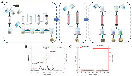
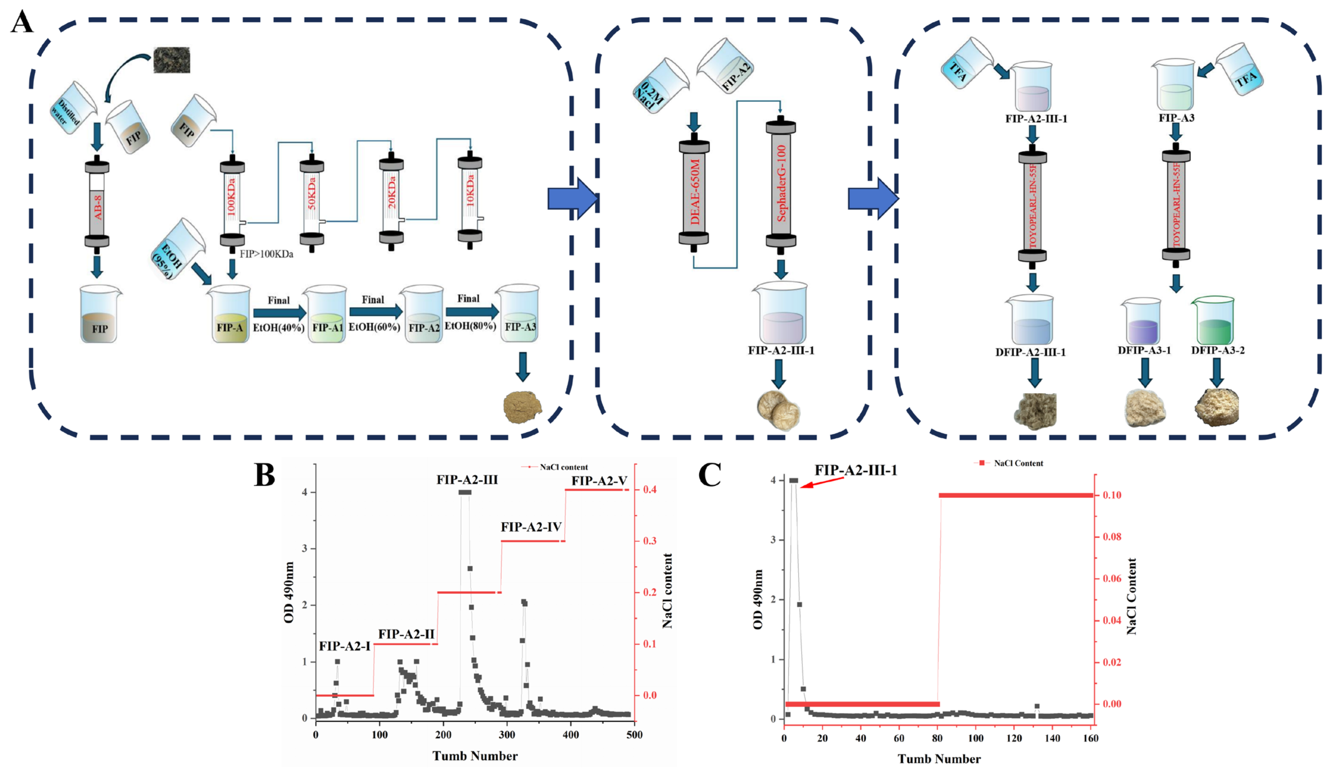
A yield of 15.03% (w/w) of the crude polysaccharides from Isatis indigotica Fort. leaves was obtained by hot water extraction followed by ethanol precipitation. Subsequently, Folium isatidis polysaccharide (FIP) was obtained using AB-8 macroporous adsorbent resin for further decolorization and deproteinization. Subsequently, five sub-fractions—designated as FIP-A (13.7%, w/w), FIP-B (1.8%, w/w), FIP-C (0.4%, w/w), FIP-D (0.1%, w/w), and FIP-E (84.0%, w/w)—were obtained by sequential ultrafiltration of FIP through membranes with molecular weight cut-offs of 100, 50, 20, and 10 kDa. FIP-A was then subjected to ethanol fractional precipitation by adjusting the ethanol concentration to 40%, 60%, and 80%. The resulting sub-fractions were named FIP-A1 (8.2%, wt%), FIP-A2 (49.2%, wt%) and FIP-A3 (42.6%, wt%), respectively. FIP-A2 on a DEAE-650M column produced five subfractions, designated FIP-A2-I through FIP-A2-V. FIP-A2-III was further purified by gel filtration on a Sephadex G-100 column (GE Healthcare Life Sciences, Uppsala, Sweden), which yielded a major subfraction named FIP-A2-III-1 with a yield of 49.27% (w/w). The separation and preparation flowchart and the elution curve are shown in Figure 1.
Figure 1.
(A) Workflow for isolation and purification of polysaccharides from leaves of Isatis indigotica Fort. (B) Elution curves of FIP-A2-I~V components. (C) Elution curves of FIP-A2-III-1.
2.2. Acid Hydrolysis of FIP-A2-III-1 and FIP-A3
Acid hydrolysis of FIP-A2-III-1 and FIP-A3 was performed with 0.5 M trifluoroacetic acid (TFA) at 80 °C for 2 h. As shown in Figure S1, following separation and purification on an HW-55F column and preparation using a Waters 515-2414 system, the pooled fractions were freeze-dried to yield DFIP-A2-III-1 (15.8%, wt%), DFIP-A3-1 (22.5%, wt%), and DFIP-A3-2 (2.8%, wt%), respectively.
2.3. Determination of Homogeneity and Molecular Weight
As shown in Figure S2, high performance gel permeation chromatography—evaporative light scattering detector (HPGPC-ELSD) analysis revealed that FIP-A2-III-1, FIP-A3, DFIP-A2-III-1, DFIP-A3-1, and DFIP-A3-2 all exhibited a single symmetrical peak, indicating that they are homogeneous polysaccharides.
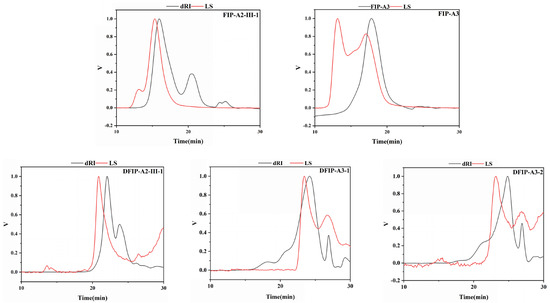
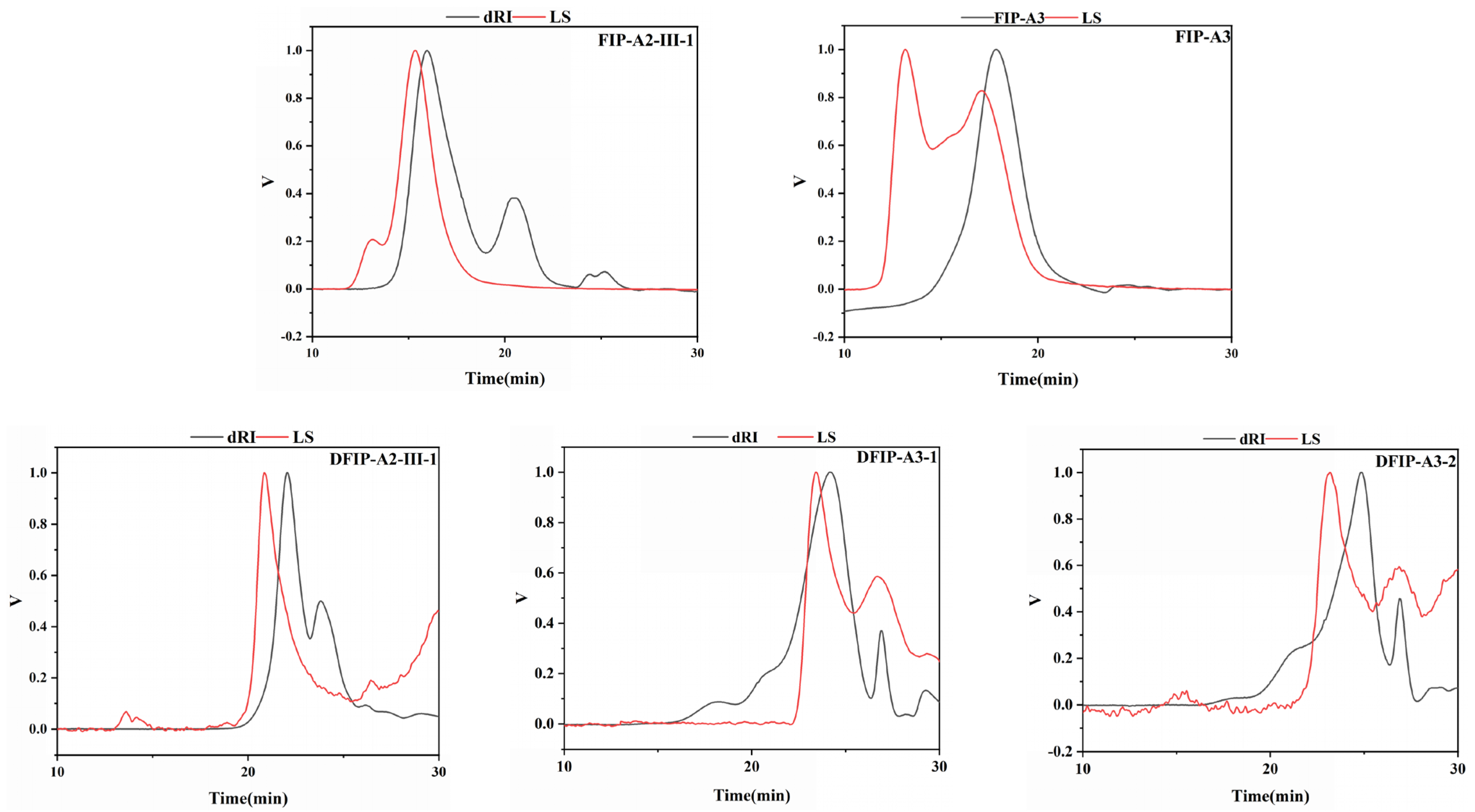
Figure 2 and Table 1 summarize the molecular weight data obtained by high performance size exclusion chromatography with multi-angle laser light scattering and refractive index detector (HPSEC-MALLS-RID). The Mw (molecular weight) of FIP-A2-III-1 decreased from 2.29 × 10⁵ g/mol before degradation to 5.11 × 10³ g/mol after degradation (DFIP-A2-III-1). Similarly, the molecular weight of FIP-A3 decreased from 9.85 × 10⁴ g/mol to 8.68 × 10³ g/mol (DFIP-A3-1) and 2.24 × 10³ g/mol (DFIP-A3-2), respectively. These results indicated a significant reduction in molecular weight after degradation. Additionally, the polydispersity indices (Mw/Mn) of the five polysaccharide samples were measured as 1.16, 1.43, 1.09, 1.54, and 1.28, corresponding to FIP-A2-III-1, DFIP-A2-III-1, FIP-A3, DFIP-A3-1, and DFIP-A3-2, respectively. The restricted molecular weight distribution of the polysaccharides was further supported by these observations. For example, the molecular weight of Belamcanda chinensis polysaccharide decreased from 2.62 × 10⁵ g/mol to 6.25 × 10⁴ g/mol after TFA [28]. Upon TFA hydrolysis, the molecular weight of the polysaccharide derived from Hericium erinaceus was reduced to 1.57–1.97 × 10⁶ Da [29]. These results collectively confirm that acid degradation is effective in regulating the molecular weight of polysaccharides.
Figure 2.
HPSEC-MALLS-RID chromatogram of FIP-A2-III-1, FIP-A3, DFIP-A2-III-1, DFIP-A3-1, and DFIP-A3-2.
Table 1.
Molecular weight data for FIP-A2-III-1, DFIP-A2-III-1, FIP-A3, DFIP-A3-1 and DFIP-A3-2.
2.4. FT-IR Analysis
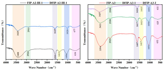
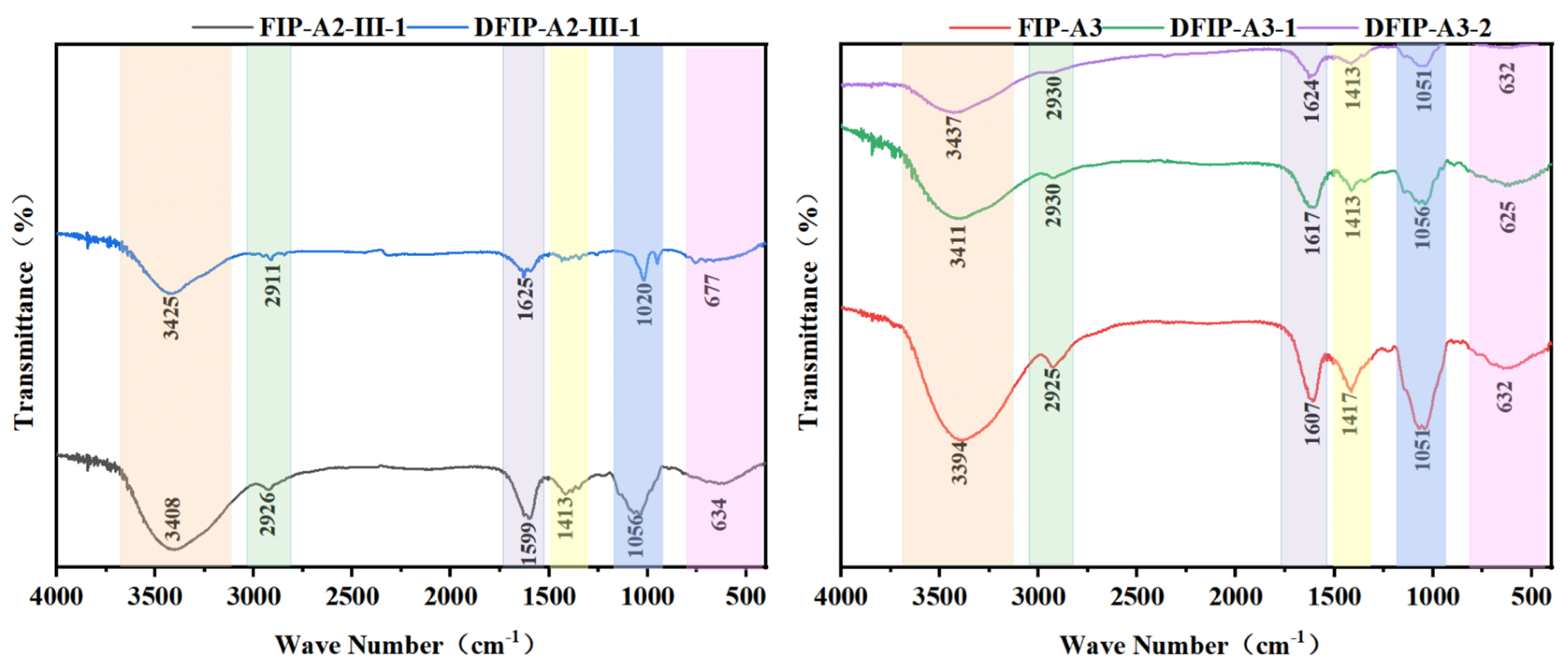
The polysaccharide functional groups were successfully characterized through analysis by Fourier transform infrared (FT-IR) spectroscopy (Figure 3). In the range of 3000–3500 cm−1, an obvious absorption peak characteristic of O–H stretching vibrations was detected [30]. Absorption bands corresponding to C–H and C=O stretching vibrations were detected at approximately 2900 cm−1 and 1700 cm−1 [31]. Furthermore, an absorption peak attributed to the C–H bending mode was recorded at approximately 1400 cm−1 [32]. An absorption signal corresponding to the C–O stretching vibration was recorded at approximately 1100 cm−1 [33]. In addition, a weak signal detected within the 520–820 cm−1 region indicated the existence of β-glycosidic bonds [34]. The presence of these absorption peaks was taken as an indication of the polysaccharide components. High similarity was observed among the absorption peaks of FIP-A2-III-1, DFIP-A2-III-1, FIP-A3, DFIP-A3-1, and DFIP-A3-2. Based on the absence of characteristic C=O stretching signals from their carboxyl groups, these polysaccharides were identified as neutral [35].
Figure 3.
The FT-IR spectra of FIP-A2-III-1, DFIP-A2-III-1, FIP-A3, DFIP-A3-1 and DFIP-A3-2.
2.5. Monosaccharide Composition Analysis
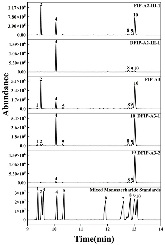
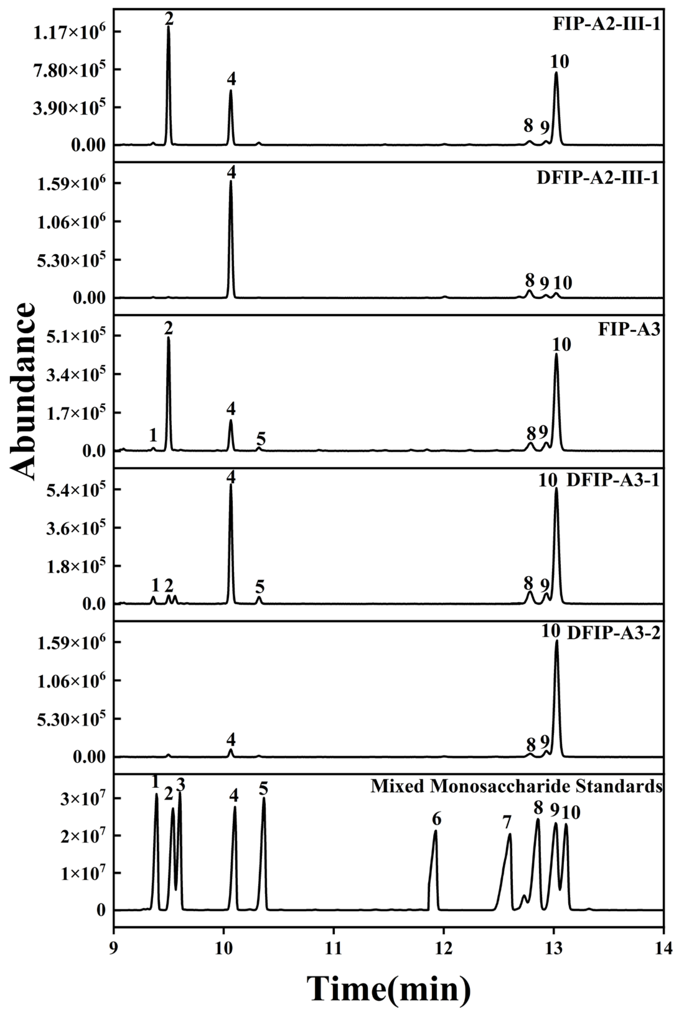
To determine their monosaccharide composition, FIP-A2-III-1, FIP-A3, DFIP-A3-1, DFIP-A3-2, and DFIP-A2-III-1 were analyzed by gas chromatography-mass spectrometry (GC-MS). The resulting data are presented in Figure 4. According to the analytical results, FIP-A2-III-1 was found to be composed of Ara, Rha, Man, Glc, and Gal, with the molar ratio of 36.000:11.317:0.054:0.050:40.415. DFIP-A2-III-1 was found to be composed of Rha, Man, Glc, and Gal, with the molar ratio of 10.041:0.051:0.005:0.029. FIP-A3 was found to be composed of Xyl, Ara, Rha, Fuc, Man, Glc, and Gal, with the molar ratio of 0.003:6.161:7.255:0.005:0.084:0.062:13.993, while DFIP-A3-1 was found to be composed of Xyl, Ara, Rha, Fuc, Man, Glc, and Gal, with the molar ratio of 0.029:0.041:10.98:0.035:0.214:0.120:22.442. DFIP-A3-2 was found to be composed of Rha, Man, Glc, and Gal, with the molar ratio of 0.042:0.012:0.033:19.507. FIP-A2-III-1, DFIP-A2-III-1, FIP-A3, DFIP-A3-1 and DFIP-A3-2 exhibited differences in monosaccharide content. A comparison of the monosaccharide compositions before and after degradation revealed that Xyl, Ara, and Fuc were highly susceptible to hydrolysis. Therefore, these results indicated that three polysaccharides were present in the side chains and that none contained glucuronic acid or galacturonic acid. Consequently, FIP-A2-III-1, DFIP-A2-III-1, FIP-A3, DFIP-A3-1 and DFIP-A3-2 were determined to be neutral polysaccharides with Rha and Gal comprising the polysaccharide backbone.
Figure 4.
Monosaccharide compositions of FIP-A2-III-1, DFIP-A2-III-1, FIP-A3, DFIP-A3-1 and DFIP-A3-2. 1: Xyl, 2: Ara, 3: Rib, 4: Rha, 5: Fuc, 6: GalA, 7: GlcA, 8: Man, 9: Glc, 10: Gal.
The previous literature has demonstrated that FIP-II, another neutral polysaccharide from Isatidis Folium, exhibits an essentially identical monosaccharide composition, with similarly high proportions of Gal, Ara, and Rha [8]. Previous studies on polysaccharides extracted from the leaves of Isatis indigotica Fort. indicated that the neutral fraction WFIP-N contained high proportions of Gal and Rha. These findings provided data support for the polysaccharides obtained in the present study [36]. Based on numerous studies on the immunological activity of polysaccharides, it could be inferred that a high proportion of Gal and Rha is a crucial structural feature for exerting immunological activity [37].
2.6. Methylation Analysis
As shown in Table 2, T-Galp, 1,6-Galp, 1,3-Galp, and 1,3,6-Galp residues were simultaneously detected in FIP-A3, DFIP-A3-1, and DFIP-A3-2. Furthermore, FIP-A3 was found to contain substantial amounts of 1,4-Galp, 1,3-Rhap, 1,2,4-Rhap, and 1,5-Araf residues, along with minor quantities of T-Araf. In DFIP-A3-1, 1,4-Galp and 1,2,4-Rhap residues were also present. These findings further confirmed the structural differences among FIP-A3, DFIP-A3-1, and DFIP-A3-2. Unlike the findings from the monosaccharide composition analysis, the low-concentration monosaccharides detected in the composition data were not reflected in the methylation results, a phenomenon closely related to their relative abundance. FIP-A3, DFIP-A3-1, and DFIP-A3-2 were found to share similar backbone structures. From the aerial parts of Astragalus membranaceus, thirteen immunologically active polysaccharides were extracted and isolated. Among them, within an arabinogalactan possessing a backbone of β-1,3,6-Gal, it was observed that the degradation of either β-1,3-Gal or β-1,6-Gal residues significantly reduced its immunological activity [38]. A novel galactose-rich GLP-1b was successfully isolated and identified from Ganoderma lucidum. Its backbone was found to be primarily composed of 1,6-Gal and various terminal residues. It was further demonstrated that GLP-1b exerted its immunomodulatory effects through binding to the TLR4 [39]. Consequently, the precise configuration of glycosidic linkages within DFIP-A3-1 might be correlated with its immunomodulatory functions, given its nature as a neutral polysaccharide.
Table 2.
The glycosidic linkage types and molar percentages of FIP-A3, DFIP-A3-1 and DFIP-A3-2 based on the methylation and GC–MS analyses.
2.7. NMR Analysis
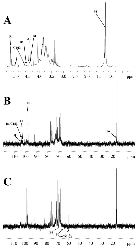
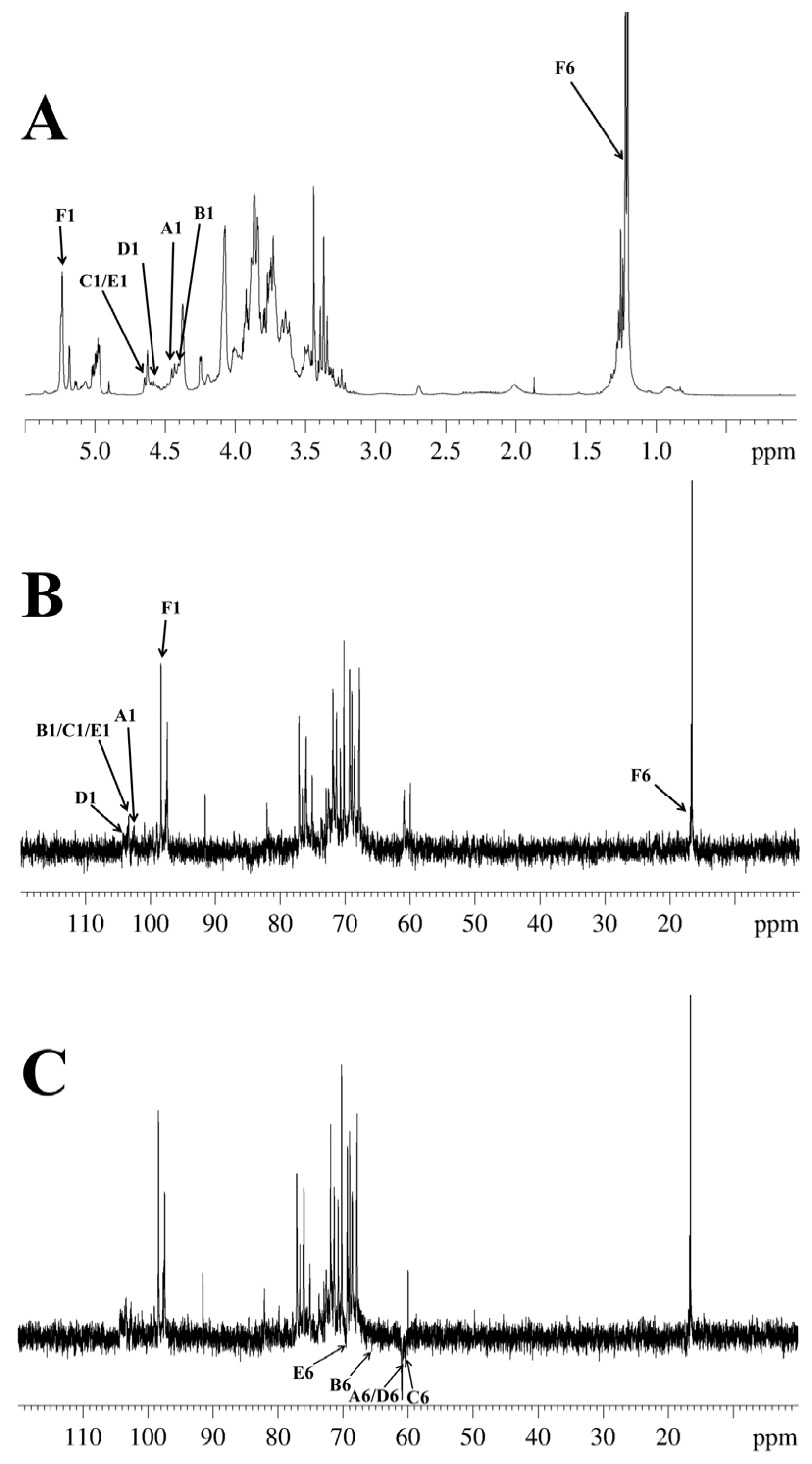
In agreement with earlier reports [40,41,42,43,44], six types of glycosidic linkages were determined in DFIP-A3-1 on the basis of anomeric proton resonances observed at δ 4.49, 4.45, 4.68, 4.58, 4.69 and 5.21. Signals for Xyl, Ara, Fuc, Man, and Glc residues were absent due to their low abundance (Figure 5A). The combined methylation and previously reported data suggested the presence of T-Galp (A), 1,6-Galp (B), 1,3-Galp (C), 1,4-Galp (D), 1,3,6-Galp (E), and 1,2,4-Rhap (F). Complete 1H and 13C signal assignments were achieved via 1D/2D NMR (1H, 13C, DEPT-135, 1H−1H COSY, HSQC, HMBC and NOESY).
Figure 5.
1D-NMR of DFIP-A3-1. (A) 1H NMR spectra (400 MHz). (B) 13C NMR spectra (100 MHz). (C) DEPT-135 spectra (100 MHz).
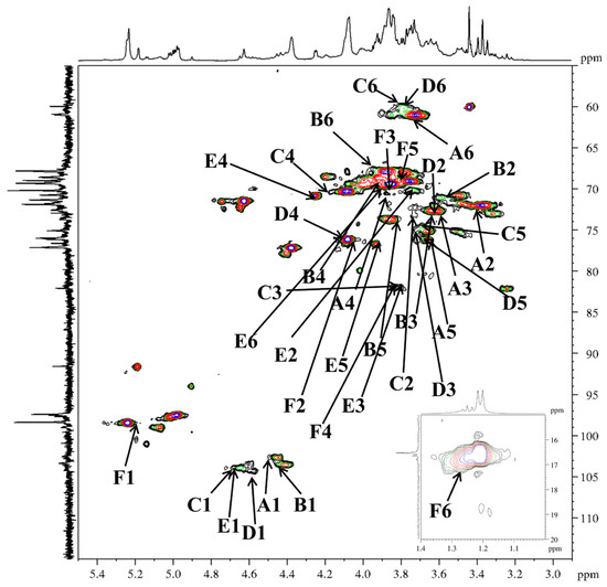
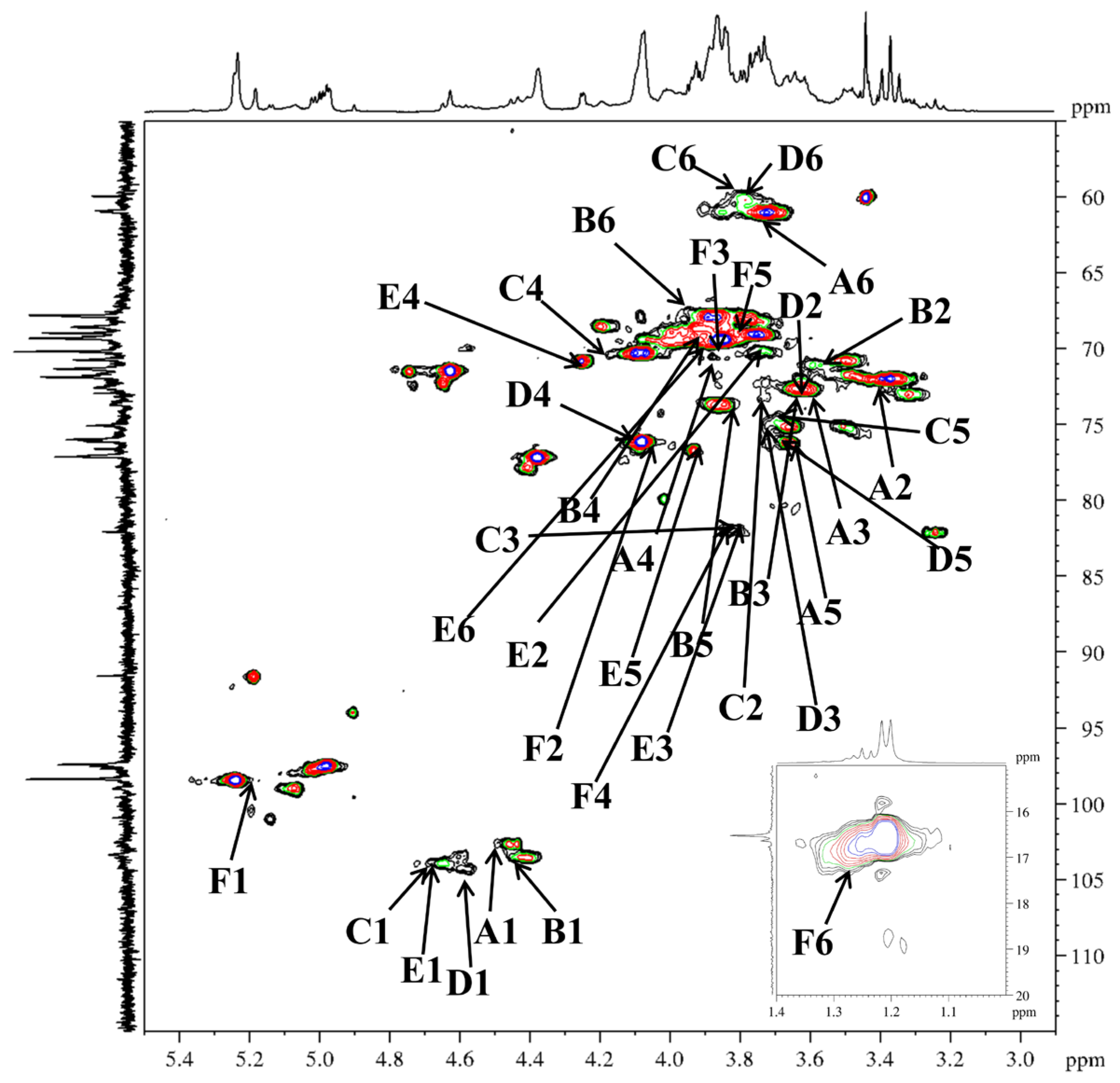
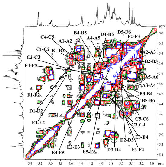
Based on the HSQC spectrum (Figure 6), the cross-peaks at δ 4.49/102.4, 4.45/103.7, 4.68/103.9, 4.58/104.4, 4.69/103.8 and 5.21/98.7 were attributed to the anomeric correlations of residues A-F, which correspond to T-Galp, 1,6-Galp, 1,3-Galp, 1,4-Galp, 1,3,6-Galp and 1,2,4-Rhap residues. Using the 13C NMR spectrum (Figure 5B), the chemical shifts in the corresponding anomeric carbons were subsequently determined. Key carbon signals for residues of −CH2 were further assigned using the DEPT-135 spectrum (Figure 5C). For instance, the inverted signals at δ 61.5, 67.4, 61.1, 61.4, and 69.8 were attributed to the C-6 atoms of residues A, B, C, D, and E. Notably, the anomeric proton chemical shifts in residues A–E were all below δ 4.95, indicating a β-configuration for these pyranose residues [45]. The anomeric proton chemical shift in residue F was observed at a relatively low field (δ 5.21), indicating an α-configuration [46]. Sequential assignment of the chemical shifts for H2–H6 and C2–C6 was accomplished by beginning with the anomeric proton signal of each residue, utilizing the scalar couplings between adjacent protons observed in the 1H−1H COSY spectrum (Figure 7) and the one-bond H−C connectivities derived from the HSQC spectrum. Beginning with the anomeric proton of residue T-β-Galp (A), a complete chain of spin-spin couplings was traced through the 1H−1H COSY spectrum. This sequential assignment established the chemical shifts for the coupled proton pairs H-1/H-2 (δ 3.45), H-2/H-3 (δ 3.59), H-3/H-4 (δ 3.89), H-4/H-5 (δ 3.65), and H-5/H-6a, H-6b (δ 3.65/3.72,3.64). The direct H−C correlations observed in the HSQC spectrum were used to assign the corresponding carbon chemical shifts, which were assigned to C-2 (δ 72.9), C-3 (δ 73.0), C-4 (δ 69.7), C-5 (δ 75.9), and C-6 (δ 61.5). By applying this well-established methodology, the full set of H and C chemical shifts for residues B through E was systematically assigned; these data are summarized in Table 3. In the 1H NMR spectrum, the H-6 protons of the 1,2,4-α-Rhap (F) residue were observed at δ 1.27, and the corresponding C-6 signal was assigned at δ 17.3 based on the HSQC correlation.
Figure 6.
HSQC spectra of DFIP-A3-1.
Figure 7.
1H−1H COSY spectra of DFIP-A3-1.
Table 3.
1H/13C chemical shifts in DFIP-A3-1 from HSQC data.
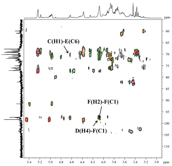
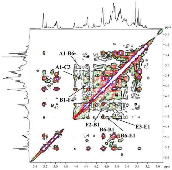
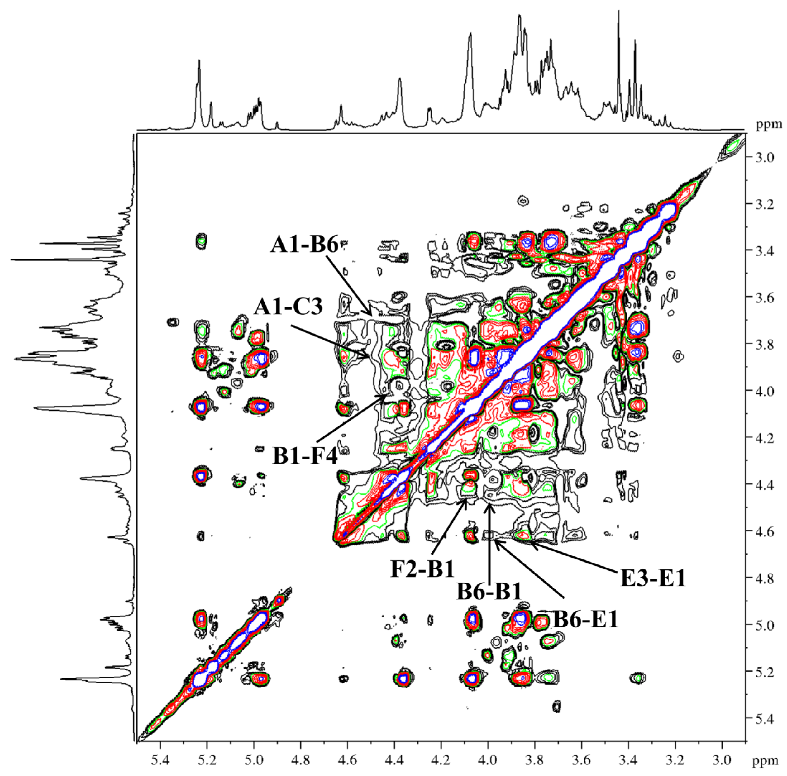
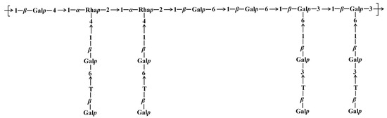
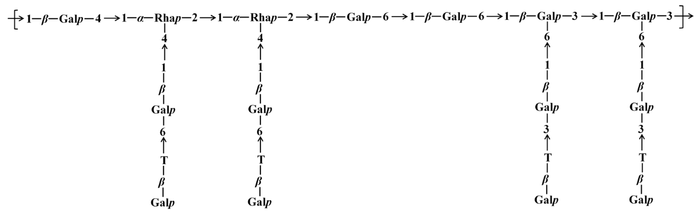
Further examination of the HMBC (Figure 8) (δ H/C) and NOESY (Figure 9) (δ H/H) correlation signals revealed that the peak correlating 1,4-β-Galp (D) H-4 with 1,2,4-α-Rhap (F) C-1 was located at δ 4.10/98.7. The correlation peak between H-2 and C-1 of 1,2,4-α-Rhap (F) was observed at δ 4.09/98.7. The correlation peak between H-2 of 1,2,4-α-Rhap (F) and H-1 of 1,6-β-Galp (B) was observed at δ 4.09/4.45. The Ha-6 and H-1 correlation peak of 1,6-β-Galp (B) was observed at δ 3.96/4.45. The correlation peak between Ha-6 of 1,6-β-Galp (B) and H-1 of 1,3,6-β-Galp (E) was observed at δ 3.96/4.69. The correlation peak between H-3 and H-1 of 1,3,6-β-Galp (E) was located at δ 3.85/4.69. It was indicated by the combined evidence that the backbone structure of DFIP-A3-1 was composed of →1)-β-Galp-(4→1,4)-α-Rhap-(2→1,4)-α-Rhap-(2→1)-β-Galp-(6→1)-β-Galp-(6→1,6)-β-Galp-(3→1,6)-β-Galp-(3→, with branches at the O-4 position of 1,2,4-α-Rhap and the O-6 position of 1,3,6-β-Galp. A cross-peak between H-1 of T-β-Galp (A) and Hb-6 of 1,6-β-Galp (B) was observed at δ 4.49/3.67. A cross-peak between H-1 of 1,6-β-Galp (B) and H-4 of 1,2,4-α-Rhap (F) was observed at δ 4.45/4.01. A correlation between H-1 of T-β-Galp (A) and H-3 of 1,3-β-Galp (C) was detected at δ 4.49/3.85. Additionally, a cross-peak between H-1 of 1,3-β-Galp (C) and C-6 of 1,3,6-β-Galp (E) was observed at δ 4.68/69.8. Collectively, these correlations reveal the branched structure of DFIP-A3-1. The putative repeating unit structure of DFIP-A3-1 is shown in Figure 10. Previous studies on plant-derived polysaccharides have also reported similar backbone and branching structures. For instance, a neutral polysaccharide (DPSW-A) was isolated from Taraxacum mongolicum Hand.-Mazz., and it was hydrolyzed with TFA to yield DPSW-A-on and DPSW-A-out. The backbone of DPSW-A-on was found to consist of →6)-β-Galp-(1→, →2,4)-α-Rhap-(1→, and →3,6)-β-Galp-(1→ residues, with branches attached at the O-4 position of 1,2,4-α-Rhap and the O-6 position of 1,3,6-β-Galp residue. The O-6 position of the →3,6)-β-Galp-(1→ residue served as the attachment site for a T-β-Galp-(1→3)-β-Galp-(1→ side chain. In a parallel manner, the dominant components of the hydrolyzed fragments were identified as T-α-Araf-(1→ and →5)-α-Araf-(1→ residues [47]. A polysaccharide, designated as REPI, was isolated from radish leaves. Its backbone was found to be composed of repeating units of →2)-Rhap-(1→4)-GalpA-(1→, along with three side chains. Among these, →4)-β-Galp-(1→ was attached at the O-4 position of →2,4)-α-Rhap-(1→ residue, and an arabino-β-3,6-galactan was also linked to the O-4 position of the same →2,4)-α-Rhap-(1→ residue [48].
Figure 8.
HMBC spectra of DFIP-A3-1.
Figure 9.
NOESY spectra of DFIP-A3-1.
Figure 10.
Putative structure of DFIP-A3-1.
2.8. Congo Red Assay Analysis
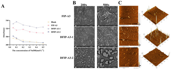
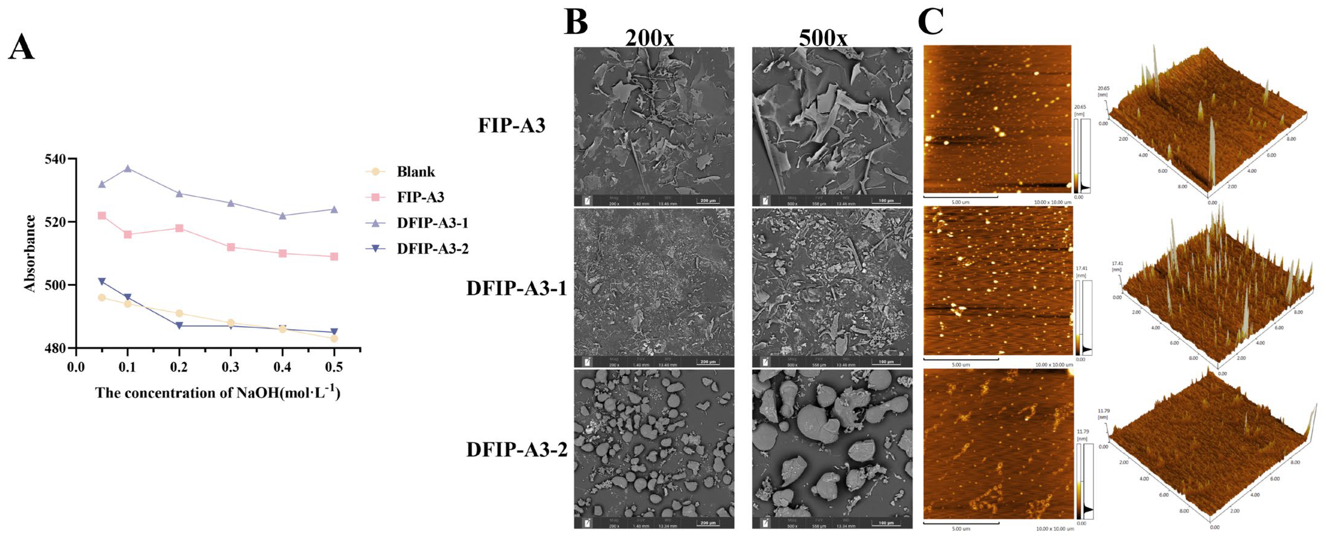
As shown in Figure 11A, the polysaccharides derived from the three-component fraction formed corresponding complexes with Congo red. As the NaOH concentration was increased, FIP-A3 and DFIP-A3-1 exhibited an initial rise followed by a sharp decline in their λmax values, suggesting the potential presence of a triple-helix conformation. In contrast, DFIP-A3-2 did not exhibit this initial increase followed by a sharp decrease, suggesting it likely lacks a triple-helix structure.
Figure 11.
Congo red staining and surface morphology analysis of FIP-A3, DFIP-A3-1 and DFIP-A3-2. (A) Congo red results for FIP-A3, DFIP-A3-1 and DFIP-A3-2. (B) 200× and 500× SEM results for FIP-A3, DFIP-A3-1 and DFIP-A3-2. (C) AFM results for FIP-A3, DFIP-A3-1 and DFIP-A3-2.
The triple-helical conformation of the polysaccharide is stabilized mainly by intermolecular hydrogen bonds and interactions with solvent molecules [49]. A comparative study demonstrated that, relative to FVPU1, FVPU2 possessed a more stable triple-helix conformation and displayed superior anti-inflammatory properties, primarily by attenuating the aberrant upregulation of inflammatory mediators, including NO, IL-1β, IL-6, and TNF-α [50]. Dendrobium officinale was the source of DLP-1 and DLP-2. Interestingly, both were found to have a triple-helix structure and had superior immunomodulatory bioactivity, promoting RAW 264.7 cell proliferation, phagocytic ability, and immune factor synthesis [51]. These results suggested that the triple-helical structure was linked to its bioactivity.
2.9. SEM Analysis
As shown in Figure 11B, distinct differences in the morphological features of FIP-A3, DFIP-A3-1, and DFIP-A3-2 were revealed by scanning electron microscopy (SEM) under 200× and 500× magnification. Upon examination, FIP-A3 displayed large, plate-shaped morphologies characterized by a disordered arrangement, subtle surface undulations, and comparatively even margins. DFIP-A3-1 was observed to have a rough surface with wrinkles and uneven edges, while DFIP-A3-2 was observed to display a rough surface with fissures, presenting a honeycomb-like structure.
2.10. AFM Analysis
As shown in Figure 11C, atomic force microscopy (AFM) analysis of the polysaccharide molecular morphology revealed that all three polysaccharide components—FIP-A3, DFIP-A3-1, and DFIP-A3-2—exhibited relatively uniform granular and network-like structures in 2D planar images. The 3D images revealed that the polysaccharide surfaces were densely covered with sharp peak-like structures, with peak heights of 20.65 nm, 17.41 nm, and 11.79 nm. Relevant studies indicated that the height of single chains in polysaccharides typically ranges from 0.1 to 1.0 nm [52]. Therefore, it was inferred that FIP-A3, DFIP-A3-1, and DFIP-A3-2 all possessed long branched chains and stacked multi-chain structures, along with good molecular aggregation properties.
2.11. Immunomodulatory Activity In Vitro of FIP
2.11.1. RAW 264.7 Macrophage Proliferation
The in vitro activities of FIP-A, FIP-B, FIP-C, FIP-D, and FIP-E, which were obtained by membrane separation, were carefully determined. As shown in Figure S3, FIP-D exhibited the strongest immune-enhancing effect, followed by FIP-A. Due to the extremely low yield of FIP-D after membrane separation, FIP-A was selected for further processing.
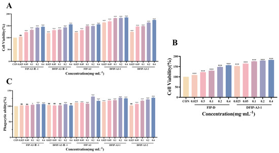
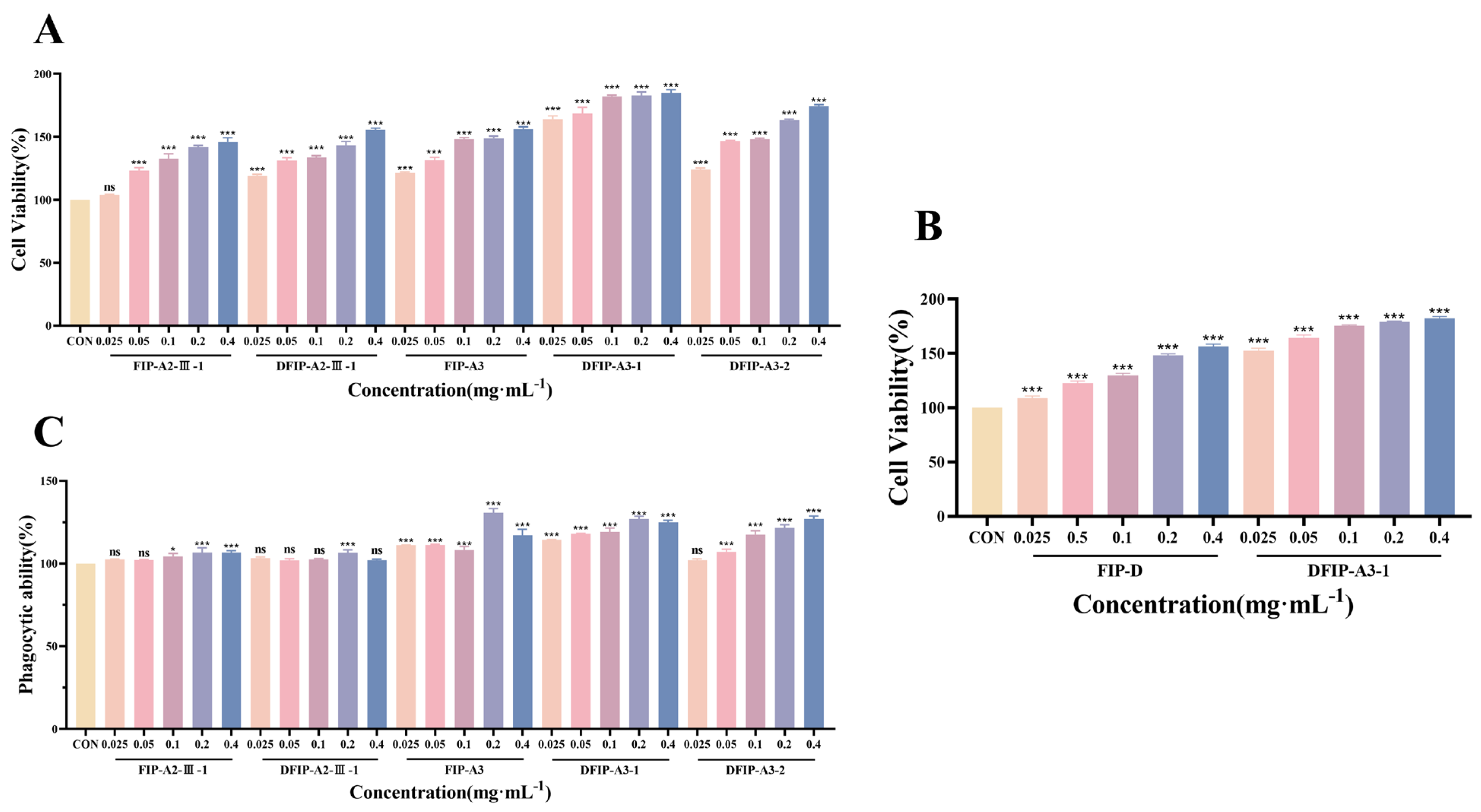
As shown in Figure 12A, the FIP-A2-III-1, DFIP-A2-III-1, FIP-A3, DFIP-A3-1 and DFIP-A3-2 were subjected to CCK-8 assays before and after degradation. The results revealed that DFIP-A3-1 exhibited significant immunostimulatory activity (p < 0.001). These data suggest that the immune-regulating potency of polysaccharides is tightly associated with their molecular weight [53,54,55,56]. Polysaccharides with higher molecular weights possess greater steric hindrance and lower solubility. Their active groups become encapsulated and cannot fully expose themselves for binding to receptors, leading to reduced bioavailability and consequently diminished immunological activity [57]. Lower-molecular-weight polysaccharides show stronger transmembrane penetration and an enhanced ability to trigger biological responses. However, an excessively low molecular weight may reduce spatial folding, compromising the maintenance of high-order structures. This results in short, irregular, highly flexible linear or globular conformations [58]. This structural feature likely explains why DFIP-A3-1 demonstrates optimal immunostimulatory effects.
Figure 12.
Screening results of immunomodulatory activity of polysaccharide fractions on RAW 264.7 cells. (A): Screening of immunological activity of FIP-A2-III-1, DFIP-A2-III-1, FIP-A3, DFIP-A3-1 and DFIP-A3-2 on RAW 264.7 cells. ( ± s; n = 3) *** p < 0.001 vs. the control and ns indicates not statistically significant. (B) Screening of immunological activity of FIP-D and DFIP-A3-1 on RAW 264.7 cells. ( ± s; n = 3) *** p < 0.001 vs. the control. (C) Effects of FIP-A2-III-1, DFIP-A2-III-1, FIP-A3, DFIP-A3-1 and DFIP-A3-2 on phagocytic activity in RAW 264.7 cells. ( ± s; n = 3) * p < 0.05, *** p < 0.001 vs. the control and ns indicates not statistically significant.
Simultaneously, as shown in Figure 12B, the most active degraded polysaccharide, DFIP-A3-1, was compared with FIP-D in terms of activity. The results indicate that the degraded polysaccharide exhibited superior immune-enhancing effects (p <0.001). This study did not directly pursue the naturally occurring, extremely low-yield, highly active component FIP-D. Instead, it utilized the high-yield, high-molecular-weight component FIP-A as a precursor. The degraded polysaccharide DFIP-A3-1 was obtained by acid degradation and was found to exhibit significantly higher immunological activity than its precursor. This approach effectively overcomes the bottleneck of limited natural high-activity polysaccharide resources.
2.11.2. Phagocytic Ability Assay
As shown in Figure 12C, phagocytosis is a hallmark of macrophage activation [59,60]. The impact of FIP-A2-III-1, DFIP-A2-III-1, FIP-A3, DFIP-A3-1, and DFIP-A3-2 on the phagocytic function of RAW264.7 cells was assessed in the present study using a neutral red phagocytosis assay. The results indicated that the polysaccharides from these fractions enhanced macrophage phagocytic rates in a concentration-dependent manner. This demonstrated that FIP-A2-III-1, DFIP-A2-III-1, FIP-A3, DFIP-A3-1, and DFIP-A3-2 effectively activated macrophages.
DFIP-A3-1 demonstrated strong phagocytic capacity overall (p < 0.001). Notably, the level of phagocytic activity recorded at 0.4 mg/mL did not differ significantly from that observed at 0.2 mg/mL, an outcome that aligns closely with the data obtained from the CCK-8 assay.
2.11.3. Determination of NO, IL-6 and TNF-α
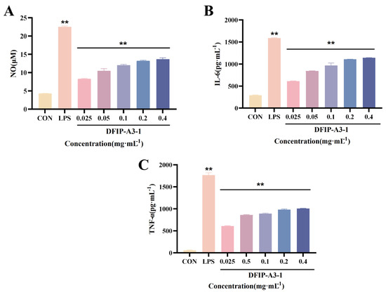
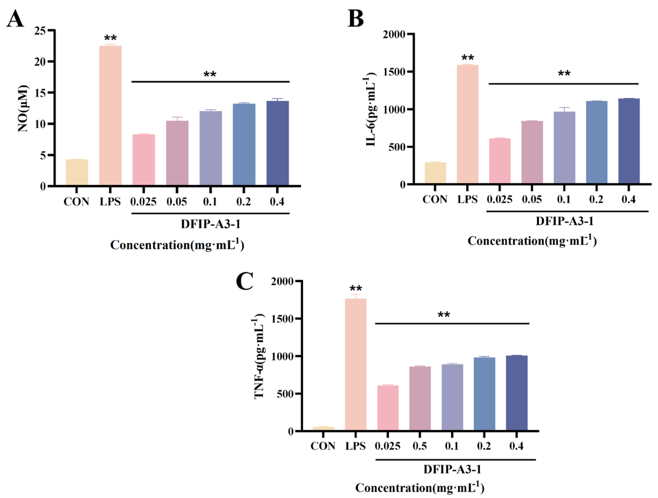
NO, IL-6, and TNF-α are key mediators produced upon macrophage activation, playing central roles in innate immunity and inflammatory responses to identify and eliminate pathogens, thereby providing essential protection for the body [61,62,63]. Following treatment, the levels of immunostimulatory cytokines released from RAW264.7 cells were quantified; the corresponding data are presented in Figure 13. Relatively low secretion of NO, IL-6, and TNF-α was detected in the control group. In contrast, the production of these cytokines was significantly increased by lipopolysaccharide (LPS) (p < 0.01). After 24 h of exposure to DFIP-A3-1, a significant concentration-dependent release of NO, IL-6, and TNF-α was detected (p < 0.01).
Figure 13.
The effects of DFIP-A3-1 on the secretion of NO, IL-6 and TNF-α in RAW 264.7 cells. (A) NO. (B) IL-6. (C) TNF-α. ( ± s; n = 3) ** p < 0.01 vs. the control.
2.11.4. NO, IL-6, and TNF-α mRNA Expression
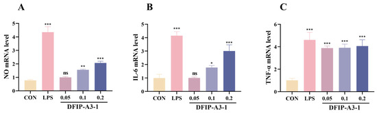
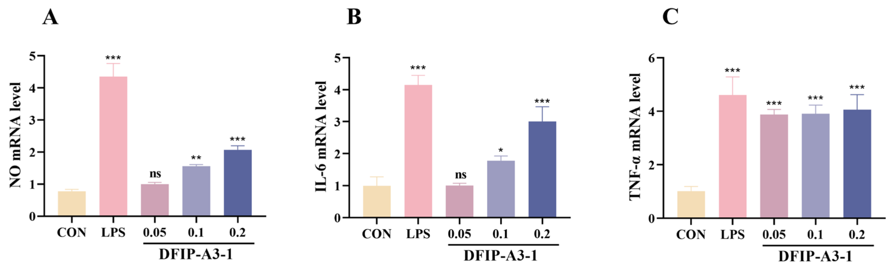
Furthermore, the mechanism by which DFIP-A3-1 stimulates macrophages to secrete immunostimulatory factors through the regulation of gene expression was investigated. After treatment with DFIP-A3-1, a significant upregulation in the expression of NO, IL-6, and TNF-α was observed, as depicted in Figure 14. These findings indicated that DFIP-A3-1 effectively induced immunomodulatory activity (p < 0.01).
Figure 14.
Expressions of NO, IL-6 and TNF-α mRNA in RAW 264.7 cells. (A) NO. (B) IL-6. (C) TNF-α. ( ± s; n = 3) * p < 0.05, ** p < 0.01, *** p < 0.001 vs. the control and ns indicates not statistically significant.
2.11.5. Mechanism of Action of DFIP-A3-1 in Activating the NF-κB Pathway
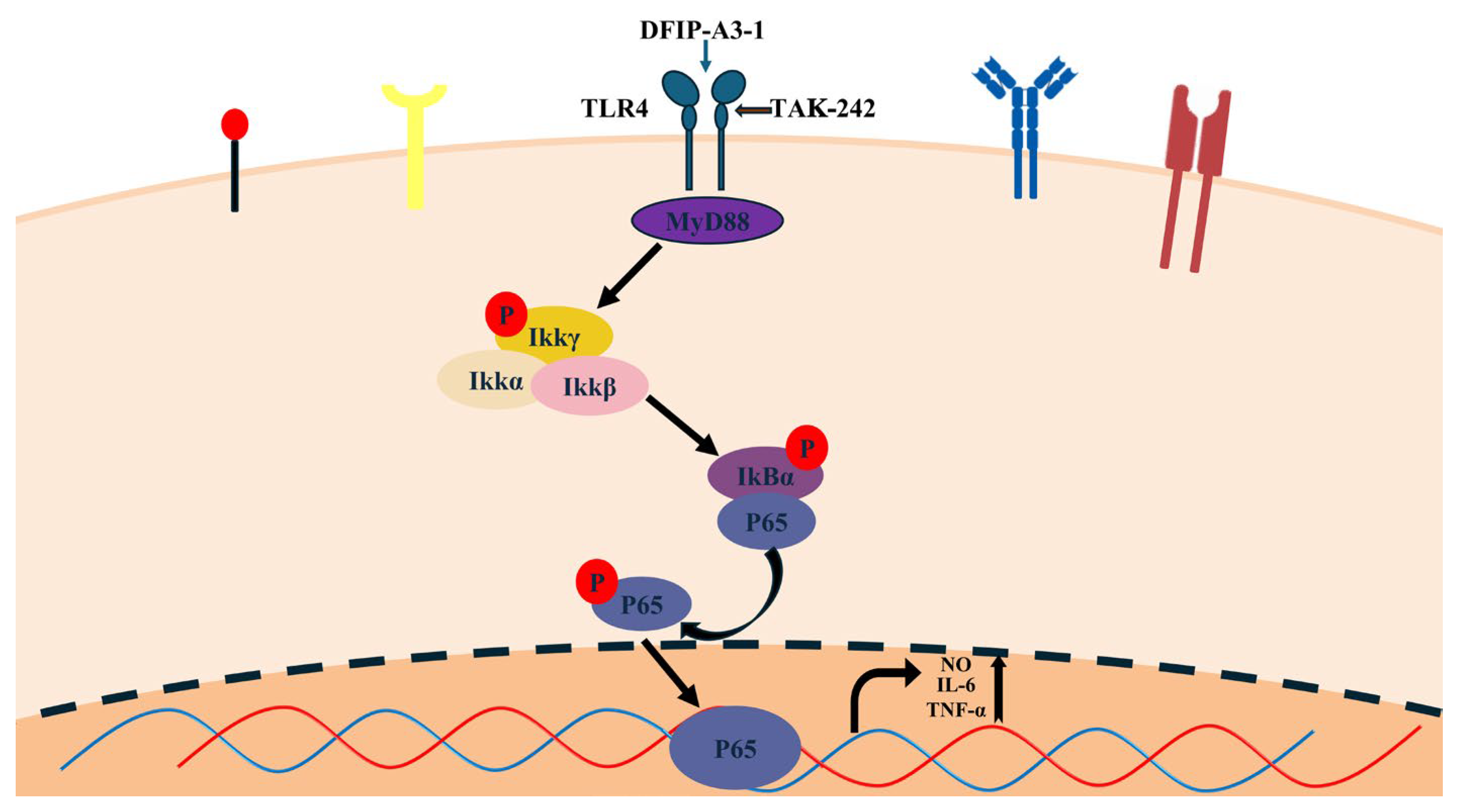
In this study, the activation of the nuclear factor kappaB (NF-κB) signaling pathway was evaluated to elucidate toll-like receptor 4 (TLR4) as the primary receptor for DFIP-A3-1 binding and to determine the mechanism by which DFIP-A3-1 induced macrophage activation. This process involves TLR4 binding to its corresponding ligand, activating downstream kinases (such as IRAKs and TRAF6), which ultimately activate the key kinase complex TAK1. Phosphorylated TAK1 activates the IKK complex, including IKKβ, which directly phosphorylates serine residues on the IκBα protein. Phosphorylated IκBα (p-IκBα) is recognized by the proteasome and rapidly degraded. The NF-κB (p65–p50) dimer is then released and translocated into the nucleus, where p65 undergoes phosphorylation, ultimately activating the NF-κB pathway [4,64,65].
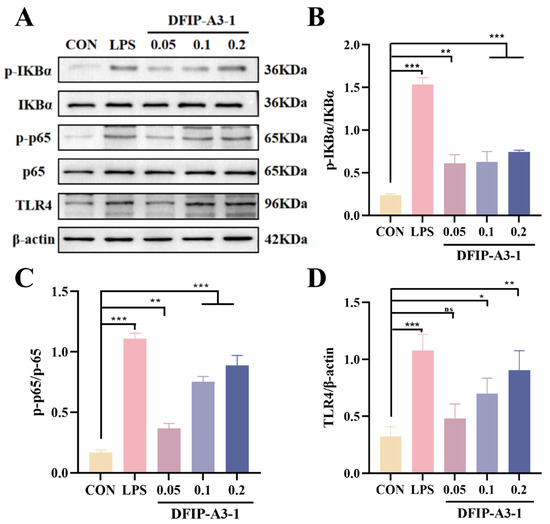
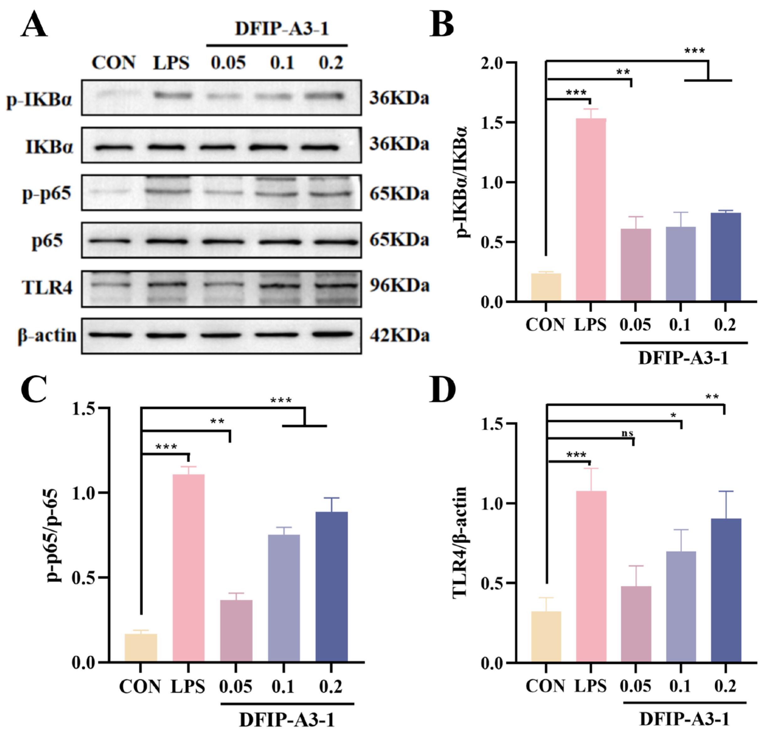
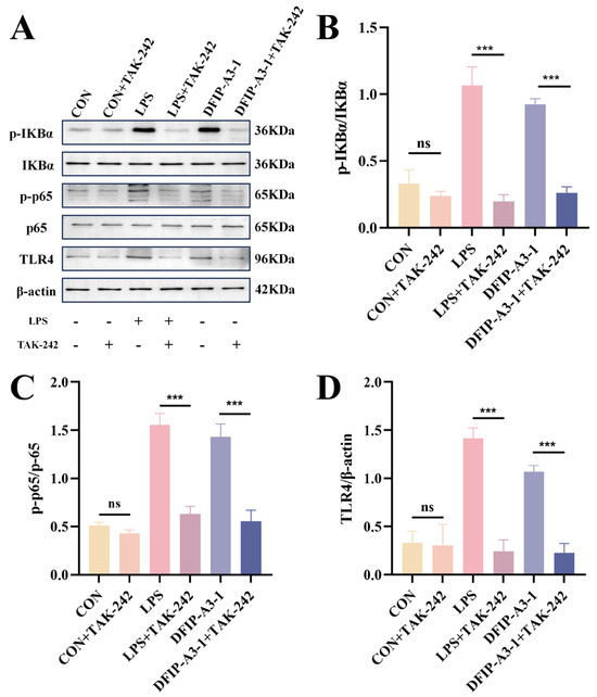
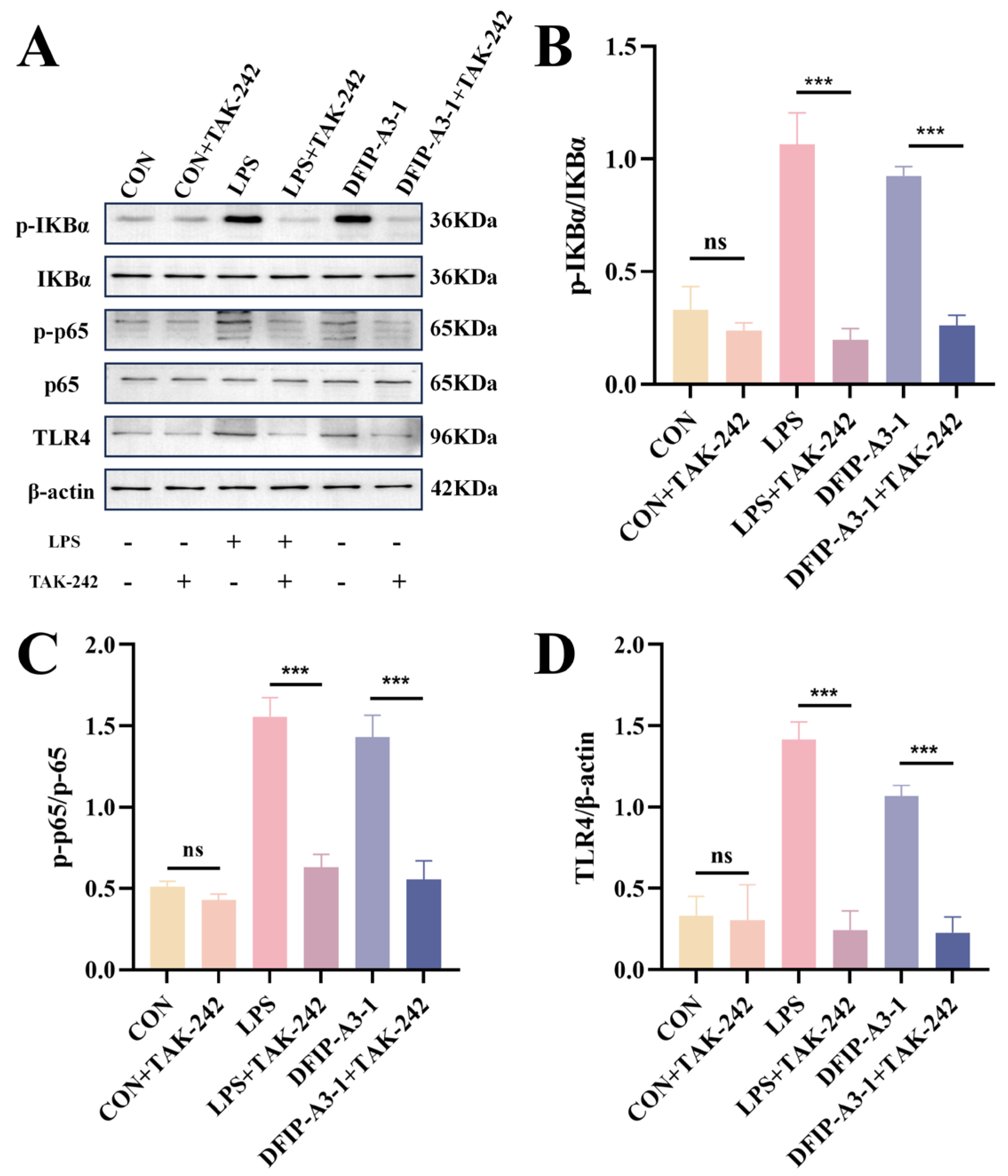
As shown in Figure 15, it was revealed by Western blot analysis that DFIP-A3-1 significantly enhanced the phosphorylation levels of IκBα and p65 (p < 0.01). This finding further suggested that TLR4 played a critical role in the DFIP-A3-1-mediated activation of RAW264.7 cells. To further verify this conclusion, the specific TLR4 inhibitor TAK-242 was added to the cells [66,67,68]. As shown in Figure 16, TAK-242 treatment significantly antagonized the DFIP-A3-1-induced upregulation of p-IκBα, p-p65, and TLR4 expression. To further clarify the immunomodulatory properties of DFIP-A3-1, a schematic diagram of its potential mechanism is provided in Figure 17.
Figure 15.
The p-IKKα, IKKα, p-p65, p65 and TLR4 protein expression levels in RAW264.7 cells. (A) The relative protein quantification of p-IκBα, p-p65, and TLR4. (B) p-IKKα and IKKα. (C) p-p65 and p65. (D) TLR4. ( ± s; n = 3), * p < 0.05, ** p < 0.01, *** p < 0.001 vs. the control and ns indicates not statistically significant.
Figure 16.
TAK242 inhibition downregulates the expression levels of p-IKKα, IKKα, p-p65, p65 and TLR4 proteins in RAW264.7 cells. (A) The relative protein quantification of p-IκBα, p-p65, and TLR4. (B) p-IKKα and IKKα. (C) p-p65 and P65. (D) TLR4. ( ± s; n = 3), *** p < 0.001 vs. the control and ns for not statistically significant.
Figure 17.
Schematic diagram of the immunomodulatory mechanism of DFIP-A3-1.
Some limitations were still present in this experiment. For instance, the stereostructural elucidation of DFIP-A3-1 was not precisely achieved. Owing to the complexity of the molecular weight and structure of polysaccharides, as well as the variability of their stereoconfigurations, the spatial conformation was difficult to determine. Similarly, due to the complexity of the signaling pathways, only one NF-κB pathway was identified through literature review in this study, and the possibility of the involvement of other pathways could not be excluded. Subsequent in vivo pharmacological experiments could be conducted to further verify the mechanism underlying its immunomodulatory activity. Meanwhile, in the context of scaled-up production and application, the experimental procedures could be refined, and effective translation into industrialization could be achieved.
3. Materials and Methods
3.1. Materials and Reagents
Isatidis Folium plants were cultivated in Bajingzi Township, Datong District, Daqing City, Heilongjiang Province. The RAW264.7 cell line was purchased from Haixing Biotechnology (Suzhou, China). All analytical reagents and standard monosaccharides utilized in this experiment were commercially available. The sources and specifications of experimental reagents were consistent with our previous published research [69].
3.2. Preparation, Isolation and Purification
The leaves of Isatis indigotica Fort. were defatted with 70% ethanol, and the residue was subsequently extracted with hot water. The resulting aqueous extract was concentrated and then lyophilized. AB-8 macroporous adsorbent resin was used for further decolorization and deproteinization, and the crude polysaccharides were obtained and designated as FIP. FIP (140 g) was dissolved and then sequentially fractionated using 100, 50, 20, and 10 kDa ultrafiltration membranes (BONA-GM-20, BONA GROUP, Jinan, China). The resulting fractions were designated as FIP-A to FIP-E. FIP-A was further subjected to ethanol fractional precipitation by adjusting the ethanol concentration to 40%, 60% and 80% respectively, and the resulting fractions were named FIP-A1~3. FIP-A2 was purified on a DEAE-650M cellulose column (5 cm × 60 cm; purchased from Tosoh Corporation, Tokyo, Japan) eluted with a linear gradient of 0–0.5 M NaCl solution, yielding sub-fractions designated as FIP-A2-I to FIP-A2-V. FIP-A2-III was further purified using Sephadex G-100 (Cytiva, Marlborough, MA, USA) gel column chromatography to obtain FIP-A2-III-1.
3.3. Acid Hydrolysis
FIP-A2-III-1 and FIP-A3 (100 mg) were dissolved in 0.5 M TFA (4 mL) and hydrolyzed in an oven at 80 °C for 2 h. The resulting products were then co-evaporated with methanol to eliminate residual acid. The samples were dissolved in ultrapure water (10 mL) and freeze-dried. Prior to use, each degraded polysaccharide was quantified and subsequently passed through a 0.8 μm membrane filter. Fractionation and isolation were subsequently achieved via column chromatography on a Toyopearl HW-55F column (Tosoh, Tokyo, Japan). The Waters 515-2414 HPLC system (Milford, MA, USA) was employed for the isolation and preparation of the degraded polysaccharides. The eluent was collected at a flow rate of 1.0 mL/min using an automatic fraction collector at intervals of 5 min per tube over a total run time of 600 min, yielding 120 fractions. After combining appropriate tubes and freeze-drying, three fractions were obtained and designated as DFIP-A2-III-1, DFIP-A3-1, and DFIP-A3-2, respectively.
3.4. Homogeneity and Molecular Weight
To evaluate homogeneity, an adapted version of a method described previously was employed [70]. Analysis was performed on an HPGPC system equipped with an UltrahydrogelTM Linear column (7.8 mm × 300 mm, Tosoh, Tokyo, Japan) connected to an HPLC-evaporative light-scattering detector (ELSD, Agilent 1260II HPLC, c.01.08.210, Agilent Technologies, Santa Clara, CA, USA).
According to a previously reported method [71], the molecular weights (Mw) of FIP-A2-III-1, DFIP-A2-III-1, FIP-A3, DFIP-A3-1, and DFIP-A3-2 were determined using HPSEC-MALLS-RID.
3.5. FT-IR Spectrum
For FT-IR spectroscopic analysis, each polysaccharide sample (3 mg) was thoroughly mixed with 200 mg of dried KBr. The mixture was subsequently compressed into a transparent disc. Spectral measurements were then performed over the range of 400–4000 cm−1 using an FTIR spectrophotometer (VERTEX-70, FTR 5.4.9, Bruker, Rheinstetten, Germany).
3.6. Monosaccharide Compositions
The monosaccharide composition of the polysaccharides was established via a method adapted from previously reported protocols [72]. Briefly, the polysaccharide samples were kindly hydrolyzed with 2 M TFA at 110 °C for 4 h, followed by derivatization together with a mixed standard of monosaccharides. The temperature program for TMSA derivatization was set as follows: an initial hold at 180 °C for 0 min, followed by ramping at 4 °C/min to 190 °C, then at 1 °C/min to 200 °C, and subsequently at 2 °C/min to 230 °C and further to 300 °C, with a final hold of 5 min.
3.7. Methylations
According to a previously reported method [73]. The lyophilized samples—FIP-A2-III-1, DFIP-A2-III-1, FIP-A3, DFIP-A3-1, and DFIP-A3-2 (2 mg)—were subjected to complete methylation. This was followed by hydrolysis, reduction, acetylation, and subsequent analysis employing a GC–MS system (Agilent 7890A-5975C, version 10.0, Agilent Technologies, Santa Clara, CA, USA) outfitted with a DB-5 silica capillary column (60 m × 0.25 mm × 0.25 µm). The temperature program employed for GC–MS analysis consisted of the following steps: an initial hold at 80 °C for 1 min, followed by a ramp to 200 °C at 5 °C/min, then to 220 °C at 2 °C/min, and finally to 270 °C at 10 °C/min, where the temperature was maintained for an additional 5 min.
3.8. 1D and 2D NMR
For NMR spectroscopic analysis, DFIP-A3-1 was solubilized in deuterium oxide (D2O). Subsequently, both one-dimensional and two-dimensional NMR spectra were acquired using a Bruker AM-400 spectrometer (operating at 400 MHz, version 11.1, Bruker, Fällanden, Switzerland).
3.9. Congo Red Test
Accurately weighed polysaccharide samples were dissolved in deionized water. Subsequently, each sample solution was combined with Congo red reagent (100 μM), and the mixture was titrated with 1 M NaOH to obtain final concentrations of NaOH of 0.05, 0.1, 0.2, 0.3, 0.4, and 0.5 M. After standing at room temperature for 10 min, the absorbance spectra of all mixtures were recorded over the 190–700 nm range with a UV-Vis spectrophotometer. The λmax and the associated absorbance for each sample were then obtained at the various NaOH concentrations tested. A binding curve was constructed by plotting the maximum absorption wavelength (nm) against the NaOH concentration (M).
3.10. Scanning Electron Microscopy
SEM analysis was employed to examine the surface features of FIP-A3, DFIP-A3-1, and DFIP-A3-2 (JSM-7800F, JEOL Ltd., Tokyo, Japan). After drying, the specimens were mounted on a conductive substrate and sputter-coated with gold using a vacuum deposition system. Observations were carried out under high vacuum conditions at an appropriate accelerating voltage.
3.11. Atomic Force Microscopy
A solution of FIP-A3, DFIP-A3-1, and DFIP-A3-2 was prepared at 5 µg/mL in deionized water and subsequently passed through a 0.22 µm cellulose filter. An aliquot of this filtrate was then deposited onto a freshly cleaved mica substrate and allowed to dry at ambient temperature. Morphological observations of the samples were conducted by AFM using an SPM-9700 instrument (SPM-9700, version 2.61, Shimadzu, Kyoto, Japan). The imaging was carried out in tapping mode, covering a region of 1.000 × 1.000 µm at a scan rate of 1.00 Hz.
3.12. Immunoregulatory Activity Test
3.12.1. Cell Culture and Treatment
RAW 264.7 cells represent a murine monocyte/macrophage lineage characterized by adherent growth and a rounded morphology. The culture environment consisted of DMEM medium containing 12% FBS and 0.1% penicillin-streptomycin, with cells maintained at 37 °C in a humidified incubator supplied with 5% CO2. Following 24 h of culture, cellular proliferation was monitored, and the medium was subsequently refreshed as needed.
3.12.2. Cell Viability Assay
For the culture procedure, RAW264.7 cells were plated in 96-well microplates at an initial density of 1 × 10⁵ cells per well. Following a 24 h adhesion period, the adherent cells were incubated for an additional 24 h with varying concentrations of FIP-A, FIP-B, FIP-C, FIP-D, and FIP-E. These concentrations spanned a range of 0.025, 0.05, 0.1, 0.2, and 0.4 mg/mL. Thereafter, a 10% CCK-8 solution was introduced, and the incubation was extended for an additional 2 h. The absorbance at 450 nm, reflecting cell viability, was recorded with a microplate reader (SpectraMax ABS Plus, version 7.1.0; Molecular Devices, San Jose, CA, USA).
The same method was employed in the subsequent in vitro immunostimulatory activity screening of FIP-A2-III-1, DFIP-A2-III-1, FIP-A3, DFIP-A3-1, and DFIP-A3-2. The optimal fraction, DFIP-A3-1, identified from these results, was then subjected to a further in vitro immunostimulatory activity assay alongside the previously evaluated FIP-D fraction, following the same procedure.
3.12.3. Phagocytosis Assay
The phagocytic activity of the cells was assessed using a neutral red assay kit. Cells were treated with varying concentrations of FIP-A2-III-1, DFIP-A2-III-1, FIP-A3, DFIP-A3-1, and DFIP-A3-2 for 24 h, with culture medium alone serving as the blank control. Subsequently, neutral red solution was added to the cells, followed by incubation for 2 h. After washing with PBS, cell lysis buffer (composed of 1% acetic acid and 50% ethanol, 1:1, v/v) was added. The absorbance was then measured at 540 nm.
3.12.4. Cytokine Detection
The levels of NO, IL-6 and TNF-α in the cell culture supernatants were determined using the Griess assay (Beyotime, Shanghai, China) and ELISA kits (Jiangsu Yutong Biotechnology Co., Ltd., Yancheng, China), respectively, strictly following the manufacturers’ instructions. Following drug stimulation, the cell culture supernatants were collected, and their absorbance was measured at the specified wavelengths using a microplate reader. All experimental procedures were performed in accordance with previously reported methods [74].
3.12.5. qRT-PCR Analysis
RNA was extracted following the supplier’s instructions, and reverse transcription was carried out according to the manufacturer’s protocol for the kit. Quantitative real-time PCR (qRT-PCR) was employed to assess mRNA expression levels. The reaction system and calculation method were kept consistent with those employed in our previous study [69] (Table 4).
Table 4.
Quantitative real-time PCR primer sequences.
3.12.6. Western Blotting
In the present investigation, Western blotting was carried out [75]. Radioimmunoprecipitation assay buffer (RIPA buffer) was used to extract total proteins from RAW264.7 cells. After fractionation by sodium dodecyl-sulfate polyacrylamide gel electrophoresis (SDS-PAGE), the resolved proteins were electrotransferred onto polyvinylidene fluoride (PVDF) membranes. These membranes were subsequently probed overnight at 4 °C with primary antibodies directed against IκBα, p-IkBα, p65, p-p65 and TLR4. The membrane was washed with tris-buffered saline with Tween-20 (TBST) buffer containing Tween-20, and the blot was imaged using an Odyssey CLx gel imaging system (Tanon-4200, 2.6.0.0, Tanon, Shanghai, China).
To determine whether TLR4 is required for the activation of the NF-κB pathway in RAW264.7 cells, experiments were performed according to a modified protocol based on established methods [76]. Cells were pre-incubated with 20 μM TAK-242, a selective TLR4 antagonist, for 6 h. Following removal of the culture supernatant, the cells were subsequently challenged with LPS and DFIP-A3-1 for an additional 24 h. Protein isolation was carried out following the same procedure described above for subsequent experimental analysis.
3.13. Statistical Analysis
To analyze the data, one-way analysis of variance (ANOVA) followed by multiple comparison test was performed. All values were expressed as the mean ± standard error of the mean ( ± s). * p < 0.05, ** p < 0.01 and *** p < 0.001 were considered statistically significant, and ns for not statistically significant. Graphical data were generated and processed using GraphPad Prism 8.0 (GraphPad Software, Boston, MA, USA) and Origin 2022.
4. Conclusions
In conclusion, two homogeneous polysaccharides, designated as FIP-A2-III-1 and FIP-A3, were successfully extracted and purified from the leaves of Isatis indigotica Fort. Subsequent degradation of these two polysaccharides yielded three degraded polysaccharides: DFIP-A2-III-1, DFIP-A3-1, and DFIP-A3-2. Chemical and spectroscopic analyses were performed on FIP-A2-III-1, DFIP-A2-III-1, FIP-A3, DFIP-A3-1, and DFIP-A3-2, and their primary structures were elucidated. As a result, they were determined to be neutral polysaccharides with molecular weights and monosaccharide compositions. Notably, DFIP-A2-III-1, DFIP-A3-1, and DFIP-A3-2 exhibited varying degrees of reduction in both molecular weight and monosaccharide species compared to their parent polysaccharides.
Furthermore, DFIP-A3-1 demonstrated potent immunostimulatory activity in preliminary screenings based on in vitro immune and phagocytic assays. It was identified as a low-molecular-weight rhamnogalactan, with glycosidic linkages comprising T-Galp, 1,6-Galp, 1,3-Galp, 1,4-Galp, 1,3,6-Galp, and 1,2,4-Rhap. NMR analysis revealed that the main chain structure was determined to be →1)-β-Galp-(4→1,4)-α-Rhap-(2→1,4)-α-Rhap-(2→1)-β-Galp-(6→1)-β-Galp-(6→1,6)-β-Galp-(3→1,6)-β-Galp-(3→, with branching occurring at the O-4 and O-6 positions of 1,2,4-β-Rhap and 1,3,6-β-Galp, respectively. The side chains were identified as T-β-Galp-(1→6)-β-Galp-(1→, and T-β-Galp-(1→3)-β-Galp-(1→. The in vitro immunostimulatory effects of DFIP-A3-1 were systematically evaluated. Within a defined concentration range, DFIP-A3-1 significantly enhanced the release of key immunomodulatory cytokines. Moreover, marked upregulation of the mRNA expression of NO, IL-6, and TNF-α was observed. Additionally, the levels of phosphorylated IκBα, phosphorylated p65, and TLR4 were markedly upregulated. This upregulation was effectively suppressed by treatment with TAK-242, thereby validating the essential involvement of the TLR4 receptor in DFIP-A3-1-induced macrophage activation and subsequent immune modulation.
Subsequent investigations should aim to evaluate the in vivo immunomodulatory efficacy of DFIP-A3-1. The present work establishes a rigorous scientific foundation for understanding the structure-activity relationship of this polysaccharide and underscores its promise as a candidate for natural immunotherapeutic applications.
Supplementary Materials
The following supporting information can be downloaded at: https://www.mdpi.com/article/10.3390/molecules31091461/s1, Figure S1: (a) Elution profile of FIP-A2-III-1 by preparative liquid chromatography. (b) Elution profile of DFIP-A3-1 and DFIP-A3-2 by preparative liquid chromatography; Figure S2: HPGPC-ELSD chromatogram of FIP-A2-III-1, FIP-A3, DFIP-A2-III-1, DFIP-A3-1 and DFIP-A3-2; Figure S3: In Vitro Immunomodulatory Screening of FIP-A to FIP-E. ( ± s; n = 3) * p < 0.05, ** p < 0.01, *** p < 0.001 vs. the control and ns for not statistically significant.
Author Contributions
Writing—original draft preparation, Y.S. and X.W.; writing—review and editing, H.Z. (Huiming Zhang) and Y.W.; visualization, Z.W.; data curation, Y.Z.; supervision, H.Z. (Hongbo Zhao). All authors have read and agreed to the published version of the manuscript.
Funding
This research was funded by the Supported by Heilongjiang Provincial Natural Science Foundation of China (no. YQ2023H001); the North Medicine and Functional Food Characteristic Subject Project in Heilongjiang Province (no. HLJTSXK-2022-03); Longjiang Technology Talent Spring Goose Support Program (no. CYQN24035); and the Basic Research Project of the Fundamental Research Business Expenses of the Education Department in Heilongjiang Province (no. 2022-KYYWF-0641).
Institutional Review Board Statement
The study did not involve humans or animals.
Informed Consent Statement
Not applicable.
Data Availability Statement
Data are contained within the article.
Conflicts of Interest
The authors declare no conflicts of interest.
Abbreviations
| FIP | Folium isatidis polysaccharide |
| TFA | trifluoroacetic acid |
| HPGPC-ELSD | high performance gel permeation chromatography—evaporative light scattering detector |
| HPSEC-MALLS-RID | high performance size exclusion chromatography with multi-angle laser light scattering and refractive index detector |
| GC-MS | gas chromatography-mass spectrometry |
| SEM | scanning electron microscope |
| AFM | atomic force microscopy |
| Mw | molecular weight |
| FT-IR | fourier transform infrared |
| RIPA buffer | Radioimmunoprecipitation assay buffer |
| SDS-PAGE | sodium dodecyl-sulfate polyacrylamide gel electrophoresis |
| PVDF | polyvinylidene fluoride |
| TBST | tris-buffered saline with Tween-20 |
| LPS | lipopolysaccharide |
| TLR4 | toll-like receptor 4 |
| qRT-PCR | Quantitative reverse-transcription PCR |
| NF-κB | nuclear factor kappaB |
References
- Colaço, H.G.; Moita, L.F. Initiation of Innate Immune Responses by Surveillance of Homeostasis Perturbations. FEBS J. 2016, 283, 2448–2457. [Google Scholar] [CrossRef] [Scilit]
- Ramirez, G.A.; Cardamone, C.; Lettieri, S.; Fredi, M.; Mormile, I. Clinical and Pathophysiological Tangles between Allergy and Autoimmunity: Deconstructing an Old Dichotomic Paradigm. Clin. Rev. Allergy Immunol. 2025, 68, 13. [Google Scholar] [CrossRef] [Scilit]
- Tang, J.; Zhao, X. Research Progress on Regulation of Immune Response by Tanshinones and Salvianolic Acids of Danshen (Salvia miltiorrhiza Bunge). Molecules 2024, 29, 1201. [Google Scholar] [CrossRef] [Scilit]
- Wang, Y.; Wang, L.; Zhang, H.; Ren, P.; Cheng, X.; Hong, F.; Liu, J.; Zhang, R.; Zhao, J.; Gou, D. Immunostimulatory Effects Mechanism of Polysaccharide Extracted from Acanthopanax senticosus on RAW 264.7 Cells through Activating the TLR/MAPK/NF-κB Signaling Pathway. Sci. Rep. 2025, 15, 13440. [Google Scholar] [CrossRef] [Scilit] [PubMed]
- Xu, W.; Zhao, M.; Fu, X.; Hou, J.; Wang, Y.; Shi, F.; Hu, S. Molecular Mechanisms Underlying Macrophage Immunomodulatory Activity of Rubus Chingii Hu Polysaccharides. Int. J. Biol. Macromol. 2021, 185, 907–916. [Google Scholar] [CrossRef] [Scilit] [PubMed]
- Zhou, W.; Hong, J.; Liu, T.; Li, M.; Jin, H.; Wang, X. Polygonatum Polysaccharide Regulates Macrophage Polarization and Improves LPS-Induced Acute Lung Injury through TLR4-MAPK/NF-κB Pathway. Can. Respir. J. 2022, 2022, 2686992. [Google Scholar] [CrossRef] [Scilit]
- Sotto, A.D.; Vitalone, A.; Giacomo, S.D. Plant-Derived Nutraceuticals and Immune System Modulation: An Evidence-Based Overview. Vaccines 2020, 8, 468. [Google Scholar] [CrossRef] [Scilit]
- Abuduwaili, A.; Nuerxiati, R.; Mutailifu, P.; Gao, Y.; Lu, C.; Yili, A. Isolation, Structural Modification, Characterization, and Bioactivity of Polysaccharides from Folium Isatidis. Ind. Crops Prod. 2022, 176, 114319. [Google Scholar] [CrossRef] [Scilit]
- Chen, J.; Zhu, Z.; Gao, T.; Chen, Y.; Yang, Q.; Fu, C.; Zhu, Y.; Wang, F.; Liao, W. Isatidis Radix and Isatidis Folium: A Systematic Review on Ethnopharmacology, Phytochemistry and Pharmacology. J. Ethnopharmacol. 2022, 283, 114648. [Google Scholar] [CrossRef] [Scilit]
- Kang, M.; Wu, H.; Yang, Q.; Huang, L.; Hu, Q.; Ma, T.; Li, Z.; Liu, J. A Chromosome-Scale Genome Assembly of Isatis Indigotica, an Important Medicinal Plant Used in Traditional Chinese Medicine: An Isatis Genome. Hortic. Res. 2020, 7, 18. [Google Scholar] [CrossRef] [Scilit]
- Min, G.-Y.; Kim, T.I.; Kim, J.-H.; Cho, W.-K.; Yang, J.-H.; Ma, J.Y. Anti-Atopic Effect of Isatidis Folium Water Extract in TNF-α/IFN-γ-Induced HaCaT Cells and DNCB-Induced Atopic Dermatitis Mouse Model. Molecules 2023, 28, 3960. [Google Scholar] [CrossRef] [Scilit]
- Wong, L.W.; Goh, C.B.S.; Tan, J.B.L. A Systemic Review for Ethnopharmacological Studies on Isatis Indigotica Fortune: Bioactive Compounds and Their Therapeutic Insights. Am. J. Chin. Med. 2022, 50, 161–207. [Google Scholar] [CrossRef] [Scilit]
- Huang, S.; Zhang, X.; Zhang, X.; Kong, L.; Ma, J.; Yang, K.; Ma, W.; Liu, X.; Liu, W. Preparation, Structure-Activity Relationship, Biological Activities and Market Applications of Isatis Indigotica Polysaccharides: A Review. Ind. Crops Prod. 2025, 234, 121552. [Google Scholar] [CrossRef] [Scilit]
- Yin, Y.; Shi, X.; Cai, X.; Liu, F.; Ni, W.; Li, B.; Wan, X.; Ren, M. Isolation Techniques, Structural Characteristics, and Pharmacological Effects of Phellinus Polysaccharides: A Review. Molecules 2024, 29, 3047. [Google Scholar] [CrossRef] [Scilit]
- Fernandes, P.A.R.; Coimbra, M.A. The Antioxidant Activity of Polysaccharides: A Structure-Function Relationship Overview. Carbohydr. Polym. 2023, 314, 120965. [Google Scholar] [CrossRef] [Scilit]
- Liu, T.; Ren, Q.; Wang, S.; Gao, J.; Shen, C.; Zhang, S.; Wang, Y.; Guan, F. Chemical Modification of Polysaccharides: A Review of Synthetic Approaches, Biological Activity and the Structure–Activity Relationship. Molecules 2023, 28, 6073. [Google Scholar] [CrossRef] [Scilit] [PubMed]
- Mohammed, A.S.A.; Naveed, M.; Jost, N. Polysaccharides; Classification, Chemical Properties, and Future Perspective Applications in Fields of Pharmacology and Biological Medicine (a Review of Current Applications and Upcoming Potentialities). J. Polym. Environ. 2021, 29, 2359–2371. [Google Scholar] [CrossRef] [Scilit]
- Chatnarin, S.; Thirabunyanon, M. Potential Bioactivities via Anticancer, Antioxidant, and Immunomodulatory Properties of Cultured Mycelial Enriched β-D-Glucan Polysaccharides from a Novel Fungus Ophiocordyceps Sinensis OS8. Front. Immunol. 2023, 14, 1150287. [Google Scholar] [CrossRef] [Scilit]
- Casas-Arrojo, V.; Arrojo Agudo, M.d.L.Á.; Cárdenas García, C.; Carrillo, P.; Pérez Manríquez, C.; Martínez-Manzanares, E.; Abdala Díaz, R.T. Antioxidant, Immunomodulatory and Potential Anticancer Capacity of Polysaccharides (Glucans) from Euglena gracilis G.A. Klebs. Pharmaceuticals 2022, 15, 1379. [Google Scholar] [CrossRef] [Scilit] [PubMed]
- Li, Y.; Guo, X.; Zhong, R.; Ye, C.; Chen, J. Structure Characterization and Biological Activities Evaluation of Two Hetero-Polysaccharides from Lepista Nuda: Cell Antioxidant, Anticancer and Immune-Modulatory Activities. Int. J. Biol. Macromol. 2023, 244, 125204. [Google Scholar] [CrossRef] [Scilit]
- Feng, Y.; Qiu, Y.; Duan, Y.; He, Y.; Xiang, H.; Sun, W.; Zhang, H.; Ma, H. Characterization, Antioxidant, Antineoplastic and Immune Activities of Selenium Modified Sagittaria sagittifolia L. Polysaccharides. Food Res. Int. 2022, 153, 110913. [Google Scholar] [CrossRef] [Scilit] [PubMed]
- Song, W.; Wang, Y.; Li, G.; Xue, S.; Zhang, G.; Dang, Y.; Wang, H. Modulating the Gut Microbiota Is Involved in the Effect of Low-Molecular-Weight Glycyrrhiza Polysaccharide on Immune Function. Gut Microbes 2023, 15, 2276814. [Google Scholar] [CrossRef] [Scilit]
- Lee, Q.; Xue, Z.; Luo, Y.; Lin, Y.; Lai, M.; Xu, H.; Liu, B.; Zheng, M.; Lv, F.; Zeng, F. Low Molecular Weight Polysaccharide of Tremella Fuciformis Exhibits Stronger Antioxidant and Immunomodulatory Activities than High Molecular Weight Polysaccharide. Int. J. Biol. Macromol. 2024, 281, 136097. [Google Scholar] [CrossRef] [Scilit]
- Lin, Y.; Yang, J.; Luo, L.; Zhang, X.; Deng, S.; Chen, X.; Li, Y.; Bekhit, A.E.-D.A.; Xu, B.; Huang, R. Ferroptosis Related Immunomodulatory Effect of a Novel Extracellular Polysaccharides from Marine Fungus Aureobasidium melanogenum. Mar. Drugs 2022, 20, 332. [Google Scholar] [CrossRef] [Scilit]
- Ip, C.C.; Manam, V.; Hepler, R.; Hennessey, J.P. Carbohydrate Composition Analysis of Bacterial Polysaccharides: Optimized Acid Hydrolysis Conditions for HPAEC-PAD Analysis. Anal. Biochem. 1992, 201, 343–349. [Google Scholar] [CrossRef] [Scilit]
- Gao, Z.; Liu, X.; Yu, J.; Li, Z.; Shi, H.; Zhang, G.; Ling, J. Structural Basis of Immunomodulation by Edible Fungal Polysaccharides: From Molecular Characteristics to Action Mechanisms. Carbohydr. Res. 2025, 555, 109591. [Google Scholar] [CrossRef] [Scilit] [PubMed]
- Pan, M.; Zhang, X.; Huang, S.; Liu, M.; Song, S.; Ma, J.; Li, C.; Ma, W.; Liu, X. Preparation, Structural Characteristics and Immune Regulatory Effects of Codonopsis Pilosula Polysaccharides: A Review. Front. Immunol. 2025, 16, 1641928. [Google Scholar] [CrossRef] [Scilit]
- Duan, Y.; Hu, Z.; Jin, L.; Zong, T.; Zhang, X.; Liu, Y.; Yang, P.; Sun, J.; Zhou, W.; Li, G. Efficient Degradation and Enhanced Anticomplementary Activity of Belamcanda chinensis (L.) DC. Polysaccharides via Trifluoroacetic Acid Treatment with Different Degrees. Int. J. Biol. Macromol. 2024, 276, 134117. [Google Scholar] [CrossRef] [Scilit]
- Li, T.; Li, C.; Wu, D.; Yang, Y.; Jin, Y. Studies on the Acid Degradation Process and in Vitro Immune Activity of the Polysaccharide H6PC20 in Hericium Erinaceus. IOP Conf. Ser. Mater. Sci. Eng. 2018, 392, 052014. [Google Scholar] [CrossRef] [Scilit]
- Gieroba, B.; Kalisz, G.; Krysa, M.; Khalavka, M.; Przekora, A. Application of Vibrational Spectroscopic Techniques in the Study of the Natural Polysaccharides and Their Cross-Linking Process. Int. J. Mol. Sci. 2023, 24, 2630. [Google Scholar] [CrossRef] [Scilit]
- Ghamgui, H.; Jarboui, R.; Jeddou, K.B.; Torchi, A.; Siala, M.; Cherif, S.; Trigui, M. Polysaccharide from Thymelaea hirsuta L. Leaves: Structural Characterization, Functional Properties and Antioxidant Evaluation. Int. J. Biol. Macromol. 2024, 262, 129244. [Google Scholar] [CrossRef] [Scilit]
- Chen, X.; Yang, T.; Huang, Q.; Li, B.; Ding, X.; Hou, Y. Comparative Studies on the Structure and Biological Activities of Two New Polysaccharides from Tricholoma sinoportentosum (TS-P) and Termitomyces albuminosus (TA-P). Polymers 2023, 15, 2227. [Google Scholar] [CrossRef] [Scilit]
- Cheng, Z.; Zheng, Q.; Duan, Y.; Cai, M.; Zhang, H. Effect of Subcritical Water Temperature on the Structure, Antioxidant Activity and Immune Activity of Polysaccharides from Glycyrrhiza Inflata Batalin. Int. J. Biol. Macromol. 2024, 261, 129591. [Google Scholar] [CrossRef] [Scilit]
- Wu, Q.; Liang, B.; Wang, J.; Dai, Y. Ultrasound-Assisted Extraction of Polysaccharides from Lyophyllum decastes: Structural Analysis and Bioactivity Assessment. Molecules 2025, 30, 961. [Google Scholar] [CrossRef] [Scilit] [PubMed]
- Fimbres-Olivarria, D.; Marquez-Escalante, J.; Martínez-Robinson, K.G.; Miranda-Arizmendi, V.; Anda-Flores, Y.D.; Rascon-Chu, A.; Brown-Bojorquez, F.; Carvajal-Millan, E. Physicochemical and Microstructural Characteristics of Sulfated Polysaccharide from Marine Microalga. Analytica 2023, 4, 527–537. [Google Scholar] [CrossRef] [Scilit]
- Pak, U.; Yu, Y.; Ning, X.; Ho, C.; Ji, L.; Mayo, K.H.; Zhou, Y.; Sun, L. Comparative Study of Water-Soluble Polysaccharides Isolated from Leaves and Roots of Isatis Indigotica Fort. Int. J. Biol. Macromol. 2022, 206, 642–652. [Google Scholar] [CrossRef] [Scilit] [PubMed]
- Choi, E.H.; Son, S.-U.; Shin, K.-S. Tumor Inhibitory Effect via Immunostimulating Activities of a Rhamnogalacturonan-I-Rich Polysaccharide Isolated from Turmeric (Curcuma longa L.). J. Food Biochem. 2022, 46, e14362. [Google Scholar] [CrossRef] [Scilit]
- Kiyohara, H.; Uchida, T.; Takakiwa, M.; Matsuzaki, T.; Hada, N.; Takeda, T.; Shibata, T.; Yamada, H. Different Contributions of Side-Chains in Beta-D-(1→3,6)-Galactans on Intestinal Peyer’s Patch-Immunomodulation by Polysaccharides from Astragalus Mongholics Bunge. Phytochemistry 2010, 71, 280–293. [Google Scholar] [CrossRef] [Scilit]
- Wu, Z.-W.; Liu, X.-C.; Quan, C.-X.; Tao, X.-Y.; Yi-Luo, N.; Zhao, X.-F.; Peng, X.-R.; Qiu, M.-H. Novel Galactose-Rich Polysaccharide from Ganoderma Lucidum: Structural Characterization and Immunomodulatory Activities. Carbohydr. Polym. 2025, 362, 123695. [Google Scholar] [CrossRef] [Scilit]
- Agrawal, P.K. NMR Spectroscopy in the Structural Elucidation of Oligosaccharides and Glycosides. Phytochemistry 1992, 31, 3307–3330. [Google Scholar] [CrossRef] [Scilit]
- Rönnols, J.; Pendrill, R.; Fontana, C.; Hamark, C.; d’Ortoli, T.A.; Engström, O.; Ståhle, J.; Zaccheus, M.V.; Säwén, E.; Hahn, L.E.; et al. Complete (1)H and (13)C NMR Chemical Shift Assignments of Mono- to Tetrasaccharides as Basis for NMR Chemical Shift Predictions of Oligosaccharides Using the Computer Program CASPER. Carbohydr. Res. 2013, 380, 156–166. [Google Scholar] [CrossRef] [Scilit]
- Suárez, E.R.; Kralovec, J.A.; Grindley, T.B. Isolation of Phosphorylated Polysaccharides from Algae: The Immunostimulatory Principle of Chlorella pyrenoidosa. Carbohydr. Res. 2010, 345, 1190–1204. [Google Scholar] [CrossRef] [Scilit]
- Xu, M.; Yan, T.; Gong, G.; Wu, B.; He, B.; Du, Y.; Xiao, F.; Jia, Y. Purification, Structural Characterization, and Cognitive Improvement Activity of a Polysaccharides from Schisandra chinensis. Int. J. Biol. Macromol. 2020, 163, 497–507. [Google Scholar] [CrossRef] [Scilit] [PubMed]
- Zhang, X.; Liu, L.; Lin, C. Structural Features, Antioxidant and Immunological Activity of a New Polysaccharide (SP1) from Sisal Residue. Int. J. Biol. Macromol. 2013, 59, 184–191. [Google Scholar] [CrossRef] [Scilit] [PubMed]
- Shen, Y.; Liang, J.; Guo, Y.-L.; Li, Y.; Kuang, H.-X.; Xia, Y.-G. Ultrafiltration Isolation, Structures and Anti-Tumor Potentials of Two Arabinose- and Galactose-Rich Pectins from Leaves of Aralia elata. Carbohydr. Polym. 2021, 255, 117326. [Google Scholar] [CrossRef] [Scilit] [PubMed]
- Ovodova, R.G.; Bushneva, O.A.; Shashkov, A.S.; Chizhov, A.O.; Ovodov, Y.S. Structural Studies on Pectin from Marsh Cinquefoil Comarum palustre L. Biochem. Biokhimiia 2005, 70, 867–877. [Google Scholar] [CrossRef] [Scilit] [PubMed]
- Chen, M.; Wu, J.; Shi, S.; Chen, Y.; Wang, H.; Fan, H.; Wang, S. Structure Analysis of a Heteropolysaccharide from Taraxacum mongolicum Hand.-Mazz. and Anticomplementary Activity of Its Sulfated Derivatives. Carbohydr. Polym. 2016, 152, 241–252. [Google Scholar] [CrossRef] [Scilit]
- Son, S.-U.; Lee, S.J.; Choi, E.H.; Shin, K.-S. Clarification of the Structural Features of Rhamnogalacturonan-I Type Polysaccharide Purified from Radish Leaves. Int. J. Biol. Macromol. 2022, 209, 923–934. [Google Scholar] [CrossRef] [Scilit]
- Guo, X.; Kang, J.; Xu, Z.; Guo, Q.; Zhang, L.; Ning, H.; Cui, S.W. Triple-Helix Polysaccharides: Formation Mechanisms and Analytical Methods. Carbohydr. Polym. 2021, 262, 117962. [Google Scholar] [CrossRef] [Scilit]
- Chen, X.; Zhang, J.; Zhan, Q.; Hu, Q.; Zhao, L. Exploring the Structure-Activity Relationship of F. Velutipes Polysaccharide: A Novel Promising Anti-Inflammatory and Intestinal Barrier-Protective Molecule Extracted by Ultrasound. J. Agric. Food Chem. 2025, 73, 10627–10643. [Google Scholar] [CrossRef] [Scilit]
- Zhong, C.; Tian, W.; Chen, H.; Yang, Y.; Xu, Y.; Chen, Y.; Chen, P.; Zhu, S.; Li, P.; Du, B. Structural Characterization and Immunoregulatory Activity of Polysaccharides from Dendrobium officinale Leaves. J. Food Biochem. 2022, 46, e14023. [Google Scholar] [CrossRef] [Scilit]
- Zhang, W.; Xiang, Q.; Zhao, J.; Mao, G.; Feng, W.; Chen, Y.; Li, Q.; Wu, X.; Yang, L.; Zhao, T. Purification, Structural Elucidation and Physicochemical Properties of a Polysaccharide from Abelmoschus esculentus L (Okra) Flowers. Int. J. Biol. Macromol. 2020, 155, 740–750. [Google Scholar] [CrossRef] [Scilit] [PubMed]
- Liu, J.; Zhang, J.; Feng, J.; Tang, C.; Yan, M.; Zhou, S.; Chen, W.; Wang, W.; Liu, Y. Multiple Fingerprint-Activity Relationship Assessment of Immunomodulatory Polysaccharides from Ganoderma Lucidum Based on Chemometric Methods. Molecules 2023, 28, 2913. [Google Scholar] [CrossRef] [Scilit]
- Li, X.; Zhao, H.; Liu, K.; Liu, M.; Qing, X.; Yu, W.; Wang, B.; Hou, Y.; Ding, X. Structural Determination, Immunomodulatory Activity, and Antitumor Activity of a Low-Molecular-Weight Polysaccharide Extracted from Lepista sordida. Int. J. Biol. Macromol. 2025, 307, 141973. [Google Scholar] [CrossRef] [Scilit]
- Peng, M.; Zhao, H.; Fu, J.; Zhang, Z.; Yuan, Z.; Zhang, B. Structural Characterization of Two Low-Molecular-Weight Polysaccharides from the Roots of Aucklandia lappa Decne. and Their Bioactivity on the Gut Microbiota of Immunocompromised Mice. J. Food Sci. 2025, 90, e70013. [Google Scholar] [CrossRef] [Scilit] [PubMed]
- Pang, G.; Zhou, K.; Gao, D.; Wang, D.; Xue, J.; Lu, T.; Chai, H. Extraction, Purification, Structural Elucidation, and Immunomodulatory Activity of Bletilla striata Polysaccharides via the NF-κB Signaling Pathway. Front. Immunol. 2026, 17, 1749545. [Google Scholar] [CrossRef] [Scilit] [PubMed]
- Huang, Y.; Zhang, C.; Xue, H.; Tan, J. Research Progress on the Structure-Activity Relationship and Action Mechanisms of Polysaccharides with Immunomodulatory Activity. J. Funct. Foods 2025, 134, 107072. [Google Scholar] [CrossRef] [Scilit]
- Guo, Y.; Zhao, X.; Xiao, S.; Lin, Y.; Xiao, Z.; Zhou, W.; Zhang, Y. Impact of Molecular Weight and Gastrointestinal Digestion on the Immunomodulatory Effects of Lycium barbarum Polysaccharides. Int. J. Biol. Macromol. 2024, 274, 133500. [Google Scholar] [CrossRef] [Scilit]
- Bond, A.; Morrissey, M.A. Biochemical and Biophysical Mechanisms Macrophages Use to Tune Phagocytic Appetite. J. Cell Sci. 2025, 138, JCS263513. [Google Scholar] [CrossRef] [Scilit]
- Chi, Y.; Jiang, H.; Yin, Y.; Zhou, X.; Shao, Y.; Li, Y.; Rao, J. Macrophage Signaling Pathways in Health and Disease: From Bench to Bedside Applications. MedComm 2025, 6, e70256. [Google Scholar] [CrossRef] [Scilit]
- Fu, Y.L.; Harrison, R.E. Microbial Phagocytic Receptors and Their Potential Involvement in Cytokine Induction in Macrophages. Front. Immunol. 2021, 12, 662063. [Google Scholar] [CrossRef] [Scilit] [PubMed]
- Arango Duque, G.; Descoteaux, A. Macrophage Cytokines: Involvement in Immunity and Infectious Diseases. Front. Immunol. 2014, 5, 491. [Google Scholar] [CrossRef] [Scilit]
- Zhao, M.; Yu, S.; Zhang, M.; Huang, Y.; Dou, Z.; Tian, B.; Yang, L.; Lu, J. Macrophages in Ulcerative Colitis: Immunomodulatory Roles, Phenotypic Switching, and Therapeutic Targeting. J. Innate Immun. 2026, 18, 85–103. [Google Scholar] [CrossRef] [Scilit] [PubMed]
- Takaesu, G.; Surabhi, R.M.; Park, K.-J.; Ninomiya-Tsuji, J.; Matsumoto, K.; Gaynor, R.B. TAK1 Is Critical for IkappaB Kinase-Mediated Activation of the NF-kappaB Pathway. J. Mol. Biol. 2003, 326, 105–115. [Google Scholar] [CrossRef] [Scilit]
- Alipourgivi, F.; Lu, T. Celebrating the 40-Year Milestone: NF-ĸB in Oncoimmunity. Cancer Lett. 2026, 636, 218087. [Google Scholar] [CrossRef] [Scilit]
- Liang, J.; Huang, Y.-X.; Zhu, X.-H.; Zhou, F.-Y.; Wu, T.-Y.; Jia, J.-F.; Liu, X.; Kuang, H.-X.; Xia, Y.-G. Structural Identification, Rheological Properties and Immunological Receptor of a Complex Galacturonoglucan from Fruits of Schisandra chinensis (Turcz.) Baill. Carbohydr. Polym. 2024, 346, 122644. [Google Scholar] [CrossRef] [Scilit] [PubMed]
- Tian, H.; Liu, Z.; Pu, Y.; Bao, Y. Immunomodulatory Effects Exerted by Poria Cocos Polysaccharides via TLR4/TRAF6/NF-κB Signaling in Vitro and in Vivo. Biomed. Pharmacother. 2019, 112, 108709. [Google Scholar] [CrossRef] [Scilit]
- Le, M.H.; Humayun, S.; Lee, H.-J.; Mi, X.-J.; Justine, E.E.; Tran, T.H.M.; Park, H.-R.; Kim, Y.-J. Structural Identification and Immunostimulatory Effect of Bacillus velezensis GV1 Polysaccharides via TLR4/NF-κB Signaling Pathway in RAW264.7 Macrophages. Int. J. Biol. Macromol. 2024, 280, 135808. [Google Scholar] [CrossRef] [Scilit]
- Shen, Y.; Wu, S.; Song, M.; Zhang, H.; Zhao, H.; Wu, L.; Zhao, H.; Qiu, H.; Zhang, Y. The Isolation, Structural Characterization and Anti-Inflammatory Potentials of Neutral Polysaccharides from the Roots of Isatis indigotica Fort. Molecules 2024, 29, 2683. [Google Scholar] [CrossRef] [Scilit]
- Chen, S.; Wang, L.; Rong, S.; Duan, Y.; Wang, H. Extraction, Purification, Chemical Characterization, and in Vitro Hypoglycemic Activity of Polysaccharides Derived from Rosa Laevigata Michx. Int. J. Biol. Macromol. 2024, 279, 135116. [Google Scholar] [CrossRef] [Scilit]
- Jiang, S.-L.; Shen, Y.; Zhang, Y.; Guo, Y.-L.; Liang, J.; Kuang, H.-X.; Xia, Y.-G. Arabinan and Galactooligosaccharide-Rich Pectin from the Stem Bark of Aralia Elata (Miq.) Seem.: Structural Elucidation and Immunomodulatory Activities. Int. J. Biol. Macromol. 2025, 309, 142853. [Google Scholar] [CrossRef] [Scilit] [PubMed]
- Xia, Y.-G.; Wang, T.-L.; Sun, H.-M.; Liang, J.; Kuang, H.-X. Gas Chromatography-Mass Spectrometry-Based Trimethylsilyl-Alditol Derivatives for Quantitation and Fingerprint Analysis of Anemarrhena asphodeloides Bunge Polysaccharides. Carbohydr. Polym. 2018, 198, 155–163. [Google Scholar] [CrossRef] [Scilit] [PubMed]
- Shen, Y.; Guo, Y.-L.; Zhang, Y.; Li, Y.; Liang, J.; Kuang, H.-X.; Xia, Y.-G. Structure and Immunological Activity of an Arabinan-Rich Acidic Polysaccharide from Atractylodes lancea (Thunb.) DC. Int. J. Biol. Macromol. 2022, 199, 24–35. [Google Scholar] [CrossRef] [Scilit] [PubMed]
- Zhao, M.; Pang, S.; Gao, Y.; Li, T.; Jiang, H. Effect of Chitooligosaccharides on TLR2/NF-κB Signaling in LPS-Stimulated RAW 264.7 Macrophages. Molecules 2025, 30, 2226. [Google Scholar] [CrossRef] [Scilit]
- Yuan, Y.; Wang, D.-N.; Zhang, W.-Y.; Sun, T.-T.; Feng, Y.-T.; Liu, M.-X.; Li, J.-P. Protective Effects of Radix Isatidis Polysaccharide Ameliorates Obesity via Promotion AMPK Pathway in High-Fat-Diet-Induced Obese Rats and 3T3-L1 Adipocyte Cells. J. Pharm. Pharmacol. 2022, 74, 1170–1179. [Google Scholar] [CrossRef] [Scilit]
- Lin, P.; Yue, W.; Xiang, H.; Liu, J.; Chen, X. Investigating the Effects of Gardenia Polysaccharides on LPS-Induced Immune Injury in Mice and Exploring the Molecular Mechanisms Underlying Its Regulatory Effect on the Immune Function of Macrophages. Foods 2025, 14, 3455. [Google Scholar] [CrossRef] [Scilit]

Disclaimer/Publisher’s Note: The statements, opinions and data contained in all publications are solely those of the individual author(s) and contributor(s) and not of MDPI and/or the editor(s). MDPI and/or the editor(s) disclaim responsibility for any injury to people or property resulting from any ideas, methods, instructions or products referred to in the content. |
© 2026 by the authors. Licensee MDPI, Basel, Switzerland. This article is an open access article distributed under the terms and conditions of the Creative Commons Attribution (CC BY) license.