


Abstract
In recent years, nanotechnology has made remarkable progress in the fight against infectious diseases. However, the development of safe and effective antiviral drugs remains a challenge, as viruses rely on host cells for replication. Plant-derived, environmentally friendly nanoparticles have gained significant attention due to their low toxicity, which enables them to target viruses without damaging host cells. In this study, we describe the synthesis of silver nanoparticles (AgNPs) using Arctostaphylos uva-ursi leaf extract and explore their potential antiviral activity. The uva-ursi AgNPs were initially characterized using nanoparticle tracking analysis (NTA) and transmission electron microscopy (TEM). We then optimized two different gellan gum/alginate film formulations (1.6:0.4 and 1.2:0.8) as delivery matrices for the AgNPs and assessed Ag+ skin permeation using a Franz diffusion cell system. The antiviral potential of the uva-ursi AgNPs—both alone and incorporated into the films—was tested against herpes simplex virus type 1 (HSV-1). Our findings indicate that uva-ursi AgNPs may directly interact with the viral envelope, disrupting the lipid membrane and/or interfering with viral surface proteins. Overall, green-synthesized uva-ursi AgNPs may represent a natural, cost-effective, and safe alternative strategy for managing herpetic infections.
1. Introduction
Herpes simplex virus (HSV) infections are among the most prevalent human viral infections worldwide, primarily transmitted through direct person-to-person contact and responsible for a wide range of mucocutaneous manifestations [1].
Following primary infection, both herpes simplex virus type 1 (HSV-1) and type 2 (HSV-2) establish lifelong latency in sensory neurons, with the potential for periodic reactivation at or near the initial site of infection. While reactivation is often asymptomatic or results in mild recurrent lesions, HSV infections can occasionally lead to severe and potentially life-threatening complications, including herpetic keratitis, herpes simplex encephalitis, and neonatal herpes.
It is estimated that a substantial proportion of the global population is seropositive for HSV-1 and/or HSV-2, with prevalence rates approaching 90% in certain populations [2]. Despite the significant global burden and clinical impact of HSV infections, the development of an effective vaccine remains a major scientific challenge due to the virus’s ability to establish latency, evade host immune responses, and reactivate intermittently [3].
Current treatments largely rely on nucleoside analogs such as acyclovir, valacyclovir, and famciclovir, which specifically target viral DNA polymerase. However, these therapies are unable to eradicate latent infections and often prove less effective against drug-resistant strains, particularly in those with compromised immune systems [4]. Despite advancements in treatment, issues of drug resistance and side effects continue to be problematic [5]. As a result, there is growing interest in using nanotechnology-based antiviral strategies that may enhance efficacy, reduce the likelihood of resistance, and enable localized delivery [6]. Among the wide range of nanomaterials investigated, silver nanoparticles (AgNPs) have emerged as particularly promising due to their broad-spectrum antimicrobial activity, notable antiviral properties, and versatility in surface functionalization for diverse biomedical applications. In recent years, the use of nanoparticles in virology has attracted increasing interest, highlighting their potential to transform both antiviral therapeutic strategies and preventive interventions [7]. By leveraging the unique physicochemical properties of nanoparticles, researchers aim to develop targeted, efficient, and multifunctional approaches to combat viral infections [8]. Green synthesis methods for AgNPs are relatively simple, cost-effective, and environmentally friendly compared to conventional chemical synthesis methods. They typically involve the reduction of silver ions using natural reducing agents present in plant extracts or microbial cultures, making them easily scalable for large-scale production [9] without the presence of harmful chemical species or the production of toxic by-products. Previous studies described the utilization of several natural extracts in the synthesis of AgNPs [10,11]. For example, eco-friendly biogenic AgNPs, produced from sources such as Penicillium spp. or Chlorella vulgaris, also exhibited significant anti-HSV-1 activity in vitro, resulting in reduced viral titers and minimal cytotoxicity [12]. Furthermore, recent research has concentrated on integrating AgNPs into topical and mucosal delivery systems, such as chitosan gels, PLGA-(poly(lactic-co-glycolic) acid) based nanoparticles, and mucoadhesive hydrogels, to enhance localized antiviral effects, improve retention time, and reduce systemic toxicity [13,14]. In addition, AgNPs have attracted significant attention due to their broad-spectrum biological activities, including antimicrobial and anticancer effects. Their multifunctional properties suggest a strong potential as innovative nanomedicine tools for the prevention, diagnosis, and treatment of infections and cancer [15].
In this study, we employed the leaf extract of Arctostaphylos uva-ursi as a natural reducing agent for the synthesis of AgNPs. Arctostaphylos uva-ursi has a long-standing history in traditional herbal medicine due to its high concentration of hydroquinone glycosides (notably arbutin), tannins, and flavonoids that contribute to its robust antioxidant and antimicrobial properties [15,16,17,18,19], which has led to its clinical use in treating uncomplicated cystitis across Europe [20,21,22,23]. Despite these well-known properties, the application of Arctostaphylos uva-ursi extract in nanobiotechnology remains unexplored. Specifically, to date, no investigation has addressed the synthesis of AgNPs using this extract or evaluated their potential antiviral efficacy. Therefore, the primary aim of this work was to evaluate the feasibility of synthesizing AgNPs using Arctostaphylos uva-ursi leaf extract and to conduct a comprehensive physicochemical characterization using UV–Visible spectroscopy, nanoparticle tracking analysis (NTA), and transmission electron microscopy (TEM). As a further step, these nanoparticles were incorporated into a topical delivery system based on natural biopolymer hydrogels, which are of significant interest for skin applications due to their biocompatibility and ability to provide controlled drug release [24,25,26,27]. Building on our previous research [28], the study further aimed to develop an advanced topical delivery system by incorporating the synthesized AgNPs into hydrogel films based on gellan gum (GG) and sodium alginate (NaALG) blends at different polymer ratios (1.6:0.4 and 1.2:0.8). The thermal behavior of these formulations was analyzed through thermogravimetric analysis (TGA) and differential scanning calorimetry (DSC). Finally, the research focused on evaluating the performance of these formulations, specifically by analyzing silver ion skin permeation via Franz diffusion cells and determining the cytotoxicity and antiviral activity of the uva-ursi AgNPs—both alone and embedded in the hydrogel films—against herpetic infections. Through this integrated approach, the study proposes a novel, biocompatible platform for the topical treatment of viral infections.
2. Results
2.1. AgNPs Synthesis and Characterization
2.1.1. Determination of AgNP Concentration Using Nanoparticle Tracking Analysis (NTA)
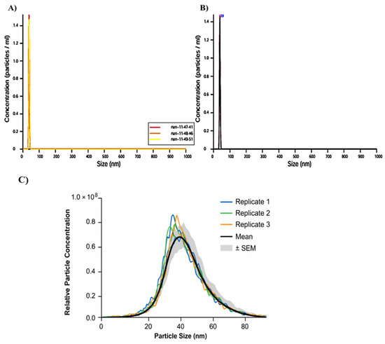
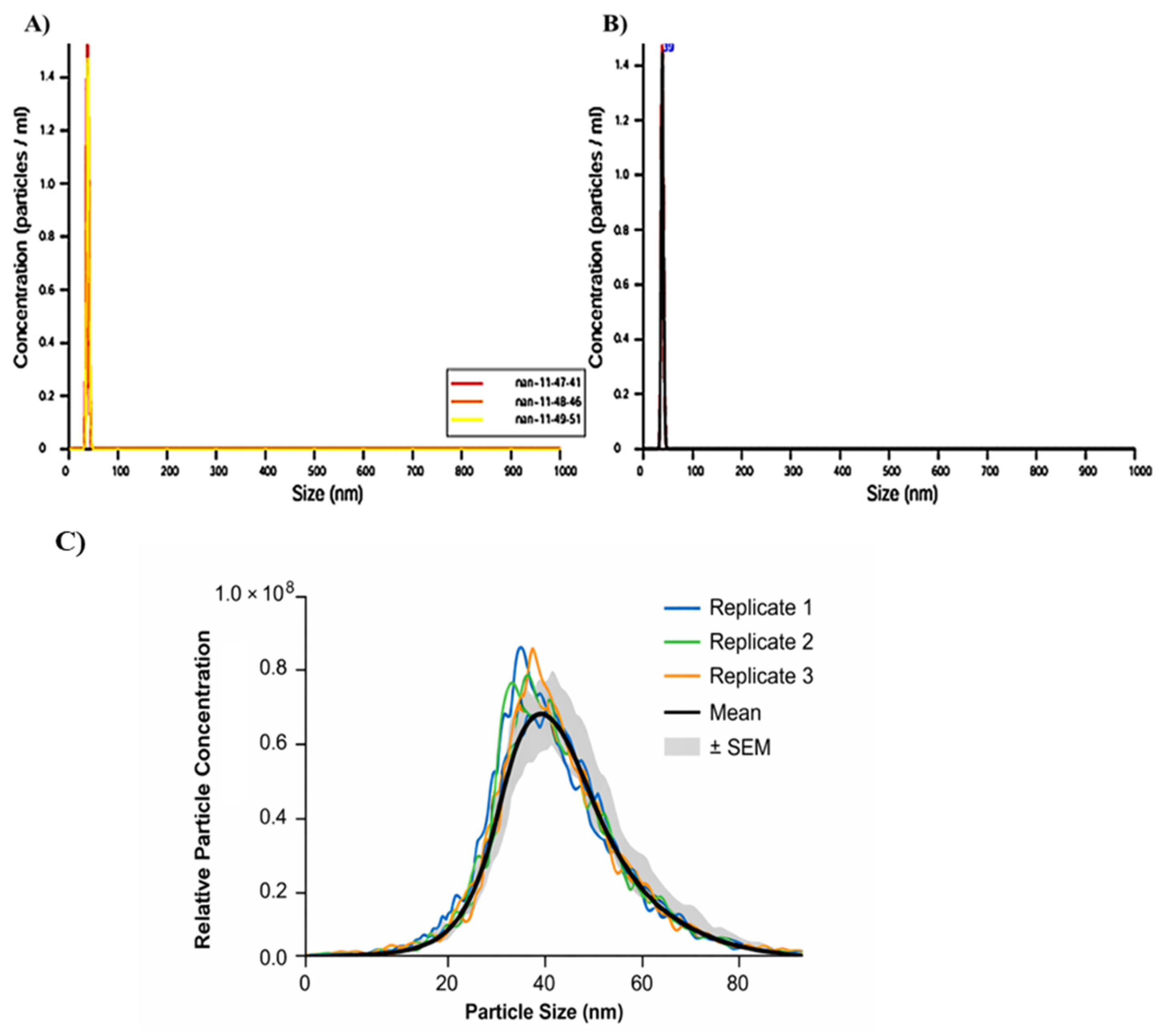
To investigate the concentration and the range size of AgNPs in 1 mL of the sample, NTA analysis was executed. The AgNPs consisted of 8.6 × 10⁸ ± 2.10 × 10⁶ N° particles/mL, with an average diameter of 38.4 ± 0.3 nm and a mode value of 38.1 ± 0.3 nm (Figure 1).
Figure 1.
Nanoparticle tracking analysis (NTA) of AgNPs in a 1 mL sample. (A) Hydrodynamic diameter size per concentration, obtained using the finite track length adjustment (FTLA) algorithm, with triplicate measurements of the AgNPs sample. (B) Average of the three measurements. All measurements were run with non-diluted samples. (C) Relative particle concentration (particles/mL) is plotted as a function of particle size (nm). Three independent measurements are shown (Replicate 1–3; colored lines), together with the mean distribution (black line) and the shaded area representing ± standard error of the mean (SEM). The particle size distribution exhibits a unimodal profile, with the majority of particles in the 30–80 nm range. The data presented represent a normalized and scaled distribution derived from particle counts and do not correspond to absolute particle concentration values. The x-axis was rescaled to 0–100 nm to improve visualization of the size distribution.
2.1.2. Morphological Characterization of AgNPs
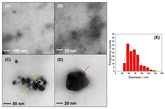
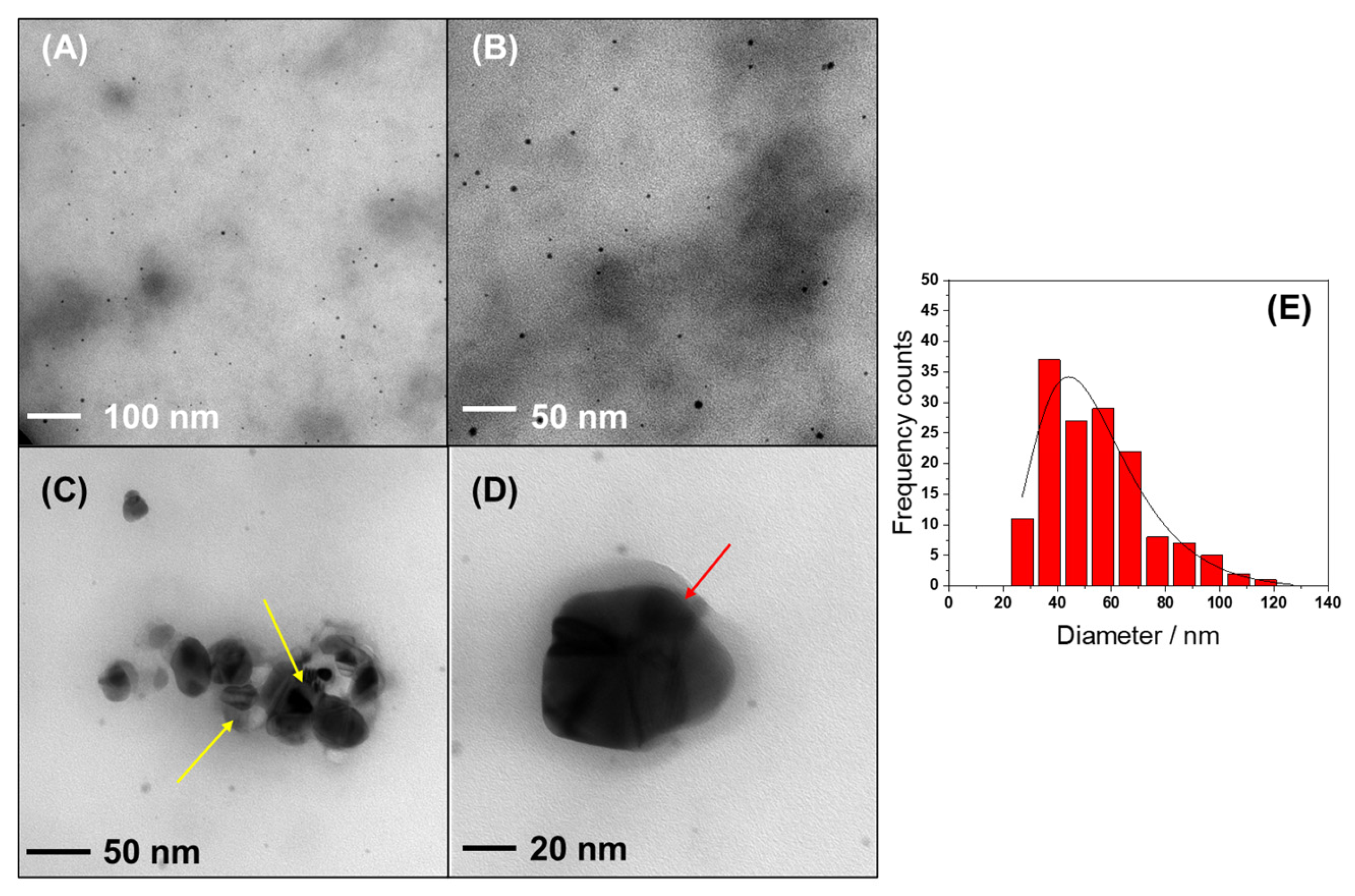
The morphological characterization of AgNPs prepared by Arctostaphylos uva-ursi extract was carried out by TEM. Typical micrographs of the synthesized particles are shown in Figure 2A–D and confirm the presence of spheroidal structures, having a log-normal distribution of measured diameters. Average particle size was equal to 46 ± 12 nm (Figure 2E).
Figure 2.
Morphological characterization of AgNPs prepared by uva-ursi by transmission electron microscopy (A–D). Size distribution histogram (E) (R2 = 0.89081) of AgNPs.
The low-contrast background of low-magnification TEM micrographs is relevant to the presence of organic moieties coming from the bio-extract. These species seem to have a dispersing effect too: minor aggregation between particles is visible (Figure 2A,B). AgNPs probably have a nano-crystalline nature, showing interference fringes in high-magnification images (Figure 2C, yellow arrows). AgNPs are sterically stabilized by the uva-ursi extract: a low-contrast halo surrounding the particle (Figure 2D, red arrow) is attributed to the presence of the plant extract [29].
2.2. Hydrogel Film Characterization
2.2.1. Swelling and Water Holding Capacity
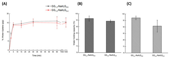
The swelling kinetics upon hydration were thoroughly investigated. As shown in Figure 3A, a plateau is reached after approximately 5 min, corresponding to about 14.0 ± 1.0% and 13.7 ± 1.0% for GG1.2-NaALG0.8 and GG1.6-NaALG0.4, respectively. The experiment was carried out at 32 °C in PBS, resulting in a rapid swelling that remained constant up to 24 h for both hydrogels. These results are in agreement with a previous study [30], in which GG- and NaALG-based hydrogels were employed as carriers for an olive leaf extract, aimed at providing protection against ultraviolet radiation generated by sunlight. In that case, the swelling kinetics of the GG1.2-NaALG0.8 and GG1.6-NaALG0.4 hydrogels were evaluated up to 60 min, while, in the present study, the water uptake behavior was observed up to 24 h. Regarding the water holding capacity test (WHC), the experiment was performed by dipping the hydrogel in PBS (pH =7.4) (Figure 3B) and SWF (Figure 3C). The results for both tests are nearly identical, since no significant differences (p < 0.05) were observed between the two buffers. Specifically, values of 96 ± 2% and 94 ± 1% were obtained in PBS and 97 ± 1% and 91 ± 5% in SWF for GG1.2-NaALG0.8 and GG1.6-NaALG0.4, respectively.
Figure 3.
Water uptake (WU%) (A), and water holding capacity percentage (WHC%) of hydrogels in PBS (B) and SWF (C).
2.2.2. DSC-TGA
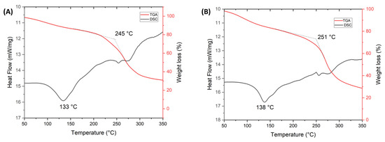
The films GG1.2-NaALG0.8/AgNPs (Figure 4A) and GG1.6-NaALG0.4/AgNPs (Figure 4B) were analyzed by DSC (black line) and TGA (red line). In the DSC curves, an endothermic peak associated with the melting process was observed, shifting from 133 °C to 138 °C as the gellan content increased. Moreover, the onset degradation temperature also increased from 245 °C to 251 °C. In the DSC thermograms, an increase in the released heat corresponded to the weight loss observed in the TGA curves.
Figure 4.
DSC (black line) and TGA (red line) curves of GG1.2-NaALG0.8 (A) and GG1.6-NaALG0.4 (B).
2.3. Silver Release Evaluation
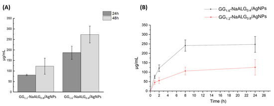
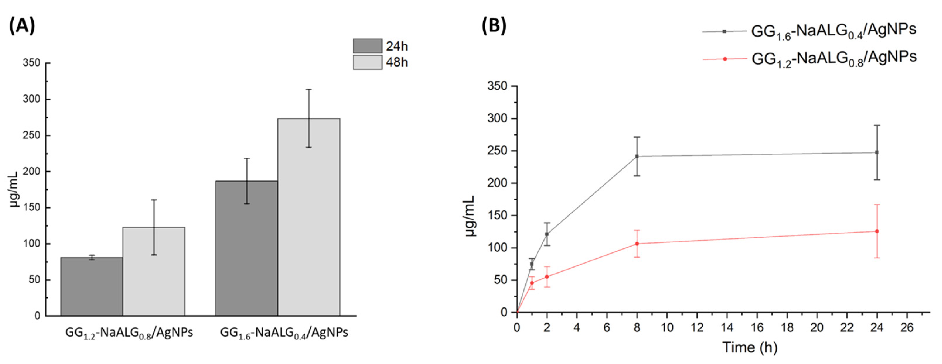
In Figure 5, the Ag+ release profiles were evaluated for both AgNP-loaded hydrogel films. The experiments were conducted using two different setups: direct immersion in PBS, monitoring silver release at 24 and 48 h (Figure 5A), and the Franz diffusion cell system (Figure 5B), monitored up to 24 h. A similar release behavior between the two analyses was observed, as the hydrogel with the higher GG content released a greater Ag+ amount. For direct immersion times in PBS, Ag+ concentrations of 80.0 ± 3.0 µg/mL and 187.0 ± 30.0 µg/mL were registered after 24 h for GG1.2-NaALG0.8/AgNPs and GG1.6-NaALG0.4/AgNPs, respectively. These values increased at 48 h to 122.0 ± 40.0 µg/mL and 270.0 ± 45.0 µg/mL for the same samples (Figure 5A). A similar trend was observed in the Franz cell experiments, where concentrations of 126.0 ± 40.0 µg/mL and 247.0 ± 42.0 µg/mL were quantified for GG1.2-NaALG0.8/AgNPs and GG1.6-NaALG0.4/AgNPs, respectively (Figure 5B).
Figure 5.
(A) Ag+ releases in PBS at 24 and 48 h, and (B) Ag+ release kinetics through a Franz diffusion cell until 24 h for AgNP-loaded hydrogel films.
2.4. Cytotoxicity of AgNPs and AgNPs Film-Conjugated
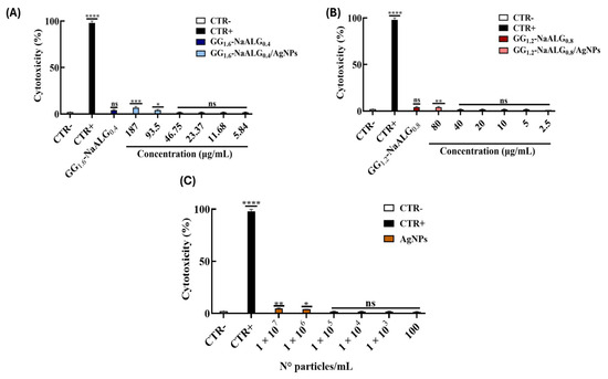
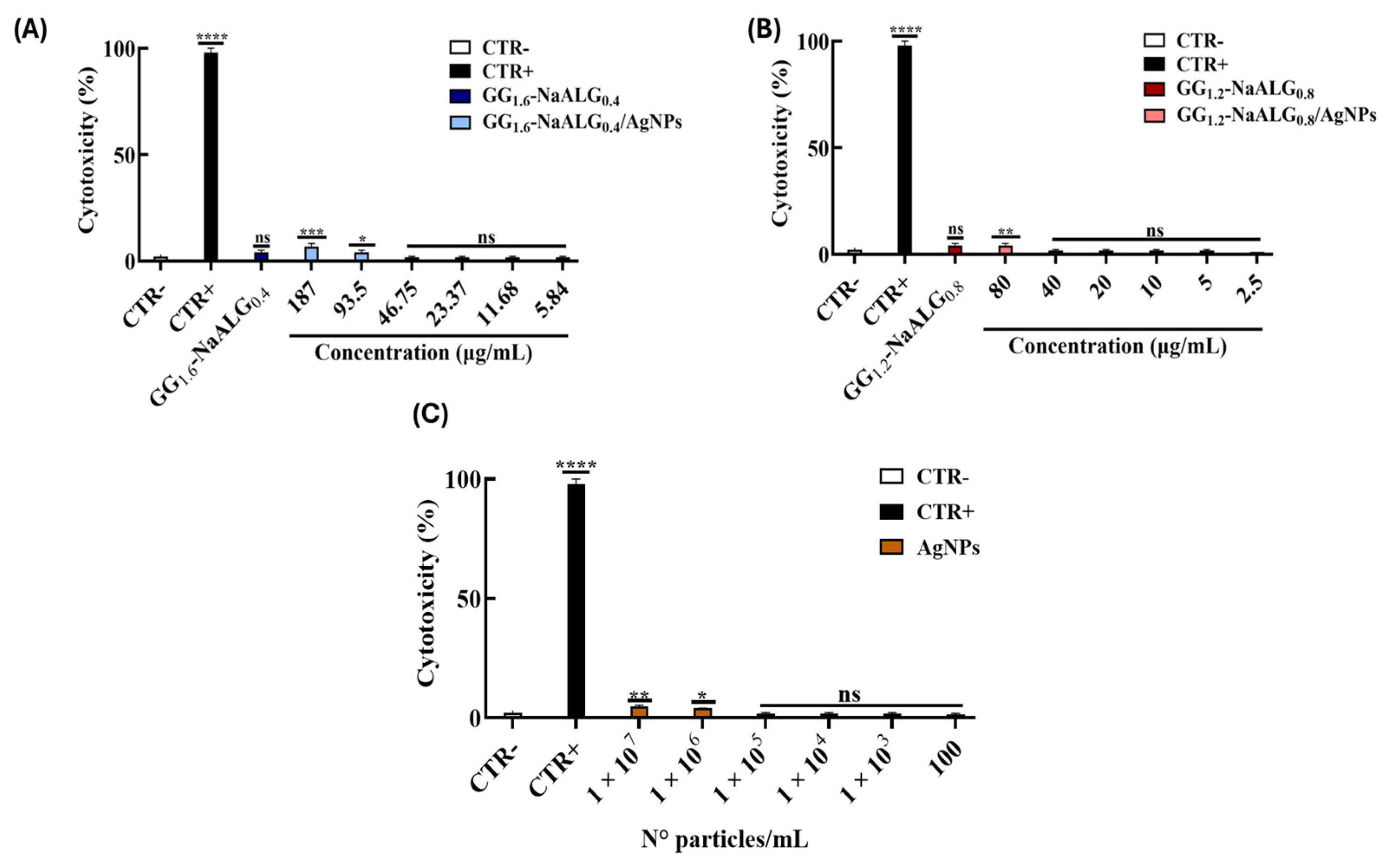
The cytotoxic effects were evaluated on the Vero-76 cell line after 24 h of treatment with several samples: UU leaf extract, AgNPs and the two AgNP-containing films, GG1.6-NaALG0.4/AgNPs and GG1.2-NaALG0.8/AgNPs. As mentioned in paragraph 2.12, the GG1.6-NaALG0.4/AgNPs and GG1.2-NaALG0.8/AgNP-containing films, together with the blank films GG1.6-NaALG0.4 and GG1.2-NaALG0.8, were first immersed in PBS for 24 h before use. The two AgNP-containing films, GG1.6-NaALG0.4/AgNPs and GG1.2-NaALG0.8/AgNPs, were evaluated at concentrations of 187 μg/mL (Figure 6A) and 80 μg/mL (Figure 6B), respectively, using 2-fold serial dilutions. AgNPs were assessed starting from 1 × 10⁷ N° particles/mL (Figure 6C) in 10-fold dilutions. UU was examined at a concentration of 1.25 mg/mL. The two films GG1.6-NaALG0.4 and GG1.2-NaALG0.8 were overloaded with 0.9 mg of UU, immersed in 1 mL of PBS for 24 h, and tested the day after in 2-fold dilutions (Supplemental Materials, Figure S1). Blank films, without AgNPs, referred to as GG1.6-NaALG0.4 and GG1.2-NaALG0.8, were tested non-diluted.
Figure 6.
Evaluation of the two AgNP-containing films, GG1.6-NaALG0.4/AgNPs (A) and GG1.2-NaALG0.8/AgNPs (B), and of AgNP (C) cytotoxicity. Cytotoxicity was assessed in Vero-76 cells using an MTT assay after 24 h of treatment with the sample suspension. Blank films, without AgNPs, referred to as GG1.6-NaALG0.4 and GG1.2-NaALG0.8, were tested non-diluted. Data are reported as mean ± SD. CTR−: untreated cells; CTR+: 100% DMSO. Statistical analysis was performed using Dunnett’s multiple comparisons test: **** p-value < 0.0001; *** p-value = 0.0004; ** p-value = 0.0042; * p-value = 0.0358; ns p-value > 0.05.
As reported in Figure 6A–C, a negligible cytotoxic effect was observed for all samples analyzed at their highest concentrations, including UU and its corresponding films (Supplemental Materials, Figure S1).
In the light of these promising results, the antiviral activity was evaluated at concentrations that did not affect cell viability, starting from 5 x 10⁶ N° particles/mL for AgNPs, from 93.5 μg/mL for GG1.6-NaALG0.4/AgNPs and from 40 μg/mL for GG1.2-NaALG0.8/AgNP-containing films, respectively.
2.5. Antiviral Activity of AgNPs and AgNP-Conjugated Film
2.5.1. Time-Shift Assays
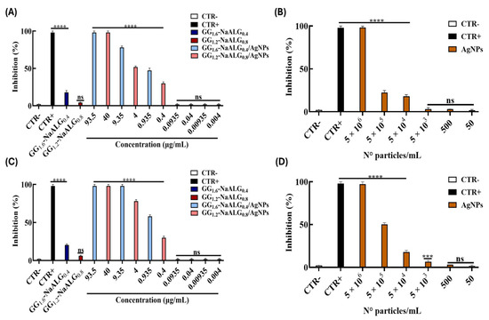
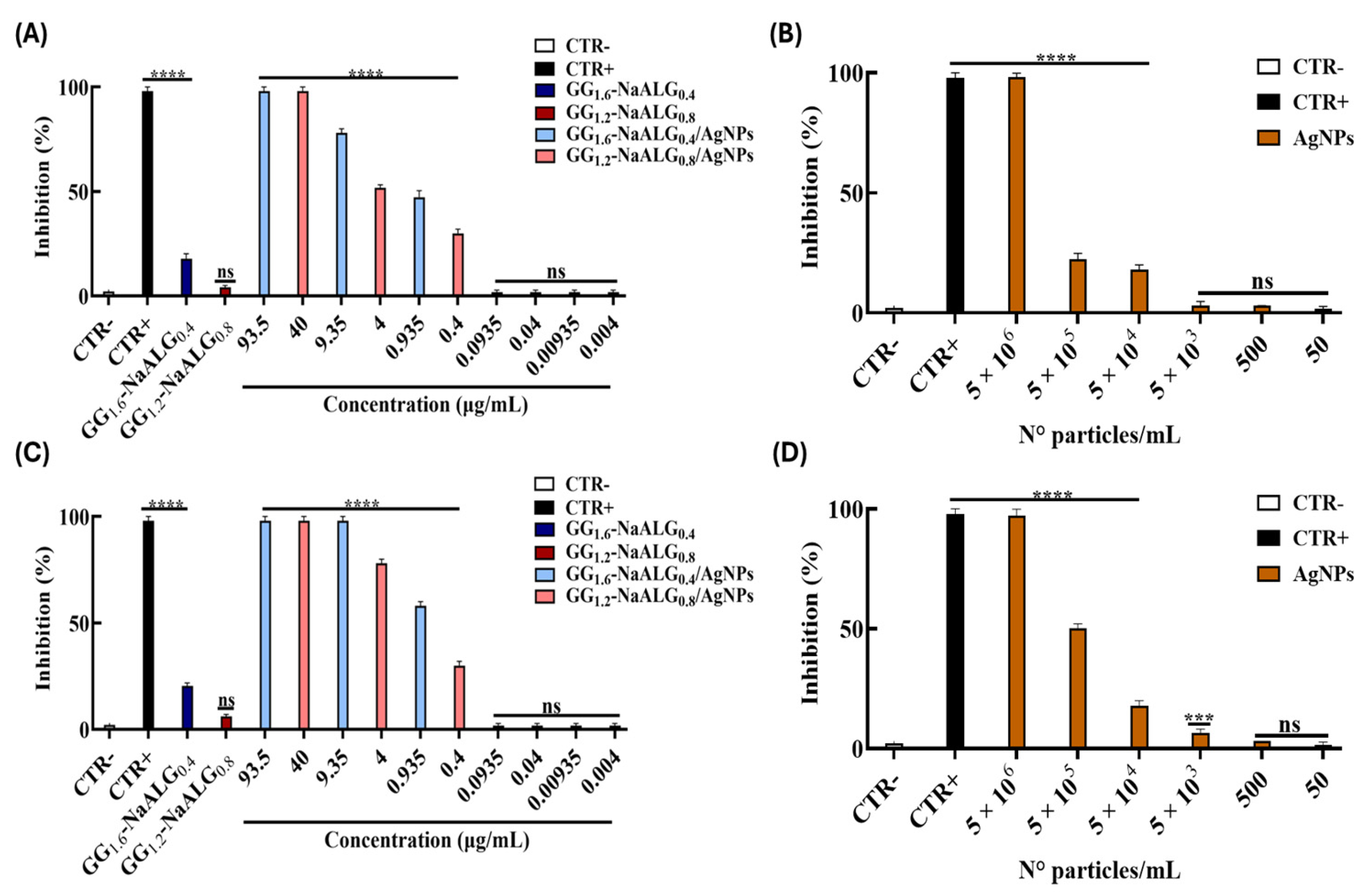
We conducted several plaque reduction assays. For the time-shift assays: in the co-treatment assay, the sample suspensions were co-incubated with the virus directly on the cellular monolayer (see Section 4.12 for more details). AgNP-conjugated film efficiently inhibited herpes infection. In particular, GG1.6-NaALG0.4/AgNPs recorded an IC50 of 1.392 µg/mL and GG1.2-NaALG0.8/AgNPs of 2.239 µg/mL, respectively (Figure 7A). Furthermore, the IC50 recorded by AgNPs alone was 9.1 × 10⁵ N° particles/mL (Figure 7B).
Figure 7.
Time-shift assays of the two AgNP-containing films (A,C), GG1.6-NaALG0.4/AgNPs and GG1.2-NaALG0.8/AgNPs, and of AgNPs (B,D) to evaluate their potential antiviral effect against HSV-1. Two different plaque reduction assays are shown. (A,B) Co-treatment assay; (C,D) virus pre-treatment assay. The untreated cells represented the negative control (CTR−). Melittin (5 µM) was used as a positive control (CTR+) in the co-treatment and virus pre-treatment. Data are reported as mean ± standard deviation (SD) of three independent experiments. **** p-value < 0.0001; *** p-value < 0.0006; ns p-value > 0.05.
To investigate the mechanism of action of sample preparations, additional experiments were carried out. Virus pre-treatment is a set experiment in which the sample suspensions and virus were first incubated together at 37 °C for 1 h. The mixture was then diluted with a medium and titrated on the Vero cell monolayers. A more substantial inhibitory effect was observed by pre-incubating the virus with AgNP-conjugated film, showing an IC50 value of 0.7820 µg/mL for GG1.6-NaALG0.4/AgNPs and of 1.018 µg/mL for GG1.2-NaALG0.8/AgNP-containing films, respectively (Figure 7C). Even AgNPs performed a greater inhibitory effect in this experimental setting (IC50 of 3.9 × 10⁵ N° particles/mL, Figure 7D). Subsequently, the Vero cells were pre-treated with both AgNPs or AgNP-conjugated films for 1 h before infection (cell pre-treatment assay, Supplemental Materials, Figure S2A,B); alternatively, the cell monolayer was infected for 1 h and then treated with AgNPs and AgNP-conjugated film (post-treatment assay, Supplemental Materials, Figure S2C,D). In both cases, any sample suspensions did not have an inhibitory effect, suggesting that any preparation was able to stop infection by interacting with the cell membrane and was not able to reach the cell’s internal compartment or to influence the virus replication once inside the cell. However, the promising results in the virus pre-treatment setting suggested that AgNPs, both alone and film-associated, could act in the extracellular phase of infection, most likely targeting the viral envelope.
To verify whether the AgNPs alone and AgNP-conjugated film have an enhanced inhibitory effect compared to the leaf extract itself, or to UU-conjugated film, GG1.6-NaALG0.4/uva ursi and GG1.2-NaALG0.8/uva ursi, we employed the same experimental settings for the latter as reported in Supplemental Materials, Figure S3. We found that even if UU, GG1.6-NaALG0.4/uva ursi and GG1.2-NaALG0.8/uva ursi showed a remarkable effect in the virus pre-treatment assay (IC50 values between 38 and 55 µg/mL; see Supplemental Materials Table S1), the effectiveness of the AgNP-conjugated films remains significantly higher, suggesting their crucial interaction within the viral particles.
2.5.2. Temperature-Shift Assays
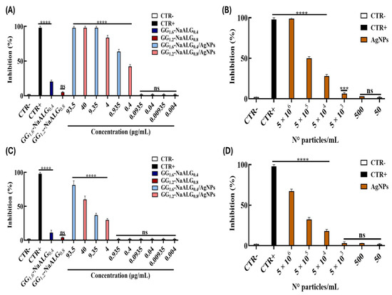
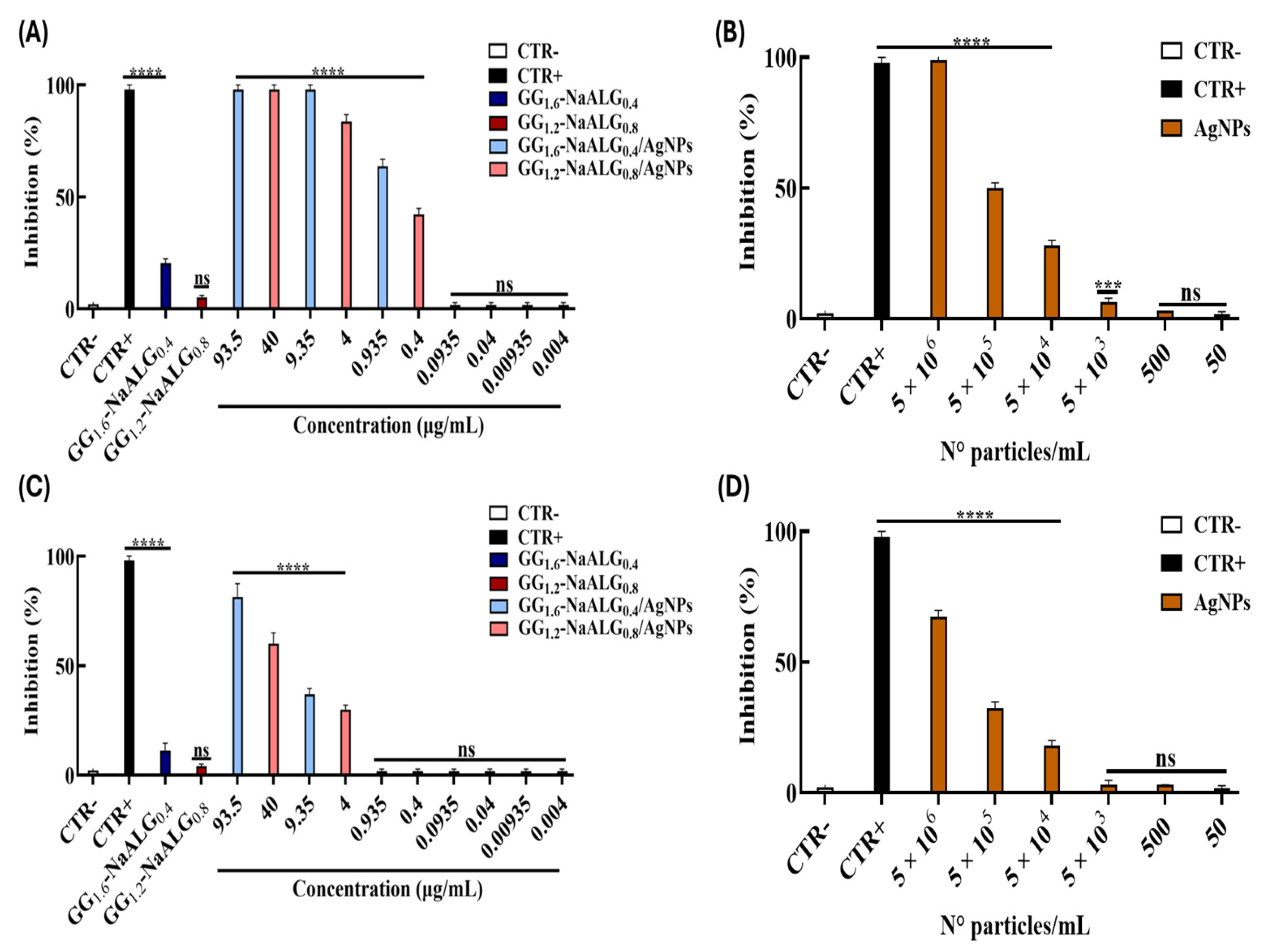
Temperature-shift assays were carried out to assess whether AgNPs were active during the virus attachment or the entry phase (Figure 8). An attachment assay evaluated the ability of the AgNPs to interfere with the virus binding to the target cell, but not its entry (Figure 8A). Alternatively, an entry assay is useful to evaluate the ability of the AgNP preparations to interfere with the intracellular entry of HSV-1 (Figure 8B).
Figure 8.
Temperature-shift assays of the two AgNP-containing films (A,C), GG1.6-NaALG0.4/AgNPs and GG1.2-NaALG0.8/AgNPs, and of AgNPs (B,D) to evaluate their potential antiviral effect against HSV-1. Two different plaque reduction assays are shown. (A,B) Attachment assay; (C,D) entry assay. Untreated cells served as the negative control (CTR−), while heparin (1 mg/mL) was used as the positive control (CTR+) in both assays. Data are reported as mean ± standard deviation (SD) of three independent experiments. Statistical significance was determined as follows: **** p-value < 0.0001; *** p-value < 0.0006; ns p-value > 0.05.
As shown in Figure 8A,B, AgNPs, alone or film-associated, could interact during the virus adhesion in a dose-dependent manner with an inhibition rate that reflected the one observed in the virus pre-treatment assay. IC50 values of 0.6116 µg/mL for GG1.6-NaALG0.4/AgNPs and 0.6971 µg/mL for GG1.2-NaALG0.8/AgNPs were the lowest recorded in all the plaque reduction assays performed. The same scenario occurred for AgNPs alone, with an IC50 recorded of 2.9 × 10⁵ N° particles/mL. On the other hand, the sample suspensions slightly interfered with the viral entry phase (Figure 8C,D).
As for the time-shift assays, we evaluated these findings in comparison with the leaf extract and its films (Supplemental Materials, Figure S4). In this instance as well, UU, itself and film-associated, demonstrated superior activity in the attachment assay among all the antiviral tests conducted (IC50 values ranging from 28 to 37 µg/mL, Supplementary Materials, Table S1); however, as previously reported, the most promising outcomes are confirmed to be linked to the film-associated AgNPs.
In summary, Table 1 reports IC50 values for each antiviral assay presented here. IC50 values were derived from the attachment assay, as it represents the most relevant condition for evaluating interference with the early stages of viral infection. The selectivity index (SI) was estimated using the highest non-cytotoxic concentration tested (SI > max non-cytotoxic concentration tested/IC50). In detail, as the highest non-cytotoxic concentration, we have considered 93.5 and 40 µg/mL for GG1.6-NaALG0.4/AgNPs and GG1.2-NaALG0.8/AgNPs, respectively, and 5 × 10⁶ particles/mL for AgNPs in suspension. Notably, GG1.6-NaALG0.4/AgNPs and GG1.2-NaALG0.8/AgNPs exhibited SI values >150 and >57, respectively, while AgNPs alone showed SI >17, indicating a favorable therapeutic window.
Table 1.
IC50 values for each antiviral assay.
2.6. Viral Gene Expression Analysis (Real-Time PCR) of GG1.6-NaALG0.4/AgNPs
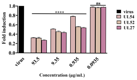
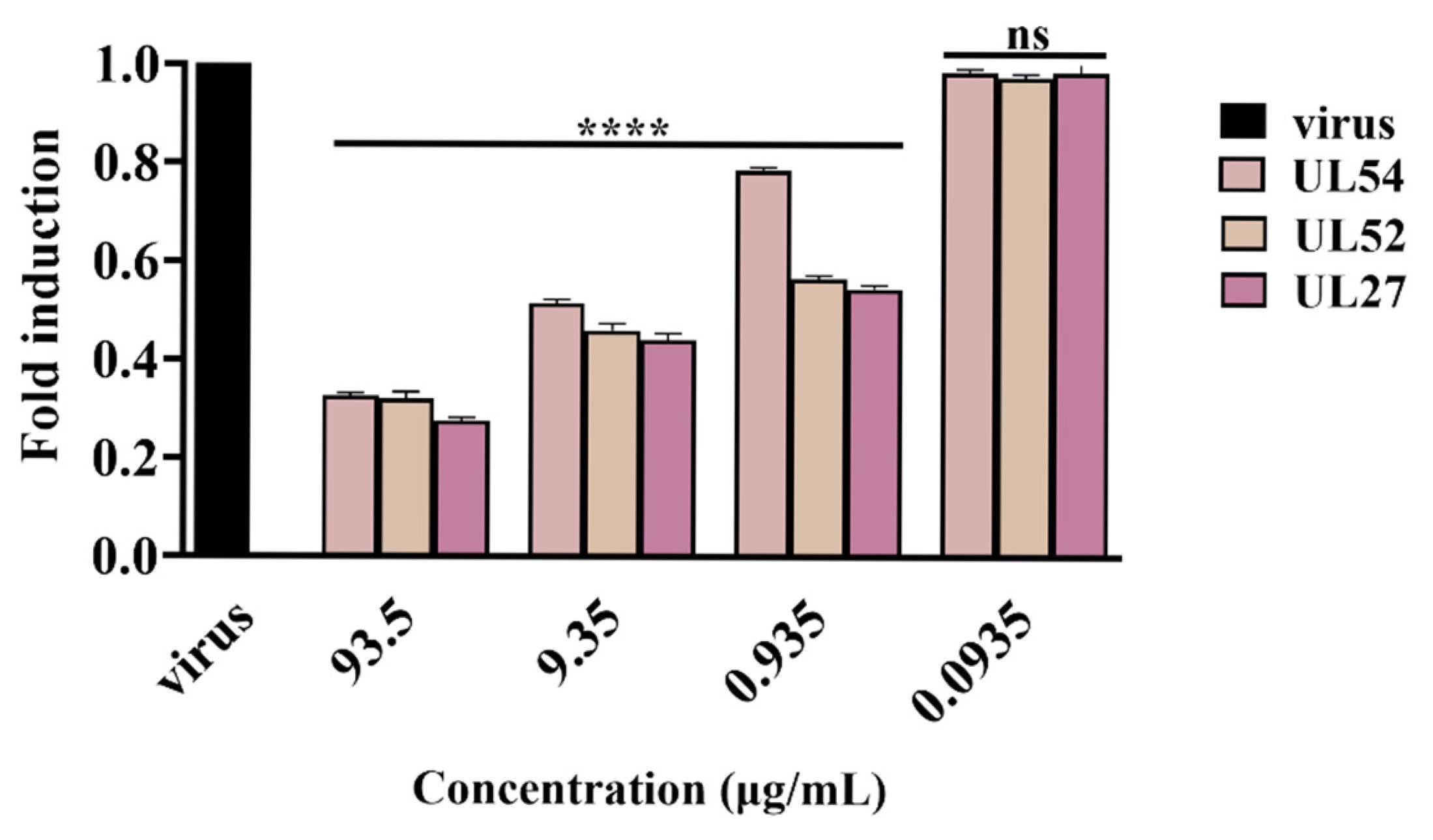
Given that the GG1.6-NaALG0.4/AgNP-conjugated film demonstrated the most effective inhibition profile in the plaque reduction tests, a qPCR was conducted to verify its potential efficacy (Figure 9). In detail, UL54 is an immediate early gene encoding the ICP27 protein involved in HSV-1 replication by inhibiting mRNA splicing at the post-transcriptional level and promoting the export of viral transcripts; UL52 is an early gene encoding DNA primase, and the UL27 gene represents a late gene coding the structural glycoprotein B (gB) [31]. A virus pre-treatment assay was performed, RNA was collected after 24 h, reverse-transcribed into cDNA, and amplified by real-time PCR.
Figure 9.
Viral gene expression analysis. Real-time PCR was performed to evaluate the effect of GG1.6-NaALG0.4/AgNP-conjugated film viral gene expression. For HSV-1, the expression levels of immediate early (UL54), early (UL52), and late (UL27) genes were analyzed. “Virus” refers to infected but not untreated cells. Data are reported as mean ± standard deviation (SD) of three independent experiments. Statistical significance was determined, comparing treated samples to ctr-, as follows: **** p-value < 0.0001, ns p-value > 0.05.
The data revealed that viral infection decreased in a dose-dependent manner, indicating the virucidal efficacy of GG1.6-NaALG0.4/AgNP-conjugated film (Figure 9). As the concentration of GG1.6-NaALG0.4/AgNPs decreased, the expression of viral genes increased, eventually reaching levels comparable to those in the virus control. These findings, together with the results of the virus pre-treatment and attachment assays, suggest that AgNPs may interfere with early stages of viral infection, likely affecting viral attachment and/or entry. However, direct evidence of the underlying molecular mechanism is beyond the scope of the present study.
3. Discussion
HSV infection is a complex, multi-step process that begins with viral adhesion to the host cell, followed by entry, replication, assembly, and eventual egress. The initial stages, adhesion and entry, are critical for establishing infection. Recent studies have investigated the antiviral potential of AgNPs, particularly those synthesized using plant extracts, as a novel strategy to block HSV infections [32,33]. These phyto-fabricated AgNPs combine the inherent antiviral properties of silver with the bioactive phytochemicals from the plant extract, often resulting in synergistic effects [34]. One of the most promising aspects of AgNPs is their ability to interfere with the early stages of viral infection, particularly viral entry. Studies have shown that AgNPs can bind to viral surface proteins, such as glycoprotein B and D, thereby preventing their interaction with host cell receptors [35]. In the same way, surface-modified AgNPs, such as those functionalized with tannic acid (TA-AgNPs), exhibited strong anti-HSV-2 effects. These ~33 nm particles prevented viral attachment and entry, significantly decreasing genital lesions in mouse models, while also boosting adaptive immune responses [12]. Another study involved sulfonate-modified AgNPs (Ag-MES), which mimic heparan sulfate proteoglycans, the natural receptors for HSV entry. These particles demonstrated selective inhibition of HSV-1 entry into host cells with low cytotoxicity [36].
Following these studies, our research presents the synthesis and characterization of AgNPs from plant extracts. In particular, we propose AgNPs embedded in GG/NaALG films as an innovative strategy for skin dressings, showcasing their potential for topical prophylaxis or therapeutic applications.
HSV-1 infection can cause laceration of the skin tissue [37]. Indeed, to promote tissue healing, adequate absorption of exudate is necessary [38]. Therefore, the use of hydrogels can be considered a promising strategy for the treatment of wounds caused by HSV-1 skin infections [35]. For this reason, it was necessary to evaluate the swelling and water-holding capacity of the hydrogels, simulating the application method on the wounds, both in terms of pH and chemical composition, using PBS and SWF [28]. High water uptake and WHC values are essential characteristics for hydrogel-based biomaterials used in wound dressings. These properties enable the hydrogel to effectively absorb wound exudate, maintaining a moisturized environment that promotes wound healing, supports tissue regeneration, and reduces the risk of infection [39,40]. For these reasons, based on the results reported in Figure 3, we positively assessed that the proposed hydrogel films (despite the different GG and NaALG ratios) are a promising strategy for exudate retention. As shown in Figure 5A,B, a higher Ag+ release for the films with a higher gellan content was observed, both for direct release in PBS and using the Franz diffusion cell. Among the two formulations, GG1.6-NaALG0.4/AgNPs released a higher amount of silver ions, which correlates with its consistently superior antiviral performance across all assays, as we will discuss later in the text. The release kinetics study using a Franz diffusion cell was carried out to simulate a potential transdermal release of Ag+, as well as to assess the amount of Ag+ remaining entrapped in the StratM® membrane.
The shift in the endothermic peak observed in Figure 4A,B is consistent with previous studies [28,41], which confirm that a higher gellan content leads to an increase in melting temperature. This increase can be attributed to an enhancement in the crystallinity of NaAlg induced by the presence of gellan [42]. The incorporation of gellan gum likely reinforces the conformation of the amorphous chains in the blend films, reducing the free volume compared to pure NaALG films, which exhibit a melting temperature of 116 °C [41]. Moreover, the presence of a single melting peak suggests good interaction between the two polymers in both blends. In addition, the onset degradation temperature also increases, in agreement with previous reports [43], indicating that higher gellan content improves the thermal stability of the material.
For all the biological experiments, the GG1.6-NaALG0.4/AgNPs and GG1.2-NaALG0.8/AgNP-containing films were first immersed in phosphate-buffered saline solution (PBS, pH 7.4) for 24 h before use. In our in vitro model (Figure 6), a negligible cytotoxic effect was observed for the two AgNP-containing films, GG1.6-NaALG0.4/AgNPs and GG1.2-NaALG0.8/AgNPs, as well as for AgNPs alone at their highest concentration. Although cytotoxicity was evaluated only in Vero-76 cells, a well-established model for HSV-1 infection, we acknowledge that the use of a single cell line represents a limitation of the study. Nevertheless, the obtained results provided an opportunity for further investigations.
Antiviral assays revealed that the AgNP-loaded films, GG1.6-NaALG0.4/AgNPs and GG1.2-NaALG0.8/AgNPs, strongly inhibited HSV-1 infection, particularly during co-treatment and, even more prominently, during virus pre-treatment (Figure 7A,C). Their ability to significantly reduce viral infectivity when incubated directly with the virus suggests a possible mechanism involving interaction with viral particles. This finding was primarily supported by the data of the attachment assay (Figure 8A,B) in which viral adhesion appears to be reduced in a dose-dependent manner, and the IC50 values recorded were 0.6116 µg/mL for GG1.6-NaALG0.4/AgNPs, 0.6971 µg/mL for GG1.2-NaALG0.8/AgNPs, respectively, and 2.9 × 10⁵ N° particles/mL for AgNPs alone. The estimation of the SI further supports the favorable safety profile of the developed system. Notably, the AgNP-loaded films exhibited high SI values (>150 and >57), indicating a strong antiviral effect at concentrations well below those associated with cytotoxicity. Between the two films, GG1.6-NaALG0.4/AgNPs film-associated showed the best inhibitory profile. The higher proportion of gellan gum in this formulation likely creates a more hydrated and porous network, facilitating ion diffusion and enabling a more efficient interaction of Ag+ or AgNPs with viral particles. This correlation is in line with previous evidence showing that the antiviral potency of silver-based systems strongly depends on the availability of ionic silver or surface-reactive AgNPs, which have been reported to interact with viral envelopes or glycoproteins, potentially leading to structural alterations and reduced infectivity [44]. For this reason, the GG1.6-NaALG0.4/AgNPs suspension was further investigated for its antiviral activity through a real-time PCR. As shown in Figure 9, the expression levels of HSV-1 immediate early (UL54), early (UL52), and late (UL27) genes were evaluated after a virus pre-treatment assay in a concentration range of 0.0935–93.5 µg/mL. The data revealed that viral infection decreased in a dose-dependent manner, supporting its potential virucidal efficacy. In general, AgNPs alone displayed a similar trend, although the film-conjugated forms consistently outperformed free nanoparticles, highlighting the beneficial impact of the polymeric matrix. For instance, tartaric acid, one of the film’s main constituents, in combination with other compounds, has been reported for its antiviral potential against HSV-1 and HSV-2 [45], thereby enhancing the suitability of our films for treating skin infections. The comparison with the uva-ursi extract and the UU-loaded films revealed that, although these preparations also exert inhibitory activity, the AgNP-loaded films remain substantially more potent. The synergy between AgNPs and the GG/NaALG matrix may further enhance nanoparticle availability, dispersion, or affinity for the viral surface.
In summary, our approach highlights the encouraging prospects of the biogenic AgNPs as innovative antiviral agents targeting HSV-1, which could be utilized for topical antiviral treatments. Continuous refinement of the NP composition, surface modification, and delivery methods will be essential to fully unlock their clinical potential. However, further studies will be necessary to directly elucidate the underlying mechanism of antiviral action.
4. Materials and Methods
4.1. Materials
Arctostaphylos Uva-Ursi leaf extract, coded as UU, containing total hydroquinone derivatives expressed as anhydrous arbutin in the range 19.0–21.0% w/w, was purchased from Farmalabor S.p.A. (Apulia, Italy). Low acyl content gellan gum (Phytagel™, molecular weight 1 × 10⁶ g/mol)—coded as GG—was purchased from Sigma-Aldrich (Merck, Milan, Italy). Sodium alginate (Cat #180947, Sigma-Aldrich, St. Louis, MO, USA, viscosity 15–25 cP, 1% in H2O), coded as NaALG, was purchased from Sigma-Aldrich (Merck, Milan, Italy). Tartaric acid and glycerol were purchased from Sigma-Aldrich (Merck, Milan, Italy). Films were prepared with ultrapure water, obtained through a Milli-Q® distillation system (Millipore-Merck, Darmstadt, Germany).
HNO3 (67%, Trace-SELECT® Ultra, for ultratrace analysis), NaH2PO4 (Trace-SELECT®, ≥99.99%, for trace analysis), and Na2HPO4 (Trace-SELECT®, ≥99.99%, for trace analysis) were from Merk Sigma-Aldrich®. Water for trace analysis at ppt level (Water RS—Ultrapure) was purchased from CARLO ERBA Reagents SAS®.
4.2. Preparation of AgNPs from Actostaphylos uva-ursi Leaf Extract
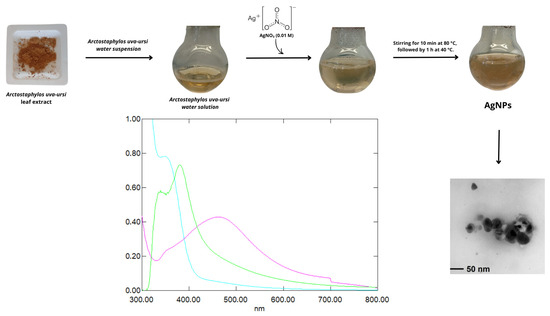
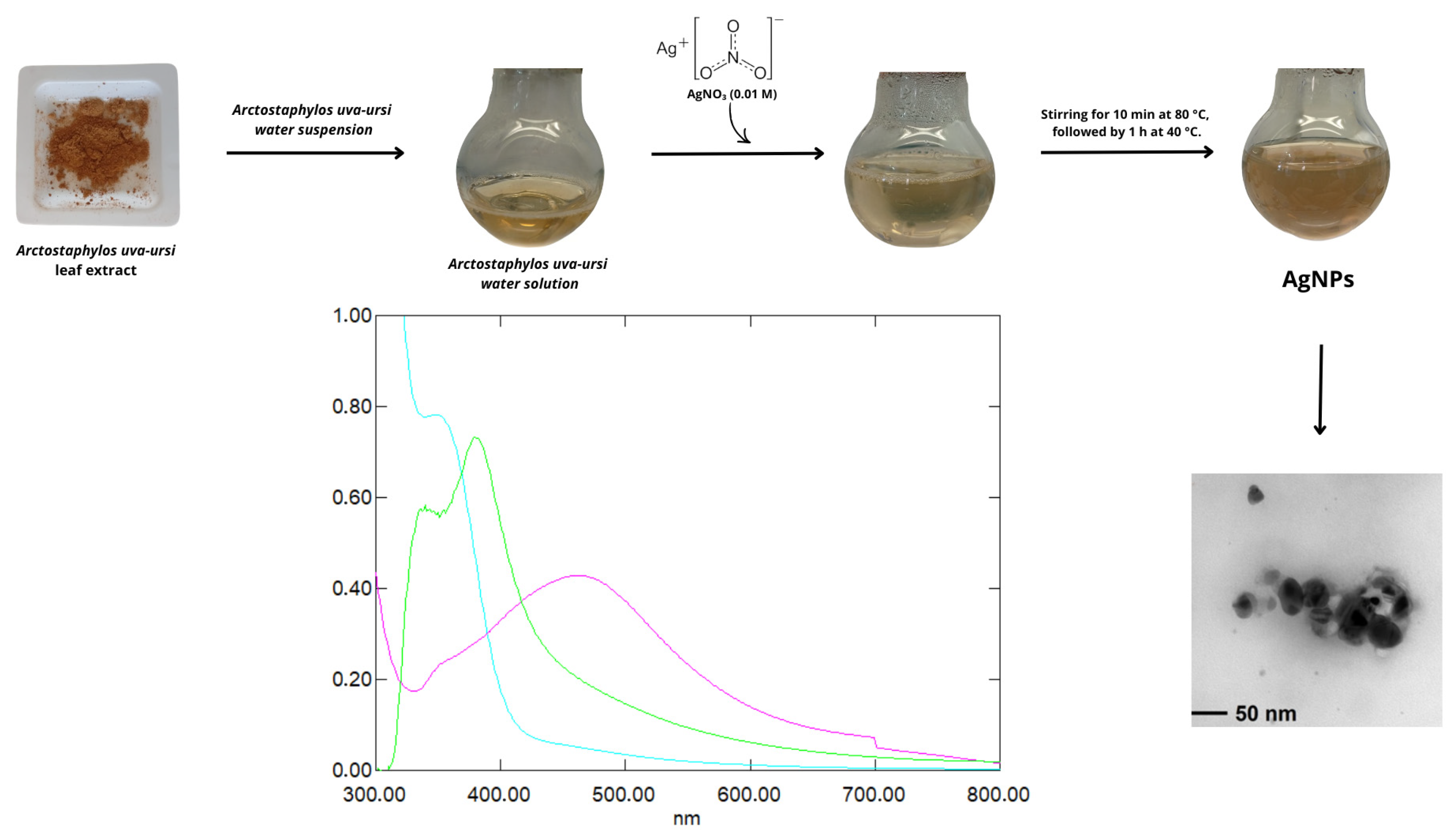
The AgNPs synthesis method was adapted from Thirumoorthy et al. with some modifications [46]. Arctostaphylos uva-ursi extract (0.006 g/mL) was centrifuged at 4000 rpm, and the resulting supernatant was collected and diluted 1:2. This solution (10 mL) was transferred to a round-bottomed flask and gently shaken at 80 °C for 10 min under dark conditions. Subsequently, 10 mL of AgNO3 (0.01 M) was added dropwise to the mixture. The reaction was maintained under stirring for 10 min at 80 °C, followed by 1 h at 40 °C. The yield of the synthesis, expressed as the amount of total silver that has been reduced to form nanoparticles, was equal to 72 ± 9%. The different steps for AgNP preparation are reported in Scheme 1, along with the UV-Vis spectra of the starting materials and nanoparticles, which allowed us to verify their formation by observing the surface plasmon resonance (SPR) band at 460 nm [29].
Scheme 1.
Schematic representation of the uva-ursi AgNP preparation steps and the UV-Vis absorption spectra of AgNO3 solution (light blue line), Arctostaphylos uva-ursi extract (green line), and uva-ursi AgNPs (fuchsia line).
The stability of AgNPs was evaluated spectrophotometrically over a period of 5 days. The UV–Vis absorption spectra remained unchanged and highly reproducible compared to freshly prepared samples, confirming the colloidal stability of the nanoparticles within this period.
4.3. Determination of AgNP Concentration by NTA
The concentration of uva-ursi AgNPs was determined using a NanoSight NS300 NTA instrument (Malvern Panalytical, Great Malvern, UK). In brief, 1 mL of uva-ursi AgNPs was inserted into the instrument with a syringe pump speed of “30” and a level chamber of “3”. Three readings were taken at room temperature (21.5 °C). 1498 frames in total were analyzed by NTA software (Malvern Instruments, version 3.4, Worcestershire, UK) using a detection threshold of 5.
4.4. Morphological Characterization of AgNPs by TEM
AgNPs were studied using transmission electron microscopy (TEM), using a FEI Tecnai 12 instrument (120 kV; filament: LaB6, Thermo Fisher Scientific, Waltham, MA, USA). Colloidal samples were deposited by drop-casting (2–3 μL) onto carbon-coated Cu grids (300 mesh, Agar Scientific Ltd., Rotherham, UK). The microscope was calibrated by using the S-106 Cross Grating (2160 lines/mm, 3.05 mm) supplied by Agar Scientific. Alignments and astigmatism correction were carried out following factory settings and fast Fourier transform (FFT) processing, respectively. The size distribution histogram was obtained from micrograph processing, using the ImageJ (version 1.8.0) software, measuring at least 250 particles. All analytical data were treated and plotted using Origin Pro© 2022 software.
4.5. Hydrogel Film Preparation
Film preparation was upgraded, starting from previously tested GG/NaALG ratios [29], maintaining the sum of the two polymer percentages equal to 2% w/v. Two different GG:NaALG ratios were considered: GG 1.6% w/v and NaALG 0.4% w/v (GG1.6-NaALG0.4) and GG 1.2% w/v and NaALG 0.8% w/v (GG1.2-NaALG0.8). Tartaric acid at 0.2% w/v was initially solubilized in distilled water at 80 °C, followed by the polymer powder addition under continuous stirring, on a magnetic plate (VELP Scientifica Srl, Usmate, Italy). Glycerol at 1% w/v was finally added to the solution as a plasticizer. When AgNP-loaded films were produced, 2 × 10⁷ NPs/mL was added to the solution after lowering the temperature to 40 °C. Smooth and homogeneous film-forming solutions were cast on Petri dishes polystyrene, 90 mm diameter (Sigma-Aldrich, St. Louis, MO, USA) and dried in an oven (PID system, MPM Instruments S.r.l., Bernareggio, Italy) at 40 °C for 24 h. Blank films, without AgNPs, were also prepared for comparison. In Table 2, a summary of the different films prepared is reported.
Table 2.
Composition (expressed as weight percent of the total weight of the components of the film) of the prepared materials; ⁂ AgNPs were added at a concentration of 2 × 10⁷ nanoparticles/mL.
4.6. Hydrogel Film Swelling and Water Holding Capacity
Dry hydrogel samples were cut into square pieces with 1 cm2 area, accurately weighed (mid), and immersed in phosphate-buffered saline solution (PBS, pH 7.4) and simulated wound fluid (SWF) prepared as reported by Svensby et al. [47], at 32 °C to determine the swelling kinetics up to 24 h. All the specimens were weighed after each time point (mit). The water uptake (expressed as grams of PBS per gram of dry film) was calculated using the following formula (Equation (1)):
Moreover, for water holding capacity measurements, the hydrated hydrogel films were centrifuged in a centrifuge equipped with a basket rotor able to deliver 1400 rpm. The centrifuged samples were weighed, and the retained water was expressed as a weight percentage of the total uptake of water (Equation (2)):
where mc, ms, and md are the weights of centrifuged, swollen, and dry hydrogels.
4.7. In Vitro Skin Permeation Studies
The in vitro skin permeation studies were performed using a Franz diffusion cell (PermeGear Inc., SES GmbH, Bechenheim, Germany) using the protocol reported in a previous work [29], with some modifications. Briefly, AgNP-loaded hydrogel films (about 1 cm2) were placed in the cell donor compartment. An O-ring joint kept the film (0.6 cm2) on the synthetic StratM® membrane (Merck KGaA, Darmstadt, Germany), characterized by skin-like porosity, diffusivity, and composition. The whole assembly was fixed with a stainless-steel clamp to maintain the tight connection between the donor and receptor compartments. A total of 1 and 5 mL of PBS (pH 7.4) were added to the donor and acceptor compartments, respectively. In the acceptor compartment, the buffer solution was continuously stirred using an ATE magnetic stirrer (VELP Scientifica Srl, Usmate, Italy). The temperature was kept constant at 32.00 ± 0.03 °C, using a CD-B5 heating circulator bath (Julabo GmbH, Seelbach, Germany). To evaluate the Ag+ release kinetics, at predetermined time points (1, 2, 8, and 24 h), PBS aliquots of 500 μL were withdrawn, replaced with fresh buffer, and analyzed by electrothermal atomic absorption spectroscopy (ETAAS) as described in Section 2.3. At the end of the experiment, the StratM® was sectioned with a scalpel into six parts and placed into 5 mL of PBS, mixed with Vortex (VELP Scientifica Srl, Usmate, Italia) for 1 min, and left overnight at 25 °C to extract and quantify the Ag+ retained by the membrane.
4.8. DSC-TGA
The thermal behavior of the films was investigated by differential scanning calorimetry (DSC) and thermogravimetric analysis (TGA).
DSC was carried out using a Perkin–Elmer DSC 7 calorimeter (Perkin-Elmer Inc., Waltham, MA, USA), putting samples into aluminum pans. The scans were performed at a rate of 10 °C/min, from 50 to 350 °C, in a N2-saturated atmosphere.
TGA was performed on a Perkin Elmer TGA-400 instrument (Perkin Elmer Inc., Waltham, MA, USA), heating 5–10 mg of samples in a N2-saturated atmosphere and setting the gas flow to 20 mL/min. The flow rate was set to 20 °C/min, heating from 50 °C to 350 °C. For each sample, thermograms (TG) with respective derivative (DTG) curves were recorded and analyzed with the TGA Pyris software.
4.9. Silver Ionic Release Evaluation by Atomic Absorption Spectroscopy
Ag+ ion release kinetics were determined by electrothermal atomic absorption spectroscopy (ETAAS). Samples were cut into 1 cm2 squares and soaked in Eppendorf® tubes (Sigma-Aldrich, St. Louis, MO, USA) containing 1 mL of PBS contact solution (pH = 6.8, ionic strength I = 0.1). This pH resembles the one used for antiviral experiments. At definite times, contact solution was sampled and properly diluted/mineralized with 0.2% w/v HNO3 to undergo ETAAS analysis. We used a Perkin Elmer PinAAcle AS900Z spectrometer, equipped with a Ag hollow cathode lamp and a transversely heated graphite furnace. A calibration curve was accomplished using proper dilutions of a Ag+ standard solution for AAS. Bare films (i.e., without AgNPs) were used as controls. All experimental points were plotted using Origin Pro© 2022 software.
4.10. Cell Line and Viral Strains
Cercopithecus aethiops kidney cells (Vero-76, ATCC CRL 1587) were purchased from the American Type Culture Collection (ATCC, Manassas, VA, USA). Cells were cultured in Dulbecco’s Modified Eagle Medium (DMEM, Microtech, Naples, Italy) with 4.5 g/L of glucose (Microtech, Naples, Italy), 2 mM of L-glutamine (Microtech), 100 IU/mL of penicillin-streptomycin solution (Himedia, Naples, Italy) and supplemented with 10% fetal bovine serum (FBS, Microgem, Naples, Italy) in a humidified atmosphere with 5% CO2 at 37 °C. As previously reported, HSV-1 (strain SC16) was propagated on Vero-76 cells [48].
4.11. Cytotoxicity Assay
For all the biological experiments that follow, the GG1.6-NaALG0.4/AgNPs and GG1.2-NaALG0.8/AgNP-containing films, together with the blank films GG1.6-NaALG0.4 and GG1.2-NaALG0.8, were first immersed in phosphate-buffered saline solution (PBS, pH 7.4) for 24 h before use.
The cytotoxicity effect of uva-ursi leaf extract, AgNPs, GG1.6-NaALG0.4/AgNPs, and GG1.2-NaALG0.8/AgNPs was evaluated through the 3-(4,5-dimethythiazol-2-yl)-2,5-diphenyltetrazole bromide (MTT) assay. A density of 2 × 10⁴ cells/well was seeded in 96-well plates, and the day after, the preparations were tested as follows: GG1.6-NaALG0.4/AgNPs at 187 μg/mL and GG1.2-NaALG0.8/AgNPs at 80 μg/mL, in 2-fold serial dilutions, respectively. AgNPs were tested from 1 × 10⁷ N°particles/mL in 10-fold dilutions. UU was tested at 1.25 mg/mL. The two films GG1.6-NaALG0.4 and GG1.2-NaALG0.8 were overloaded with 0.9 mg of UU, immersed in 1 mL of PBS for 24 h, and tested the day after in 2-fold dilutions (Supplementary Materials, Figure S1). Blank films, without AgNPs, referred to as GG1.6-NaALG0.4 and GG1.2-NaALG0.8, were tested non-diluted. Untreated and treated cells with 100% DMSO represented CTR− and CTR+. After 24 h of exposure, 100 μL of MTT solution was added to each well for 3 h at 37 °C, then the solution was solubilized, and the absorbance was read at 570 nm using a microplate reader (Tecan, Männedorf, Switzerland). The cytotoxicity percentage was calculated according to the following formula (Equation (3)):
4.12. Antiviral Activity
The antiviral potential of UU leaf extract, AgNPs, GG1.6-NaALG0.4/AgNPs, and GG1.2-NaALG0.8/AgNPs was investigated. Different plaque-reduction assays were conducted as previously reported [49]. Vero-76 cells were seeded in 24-well plates (1.2 × 10⁵ cells/well) and grown for 24 h at 37 °C in 5% CO2. Samples were tested as follows: GG1.6-NaALG0.4/AgNPs at 93.5 μg/mL and GG1.2-NaALG0.8/AgNPs at 40 μg/mL in 10-fold serial dilutions, respectively. AgNPs were tested from 5 × 10⁶ N°particles/mL in 10-fold dilutions. Uva ursi was tested at concentrations ranging from 625 μg/mL in a 10-fold serial dilution. The two films GG1.6-NaALG0.4 and GG1.2-NaALG0.8 were overloaded with 0.9 mg of UU, immersed in 1 mL of PBS for 24 h, and tested the day after from 450 μg/mL in 10-fold dilutions (Supplementary Materials). Blank films, GG1.6-NaALG0.4 and GG1.2-NaALG0.8, were tested non-diluted. The viral strain was used at a multiplicity of infection (MOI) of 0.01. After treatment, the monolayer was washed, covered with the complete medium (10% FBS) supplemented with 3% carboxymethylcellulose (CMC) (Sigma-Aldrich, C5678, C5013, St. Louis, MO, USA), and incubated for 48 h at 37 °C and 5% CO2. Finally, cells were fixed with 4% formaldehyde (Sigma-Aldrich, St. Louis, MO, USA) and stained with crystal violet solution. For each sample, the antiviral assays were performed as follows [49].
4.12.1. Time-Shift Assays
(i) Co-treatment assay: Sample suspensions and virus were simultaneously incubated on Vero-76 cells for 1 h at 37 °C. (ii) Cell pre-treatment assay: First, the sample, in its concentration range, was added to Vero-76 cells for 1 h. Then, it was removed, and cells were infected with viral suspensions for 1 h at 37 °C. (iii) Virus pre-treatment assay: Sample suspensions and virus were incubated together for 1 h at 37 °C at an MOI of 0.1 pfu/mL. This initial MOI was used to promote effective interaction between viral particles and the tested compounds during the pre-incubation step, ensuring sufficient virus–compound contact while allowing the antiviral agent to be diluted to a concentration that is not active per se in standard antiviral conditions. Following this incubation, the mixture (sample + virus) was diluted to a final MOI of 0.01 before infection. This dilution was performed to achieve optimal plaque resolution, avoid saturation effects, and enable accurate and reliable quantification of antiviral activity. Dilutions were added to the Vero-76 monolayer for 1 h at 37 °C and 5% CO2. (iv) Post-treatment assay: Cells were previously infected with the viral suspension in DMEM w/o FBS for 1 h at 37 °C. Then, the cell monolayer was washed to remove extracellular virions and treated with the sample suspensions for 1 h at 37 °C.
4.12.2. Temperature-Shift Assays
(i) Entry assay: Pre-cooled Vero-76 cells were first infected for 1 h at 4 °C to permit viral attachment. Then, cells were washed and treated with sample suspensions for 1 h at 37 °C. After the treatment, cells were covered with CMC and incubated for 48 h at 37 °C and 5% CO2. (ii) Attachment assay: Pre-cooled Vero-76 cell monolayers were infected in the presence of samples for 1 h at 4 °C. Subsequently, cells were washed to remove unabsorbed viruses, overlaid with CMC, and incubated for 48 h at 37 °C and 5% CO2.
The untreated cells represented the negative control (CTR−). At the same time, several compounds were used as positive controls (CTR+): melittin (5 µM) in the co-treatment and virus pre-treatment, dextran-sulfate (1 µM) in cell pre-treatment, and aciclovir (5 µM) in post-treatment [49]; heparin (1 mg/mL) for the attachment and entry assays [50].
The inhibition rate of viral infectivity was calculated by comparing the number of plaques counted in cells treated with sample suspensions to the plaques detected in the CTR−, according to the following formula (Equation (4)):
The 50% inhibitory concentration (IC50) was calculated by linear regression analysis.
4.13. Viral Gene Expression Analysis (Real-Time PCR)
Since the GG1.6-NaALG0.4/AgNPs sample showed the best inhibition profile in the plaque reduction assays, a qPCR was performed to confirm its promising activity. The virus pre-treatment scheme was conducted as described above. After 30 h of incubation, the total RNA was extracted from the cells using TRIzol reagent (Thermo Fisher, Waltham, MA, USA). 1 µg of the obtained RNA was retrotranscribed into cDNA, according to the instructions of 5× All-In-One RT MasterMix (Applied Biological Materials, Richmond, BC, Canada). The qPCR was performed in a total volume of 20 µL containing 0.1 µM of each primer, BrightGreen 2× qPCR MasterMix-No Dye (Applied Biological Materials), and 100 ng of cDNA. Amplification was conducted in Thermal Cycler UNO96 (VWR International, Radnor, PA, USA) using the following amplification program: denaturation at 95 °C for 15 s, annealing at 60 °C for 60 s, and extension at 72 °C for 20 s (40 cycles). The expression of specific viral genes UL54 (immediate early gene), UL52 (early gene), and UL27 (late gene) was evaluated using the primer sequences (Euforins, Milan, Italy) reported in Table 3.
Table 3.
Oligos sequences for viral genes.
The target threshold cycle (Ct) values were normalized to glyceraldehyde 3-phosphate dehydrogenase (GAPDH), which was used as a housekeeping gene control. Gene expression was examined by the 2−ΔΔCt method [51].
4.14. Statistical Analysis
Each antiviral test was performed in triplicate and expressed as mean ± standard deviation (SD). One-way ANOVA was followed by the Dunnett’s multiple comparisons test, and all the graphs were generated by GraphPad Prism (version 8.0.1; software for 2D graphing and statistics; GraphPad Software Inc., San Diego, CA, USA, 2018). The significance of the difference between treated and CTR− samples was obtained with Dunnett’s test and ANOVA analysis. The p-value < 0.05 was considered significant.
5. Conclusions
In this work, we successfully developed and characterized plant-mediated AgNPs embedded within GG/NaALG composite films, demonstrating their potential as innovative biomaterials for topical applications against HSV-1. The hydrogels exhibited excellent swelling behavior, water holding capacity, and thermal stability, key properties for wound dressings designed to manage exudate and support tissue healing in HSV-1–associated skin lesions. Notably, the GG1.6-NaALG0.4/AgNPs formulation released the highest amount of Ag+ ions, a factor that correlated directly with its superior antiviral performance. Across all antiviral assays, AgNP-loaded films significantly inhibited HSV-1 infection, with the most potent effects observed during virus pre-treatment and attachment phases. These findings suggest a possible interaction of AgNPs with viral particles during the early stages of infection, potentially involving the viral envelope or surface glycoproteins, thereby reducing viral adhesion. However, no direct mechanistic evidence (e.g., binding or imaging studies) was provided in this work, and further investigations are required to confirm this hypothesis. The improved antiviral efficacy of the films compared to free AgNPs or UU–based formulations further highlights the synergistic contribution of the polymeric network, which enhances nanoparticle stability, availability, and bioactivity. Overall, the combination of favorable physicochemical properties, controlled silver release, and potent antiviral activity positions these AgNP-based hydrogels as promising candidates for topical and wound care new strategies. Future work should focus on exploring functional surface modifications and validating therapeutic potential in vivo models to advance their translational applicability.
Supplementary Materials
The following supporting information can be downloaded at https://www.mdpi.com/article/10.3390/molecules31091459/s1: Figure S1. Evaluation of the two UU films, referred to as GG1.6-NaALG0.4/uva ursi and GG1.2-NaALG0.8/uva ursi, and of UU cytotoxicity; Figure S2. Time-shift assays of the two AgNPs films (A,C), GG1.6-NaALG0.4/AgNPs and GG1.2-NaALG0.8/AgNPs, and of AgNPs (B,D) to evaluate their potential antiviral effect against HSV-1; Figure S3. Time-shift assay of the two UU films, referred to as GG1.6-NaALG0.4/uva ursi and GG1.2-NaALG0.8/uva ursi, and of UU to evaluate their potential antiviral effect against HSV-1; Figure S4. Temperature-shift assay of the two UU films, referred to as GG1.6-NaALG0.4/uva ursi and GG1.2-NaALG0.8/uva ursi, and of UU to evaluate their potential antiviral effect against HSV-1; Table S1. IC50 values for each antiviral assay of the two UU films, referred to as GG1.6-NaALG0.4/uva ursi and GG1.2-NaALG0.8/uva ursi, and of UU.
Author Contributions
Conceptualization, E.D.G. and A.D.F.; methodology, F.B., S.L., E.D.G., M.C.S., R.D.M. and C.Z.; validation, C.Z.; visualization, M.S. and S.B.; formal analysis, F.B. and S.L.; investigation, F.B., S.L., M.C.S., R.D.M. and C.Z.; data curation, R.D.M., F.B., C.Z., S.L., M.C.S., M.S. and S.B.; writing—original draft preparation, F.B., S.L., E.D.G., M.C.S., R.D.M. and C.Z.; writing—review and editing, E.D.G., M.C.S., M.G. and A.D.F.; supervision, E.D.G., M.G. and A.D.F.; funding acquisition, E.D.G.; F.B. and S.L. prepared AgNPs along with Ag-modified GG/NaALG films and corresponding control samples. F.B., S.L. and E.D.G. performed the following characterizations on the composite films: swelling and water holding capacity, in vitro skin permeation studies, and DSC-TGA. F.B. and M.C.S. performed Ag+ ionic release evaluation by atomic absorption spectroscopy. M.C.S. performed morphological characterization of AgNPs by transmission electron microscopy. R.D.M. and C.Z. performed all biological assays: cytotoxicity and antiviral assays. All authors have read and agreed to the published version of the manuscript.
Funding
This research received no external funding.
Institutional Review Board Statement
Not applicable.
Informed Consent Statement
Not applicable.
Data Availability Statement
The data presented in this study are available upon request from the corresponding author. Authors can confirm that all relevant data are included in the article.
Conflicts of Interest
The authors declare no conflicts of interest.
Abbreviations
The following abbreviations are used in this manuscript:
| Ag+ | Silver ion |
| AgNPs | Silver nanoparticles |
| AgNO3 | Silver nitrate |
| CMC | Carboxymethylcellulose |
| CTR−/CTR+ | Negative control/positive control |
| cP | Centipoise |
| Ct | Cycle threshold |
| DMSO | Dimethyl sulfoxide |
| DSC | Differential scanning calorimetry |
| FBS | Fetal bovine serum |
| FTLA | Finite track length adjustment |
| GG | Gellan gum |
| HNO3 | Nitric acid |
| HSV-1/HSV-2 | Herpes simplex virus (type 1/2) |
| IC50 | Inhibitory concentration 50% |
| Milli-Q® | Ultrapure water system |
| MOI | Multiplicity of infection |
| MTT | 3-(4,5-dimethylthiazol-2-yl)-2,5-diphenyltetrazolium bromide |
| NaALG | Sodium alginate |
| NaH2PO4 | Sodium dihydrogen phosphate |
| Na2HPO4 | Disodium hydrogen phosphate |
| nm | Nanometers |
| NTA | Nanoparticle tracking analysis |
| PBS | Phosphate-buffered saline |
| pfu/mL | Plaque-forming units per milliliter |
| RT-PCR | Real-Time Polymerase Chain Reaction |
| rpm | Rotations per minute |
| SD | Standard deviation |
| SWF | Simulated wound fluid |
| TGA | Thermogravimetric analysis |
| TEM | Transmission electron microscopy |
| UU | Arctostaphylos uva-ursi leaf extract |
| WHC | Water holding capacity |
| WU% | Water uptake percentage |
| XRD | X-ray Diffraction |
References
- Zhu, S.; Viejo-Borbolla, A. Pathogenesis and virulence of herpes simplex virus. Virulence 2021, 12, 2670–2702. [Google Scholar] [CrossRef] [Scilit]
- Cohen, E.M.; Avital, N.; Shamay, M.; Kobiler, O. Abortive herpes simplex virus infection of nonneuronal cells results in quiescent viral genomes that can reactivate. Proc. Natl. Acad. Sci. USA 2020, 117, 635–640. [Google Scholar] [CrossRef] [Scilit]
- Xu, X.; Zhang, Y.; Li, Q. Characteristics of herpes simplex virus infection and pathogenesis suggest a strategy for vaccine development. Rev. Med. Virol. 2019, 29, e2054. [Google Scholar] [CrossRef] [Scilit]
- Poole, C.L.; James, S.H. Antiviral Therapies for Herpesviruses: Current Agents and New Directions. Clin. Ther. 2018, 40, 1282–1298. [Google Scholar] [CrossRef] [Scilit]
- Birkmann, A.; Zimmermann, H. HSV antivirals—Current and future treatment options. Curr. Opin. Virol. 2016, 18, 9–13. [Google Scholar] [CrossRef] [Scilit] [PubMed]
- Chakravarty, M.; Vora, A. Nanotechnology-based antiviral therapeutics. Drug Deliv. Transl. Res. 2021, 11, 748–787. [Google Scholar] [CrossRef] [Scilit]
- Ratan, Z.A.; Mashrur, F.R.; Chhoan, A.P.; Shahriar, S.M.; Haidere, M.F.; Runa, N.J.; Kim, S.; Kweon, D.H.; Hosseinzadeh, H.; Cho, J.Y. Silver Nanoparticles as Potential Antiviral Agents. Pharmaceutics 2021, 13, 2034. [Google Scholar] [CrossRef] [Scilit] [PubMed]
- Luceri, A.; Francese, R.; Lembo, D.; Ferraris, M.; Balagna, C. Silver Nanoparticles: Review of Antiviral Properties, Mechanism of Action and Applications. Microorganisms 2023, 11, 629. [Google Scholar] [CrossRef] [Scilit]
- Haggag, E.G.; Elshamy, A.M.; Rabeh, M.A.; Gabr, N.M.; Salem, M.; Youssif, K.A.; Samir, A.; Bin Muhsinah, A.; Alsayari, A.; Abdelmohsen, U.R. Antiviral potential of green synthesized silver nanoparticles of Lampranthus coccineus and Malephora lutea. Int. J. Nanomed. 2019, 14, 6217–6229. [Google Scholar] [CrossRef] [Scilit] [PubMed]
- Shahzadi, S.; Fatima, S.; Ul Ain, Q.; Shafiq, Z.; Janjua, M. A review on green synthesis of silver nanoparticles (SNPs) using plant extracts: A multifaceted approach in photocatalysis, environmental remediation, and biomedicine. RSC Adv. 2025, 15, 3858–3903. [Google Scholar] [CrossRef] [Scilit]
- Palanisamy, J.; Palanichamy, V.S.; Vellaichamy, G.; Perumal, P.; Vinayagam, J.; Gunalan, S.; Prabhakaran, S.G.; Thiraviam, P.P.; Musthafa, F.; Balaraman, A.K.; et al. A comprehensive review on the green synthesis of silver nanoparticles from marine sources. Naunyn-Schmiedeberg’s Arch. Pharmacol. 2025, 398, 3409–3432. [Google Scholar] [CrossRef] [Scilit]
- Orlowski, P.; Kowalczyk, A.; Tomaszewska, E.; Ranoszek-Soliwoda, K.; Wegrzyn, A.; Grzesiak, J.; Celichowski, G.; Grobelny, J.; Eriksson, K.; Krzyzowska, M. Antiviral Activity of Tannic Acid Modified Silver Nanoparticles: Potential to Activate Immune Response in Herpes Genitalis. Viruses 2018, 10, 524. [Google Scholar] [CrossRef] [Scilit]
- Hua, J.; Björling, M.; Larsson, R.; Shi, Y. Friction Control of Chitosan-Ag Hydrogel by Silver Ion. ES Mater. Manuf. 2021, 26, 30–36. [Google Scholar] [CrossRef] [Scilit]
- Wu, Y.; Chen, E.; Weng, X.; He, Z.; Chang, G.; Pan, X.; Liu, J.; Huang, K.; Huang, K.; Lei, M. Conductive Polyvinyl Alcohol/Silver Nanoparticles Hydrogel Sensor with Large Draw Ratio, High Sensitivity and High Stability for Human Behavior Monitoring. Eng. Sci. 2022, 18, 113–120. [Google Scholar] [CrossRef] [Scilit]
- Rai, M.; Deshmukh, S.D.; Ingle, A.P.; Gupta, I.R.; Galdiero, M.; Galdiero, S. Metal nanoparticles: The protective nanoshield against virus infection. Crit. Rev. Microbiol. 2016, 42, 46–56. [Google Scholar] [CrossRef] [Scilit] [PubMed]
- Sugier, P.; Seczyk, L.; Sugier, D.; Krawczyk, R.; Wojcik, M.; Czarnecka, J.; Okon, S.; Plak, A. Chemical Characteristics and Antioxidant Activity of Arctostaphylos uva-ursi L. Spreng. at the Southern Border of the Geographical Range of the Species in Europe. Molecules 2021, 26, 7692. [Google Scholar] [CrossRef] [Scilit]
- Snowden, R.; Harrington, H.; Morrill, K.; Jeane, L.; Garrity, J.; Orian, M.; Lopez, E.; Rezaie, S.; Hassberger, K.; Familoni, D.; et al. A comparison of the anti-Staphylococcus aureus activity of extracts from commonly used medicinal plants. J. Altern. Complement. Med. 2014, 20, 375–382. [Google Scholar] [CrossRef] [Scilit] [PubMed]
- Dell’Annunziata, F.; Cometa, S.; Della Marca, R.; Busto, F.; Folliero, V.; Franci, G.; Galdiero, M.; De Giglio, E.; De Filippis, A. In Vitro Antibacterial and Anti-Inflammatory Activity of Arctostaphylos uva-ursi Leaf Extract against Cutibacterium acnes. Pharmaceutics 2022, 14, 1952. [Google Scholar] [CrossRef] [Scilit] [PubMed]
- Shimizu, M.; Shiota, S.; Mizushima, T.; Ito, H.; Hatano, T.; Yoshida, T.; Tsuchiya, T. Marked potentiation of activity of beta-lactams against methicillin-resistant Staphylococcus aureus by corilagin. Antimicrob. Agents Chemother. 2001, 45, 3198–3201. [Google Scholar] [CrossRef] [Scilit]
- Samoilova, Z.; Muzyka, N.; Lepekhina, E.; Oktyabrsky, O.; Smirnova, G. Medicinal plant extracts can variously modify biofilm formation in Escherichia coli. Antonie Van. Leeuwenhoek 2014, 105, 709–722. [Google Scholar] [CrossRef] [Scilit]
- Trill, J.; Simpson, C.; Webley, F.; Radford, M.; Stanton, L.; Maishman, T.; Galanopoulou, A.; Flower, A.; Eyles, C.; Willcox, M.; et al. Uva-ursi extract and ibuprofen as alternative treatments of adult female urinary tract infection (ATAFUTI): Study protocol for a randomised controlled trial. Trials 2017, 18, 421. [Google Scholar] [CrossRef] [Scilit]
- Datta, R.; Juthani-Mehta, M. Antibiotic-sparing agents for uncomplicated cystitis: Uva-ursi and ibuprofen not ready for primetime. Clin. Microbiol. Infect. 2019, 25, 922–924. [Google Scholar] [CrossRef] [Scilit] [PubMed]
- Gagyor, I.; Hummers, E.; Schmiemann, G.; Friede, T.; Pfeiffer, S.; Afshar, K.; Bleidorn, J. Herbal treatment with uva ursi extract versus fosfomycin in women with uncomplicated urinary tract infection in primary care: A randomized controlled trial. Clin. Microbiol. Infect. 2021, 27, 1441–1447. [Google Scholar] [CrossRef] [Scilit]
- Afshar, K.; Fleischmann, N.; Schmiemann, G.; Bleidorn, J.; Hummers-Pradier, E.; Friede, T.; Wegscheider, K.; Moore, M.; Gagyor, I. Reducing antibiotic use for uncomplicated urinary tract infection in general practice by treatment with uva-ursi (REGATTA)—A double-blind, randomized, controlled comparative effectiveness trial. BMC Complement. Altern. Med. 2018, 18, 203. [Google Scholar] [CrossRef] [Scilit] [PubMed]
- Wang, F.; Li, S.; Wang, X.; Yang, Q.; Duan, J.; Yang, Y.; Mu, H. Gellan gum-based multifunctional hydrogel with enduring sterilization and ROS scavenging for infected wound healing. Int. J. Biol. Macromol. 2024, 282, 136888. [Google Scholar] [CrossRef] [Scilit] [PubMed]
- Qin, S.; Li, H.; Liu, X.; Zheng, X.; Zhao, X.; Wen, S.; Wang, Y.; Wen, J.; Sun, D. Supramolecular nanofiber network hydrogel dressing for promoting wound healing with low swelling and mechanical stability properties. Colloids Surf. B Biointerfaces 2025, 245, 114345. [Google Scholar] [CrossRef] [Scilit]
- Sosa, A.M.; Cottet, C.; Berin, B.E.; Martinez, L.M.; Peltzer, M.A.; Prieto, M.J.; Martinez, C.S. Development of Films for Wound Healing Based on Gelatin and Oil/Water Emulsions as Carriers of Bioactive Compounds. Pharmaceutics 2025, 17, 357. [Google Scholar] [CrossRef] [Scilit]
- Busto, F.; Licini, C.; Cometa, S.; Liotino, S.; Damiani, E.; Bacchetti, T.; Kleider, I.; La Contana, A.; Mattioli-Belmonte, M.; De Giglio, E. Pectin/Gellan Gum Hydrogels Loaded with Crocus sativus Tepal Extract for In Situ Modulation of Pro-Inflammatory Pathways Affecting Wound Healing. Polymers 2025, 17, 814. [Google Scholar] [CrossRef] [Scilit]
- Fahim, M.; Shahzaib, A.; Nishat, N.; Jahan, A.; Bhat, T.A.; Inam, A. Green synthesis of silver nanoparticles: A comprehensive review of methods, influencing factors, and applications. JCIS Open 2024, 16, 100125. [Google Scholar] [CrossRef] [Scilit]
- Busto, F.; Licini, C.; Luccarini, A.; Damiani, E.; Mattioli-Belmonte, M.; Cometa, S.; De Giglio, E. Oleuropein-Rich Gellan Gum/Alginate Films as Innovative Treatments against Photo-Induced Skin Aging. Molecules 2023, 28, 4352. [Google Scholar] [CrossRef] [Scilit]
- Wang, Y.Y.; Lyu, Y.N.; Xin, H.Y.; Cheng, J.T.; Liu, X.Q.; Wang, X.W.; Peng, X.C.; Xiang, Y.; Xin, V.W.; Lu, C.B.; et al. Identification of Putative UL54 (ICP27) Transcription Regulatory Sequences Binding to Oct-1, v-Myb, Pax-6 and Hairy in Herpes Simplex Viruses. J. Cancer 2019, 10, 430–440. [Google Scholar] [CrossRef] [Scilit]
- Tomaszewska, E.; Bednarczyk, K.; Janicka, M.; Chodkowski, M.; Krzyzowska, M.; Celichowski, G.; Grobelny, J.; Ranoszek-Soliwoda, K. The Influence of the AgNPs Ligand on the Antiviral Activity Against HSV-2. Int. J. Nanomed. 2025, 20, 2659–2671. [Google Scholar] [CrossRef] [Scilit]
- Dell’Annunziata, F.; Mosidze, E.; Folliero, V.; Lamparelli, E.P.; Lopardo, V.; Pagliano, P.; Porta, G.D.; Galdiero, M.; Bakuridze, A.D.; Franci, G. Eco-friendly synthesis of silver nanoparticles from peel and juice C. limon and their antiviral efficacy against HSV-1 and SARS-CoV-2. Virus Res. 2024, 349, 199455. [Google Scholar] [CrossRef] [Scilit] [PubMed]
- Corciova, A.; Mircea, C.; Fifere, A.; Turin Moleavin, I.A.; Burlec, A.F.; Ivanescu, B.; Vlase, A.M.; Hancianu, M.; Macovei, I. A Green Integrated Approach to Multifunctional Silver Nanoparticles Derived from Aronia melanocarpa. Pharmaceutics 2025, 17, 669. [Google Scholar] [CrossRef] [Scilit] [PubMed]
- Pan, X.; Zhang, Y.; Zhao, Y.; Yao, S.; Guan, C.; Wang, L.; Chen, L. Inhibitory activity and mechanism of silver nanoparticles against herpes simplex virus type 1. Arch. Virol. 2022, 167, 1619–1636. [Google Scholar] [CrossRef] [Scilit] [PubMed]
- Baram-Pinto, D.; Shukla, S.; Perkas, N.; Gedanken, A.; Sarid, R. Inhibition of herpes simplex virus type 1 infection by silver nanoparticles capped with mercaptoethane sulfonate. Bioconjugate Chem. 2009, 20, 1497–1502. [Google Scholar] [CrossRef] [Scilit]
- Rechenchoski, D.Z.; Faccin-Galhardi, L.C.; Linhares, R.E.C.; Nozawa, C. Herpesvirus: An underestimated virus. Folia Microbiol. 2017, 62, 151–156. [Google Scholar] [CrossRef] [Scilit]
- Zhang, L.; Yin, H.; Lei, X.; Lau, J.N.Y.; Yuan, M.; Wang, X.; Zhang, F.; Zhou, F.; Qi, S.; Shu, B.; et al. A Systematic Review and Meta-Analysis of Clinical Effectiveness and Safety of Hydrogel Dressings in the Management of Skin Wounds. Front. Bioeng. Biotechnol. 2019, 7, 342. [Google Scholar] [CrossRef] [Scilit]
- Gounden, V.; Singh, M. Hydrogels and Wound Healing: Current and Future Prospects. Gels 2024, 10, 43. [Google Scholar] [CrossRef] [Scilit]
- Su, J.; Li, J.; Liang, J.; Zhang, K.; Li, J. Hydrogel Preparation Methods and Biomaterials for Wound Dressing. Life 2021, 11, 1016. [Google Scholar] [CrossRef] [Scilit]
- Zhang, N.; Xu, J.; Gao, X.; Fu, X.; Zheng, D. Factors affecting water resistance of alginate/gellan blend films on paper cups for hot drinks. Carbohydr. Polym. 2017, 156, 435–442. [Google Scholar] [CrossRef] [Scilit]
- Yang, J.-M.; Wang, N.-C.; Chiu, H.-C. Preparation and characterization of poly(vinyl alcohol)/sodium alginate blended membrane for alkaline solid polymer electrolytes membrane. J. Membr. Sci. 2014, 457, 139–148. [Google Scholar] [CrossRef] [Scilit]
- Naji-Tabasi, S.; Shahidi-Noghabi, M.; Dovom, A.M. Investigating the fabrication and functional properties of new composite hydrogels containing gellan/alginate/xanthan gum. J. Sol-Gel Sci. Technol. 2023, 105, 637–649. [Google Scholar] [CrossRef] [Scilit]
- Lara, H.H.; Ayala-Nunez, N.V.; Ixtepan-Turrent, L.; Rodriguez-Padilla, C. Mode of antiviral action of silver nanoparticles against HIV-1. J. Nanobiotechnol. 2010, 8, 1. [Google Scholar] [CrossRef] [Scilit] [PubMed]
- Sutar, Y.; Yadavalli, T.; Paul, S.K.; Mallick, S.; Koganti, R.; Chauhan, H.; Date, A.A.; Shukla, D. BX795-Organic Acid Coevaporates: Evaluation of Solid-State Characteristics, In Vitro Cytocompatibility and In Vitro Activity against HSV-1 and HSV-2. Pharmaceutics 2021, 13, 1920. [Google Scholar] [CrossRef] [Scilit]
- Thirumoorthy, G.; Balasubramanian, B.; George, J.A.; Nizam, A.; Nagella, P.; Srinatha, N.; Pappuswamy, M.; Alanazi, A.M.; Meyyazhagan, A.; Rengasamy, K.R.R.; et al. Phytofabricated bimetallic synthesis of silver-copper nanoparticles using Aerva lanata extract to evaluate their potential cytotoxic and antimicrobial activities. Sci. Rep. 2024, 14, 1270. [Google Scholar] [CrossRef] [Scilit]
- Svensby, A.U.; Nygren, E.; Gefen, A.; Cullen, B.; Ronkvist, A.M.; Gergely, A.; Craig, M.D. The importance of the simulated wound fluid composition and properties in the determination of the fluid handling performance of wound dressings. Int. Wound J. 2024, 21, e14861. [Google Scholar] [CrossRef] [Scilit]
- Cometa, S.; Zannella, C.; Busto, F.; De Filippis, A.; Franci, G.; Galdiero, M.; De Giglio, E. Natural Formulations Based on Olea europaea L. Fruit Extract for the Topical Treatment of HSV-1 Infections. Molecules 2022, 27, 4273. [Google Scholar] [CrossRef] [Scilit]
- Zannella, C.; Chianese, A.; Monti, A.; Giugliano, R.; Morone, M.V.; Secci, F.; Sanna, G.; Manzin, A.; De Filippis, A.; Doti, N.; et al. SARS-CoV-2 Fusion Peptide Conjugated to a Tetravalent Dendrimer Selectively Inhibits Viral Infection. Pharmaceutics 2023, 15, 2791. [Google Scholar] [CrossRef] [Scilit] [PubMed]
- Tai, C.J.; Li, C.L.; Tai, C.J.; Wang, C.K.; Lin, L.T. Early Viral Entry Assays for the Identification and Evaluation of Antiviral Compounds. J. Vis. Exp. 2015, 104, e53124. [Google Scholar] [CrossRef] [Scilit]
- Zannella, C.; Chianese, A.; Annunziata, G.; Ambrosino, A.; De Filippis, A.; Tenore, G.C.; Novellino, E.; Stornaiuolo, M.; Galdiero, M. Antiherpetic Activity of Taurisolo®, a Grape Pomace Polyphenolic Extract. Microorganisms 2023, 11, 1346. [Google Scholar] [CrossRef] [Scilit]

Disclaimer/Publisher’s Note: The statements, opinions and data contained in all publications are solely those of the individual author(s) and contributor(s) and not of MDPI and/or the editor(s). MDPI and/or the editor(s) disclaim responsibility for any injury to people or property resulting from any ideas, methods, instructions or products referred to in the content. |
© 2026 by the authors. Licensee MDPI, Basel, Switzerland. This article is an open access article distributed under the terms and conditions of the Creative Commons Attribution (CC BY) license.