


Abstract
The search for new anticancer agents with improved efficacy and reduced toxicity has intensified interest in metal-based compounds. In this study, two novel palladium(II) complexes, synthesized from Schiff base ligands derived from 5-chloro-salicylaldehyde and p-hydroxybenzylamine or tyramine, were chemically characterized and biologically evaluated. Both complexes exhibited significant cytotoxic activity against the MCF-7 breast cancer cell line in a dose- and time-dependent manner, with Pd2 showing slightly higher potency. Morphological analysis of treated cells indicated that apoptosis is the predominant mechanism of cell death. To gain deeper insight into the potential mechanisms underlying the observed anticancer activity, several biologically relevant targets were investigated. Enzyme kinetics revealed that the complexes act as uncompetitive inhibitors of liver catalase, suggesting a possible role in the induction of oxidative stress. Fluorescence studies demonstrated that Pd2 interacts with CT-DNA through combined intercalative and minor groove binding modes and exhibits significant binding affinity toward human serum albumin, predominantly at Sudlow’s site I. Molecular docking analysis further supported favorable interactions with catalase, estrogen receptor α, and B-form DNA, providing structural insight into the experimentally observed biological effects. Overall, the study explores multiple potential mechanisms of anticancer action, underscoring the promising therapeutic potential of these palladium(II) complexes, while antitumor activity has been initially assessed using a MCF-7 cell line as a preliminary model.
1. Introduction
The design and synthesis of novel antitumor drugs with improved selectivity and low toxicity is one of the major goals of modern science. Metallo-drugs deserve special attention in this area, mainly due to the unique chemical properties reflected in a variety of coordination modes and redox properties [1,2]. Platinum-based agents have long played a central role in cancer chemotherapy, with Cisplatin representing the prototype of this class. Although initially synthesized as early as 1844 by Michele Peyrone, its biological activity remained unrecognized until Barnett Rosenberg reported its antitumor properties in 1965. This discovery occurred serendipitously during experiments investigating the effects of electric currents on the growth of Escherichia coli, where inhibition of cell division was traced back to the formation of cisplatin from platinum electrodes. Subsequent in vivo studies led to clinical trials initiated in the early 1970s, culminating in the approval of cisplatin in 1978 for the treatment of testicular and ovarian cancers. Since then, its clinical use has expanded to a wide range of malignancies, including bladder, lung, cervical, and head and neck cancers. Although the platinum compounds have been applied as adjuvant therapy in the treatment of a large spectrum of cancers [3,4,5], their side effects and drug resistance are imposing the need for novel antitumor solutions from the metallo-drug area [6]. In the search for improved anticancer agents, transition metal complexes have attracted considerable attention, particularly those based on palladium [7,8,9,10,11]. Palladium complexes share notable chemical similarities with their platinum counterparts, as both metals belong to the platinum group elements and commonly exist in comparable oxidation states. Structural parallels are reflected in similar bond lengths and coordination geometries. However, palladium compounds exhibit significantly faster ligand exchange kinetics, making them substantially more reactive. This increased reactivity necessitates stabilization through appropriate ligand design, often involving chelating systems. The differences in behavior between platinum and palladium can be attributed to their electronic configurations, which influence bond strength, ionization potential, and overall stability of the resulting complexes. Palladium has found its place in medicine, such as 103Pd radioactive plaque radiotherapy of intraocular melanoma [12] or as alloys for dental applications [13]. Due to the similar electronic configuration and significant structural resemblance to platinum, palladium has inspired the development of Pd(II) complexes as more effective and less toxic alternatives to the platinum-based antitumor agents [14,15,16,17]. All these studies have helped in the deeper understanding of the underlying relationships between the complex structures and exhibited activities. Although platinum-based drugs remain widely used in clinical practice, their limitations—including poor water solubility, significant side effects, and resistance development—have driven ongoing efforts to identify alternative metal-based therapeutics. In this context, palladium complexes have emerged as promising candidates, motivating further investigation into their chemical properties and biological activity. The key step in the design of metallo-drugs suitable to serve as antitumor agents is the proper choice of organic ligand, since the delicate changes in the ligand structure and introduction of adequate substituents can provide desired pharmacological and physicochemical properties to metal complexes. Schiff base ligands received enormous scientific attention due to the versatile coordination modes and wide range of physicochemical properties [18,19,20,21,22,23,24]. Among them, particularly those bearing a salicylic structural motif have been studied intensively during the last years [25,26,27,28,29]. The heightened interest arises from their structural flexibility, ease of functional modification, and strong propensity to form stable metal complexes that often exhibit enhanced biological activity relative to the free ligands.
One of the potential mechanisms underlying the antitumor activity of drugs is catalase inhibition, which leads to elevated intracellular oxidative stress, increased susceptibility of cancer cells to damage, and consequently enhanced antitumor efficacy. Palladium complexes capable of inhibiting catalase have attracted increasing attention due to their potential biological and therapeutic relevance [30,31,32,33]. Catalase is a key antioxidant enzyme responsible for the decomposition of hydrogen peroxide, and its inhibition can lead to elevated intracellular reactive oxygen species (ROS) levels. This catalase inhibition is biologically significant because increased ROS levels can promote oxidative stress in cancer cells, which are often more vulnerable to redox imbalance than normal cells. As a result, palladium complexes that inhibit catalase may enhance antitumor activity by triggering apoptosis, disrupting cellular metabolism, and sensitizing tumor cells to oxidative damage. These properties position catalase-inhibiting palladium complexes as promising candidates for the development of novel metal-based anticancer agents.
In light of the above and our previous experience in this field [26,34,35], this study reports the synthesis, structural characterization, and in vitro cytotoxic activity against MCF-7 cells of two novel palladium(II) complexes, Pd1 and Pd2, derived from Schiff bases 1 and 2 formed by condensation of 5-chloro-substituted salicylaldehyde with p-hydroxybenzylamine or tyramine. The incorporation of a chloro substituent was expected to enhance lipophilicity and promote hydrophobic interactions with potential biological targets, thereby improving the biological activity of the ligands [36]. In addition, the structural difference between p-hydroxybenzylamine and tyramine, consisting of the presence or absence of a methylene linkage between the aromatic ring and the amino group, may influence molecular flexibility, binding orientation, and overall biological effectiveness. To gain deeper insight into the potential anticancer mechanisms of these complexes, several biologically relevant interactions were investigated, including the inhibitory effect on catalase at different complex concentrations, as well as interactions with DNA and human serum albumin. Furthermore, considering the estrogen-dependent nature of MCF-7 cells, the potential interaction with estrogen receptor α was also examined. Complementary molecular docking studies were also conducted to provide structural insight into the observed biological activity and to explore possible interactions with catalase, estrogen receptor α, and DNA. Overall, the multimodal anticancer mechanisms of the synthesized palladium(II) complexes were investigated, including cytotoxicity assessment, catalase inhibition, DNA interaction studies, and molecular docking analyses to explore biomolecular binding behavior. Experimental biological evaluation was performed using the MCF7 cell line as an initial model system.
2. Results and Discussion
2.1. Synthesis of Complexes Pd1 and Pd2
Schiff base ligands have been synthetized in one-pot procedure consisting of absolute ethanol-driven reaction between equimolar amounts of 5-chlorosalicylic aldehyde and para hydroxy benzyl amine/or tyramine. Of the ligands studied, Schiff base 2 is known from the literature [37], whereas compound 1 is a novel compound, hitherto unreported. The general procedure for the synthesis of all compounds and their spectral data is described in the Experimental part, and all spectra are given in the Supplementary Material. Further, these ligand compounds have been utilized for the preparation of palladium(II) complex species, as depicted in Scheme 1. The reaction procedure encompasses the reaction between the corresponding ligand compound (1 or 2) with palladium chloride in a molar ratio of 2:1, in acetonitrile, at room temperature. The obtained yellow powders were characterized using NMR and IR spectroscopy, mass, and elemental analysis.
Scheme 1.
Synthesis of palladium complexes Pd1 and Pd2.
The synthesized complex involves a Schiff base ligand acting as a bidentate N,O-donor, coordinating to the Pd(II) center through the imine nitrogen and phenolic oxygen atoms. With two such ligands bound to the metal center, a square-planar coordination environment is formed, which is typical for Pd(II) complexes. The preference for the trans configuration is consistent with previously reported Pd(II) complexes containing similar N,O-bidentate Schiff base ligands, where steric and electronic factors favor the trans arrangement over the cis one [38]. The obtained complexes exhibit a 1:2 metal-to-ligand stoichiometry, as confirmed by mass spectrometric analysis. This can be attributed to the preferred square-planar coordination geometry of palladium(II), which allows coordination of two ligand molecules. Under the applied reaction conditions, the equilibrium is shifted toward the formation of the bis-ligand complex, resulting in the isolation of the PdL2 species.
2.2. Structural Characterization of Complex Compounds Pd1 and Pd2
In the FT-IR spectrum, the characteristic imine (C=N) stretching band, originally observed around 1620–1650 cm−1 in the free ligand 1 and 2 (1644 and 1625, respectively), exhibited a noticeable shift to lower wavenumbers (1625 and 1621), indicating coordination of the nitrogen atom to the palladium center (Table 1). Similarly, the phenolic –OH stretching band, typically appearing near 3400–3450 cm−1 in the ligand compounds, was either significantly broadened or shifted, suggesting involvement of the hydroxyl group in metal binding.
Table 1.
Characteristic NMR chemical shifts and IR absorption bands of free ligand compounds and corresponding palladium complexes.
The 1H NMR spectra provided clear evidence of coordination, as the –OH proton signals at 13.57 ppm (Schiff base 1) and 13.64 ppm (Schiff base 2) disappeared due to deprotonation and metal binding, while the imine proton signals at 8.63 ppm and 8.44 ppm (for 1 and 2, respectively) were slightly deshielded to 7.99 and 7.89 ppm in complexes, confirming involvement of both the hydroxyl oxygen and imine nitrogen in complex formation. The characteristic chemical shifts summarized in Table 1 are also clearly observed in the representative example of the NMR spectra presented in Figures S5 and S9 in the Supplementary Materials.
The mass spectrometric analysis of the palladium complex provided further insight into its stoichiometry. The molecular ion peaks [M + H]+ from 629.0054 (for Pd1) and 657.0373 (for Pd2) corresponded to a molecular weight consistent with a 1:2 metal-to-ligand ratio, indicating that two Schiff base ligands are coordinated to a single palladium center (Supplementary Materials, Figures S8 and S12). Fragmentation patterns further supported this assignment, showing characteristic losses corresponding to ligand moieties while preserving the integrity of the coordinated complex. These results confirm that the palladium ion is chelated by two ligand molecules, forming a stable bidentate complex.
The molar conductivities of complexes Pd1 and Pd2 were determined in DMSO and found to belong to the values of 15.2 and 16.9 S cm2 mol−1, respectively. These low conductivity values are consistent with literature reports for palladium complexes coordinated by two ligand molecules, indicating a neutral nature and nonelectrolytic behavior in solution [39].
2.3. Assessment of Cytotoxic Activity
2.3.1. MTT Assay on MCF-7 Cell Line
Using the MTT assay, the MCF-7 cell viability was initially determined and presented in Figure 1. The Pd complexes induced a significant (p < 0.05 in all applied doses compared to control) dose-dependent decrease in MCF-7 cell viability.
Figure 1.
The MCF-7 cell viability in 24 and 72 h treatments with Pd complexes.
Based on observed viabilities, the IC50 values (concentration that induce 50% of cell death) were calculated and presented in Table 2. It is a commonly used value as a parameter for cytotoxicity in preclinical testing. These values for Pd complexes firstly indicate their pronounced cytotoxicity on MCF-7 cells. Time-dependent effects for both investigated complexes are also important, suggesting that cells do not recover after prolonged exposure. After 24 h, Pd2 demonstrated higher cell viability (63.2 ± 1.5%) compared to Pd1 (48.6 ± 1.6%), indicating a lower initial cytotoxic effect and better short-term biocompatibility. This suggests that Pd2 is less aggressive toward the tested cells at earlier exposure times.
Table 2.
The IC50 values (µM) represent the cytotoxicity of Pd complexes on MCF-7 cells.
However, after 72 h, a different trend was observed. Cell viability decreased for both complexes, with Pd2 showing a more pronounced reduction (40.7 ± 1.0%) compared to Pd1 (46.4 ± 1.7%). This indicates that Pd2 exhibits a stronger time-dependent cytotoxic effect, leading to reduced cell survival over prolonged exposure.
Overall, the results suggest that Pd2 has a more favorable short-term profile in terms of cell viability, while Pd1 maintains more stable cell viability over time. These findings indicate that the biological effects of Pd complexes are strongly dependent on exposure duration, highlighting the importance of time-dependent assessment when evaluating their cytotoxic potential.
2.3.2. Analysis of Cell Morphology
After observing noticeable cytotoxicity of the investigated Pd complexes, analysis of cell morphology was conducted to determine the predominant type of induced cell death. MTT assay gives information about cell proliferation and viability, but the following observation provides insight into the mechanism by which cytotoxicity is induced. This is crucial to ensure that no necrotic effects occur. The morphological changes observed in the treated samples included loss of adhesion and detachment from the culture plate, alterations in nuclear morphology such as chromatin condensation and nuclear fragmentation, and a stronger green, fluorescent signal (Figure 2). In addition, membrane blebbing and formation of apoptotic bodies were evident, all of which are characteristic features of apoptosis. These observations indicate that apoptosis is the primary mechanism of cell death induced by both Pd complexes. Necrotic events were rare and occurred only sporadically.
Figure 2.
Morphological changes in MCF-7 cells occur under the influence of Pd complexes.
An important aspect of the obtained results is the mechanism of cell death induced by the investigated compounds. Experimental observations indicate that the cytotoxic effect is primarily associated with apoptosis rather than necrosis. Apoptosis represents a regulated and controlled form of programmed cell death, which is generally considered more desirable in anticancer therapy because it does not trigger inflammatory responses in surrounding tissues. In contrast, necrosis is an uncontrolled process that often leads to inflammation and damage to neighboring cells [42].
Therefore, the ability of the investigated palladium complexes to induce apoptotic cell death represents a particularly significant finding. This mechanism suggests a more selective and therapeutically favorable mode of action, which may reduce undesirable side effects commonly associated with many anticancer agents. Overall, the combination of very low IC50 values and the induction of apoptosis highlights the strong potential of these palladium complexes as promising candidates for further investigation in anticancer drug development.
2.3.3. Influence of Structural Variation and Lipophilicity on Biological Activity
To examine the effect of structural variations—specifically, the single –CH2– difference between Pd1 and Pd2, as well as a chloro-free model of Pd1—lipophilicity parameters (Log Po/w) were predicted using the WebADMET platform. Multiple computational models implemented within this tool (iLOGP, XLOGP3, WLOGP, MLOGP, and SILICOS-IT) were applied, and the reported consensus Log P values were used for discussion (Table 3), as they provide a more reliable assessment by combining different predictive approaches.
Table 3.
Predicted lipophilicity (Log Po/w) of Pd1 and Pd2 complexes and Pd1 chloro-free model.
The lipophilicity of the investigated systems shows a clear and systematic trend across the series: Pd1 (chloro-free model structure) < Pd1 < Pd2, with consensus Log Po/w values of 2.79, 3.74, and 4.18, respectively. This progression reflects the cumulative impact of structural modifications on the overall hydrophobic character of the complexes.
The comparison between Pd1 (chloro-free model) and Pd1 highlights the significant contribution of chloro substituents to lipophilicity. The increase in Log P (Δ ≈ 0.95) upon introduction of chloro groups indicates their strong electron-withdrawing and hydrophobic nature, which enhances the affinity of the complex for lipid environments.
A further increase in lipophilicity is observed from Pd1 to Pd2 (ΔLog P ≈ 0.44), despite the seemingly minor structural difference in a single –CH2– group. This result demonstrates that even small structural modifications can measurably influence physicochemical properties. The additional –CH2– group increases the hydrophobic surface area and molecular flexibility, contributing to higher lipophilicity.
From a biological perspective, this gradual increase in lipophilicity is highly relevant. Enhanced lipophilicity is generally associated with improved membrane permeability and more efficient cellular uptake. Therefore, the higher biological activity observed for Pd2 compared to Pd1 can be rationalized by its increased lipophilicity. Similarly, the significantly lower lipophilicity of the chloro-free model suggests reduced membrane affinity, supporting the importance of chloro substituents in modulating biological performance.
Overall, these results demonstrate that both the presence of chloro substituents and subtle structural variations, such as the introduction of a –CH2– group, play a crucial role in tuning lipophilicity and, consequently, the biological activity of Pd(II) complexes.
2.4. Inhibition of Liver Catalase by Pd1 and Pd2 Complexes
In addition to the evaluation of cytotoxic activity, the inhibition of catalase was also investigated in order to obtain deeper insight into the possible mechanisms responsible for the observed antiproliferative effects. Catalase is a key antioxidant enzyme involved in the decomposition of hydrogen peroxide and the regulation of intracellular reactive oxygen species (ROS) [43]. Alterations in catalase activity can significantly affect the redox balance within cancer cells, potentially leading to oxidative stress and the activation of apoptotic pathways. Therefore, studying the type of catalase inhibition provides valuable information about the possible biochemical interactions of the investigated palladium complexes and may help to explain, at least in part, the pronounced cytotoxic effects observed in MCF-7 cells. To investigate the type of inhibitory effect of different concentrations of Pd complexes on enzyme activity over a range of hydrogen peroxide substrate concentrations (1.5–7.6 mM), the Michaelis–Menten kinetic model was employed. The Lineweaver–Burk plots (Figure 3 and Figure 4) indicate an uncompetitive type of enzyme inhibition, as evidenced by the presence of parallel lines. Inhibitors of this type require the prior formation of the enzyme–substrate (ES) complex for binding and inhibition. Consequently, such inhibitors affect catalytic steps that occur after the initial noncovalent binding of the substrate to the enzyme’s active site.
Figure 3.
Lineweaver–Burk plot for determining Vmax and Km in the absence and presence of different concentrations of a Pd complex (Pd1): 0 mg/mL, 0.07 mg/mL, 0.11 mg/mL, 0.25 mg/mL.
Figure 4.
Lineweaver–Burk plot for determining Vmax and Km in the absence and presence of different concentrations of a Pd complex (Pd2): 0 mg/mL, 0.07 mg/mL, 0.11 mg/mL, 0.25 mg/mL.
The binding of the Pd complex to the ES complex proceeds according to the following mechanism. In the first step, the substrate binds to the active site of the enzyme, followed by coordination of the Pd complex to the ES complex, resulting in the formation of an enzyme–substrate–palladium complex. Palladium (Pd) in these complexes typically exists in the +2 or 0 oxidation states and is coordinated by ligands. Pd complexes are known for their ability to coordinate with donor atoms (N, S, and O) in proteins, thereby interfering with the enzyme’s active site or inducing conformational changes in the protein structure. Palladium complexes are being investigated as antitumor agents due to their ability to deactivate enzymes such as catalase, leading to increased oxidative stress in tumor cells. However, inhibition of catalase may also result in toxicity, as accumulation of H2O2 can cause cellular damage.
Using the Lineweaver–Burk Equation (1), the kinetic parameters, i.e., the constants Km and Vmax, were calculated for the corresponding concentrations of the Pd complex:
The values obtained for Vmax and Km change because this is an uncompetitive type of inhibition. For the uninhibited reaction, the value of Vmax is 0.81 × 10⁻⁴ while Km is 0.88 mM. The Km values in the presence of the Pd complex Pd1 at different concentrations decrease from 2.37 mM to 0.24 mM, indicating the binding affinity of the Pd complex to the enzyme–substrate complex. The Vmax values also decrease (3.66, 0.81, 0.34 × 10⁻⁴ Mmin−1) for the same complex and the concentration range of 0.07, 0.11, and 0.25 mg/mL.
At a fixed substrate concentration of 0.01 mM, the palladium complex Pd1 shows 54.42% inhibition at a complex concentration of 0.11 mg/mL. In a comparative study, the IC50 value of the Pd(II) complex is very close to those of traditional AChE inhibitors (tacrine, rivastigmine, and galantamine) [44,45,46].
The Km values in the presence of the Pd complex Pd2 at different concentrations decrease to 3.38, 0.78, and 0.10 mM, as do the maximum reaction rates (4.56, 1.23, and 0.13 × 10⁻⁴ M min−1). When comparing the Km values of the complexes, it can be seen that the palladium complex Pd2 has a stronger binding affinity for catalase. For the same substrate concentration and a fixed complex concentration, the IC50 value is 32.20%.
The cytotoxic effects of the investigated Pd complexes are closely associated with their inhibition of catalase. Both Pd1 and Pd2 act as uncompetitive inhibitors, binding preferentially to the enzyme–substrate complex and reducing Vmax and Km values. Pd2 exhibits slightly stronger binding affinity than Pd1, consistent with its lower IC50 values. Morphological analysis of treated cells revealed hallmarks of apoptosis, including chromatin condensation, nuclear fragmentation, membrane blebbing, and formation of apoptotic bodies, while necrotic events were minimal. These findings suggest that catalase inhibition by the Pd complexes may lead to increased intracellular H2O2 levels, triggering regulated apoptotic cell death. Although direct ROS measurements were not performed, the combination of enzymatic inhibition and apoptosis strongly supports an oxidative stress-mediated mechanism, highlighting the potential of these Pd complexes as selective anticancer agents.
2.5. Biomolecular Interactions: DNA and HSA Binding Studies
2.5.1. DNA Binding Studies
Numerous metal-based anticancer compounds have DNA as their primary biological target. Therefore, understanding the binding behavior of various transition metal ion complexes is of great importance. In general, these complexes can interact with DNA through two main types of binding: covalent and non-covalent. Covalent interactions involve the substitution of a labile ligand in the metal complex by a nitrogen atom from a DNA base, whereas non-covalent interactions include intercalation, electrostatic attraction, and groove binding. Fluorescence quenching is one of the most commonly applied techniques for determining the binding mode between metal complexes and DNA [47]. In this study, competitive binding experiments were carried out using ethidium bromide (EB) and Hoechst 33258 (Hoe) through fluorescence quenching measurements [48,49,50]. For the subsequent biological evaluations, complex Pd2 was selected for detailed discussion due to its slightly higher cytotoxic activity compared to Pd1. Although the IC50 values of both complexes at 72 h are very similar, the marginally greater activity of Pd2 allowed for a clearer illustration of the observed trends.
Ethidium bromide (EB) is a well-known classical intercalating agent that exhibits strong fluorescence emission upon insertion between DNA base pairs [51]. The fluorescence titration of the EB-DNA system with progressively increasing concentrations of complex Pd2 is presented in Figure 5. The observed reduction in emission intensity at 614 nm indicates that complex Pd2 is capable of displacing EB from the EB-DNA adduct, suggesting that it binds to DNA via an intercalative mechanism [52,53].
Figure 5.
(a) Emission spectra of EB-DNA in the absence and presence of complex Pd2. pH = 7.4; λex = 550 nm; (b) plots of I0/I versus concentration of quencher [Pd2].
The Stern–Volmer quenching constants (Ksv) are calculated from the slopes of the plots of I0/I vs. [4] using Equation (2) [54,55]:
and presented in Table 4. Quenching rate constants, kq, are calculated through the correlation (3), observing that the average fluorescence lifetime of the DNA without a quencher (τ0) is 10−8 s [56,57].
I0/I = 1 + Ksv[Q]
Ksv = kq × τ0
Table 4.
The bimolecular quenching rate constant (kq), Stern–Volmer constant (Ksv), and correlation coefficient (R) for complex Pd2 as a quencher.
The obtained values for Ksv suggest that Pd2 can interact with the DNA molecule in an intercalative mode [58].
To further elucidate the potential binding mechanism of complex Pd2 with CT-DNA, additional competitive experiments were conducted using Hoechst 33258 (Hoe). Hoe is a synthetic derivative of N-methylpiperazine known for its ability to bind within the minor groove of DNA [59]. Minor groove binding represents a type of non-covalent interaction that is typical for many small bioactive molecules, including various therapeutic agents [60].
The fluorescence titration curve of the Hoe–DNA system upon gradual addition of complex Pd2 is shown in Figure 6.
Figure 6.
(a) Emission spectra of Hoe–DNA in the absence and presence of complex Pd2. pH = 7.4; λex = 346 nm; (b) plots of I0/I versus concentration of quencher [Pd2].
As illustrated in Figure 6, the observed reduction in fluorescence intensity indicates that the palladium complex Pd2 is able to displace Hoe from the Hoe–DNA adduct. The Stern–Volmer quenching constants (Ksv) were calculated (Table 4), and the obtained values support this assumption.
Comparison of the Stern–Volmer quenching constants obtained from the EB and Hoe competitive experiments (Table 4) provides further insight into the binding behavior of complex Pd2 toward double-stranded DNA. The results indicate that the complex is capable of interacting with DNA through both intercalative and minor groove binding modes.
Nevertheless, a bit higher Ksv value determined for the displacement of Hoe relative to that obtained for EB suggests a stronger affinity of complex Pd2 for the minor groove region of DNA. This difference in quenching efficiency implies that, although intercalation does occur, minor groove binding represents the most common interaction pathway under the investigated experimental conditions.
2.5.2. HSA Binding Studies
Serum albumin is one of the most pharmacologically significant proteins present in the bloodstream, as it plays a key role in the transport and distribution of numerous therapeutic agents to their specific target sites. Therefore, investigating the interactions between various compounds and serum albumin is essential for gaining deeper insight into the pharmacokinetic behavior and mechanism of action of potential drugs.
To investigate the binding properties of the Pd2 complex toward serum albumin and to verify the formation of a protein complex adduct with HSA, fluorescence quenching measurements were performed.
When excited at 290 nm in buffer solution, human serum albumin (HSA) exhibits a characteristic emission maximum at 359 nm, which predominantly arises from its tryptophan residues [61]. Upon gradual addition of increasing concentrations of the Pd2 complex to the HSA solution, a systematic reduction in fluorescence intensity was observed, as illustrated in Figure 7. This decrease in emission supports the occurrence of an interaction between the complex and the protein.
Figure 7.
(a) Emission spectra of HSA in the absence and presence of Pd2. pH = 7.4; λex = 295 nm; (b) plots of I0/I versus [Pd2] in mol/dm3; (c) plots of log(I0 − I/I) versus log [Pd2].
The fluorescence quenching constant (Ksv) and the bimolecular quenching rate constant (kq) were determined using the Stern–Volmer equation (Equation (2)). The calculated parameters are presented in Table 5. The obtained kq value from Equation (3) (3.9 × 10¹² M−1 s−1) is approximately two orders of magnitude higher than the maximum value expected for a purely dynamic quenching process (2.0 × 10¹⁰ M−1 s−1). Such a significant difference indicates that the quenching of HSA fluorescence by the Pd2 complex proceeds predominantly through a static mechanism, involving the formation of a ground-state complex between the protein and the quencher [62].
Table 5.
The bimolecular quenching rate constant (kq), Stern–Volmer constant (Ksv), the binding parameter (Kb), and the correlation coefficient (R) for the interaction of the tested compound with HSA.
In cases where static quenching occurs, the dependence of fluorescence intensity on quencher concentration can be further analyzed using the Scatchard equation (Equation (4)) [63,64].
where Kb represents the binding constant. These parameters were determined from the slope and intercept of the linear plot of log[(I0 − I)/I] versus log[Q], providing additional insight into the strength of the interaction between the Pd2 complex and HSA. As the optimal values of binding constants Kb are between 104 and 106 M−1, it can be concluded that the compound Pd2 has significant binding ability to the HSA molecule [65].
log [(I0 − I)/I] = log Kb + n · log [Q]
2.5.3. Site-Selective Binding of the Pd2 Complex to HSA
HSA is a globular protein composed of three homologous domains (I–III), each divided into subdomains A and B [66]. The main ligand-binding regions of albumin are located in subdomains IIA (Sudlow’s site I) and IIIA (Sudlow’s site II) [67]. To determine whether the Pd2 complex preferentially binds to site I and/or site II, competitive displacement experiments were performed using specific site markers. Eosin Y and Ibuprofen were employed as selective probes for Sudlow’s site I and site II, respectively [68,69]. The binding location of the Pd complex was evaluated by monitoring changes in the fluorescence emission of the HSA–complex system in the absence and presence of each marker individually.
As illustrated in Figure 8a, the characteristic tryptophan emission band at 361 nm decreases upon the addition of Ibuprofen, confirming that Ibuprofen effectively occupies its binding site on HSA. When the Pd2 complex was subsequently introduced into the Ibuprofen–HSA system, competition between the complex and Ibuprofen would be expected only if both species target the same binding region. An analogous experimental approach was applied using eosin Y (Figure 8b).
Figure 8.
Fluorescence spectra of HSA in the absence and presence of the site marker (a) Ibuprofen, and (b) eosin Y in the presence of the various concentrations of the Pd2 complex.
The fluorescence quenching data obtained from these competitive experiments were analyzed using the double-logarithmic equation (Equation (4)). As summarized in Table 6, the binding constant of the Pd2 complex is significantly reduced in the presence of eosin Y, indicating competitive interaction at subdomain IIA (site I). In contrast, the binding constant remains essentially unchanged in the presence of Ibuprofen, suggesting that the complex does not compete for site II [70].
Table 6.
Binding constant (Kb) and correlation coefficient (R) for the interaction of the Pd2 complex with HSA in the absence and presence of the site marker probes.
These findings demonstrate that the Pd2 complex preferentially binds to Sudlow’s site I of HSA, most likely driven by hydrophobic interactions within subdomain IIA. Nevertheless, additional stabilizing forces, such as hydrogen bonding, electrostatic attractions, van der Waals forces, and steric effects, may also contribute to the overall binding process [71].
2.5.4. Stability of the Complex Pd2 Under Physiological Conditions
The stability of the Pd(II) complex Pd2 under physiological conditions was evaluated spectrophotometrically in DMSO in the presence of PBS to ensure its suitability for biological studies. The UV–Vis spectra recorded over time (Figure S13, 0 h, 24 h, 48 h) showed no significant changes in the characteristic absorption bands, indicating that the complex remains stable in the tested medium. These results suggest that no substantial decomposition or structural alteration occurs under the conditions used for biological assays, confirming that the observed cytotoxic activity can be attributed to the intact complexes.
2.6. Molecular Basis of Dual Targeting of Catalase and Estrogen Receptor α by Pd(II) Complexes
The molecular docking analysis was performed to provide structural insight into the experimentally observed biological activity of the investigated Pd(II) complexes, which demonstrated pronounced antiproliferative effects in MCF-7 breast cancer cells and the ability to inhibit catalase activity. Given the estrogen-dependent nature of MCF-7 cells, ERα represents a biologically relevant target for evaluating potential receptor–ligand interactions associated with modulation of proliferative signaling. In parallel, CAT was included as a docking target due to its experimentally confirmed inhibition, enabling structural interpretation of enzyme–complex association at the molecular level. Molecular docking simulations were therefore carried out to evaluate the binding affinity, preferred binding orientations, and stabilizing intermolecular interactions of the Pd(II) complexes within these biologically relevant protein environments.
A comparative analysis of the docking-derived energetic parameters demonstrates that both Pd(II) complexes are capable of establishing energetically favorable interactions with the investigated protein targets. The calculated binding free energies (ΔGbind), inhibition constant (Ki), and corresponding energetic contributions are summarized in Table 7. Both Pd1 and Pd2 exhibited predicted affinities within the micromolar range, indicating structurally stable association with both protein environments. Notably, Pd2 demonstrated the most favorable overall binding profile, reaching a ΔGbind of −8.13 kcal mol−1 (Ki = 1.10 × 100 µM) for CAT, while maintaining a similarly strong interaction with ERα = −7.94 kcal mol−1; Ki = 1.50 × 100 µM). Pd1 displayed a comparable trend, with ΔGbind of −8.02 kcal mol−1 (Ki = 1.33 × 100 µM) for CAT and −7.58 kcal mol−1 (Ki = 2.79 × 100 µM) for ERα. Although the co-crystallized ERα antagonist ligand 4-D ((2S,3R)-2-(4-(2-(piperidin-1-yl)ethoxy)phenyl)-2,3-dihydro-3-(4-hydroxyphenyl)benzo[b][1,4]oxathiin-6-ol) exhibited stronger binding within the receptor ligand-binding domain, the Pd(II) complexes demonstrated a more balanced affinity toward both investigated protein targets. In contrast, the reference CAT inhibitor 3-amino-1,2,4-triazole (3AT) displayed significantly weaker predicted binding, corresponding to substantially lower stabilization within the enzyme cavity under identical docking conditions. The comparatively favorable binding energies observed for both Pd complexes toward CAT provide structural support for their experimentally observed inhibitory activity and indicate efficient accommodation of the coordination frameworks within the catalytically relevant region of the enzyme (the structures of 3AT and 4-D are given in Figure S14).
Table 7.
Molecular docking binding parameters of Pd1 and Pd2 complexes and reference inhibitors within the ligand-binding domains of human estrogen receptor alpha (ERα) and human catalase (CAT). The calculated energetic profile of the docking-derived complex–protein associations is summarized in the table, including the predicted binding free energy (ΔGbind, kcal mol−1) and the corresponding inhibition constant (Ki, µM). Furthermore, the individual energetic contributions to the overall binding process are reported, comprising the total internal energy (ΔGtotal), torsional energy (ΔGtor), unbound system energy (ΔGunb), electrostatic interactions (ΔGelec), the sum of dispersion and repulsion forces (ΔGvdw), hydrogen bonding (ΔGhbond), and desolvation energy (ΔGdesolv).
Inspection of the energetic contributions reported in Table 7 indicates that stabilization of the Pd(II) complexes within both protein targets is primarily governed by van der Waals interactions, hydrogen bonding, and desolvation effects, while electrostatic contributions are less pronounced. This energetic profile reflects favorable steric complementarity between the coordination frameworks and the protein-binding cavities, enabling stable accommodation of the complexes within structurally compatible regions. The square-planar geometry of the Pd(II) core facilitates efficient packing within hydrophobic environments, contributing to overall complex stabilization and supporting the predicted binding affinities toward both ERα and CAT.
The structural basis of the predicted interaction with ERα is illustrated in Figure 9. The representative docking conformations of Pd1 and Pd2 within the ERα ligand-binding domain demonstrate stable accommodation of the coordination frameworks within the receptor cavity, indicating structural compatibility with the receptor environment and supporting the favorable energetic profile observed in the docking analysis.
Figure 9.
Predicted binding modes of Pd1 (a) and Pd2 (b) within the ligand-binding domain of human estrogen receptor alpha (ERα). Key intermolecular interactions are depicted, and selected contact distances are indicated in Å (angstrom). Interaction types are defined according to the legend.
Detailed examination of the docking conformations reveals that both Pd(II) complexes establish multiple stabilizing interactions within the ligand-binding domain of ERα. Conventional hydrogen bonds, which play an essential role in ligand stabilization and binding specificity [72,73], are formed between the hydroxyl groups of the coordination frameworks and polar amino acid residues within the receptor cavity, including A:ASP 351, A:LEU 525, and A:MET 528. Additionally, Pd2 forms a carbon–hydrogen bond with A:VAL 534 (Figure 9b), further contributing to stabilization and favorable orientation of the complex within the binding site.
Hydrophobic interactions also play an important role in complex stabilization. Both Pd1 and Pd2 establish alkyl and π–alkyl contacts with hydrophobic residues such as A:ALA 350, A:LEU 354, and A:TRP 383, supporting efficient accommodation within nonpolar regions of the receptor cavity. In addition, Pd2 forms a halogen bond between its coordinated Cl atom and residue A:ALA 350, representing a directional noncovalent interaction arising from the electropositive σ-hole region of the chlorine atom and an electron-rich site of the protein backbone, further contributing to stabilization of the complex within the receptor binding site [74]. Furthermore, Pd1 forms a π–π stacking interaction with A:TRP 383 through its aromatic moiety (Figure 9a), arising from overlap between π-electron systems and contributing to stabilization through dispersion-driven interactions [75,76]. Additional stabilization of Pd1 is provided by a π–sulfur interaction with A:CYS 530, involving favorable contacts between the sulfur atom of the amino acid residue and the aromatic system of the coordination framework [77].
Collectively, the observed hydrogen bonding, hydrophobic contacts, and aromatic interactions provide a structural basis for stable association of the Pd(II) complexes with the ERα ligand-binding domain, supporting the predicted binding affinities and confirming structural compatibility with the receptor environment.
The interaction of the Pd(II) complexes with CAT is illustrated in Figure 10. Both Pd1 and Pd2 adopt energetically favorable conformations within the enzyme binding cavity, suggesting efficient accommodation of the coordination frameworks within the protein environment. The predicted binding orientations support the favorable binding affinities obtained from docking analysis and indicate the ability of the Pd(II) complexes to establish structurally stable associations within the CAT active-site region.
Figure 10.
Predicted binding modes of Pd1 (a) and Pd2 (b) within the binding cavity of human catalase (CAT). Key intermolecular interactions are depicted, and selected contact distances are indicated in Å (angstrom). Interaction types are defined according to the legend.
Analysis of the docking conformations within CAT indicates that both Pd(II) complexes occupy well-defined regions of the enzyme binding cavity, adopting orientations that enable effective integration of the coordination frameworks within the surrounding protein environment. The square-planar geometry of the Pd(II) center allows the complexes to align favorably with the structural contours of the binding pocket, promoting stable association through multiple complementary interactions.
A significant contribution to stabilization arises from interactions with hydrophobic regions lining the enzyme cavity. The Pd1 complex forms alkyl and π–alkyl contacts with residues such as A:PRO 374 and A:MET 394, while additional interactions involving its aromatic moiety are observed with A:ALA 327. In particular, the π–π stacking interaction with A:PHE 326 reflects favorable overlap between aromatic systems, contributing to stabilization through dispersion-driven effects and reinforcing the structural integrity of the enzyme–complex association.
Polar interactions further enhance stabilization of the complexes within the CAT environment. Hydrogen bonds are established between the hydroxyl groups of Pd1 and Pd2 and polar residues, including A:ARG 66, A:ASN 324, A:ASN 369, and A:MET 392, contributing to positional stabilization within the enzyme cavity. Additionally, Pd2 forms carbon–hydrogen bonds with A:ARG 365 and A:GLY 367, supporting favorable spatial orientation and further reinforcing the stability of the bound conformation.
These combined interactions are consistent with stable accommodation of the Pd(II) complexes within the CAT binding cavity and provide a structural basis for their experimentally observed inhibitory activity, consistent with the favorable binding affinities predicted by molecular docking.
Molecular Basis of Pd(II) Complex Binding to Canonical and Intercalated B-Form DNA
Given the established ability of metal-based coordination compounds to interact with nucleic acids, molecular docking analysis was performed to examine the potential association of the investigated Pd(II) complexes with structurally distinct B-form DNA models. The intercalated 1Z3F and the canonical B-DNA duplex were selected to evaluate different modes of DNA association, including groove binding and intercalation-associated interactions. The calculated energetic parameters describing the predicted DNA–complex interactions are summarized in Table 8.
Table 8.
Molecular docking binding parameters of Pd1 and Pd2 complexes with the intercalated B-form DNA duplex (1Z3F) and canonical B-form DNA duplex (B-DNA). The calculated energetic profile of the docking-derived complex–DNA associations is summarized in the table, including the predicted binding free energy (ΔGbind, kcal mol−1) and the corresponding inhibition constant (Ki, µM). Furthermore, the individual energetic contributions to the overall binding process are reported, comprising the total internal energy (ΔGtotal), torsional energy (ΔGtor), unbound system energy (ΔGunb), electrostatic interactions (ΔGelec), the sum of dispersion and repulsion forces (ΔGvdw), hydrogen bonding (ΔGhbond), and desolvation energy (ΔGdesolv).
The docking results reveal favorable binding of both Pd(II) complexes to B-form DNA, with a pronounced preference for intercalated 1Z3F. In particular, Pd2 exhibited the strongest predicted affinity, reaching a ΔGbind of −9.71 kcal mol−1, corresponding to a Ki of 0.08 µM. Pd1 also demonstrated substantial stabilization within the same DNA environment, with a ΔGbind of −8.63 kcal mol−1. These results indicate efficient accommodation of the coordination frameworks within DNA structures that permit intercalation-associated binding modes.
In contrast, interactions with the canonical B-DNA duplex were characterized by moderately lower binding affinities, with Pd1 exhibiting a ΔGbind of −7.51 kcal mol−1 and Pd2 −6.45 kcal mol−1. This energetic distinction suggests that the structural environment of 1Z3F provides more favorable conditions for stabilization of the Pd(II) coordination frameworks.
According to the energetic contributions presented in Table 8, stabilization of the Pd(II) complexes within both DNA models is predominantly controlled by dispersion interactions, hydrogen bonding, and desolvation effects, with comparatively minor electrostatic contributions. This energetic profile reflects favorable structural complementarity between the coordination frameworks and the DNA environment, enabling stable association through interactions with nucleoside residues and surrounding regions of the double helix. These findings demonstrate the ability of the investigated Pd(II) complexes to establish structurally stable interactions with DNA.
The structural determinants underlying the predicted interaction with the intercalated 1Z3F are illustrated in Figure 11. The representative docking conformations suggest stable positioning of the coordination frameworks within regions compatible with intercalation-associated binding. The observed orientations enable effective spatial complementarity with surrounding nucleoside residues, facilitating stabilizing intermolecular contacts and supporting the favorable energetic profile obtained from docking analysis.
Figure 11.
Predicted binding modes of Pd1 (a) and Pd2 (b) within the intercalated B-form DNA duplex (1Z3F). Key intermolecular interactions are depicted, and interacting nucleosides are labeled as DC (deoxycytidine), DA (deoxyadenosine), and DG (deoxyguanosine). Selected contact distances are indicated in Å (angstrom), and interaction types are defined according to the legend.
The 1Z3F provides a structurally favorable environment for evaluating the ability of the Pd(II) complexes to establish stabilizing interactions within regions compatible with intercalation-associated binding. Within this framework, Pd2 forms conventional hydrogen bonds with nucleoside residues DA3 and DG6 through its hydroxyl groups, contributing to positional stabilization of the coordination framework within the DNA structure. In parallel, Pd1 establishes a carbon–hydrogen bond with DC1, further supporting structural stabilization and favorable alignment relative to adjacent nucleosides.
In addition to hydrogen bonding, both complexes engage in stabilizing noncovalent interactions with surrounding nucleoside residues. The coordination environment of Pd2 enables alkyl and π–alkyl contacts with DC1, facilitating efficient accommodation within the DNA environment. Furthermore, the aromatic moiety of Pd2 participates in π–π stacking interactions with DC5 and DG6, arising from overlap between aromatic π-electron systems and contributing to stabilization through dispersion-driven effects. Additional stabilizing contributions arise from interactions involving aromatic electron density and neighboring structural elements. π–sigma interactions are observed between Pd1 and DC5, reflecting favorable overlap between aromatic π-systems and adjacent σ-bonds [77]. Moreover, π–lone pair interactions [77,78] are identified between DG6 and the oxygen atom of Pd1, as well as between DA3 and the aromatic moiety of Pd2, further enhancing structural compatibility between the coordination frameworks and the DNA structure.
Collectively, the combination of hydrogen bonding and multiple aromatic and hydrophobic interactions enables stable accommodation of the Pd(II) complexes within the 1Z3F environment, supporting the favorable binding affinities predicted by docking analysis.
Docking conformations obtained for the canonical B-DNA duplex indicate that the Pd(II) complexes preferentially associate with groove-accessible regions of the double helix, rather than inserting between adjacent nucleosides. As illustrated in Figure 12, both complexes adopt energetically favorable orientations along the DNA structure, enabling stabilizing interactions with exposed nucleoside residues and surrounding elements of the DNA backbone. The coordination frameworks appear to be accommodated in structurally accessible regions of the helix, supporting stable ligand–DNA association and reflecting the binding affinities predicted by docking analysis.
Figure 12.
Predicted binding modes of Pd1 (a) and Pd2 (b) within the canonical B-form DNA duplex (B-DNA). Key intermolecular interactions are depicted, and interacting nucleosides are labeled as DC (deoxycytidine), DA (deoxyadenosine), and DG (deoxyguanosine). Selected contact distances are indicated in Å (angstrom), and interaction types are defined according to the legend.
Examination of the docking conformations within the B-DNA reveals that both Pd(II) complexes establish stabilizing interactions with nucleoside residues located along groove-accessible regions of the double helix. Aromatic and hydrophobic interactions play an important role in the stabilization of the coordination frameworks within the DNA environment. In particular, Pd1 forms a π–π stacking interaction with nucleoside DC11 via its aromatic moiety (Figure 12a). This interaction arises from overlap between aromatic π-electron systems and contributes to stabilization through dispersion-driven effects, supporting favorable accommodation of the coordination framework along the DNA groove.
Additional structural support for Pd2 is achieved through the formation of a carbon–hydrogen bond with DC21, further reinforcing the favorable positioning of the coordination framework relative to adjacent nucleosides (Figure 12b). Hydrogen bonding interactions also contribute significantly to structural stabilization, with conventional hydrogen bonds observed between the hydroxyl groups of the Pd(II) complexes and nucleosides DA5, DC15, DG16, and DA17. These interactions help anchor the coordination frameworks within accessible regions of the DNA structure and support stable ligand–DNA association.
The combined contribution of aromatic interactions, hydrophobic contacts, and hydrogen bonding enables stable accommodation of the Pd(II) complexes along the B-DNA, consistent with the predicted binding affinities obtained from docking analysis.
Collectively, the docking analysis reveals that the investigated Pd(II) complexes exhibit a pronounced ability to establish stable and energetically favorable interactions with ERα, CAT, and B-form DNA structures. The predicted binding modes suggest effective structural accommodation of the coordination frameworks within protein-binding cavities and DNA environments. The particularly favorable interactions observed with CAT and 1Z3F highlight the potential structural compatibility of the Pd(II) coordination cores with biologically relevant molecular environments. These findings provide a structural context for the experimentally observed cytotoxic activity in MCF-7 cells and CAT inhibition, supporting the ability of the complexes to interact with multiple biomolecular targets. Such a dual protein–DNA interaction profile underscores the potential of the investigated Pd(II) complexes to engage diverse molecular components associated with cancer cell function and oxidative stress regulation.
Although the synthesized palladium(II) complexes demonstrated promising in vitro anticancer activity and dual-targeting potential toward catalase and DNA, this study is limited to cellular and computational models. Future investigations should focus on in vivo evaluation, deeper mechanistic studies, and structural optimization to enhance selectivity, potency, and therapeutic efficacy. The MCF7 cell line was employed as an initial and widely used in vitro model for preliminary assessment of anticancer activity; nevertheless, broader validation across multiple cancer cell lines will be necessary in future studies to confirm the generality of the observed effects. In addition, as antitumor activity was assessed in a single cell line and ROS levels were not directly evaluated, a comprehensive correlation analysis between these parameters was beyond the scope of the present study and remains to be addressed in future investigations.
3. Materials and Methods
3.1. General Procedures and Materials
All chemicals were commercially available and were used as received, without further purification. NMR spectra were recorded in DMSO-d6 as the solvent using a Varian Bruker Avance III 300 MHz spectrometer (Billerica, MA, USA). Elemental analyses (C, H, O, N) were performed with an Elementar (Langenselbold, Germany) Vario MICRO elemental analyzer. IR spectra were collected using a PerkinElmer (Waltham, MA, USA) FT-IR spectrometer equipped with a DTGS detector. Mass spectra were analyzed using a high-resolution 6546 LC/Q-TOF mass spectrometer (Agilent, Santa Clara, CA, USA) coupled to a 1290 Infinity II HPLC liquid chromatograph (Agilent, Santa Clara, CA, USA). The samples were dissolved in a mixture of 100% methanol and 100% acetonitrile (1:1) and then analyzed. Sample spectra were acquired under positive ionization in high-resolution positive mode. Data were collected every second over a mass range of 100–1000 Da. Molar conductance of the Pd(II) complexes was measured on freshly prepared 10–3 M solutions in DMSO at room temperature using Crison (Alella, Spain) EC-Meter basic 30+conductivity cell.
Dulbecco’s Modified Eagle Medium for cell culturing (DMEM), MTT (4,5-dimethylthiazol-2-yl)-2,5-diphenyltetrazolium bromide) and dimethyl sulfoxide (DMSO) were obtained from GIBCO, Invitrogen, Norristown, PA, USA. Ethidium bromide (EA) was obtained from SERVA, Germany, and acridine orange (AO) from Acros Organics, Fair Lawn, NJ, USA.
HAS/DNA solutions were prepared in 0.01 M phosphate-buffered saline buffer at pH = 7.4. The fluorescence spectroscopy on an RF-1501 PC spectrofluorometer (Shimadzu, Tokyo, Japan) was used for analysis of the results of the interaction between HAS/DNA and the tested compound.
3.1.1. General Procedure for the Synthesis of Schiff Base Ligands
p-Hydroxybenzylamine/or tyramine (1 mmol) was dissolved in 15 mL of absolute ethanol, after which an equimolar amount (1 mmol) of the 5-chlorosalicylic aldehyde was added. The reaction mixture was stirred at room temperature overnight. Upon completion of the reaction, the resulting solid products were collected by filtration, washed with cold ethanol, and dried under vacuum. The corresponding spectra (Supplementary Material, Figures S1–S4) for compound 1 are provided in the Supplementary Material.
3.1.2. General Procedure for the Synthesis of Pd1 and Pd2
One millimole of the appropriate ligand was dissolved in 10 mL of acetonitrile, after which 0.5 mmol of PdCl2 was added. The reaction mixture was stirred at room temperature for 30 min, leading to the formation of orange to red–orange precipitates. The resulting solids were isolated by filtration, washed with cold acetonitrile, and dried under vacuum.
(E)-4-chloro-2-(((4-hydroxybenzyl)imino)methyl)phenol 1. Yellow powder, yield 86%, 1H NMR (300 MHz, DMSO-d6): δ 13.57 (s, 1H, -O-H), 9.42 (s, 1H, -O-H), 8.63 (s, 1H, -N=C-H), 7.56 (d, J = 2.7 Hz, 1H, Ar-H), 7.34 (dd, J = 8.8, 2.7 Hz, 1H, Ar-H), 7.24–7.05 (m, 2H, Ar-H), 6.89 (d, J = 8.8 Hz, 1H, Ar-H), 6.82–6.57 (m, 2H, Ar-H), 4.68 (s, 2H, -CH2-). 13C NMR (75 MHz, DMSO-d6): δ 164.70 (-C=N-), 159.82, 156.71, 132.07, 130.62, 129.24, 128.35, 121.78, 119.71, 118.67, 115.38, 61.41. IR (cm−1): 1644 (ν(C=N). HRMS (ESI+): m/z calculated for C14H12ClNO2 [M + H] + 262.0629; found 262.0630. Anal. Found: C, 63.74; H, 4.71; N, 5.29. Calc. for C14H12ClNO2: C, 64.25; H, 4.62; N, 5.35; O, 12.23.
(E)-4-Chloro-2-((4-hydroxyphenethylimino)methyl) phenol 2. Orange crystals, yield 93%. 1H NMR (300 MHz, DMSO-d6): δ 13.61 (s, 1H, -O-H), 9.21 (s, 1H, -O-H), 8.43 (s, 1H,-N=C-H), 7.47 (d, J = 2.7 Hz, 1H, Ar-H), 7.32 (dd, J = 8.8, 2.7 Hz, 1H, Ar-H), 7.02 (d, J = 8.5 Hz, 2H, Ar-H), 6.87 (d, J = 8.8 Hz, 1H, Ar-H), 6.67 (d, J = 8.5 Hz, 2H, Ar-H), 3.77 (t, J = 6.8 Hz, 2H, -CH2-), 2.82 (t, J = 7.0 Hz, 2H, -CH2-). 13C NMR (75 MHz, DMSO-d6): δ 164.97, 160.25 (-C=N-), 155.87, 132.17, 130.68, 129.91, 129.41, 121.74, 119.68, 118.92, 115.33, 60.02, 35.89.
Complex Pd1. Yellow powder, yield 72%. 1H NMR (300 MHz, DMSO-d6): δ 9.62 (s, 1H, -OH), 8.01 (s, 1H,-N=C-H), 7.64–7.47 (m, 2H, Ar-H), 7.25 (d, J = 8.0 Hz, 2H, Ar-H), 6.77 (t, J = 9.3 Hz, 2H, Ar-H), 3.89 (d, J = 5.3 Hz, 2H, -CH2-). 13C NMR (75 MHz, DMSO-d6): δ 189.88, 159.73 (-C=N-), 157.90, 135.97, 130.73, 129.53, 127.52, 124.27, 123.52, 119.74, 118.35, 115.52, 42.18. HRMS (ESI+): m/z calculated for C28H22Cl2N2O4Pd [M + H]+ 629.0059 (100%); found 629.0054. Anal. Calcd for C28H22Cl2N2O4Pd: C, 53.57; H, 3.53; N, 4.46; O, 10.19. Found: C, 53.68; H, 3.41; N, 4.59.
Complex Pd2. Yellow powder, yield 79%. 1H NMR (300 MHz, DMSO-d6): δ 9.23 (s, 1H, -OH), 7.89 (s, 1H, -N=C-H), 7.36 (d, J = 2.8 Hz, 1H, Ar-H), 7.31–7.22 (m, 1H, Ar-H), 7.07 (d, J = 8.5 Hz, 2H, Ar-H), 6.79 (d, J = 9.0 Hz, 1H, Ar-H), 6.68 (d, J = 8.5 Hz, 2H, Ar-H), 3.84 (t, J = 7.3 Hz, 2H, -CH2-), 2.90 (t, J = 7.4 Hz, 2H, -CH2-). 13C NMR (75 MHz, DMSO-d6): δ 162.53 (-C=N-), 162.12, 155.78, 134.24, 132.72, 129.81, 128.61, 121.50, 121.11, 117.86, 115.23, 58.67. HRMS (ESI+): m/z calculated for C30H26Cl2N2O4Pd [M + H] 657.0373 (100%), found 657.0373. Anal. Calcd for C30H26Cl2N2O4Pd: C, 54.94; H, 4.00; N, 4.27; O, 9.76. Found: C, 54.88; H, 4.12; N, 4.34.
3.2. Cell Culturing
The breast cancer cell line (MCF-7) was obtained from the American Tissue Culture Collection (Manassas, VA, USA), maintained in laboratory conditions, and prepared for experiments according to standard protocols [79].
3.2.1. Cytotoxic Activity—MTT Assay
Determination of Pd complexes cytotoxicity on MCF-7 cancer cells was conducted using MTT assay [80], based on the reduction in MTT to formazan crystals by metabolically active (viable) cells. Dissolved crystals in DMSO were solubilized and quantified spectrophotometrically at 550 nm. For this assay, cells were seeded in a 96-well plate (104 cells/well), after incubation for 24 h, followed by treatments with Pd complexes in a concentration range of 1–200 µM, and medium only for the control cells. The result was expressed initially across cell viability in all applied concentrations after 24 and 72 h, where the control absorbances were considered as 100% cell viability, and then the cytotoxicity was assessed based on calculated IC50 values in the CalcuSyn program.
3.2.2. Cell Morphology Analysis
Cell morphology of MCF-7 control and cells treated with 20 µM concentration of Pd complexes on a fluorescent microscope was observed after double staining with acridine orange/ethidium bromide (AO/EB) [81]. The morphology of cells and different colored fluorescent signals were considered in the analysis. Based on this observation and micrographs of samples, the predominant type of cell death was determined. For this assay, cells were seeded in a 96-well plate (104 cells/well), incubated overnight, and then treated for 24 h. The cells were observed under the fluorescent microscope (Ti-Eclipse, Nikon, Tokyo, Japan) at 400× magnification immediately after the addition of the dyes.
3.2.3. Statistical Analysis
All data are expressed as mean ± standard error (SE) and were obtained in three individual experiments. Statistical significance was determined by Student’s t-test for comparisons of control values vs. values in treatments. A p-value < 0.05 was considered significant. The magnitude of the correlation between variables was calculated using software (SPSS for Windows, ver. 17, 2008, Chicago, IL, USA).
3.3. Catalase Inhibition
The inhibition of catalase was examined using a spectrophotometric method, in which the obtained absorbance values were converted into corresponding reaction rates based on kinetic principles.
To determine the effect of palladium complexes on catalase activity, the Michaelis–Menten equation was used. Measurements were carried out at 240 nm and at a temperature of 37 °C. The reaction mixture (total volume 130 μL) consisted of phosphate buffer pH 7.00 (100 mM), 10 μL of enzyme solution, various concentrations of hydrogen peroxide ranging from 0.0015 M to 0.0076 M, and three different concentrations of Pd complex: 0.07 mg/mL, 0.11 mg/mL, and 0.25 mg/mL.
3.4. Albumin-Binding Studies
The protein-binding studies were made by using a fixed HSA concentration (2 µM) and varying the concentration of the analyzed compound (0–9.1 µM). The site competitive replacement experiments were performed in the presence of two site markers (eosin Y and Ibuprofen) using the fluorescence titration methods to identify the binding location of the Pd2 complex on HSA. Equimolar concentrations (2 µM) of HSA and eosin Y/Ibuprofen were used, and the Pd2 complex was then added gradually. Spectra were recorded in the range between 300 and 500 nm, with an excitation wavelength of 295 nm.
DNA-Binding Studies
Competitive binding experiments involving EB and Hoe were carried out for complex Pd2 using fluorescence emission spectroscopy. The DNA–EB and DNA–Hoe systems were prepared by combining 0.1 mM EB or Hoe with 2 mM CT-DNA in phosphate-buffered saline (PBS) at pH 7.4.
Fluorescence measurements were performed immediately after the stepwise addition of complex Pd2 (0–30 µM) to the preformed DNA–EB or DNA–Hoe solutions in order to evaluate the potential binding interactions of the investigated compound. The excitation wavelength was set at 527 nm for EB and 346 nm for Hoe. Emission spectra were recorded over the range of 550–750 nm for the EB system and 360–600 nm for the Hoe system.
3.5. Computational Investigation of Pd(II) Complexes: Density Functional Theory Optimization and Molecular Docking Analysis
While experimental studies demonstrated significant cytotoxic activity of the investigated Pd(II) complexes against MCF-7 breast cancer cells, as well as their ability to inhibit catalase activity, the molecular basis of these effects is governed by structure-dependent interactions at the atomic level. To examine such interactions from a structural perspective, molecular docking simulations were performed using the DFT-optimized geometries of the Pd(II) complexes. The objective of the computational analysis was to characterize potential binding orientations, evaluate spatial complementarity with selected macromolecular targets, and identify key intermolecular contacts relevant to ligand–target association.
The molecular geometries of the Pd(II) complexes were optimized by density functional theory (DFT) calculations using the Gaussian16 program package [82] at the B3LYP-D3BJ level of theory. The 6-311+G(d,p) basis set was applied for C, H, N, O, and Cl atoms, while the Pd center was described using the LANL2DZ basis set together with the corresponding effective core potential [83]. Vibrational frequency analysis confirmed the absence of imaginary frequencies, verifying that the optimized geometries correspond to a true minimum on the potential energy surface. These optimized structures were subsequently used as input geometries for molecular docking simulations. The crystallographic structures of human estrogen receptor alpha (ERα, PDB ID: 1SJ0) [84] and human catalase (CAT, PDB ID: 1QQW) [85] were obtained from the RCSB Protein Data Bank and used as enzymatic targets. The intercalated DNA duplex (PDB ID: 1Z3F) [86] and the canonical B-DNA dodecamer d(CGCGAATTCGCG)2 (PDB ID: 1BNA) [87] were retrieved from the same source and used as nucleic acid targets. Macromolecular target preparation was performed using BIOVIA Discovery Studio 4.0 [88]. Crystallographic water molecules, buffer components, and non-essential heteroatoms were removed to ensure a consistent representation of the binding environments. Polar hydrogen atoms were added, and Kollman united-atom charges were assigned according to the standard AutoDock protocol. For CAT, the heme prosthetic group was retained due to its structural role within enzyme architecture. DNA structures were prepared using the same procedure, preserving the integrity of the double-helical framework. All macromolecular targets were treated as rigid structures during docking calculations. Ligands were prepared according to the same protocol. Gasteiger partial charges were assigned, and nonpolar hydrogen atoms were merged. Conformational flexibility was allowed within the organic ligand framework, while the square-planar Pd(II) coordination geometry was preserved to maintain consistency with the DFT-optimized structures. Molecular docking simulations were performed using AutoDock Tools 1.5.7 and AutoDock 4.2.6 [89,90]. The conformational space of the ligands was explored employing the Lamarckian Genetic Algorithm (LGA) [90,91]. Grid boxes were defined as encompassing the relevant binding regions of each target structure. For the canonical B-DNA duplex, the grid center was positioned at coordinates 15.81 × 21.31 × 9.88 Å, and a grid box of dimensions 60 × 74 × 120 Å was used to cover the entire DNA structure. The genetic algorithm parameters included a population size of 150 individuals, a maximum of 2,500,000 energy evaluations, and 27,000 generations per run, with mutation and crossover rates set to 0.02 and 0.8, respectively. Multiple independent docking runs were performed to ensure reproducibility and convergence of the predicted binding conformations.
Docked conformations were ranked according to predicted binding free energies calculated using the AutoDock empirical free energy scoring function. The most favorable binding poses were further analyzed using BIOVIA Discovery Studio 4.0 [88] to identify hydrogen bonding, hydrophobic interactions, π-interactions, electrostatic contributions, and overall structural complementarity between the Pd(II) complexes and the investigated targets.
4. Conclusions
In this study, two novel palladium(II) complexes derived from salicylic Schiff base ligands containing chloro-substituted salicylaldehyde moieties were successfully synthesized and structurally characterized using spectroscopic techniques, mass spectrometry, and elemental analysis. The obtained results confirmed the coordination of the ligands to the Pd(II) center through the imine nitrogen and phenolic oxygen atoms, leading to the formation of stable square-planar complexes with a 1:2 metal-to-ligand stoichiometry.
Biological evaluation revealed that both complexes exhibited pronounced cytotoxic activity against the MCF-7 breast cancer cell line in a dose- and time-dependent manner, particularly Pd2. Morphological analysis of treated cells showed that cytotoxicity was achieved by apoptosis, as indicated by morphological changes, while necrosis was present in traces. The inhibition studies of liver catalase demonstrated that both Pd complexes act as uncompetitive inhibitors, binding preferentially to the enzyme–substrate complex and significantly affecting the catalytic activity of catalase. This inhibition may contribute to increased intracellular oxidative stress through the accumulation of hydrogen peroxide and elevated levels of reactive oxygen species, which can promote apoptotic pathways in cancer cells. Fluorescence quenching experiments further revealed that complex Pd2 interacts with CT-DNA through a combination of intercalative and minor groove binding modes, with a slightly stronger affinity toward the minor groove region. Additionally, the complex exhibited significant binding affinity toward human serum albumin, with fluorescence quenching analysis indicating a predominantly static mechanism of interaction and formation of a stable protein–complex adduct. Competitive binding studies demonstrated that Pd2 preferentially associates with Sudlow’s site I within subdomain IIA of HSA. Molecular docking analysis provided structural insight into the experimentally observed biological effects. The investigated Pd(II) complexes demonstrated favorable binding affinities toward catalase, estrogen receptor α, and B-form DNA models. The predicted interactions were primarily stabilized by van der Waals forces, hydrogen bonding, and hydrophobic contacts, enabling efficient accommodation of the coordination frameworks within the corresponding biomolecular environments. Notably, Pd2 exhibited the most favorable docking profile toward catalase and intercalated DNA structures, supporting the experimentally observed biological activity.
Overall, the combined experimental and computational results demonstrate that the synthesized palladium(II) complexes possess significant antiproliferative activity and the ability to interact with multiple biologically relevant targets, including catalase, DNA, and serum albumin. These findings highlight the potential of salicylic Schiff base-derived Pd(II) complexes as promising candidates for further development of metal-based anticancer agents. Future studies should focus on detailed mechanistic investigations, evaluation in additional cancer models, and assessment of their in vivo therapeutic potential.
Supplementary Materials
The following supporting information can be downloaded at https://www.mdpi.com/article/10.3390/molecules31081370/s1.
Author Contributions
Conceptualization, Ž.M., M.M., S.H., V.M.D. and M.D.K.; Methodology, J.S.D. and M.D.K.; Validation, V.M.D. and M.D.K.; Formal analysis, J.S.D., Ž.M., K.M., M.M., S.H., E.H., N.V. and M.D.K.; Resources, M.D.K.; Writing—original draft, J.S.D., Ž.M., K.M., M.M., S.H. and M.D.K. All authors have read and agreed to the published version of the manuscript.
Funding
This work was supported by the Ministry of Science, Technological Development, and Innovations of the Republic of Serbia [Agreements Nos. 451-03-33/2026-03/200378, 451-03-34/2026-03/200122 and 451-03-33/2026-03/200122].
Institutional Review Board Statement
Not applicable.
Informed Consent Statement
Not applicable.
Data Availability Statement
The original contributions presented in this study are included in the article/Supplementary Material. Further inquiries can be directed to the corresponding author.
Conflicts of Interest
The authors declare that the research was conducted in the absence of any commercial or financial relationships that could be construed as a potential conflict of interest.
References
- Joksimović, N.; Janković, N.; Petronijević, J.; Baskić, D.; Popovic, S.; Todorović, D.; Zarić, M.; Klisurić, O.; Vraneš, M.; Tot, A.; et al. Synthesis, Anticancer Evaluation and Synergistic Effects with Cis Platin of Novel Palladium Complexes: DNA, BSA Interactions and Molecular Docking Study. Med. Chem. 2020, 16, 78–92. [Google Scholar] [CrossRef] [Scilit] [PubMed]
- Petronijević, J.; Joksimović, N.; Milović, E.; Crnogorac, M.Đ.; Petrović, N.; Stanojković, T.; Milivojević, D.; Janković, N. Antitumor Activity, DNA and BSA Interactions of Novel Copper(II) Complexes with 3,4-Dihydro-2(1H)-Quinoxalinones. Chem.-Biol. Interact. 2021, 348, 109647. [Google Scholar] [CrossRef] [Scilit] [PubMed]
- Oun, R.; Moussa, Y.E.; Wheate, N.J. The side effects of platinum-based chemotherapy drugs: A review for chemists. Dalton Trans. 2018, 47, 6645–6653. [Google Scholar] [CrossRef] [Scilit] [PubMed]
- Ali, I.; Wani, W.A.; Saleem, K.; Haque, A. Platinum compounds: A hope for future cancer chemotherapy. Anti-Cancer Agents Med. Chem. 2013, 13, 296–306. [Google Scholar] [CrossRef] [Scilit]
- Barefoot, R.R. Speciation of platinum compounds: A review of recent applications in studies of platinum anticancer drugs. J. Chromatogr. B Biomed. Sci. Appl. 2001, 751, 205–211. [Google Scholar] [CrossRef] [Scilit]
- Lucaciu, R.L.; Hangan, A.C.; Sevastre, B.; Oprean, L.S. Metallo-Drugs in Cancer Therapy: Past, Present and Future. Molecules 2022, 27, 6485. [Google Scholar] [CrossRef] [Scilit]
- Czarnomysy, R.; Radomska, D.; Szewczyk, O.K.; Roszczenko, P.; Bielawski, K. Platinum and Palladium Complexes as Promising Sources for Antitumor Treatments. Int. J. Mol. Sci. 2021, 22, 8271. [Google Scholar] [CrossRef] [Scilit]
- Jahromi, E.Z.; Divsalar, A.; Saboury, A.A.; Khaleghizadeh, S.; Mansouri-Torshizi, H.; Kostova, I. Palladium complexes: New candidates for anti-cancer drugs. J. Iran. Chem. Soc. 2016, 13, 967–989. [Google Scholar] [CrossRef] [Scilit]
- Gao, E.; Liu, C.; Zhu, M.; Lin, H.; Wu, Q.; Liu, L. Current development of Pd(II) complexes as potential antitumor agents. Anti-Cancer Agents Med. Chem. 2009, 9, 356–368. [Google Scholar] [CrossRef] [Scilit]
- Carneiro, T.J.; Martins, A.S.; Marques, M.P.M.; Gil, A.M. Metabolic Aspects of Palladium(II) Potential Anti-Cancer Drugs. Front. Oncol. 2020, 10, 590970. [Google Scholar] [CrossRef] [Scilit]
- Scattolin, T.; Voloshkin, V.A.; Visentin, F.; Nolan, S.P. A critical review of palladium organometallic anticancer agents. Cell Rep. Phys. Sci. 2021, 2, 100446. [Google Scholar] [CrossRef] [Scilit]
- Leonard, K.L.; Gagne, N.L.; Mignano, J.E.; Duker, J.S.; Bannon, E.A.; Rivard, M.J. A 17-Year Retrospective Study of Institutional Results for Eye Plaque Brachytherapy of Uveal Melanoma Using 125I, 103Pd, and 131Cs and Historical Perspective. Brachytherapy 2011, 10, 331–339. [Google Scholar] [CrossRef] [Scilit] [PubMed]
- Wataha, J.C.; Shor, K. Palladium Alloys for Biomedical Devices. Expert Rev. Med. Devices 2010, 7, 489–501. [Google Scholar] [CrossRef] [Scilit] [PubMed]
- Tanwar, D.; Kaur, T.; Sudheendranath, A.; Kumar, U.; Sharma, D. Pd(II) Complexes Bearing NNS Pincer Ligands: Unveiling Potent Cytotoxicity against Breast and Pancreatic Cancer. Dalton Trans. 2024, 53, 9798–9811. [Google Scholar] [CrossRef] [Scilit]
- Jiang, M.; Li, W.; Liang, J.; Pang, M.; Li, S.; Xu, G.; Zhu, M.; Liang, H.; Zhang, Z.; Yang, F. Developing a Palladium(II) Agent to Overcome Multidrug Resistance and Metastasis of Liver Tumor by Targeted Multiacting on Tumor Cell, Inactivating Cancer-Associated Fibroblast and Activating Immune Response. J. Med. Chem. 2024, 67, 16296–16310. [Google Scholar] [CrossRef] [Scilit]
- Glišić, M.; Čanović, P.P.; Zarić, M.M.; Živković Zarić, R.S.; Franich, A.; Rajković, S.; Živković, M. The Cytotoxicity of Palladium (II) Complexes Containing 1,2- or 1,4-diazine Bridging Ligands on Squamous Cell Carcinoma Cells In Vitro: Insights in the Mechanisms of Action. Appl. Organomet. Chem. 2024, 38, e7449. [Google Scholar] [CrossRef] [Scilit]
- Pereira, T.H.R.; de Moura, T.R.; Santos, M.R.M.; Zamarioli, L.d.S.; Erustes, A.G.; Smaili, S.S.; Pereira, G.J.S.; Godoy Netto, A.V.d.; Bincoletto, C. Palladium (II) Compounds Containing Oximes as Promising Antitumor Agents for the Treatment of Osteosarcoma: An In Vitro and In Vivo Comparative Study with Cisplatin. Eur. J. Med. Chem. 2024, 264, 116034. [Google Scholar] [CrossRef] [Scilit]
- Kumar, M.; Singh, A.K.; Singh, V.K.; Yadav, R.K.; Singh, A.P.; Singh, S. Recent Developments in the Biological Activities of 3d-Metal Complexes with Salicylaldehyde-Based N, O-Donor Schiff Base Ligands. Coord. Chem. Rev. 2024, 505, 215663. [Google Scholar] [CrossRef] [Scilit]
- Pervaiz, M.; Shahin, M.; Ejaz, A.; Quratulain, R.; Saeed, Z.; Ashraf, A.; Rashad Mahmood Khan, R.; Majid Bukhari, S.; Ullah, S.; Younas, U. An Overview of Aniline-Based Schiff Base Metal Complexes: Synthesis, Characterization and Biological Activities—A Review. Inorg. Chem. Commun. 2024, 159, 111851. [Google Scholar] [CrossRef] [Scilit]
- Rana, M.S.; Islam, M.A.; Islam, M.S.; Sarafi, M.S.; Kudrat-E-Zahan, M.; Hossen, M.F.; Asraf, M.A. A Review on Synthesis and Biological Activities of 2-Aminophenol-Based Schiff Bases and Their Transition Metal Complexes. Appl. Organomet. Chem. 2024, 38, e7724. [Google Scholar] [CrossRef] [Scilit]
- Thiriveedhi, A.; Nadh, R.V.; Srinivasu, N.; Bobde, Y.; Ghosh, B.; Sekhar, K.V.G.C. Design, Synthesis and Anti-Tumour Activity of New Pyrimidine-Pyrrole Appended Triazoles. Toxicol. Vitr. 2019, 60, 87–96. [Google Scholar] [CrossRef] [Scilit] [PubMed]
- Venkatesh, G.; Vennila, P.; Kaya, S.; Ahmed, S.B.; Sumathi, P.; Siva, V.; Rajendran, P.; Kamal, C. Synthesis and Spectroscopic Characterization of Schiff Base Metal Complexes, Biological Activity, and Molecular Docking Studies. ACS Omega 2024, 9, 8123–8138. [Google Scholar] [CrossRef] [Scilit] [PubMed]
- Obradović, R.; Joksimović, N.; Janković, N.; Kosanić, M.; Matić, J.; Milović, E.; Bogdanović, G.A.; Petronijević, J. Discovery of Schiff Bases as Potent Antibacterial and Antifungal Agents. New J. Chem. 2024, 48, 17492–17499. [Google Scholar] [CrossRef] [Scilit]
- Joksimović, N.; Petronijević, J.; Ćoćić, D.; Janković, N.; Milović, E.; Kosanić, M.; Petrović, N. Synthesis, Characterization, Biological Evaluation, BSA Binding Properties, Density Functional Theory and Molecular Docking Study of Schiff Bases. J. Mol. Struct. 2021, 1244, 130952. [Google Scholar] [CrossRef] [Scilit]
- Nowak, P.; Sikorski, A. Synthesis, Structural Characterization, and In Silico Evaluation of the Salicylidene Schiff Base 4-{(E)-[(2,3-Dihydroxyphenyl)Methylidene]Amino}-2-Hydroxybenzoic Acid as a Promising Scaffold for Human Transthyretin Inhibitor. ACS Omega 2026, 11, 5154–5169. [Google Scholar] [CrossRef] [Scilit]
- Pantic, D.; Mirkovic, N.; Vulovic, T.; Jovanovic, D.; Jakovljevic, S.; Canovic, P.; Zaric, M.; Zaric, R.Z.; Kostic, M.; Dragojevic, J.; et al. Evaluation of Newly Synthesized Schiff Base Pd(II) Complexes for Prostate Cancer Treatment Through In Vitro Cytotoxicity and Molecular Mechanistic Studies. Front. Chem. 2025, 13, 1636477. [Google Scholar] [CrossRef] [Scilit]
- Alfonso-Herrera, L.A.; Hernández-Romero, D.; Cruz-Navarro, J.A.; Ramos-Ligonio, Á.; López-Monteon, A.; Rivera-Villanueva, J.M.; Morales-Morales, D.; Colorado-Peralta, R. Transition Metal Complexes with Tetradentate Schiff Bases (N2O2) Obtained from Salicylaldehyde: A Review of Their Possible Anticancer Properties. Coord. Chem. Rev. 2024, 505, 215698. [Google Scholar] [CrossRef] [Scilit]
- Alfonso-Herrera, L.A.; Rosete-Luna, S.; Hernández-Romero, D.; Rivera-Villanueva, J.M.; Olivares-Romero, J.L.; Cruz-Navarro, J.A.; Soto-Contreras, A.; Arenaza-Corona, A.; Morales-Morales, D.; Colorado-Peralta, R. Transition Metal Complexes with Tridentate Schiff Bases (O N O and O N N) Derived from Salicylaldehyde: An Analysis of Their Potential Anticancer Activity. ChemMedChem 2022, 17, e202200367. [Google Scholar] [CrossRef] [Scilit]
- Samaszko-Fiertek, J.; Dmochowska, B.; Madaj, J. Anticancer Activity of Schiff Base Metal Complexes Against MCF-7 Breast Cancer Cell Line. Int. J. Mol. Sci. 2026, 27, 678. [Google Scholar] [CrossRef] [Scilit]
- Shahraki, S.; Dehghanian, E.; Shahraki, K. Evaluation of Anticancer Capacity, Catalase Interactions, Molecular Docking, and Antioxidant Studies of Some Diamagnetic (Pd, Pt, and Zn) Schiff Base Complexes. Inorganica Chim. Acta 2025, 581, 122642. [Google Scholar] [CrossRef] [Scilit]
- Oveisi Keikha, A.; Shahraki, S.; Dehghanian, E.; Mansouri-Torshizi, H. Effect of Central Metal Ion on Some Pharmacological Properties of New Schiff Base Complexes. Anticancer, Antioxidant, Kinetic/Thermodynamic and Computational Studies. Spectrochim. Acta Part A Mol. Biomol. Spectrosc. 2025, 325, 125034. [Google Scholar] [CrossRef] [Scilit]
- Fahmy, H.M.; Abdel-Rahman, F.M.; El-Sayed, A.A.; El-Sherif, A.A. Study of Novel Bidentate Heterocyclic Amine-Based Metal Complexes and Their Biological Activities: Cytotoxicity and Antimicrobial Activity Evaluation. BMC Chem. 2023, 17, 78. [Google Scholar] [CrossRef] [Scilit] [PubMed]
- Oveisi Keikha, A.; Mansouri-Torshizi, H.; Shahraki, S.; Dehghanian, E. Zn(II) and Pd(II) Complexes Derived from Novel Benzohydrazide-Based Schiff Base Ligand as Multi-Target Agents. J. Mol. Liq. 2023, 391, 123272. [Google Scholar] [CrossRef] [Scilit]
- Babić, S.; Marjanović, J.S.; Divac, V.M.; Klisurić, O.R.; Milivojević, D.; Bogojeski, J.V.; Raković, I.; Zarić, M.; Jovanović, M.; Živković Zarić, R.; et al. Molecular Docking Study and In Vitro Evaluation of Apoptotic Effect of Biogenic-Amine-Based N, O-Cu(II) Complexes as Potent Antitumor Agents. J. Coord. Chem. 2025, 78, 1007–1026. [Google Scholar] [CrossRef] [Scilit]
- Divac, V.M.; Mijatović, A.; Kostić, M.D.; Bogojeski, J. The Interaction of Organoselenium Trans-Palladium(II) Complexes toward Small-Biomolecules and CT-DNA. Inorganica Chim. Acta 2017, 466, 464–469. [Google Scholar] [CrossRef] [Scilit]
- Hernandes, M.; Cavalcanti, S.M.; Moreira, D.R.; de Azevedo Junior, W.; Leite, A.C. Halogen Atoms in the Modern Medicinal Chemistry: Hints for the Drug Design. Curr. Drug Targets 2010, 11, 303–314. [Google Scholar] [CrossRef] [Scilit]
- Shi, L.; Ge, H.-M.; Tan, S.-H.; Li, H.-Q.; Song, Y.-C.; Zhu, H.-L.; Tan, R.-X. Synthesis and Antimicrobial Activities of Schiff Bases Derived from 5-Chloro-Salicylaldehyde. Eur. J. Med. Chem. 2007, 42, 558–564. [Google Scholar] [CrossRef] [Scilit]
- Amaral, L.d.O.; Santiago, L.R.; Ferreira, W.V.; da Silva, J.D.S.; Bortoluzzi, A.J.; Marques, M.B.; Costa, M.J.F.; Sette-de-Souza, P.H.; Soares, S.M. Novel Palladium(II) Complexes with Schiff Bases Derived from 4,5-Methylenedioxy-2-Nitrobenzaldehyde and 5-Bromo-2-Hydroxybenzaldehyde as Potential Antimicrobial Agents: Synthesis, Characterization and Studies In Vitro. J. Mol. Struct. 2025, 1328, 141403. [Google Scholar] [CrossRef] [Scilit]
- Ali, I.; Wani, W.A.; Saleem, K. Empirical Formulae to Molecular Structures of Metal Complexes by Molar Conductance. Synth. React. Inorg. Met.-Org. Nano-Met. Chem. 2013, 43, 1162–1170. [Google Scholar] [CrossRef] [Scilit]
- Suberu, J.O.; Romero-Canelón, I.; Sullivan, N.; Lapkin, A.A.; Barker, G.C. Comparative cytotoxicity of artemisinin and cisplatin and their interactions with chlorogenic acids in MCF7 breast cancer cells. ChemMedChem 2014, 12, 2791–2797. [Google Scholar] [CrossRef] [Scilit]
- Ćwiklińska-Jurkowska, M.; Wiese-Szadkowska, M.; Janciauskiene, S.; Paprocka, R. Disparities in Cisplatin-Induced Cytotoxicity-A Meta-Analysis of Selected Cancer Cell Lines. Molecules 2023, 28, 5761. [Google Scholar] [CrossRef] [Scilit]
- Carneiro, B.A.; El-Deiry, W.S. Targeting Apoptosis in Cancer Therapy. Nat. Rev. Clin. Oncol. 2020, 17, 395–417. [Google Scholar] [CrossRef] [Scilit] [PubMed]
- Nandi, A.; Yan, L.-J.; Jana, C.K.; Das, N. Role of Catalase in Oxidative Stress- and Age-Associated Degenerative Diseases. Oxidative Med. Cell. Longev. 2019, 2019, 9613090. [Google Scholar] [CrossRef] [Scilit] [PubMed]
- Lahiri, D.; Rogers, J.; Greig, N.; Sambamurti, K. Rationale for the Development of Cholinesterase Inhibitors as Anti- Alzheimer Agents. Curr. Pharm. Des. 2004, 10, 3111–3119. [Google Scholar] [CrossRef] [Scilit] [PubMed]
- O’Brien, J.T.; Ballard, C.G. Drugs for Alzheimer’s Disease. BMJ 2001, 323, 123–124. [Google Scholar] [CrossRef] [Scilit]
- Ahmed, M.; Khan, S.Z.; Sher, N.; Rehman, Z.U.; Mushtaq, N.; Khan, R.A. Kinetic and Toxicological Effects of Synthesized Palladium(II) Complex on Snake Venom (Bungarus sindanus) Acetylcholinesterase. J. Venom. Anim. Toxins Incl. Trop. Dis. 2021, 27, e20200047. [Google Scholar] [CrossRef] [Scilit]
- Recio Despaigne, A.; Da Silva, J.; Da Costa, P.; Dos Santos, R.; Beraldo, H. ROS-Mediated Cytotoxic Effect of Copper(II) Hydrazone Complexes Against Human Glioma Cells. Molecules 2014, 19, 17202–17220. [Google Scholar] [CrossRef] [Scilit]
- Vesović, M.; Jelić, R.; Nikolić, M.; Nedeljković, N.; Živanović, A.; Bukonjić, A.; Mrkalić, E.; Radić, G.; Ratković, Z.; Kljun, J.; et al. Investigation of the Interaction Between S-Isoalkyl Derivatives of the Thiosalicylic Acid and Human Serum Albumin. J. Biomol. Struct. Dyn. 2025, 43, 4081–4094. [Google Scholar] [CrossRef] [Scilit]
- Živković, N.; Mrkalić, E.; Jelić, R.; Tomović, J.; Odović, J.; Serafinović, M.Ć.; Sovrlić, M. The Molecular Recognition of Lurasidone by Human Serum Albumin: A Combined Experimental and Computational Approach. Molecules 2025, 30, 1420. [Google Scholar] [CrossRef] [Scilit]
- Djurović, A.; Mrkalić, E.; Milanović, Ž.; Serafinović, M.Ć.; Odović, J.; Milovanović, D.; Jelić, R. Unraveling the Binding Mechanism of Olanzapine with Human Serum Transferrin: A Multispectroscopic and Computational Investigation. Mol. Divers. 2026, 30, 1139–1157. [Google Scholar] [CrossRef] [Scilit]
- Petronijević, J.; Janković, N.; Stanojković, T.P.; Joksimović, N.; Grozdanić, N.Đ.; Vraneš, M.; Tot, A.; Bugarčić, Z. Biological Evaluation of Selected 3,4-dihydro-2(1 H)-quinoxalinones and 3,4-dihydro-1,4-benzoxazin-2-ones: Molecular Docking Study. Arch. Pharm. 2018, 351, 1700308. [Google Scholar] [CrossRef] [Scilit]
- Tarushi, A.; Raptopoulou, C.P.; Psycharis, V.; Terzis, A.; Psomas, G.; Kessissoglou, D.P. Zinc(II) Complexes of the Second-Generation Quinolone Antibacterial Drug Enrofloxacin: Structure and DNA or Albumin Interaction. Bioorganic Med. Chem. 2010, 18, 2678–2685. [Google Scholar] [CrossRef] [Scilit] [PubMed]
- Tarushi, A.; Lafazanis, K.; Kljun, J.; Turel, I.; Pantazaki, A.A.; Psomas, G.; Kessissoglou, D.P. First- and Second-Generation Quinolone Antibacterial Drugs Interacting with Zinc(II): Structure and Biological Perspectives. J. Inorg. Biochem. 2013, 121, 53–65. [Google Scholar] [CrossRef] [Scilit] [PubMed]
- Motz, R.N.; Sun, A.C.; Lehnherr, D.; Ruccolo, S. High-Throughput Determination of Stern–Volmer Quenching Constants for Common Photocatalysts and Quenchers. ACS Org. Inorg. Au 2023, 3, 266–273. [Google Scholar] [CrossRef] [Scilit] [PubMed]
- Lakowicz, J.R.; Weber, G. Quenching of Fluorescence by Oxygen. Probe for Structural Fluctuations in Macromolecules. Biochemistry 1973, 12, 4161–4170. [Google Scholar] [CrossRef] [Scilit]
- Lakowicz, J.R.; Gryczynski, I.; Gryczynski, Z.; Dattelbaum, J.D. Anisotropy-Based Sensing with Reference Fluorophores. Anal. Biochem. 1999, 267, 397–405. [Google Scholar] [CrossRef] [Scilit]
- Arık, M.; Çelebi, N.; Onganer, Y. Fluorescence quenching of fluorescein with molecular oxygen in solution. J. Photochem. Photobiol. A Chem. 2005, 170, 105–111. [Google Scholar] [CrossRef] [Scilit]
- Tarushi, A.; Kljun, J.; Turel, I.; Pantazaki, A.A.; Psomas, G.; Kessissoglou, D.P. Zinc(II) Complexes with the Quinolone Antibacterial Drug Flumequine: Structure, DNA- and Albumin-Binding. New J. Chem. 2013, 37, 342–355. [Google Scholar] [CrossRef] [Scilit]
- Fornander, L.H.; Wu, L.; Billeter, M.; Lincoln, P.; Nordén, B. Minor-Groove Binding Drugs: Where Is the Second Hoechst 33258 Molecule? J. Phys. Chem. B 2013, 117, 5820–5830. [Google Scholar] [CrossRef] [Scilit]
- Shahabadi, N.; Maghsudi, M. Multi-Spectroscopic and Molecular Modeling Studies on the Interaction of Antihypertensive Drug; Methyldopa with Calf Thymus DNA. Mol. Biosyst. 2013, 10, 338–347. [Google Scholar] [CrossRef] [Scilit]
- Farrokhpour, H.; Hadadzadeh, H.; Darabi, F.; Abyar, F.; Rudbari, H.A.; Ahmadi-Bagheri, T. A Rare Dihydroxo Copper(II) Complex with Ciprofloxacin; a Combined Experimental and ONIOM Computational Study of the Interaction of the Complex with DNA and BSA. RSC Adv. 2014, 4, 35390. [Google Scholar] [CrossRef] [Scilit]
- Li, C.-H.; Jiang, J.-H.; Li, X.; Tao, L.-M.; Xiao, S.-X.; Gu, H.-W.; Zhang, H.; Jiang, C.; Xie, J.-Q.; Peng, M.-N.; et al. Synthesis, Crystal Structure and Biological Properties of a Bismuth(III) Schiff-Base Complex. RSC Adv. 2015, 5, 94267–94275. [Google Scholar] [CrossRef] [Scilit]
- Wang, N.; Ye, L.; Yan, F.; Xu, R. Spectroscopic Studies on the Interaction of Azelnidipine with Bovine Serum Albumin. Int. J. Pharm. 2008, 351, 55–60. [Google Scholar] [CrossRef] [Scilit] [PubMed]
- Liu, J.; He, Y.; Liu, D.; He, Y.; Tang, Z.; Lou, H.; Huo, Y.; Cao, X. Characterizing the binding interaction of astilbin with bovine serum albumin: A spectroscopic study in combination with molecular docking technology. RSC Adv. 2018, 8, 7280–7286. [Google Scholar] [CrossRef] [Scilit] [PubMed]
- Timerbaev, A.R.; Hartinger, C.G.; Aleksenko, S.S.; Keppler, B.K. Interactions of Antitumor Metallodrugs with Serum Proteins: Advances in Characterization Using Modern Analytical Methodology. Chem. Rev. 2006, 106, 2224–2248. [Google Scholar] [CrossRef] [Scilit]
- Song, Y.; Liu, Y.; Liu, W.; Villamena, F.A.; Zweier, J.L. Characterization of the Binding of the Finland Trityl Radical with Bovine Serum Albumin. RSC Adv. 2014, 4, 47649–47656. [Google Scholar] [CrossRef] [Scilit]
- Sudlow, G.; Birkett, D.J.; Wade, D.N. The Characterization of Two Specific Drug Binding Sites on Human Serum Albumin. Mol. Pharmacol. 1975, 11, 824–832. [Google Scholar] [CrossRef] [Scilit]
- Zhang, G.; Ma, Y. Mechanistic and Conformational Studies on the Interaction of Food Dye Amaranth with Human Serum Albumin by Multispectroscopic Methods. Food Chem. 2013, 136, 442–449. [Google Scholar] [CrossRef] [Scilit]
- He, X.M.; Carter, D.C. Atomic Structure and Chemistry of Human Serum Albumin. Nature 1992, 358, 209–215. [Google Scholar] [CrossRef] [Scilit]
- Kumar, H.; Devaraji, V.; Joshi, R.; Jadhao, M.; Ahirkar, P.; Prasath, R.; Bhavana, P.; Ghosh, S.K. Antihypertensive Activity of a Quinoline Appended Chalcone Derivative and Its Site Specific Binding Interaction with a Relevant Target Carrier Protein. RSC Adv. 2015, 5, 65496–65513. [Google Scholar] [CrossRef] [Scilit]
- Sharma, A.S.; Anandakumar, S.; Ilanchelian, M. In Vitro Investigation of Domain Specific Interactions of Phenothiazine Dye with Serum Proteins by Spectroscopic and Molecular Docking Approaches. RSC Adv. 2014, 4, 36267–36281. [Google Scholar] [CrossRef] [Scilit]
- Panigrahi, S.K.; Desiraju, G.R. Strong and Weak Hydrogen Bonds in the Protein-Ligand Interface. Proteins 2007, 67, 128–141. [Google Scholar] [CrossRef] [Scilit] [PubMed]
- Bitencourt-Ferreira, G.; Veit-Acosta, M.; de Azevedo, W.F. Hydrogen Bonds in Protein-Ligand Complexes. In Docking Screens for Drug Discovery; Methods in Molecular Biology; Humana: New York, NY, USA, 2019; Volume 2053, pp. 93–107. [Google Scholar] [CrossRef] [Scilit]
- Auffinger, P.; Hays, F.A.; Westhof, E.; Ho, P.S. Halogen Bonds in Biological Molecules. Proc. Natl. Acad. Sci. USA 2004, 101, 16789–16794. [Google Scholar] [CrossRef] [Scilit] [PubMed]
- Meyer, E.A.; Castellano, R.K.; Diederich, F. Interactions with Aromatic Rings in Chemical and Biological Recognition. Angew. Chem. Int. Ed. 2003, 42, 1210–1250. [Google Scholar] [CrossRef] [Scilit]
- Tsuzuki, S.; Honda, K.; Uchimaru, T.; Mikami, M.; Tanabe, K. Origin of Attraction and Directionality of the Pi/Pi Interaction: Model Chemistry Calculations of Benzene Dimer Interaction. J. Am. Chem. Soc. 2002, 124, 104–112. [Google Scholar] [CrossRef] [Scilit]
- Bartlett, G.J.; Choudhary, A.; Raines, R.T.; Woolfson, D.N. n→π* Interactions in Proteins. Nat. Chem. Biol. 2010, 6, 615–620. [Google Scholar] [CrossRef] [Scilit]
- Newberry, R.W.; Raines, R.T. The n→π* Interaction. Acc. Chem. Res. 2017, 50, 1838–1846. [Google Scholar] [CrossRef] [Scilit]
- Milutinović, M.; Čurović, D.; Nikodijević, D.; Vukajlović, F.; Predojević, D.; Marković, S.; Pešić, S. The Silk of Plodia interpunctella as a Potential Biomaterial and Its Cytotoxic Effect on Cancer Cells. Anim. Biotechnol. 2020, 31, 195–202. [Google Scholar] [CrossRef] [Scilit]
- Mosmann, T. Rapid Colorimetric Assay for Cellular Growth and Survival: Application to Proliferation and Cytotoxicity Assays. J. Immunol. Methods 1983, 65, 55–63. [Google Scholar] [CrossRef] [Scilit]
- Baskic, D.; Popovic, S.; Ristic, P.; Arsenijevic, N. Analysis of Cycloheximide-Induced Apoptosis in Human Leukocytes: Fluorescence Microscopy Using Annexin V/Propidium Iodide versus Acridin Orange/Ethidium Bromide. Cell Biol. Int. 2006, 30, 924–932. [Google Scholar] [CrossRef] [Scilit]
- Frisch, M.J.; Trucks, G.W.; Schlegel, H.B.; Scuseria, G.E.; Robb, M.A.; Cheeseman, J.R.; Scalmani, G.; Barone, V.; Petersson, G.A.; Nakatsuji, H.; et al. Gaussian 16 2019, Gaussian 16; Revision C.01; Gaussian, Inc.: Wallingford, CT, USA, 2019. [Google Scholar]
- Hay, P.J.; Wadt, W.R. Ab Initio Effective Core Potentials for Molecular Calculations. Potentials for the Transition Metal Atoms Sc to Hg. J. Chem. Phys. 1985, 82, 270–283. [Google Scholar] [CrossRef] [Scilit]
- Kim, S.; Wu, J.Y.; Birzin, E.T.; Frisch, K.; Chan, W.; Pai, L.-Y.; Yang, Y.T.; Mosley, R.T.; Fitzgerald, P.M.D.; Sharma, N.; et al. Estrogen Receptor Ligands. II. Discovery of Benzoxathiins as Potent, Selective Estrogen Receptor Alpha Modulators. J. Med. Chem. 2004, 47, 2171–2175. [Google Scholar] [CrossRef] [Scilit] [PubMed]
- Ko, T.P.; Safo, M.K.; Musayev, F.N.; Di Salvo, M.L.; Wang, C.; Wu, S.H.; Abraham, D.J. Structure of Human Erythrocyte Catalase. Acta Crystallogr. Sect. D Biol. Crystallogr. 2000, 56, 241–245. [Google Scholar] [CrossRef] [Scilit] [PubMed]
- Canals, A.; Purciolas, M.; Aymamí, J.; Coll, M. The Anticancer Agent Ellipticine Unwinds DNA by Intercalative Binding in an Orientation Parallel to Base Pairs. Acta Crystallogr. Sect. D Biol. Crystallogr. 2005, 61, 1009–1012. [Google Scholar] [CrossRef] [Scilit]
- Drew, H.R.; Wing, R.M.; Takano, T.; Broka, C.; Tanaka, S.; Itakura, K.; Dickerson, R.E. Structure of a B-DNA Dodecamer: Conformation and Dynamics. Proc. Natl. Acad. Sci. USA 1981, 78, 2179–2183. [Google Scholar] [CrossRef] [Scilit]
- Dassault Systèmes. BIOVIA; 2024; Dassault Systèmes: Vélizy-Villacoublay, France, 2023. [Google Scholar]
- Morris, G.M.; Goodsell, D.S.; Halliday, R.S.; Huey, R.; Hart, W.E.; Belew, R.K.; Olson, A.J. Automated Docking Using a Lamarckian Genetic Algorithm and an Empirical Binding Free Energy Function. J. Comput. Chem. 1998, 19, 1639–1662. [Google Scholar] [CrossRef] [Scilit]
- Morris, G.M.; Huey, R.; Lindstrom, W.; Sanner, M.F.; Belew, R.K.; Goodsell, D.S.; Olson, A.J. AutoDock4 and AutoDockTools4: Automated Docking with Selective Receptor Flexibility. J. Comput. Chem. 2009, 30, 2785–2791. [Google Scholar] [CrossRef] [Scilit]
- Fuhrmann, J.; Rurainski, A.; Lenhof, H.; Neumann, D. A New Lamarckian Genetic Algorithm for Flexible Ligand-receptor Docking. J. Comput. Chem. 2010, 31, 1911–1918. [Google Scholar] [CrossRef] [Scilit]
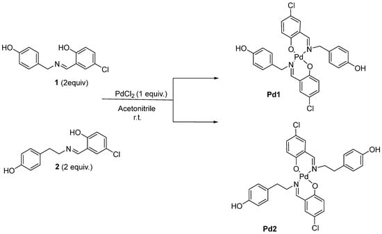
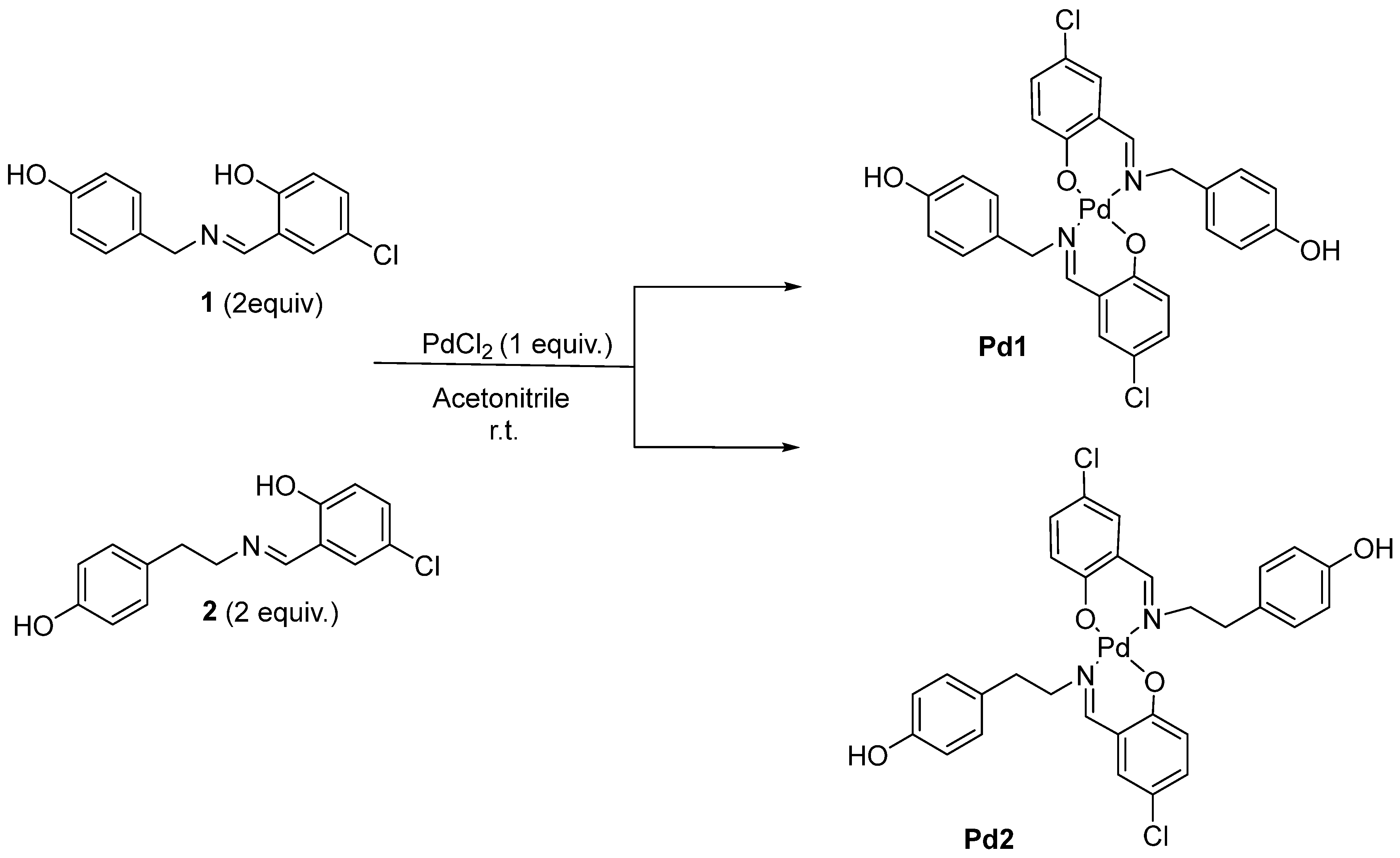
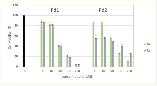
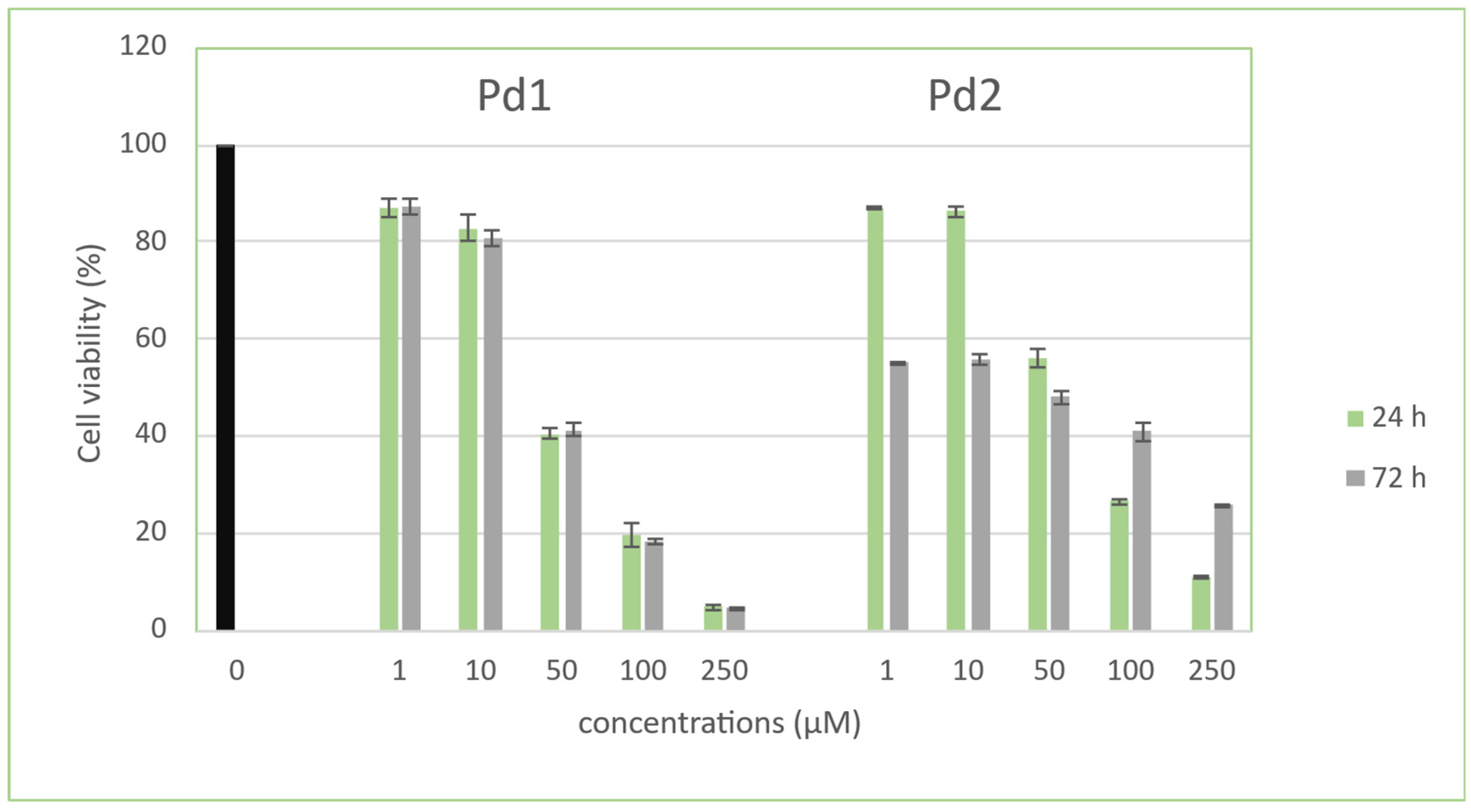
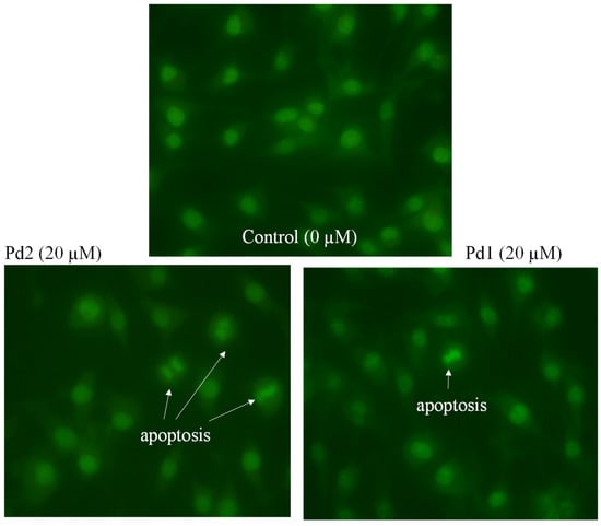
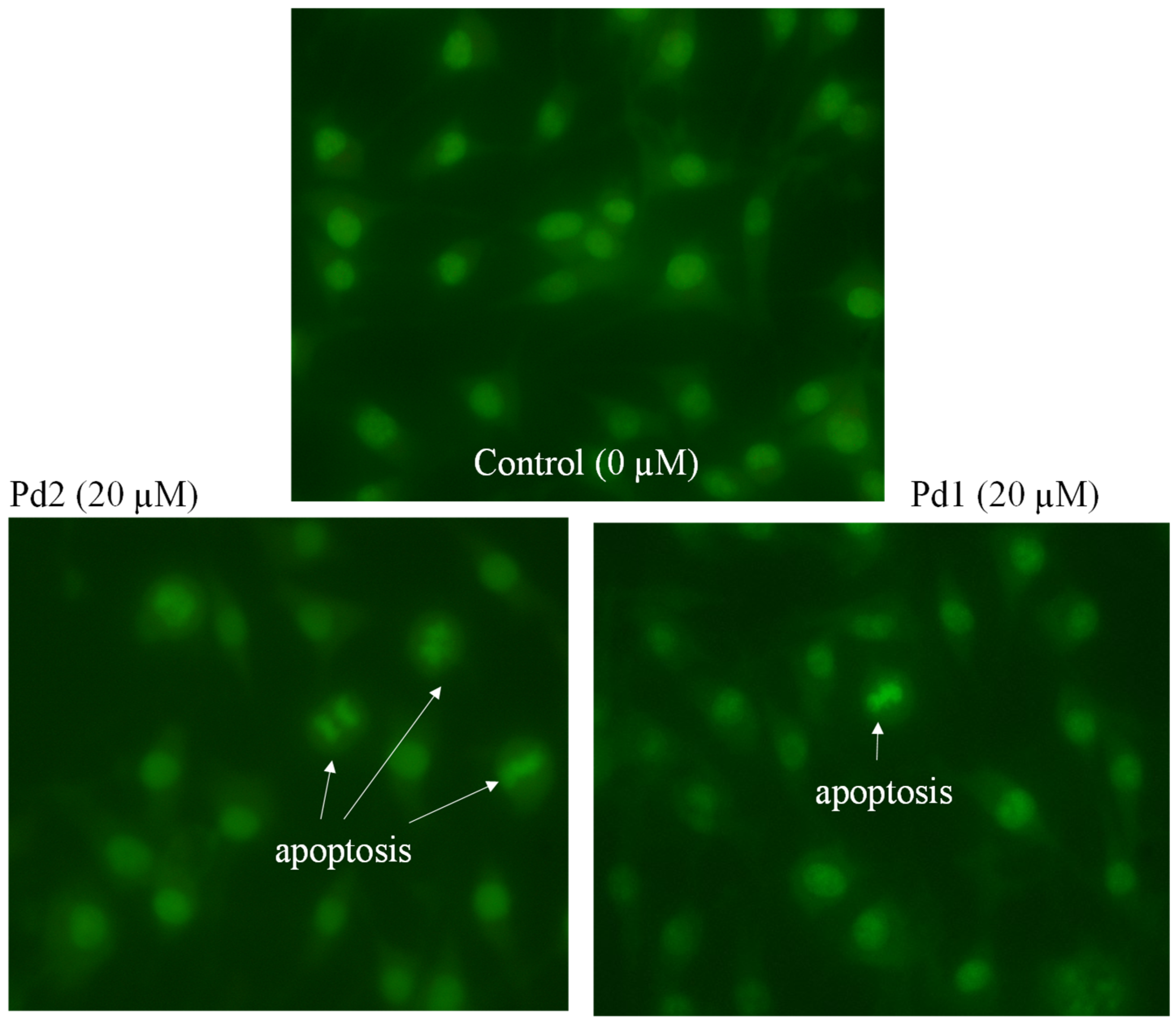
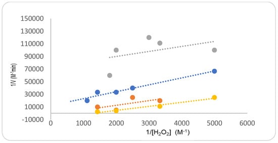
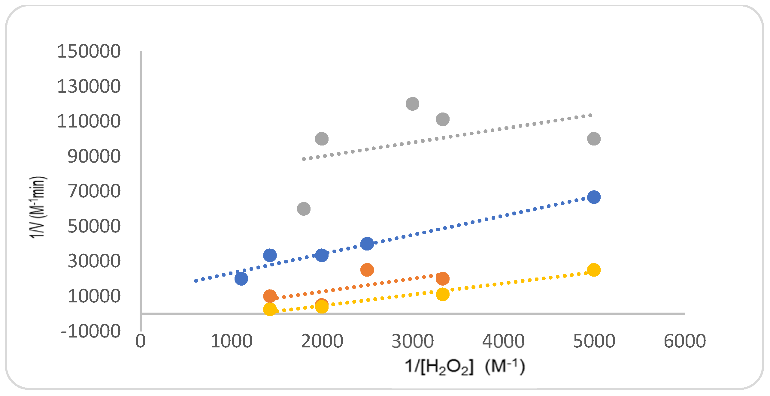
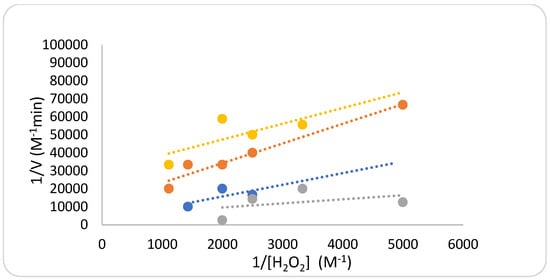
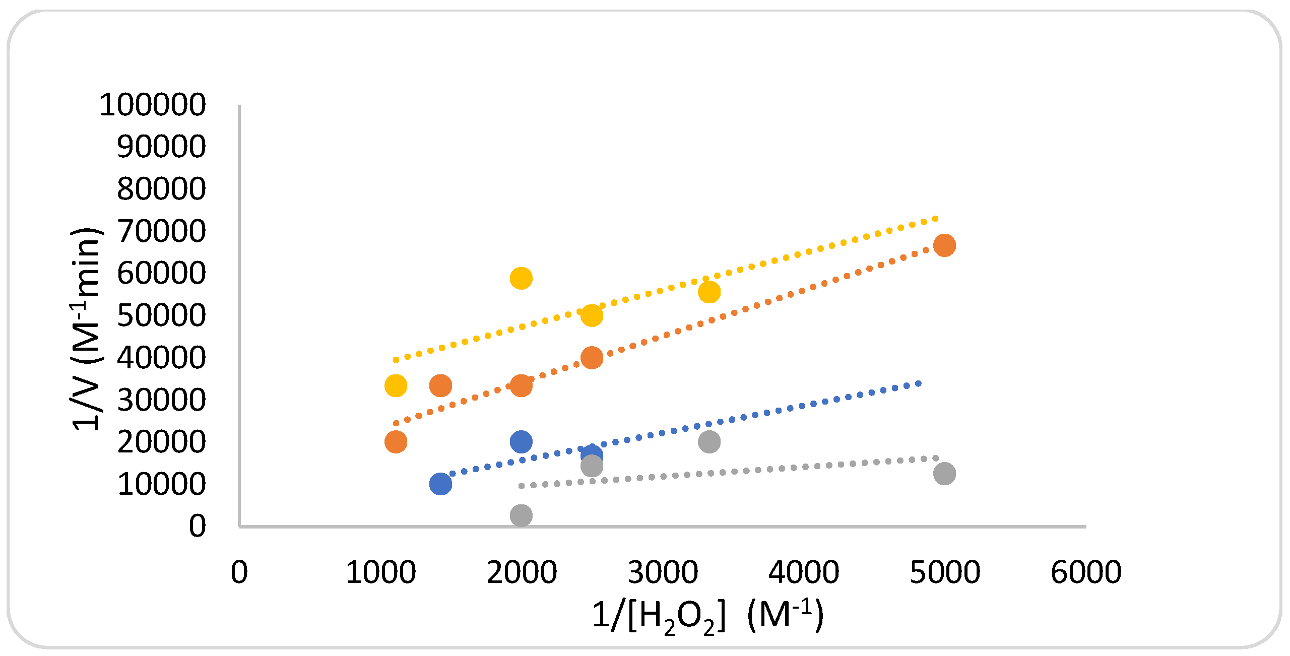
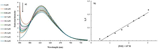
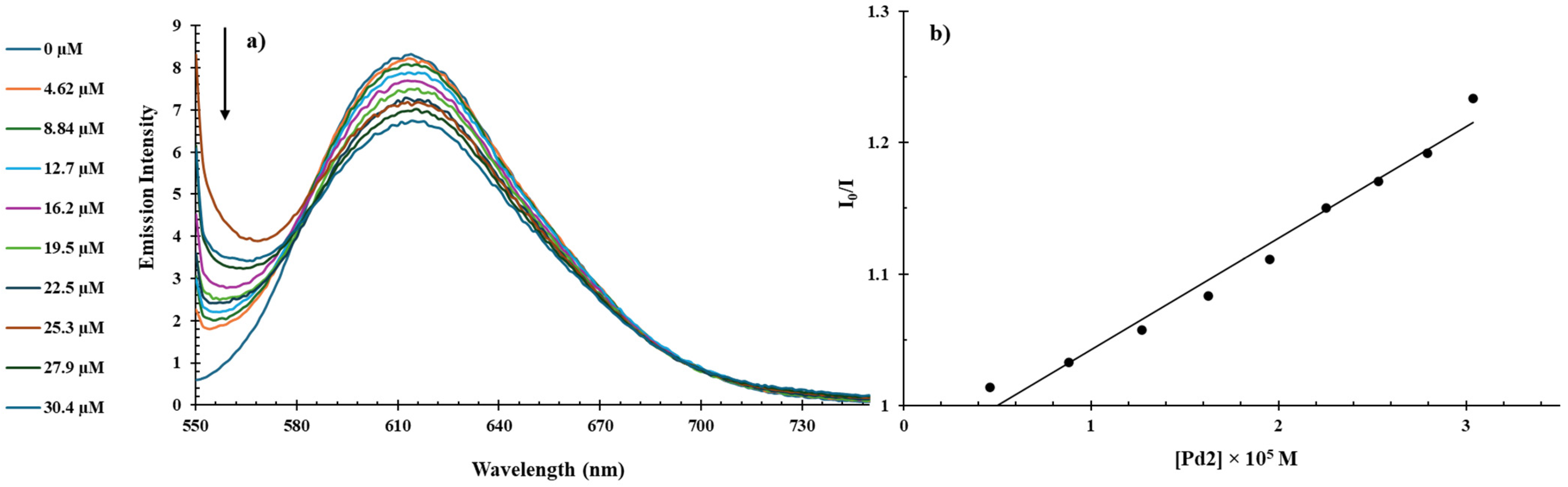
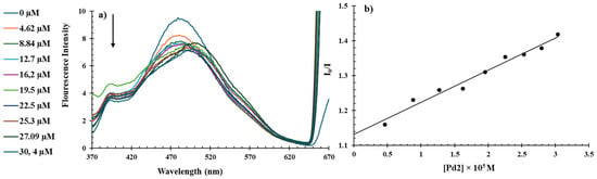
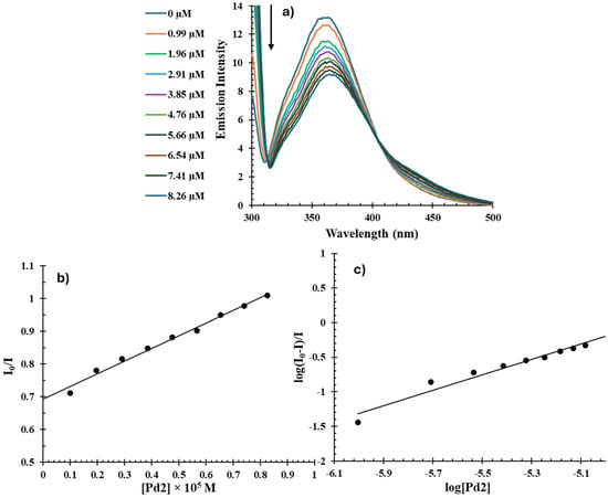
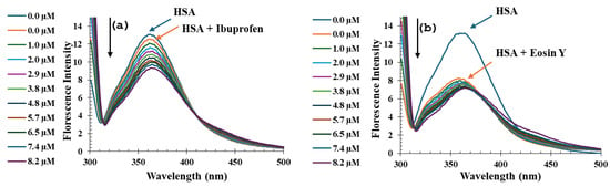
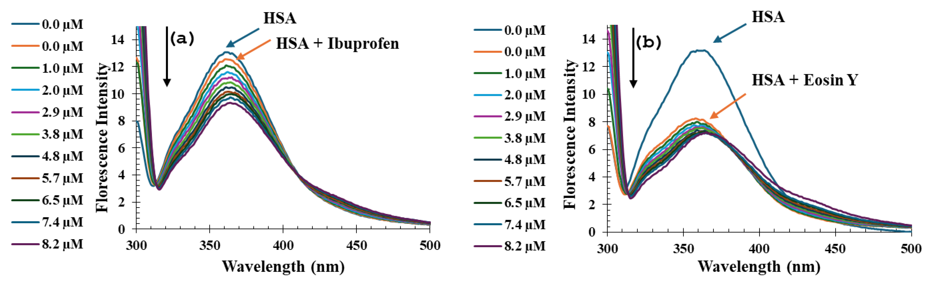
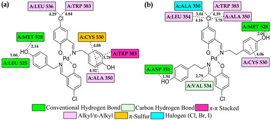
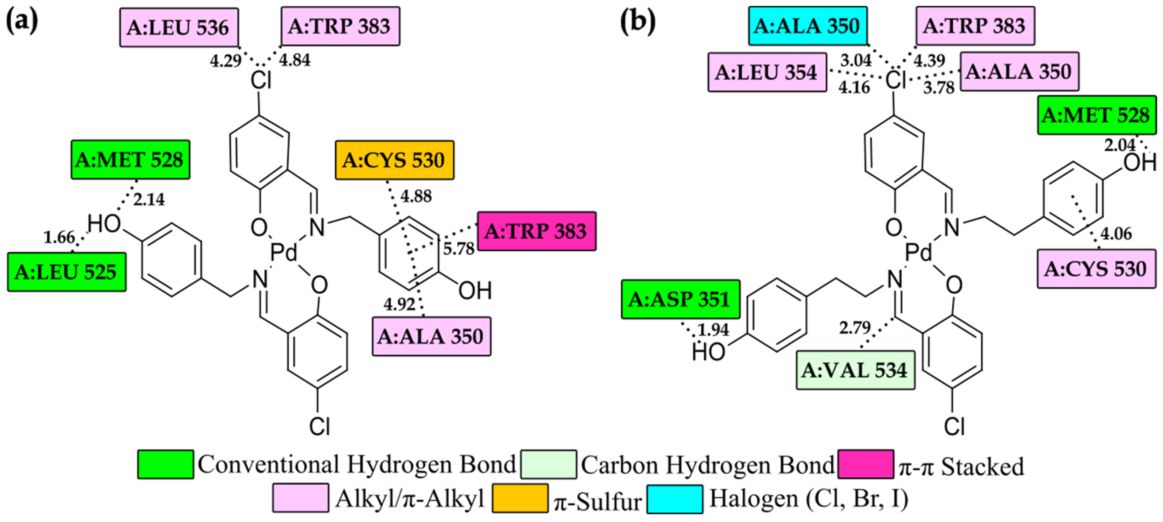
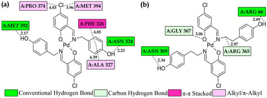
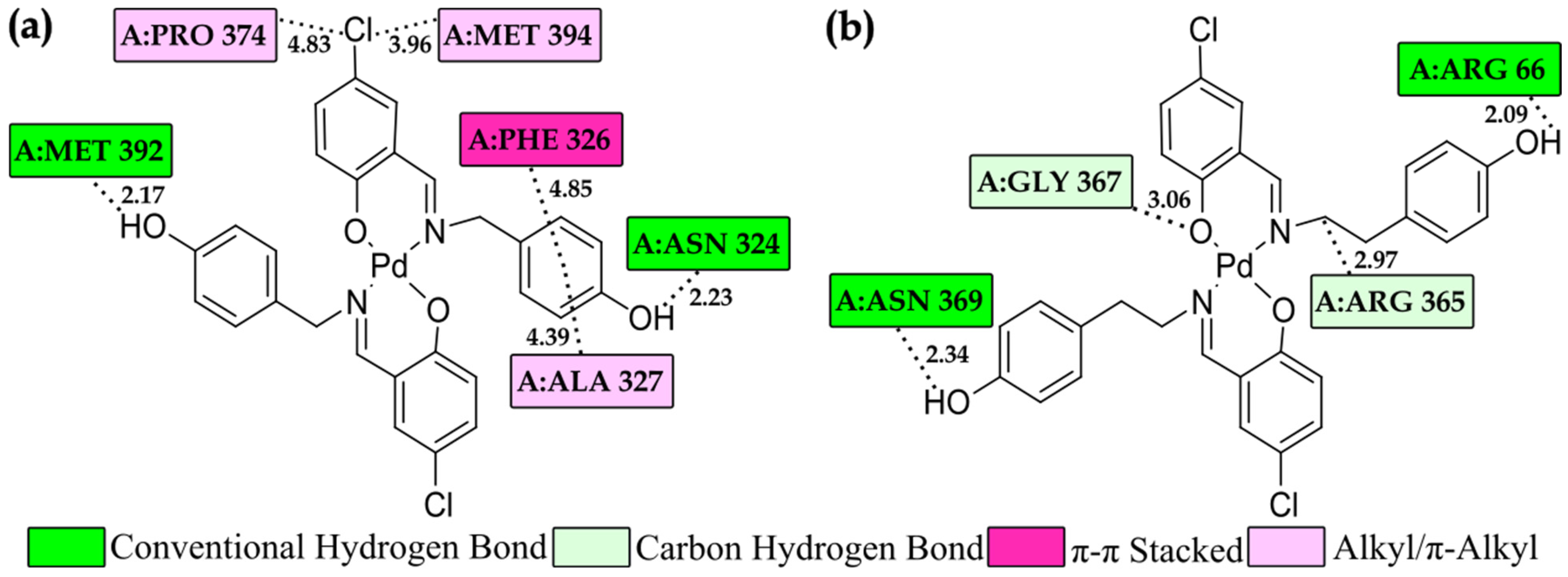
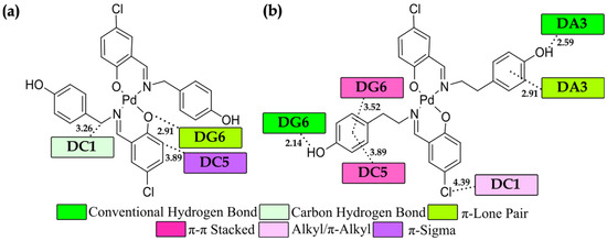
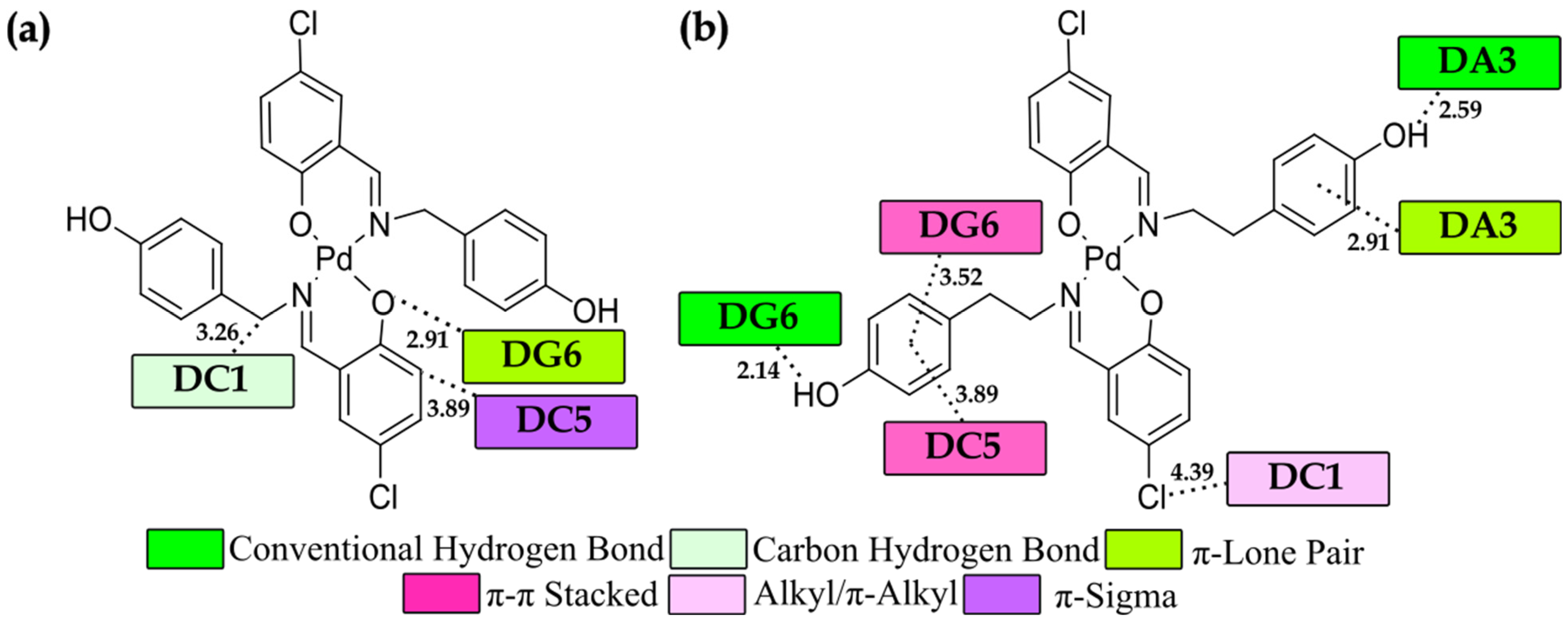
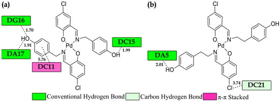
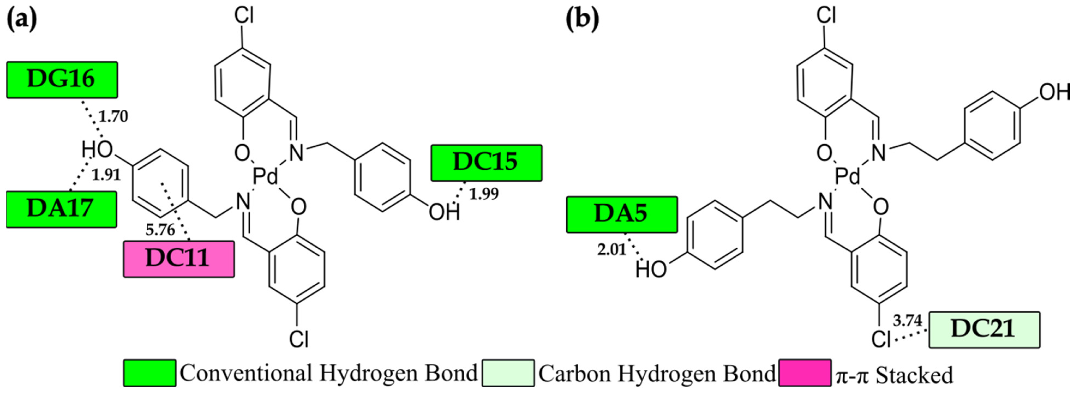

Disclaimer/Publisher’s Note: The statements, opinions and data contained in all publications are solely those of the individual author(s) and contributor(s) and not of MDPI and/or the editor(s). MDPI and/or the editor(s) disclaim responsibility for any injury to people or property resulting from any ideas, methods, instructions or products referred to in the content. |
© 2026 by the authors. Licensee MDPI, Basel, Switzerland. This article is an open access article distributed under the terms and conditions of the Creative Commons Attribution (CC BY) license.