


Abstract
Sclerotinia sclerotiorum is a soilborne fungal pathogen, and it is a major threat to lettuce production, causing lettuce drop. This study evaluated the antifungal effectiveness of five essential oils (EOs) (Rosmarinus officinalis, R. officinalis var. verbenone, Lavandula hybrida, Origanum majorana, and Thymus vulgaris) at 0.1%, 1%, and 10%, along with their phytotoxic effect in the field on three different crops (lettuce, tomato, and chard) following foliar application. T. vulgaris EO completely inhibited S. sclerotiorum mycelial growth at all tested concentrations. R. officinalis, L. hybrida, and O. majorana also showed full inhibition at 1% and 10%, while R. officinalis var. verbenone achieved 80–100% inhibition. R. officinalis had the least phytotoxic effects, with only a minimal effect on chard at 1%. R. officinalis var. verbenone caused low/moderate phytotoxicity in lettuce and chard but had no toxic effect on tomato. L. hybrida and O. majorana had moderate to low effects, while T. vulgaris was the most phytotoxic, significantly affecting lettuce and tomato at 1%. Further field trials are needed to define EO application protocols toward sustainable lettuce drop management without risks of phytotoxicity.
1. Introduction
Lettuce (Lactuca sativa) is an important salad crop that is produced all over the world [1]. According to FAO estimates, global lettuce production is led by China, with approximately 14.98 million tonnes, followed by India at 1.16 million tonnes. In Europe, the key producers are Spain and Italy, yielding 969,190 tonnes and 638,180 tonnes, respectively [2].
Lettuce crops suffer yield losses due to Sclerotinia spp., typically ranging from 10% to 45%, with severe cases reaching up to 70% under favorable conditions for the pathogen [3,4,5]. Sclerotinia minor and Sclerotinia sclerotiorum are the main species causing lettuce drop [6]. S. minor, a soilborne pathogen, infects lettuce roots and leaves via mycelia and can also contaminate seeds under certain conditions. S. sclerotiorum primarily infects lettuce plants through ascospores but can also contaminate seeds in favorable environments [7]. S. sclerotiorum infections typically begin near the stem, leading to root rot and a watery appearance of the crop [8,9]. S. minor infection is characterized by watery decay, browning, and plant collapse due to crown tissue damage with white mycelial mats forming small sclerotia on underground tissues. Infection by S. minor starts with sclerotia germination, unlike the airborne spread typical of S. sclerotiorum [10]. They can be distinguished by sclerotia size, with S. minor producing smaller (1–2 mm) and S. sclerotiorum larger (3–10 mm) sclerotia [11,12]. While S. sclerotiorum has a wide host range, S. minor is more host specific [13]. Secondary spread occurs when diseased tissues contact healthy tissues on adjacent plants. These fungal pathogens threaten lettuce production, causing significant losses, characteristic decay and wilting symptoms, with variations in their life cycles [14,15]. The management of Sclerotinia spp. has traditionally relied on the use of synthetic fungicides, with azoxystrobin, fludioxonil, and tebuconazole being among the most used agents [16,17]. These fungicides have proven effective in controlling Sclerotinia spp. across various plant species, but widespread use has raised significant environmental concerns about sustainability challenges [18]. In alignment with the European Green Deal and Farm to Fork Strategy, reducing dependence on synthetic pesticides has become mandatory for future agriculture, and many widely used fungicides are facing heavy limitations, leaving growers with fewer tools against crop diseases [19,20]. Consequently, it is important to search for alternative, environmentally friendly strategies for managing Sclerotinia spp. and focus on moving toward more sustainable farming practices.
These alternatives encompass biological control agents [21], basic substances [22], and the evaluation of plant extracts, including their nanoforms [23]. Among these, essential oils (EOs) are mixtures of natural organic compounds with inherent antifungal and antibacterial properties. They also stimulate host defenses, enhancing disease control on plants [24]. Rosmarinus officinalis EO demonstrated significant antifungal properties against pathogenic fungi, including Sclerotinia species. R. officinalis EO was evaluated in vitro and field testing, revealing its capacity to effectively inhibit the activity of Sclerotinia spp. on various plants [25,26]. Thymus vulgaris EO is known for its antifungal properties due to its composition of several bioactive compounds [27]. Previous studies have reported that EOs of Thymus spp. and Lavandula spp. exhibited dose-dependent antifungal activity against Sclerotinia spp. isolates obtained from different sources [28]. EOs are generally regarded as safe (GRAS) compounds for the environment due to their natural origins, rapid degradation, and low toxicity. The phytotoxicity of EOs is a complex phenomenon influenced by their chemical composition and application conditions [29]. Lettuce is the primary test organism due to its rapid germination and documented sensitivity to phytotoxic compounds, making it a standard bioassay species for preliminary toxicity screening [30]. Tomato (Solanum lycopersicum), a globally significant crop with significant postharvest losses caused by fungal pathogens, requires that any antifungal treatment be non-phytotoxic to preserve fruit quality and marketability [31,32]. Swiss chard (Beta vulgaris subsp. cicla), characterized by its delicate leaf structure and pronounced sensitivity to environmental and chemical stressors, serves as an effective bioindicator for detecting subtle phytotoxic effects. Including these contrasting species allowed for a broader and more realistic assessment of EO safety across horticultural systems. Furthermore, within the European Union regulatory framework, the “basic substances” concept under Regulation (EC) No 1107/2009 offers a pathway for authorizing low-risk, naturally derived compounds [33]. Several EOs and plant-based extracts, such as onion oil, grape seed extract, Equisetum arvense L., and sweet orange EO, are already approved under this category [34]. Demonstrating both effectiveness and safety could enable the use of EOs, supporting their integration into sustainable plant protection programs.
The objectives of this study were (i) to investigate and compare the in vitro antifungal activity of five different EOs with three different concentrations to control Sclerotinia spp., and (ii) to assess the phytotoxic effects of the five EOs on lettuce, tomato and chard under field conditions, using concentrations that maintained antifungal activity in vitro, in order to minimize potential phytotoxicity.
2. Results
2.1. Sclerotinia spp. Isolates from Lettuce Sample
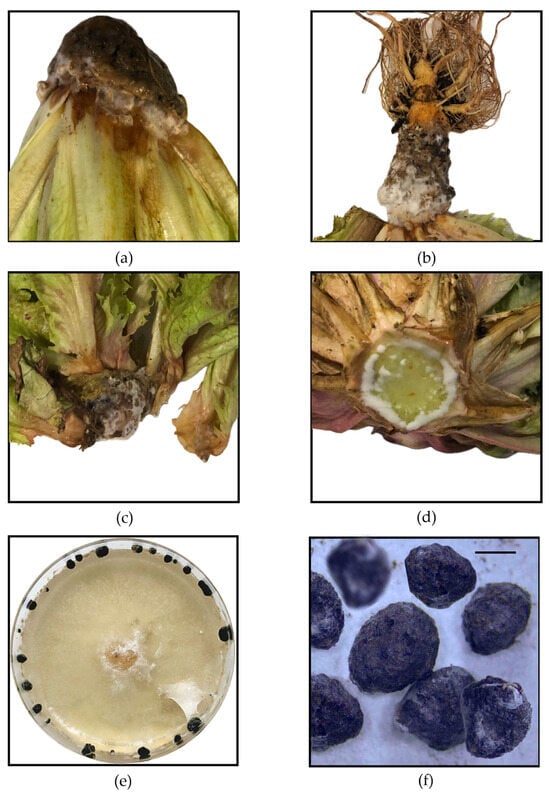
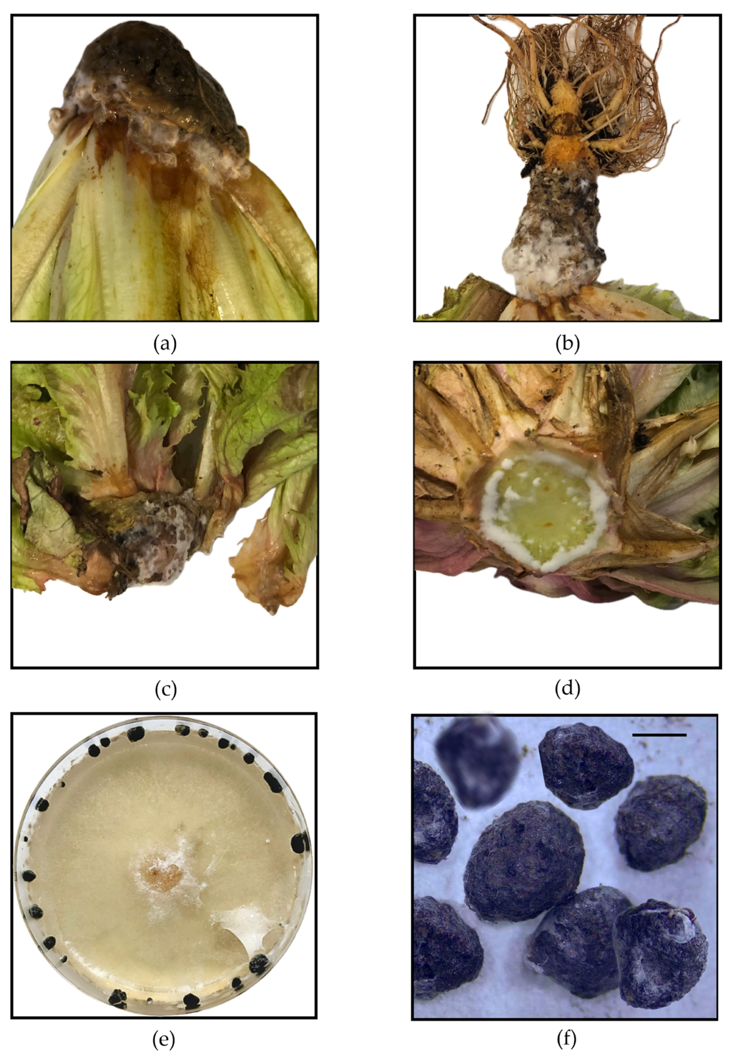
Among seven suspected lettuce samples, four were confirmed to be infected with S. sclerotiorum through morphological identification of the isolated causal organism. Infected lettuce showed severe root and basal stem rot, with water-soaked, soft and mushy tissues. The outer surface appears discolored and becomes brown and dark as the rot progresses. A dense, white, cottony mycelium covers the affected areas and spreads rapidly. Inside the hollow stem, the tissue is completely degraded, leaving a soft, collapsed structure. Small, initially transparent sclerotia begin to form within the decayed tissue, later maturing into hard, black structures. Lettuce isolates of Sclerotinia spp. exhibited rapid growth on PDA medium, producing prolific, white, cottony mycelia that completely covered the Petri dishes within two to three days at 21 ± 2 °C. Mycelia initially appeared soft and fluffy, but became more compact and regular as growth progressed. Sclerotia began to form, first appearing as small, transparent structures and gradually darkening into black, hard, irregularly shaped bodies (Figure 1). Mature sclerotia exhibited a hard outer surface with a consistent size range. Morphological analysis of 51 sclerotia, including measurements under the microscope, confirmed the identification of the species. Sclerotium sizes were recorded among the four lettuce isolates and varied slightly between isolates, with measurements presented in Table 1. The treatments were inoculated using these four isolates, referred to as Sc1, Sc2, Sc3, and Sc4.
Figure 1.
(a) Lettuce sample exhibiting characteristic symptoms of Sclerotinia sclerotiorum infection, including white cottony mycelium and water-soaked lesions near the base. (b) Advanced infection with extensive mycelial growth and black sclerotia formation on the roots. (c) Necrotic and decayed lettuce leaves with visible sclerotia embedded in the infected tissue. (d) Cross-section of an infected lettuce stem showing internal tissue colonization and soft rot. (e) Sclerotinia sclerotiorum isolate grown on PDA medium for seven days, displaying characteristic mycelial growth and sclerotia formation along the Petri dish edges. (f) Black sclerotia observed under a stereomicroscope. Scale bar: 100 µm.
Table 1.
Average sclerotium diameter in micrometers (µm) and standard deviation among lettuce isolates.
2.2. In Vitro Inhibition of Fungal Growth by the Five Essential Oils
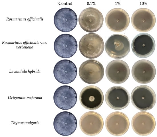
The antifungal activity of five EOs was evaluated against four S. sclerotiorum isolates (Sc1–Sc4) at three concentrations (0.1%, 1%, and 10%), as can be seen in Figure 2 and Table 2. Inhibition percentages were calculated relative to the control (8.3 cm mycelial growth). The findings demonstrate varied degrees of antifungal activity among the EOs, with a general trend of higher concentrations correlating with higher inhibition of fungal growth in a dose-dependent manner. T. vulgaris EO was the most effective, achieving complete inhibition (100%) against all isolates at all concentrations tested, including 0.1%. At 1% and 10% concentrations, R. officinalis EO achieved complete inhibition (100%) across all isolates. R. officinalis EO has shown limited antifungal activity at 0.1%, with an inhibition percentage of 29.3% and 13.2% against Sc2 and Sc3, respectively, while low inhibition ranging from 0 to 1.0% was observed against Sc1 and Sc4. At 10%, R. officinalis var. verbenone EO achieved 100% inhibition across all isolates. At 1%, inhibition ranged from 57.0% for Sc1 to 68.8% for Sc4, with complete inhibition (100%) recorded for Sc2 and Sc3. At 0.1% concentration, partial inhibition was observed at 26.30% and 27.7% for isolates Sc2 and Sc3, respectively. In contrast, isolates Sc1 and Sc4 exhibited minimal inhibition, with values ranging from 0% to 1%. At 1% and 10%, L. hybrida EOs completely inhibited (100%) fungal growth across all isolates. The same EO demonstrated moderate antifungal activity at 0.1%, achieving inhibition values of 56.2% and 56.9% against Sc2 and Sc3, respectively, 1.0% against Sc4, and low inhibition against Sc1. At both 1% and 10%, O. majorana EO achieved complete inhibition (100%) across all isolates. O. majorana EO exhibited notable antifungal activity at 0.1%, achieving inhibition values of 56.4%, 62.6%, 90.9%, and 100% against Sc1, Sc4, Sc3, and Sc2, respectively.
Figure 2.
Representative experiment showing the mycelial growth of Sclerotinia sclerotiorum isolate 1 after treatment with five essential oils (Rosmarinus officinalis, R. officinalis var. verbenone, Lavandula hybrida, Origanum majorana, and Thymus vulgaris) at 0.1%, 1%, and 10%, compared to the control (PDA amended with Tween 20). Pictures were taken after seven days of plate incubation at 22 ± 2 °C.
Table 2.
Mycelial radial growth (cm) of four Sclerotinia sclerotiorum isolates treated with five essential oils. Measurements were recorded after 7 days of inoculation at 22 ± 2 °C, when the colonies of the control reached the edge of the Petri dishes.
2.3. Fungicidal and Fungistatic Effects of Essential Oils Against Sclerotinia sclerotiorum
The fungicidal and fungistatic activities of five EOs (Rosmarinus officinalis, R. officinalis var. verbenone, Lavandula hybrida, Origanum majorana, and Thymus vulgaris) were evaluated on four S. sclerotiorum isolates (Sc1–Sc4), at concentrations of 0.1%, 1%, and 10% (v/v). The results are summarized in Table 3. R. officinalis EO exhibited a concentration-dependent pattern. At 1%, all isolates that showed complete growth inhibition were able to regrow after re-inoculation, indicating a fungistatic effect. At 10%, a fungicidal effect was observed against isolates Sc3 and Sc4. R. officinalis var. verbenone EO mainly displayed fungistatic activity. At 10%, fungicidal action was recorded only for Sc3 and Sc4, while the remaining isolates showed regrowth, confirming their predominantly fungistatic nature. L. hybrida EO showed a mixed response depending on both concentration and isolate. At 1%, isolates Sc1 and Sc2 exhibited fungistatic behavior, while Sc3 and Sc4 were completely inhibited, showing a fungicidal effect. At 10%, all isolates exhibited fungicidal activity. O. majorana EO exhibited both fungistatic and fungicidal effects depending on the concentration and isolate. At 0.1%, it was mostly fungistatic, while at 1% and 10%, fungicidal activity was observed on several isolates. T. vulgaris EO was the most active among the tested oils, showing fungicidal effects against all isolates at all concentrations tested.
Table 3.
Fungicidal and fungistatic activities of five essential oils (Rosmarinus officinalis, R. officinalis var. verbenone, Lavandula hybrida, Origanum majorana, and Thymus vulgaris) on four Sclerotinia sclerotiorum isolates (Sc1–Sc4), at three concentrations (0.1%, 1%, and 10%, v/v). The effects were determined after seven days of incubation at 22 ± 2 °C.
2.4. Phytotoxicity Evaluation of the Five Essential Oils in Lettuce, Tomato, and Chard
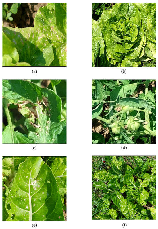
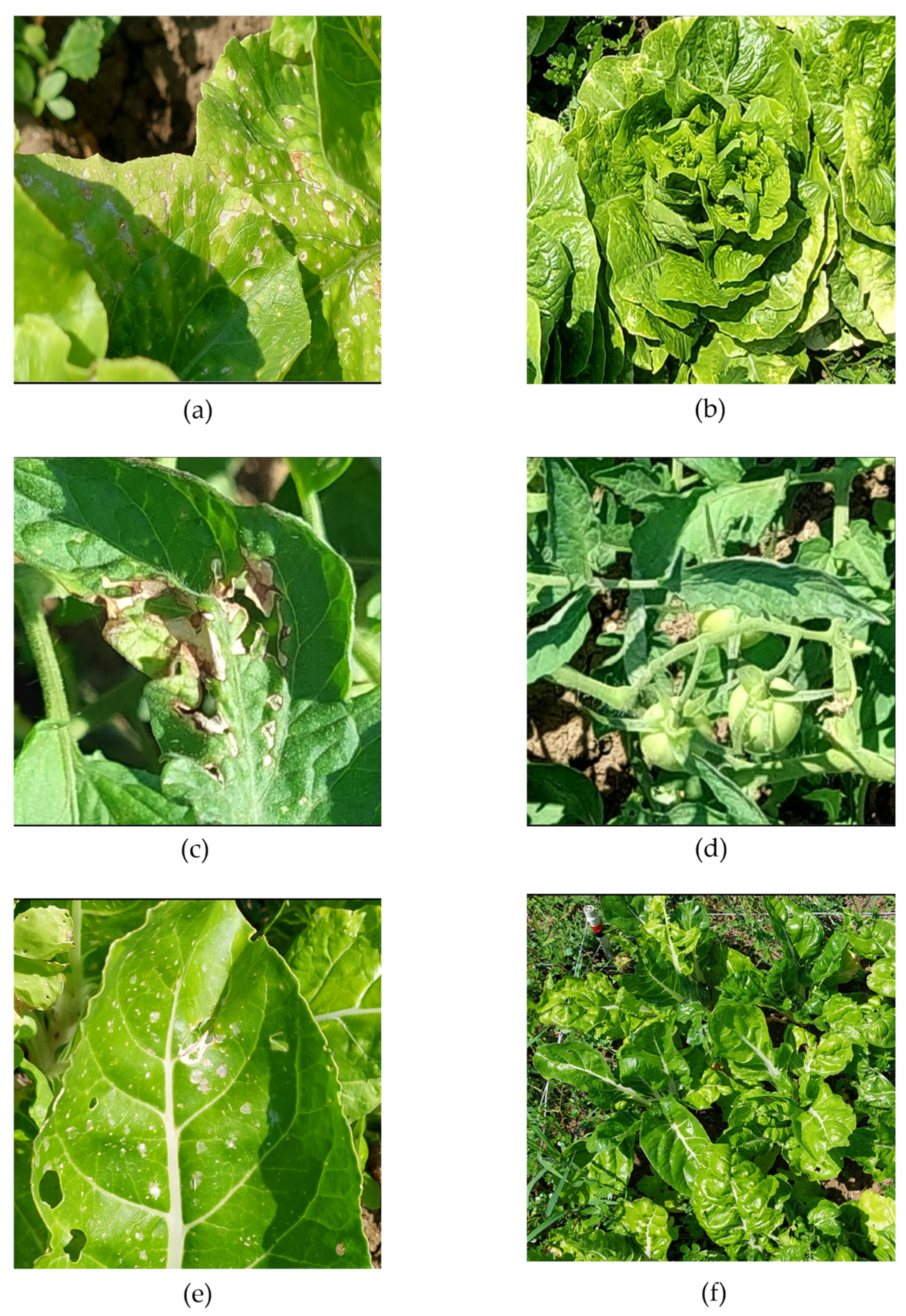
Visible phytotoxic responses were observed on lettuce, tomato and chard treated with five EOs (Rosmarinus officinalis, R. officinalis var. verbenone, Lavandula hybrida, Origanum majorana, and Thymus vulgaris) at concentrations of 0.1 and 1% (v/v). Photos of lettuce plants treated with different EOs are reported in Figure 3 and Figure S1 and Table 4 to summarize the results of the phytotoxicity evaluation for all the crops.
Figure 3.
Representative phytotoxic symptoms caused by Thymus vulgaris EO (1%), the most phytotoxic treatment, showing yellow spots, leaf burn, and necrosis on (a) lettuce compared with (b) control, (c) tomato compared with (d) control, and (e) chard compared with (f) control. Additional pictures for the other essential oils are presented in Figure S2.
Table 4.
Phytotoxicity of five essential oils (Rosmarinus officinalis, Rosmarinus officinalis var. verbenone, Lavandula hybrida, Origanum majorana, Thymus vulgaris) on lettuce, tomato, and chard plants treated with 0.1% and 1% concentrations (v/v) and evaluated at 11 and 22 days after application (DAA). Symptoms were assessed individually by plant species, and an overall score was assigned based on the appearance of 9 plants per treatment (NS, no phytotoxic symptoms; +, low phytotoxicity (1–20% of plants showing symptoms); ++, medium phytotoxicity (21–50% of plants showing symptoms); +++, high phytotoxicity (>51% of plants showing symptoms). Changes over time are shown as first/second assessment (e.g., NS/+, if the plant was asymptomatic in the first assessment and started to show symptoms in the second assessment).
2.4.1. Lettuce
In lettuce, plants treated with R. officinalis EO showed no visible phytotoxicity at either concentration (0.1% and 1%); leaves remained similar to those of the untreated control plants (Figure S1a,b). Plants treated with R. officinalis var. verbenone EO caused low phytotoxicity at both concentrations, affecting up to 20% of plants with small, scattered yellow spots on the leaf surface (Figure S1c,d). Using L. hybrida EO, clear differences were observed between concentrations. At 0.1%, medium phytotoxicity symptoms were visible, with approximately 20–50% of plants exhibiting yellow spots and slight leaf burn, at 1%, the phytotoxicity level was lower, affecting up to 20% of plants (Figure S1e,f). No visible symptoms were detected in plants treated with O. majorana EO at all concentrations, and their general appearance was like the untreated controls (Figure S1g,h). Plants treated with T. vulgaris EO showed the highest level of phytotoxicity among all treatments. At 1%, over 50% of the plants were severely affected, exhibiting widespread yellow spots, leaf burn (Figure S1i,j), while no symptoms were visible at 0.1%. All treatments were compared with the untreated control plants (Figure S1k,l).
2.4.2. Tomato
In tomato plants, no visible phytotoxicity was detected in any of the treatments with R. officinalis, R. officinalis var. verbenone, or L. hybrida EOs at both concentrations (0.1% and 1%). Similarly, O. majorana EO did not cause visible phytotoxic symptoms at 0.1%, and only a few plants showed low phytotoxicity at 1%, with yellow spots observed on some leaves during the second assessment. In contrast, plants treated with T. vulgaris EO at 1% exhibited high phytotoxicity, affecting more than 50% of the plants, which displayed widespread yellow spots, leaf burn, and necrosis. At 0.1%, no visible phytotoxicity symptoms were observed, and treated plants remained comparable to the controls. T. vulgaris EO was the only treatment that induced a severe phytotoxic response in tomato, while the other oils were well tolerated by the plants.
2.4.3. Chard
In chard plants, phytotoxic responses varied depending on the EO and concentration applied. Plants treated with R. officinalis EO showed no visible phytotoxicity at 0.1%, whereas low phytotoxicity was observed at 1% during the second assessment, with up to 20% of plants displaying scattered yellow spots on the leaf surface. Treatment with R. officinalis var. verbenone EO caused medium phytotoxicity at both concentrations, affecting 20–50% of plants, which showed yellow spots and slight leaf burns. In plants treated with L. hybrida EO, the phytotoxic response increased with concentration, with low phytotoxicity observed at 0.1%, and medium symptoms developed at 1%, with up to 50% of plants showing partially burnt leaves. O. majorana and T. vulgaris EOs both induced low phytotoxicity at 0.1% and 1%, with 1–20% of plants showing small yellow spots and minor leaf burn, but no severe damage was detected. R. officinalis var. verbenone and L. hybrida EOs produced the most evident phytotoxic effects in chard, while O. majorana and T. vulgaris EOs were the least harmful treatments.
3. Discussion
S. sclerotiorum is a highly destructive fungal pathogen, causing significant losses in lettuce production worldwide. Considering the limitations of conventional control methods, the research of alternative strategies is crucial for sustainable disease management. Morphological identification provides an initial approach for characterizing S. sclerotiorum, confirming its colony morphology, hyphal structure, and sclerotia formation [35,36,37]. In our study, microscopic analysis confirmed the presence of septate hyphae with characteristic branching patterns [38]. Sclerotia size measurements across all four isolates showed uniformity, ranging from 2.5 to 2.8 mm, which aligns with previous studies. This consistency supports the reliability of morphological identification methods for diagnosing S. sclerotiorum [39,40,41].
Our in vitro findings demonstrate the effectiveness of EOs tested and provide a new insight beyond simple inhibition. A key result was that T. vulgaris EO completely inhibited all S. sclerotiorum isolates at all tested concentrations (0.1%, 1% and 10%). This broad-spectrum activity is consistent with its fungicidal properties, which can be explained by the high content of phenolic compounds such as thymol and carvacrol, which are known to disrupt cell membranes and metabolic pathways [42,43]. O. majorana EO was the second most effective treatment, exhibiting dose-dependent activity that varied between isolates. Sc2 was inhibited at 0.1% while Sc3 and Sc4 required higher concentrations for fungicidal effects. This observation aligns with previous findings, which reported dose-dependent antifungal activity of O. majorana EO against various fungal pathogens [44]. The activity of R. officinalis and L. hybrida EOs was strongly concentration-dependent, with consistent fungicidal activity achieved only at 1% and 10%. The requirement for a higher concentration of R. officinalis var. verbenone EO in our study, compared to reports of complete inhibition at 0.1% for other chemotypes, suggests that the antifungal activity is linked to the specific chemical profile of the EO. This chemotypic variation is a key factor explaining variation between studies and emphasizes the need for standardized chemical characterization in bioactivity research [45,46]. The chemical composition of each EO plays an important role in determining its mechanism of antifungal activity. As reported in Table 5, the EOs of R. officinalis and L. hybrida are particularly rich in key compounds such as α-pinene, 1,8-cineole, camphene, and limonene. According to previous studies, oxygenated monoterpenes, including α-pinene, linalool, and 1,8-cineole, which are dominant constituents in L. hybrida and R. officinalis EOs, have demonstrated antifungal activity against major plant pathogens like Alternaria alternata and Fusarium oxysporum [47,48]. Phenolic monoterpenes such as thymol and carvacrol, which are abundant in T. vulgaris EO, are widely recognized as highly potent antifungal agents. Their effectiveness against destructive pathogens such as Botrytis cinerea and S. sclerotiorum has been extensively reported [49,50]. These compounds express their antifungal action mainly through the disruption of fungal cell membranes, leading to leakage of intracellular contents and ultimately inhibiting fungal growth [49,50].
Table 5.
Essential oils assessed for their in vitro antifungal activity against Sclerotinia sclerotiorum. The table reports the plant species of origin, common names, commercial providers, and the main chemical constituents of each essential oil according to the supplier’s specifications.
The core innovation of our study lies in the direct linkage of antifungal effectiveness with a simultaneous, multi-crop phytotoxicity assessment. This integrated approach is important for identifying EOs with a viable safety profile. Studies have demonstrated that the phytotoxic effects of EOs vary depending on plant species, developmental stages, and concentrations [51]. The most potent fungicide, T. vulgaris EO, has the highest phytotoxic risk to lettuce and tomato at 1% concentration. R. officinalis EO demonstrated the most promising balance, combining significant antifungal activity with minimal phytotoxicity, causing only slight symptoms on chard at 1%, a finding supported by its relative safety on other crops [52,53]. O. majorana and L. hybrida induced low to moderate effects that varied by crop species. A particularly significant and novel finding was the high sensitivity of chard to most EOs, a previously unreported phenomenon that provides crucial new information for growers of this crop [54,55]. This underlines that phytotoxicity is not a universal trait but is highly specific to both the EO and the crop species, necessitating individual evaluation.
Our research goes beyond simple in vitro screening, providing an integrated risk-benefit analysis. We identified T. vulgaris as the most potent antifungal EO, but its phytotoxic risk limits its practical use. In contrast, R. officinalis and O. majorana EOs represent the most promising candidates, offering effective pathogen control with an acceptable safety profile for tomato and lettuce. The practical importance of these results is significantly amplified within the regulatory framework of the European Union for “basic substances” (Regulation (EC) No 1107/2009). This classification is designed for safe, naturally occurring substances mainly used for purposes other than plant protection (for example, as foodstuffs or medicinal products). EOs are good candidates for this pathway since they are considered generally recognized as safe (GRAS) compounds. The approval of EOs and other plant extracts, such as onion oil, garlic extract, and mustard seed powder, as basic substances, provides a clear background. Obtaining basic substance status for the EOs identified in this study has profound implications. Firstly, it offers a faster and more cost-effective route to commercialization compared to conventional synthetic pesticides. Secondly, basic substances count as “0” in the calculation of the Harmonized Risk Indicator 1 (HRI-1), a key parameter for the EU directive on sustainable use of pesticides. This provides a strong incentive for farmers to adopt these low-risk alternatives and contributes to achieving policy goals for reducing the use of chemical pesticides. Therefore, our work lays the essential groundwork for future applied research. The promising effectiveness and safety profile of EOs justifies immediate field validation to confirm their performance under real conditions. This research pipeline, from in vitro effectiveness and phytotoxicity to field studies, is fundamental for creating the scientific dossier required to support future applications of these EOs as potential basic substances, thereby expanding the range of sustainable and accessible options for modern agriculture.
4. Materials and Methods
4.1. Isolation of Lettuce
Seven lettuce (Lactuca sativa var. longifolia) samples, suspected of Sclerotinia spp. infection, were obtained from the Marche region, Italy. Five lettuce samples were from greenhouse cultivation, and two were from field cultivation. All samples were examined under a stereomicroscope (Leica M125, Leica Microsystems CMS, Wetzlar, Germany) to verify the presence of Sclerotinia spp. by assessing the presence of sclerotia or mycelia. Each affected plant was sliced into small fragments (less than 5 mm) using flame-sterilized scalpels. These fragments were immersed in 1% sodium hypochlorite for 1 min, rinsed three times with sterile distilled water, and dried on sterile blotter paper in a laminar flow hood. The fragments were then placed on potato dextrose agar (PDA, 42 g/L; Liofilchem Srl, Roseto degli Abruzzi, Italy) and incubated at 22 ± 2 °C for seven days. The plates were checked daily, and colonies grown were transferred to fresh PDA plates to obtain pure Sclerotinia spp. cultures. Morphological identification was performed by growth type, sclerotia characteristics, and the colors and shapes of the colonies. Fungal structures were measured using LAS V3.8 software (Leica DFC 295) from 51 units of each structure for each fungal isolate. Species identification was conducted using the taxonomic keys provided [36,37].
4.2. In Vitro Antifungal Activity on Mycelial Growth
The antifungal activity of five EOs (R. officinalis, R. officinalis var. verbenone, L. hybrida, O. majorana and T. vulgaris) was assessed against four S. sclerotiorum isolates derived from various lettuce samples. EOs were purchased from Verde Naturale Company (Corinaldo, Ancona, Italy). According to the manufacturer, the EOs were 100% pure and obtained by steam distillation (Table 5). The EOs were kept in sealed amber vials and stored at 4 °C until used in all experiments. The EOs were assessed against four S. sclerotiorum isolates derived from various lettuce samples. To prepare homogeneous emulsions, EOs were dissolved in sterile distilled water containing 0.1% (v/v) Tween 20 (Sigma Aldrich, Steinheim, Germany). The emulsions were then incorporated into potato dextrose agar (PDA) medium, cooled to 40 °C, to achieve final EO concentrations of 0.1%, 1%, and 10%. Petri dishes (90 mm in diameter) were filled with 15 mL of PDA amended with EO emulsions, while PDA containing only 0.1% (v/v) Tween 20 served as the control. Each Petri dish was inoculated with a 6 mm plug of S. sclerotiorum, and five replicates were prepared for each EO concentration. Plates were sealed with parafilm and incubated at 22 ± 2 °C for seven days. Fungal colony diameters (measured perpendicularly) were recorded daily until mycelial growth reached the edge of the control plates. Mycelial growth inhibition was calculated using Equation (1):
where “” is the mean diameter of the control colonies and “” is the mean diameter of the treated colonies.
To differentiate between fungicidal and fungistatic effects, the methodology outlined by Moumni et al. [56] was followed. Mycelial plugs showing complete growth inhibition on EO-amended media were transferred onto fresh PDA plates without EOs and incubated at 22 ± 2 °C for seven days. The absence of fungal regrowth was considered indicative of a fungicidal effect, whereas resumed mycelial growth was interpreted as fungistatic activity.
4.3. Phytotoxicity Assessment of the Five Essential Oils in Lettuce, Tomato and Chard
Phytotoxicity of the EOs was evaluated following foliar application in open-field conditions in Senigallia, Italy (Latitude: 43.697778° N; Longitude: 13.242778° E). Lettuce, tomato, and chard plants were transplanted on 1 June 2023, and treatments were carried out 22 days later. EOs were applied at concentrations of 0.1% and 1%, selected based on confirmed effectiveness in in vitro studies, to assess potential side effects. Treatments were applied to one plot per treatment per crop, with each plot containing nine plants. A spraying volume of 1 L per plot was used. Control plots were sprayed with water containing 0.1% Tween 20.
Phytotoxicity assessments were conducted on 2 July 2023 (11 days after application) and 14 July 2023 (22 days after application). Phytotoxicity was assessed visually on individual plants by observing foliar changes such as yellow spotting and leaf burning in comparison with untreated controls. In several cases, these symptoms were associated with reduced plant growth, and in the most severe instances, plant death was observed. The extent of phytotoxic effects was classified using an empirical scale: no symptoms (NS); low (1–20% of plants showing phytotoxicity); moderate (21–50%); and high (>51%).
4.4. Statistical Analysis
Data were expressed as mean ± standard deviation (SD) and analyzed using SPSS software (version 21.0; SPSS Inc., Chicago, IL, USA). Analysis of variance (ANOVA) was performed to determine significant differences among treatments, and means were compared using Tukey’s test at a significance level of p ≤ 0.05.
5. Conclusions
This research highlighted the potential of five EOs, R. officinalis, R. officinalis var. verbenone, L. hybrida, O. majorana, and. T. vulgaris, in inhibiting the mycelial growth of S. sclerotiorum. T. vulgaris EO demonstrated complete inhibition of mycelial growth across all tested isolates and exhibited consistent fungicidal activity at all tested concentrations, marking it as a highly effective candidate for disease management. At a low concentration (0.1%), T. vulgaris EO showed no phytotoxic effects on lettuce, reinforcing its potential for safe application. Its effectiveness at low concentration is particularly significant given the high cost of EOs and the risks of phytotoxicity on fresh produce, making it a promising and economically viable alternative to synthetic fungicides.
Future research should build upon these findings by assessing the performance of these EOs beyond controlled conditions, particularly in greenhouse and field trials, to further validate their effectiveness and practical applicability. As the demand for sustainable agricultural solutions grows, EOs offer a compelling, eco-friendly alternative for controlling S. sclerotiorum infections.
Supplementary Materials
The following supporting information can be downloaded at: https://www.mdpi.com/article/10.3390/molecules31040682/s1. Figure S1. Effects of essential oils in lettuce culture, (a) Rosmarinus officinalis 0.1%, (b) R. officinalis 1%, (c) R. officinalis var. verbenone 0.1%, (d) R. officinalis var. verbenone 1%, (e) Lavandula hybrida 0.1%, (f) L. hybrida 1%, (g) Origanum majorana 0.1%, (h) O. majorana 1%, (i) Thymus vulgaris 0.1%, (j) T. vulgaris 1%, (k, l) control. Figure S2. Phytotoxicity evaluation of Rosmarinus officinalis, Rosmarinus officinalis var. verbenone, Lavandula hybrida, Origanum majorana, and Thymus vulgaris essential oils on different crops following foliar application. The panel illustrates representative visible morphological responses (including yellow spots, leaf necrosis and growth inhibition) observed on different crops with all the tested essential oils. Each essential oil treatment was evaluated based on span changes according to the different plant and symptom severity.
Author Contributions
Conceptualization, G.R.; methodology, M.M.; software, S.P. and S.M.M.; validation, M.M. and S.P.; formal analysis, M.T.; investigation, M.T., S.M.M. and A.L.D.; resources, S.S., L.P., E.G., F.B. and G.R.; data curation, M.T.; writing—original draft preparation, M.T.; writing—review and editing, S.P., S.M.M., A.L.D., M.M., S.S., E.G., L.P., F.B. and G.R.; visualization, M.T. and E.G.; supervision, G.R.; project administration, G.R.; funding acquisition, G.R. All authors have read and agreed to the published version of the manuscript.
Funding
This research was funded by Regione Marche (Rural Development Programme 2014–2022, Measure 16.1.A.2, project “OliEssBio”, ID 59670).
Data Availability Statement
The data presented in this study are available on request from the corresponding author.
Conflicts of Interest
The authors declare no conflicts of interest.
References
- Sharma, S.; Gautam, N.; Thakur, A.K.; Srivastava, D.K. Transgenic lettuce (Lactuca sativa L.) harboring chitinase gene expressed resistance against a devastating fungus, Sclerotinia sclerotiorum. Vegetos 2022, 36, 1265–1274. [Google Scholar] [CrossRef] [Scilit]
- Food and Agriculture Organization of the United Nations. FAOSTAT Database. 2022. Available online: https://www.fao.org/faostat (accessed on 28 December 2025).
- O’Neill, T. Diseases of Lettuce Crops; Agriculture and Horticulture Development Board: Coventry, UK, 2019; Available online: https://archive.ahdb.org.uk/knowledge-library/diseases-of-lettuce-crops (accessed on 28 December 2025).
- Tang, L.; Yang, G.; Ma, M.; Liu, X.; Li, B.; Xie, J.; Fu, Y.; Chen, T.; Yu, Y.; Chen, W.; et al. An effector of a necrotrophic fungal pathogen targets the calcium-sensing receptor in chloroplasts to inhibit host resistance. Mol. Plant Pathol. 2020, 21, 686–701. [Google Scholar] [CrossRef] [Scilit]
- O’Sullivan, C.A.; Katharina, B.; Louise, F.T. Tackling control of a cosmopolitan phytopathogen: Sclerotinia. Front. Plant Sci. 2021, 12, 707509. [Google Scholar] [CrossRef] [Scilit]
- Abd-Elmagid, A.; Garrido, P.A.; Hunger, R.; Lyles, J.L.; Mansfield, M.A.; Gugino, B.K.; Smith, D.L.; Melouk, H.A.; Garzon, C.D. Discriminatory simplex and multiplex PCR for four species of the genus Sclerotinia. J. Microbiol. Methods 2013, 92, 293–300. [Google Scholar] [CrossRef] [Scilit]
- Raid, R.N. Lettuce Diseases and Their Management. In Diseases of Fruits and Vegetables; Naqvi, S.A.M.H., Ed.; Springer: Dordrecht, The Netherlands, 2004; Volume II. [Google Scholar] [CrossRef] [Scilit]
- Young, C.S.; Clarkson, J.P.; Smith, J.A.; Watling, M.; Phelps, K.; Whipps, J.M. Environmental conditions influencing Sclerotinia sclerotiorum infection and disease development in lettuce. Plant Pathol. 2004, 53, 387–397. [Google Scholar] [CrossRef] [Scilit]
- Macioszek, V.K.; Marciniak, P.; Kononowicz, A.K. Impact of Sclerotinia sclerotiorum infection on lettuce (Lactuca sativa L.) survival and phenolics content—A case study in a horticulture farm in Poland. Pathogens 2023, 12, 1416. [Google Scholar] [CrossRef] [Scilit] [PubMed]
- Hao, J.J.; Subbarao, K.V. Comparative analyses of lettuce drop epidemics caused by Sclerotinia minor and S. sclerotiorum. Plant Dis. 2005, 89, 717–725. [Google Scholar] [CrossRef] [Scilit] [PubMed]
- Hao, J.J.; Subbarao, K.V.; Duniway, J.M. Germination of Sclerotinia minor and S. sclerotiorum sclerotia under various soil moisture and temperature combinations. Phytopathology 2003, 93, 443–450. [Google Scholar] [CrossRef] [Scilit]
- Ferreira, L.M.; Xavier, M.G.A.; Dias, P.R.R.; Vieira, G.H.D.C. Extracts of cinnamon (Cinnamomum cassia) and mint (Mentha arvensis) for the fungi control of Sclerotinia sclerotiorum. Rev. Agric. Neotrop. 2021, 8, 6161. [Google Scholar] [CrossRef] [Scilit]
- Xu, T.; Yao, F.; Liang, W.-S.; Li, Y.-H.; Li, D.-R.; Wang, H.; Wang, Z.-Y. Involvement of alternative oxidase in the regulation of growth, development, and resistance to oxidative stress of Sclerotinia sclerotiorum. J. Microbiol. 2012, 50, 594–602. [Google Scholar] [CrossRef] [Scilit]
- Steadman, J.R. Control of plant diseases caused by Sclerotinia species. Phytopathology 1979, 69, 904. [Google Scholar] [CrossRef] [Scilit]
- Villalta, O.; Wite, D.; Hunt, J.; Stewart, A.; Porter, I. Biological control of Sclerotinia minor on lettuce using Trichoderma and Coniothyrium species. Acta Hortic. 2012, 944, 51–58. [Google Scholar] [CrossRef] [Scilit]
- Zamani-Noor, N. Baseline sensitivity and control efficacy of various group of fungicides against Sclerotinia sclerotiorum in oilseed rape cultivation. Agronomy 2021, 11, 1758. [Google Scholar] [CrossRef] [Scilit]
- Li, P.; Peng, F.; Zhang, R.; Yong, Y.; Yu, Y.; Bi, C. Monitoring and molecular mechanism of boscalid resistance of Sclerotinia sclerotiorum in Southwest China. J. Plant Dis. Prot. 2022, 129, 1297–1304. [Google Scholar] [CrossRef] [Scilit]
- Willbur, J.F.; McCaghey, M.; Kabbage, M.; Smith, D. An overview of the Sclerotinia sclerotiorum pathosystem in soybean: Impact, fungal biology, and current management strategies. Trop. Plant Pathol. 2019, 44, 3–11. [Google Scholar] [CrossRef] [Scilit]
- Feliziani, E.; Landi, L.; Romanazzi, G. Preharvest treatments with chitosan and other alternatives to conventional fungicides to control postharvest decay of strawberry. Carbohydr. Polym. 2015, 132, 111–117. [Google Scholar] [CrossRef] [Scilit]
- Beckerman, J.; Palmer, C.; Tedford, E.; Ypema, H. Fifty years of fungicide development, deployment, and future use. Phytopathology 2023, 113, 694–706. [Google Scholar] [CrossRef] [Scilit]
- Jain, A.; Singh, A.; Singh, S.; Singh, H.B. Biological management of Sclerotinia sclerotiorum in pea using plant growth promoting microbial consortium. J. Basic Microbiol. 2015, 55, 961–972. [Google Scholar] [CrossRef] [Scilit]
- Romanazzi, G.; Erica, F.; Sivakumar, D. Chitosan, a biopolymer with triple action on postharvest decay of fruit and vegetables: Eliciting, antimicrobial and film-forming properties. Front. Microbiol. 2018, 9, 2745. [Google Scholar] [CrossRef] [Scilit]
- Al-Samarrai, A.M. Nanoparticles as alternative to pesticides in management plant diseases—A review. Int. J. Sci. Res. 2012, 2, 261–264. [Google Scholar]
- Romanazzi, G.; Sanzani, S.M.; Bi, Y.; Tian, S.; Gutiérrez Martínez, P.; Alkan, N. Induced resistance to control postharvest decay of fruit and vegetables. Postharvest Biol. Technol. 2016, 122, 82–94. [Google Scholar] [CrossRef] [Scilit]
- Putnam, M.L. First report of stem rot of rosemary caused by Sclerotinia sclerotiorum in the United States. Plant Pathol. 2004, 53, 252. [Google Scholar] [CrossRef] [Scilit]
- Ojaghian, M.; Chen, Y.; Chen, S.; Cui, Z.Q.; Xie, G.L.; Zhang, J. Antifungal and enzymatic evaluation of plant crude extracts derived from cinnamon and rosemary against Sclerotinia carrot rot. Ann. Appl. Biol. 2014, 164, 415–429. [Google Scholar] [CrossRef] [Scilit]
- Aksit, H.; Bayar, Y.; Simsek, S.; Ulutas, Y. Chemical composition and antifungal activities of the essential oils of Thymus species (Thymus pectinatus, Thymus convolutus, Thymus vulgaris) against plant pathogen. J. Essent. Oil Bear. Plants 2022, 25, 200–207. [Google Scholar] [CrossRef] [Scilit]
- Ojaghian, S.; Wang, L.; Xie, G.; Zhang, J. Inhibitory efficacy of different essential oils against storage carrot rot with antifungal and resistance-inducing potential. J. Phytopathol. 2019, 167, 490–500. [Google Scholar] [CrossRef] [Scilit]
- Moon, T.; Cavanagh, H.M.; Wilkinson, J.M. Antifungal activity of Australian grown Lavandula spp. essential oils against Aspergillus nidulans, Trichophyton mentagrophytes, Leptosphaeria maculans and Sclerotinia sclerotiorum. J. Essent. Oil Res. 2007, 19, 171–175. [Google Scholar] [CrossRef] [Scilit]
- ISO 11269-1:2012; Soil Quality—Determination of the Effects of Pollutants on Soil Flora—Part 1: Method for the Measurement of Inhibition of Root Growth. International Organization for Standardization (ISO): Geneva, Switzerland, 2012.
- Food and Agriculture Organization of the United Nations. FAOSTAT Database. 2023. Available online: https://www.fao.org/faostat (accessed on 28 December 2025).
- Ricupero, M.; Biondi, A.; Cincotta, F.; Condurso, C.; Palmeri, V.; Verzera, A.; Zappalà, L.; Campolo, O. Bioactivity and physico-chemistry of garlic essential oil nanoemulsion in tomato. Entomol. Gen. 2022, 42, 921–930. [Google Scholar] [CrossRef] [Scilit]
- Romanazzi, G.; Orçonneau, Y.; Moumni, M.; Davillerd, Y.; Marchand, P.A. Basic substances, a sustainable tool to complement and eventually replace synthetic pesticides in the management of pre and postharvest diseases: Reviewed instructions for users. Molecules 2022, 27, 3484. [Google Scholar] [CrossRef] [Scilit]
- Toffolatti, S.L.; Davillerd, Y.; D’Isita, I.; Facchinelli, C.; Germinara, G.S.; Ippolito, A.; Khamis, Y.; Kowalska, J.; Maddalena, G.; Marchand, P.; et al. Are basic substances a key to sustainable pest and disease management in agriculture? An open field perspective. Plants 2023, 12, 3152. [Google Scholar] [CrossRef] [Scilit]
- Shaltiel-Harpaz, L.; Kreimer, T.; Dudai, N.; Kaspi, R.; Ben-Yakir, D.; Rytwo, G. Sepiolite–Rosemary Oil combination as an environmentally oriented insecticide. Appl. Clay Sci. 2023, 234, 106838. [Google Scholar] [CrossRef] [Scilit]
- Willetts, H.J.; Wong, J.A.L. The biology of Sclerotinia sclerotiorum, S. trifoliorum, and S. minor with emphasis on specific nomenclature. Bot. Rev. 1980, 46, 101–165. [Google Scholar] [CrossRef] [Scilit]
- Ekins, M.G.; Aitken, E.A.B.; Goulter, K.C. Identification of Sclerotinia species. Austral. Plant Pathol. 2005, 34, 549. [Google Scholar] [CrossRef] [Scilit]
- Melo, B.S.; Voltan, A.R.; Arruda, W.; Lopes, F.A.C.; Georg, R.C.; Ulhoa, C.J. Morphological and molecular aspects of sclerotial development in the phytopathogenic fungus Sclerotinia sclerotiorum. Microbiol. Res. 2019, 229, 126326. [Google Scholar] [CrossRef] [Scilit]
- Yaderets, V.V.; Karpova, N.V.; Glagoleva, E.V.; Ovchinnikov, A.I.; Petrova, K.S.; Dzhavakhiya, V.V. Inhibition of the growth and development of Sclerotinia sclerotiorum (Lib.) De Bary by combining Azoxystrobin, Penicillium chrysogenum VKM F-4876d, and Bacillus Strains. Agronomy 2021, 11, 2520. [Google Scholar] [CrossRef] [Scilit]
- Garibaldi, A.; Minuto, A.; Gullino, M.L. First report of white mold caused by Sclerotinia sclerotiorum on Thymus × citriodorus in Italy. Plant Dis. 2004, 88, 1044. [Google Scholar] [CrossRef] [Scilit]
- Mahalingam, T.; Guruge, B.M.A.; Somachandra, K.P.; Rajapakse, C.S.; Attanayake, R.N. First report of white mold caused by Sclerotinia sclerotiorum on cabbage in Sri Lanka. Plant Dis. 2016, 101, 249. [Google Scholar] [CrossRef] [Scilit]
- Hou, T.; Sana, S.S.; Li, H.; Xing, Y.; Nanda, A.; Netala, V.R.; Zhang, Z. Essential oils and its antibacterial, antifungal and antioxidant activity applications: A review. Food Biosci. 2022, 47, 101716. [Google Scholar] [CrossRef] [Scilit]
- Boukhalfa, R.; Ruta, C.; Messgo-Moumene, S.; Calabrese, G.J.; Argentieri, M.P.; De Mastro, G. Valorization of Mediterranean species of thyme for the formulation of bio-herbicides. Agronomy 2024, 14, 2077. [Google Scholar] [CrossRef] [Scilit]
- Moumni, M.; Romanazzi, G.; Najar, B.; Pistelli, L.; Ben Amara, H.; Mezrioui, K.; Karous, O.; Chaieb, I.; Allagui, M.B. Antifungal activity and chemical composition of seven essential oils to control the main seedborne fungi of cucurbits. Antibiotics 2021, 10, 104. [Google Scholar] [CrossRef] [Scilit]
- Álvarez-García, S.; Moumni, M.; Romanazzi, G. Antifungal activity of volatile organic compounds from essential oils against the postharvest pathogens Botrytis cinerea, Monilinia fructicola, Monilinia fructigena, and Monilinia laxa. Front. Plant Sci. 2023, 14, 1274770. [Google Scholar] [CrossRef] [Scilit]
- Perez-Sanchez, R.; Infante, F.; Galvez, C.; Ubera, J.L. Fungitoxic activity against phytopathogenic fungi and the chemical composition of Thymus zygis essential oils. Food Sci. Technol. Int. 2007, 13, 341–347. [Google Scholar] [CrossRef] [Scilit]
- Kaab, S.B.; Rebey, I.B.; Hanafi, M.; Berhal, C.; Fauconnier, M.L.; De Clerck, C.; Ksouri, R.; Jijakli, H. Rosmarinus officinalis essential oil as an effective antifungal and herbicidal agent. Span. J. Agric. Res. 2019, 17, e1006. [Google Scholar] [CrossRef] [Scilit]
- Zhang, H.; Zhou, Y.; Yang, Y.; Zhao, L.; Li, H.; Long, L.; Wang, J.; Wu, X.; Wei, Z. The role of linalool in managing Alternaria alternata infection and delaying black mold rot in goji berry. Postharvest Biol. Technol. 2024, 219, 113240. [Google Scholar] [CrossRef] [Scilit]
- Maral, H.; Oğuz, M.; Türkmen, M.; Soylu, S. Chemical profile and bioactivity of essential oils from five Turkish thyme species against white mold fungal disease agent Sclerotinia sclerotiorum. Sci. Rep. 2025, 15, 35699. [Google Scholar] [CrossRef] [Scilit] [PubMed]
- Leiva-Mora, M.; Bustillos, D.; Arteaga, C.; Hidalgo, K.; Guevara-Freire, D.; López-Hernández, O.; Saa, L.R.; Padilla, P.S.; Bustillos, A. Antifungal mechanisms of plant essential oils: A Comprehensive literature review for biofungicide development. Agriculture 2025, 15, 2303. [Google Scholar] [CrossRef] [Scilit]
- Ibáñez, M.D.; Blázquez, M.A. Phytotoxic effects of commercial essential oils on selected vegetable crops: Cucumber and tomato. Sustain. Chem. Pharm. 2019, 15, 100209. [Google Scholar] [CrossRef] [Scilit]
- Hussein, K.A.; Lee, Y.D.; Joo, J.H. Effect of rosemary essential oil and Trichoderma koningiopsis VOCs on pathogenic fungi responsible for ginseng root-rot disease. J. Microbiol. Biotechnol. 2020, 30, 1018–1026. [Google Scholar] [CrossRef] [Scilit]
- Alipour, M.; Saharkhiz, M.J. Phytotoxic activity and variation in essential oil content and composition of rosemary (Rosmarinus officinalis L.) during different phenological growth stages. Biocatal. Agric. Biotechnol. 2016, 7, 271–278. [Google Scholar] [CrossRef] [Scilit]
- Khaldari, I.; Naghavi, M.R.; Motamedi, E. Synthesis of green and pure copper oxide nanoparticles using two plant resources via solid-state route and their phytotoxicity assessment. RSC Adv. 2021, 11, 3346–3353. [Google Scholar] [CrossRef] [Scilit]
- Qiao, Y.; Zhang, M.; Cao, Y.; Mi, Q.; Liang, S.; Feng, J.; Wang, Y. Postharvest Sclerotinia rot control in carrot by the natural product hinokitiol and the potential mechanisms involved. Int. J. Food Microbiol. 2022, 383, 109939. [Google Scholar] [CrossRef] [Scilit]
- Moumni, M.; Allagui, M.B.; Mezrioui, K.; Ben Amara, H.; Romanazzi, G. Evaluation of seven essential oils as seed treatments against seedborne fungal pathogens of Cucurbita maxima. Molecules 2021, 26, 2354. [Google Scholar] [CrossRef] [Scilit]

Disclaimer/Publisher’s Note: The statements, opinions and data contained in all publications are solely those of the individual author(s) and contributor(s) and not of MDPI and/or the editor(s). MDPI and/or the editor(s) disclaim responsibility for any injury to people or property resulting from any ideas, methods, instructions or products referred to in the content. |
© 2026 by the authors. Licensee MDPI, Basel, Switzerland. This article is an open access article distributed under the terms and conditions of the Creative Commons Attribution (CC BY) license.