


Abstract
Rifampicin was selectively quaternized at the distal methyl piperazine nitrogen with the chemical compound 3-bromo-1-(trimethylsilyl)-1-propyne in >99% purity. A nitrogen ylide-driven rearrangement finally left isolated 4′-[(1S)-2-(trimethylsilyl)cycloprop-2-en-1-yl]rifampicinium-4-olate (TPR). The intermediate product 4′-[3-(trimethylsilyl)prop-2-yn-1-yl]rifampicinium-4-olate was not isolable. TPR was tested against the Mycobacterium tuberculosis laboratory strain H37Rv, and against a panel of six clinical isolates of M. tuberculosis covering acquired resistance towards isoniazid, rifampicin, ethambutol, pyrazinamide, streptomycin, kanamycin A, amikacin, capreomycin/viomycin, and ethionamide/prothionamide. TPR was (3.7–1.8)-fold (molar) more potent than rifampicin against M. tuberculosis H37Rv, and was elucidated to act bactericidal, extra- and intracellularly, and under low-oxygen conditions versus M. tuberculosis, while exerting no cell cytotoxicity. TPR was highly active against an MDR-TB clinical isolate carrying the bacterial DNA-dependent RNA polymerase β subunit enzyme protein RpoB Asp435(516)Val mutation conferring resistance to rifampicin. TPR in co-administration with the steroidal (gluco)cortico(stero)id nuclear receptor antagonist ORG34517 was highly active versus Staphylococcus aureus (MSSA and MRSA) and Streptococcus pneumoniae [including penicillin- and (lev)ofloxacin-resistant isolates]. Bacillus sp. are very susceptible to TPR, predicting TPR’s activity towards sporulating B. anthracis (anthrax). In the Gram-negative sector, TPR showed high activity versus drug-sensitive Neisseria gonorrhoeae, Vibrio cholerae serogroup O1 biovar El Tor serotype Inaba, and Liberibacter crescens BT-1T. TPR showed remarkable in vivo activity against Francisella tularensis, far superior to the resistance-inducing and disease-relapse-suffering rifampicin. The efficacious in vivo activity of TPR versus F. tularensis Schu S4 was shown to be synergistic with doxycycline, to be devoid of any induction of resistance development, and to evoke 100% cure of murine tularemia without any disease relapse off-treatment. The superior antimicrobial in vivo activity of TPR in comparison to rifampicin (and other antibiotics) is proposed to rely on covalent inactivation of the bacterial DNA-dependent RNA polymerase β subunit RpoB enzyme protein by the TPR aromatic split-off product (trimethylsilyl)cyclopropen(yl)ium.
1. Introduction
The emergence of multidrug-resistant tuberculosis (MDR-TB) caused by Mycobacterium tuberculosis strains exhibiting resistance to treatment with the two fundamental anti-mycobacterial drugs isoniazid and rifampicin (USAN: rifampin) constitutes a considerable global health concern [1,2]. Moreover, the emergence of extensively drug-resistant tuberculosis (XDR-TB) [2] caused by MDR-TB M. tuberculosis strains with additional resistance to treatment with any fluoroquinolone [ciprofloxacin, (lev)ofloxacin, moxifloxacin, gatifloxacin] and at least one injectable second-line drug (kanamycin A, amikacin, capreomycin, viomycin), represents a threat to worldwide efforts to control the spread of tuberculosis [1,2].
Isoniazid was first synthesized in 1912 by Hans Meyer and Josef Mally [3], and was elucidated as an antitubercular chemotherapeutic drug in 1952 by Gerhard Domagk and colleagues [4]. Rifampicin was first reported in 1966 [5], and was prepared from 3-formylrifamycin SV [6], which, in turn, was semi-synthesized from rifamycin B produced by the Gram-positive actinobacterium Amycolatopsis rifamycinica [7]. A. rifamycinica was originally isolated as Streptomyces mediterranei in 1957 by Piero Sensi, Pinhas Margalith, and Maria Teresa Timbal from a soil sample obtained at the French Riviera (near Saint-Raphaël, Côte d’Azur) [8], and the rifamycins were so-named after the then (1955) popular movie Rififi [9]. Rifampicin, following its introduction to clinical practice in 1968, soon represented a major advance in the treatment of tuberculosis because of its very high antibacterial potency, its fast bactericidal action, its oral bioavailability, its deep tissue penetration, including the liquor cerebrospinalis/brain, and its extra- and intracellular antibacterial efficacy both under aerobic and low-oxygen (hypoxia) conditions.
The new rifampicin derivative TPR showed desirable properties expected from a new rifamycin antibiotic suitable for the treatment of complicated tuberculosis, including MDR-TB. It needs to be examined further in Preclinical and Clinical Phase studies for its pharmacokinetics, toxicity profile, and its clinical utility for the treatment of susceptible bacteria, encompassing important drug-resistant (myco)bacteria. This study with TPR emphasizes that it is possible to semi-synthetically modify well-known antibiotics like rifampicin to overcome developed/acquired antibiotic resistance. This is important, since the discovery of completely new antibiotics from scratch is very tedious, costly, and time-consuming. The strategy of chemical modification of classical antibiotics to achieve exciting new antibacterial activities was already described for the chemical modification of minocycline to yield tigecycline (GAR-936) [10], and for the chemical modification of spectinomycin to yield spectinamides [11]. In the latter case, the spectinamides showed interesting antitubercular activities, whereas the parent antibiotic spectinomycin did not [11].
Infections with staphylococci and streptococci such as S. aureus and S. pneumoniae remain predominant causes of mortality during hospitalization [12,13], and TPR is exceptionally active against these community- and nosocomially-acquired pathogens. Methicillin-resistant S. aureus (MRSA)—any S. aureus strain that has acquired resistance to anti-staphylococcal β-lactam antibiotics [14]—is conventionally treated with the glycopeptides vancomycin and teicoplanin, which, owing to their very low oral bioavailability, can be administered only intravenously to control systemic infection, and which are less efficacious against MRSA than β-lactams are against methicillin-susceptible S. aureus (MSSA). Glycopeptide-intermediate strains (GISA, or vancomycin-intermediate S. aureus, VISA; MIC 4–8 μg/mL) emerged in the late 1990s, and the first fully vancomycin-resistant S. aureus (VRSA; MIC > 16 μg/mL) was reported in the United States in 2002 [15]. TPR therefore merits evaluation against VISA and VRSA strains. TPR is likewise highly active against S. pneumoniae, a major agent of bacterial meningitis and pneumonia, including penicillin- and (lev)ofloxacin-resistant isolates, mirroring the established efficacy of rifampicin against N. meningitidis [16].
In the Gram-positive field, the activity of TPR versus sporulating Bacillus sp. needs to be further examined. TPR has been shown in preliminary experiments to be active against B. subtilis and B. megaterium, which suggests potentially very important antibiotic activity against B. anthracis, the etiologic agent of anthrax (‘Milzbrand’), which is a National Institute of Allergy and Infectious Diseases (NIAID) Category A priority pathogen suitable as a biological weapon.
In the Gram-negative sector, interesting TPR treatment applications are for N. gonorrhoeae (Gonococci), N. meningitidis (Meningococci), V. cholerae (the etiologic agent of cholera), and F. tularensis (rabbit fever, ‘Hasenpest’). An important use of rifampicin is the treatment of antimicrobial drug-resistant N. gonorrhoeae, which is exceptionally sensitive to rifampicin [16,17]. Importantly, TPR has enhanced potency against these organisms and may have a role in managing gonococcal and meningococcal infections. In approximately the last 80 years, N. gonorrhoeae has developed resistance to sulfonamides (co-trimoxazole = trimethoprim/sulfamethoxazole), penicillin G (benzylpenicillin), tetracyclines, fluoroquinolones, rosoxacin, and spectinomycin, and the treatment of gonococcal infections currently relies on a single therapeutic antimicrobial class, the cephalosporins, notably ceftriaxone and cefixime [16,18]. Historically, rifampicin was found to be highly efficacious and safe in curing uncomplicated gonococcal urethritis, either as a single agent [19] or in combination with erythromycin [20,21]. TPR could be considered as a co-administration partner, being more potent than rifampicin, in co-administration with ceftriaxone or cefixime, and doxycycline or azithromycin, for treatment of antimicrobial drug-resistant gonorrhoea. Meningococci are exceptionally sensitive to rifampicin [17,22,23]. However, rifampicin-resistant N. meningitidis strains were reported due to rpoB gene product amino acid exchanges Ser548(522) [441]Phe and His552(526) [445]Tyr [23]. Interestingly, rifampicin-resistant N. meningitidis strains also carry the Asp542(516) [435]Val {N. meningitidis RpoB protein sequence notation, in round parentheses (…) E. coli RpoB protein sequence notation, in brackets […] M. tuberculosis RpoB protein sequence notation} RpoB protein amino acid exchange [24] already described for M. tuberculosis clinical MDR-TB isolate CCM.11.1. TPR is active against this M. tuberculosis clinical isolate, and this could also be valid for these rifampicin-resistant N. meningitidis rpoB/RpoB mutants, which in turn should be sensitive to TPR.
Rifampicin is also active against the Gram-negative bacterium Haemophilus influenzae [17], the third major cause of bacterial meningitis, against Listeria monocytogenes (the Gram-positive bacterial causative agent of listeriosis) and Legionella pneumophila (the Gram-negative bacterial causative agent of Legionnaires’ disease atypical pneumonia) [17]. These pathogens should also be considered as potential antibiotic application targets of TPR.
TPR is highly active against the Gram-negative (Rhizobiaceae) Liberibacter crescens BT-1T, a cultivable surrogate for the non-cultivable ‘Candidatus Liberibacter’ bacteria (Ca. Liberibacter asiaticus, Ca. Liberibacter africanus, Ca. Liberibacter americanus) causing the disastrous ‘citrus greening’ disease (Chinese: 黃龍病; pinyin: huánglóngbìng; literally: “yellow dragon disease”; or HLB) in economically important Citrus plants. Ca. Liberibacter spp. are fastidious, phloem-restricted bacteria. The disease is vectored and transmitted by the (insects) Asian citrus psyllid, Diaphorina citri, and the African citrus psyllid, Trioza erytreae. The latter insect is also known as the two-spotted citrus psyllid. The disease has also been shown to be graft-transmissible. Three different types of HLB are currently known: The heat-tolerant Asian form and the heat-sensitive African and American forms. The excellent activity of TPR against L. crescens should be considered in agricultural economics for prevention and/or treatment of HLB.
2. Results
2.1. The Chemistry of Antibiotic TPR
2.1.1. Water Content Analysis of the Commercial Rifampicin Utilized for the Synthesis of TPR
It was determined by elemental analysis (Table 1, Table 2 and Table 3) that the commercial rifampicin (Figure 1A) utilized for the synthesis of TPR contained 0.24 H2O molecules per molecule of rifampicin (Table 1, Table 2 and Table 3). The calculation in Table 1 resulted in the comparison of the analytical parameters of the commercial bulk rifampicin n-hydrate utilized for the synthesis of TPR, as obtained by our elemental analysis (this work) and a commercial certificate of analysis (Table 2). For the right stoichiometry applied in the synthesis of TPR, the molar mass of rifampicin n-hydrate was calculated with M = 827.26 g/mol for the rifampicin 0.24 hydrate (Table 2 and Table 3).
Table 1.
The determination of the water content of the utilized commercial bulk rifampicin = (2S,12Z,14E,16S,17S,18R,19R,20R,21S,22R,23S,24E)-5,6,9,17,19-pentahydroxy-23-methoxy-2,4,12,16,18,20,22-heptamethyl-8-{(E)-[(4-methylpiperazin-1-yl)imino]methyl}-1,11-dioxo-1,2-dihydro-2,7-(epoxypentadeca [1,11,13]trienoimino)naphtho [2,1-b]furan-21-yl acetate.
Table 2.
The elemental analysis of the commercial bulk rifampicin n hydrate utilized for the synthesis of TPR, as obtained by our elemental analysis.
Table 3.
The analytical parameters of the commercial bulk rifampicin n-hydrate utilized for the synthesis of TPR, as obtained by our elemental analysis and compared to the commercial certificate of analysis.
Figure 1.
(A) The organic chemical structure formula of rifampicin with numbering of atoms used in the interpretation of the 1H NMR spectra. (B) The organic chemical structure formulae of rifamycin SV (R = −H) and 3-formylrifamycin SV (R = −CH=O) with indication of the ansa bridge (in blue) and the naphtho [2,1-b]furan heterocyclic core (in green).
2.1.2. The Synthesis of 4′-[(1S)-2-(Trimethylsilyl)cycloprop-2-en-1-yl]rifampicinium-4-olate × 0.75 HBr × 2.375 H2O (TPR)
Rifampicin (rifampin) 0.24 hydrate (it is imperative to always determine the water content of rifampicin by elemental analysis or Karl Fischer titration) was dissolved in ethyl acetate (EtOAc) at room temperature (RT). An equimolar dilution of 3-bromo-1-(trimethylsilyl)-1-propyne in acetone was added under stirring at RT. The resulting deep red solution was heated to 40–50 °C (heat gun). The deep red solution was then left standing at RT for 1½ h. Afterwards, the volume of the deep dark red solution was reduced in vacuo at the lowest possible temperature (RT). Then, the reddish-black concentrate was transferred with acetone into an open crystallization dish. The latter transferred concentrate was evaporated to dryness over CaCl2 in vacuo in a vacuum desiccator [include at least two boiling capillaries, since the suspension tends to hit because of boiling retardation; it is appropriate to use a bent syringe cannula (Figure 2) as a boiling capillary]. The obtained blackish-red semi-crystalline material was thoroughly dried over CaCl2 in vacuo.
Figure 2.
Two bent syringe cannulas projecting into a 150 mL crystallization dish serving as boiling capillaries, to prevent the TPR suspension from a boiling retardation hit in vacuo.
This procedure is based on the selective Menshutkin reaction [25] of a reactive bromide with the distal piperazine nitrogen of rifampicin by quaternization. To the best of our knowledge, such a reaction product of rifampicin has only been described twice [26,27], the first being the dimethyl piperazinium iodide (4′-N-methylrifampicinium iodide) [26], the second being the zwitterionic 2-(piperazinium-N-yl)ethanolate [2-(4′-N-ylrifampicinium)ethanolate] resulting from the selective reaction of rifampicin with ethylene oxide (oxirane) [27]. It is crucial to use a bromide or iodide in this reaction (not a chloride), and to apply an intrinsically activated bromide (allyl, propargyl, benzyl), to achieve a complete Menshutkin reaction [25] based on the smooth evaporation procedure.
2.1.3. The 1H Nuclear Magnetic Resonance (1H NMR) Spectrum of TPR
The TPR substance dissolved in deuterated chloroform (chloroform-d, CDCl3) was subjected to 500 MHz 1H nuclear magnetic resonance (1H NMR) spectroscopy (Figure 3). The results are tabulated and summarized (this Section 2.1.3. and Experimental Section 5.3.2.). The 1H NMR spectrum showed a split duplication of resonances for the two discernible, co-existing ionic forms of TPR, the zwitterion and the hydrobromide of the zwitterion. In 2014, this peculiar property of two discernible, co-existing species was proved for rifampicin [28]. The Menshutkin reaction was 100% selective for the distal piperazine nitrogen, as judged from the analysis of the 1H NMR spectrum of TPR (Figure 3) in comparison to the 1H NMR spectrum of rifampicin (Figure 4).
Figure 3.
The 499.86 MHz 1H NMR spectrum of the 4′-[(1S)-2-(trimethylsilyl)cycloprop-2-en-1-yl]rifampicinium-4-olate × 0.75 HBr × 2.375 H2O (TPR) recorded in chloroform-d [residual, non-deuterated CHCl3 singlet at δ 7.27 ppm]. Resonance assignments are listed in Table S4 (Supplementary Information).
Figure 4.
The 700.35 MHz 1H NMR spectrum of rifampicin 0.1375 hydrate (C43H58N4O12 × 0.1375 H2O) (AppliChem batch) recorded in chloroform-d [residual, non-deuterated CHCl3 singlet at δ 7.26 ppm]. Resonance assignments are listed in Table S4 (Supplementary Information).
The purity of TPR proved to be >99% (n/n) (1H NMR) (except for traces of the solvents acetone and ethyl acetate), as the characteristic aldehyde proton (δ 10.65 ppm) [28] of the brick-red 3-formylrifamycin SV was not detected in any trace in the 1H NMR spectrum of TPR dissolved in deuterated chloroform (CDCl3) (Figure 3). Moreover, none of the starting material rifampicin was present in TPR, since the characteristic singlet of the piperazine 4′-N-methyl group (δ 2.34 ppm) of rifampicin (Figure 4) was not detected in any trace in the 1H NMR spectrum of TPR. Also, no ester hydrolysis of the 25-O-acetyl group was observed (see Figure 1). The analysis (see next paragraph and Experimental Section 5.3.2.) of the 1H NMR spectrum of TPR dissolved in CDCl3 (Figure 3) revealed that at the piperazine 4′-ammonium nitrogen, both the 4′-N-methyl group and the 4′-N-[(1S)-2-(trimethylsilyl)cycloprop-2-en-1-yl] substituent were involved in ammonium ylide [29] formation induced by proton transfer in the zwitterion.
A direct comparison of the 1H NMR spectrum of TPR (Figure 3) with the 1H NMR reference spectrum of rifampicin (Figure 4), tabulated side by side in Table S4 (Supplementary Information), establishes both the site and the completeness of the modification. The rifampicin piperazine 4′-N-CH3 singlet at δ 2.34 is absent from the spectrum of TPR without detectable trace, while two new quaternary ammonium N+–CH3 singlets appear at δ 3.65 (hydrobromide) and δ 3.91 (zwitterion), displaced downfield by ca. 1.3–1.6 ppm as expected upon quaternization of the piperazine nitrogen, and a new R3N+–CH2− ylide multiplet appears at δ 3.95–4.08. Two new R–Si(CH3)3 singlets appear at δ 0.18 and δ 0.22 in an intensity ratio of 1:3, which fixes the proportion of the zwitterion to its hydrobromide at approximately 25:75; the cyclopropene ring protons are resolved at δ 5.02/5.05 (1-CH) and δ 5.10/5.14 (3-CH). The piperazine 3′,5′- and 2′,6′-CH2 resonances of rifampicin (δ 2.56 and δ 3.13) are shifted and substantially broadened in TPR, as anticipated adjacent to a quaternary ammonium center. The duplication of resonances arising from the two co-existing ionic forms is further evident in the azomethine α-CH=NR singlet (δ 8.29/8.44) and in the 8-O−H resonance (δ 12.02/12.20). Conversely, the resonances of the ansa-macrocyclic and naphtho [2,1-b]furan framework are essentially unchanged between the two compounds—34-CH3 at δ −0.30, methoxy 37-OCH3 at δ 3.05, and the 17-, 18-, 19-, 25-, 27-, 28- and 29-CH methine and olefinic resonances all within 0.02 ppm of their rifampicin values—confirming that the quaternization and the subsequent ylide-driven rearrangement leave the rifamycin scaffold intact.
The 1H NMR spectrum of TPR (Figure 3) was interpreted with the help of literature data [30], with the reference spectrum of rifampicin in CDCl3 (Section 2.1.4., Figure 4), and by our own effort, to the point that it was possible to assign resonances without doubt. 1H resonances of TPR dissolved in CDCl3 could be differentiated as (with numbering of atoms given in Figure 1A): δ −0.30 (3 H, d; 3J (H,H) = 6.7 Hz; 34-CH3, all forms), 0.18 (2.25 H, s; R–Si(CH3)3, zwitterion), 0.22 (6.75 H, s; R–Si(CH3)3, hydrobromide), 0.59 (2.25 H, d; 3J (H,H) = 4.9 Hz; 33-CH3, hydrobromide), 0.60 (0.75 H, d; 3J (H,H) = 5.2 Hz; 33-CH3, zwitterion), 0.86 (0.75 H, d; 3J (H,H) = 7.0 Hz; 31-CH3, zwitterion), 0.88 (2.25 H, d; 3J (H,H) = 7.0 Hz; 31-CH3, hydrobromide), 1.01 (0.75 H, d; 3J (H,H) = 7.0 Hz; 32-CH3, zwitterion), 1.02 (2.25 H, d; 3J (H,H) = 7.0 Hz; 32-CH3, hydrobromide), 1.26 (1.2 H, t; 3J (H,H) = 7.0 Hz; EtOAc ethyl CH3), 1.36 (1 H, m; 26-CH, all forms), 1.53 (1 H, m; 24-CH, all forms), 1.71 (1 H, m; 22-CH, all forms), 1.80 (2.25 H, s; 13-CH3, hydrobromide), 1.81 (0.75 H, s; 13-CH3, zwitterion), 2.05 (1.2 H, s; EtOAc acetyl CH3), 2.07 (2.25 H, s; acetyl 36-CH3, hydrobromide), 2.07 (0.75 H, s; acetyl 36-CH3, zwitterion), 2.09 (2.25 H, br s; 30-CH3, hydrobromide), 2.10 (0.75 H, br s; 30-CH3, zwitterion), 2.17 (0.14 H, s; acetone CH3), 2.23 (3 H, s; 14-CH3, all forms), 2.38 (1 H, s; 20-CH, all forms), 2.41 (1 H, br m; piperazine 3′,5′-CH2, zwitterion), 2.62 (3 H, br m; piperazine 3′,5′-CH2, hydrobromide), 3.01–3.04 (1 H, m; 23-CH, all forms), 3.05 (3 H, s; methoxy 37-OCH3, all forms), 3.13 (1.5 H, br s; piperazine 2′,6′-CH2, hydrobromide), 3.22 (1.5 H, br s; piperazine 2′,6′-CH2, hydrobromide), 3.35 (0.5 H, m; piperazine 2′,6′-CH2, zwitterion), 3.43 (0.5 H, m; piperazine 2′,6′-CH2, zwitterion), 3.48 (1 H, dt; 3J (H,H) = 6.6 Hz, 3J (H,H) = 1.1 Hz; 27-CH, all forms), 3.51 (0.75 H, br s; hydroxyl 21-O–H, hydrobromide), 3.54 (0.25 H, br s; hydroxyl 21-O–H, zwitterion), 3.57 (0.75 H, br s; hydroxyl 23-O–H, hydrobromide), 3.63 (0.25 H, br s; hydroxyl 23-O–H, zwitterion), 3.65 (1.5 H, br s; 66.6% 4′-piperazine N+–CH3, hydrobromide), 3.73 (0.75 H, d; 3J (H,H) = 9.8 Hz; 21-CH, hydrobromide), 3.77 (0.25 H, d; 3J (H,H) = 9.5 Hz; 21-CH, zwitterion), 3.91 (0.5 H, s; 66.6% 4′-piperazine N+–CH3, zwitterion), 3.95–4.08 (0.66 H, br m; R3N+–CH2–, 33.3% nitrogen ylide at piperazine), 4.12 (0.8 H, q; 3J (H,H) = 7.1 Hz; EtOAc ethyl CH2), 4.94 (1 H, t; 3J (H,H) = 10.2 Hz; 25-CH, all forms), 5.02 (0.15 H, br s; 2-(trimethylsilyl)cycloprop-2-en-1-yl, 1-CH, hydrobromide), 5.05 (0.25 H, br s; 2-(trimethylsilyl)cycloprop-2-en-1-yl, 1-CH, zwitterion), 5.10 (1 H, dt; 3J (H,H) = 12.7 Hz, 3J (H,H) = 6.3 Hz; 28-CH, all forms), 5.10 (0.25 H, br s; 2-(trimethylsilyl)cycloprop-2-en-1-yl, 3-CH, zwitterion), 5.14 (0.15 H, br s; 2-(trimethylsilyl)cycloprop-2-en-1-yl, 3-CH, hydrobromide), 5.95 (1 H, dt; 3J (H,H) = 15.5 Hz, 3J (H,H) = 5.0 Hz; 19-CH, all forms), 6.17 (0.75 H, dd; 3J (H,H) = 12.7 Hz, 3J (H,H) = 1.1 Hz; 29-CH, zwitterion), 6.20 (0.25 H, dd; 3J (H,H) = 12.7 Hz, 3J (H,H) = 1.1 Hz; 29-CH, hydrobromide), 6.41 (1 H, t; 3J (H,H) = 12.4 Hz; 17-CH, all forms), 6.58 (1 H, m; 18-CH, all forms), 8.29 (0.25 H, s; azomethine α-CH=NR, hydrobromide), 8.44 (0.75 H, s; azomethine α-CH=NR, zwitterion), 12.02 (0.75 H, s; hydroxyl 8-O–H, hydrobromide), 12.20 (0.25 H, s; hydroxyl 8-O–H, zwitterion), 12.32 (0.75 H, br s; 4-O–H, hydrobromide), 13.16 (0.75 H, br s; amide 15-N–H, hydrobromide), 13.29 (1.25 H, br s; hydroxyl 1-O–H, all forms, + amide 15-N–H, zwitterion).
13C NMR and two-dimensional (COSY, HSQC, HMBC) NMR spectra of TPR were not acquired. Natural-abundance 13C NMR of a compound of M = 1036.64 g/mol existing in solution as two discernible co-existing ionic species, each carrying a fraction of the total resonance intensity, requires considerably more material and longer acquisition than the 1H experiments reported here; the available TPR was committed in its entirety to the structural characterization and to the antibacterial evaluation reported in Section 3, and the oxidative lability of the hydroquinone chromophore documented in Section 2.1.7. places a further constraint on long acquisitions. No material from either TPR batch remained available for measurement. The constitutional assignment of TPR therefore rests on the combination of the 1H NMR analysis presented above and in Table S4, the ESI-ToF accurate-mass determination (Section 2.1.8.), the quantitative CHNS–O elemental analysis establishing the hydrate and hydrobromide stoichiometry, and the ATR FT−IR evidence (Section 2.1.5.)—the last of which independently establishes both the presence of the R–Si(CH3)3 group and the absence of the propargylic ν C≡C absorption expected at 2174 cm−1, and therefore the completion of the alkyne-to-cyclopropene rearrangement. 13C NMR data for rifampicin and for related N-alkylated rifamycin derivatives, including the phenomenon of two discernible co-existing species, are available in the literature [28] for comparison.
2.1.4. The 1H Nuclear Magnetic Resonance (1H NMR) Reference Spectrum of Rifampicin
The 1H NMR spectrum (Figure 4) of rifampicin 0.1375 hydrate (AppliChem batch) was interpreted with the help of literature data [30] and by our own effort, to the point that it was possible to assign resonances without doubt. 1H resonances of rifampicin 0.1375 hydrate (M = 822.94 g/mol + 2.4771 g/mol = 825.42 g/mol) dissolved in CDCl3 could be differentiated as (with numbering of atoms given in Figure 1A): δ −0.30 (3 H, d; 3J (H,H) = 7.1 Hz; 34-CH3), 0.60 (3 H, d; 3J (H,H) = 7.1 Hz; 33-CH3), 0.88 (3 H, d; 3J (H,H) = 7.1 Hz; 31-CH3), 1.02 (3 H, d; 3J (H,H) = 7.1 Hz; 32-CH3), 1.36 (1 H, m; 26-CH), 1.54 (1 H, m; 24-CH), 1.71 (1 H, m; 22-CH), 1.80 (3 H, s; 13-CH3), 2.07 (3 H, s; acetyl 36-CH3), 2.08 (3 H, s; 30-CH3), 2.23 (3 H, s; 14-CH3), 2.34 (3 H, s; piperazine 4′-N-CH3), 2.38 (1 H, m; 20-CH), 2.56 (4 H, m; piperazine 3′,5′-CH2), 3.02 (1 H, dd; 3J (H,H) = 10.3 Hz, 3J (H,H) = 3.8 Hz; 23-CH), 3.05 (3 H, s; methoxy 37-OCH3), 3.13 (4 H, m; piperazine 2′,6′-CH2), 3.46 (1 H, br s; hydroxyl 21-O–H), 3.48 (1 H, d; 3J (H,H) = 7.1 Hz; 27-CH), 3.63 (1 H, d; 3J (H,H) = 5.1 Hz; hydroxyl 23-O–H), 3.77 (1 H, d; 3J (H,H) = 9.6 Hz; 21-CH), 4.94 (1 H, d; 3J (H,H) = 10.9 Hz; 25-CH), 5.11 (1 H, dd; 3Jtrans (H,H) = 12.8 Hz, 3J (H,H) = 7.1 Hz; 28-CH), 5.94 (1 H, dd; 3Jtrans (H,H) = 15.4 Hz, 3J (H,H) = 5.1 Hz; 19-CH), 6.17 (1 H, d; 3Jtrans (H,H) = 12.8 Hz; 29-CH), 6.40 (1 H, d; 3J (H,H) = 10.9 Hz; 17-CH), 6.59 (1 H, dd; 3Jtrans (H,H) = 15.4 Hz, 3J (H,H) = 11.5 Hz; 18-CH), 8.28 (1 H, s; α-H azomethine –CH=NR), 12.02 (1 H, s; hydroxyl 8-O–H), 13.23 (1 H, s; amide 15-N–H), 13.24–13.78 (2 H, br s; hydroxyl 1-O–H + 4-O–H).
2.1.5. The Fourier Transform Infrared (FT−IR) Spectrum of TPR
The ATR FT−IR spectrum of TPR (Figure 5A) in comparison to the ATR FT−IR reference spectrum of rifampicin × 0.24 H2O (Figure 5B) was recorded applying attenuated total reflectance (ATR). The two ATR FT−IR spectra were interpreted with the help of literature data [31] on the infrared absorptions of rifamycins, including rifampicin.
Figure 5.
The FT−IR spectra of TPR and rifampicin × 0.24 H2O were recorded using attenuated total reflectance (ATR). (A) The ATR FT−IR spectrum of TPR. (B) The ATR FT−IR reference spectrum of rifampicin × 0.24 H2O.
The absorbance peaks in the ATR FT−IR spectrum of TPR (Figure 5A) are listed as wavenumbers (cm−1): 2971 (br m; ν N−H, amide), 1718 (br m; ν C=O, 25-O-acetyl ester and furanoic ν C=O, C11), 1646 (m; ν C=O, amide I, C15), 1561 (m; ν C=C), 1522 (m; δ N−H, amide II), 1431 (m; δ O−H), 1372 (str; mainly δ O−H mixed with ν C1−O/C8−O), 1328 (m; mixed vibration ν N−C of chromophore δ N−H), 1238 [str; δsym Si−CH3, R−Si(CH3)3 and δ O−H mixed with ν C1−O/C4−O], 1158 (m; ν C−N, amide III), 1119 (w), 1086 (w), 1045 (str; νsym C−O−C, ether), 972 (str), 946 (m), 890 (m), 842 [str; γ Si−CH3, R−Si(CH3)3], 802 (w; chromophoric ring vibration), 759 [str; γ Si−CH3, R−Si(CH3)3], 723 (w), 704 (w), 640 (m).
The absorbance peaks in the ATR FT−IR reference spectrum of rifampicin (Figure 5B) are listed as wavenumbers (cm−1): 2968 (br m; ν N−H, amide), 2932 (br m; ν O−H), 1732 (m; ν C=O, 25-O-acetyl ester), 1710 (m; furanoic ν C=O, C11), 1648 (m; ν C=O, amide I, C15), 1561 (m; ν C=C), 1522 (m; δ N−H, amide II), 1449 (w), 1428 (m; δ O−H), 1372 (str; mainly δ O−H mixed with ν C1−O/C8−O), 1328 (m; mixed vibration ν N−C of chromophore δ N−H), 1290 (w; mixed vibrations δ O−H and ν C−O), 1237 (str; δ O−H mixed with ν C1−O/C4−O), 1215 (str; δ O−H mixed with ν C1−O/C4−O), 1156 (m; ν C−N, amide III), 1120 (w), 1062 (w), 1045 (str; νsym C−O−C, ether), 1020 (str; νsym C−O−C, ether), 1000 (m), 969 (str), 947 (m), 924 (m), 892 (str), 841 (w), 801 (m; chromophoric ring vibration), 773 (m), 757 (w), 724 (w), 625 (m).
2.1.6. The Specific Optical Rotation of TPR
The specific optical rotation of rifampicin is given as = +10.6° (c = 0.5 in deuterated chloroform CDCl3) [33]. This is clearly wrong. The rifampicin solution in CDCl3 at 0.5% (m/v) is too deeply colored to be measurable with a standard polarimeter. After 1:5 dilution, the solution could be measured with a high-precision polarimeter. This gave for rifampicin = +145° (c = 0.1 in CDCl3) and for TPR = +187° (c = 0.1 in CDCl3). The corresponding specific optical rotation values ± s.d. (n = 10) in chloroform (CHCl3) are for rifampicin (×0.24 H2O) = +103.30 ± 2.85° (c = 0.1032 in CHCl3) and for TPR = +120.67 ± 4.24° (c = 0.1055 in CHCl3).
2.1.7. The Liquid Chromatographic (HPLC) Investigation of TPR
TPR was investigated by high-performance liquid chromatography (HPLC) (Figure 6A,B) performed with a reversed-phase C8 (RP8, n-octyl) column and gradient elution with eluent A = water/0.1% (v/v) formic acid (HCOOH) and eluent B = acetonitrile/0.1% (v/v) formic acid (HCOOH). The flow rate was 0.5 mL/min, and the linear eluent gradient was from t0min = 95% eluent A/5% eluent B to t13min = 5% eluent A/95% eluent B, t16min = stop (eluent B has pH 4.1). A 5 µL volume of a 50 µM TPR solution in acetonitrile (N≡C−CH3) was injected (0.25 nmol, 259.16 ng). UV/VIS detection was set simultaneously at the four wavelengths λ = 214 nm, 254 nm, 335 nm, and 475 nm.
Figure 6.
(A) The total ion current (TIC) chromatogram of the liquid chromatography (HPLC)/electrospray ionization (ESI) time-of-flight (ToF) mass spectrometric investigation of TPR. The HPLC of TPR was performed on a reversed-phase C8 (RP8, n-octyl) column with eluent A = water/0.1% (v/v) formic acid (HCOOH) and eluent B = acetonitrile/0.1% (v/v) formic acid (HCOOH). The linear eluent gradient was from t0min = 95% eluent A/5% eluent B to t13min = 5% eluent A/95% eluent B, t16min = stop. The respective 1,4-naphthoquinone forms of rifampicin and TPR are marked in orange in the TIC chromatogram [retention time 8.964 min (rifampicin 1,4-naphthoquinone) and 10.768 min (TPR 1,4-naphthoquinone)]. The impurities at the trace TIC chromatogram peak 10.443 min (marked blue) correspond to protonated rifampicin-4′-N-oxide and the acidic HPLC eluent-generated split product [2-(trimethylsilyl)cycloprop-2-en-1-ylium] formiate. (B) The analogous liquid chromatographic (HPLC) investigation of TPR with UV−A detection at λ = 335 nm.
The total ion current (TIC) chromatogram is shown in Figure 6A, and the chromatogram with UV detection at λ = 335 nm is depicted in Figure 6B. TPR [retention time 11.499 min (Figure 6A) and 11.413 min (Figure 6B)] dissolved in acetonitrile (N≡C−CH3) mixed at 0.1% (v/v) with HCOOH (this mixture has pH 4.1) replicates rifampicin [retention time 9.729 min (Figure 6A) and 9.647 min (Figure 6B)]. Regarding impurities, only very small traces of the respective 1,4-naphthoquinone forms {2,5-dihydroxy-2,4-dimethylnaphtho [2,1-b]furan-1,6,9(2H)-trione core chromophor} of the two substances can be detected in the total ion count (TIC) chromatogram [rifampicin 1,4-naphthoquinone: retention time 8.964 min; calculated m/z 821.3968; found m/z 821.3988; accuracy 2.43 ppm (Figure S1A)] [TPR 1,4-naphthoquinone: retention time 10.768 min; calculated m/z 931.4519; found m/z 931.4540; accuracy 2.25 ppm (Figure S1B)].
Rifampicin and TPR are slowly oxidized by atmospheric oxygen (O2) from their genuine 1,4-naphthohydroquinone state to their corresponding 1,4-naphthoquinone congeners. The trace TIC chromatogram peak at 10.443 min corresponds to the impurity protonated rifampicin-4′-N-oxide (calculated m/z 839.4073; found m/z 839.4084; accuracy 1.31 ppm) (Figure 6A, Figure 7C and Figure S2) and the HPLC eluent-generated impurity [2-(trimethylsilyl)cycloprop-2-en-1-ylium] formiate which is electrospray-ionized to [2-(trimethylsilyl)cycloprop-2-en-1-ylium]+ × [2-(trimethylsilyl)cycloprop-2-en-1-yl]• × [(trimethylsilyl)-1-ethenyl]• diradical cation cluster (calculated m/z 321.1885; found m/z 321.2181; accuracy 92.16 ppm) (Figure 6A, Figure 7D and Figure S2, see Table 4 and Table S3). TPR 1,4-naphthoquinone, rifampicin-4′-N-oxide, rifampicin 1,4-naphthoquinone, and [2-(trimethylsilyl)cycloprop-2-en-1-ylium] formiate are not contained in the synthesized TPR material (see Figure 3), but are generated in the HPLC eluents by the impact of formic acid (HCOOH) and atmospheric oxygen (O2) oxidation.
Figure 7.
The HPLC eluent- and air oxygen (O2)-generated impurities of TPR. (A) Rifampicin 1,4-naphthoquinone. (B) TPR 1,4-naphthoquinone. (C) Protonated rifampicin 4′-N-oxide. (D) [2-(Trimethylsilyl)cycloprop-2-en-1-ylium] formiate.
Table 4.
The liquid chromatography (HPLC)/electrospray ionization (ESI) time-of-flight (ToF) mass spectrometric investigation of TPR. The HPLC rifampicin peak region 9.662–9.795 min in the total ion current (TIC) chromatogram (Figure 6A) was scanned in the ESI mass spectrometer and returned the observed fragmentation peaks. The presumed ionization identities of the ionized species (carbocations, radical cations, carbenium cation + radical clusters) of TPR chromatographed in acetonitrile with an admixture of 0.1% (v/v) HCOOH in the HPLC eluent B (at pH 4.1) are tabulated. The mass measurement error (or accuracy) [(mi − ma)/ma × 10−6] in ppm was calculated according to the literature [34]. See Supplementary Information (Figures S3A,B and S4) for the enlarged mass spectra. The double-headed arrow (↔) indicates resonance contributors of the same species; the raised dot (•) denotes an unpaired electron (radical species).
2.1.8. The Electrospray Ionization (ESI) Mass Spectrometric Investigation of TPR After HPLC Separation
The HPLC peak at 9.729 min (HPLC region 9.671–9.804 min) (Figure 6A, Table 4) gave the single mass m/z 823.4154 (Supplementary Information Figure S5A) of [rifampicinium]+ = [rifampicin + H]+. The calculated m/z for the sum formula [C43H59N4O12]+ is 823.4124, which corresponds to a mass measurement error (or accuracy) of 3.64 ppm (Table 4). The m/z values were taken from the Excel tabulation (Table S1) of the peaks in the ESI mass spectrum of the rifampicin peak region 9.662–9.812 min (Figure 6A) in the total ion current (TIC) chromatogram of TPR. The HPLC peak at 11.499 min (HPLC region 11.424–11.623 min) (Figure 6A) gave the single mass m/z 933.4717 (Supplementary Information Figure S5B) of 4′-[3-(trimethylsilyl)prop-2-yn-1-yl]rifampicinium or 4′-[(1S)-2-(trimethylsilyl)cycloprop-2-en-1-yl]rifampicinium (TPR), the two possibilities for the identity of TPR. The calculated m/z for the sum formula [C49H69N4O12Si]+ is 933.4675, which corresponds to a mass measurement error (or accuracy) of 4.50 ppm. The m/z values were taken from the Excel tabulation (Table S2) of the peaks in the ESI mass spectrum of the TPR peak region 11.407–11.665 min (Figure 6A) in the total ion current (TIC) chromatogram of TPR.
These results indicate that (i) the identity of TPR is either 4′-[3-(trimethylsilyl)prop-2-yn-1-yl]rifampicinium or 4′-[(1S)-2-(trimethylsilyl)cycloprop-2-en-1-yl]rifampicinium, (ii) TPR undergoes 1:1 (n/n) re-splitting (ca. 50% of TPR) to protonated rifampicin [rifampicin + H]+ and the [2-(trimethylsilyl)cycloprop-2-en-1-ylium]+ carbocation/carbenium ion in acidic media (pH 4.1) like the utilized HPLC eluents [eluent A = water/0.1% (v/v) formic acid (HCOOH) and eluent B = acetonitrile/0.1% (v/v) formic acid (HCOOH)], and (iii) according to the mass spectrometric results (Table 4 and Table S3) regarding the expelling of [2-(trimethylsilyl)cycloprop-2-en-1-ylium]+, a three-membered aromatic carbocation/carbenium ion, and the elemental analysis of TPR, the identity of TPR must be 4′-[2-(trimethylsilyl)cycloprop-2-en-1-yl]rifampicinium-4-olate × ¾ HBr × 2.375 H2O (C49H68N4O12Si × ¾ HBr × 2.375 H2O).
2.1.9. The Chemical Identity of TPR
Based on the above results (Section 2.1.3; the 1H NMR Spectrum of TPR, Section 2.1.5; the FT−IR Spectrum of TPR, Section 2.1.6; the Specific Optical Rotation of TPR, Section 2.1.7; the HPLC Investigation of TPR, Section 2.1.8; the ESI Mass Spectrometric Investigation of TPR after HPLC Separation) and the elemental analysis (Experimental Section 5.3.2.), TPR’s chemical identity can be deduced. It is not 4′-[3-(trimethylsilyl)prop-2-yn-1-yl]rifampicinium-4-olate × ¾ HBr × 2.375 H2O (C49H68N4O12Si × ¾ HBr × 2.375 H2O). Instead, it is 4′-[2-(trimethylsilyl)cycloprop-2-en-1-yl]rifampicinium-4-olate × ¾ HBr × 2.375 H2O (C49H68N4O12Si × ¾ HBr × 2.375 H2O) (Scheme 1). Rifampicin is treated with 3-bromo-1-(trimethylsilyl)-1-propyne resulting in 4′-[3-(trimethylsilyl)prop-2-yn-1-yl]rifampicinium (bromide). The cation can be α-protonated to a carbocation ammonium (piperazinium) dication, which loses a proton to give the nitrogen ylide carbocation I. The latter is involved in a ylide-driven proton rearrangement yielding β-protonated nitrogen ylide carbocation II, cyclizing to 4′-[2-(trimethylsilyl)cycloprop-2-en-1-yl]rifampicinium, isolated as a hydrated (partial) salt (×¾ HBr) (TPR) (Scheme 1). The [2-(trimethylsilyl)cycloprop-2-en-1-yl] substituent can generate the three-membered aromatic ring carbocation/carbenium ion 2-(trimethylsilyl)cycloprop-2-en-1-(yl)ium (Scheme 2), which was proved by the ESI−MS investigation of TPR (Figure 6 and Figures S2–S4, Table 4 and Table S1). 2-(Trimethylsilyl)cycloprop-2-en-1-(yl)ium is a species of the smallest aromatic compound, cyclopropenium (or cyclopropenylium) [35,36].
Scheme 1.
The presumed synthesis of TPR from rifampicin. Rifampicin is treated with 3-bromo-1-(trimethylsilyl)-1-propyne resulting in a propargyl ammonium (piperazinium) cation 4′-[3-(trimethylsilyl)prop-2-yn-1-yl]rifampicinium. This cation can be protonated to a carbocation ammonium (piperazinium) dication which loses a proton to give the nitrogen ylide carbocation I. The latter is involved in a ylide-driven proton rearrangement yielding nitrogen ylide carbocation II, cyclizing to TPR, a hydrated (partial) salt (×¾ HBr) form of 4′-[2-(trimethylsilyl)cycloprop-2-en-1-yl]rifampicinium. The stabilization of a positive charge in the β-position to the silicon substituent is indicated (middle bottom). A filled-in C−Si σ-orbital interacts with the vacant p-orbital of a carbocation/carbenium ion in β-position (β-stabilization) [37].
Scheme 2.
The presumed absolute (1S)-(+)-configuration of the [2-(trimethylsilyl)cycloprop-2-en-1-yl] substituent in 4′-[(1S)-2-(trimethylsilyl)cycloprop-2-en-1-yl]rifampicinium-4-olate (×¾ HBr × 2.375 H2O = TPR), and the splitting of TPR into rifampicin and 2-(trimethylsilyl)cycloprop-2-en-1-(yl)ium as a consequence of protonation in acidic media.
2.1.10. The Absolute Configuration of TPR
According to a recent publication [38], the optical-rotation sign of a chiral molecule can be deduced from the priority listing of the para-Hammett constants σp of the substituents at the chiral center. Assign the priority value of each moiety connected to the stereocenter as given by their electron-withdrawing power (priority-1 = most positive para-Hammett constant σp, to priority-4 = least positive para-Hammett constant σp). See the lowest priority-4 group at the back of the stereocenter. Order the other three priorities from priority-1 to priority-3. The clockwise-priority sequence is predicted to be dextrorotatory, and the counterclockwise-priority sequential order is predicted to be levorotatory [38]. Applied on the [2-(trimethylsilyl)cycloprop-2-en-1-yl] substituent in 4′-[2-(trimethylsilyl)cycloprop-2-en-1-yl]rifampicinium-4-olate × ¾ HBr × 2.375 H2O (TPR) (Figure 8), the absolute configuration of the [2-(trimethylsilyl)cycloprop-2-en-1-yl] substituent in TPR must be (1S)-(+), since the new substituent makes the TPR molecule more dextrorotatory than its parent molecule rifampicin (Section 2.1.6.) (Scheme 2, Figure 8). This assumption is supported by molecular modeling of TPR without the hydrogen bromide and the water of crystallization (2.375 hydrate) (Figure 9). The para-Hammett constants σp of the substituents at the chiral center HC-1 of the [2-(trimethylsilyl)cycloprop-2-en-1-yl] substituent in TPR were taken from the literature [38,39].
Figure 8.
The deduction of the absolute (1S)-(+)-configuration of the [2-(trimethylsilyl)cycloprop-2-en-1-yl] substituent in 4′-[(1S)-2-(trimethylsilyl)cycloprop-2-en-1-yl]rifampicinium-4-olate × ¾ HBr × 2.375 H2O (TPR). (A) The absolute configuration of the TPR core 3-[N-{(4r)-4-methyl-4-[(1S)-2-(trimethylsilyl)cycloprop-2-en-1-yl]piperazinium-1-yl}-(E)-iminomethyl]rifamycin SV (R = rifamycin SV-3-yl). The 4-methyl group of the 4-methylpiperazinium ring is prochiral (pseudoasymmetric), according to molecular modeling (Figure 9), being (4r) or (pro-R). (B) The para-Hammett constants σp of the substituents at the model chiral center HC-1 of the [2-(trimethylsilyl)cycloprop-2-en-1-yl] substituent in TPR were retrieved from the literature [38,39]. They were taken as: σp [−N(R)3+] +0.82; σp (H) 0.0; σp (−CH=CH2) −0.04; σp [−Si(CH3)3] −0.07. Through application of literature priority rules [38], the levorotatory enantiomer can be modeled as (1R)-(−), and the dextrorotatory as (1S)-(+). R1 symbolizes the imino-di(ethane-1,2-diyl) moiety of the piperazinium ring.
Figure 9.
Molecular modeling [ACD/Chem Sketch version 2022.1.0 with integrated ACD/3D Viewer (Advanced Chemistry Development, Inc., Toronto, ON, Canada)] processed with Mercury 2022.3.0 [The Cambridge Crystallographic Data Centre (CCDC), Cambridge, United Kingdom] of TPR = 4′-[2-(trimethylsilyl)cycloprop-2-en-1-yl]rifampicinium-4-olate × ¾ HBr × 2.375 H2O (C49H68N4O12Si × ¾ HBr × 2.375 H2O) (M = 1036.64 g/mol) without the hydrogen bromide and the water of crystallization. (A) Capped sticks model. (B) 3D, space-filling model.
2.2. The Chemistry of the Known Antibiotic 3-Formylrifamycin SV (E)-Oxime (OR)
The Synthesis of 3-Formylrifamycin SV (E)-Oxime 1.2 Hydrate (OR)
3-Formylrifamycin SV (E)-oxime [40,41,42,43] (Figure 10A) 1.2 hydrate (OR) was synthesized from rifampicin and 47–53% (m/m) aqueous hydroxylamine solution, pH 10.6 (H2O, 20 °C). Thereby, rifampicin (AppliChem batch) was hydrolyzed to 3-formylrifamycin SV and 1-amino-4-methylpiperazine by treatment with an 8.48-fold molar excess of 10.27 M [32% (m/m)] hydrochloric acid (HCl) in 90% (v/v) aqueous ethanol, and OR was formed by simultaneous treatment with 47–53% (m/m) aqueous hydroxylamine solution. The elemental analysis for the 1.2 hydrate was determined (Experimental Section 5.3.3.). OR was analyzed by 1H NMR spectroscopy (Figure S6) and FT−IR spectroscopy (Figure S7), and found to be quite pure. No rifampicin, 1-amino-4-methylpiperazine, and/or 3-formylrifamycin SV could be detected in OR (Figure S6).
Figure 10.
Organic chemical structure formulae of OR and MR. Only the oxime (E)-stereoisomers were observed in their corresponding 1H NMR spectra. (A) 3-Formylrifamycin SV (E)-oxime (OR) (without the 1.2 hydrate water) with atom numbering scheme in blue. (B) 3-Formylrifamycin SV (E)-O-meth(yl)oxime (MR).
2.3. The Chemistry of the Known Antibiotic 3-Formylrifamycin SV (E)-O-Meth(yl)oxime (MR)
The Synthesis of 3-Formylrifamycin SV (E)-O-Meth(yl)oxime (MR)
3-Formylrifamycin SV (E)-O-meth(yl)oxime (MR) [40,41,42,43] (Figure 10B) was synthesized from rifampicin and a 5.31-fold molar excess of 24.0–26.0% (m/m) aqueous O-methyl hydroxylammonium chloride solution in 17.0–19.0% (m/m) aqueous hydrochloric acid. Thereby, rifampicin (AppliChem batch) was hydrolyzed to 3-formylrifamycin SV and 1-amino-4-methylpiperazine by treatment with the hydrochloric acid (HCl) content of the aqueous O-methyl hydroxylammonium chloride solution, and MR was formed simultaneously. The elemental analysis of MR was determined (Experimental Section 5.3.4.). MR was analyzed by 1H NMR spectroscopy (Figure S8) and FT−IR spectroscopy (Figure S9). It was found to be quite pure. No rifampicin, 1-amino-4-methylpiperazine, and/or 3-formylrifamycin SV could be detected in MR (Figure S8).
2.4. The Chemistry of the Equimolar Mixture of Antibiotic TPR with the (Gluco)cortico(ster)oid Nuclear Receptor Antagonist ORG34517 (TPR/ORG34517)
2.4.1. The Preparation of TPR/ORG34517
The steroidal (gluco)cortico(stero)id nuclear receptor antagonist ORG34517, the (11β,17β)-17-hydroxy-11-[3,4-(methylenedioxy)phenyl]-17-(1-propyn-1-yl)estra-4,9-dien-3-one (C28H30O4) (M = 430.54 g/mol, 215 mg, 499.3729 µmol) (Figure 11), was mixed as a powder with the first batch of TPR (C49H68N4O12Si × ¾ HBr × H2O) (M = 1011.87 g/mol, 505 mg, 499.0760 µmol). The two solid substances were carefully weighed and thoroughly mixed as solid powders with a spatula. The mixture was then dried over CaCl2 in vacuo to yield TPR/ORG34517 [(C28H30O4) × (C49H68N4O12Si × ¾ HBr × H2O)] (M = 1443.41 g/mol, 720 mg) as a brownish-maroon amorphous powder.
Figure 11.
The (physical) equimolar mixture of the steroidal (gluco)cortico(stero)id nuclear receptor antagonist ORG34517, the (11β,17β)-17-hydroxy-11-[3,4-(methylenedioxy)phenyl]-17-(1-propyn-1-yl)estra-4,9-dien-3-one, with the first batch of TPR (C49H68N4O12Si × ¾ HBr × H2O) yielding TPR/ORG34517.
2.4.2. The 1H NMR Spectrum of TPR/ORG34517
The 700.35 MHz 1H nuclear magnetic resonance (1H NMR) spectrum of TPR/ORG34517 dissolved in CDCl3 is pictured (Figure 12). No chemical reaction took place between the two mixture ingredients, as was proven by the 1H NMR spectrum (Figure 12).
Figure 12.
The 700.35 MHz 1H NMR spectrum of the equimolar mixture of the steroidal (gluco)cortico(stero)id nuclear receptor antagonist ORG34517, the (11β,17β)-17-hydroxy-11-[3,4-(methylenedioxy)phenyl]-17-(1-propyn-1-yl)estra-4,9-dien-3-one, with the first batch of TPR (C49H68N4O12Si × ¾ HBr × H2O) yielding TPR/ORG34517. The spectrum was recorded in chloroform-d [residual, non-deuterated CHCl3 singlet at δ 7.27 ppm].
2.5. The Antitubercular Action of Antibiotic TPR on M. tuberculosis
2.5.1. The Antitubercular Effect of Rifamycin Antibiotics Comprising OR, MR, TPR, and Rifampicin on Fully Drug-Sensitive M. tuberculosis Laboratory Strain H37Rv
In the applied test system, the reference antibiotic rifampicin (rifampin) showed a MIC99 [MIC99 is the minimum inhibitory concentration (MIC) at which 99% of the bacterial growth is inhibited by a specific compound or drug [44]] versus M. tuberculosis H37Rv of 0.06 µg/mL (72.9 nM), whereas the first batch of TPR (C49H68N4O12Si × ¾ HBr × H2O) exhibited a MIC99 of 0.02–0.04 µg/mL (19.8–39.5 nM). Obviously, TPR was more potent (3.7–1.8 × on a molar basis) than rifampicin against this M. tuberculosis laboratory strain H37Rv [45]. 3-Formylrifamycin SV (E)-oxime (OR) and 3-formylrifamycin SV (E)-O-meth(yl)oxime (MR) were tested accordingly in this resazurin assay (Table 5). Details of the resazurin assay procedure are given in Experimental Section 5.2.1. and Figure S10 (Supplementary Information).
Table 5.
Antitubercular properties of rifamycin antibiotics in the standard resazurin assay against M. tuberculosis H37Rv. 3-Formylrifamycin SV (E)-oxime (OR), 3-formylrifamycin SV (E)-O-meth(yl)oxime (MR) and 4′-[(1S)-2-(trimethylsilyl)cycloprop-2-en-1-yl]rifampicinium-4-olate × ¾ HBr × H2O (first batch of TPR) were tested in the standard resazurin assay (Section 5.2.1. and Figure S10) as compared to the reference drug rifampicin (rifampin).
2.5.2. The Antitubercular Effect of TPR on Clinical Isolates of M. tuberculosis with or Without Acquired Drug Resistances
4′-[(1S)-2-(Trimethylsilyl)cycloprop-2-en-1-yl]rifampicinium-4-olate × ¾ HBr × H2O (first batch of TPR) was then tested against a panel of six recently isolated clinical isolates (CCM.11.1, CCM.10.1, CCM.10.4, CCM.10.2, CCM.4.2, CCM.4.1) (Table 6). TPR was active [MIC99 (TPR) 0.08 µg/mL (79.1 nM)] against the MDR-TB isolate CCM.11.1 from Lineage 2 East-Asian-Beijing RD105, which was additionally resistant to ethambutol and ethionamide/prothionamide (Table 6). This isolate carried the Asp435(516)Val (M. tuberculosis RpoB protein sequence notation, in parentheses E. coli RpoB protein sequence notation) mutation in the rpoB gene (GAC → GTC) product, which is well-known to induce resistance to rifampicin [46,47,48,49]. This mutation was identified in 1994 during sequencing of rifampicin-resistant North American isolates (from New York or Texas) [46], but is increasingly detected in MDR-TB and XDR-TB isolates from, for example, Russia (Tomsk, Siberia) [47]. TPR was exceptionally active [MIC99 (TPR) 2.5 ng/mL (2.47 nM)] versus a fully drug-sensitive isolate CCM.10.1 from Lineage 3 East-African-Indian CAS RD750.
Table 6.
Antibiotic activities of TPR (first batch) versus M. tuberculosis clinical isolates. Legend: RD, region of difference (large sequence polymorphism); MDR-TB, multidrug-resistant tuberculosis; INH, isoniazid; katG, Rv1908c; inhA, Rv1484; ahpC, Rv2428; RIF, rifampicin (USAN: rifampin); rpoB, Rv0667; ETB, ethambutol; embB, Rv3795; ETH, ethionamide; ethA, Rv3854c; embA, Rv3794; embC, Rv3793; rrs, MTB000019; A514C, 500 rrs promoter region nucleotide exchange; tlyA, Rv1694; gyrA, Rv0006; FDS, fully drug-sensitive; pncA, Rv2043c; CAS, Central Asian; PZA, pyrazinamide; SM, streptomycin; rpsL, Rv0682; KAN, kanamycin A; AMK, amikacin; CAP, capreomycin; rpoC, Rv0668; A1401G, 1400 rrs promoter region nucleotide exchange; rpsA, Rv1630; RIF-R, rifampicin-resistant; INH-R, isoniazid-resistant; LAM, Latin American and Mediterranean; H, Haarlem.
TPR was inactive [MIC99 (TPR) > 8 µg/mL (>7.9 µM)] against the MDR-TB isolate CCM.10.4 from Lineage 2 East-Asian-Beijing RD105, which was additionally resistant to ethambutol, pyrazinamide, streptomycin, and kanamycin A/amikacin/capreomycin/viomycin (Table 6). This MDR-TB isolate carried the canonical KatG protein Ser315Thr exchange [48,49], conferring resistance to isoniazid, and the frequent Ser450(531)Leu mutation in the rpoB gene (TCG → TTG) product, which is well-known to induce resistance to rifampicin [48,49,50] (Table 6).
Serine 450 in the M. tuberculosis RpoB protein binds to the hydroxy group 8-O−H at the naphtho [2,1-b]furan hydroquinone scaffold of rifamycins, as was elucidated in the rifampicin-co-crystallized X-ray crystallographically determined structure of Thermus aquaticus RNA polymerase β subunit [51]. In this structure, the T. aquaticus RpoB protein Ser411 (=Ser531 in M. tuberculosis RpoB protein) contacts a principal residue in all rifamycins (Figure 13, Figures S11 and S12). Consequently, this resistance mutation is difficult for new rifamycin/rifampicin derivatives like TPR to circumvent. However, TPR is inferred to circumvent the Asp435Val (D435V) mutation in MDR-TB isolate CCM.11.1 through a newly permitted hydrophobic interaction (a structural inference from the published rifampicin–RpoB binding mode [51] and from the measured susceptibility of isolate CCM.11.1, not established computationally or crystallographically) between the mutant, hydrophobic valine 435 and the [2-(trimethylsilyl)cycloprop-2-en-1-yl] substituent in 4′-[(1S)-2-(trimethylsilyl)cycloprop-2-en-1-yl]rifampicinium-4-olate (TPR). In the non-mutated sequence aspartate 435 [= aspartate 396 (D396) in T. aquaticus RpoB protein] contacts the protonated 4′-N-methylpiperazine nitrogen of zwitterionic rifampicin (Figure 13, Figures S11 and S12).
Figure 13.
Adapted from the published crystallographic analysis of Campbell et al. [51]; no molecular docking or other computational analysis was performed by the present authors. Detailed interactions of rifampicin with Thermus aquaticus DNA-dependent RNA polymerase (bacterial subunit composition α2ββ’ω) β subunit. Residues forming van der Waals interactions are indicated; those participating in hydrogen bonds are shown in a ball-and-stick representation, with hydrogen bonds depicted as dashed lines. Carbon atoms of the protein are black, while carbon atoms of rifampicin are orange. Oxygen atoms are red, and nitrogen atoms are blue. In this structure, the T. aquaticus RpoB protein Ser411 (=Ser450 in M. tuberculosis RpoB protein) contacts a principal residue in all rifamycins, the hydroxy group 8-O−H at the naphtho [2,1-b]furan hydroquinone scaffold. If serine 450 is hydrophobically mutated in MDR-TB strains, this resistance mutation is difficult for new rifamycin/rifampicin derivatives like TPR to circumvent. However, TPR is inferred to circumvent an Asp435Val mutation (=Asp396Val in T. aquaticus RpoB protein) in MDR-TB strains through a newly permitted hydrophobic interaction between the mutant, hydrophobic valine 435 and the hydrophobic 4′-methylpiperazinium substituent [2-(trimethylsilyl)cycloprop-2-en-1-yl] in 4′-[(1S)-2-(trimethylsilyl)cycloprop-2-en-1-yl]rifampicinium-4-olate (TPR).
TPR was inactive [MIC99 (TPR) > 8 µg/mL (>7.9 µM)] against the MDR-TB isolate CCM.10.2 from Lineage 2 East-Asian-Beijing RD105, which was additionally resistant to ethambutol, pyrazinamide, and streptomycin (Table 6). This MDR-TB isolate carried the canonical KatG protein Ser315Thr exchange [48,49], conferring resistance to isoniazid and a new combination of three RpoB protein mutations, Phe424(505)Leu, Leu430(511)Pro, and His445(526)Gln (Table 6). The single Phe424(505)Leu mutation in the rpoB gene (TTC→TTA/TTG) was detected in 1999 [52], and confers cross-resistance to rifampicin and rifabutin [52]. The combination of Phe424(505)Leu and Leu430(511)Pro RpoB protein amino acid exchanges was detected in 2014 [53], and also confers resistance to, at least, rifampicin [53]. The combination of Leu430(511)Pro RpoB and His445(526)Gln RpoB protein amino acid exchanges may confer resistance towards rifampicin but the situation is not defined clearly [54]. The Leu430(511)Pro RpoB or the His445(526)Gln RpoB protein amino acid exchange per se seems not to induce rifampicin resistance, and requires at least another mutation in rpoB gene product to confer resistance towards rifampicin [46,48,49].
TPR was inactive [MIC99 (TPR) > 8 µg/mL (>7.9 µM)] against the RIF-resistant isolate CCM.4.2 from Lineage 3 East-African–Indian CAS RD750 carrying the frequent Ser450(531)Leu mutation in the rpoB gene (TCG→TTG) product, which is well-known to induce resistance to rifampicin [48,49,50] (Table 6). Finally, TPR was exceptionally active [MIC99 (TPR) < 0.6 ng/mL (< 0.59 nM)] versus the isoniazid-resistant (canonical katG gene product Ser315Thr exchange) [48,49] isolate CCM.4.1 from Lineage 4 Euro-American (Table 6).
In summary, TPR was more potent than rifampicin towards M. tuberculosis strains. TPR was highly active against an MDR-TB clinical isolate carrying the Asp435(516)Val mutation. MDR-TB isolates carrying the Ser450(531)Leu single mutation or the Phe424(505)Leu/Leu430(511)Pro double mutation in the rpoB gene product were resistant to TPR. The mutation dependency of the M. tuberculosis DNA-dependent RNA polymerase β subunit in gaining survival benefit is detailed in three Figures (Figure 13, Figures S11 and S12).
2.5.3. The Nature of the Antitubercular Effect of TPR on M. tuberculosis Strain H37Rv
4′-[(1S)-2-(Trimethylsilyl)cycloprop-2-en-1-yl]rifampicinium-4-olate × ¾ HBr × H2O (first batch of TPR) was active against M. tuberculosis fully drug-sensitive laboratory strain H37Rv both under aerobic conditions and hypoxia. The activity of TPR against a luminescent clone (which constitutively expresses the luxABCDE operon) of M. tuberculosis H37Rv grown under hypoxic conditions was assessed using the low-oxygen-recovery assay (LORA) [55]. TPR showed a LORA inhibitory concentration 50% (IC50) of 0.0044 µg/mL (4.35 nM), whereas rifampicin showed a LORA IC50 of 0.0020 µg/mL (2.43 nM). In comparison, TPR showed a normal oxygen IC50 of 0.0075 µg/mL (7.41 nM), whereas rifampicin showed a normal oxygen IC50 of 0.0012 µg/mL (1.46 nM). Therefore, TPR was more active under low-oxygen conditions than under aerobic conditions.
TPR was active against M. tuberculosis intracellularly in THP-1 macrophage-like cells. The intracellular activity of TPR was measured using human acute monocytic leukemia THP-1 cells. THP-1 cells were differentiated into monocytic, macrophage-like cells by treatment with phorbol 12-O-myristate 13-O-acetate [56] and infected with a luminescent clone (which constitutively expresses the luxABCDE operon) of M. tuberculosis H37Rv. Viable bacterial counts were measured using luminescence as a measure of intracellular growth. TPR showed an intracellular IC50 of < 0.0992 µg/mL (<0.098 µM), whereas the control compound isoniazid showed an intracellular IC50 of 0.23 µg/mL (0.23 µM).
TPR was found to act as a bactericidal agent against M. tuberculosis. To assess the bactericidal activity of TPR, M. tuberculosis H37Rv was grown aerobically to logarithmic phase and inoculated into 7H9–Tw–OADC liquid medium. Cultures were exposed to TPR for 21 days, and cell viability was measured by enumerating colony-forming units (CFUs) on agar plates. The minimum bactericidal concentration (MBC) was defined as the minimum concentration required to achieve a 2–log10 kill in 21 days. TPR showed an MBC of 0.57 µg/mL (0.56 µM), whereas TPR showed a normal control MIC99 of 0.057 µg/mL (0.056 µM). The MBC of TPR was ten times the MIC99 on a molar basis.
The cytotoxic concentration 50% [CC50 (in round parentheses the reduction of cell viability in % at the fixed concentration 100 µM)] results for TPR (first batch) were freshly explanted, primary, human peripheral blood mononuclear cells (PBMCs) [CC50 (TPR) > 100 µM (−6.9%)], CCRF−CEM [CC50 (TPR) 80.1 µM], and Vero [CC50 (TPR) > 100 µM (–16.2%)] (Table 7). Furthermore, the values of CC50 mediated by TPR (first batch) on HepG2 cells, THP-1 cells, and HeLa cells were all determined to be >128 µg/mL [CC50 (TPR) > 126.5 µM] [57] (Table 7). This clearly indicated that TPR is not cytotoxic towards non-tumorigenic, primary human blood, and simian kidney cells up to the highest tested concentration of 100 µM. The result in CCRF−CEM cells reflected an artifact antineoplastic effect towards cultured malignant human cells. This also clearly indicated that TPR does not belong to the class of semi-synthetic rifamycins inhibiting reverse transcriptases of Retroviridae and human DNA-dependent DNA polymerases. It is well known that some rifamycin semi-synthetic derivatives can inhibit cellular proliferation through inhibition of human DNA polymerases, including human DNA polymerase α [26,58].
Table 7.
The cytotoxic concentration 50% (CC50) exerted by TPR (first batch) on representative human or animal cell types as an indication of potential toxicities exerted by TPR.
2.5.4. The Effect of TPR on Atypical Mycobacteria
4′-[(1S)-2-(Trimethylsilyl)cycloprop-2-en-1-yl]rifampicinium-4-olate × ¾ HBr × H2O (first batch of TPR) was active towards M. avium subsp. avium 2285 (S) [MIC99 (TPR) 0.16 µg/mL (0.16 µM); MIC99 (rifampicin) 0.064 µg/mL (0.078 µM)], and only marginally active versus M. abscessus subsp. bolletii 103 [MIC99 (TPR) 14.2 µg/mL (14.0 µM); MIC99 (rifampicin) 6.1 µg/mL (7.4 µM)]. TPR was less potent than rifampicin on both occasions (TPR about half as potent as rifampicin on a molar basis). These two Mycobacteriaceae belong to the group of non-tuberculous mycobacteria (atypical mycobacteria) that can cause human disease [59,60]. M. abscessus subsp. bolletii must be classified as intrinsically resistant to rifampicin [60], and, as found here, cross-resistant with rifampicin to TPR.
2.5.5. The Effect of TPR/ORG34517 on Non-Tuberculous Gram-Positive and Gram-Negative Bacteria
4′-[(1S)-2-(Trimethylsilyl)cycloprop-2-en-1-yl]rifampicinium-4-olate × ¾ HBr × H2O in 1:1 (n/n) mixture with (11β,17β)-17-hydroxy-11-[3,4-(methylenedioxy)phenyl]-17-(1-propyn-1-yl)estra-4,9-dien-3-one (TPR/ORG34517) was highly active (MIC99 ≤ 0.25 µg/mL) versus S. aureus s(ub)sp. aureus Rosenbach (ATCC® 29213™), methicillin-susceptible S. aureus (MSSA) isolates VGP16-100 and VGP16-101, methicillin-resistant S. aureus (MRSA) isolates 6215 and 6229, and methicillin-resistant S. aureus s(ub)sp. aureus Rosenbach (MRSA) (ATCC® 43300™) (Table 8). All these S. aureus strains/isolates were found to be sensitive to vancomycin (MIC99 0.5–1.0 µg/mL).
Table 8.
The Effect of TPR/ORG34517 on the growth of non-tuberculous Gram-positive and Gram-negative bacteria. TPR/ORG34517 concentration range tested: 0.25–128 µg/mL. Legend: S. aureus, Staphylococcus aureus; E. faecalis, Enterococcus faecalis; E. faecium, Enterococcus faecium; S. pneumoniae, Streptococcus pneumoniae; P. aeruginosa, Pseudomonas aeruginosa; E. coli, Escherichia coli; MRSA, methicillin-resistant S. aureus; MSSA, methicillin-sensitive S. aureus; VanA, vancomycin/teicoplanin resistance gene cluster; Van-R, vancomycin-resistant; VanB, vancomycin resistance gene cluster; Pen-R, penicillin-resistant; Levo-R, (lev)ofloxacin-resistant; FQ-R, fluoroquinolone-resistant; Mero-R, meropenem-resistant; WT, wild-type; TolR, inner membrane protein subunit of Tol-Pal stator protein complex in Gram-negative bacteria.
TPR/ORG34517 was highly active (MIC99 ≤ 0.25 µg/mL) versus S. pneumoniae (Klein) Chester (ATCC® 49619™), penicillin-resistant S. pneumoniae isolate MD77773, and (lev)ofloxacin-resistant S. pneumoniae isolate CO314937 [these S. pneumoniae strains/isolates were found to be sensitive towards vancomycin (MIC99 ≤ 0.125 µg/mL, 0.5 µg/mL, 0.25 µg/mL, respectively)]. TPR/ORG34517 was active versus E. faecalis (Andrewes and Horder) Schleifer and Kilpper-Balz (ATCC® 29212™), but inactive versus vancomycin-resistant (VanB) E. faecalis (VRE) isolate 1768 and vancomycin/teicoplanin-resistant (VanA) E. faecium (VRE) isolate 1593 [these Enterococcus strains/isolates were found to be sensitive towards daptomycin (MIC99 4.0 µg/mL, 1.0 µg/mL, 4.0 µg/mL, respectively)].
In the Gram-negative sector, TPR/ORG34517 showed inactivity towards fluoroquinolone-resistant E. coli isolate 2004-51, wild-type TolR+-parent E. coli isolate BW25113 (7636), and TolR− E. coli isolate JW5503-1 (11430) [the three E. coli strains/isolates were found to be sensitive towards meropenem (MIC99 ≤ 0.125 µg/mL)]. TPR/ORG34517 was completely inactive versus P. aeruginosa (Schroeter) Migula (ATCC® 27853™) [MIC99 (meropenem) 0.25 µg/mL], fluoroquinolone-resistant P. aeruginosa isolate BB2013-100 [MIC99 (meropenem) 1.0 µg/mL], and meropenem-resistant P. aeruginosa isolate J4228 [MIC99 (meropenem) > 64 µg/mL].
2.5.6. The Effect of TPR and TPR/ORG34517 on Sporulating Gram-Positive Bacillus sp.
4′-[(1S)-2-(Trimethylsilyl)cycloprop-2-en-1-yl]rifampicinium-4-olate × ¾ HBr × H2O (first batch of TPR) and 4′-[(1S)-2-(Trimethylsilyl)cycloprop-2-en-1-yl]rifampicinium-4-olate × ¾ HBr × H2O in 1:1 (n/n) mixture with (11β,17β)-17-hydroxy-11-[3,4-(methylenedioxy)phenyl]-17-(1-propyn-1-yl)estra-4,9-dien-3-one (TPR/ORG34517) were highly active against the sporulating bacilli B. subtilis subsp. subtilis (Ehrenberg) Cohn (ATCC® 6051™) and B. megaterium de Bary (ATCC® 14581™) in a Mueller–Hinton agar disk diffusion assay. This employed paper disks with 0.5 ng, 10 ng, and 10 µg TPR or TPR/ORG34517, respectively (Table 9). The control was a paper disk with 5 µg ciprofloxacin (Table 9). The same experiment for B. megaterium de Bary gave similar results.
Table 9.
The effect of TPR or TPR/ORG34517 on the growth of Gram-positive sporulating Bacillus subtilis in a Mueller–Hinton broth disk diffusion assay on Mueller–Hinton agar plates. Control was ciprofloxacin.
2.5.7. The Effect of TPR on Gram-Negative Gonococci (Neisseria gonorrhoeae)
4′-[(1S)-2-(Trimethylsilyl)cycloprop-2-en-1-yl]rifampicinium-4-olate × ¾ HBr × H2O (first batch of TPR) and 4′-[(1S)-2-(Trimethylsilyl)cycloprop-2-en-1-yl]rifampicinium-4-olate × ¾ HBr × H2O in 1:1 (n/n) mixture with (11β,17β)-17-hydroxy-11-[3,4-(methylenedioxy)phenyl]-17-(1-propyn-1-yl)estra-4,9-dien-3-one (TPR/ORG34517) were active versus drug-sensitive N. gonorrhoeae (Zopf) Trevisan (ATCC® 19424™) in a Mueller–Hinton agar disk diffusion assay. This employed paper disks with 1 µg and 10 µg TPR or TPR/ORG34517, respectively (Table 10). The control was a paper disk with 5 µg ciprofloxacin (Table 10).
Table 10.
The effect of TPR or TPR/ORG34517 on the growth of Gram-negative Neisseria gonorrhoeae (Zopf) Trevisan in a Mueller–Hinton broth disk diffusion assay on Mueller–Hinton agar plates. Control was ciprofloxacin.
2.5.8. The Effect of TPR on Gram-Negative γ-Proteobacterium and NIAID Category A Priority Pathogen Francisella tularensis (Rabbit Fever, ‘Hasenpest’)
4′-[(1S)-2-(Trimethylsilyl)cycloprop-2-en-1-yl]rifampicinium-4-olate × ¾ HBr × H2O (first batch of TPR) proved to be efficacious against F. tularensis laboratory reference strain Schu S4 and a panel of F. tularensis clinical strains [57] (Table 11). MIC99 values were determined according to Clinical and Laboratory Standards Institute (CLSI, Malvern, PA, USA) guidelines M07/M45 as described [57] (Table 11) and were taken from the literature [57].
Table 11.
Taken from the literature [57]. The MIC99 values of 4′-[(1S)-2-(trimethylsilyl)cycloprop-2-en-1-yl]rifampicinium-4-olate × ¾ HBr × H2O (first batch of TPR) on the growth of Gram-negative NIAID Category A priority pathogen Francisella tularensis (rabbit fever, ‘Hasenpest’). Reference agents included were rifampicin, doxycycline, and gentamicin. Legend: BEI, BEI Resources [established by NIAID (NIH) and managed by American Type Culture Collection (ATCC®)].
TPR represents a convenient inhibitor of F. tularensis with no cytotoxicity [57]. It has an MIC99 range of 0.125–4 µg/mL against F. tularensis laboratory reference strain Schu S4 and a selection of F. tularensis clinical strains with varying susceptibilities to standard-of-care drugs rifampicin, doxycycline, and gentamicin [57] (Table 11). This MIC99 range is characteristic of clinical drugs with bactericidal activity against F. tularensis and is within the range for a new antibiotic to have clinical efficacy. Most significantly, TPR demonstrated antibacterial effects comparable to current clinically used drugs to treat F. tularensis infections with no observable development of spontaneous drug resistance [57].
TPR demonstrated antibiotic efficacy and controlled bacterial dissemination in the standard F. tularensis Schu S4 murine model of infection [57]. For these studies, BALB/c mice were infected with F. tularensis Schu S4, and TPR was administered once a day for 5 days beginning at day 1 and monitored for a total of 28 days for morbidity, mortality, and disease relapse. The effectiveness of TPR was assessed at 40 mg/kg and 80 mg/kg. Both levels of TPR dosing demonstrated a reduction in bacterial burden in the lungs and spleen compared with untreated controls. When administered at 40 mg/kg, TPR reduced bacterial growth in the lungs, and no bacteria were detected in the spleen. The observed partial efficacy of TPR delivered at 40 mg/kg is consistent with the observed delayed weight loss and increased median survival of 8 days. Durable cure was achieved when TPR was delivered at 80 mg/kg based on no detectable bacteria in the lungs or spleen, reversible weight loss, and 100% survival until the study endpoint [57].
TPR and doxycycline co-administration at sub-therapeutic doses demonstrated efficacy [57]. TPR was assessed as a co-therapeutic with the standard-of-care drug doxycycline to determine its use as a new agent in a combination therapy regimen. Dosing at 40 mg/kg doxycycline reduced the bacterial burden by ~1 − log10 colony-forming units (CFUs) in the lungs and ~4 − log10 CFU in the spleen in comparison to untreated controls, which demonstrated only partial efficacy at this dose. Co-administration of sub-efficacious doses of TPR (40 mg/kg) and doxycycline (40 mg/kg) demonstrated efficacy upon treatment withdrawal after 4 days of treatment, and 100% survival until the study endpoint of 28 days. Mice that reached the study endpoint were shown to have undetectable levels of bacteria in both the lung and spleen. Importantly, this demonstrates that the combination of TPR and doxycycline has an additive effect and is capable of obtaining a ‘durable’ cure, even when co-administered at single sub-efficacious doses regarding the isolated drugs.
These experiments [57] demonstrated that TPR possesses antibacterial activity against a panel of F. tularensis strains that included clinical strains with diverse susceptibilities to current standard-of-care drugs. TPR was shown to have in vivo efficacy in a murine model of tularemia. The improved efficiency of TPR alone and in combination with doxycycline, in the acute model of tularemia with no detected resistance induction and cytotoxicity, clearly indicated that TPR has excellent potential as a next-generation antibiotic agent against tularemia [57].
2.5.9. The Effect of TPR on Gram-Negative γ-Proteobacterium and NIAID Category B Priority Pathogen Vibrio cholerae Serogroup O1 Biotype/Biovar El Tor Serotype Inaba Strain 2125 (ATCC® 39050™) (One Etiologic Agent of Cholera)
4′-[(1S)-2-(Trimethylsilyl)cycloprop-2-en-1-yl]rifampicinium-4-olate × ¾ HBr × H2O (first batch of TPR) exhibited good activity against Gram-negative NIAID Category B priority pathogen V. cholerae serogroup O1 biotype/biovar El Tor serotype Inaba strain 2125 (ATCC® 39050™). TPR’s MIC99 for V. cholerae was determined aerobically in 96-well plates. Culture was initiated in Tryptic Soy broth and diluted in 96-well plates with Mueller–Hinton broth (broth micro-dilution) and diluted at time zero with sterile phosphate-buffered saline (PBS). Starting CFU enumeration for V. cholerae was 2.633 ± 0.403 × 10⁵ CFU/mL. The 96-well plates were incubated for 24 h at 36 ± 1 °C. TPR was dissolved in dimethyl sulfoxide (DMSO) and added to the inoculum at broth micro-dilution. The determined minimum inhibitory concentration of TPR versus V. cholerae serogroup O1 biotype/biovar El Tor serotype Inaba strain 2125 (ATCC® 39050™) was MIC99 3.125–1.563 µg/mL (3.015–1.507 µM). For comparison, the MIC99 of rifampicin on V. cholerae serogroup O1 biotype/biovar El Tor serotype Inaba strain N16961 (ATCC® 39315™) was reported as MIC99 1.3 µg/mL (1.58 µM) [61]. The DNA sequence of the latter organism, which comprises 4,033,460 base pairs (bp), was determined in 2000 [62]. The genome consists of two circular chromosomes of 2,961,146 bp and 1,072,314 bp that together encode 3885 open reading frames.
2.5.10. The Effect of TPR and TPR/ORG34517 on Gram-Negative (Rhizobiaceae) Liberibacter crescens BT-1T, a Cultivable Surrogate for the Non-Cultivable ‘Candidatus Liberibacter’ Bacteria
Liberibacter crescens BT-1T [63] was cultivated in BM7 medium [64]. The tested drugs, TPR (first batch) and TPR/ORG34517, were dispensed into 96-well plates; one plate with L. crescens, one with plain medium. Total volume in each well was 200 µL. Each tested compound at each concentration (0.01, 0.05, 0.10, 0.50, 1.00, 5.00, 10.00 µg/mL) (Figure 14) was tested in quadruplicate. Plates were sealed with clear, adhesive sheet covers and placed at ϑ = 28 °C in a shaking incubator at 150 rpm for three days. Afterwards, an aliquot from each well was added to PrestoBlue™ Cell Viability Reagent (fluorescent resazurin/resorufin assay) in a new 96-well black plate, wrapped in foil, and allowed to sit at RT. Then, the plates were read with a fluorimetric microplate reader for fluorescence with excitation at λ = 530 nm and emission at λ = 615 nm to determine bacterial growth with each drug compared to the control without drug, respectively. The MIC99 of TPR on L. crescens growth was determined to be 1.0–0.5 µg/mL (0.965–0.482 µM), and for TPR/ORG34517 it was determined to be 1.0–0.5 µg/mL (0.693–0.346 µM) (Figure 14).
Figure 14.
Determination of the minimum inhibitory concentration 99% (MIC99) of TPR (first batch) and TPR/ORG34517 on the growth of Liberibacter crescens BT-1T [63]. The red line marks the 99% level of inhibition.
3. Discussion
The new antibiotic molecule TPR is demonstrated to be easily synthesized and to exhibit very interesting chemical features like the chemistry of nitrogen ylides, carbanions, carbocations/carbenium ions, carbenes, and radicals. The ESI−MS spectra of TPR dissolved and analyzed in acidic media proved the positive ion mode mass spectrometric existence of (oligo)radical (oligo)cation clusters m/z: (X, cation; z, number of cations; Y, carbon/nitrogen radical; q, number of carbon/nitrogen radicals; Z, neutral molecule; r, number of neutral molecules). Such clusters are known to be created by electron impact [65,66,67,68,69]. Nevertheless, the property of the TPR split product [2-(trimethylsilyl)cycloprop-2-en-1-ylium] formiate to build such clusters upon electrospray ionization in acidic, aqueous acetonitrile eluent is new in this special case; however, it is known in principle for the association of acetonitrile/hydrogen cyanide clusters with the benzene radical cation [70].
Atmospheric air oxygen (O2) transforms TPR into its 1,4-naphthoquinone form (autoxidation). This characteristic property was also reported for rifampicin [5,33,71,72]. The 1,4-naphthoquinone of the 1,4-naphthohydroquinone rifamycin SV (Figure 1B), the rifamycin S, was elucidated to be equally active in vitro against typical rifamycin-susceptible bacteria in comparison to rifamycin SV [73]. It could be shown that the observed impurities detected by the HPLC/ESI−MS investigation of TPR (Section 2.1.7. and Section 2.1.8.) are produced in the acidic (pH 4.1) HPLC eluent and are not part of TPR solid substance. TPR 1,4-naphthoquinone, rifampicin-4′-N-oxide (a known compound [33,74,75]), rifampicin 1,4-naphthoquinone [5,33,71,72], and [2-(trimethylsilyl)cycloprop-2-en-1-ylium] formiate are not contained in the synthesized TPR material (proved with Figure 3), but are generated in the HPLC eluents by impact of formic acid (HCOOH) and atmospheric oxygen (O2) autoxidation.
On the capture of one proton, TPR splits off the aromatic cation (trimethylsilyl)cyclopropen(yl)ium (Scheme 2, Scheme 3), a species of the smallest aromatic entity cyclopropen(yl)ium [35,36]. It is postulated that the proven [57] superior antibacterial in vivo activity of TPR over rifampicin and other antibiotics stems from the covalent trapping (Scheme 3) of the β subunit of bacterial DNA-dependent RNA polymerase protein (RpoB) by TPR and/or (trimethylsilyl)cyclopropen(yl)ium. By this mechanism, the enzyme protein RpoB is covalently inactivated at L-aspartic acid (Asp, D), L-glutamic acid (Glu, E), and L-tyrosine (Tyr, Y) side chain residues by ester or ether formation to (1RS)-2-(trimethylsilyl)cycloprop-2-en-1-yl esters or ethers (Scheme 3). This results clinically in a durable therapeutic response without disease relapse off-therapy, and, interestingly, seems to prevent resistance development [57].
Scheme 3.
Postulated covalent reaction between (trimethylsilyl)cyclopropen(yl)ium and L-aspartate (Asp, D), L-glutamate (Glu, E) or L-tyrosinate (Tyr, Y) protein side chain residues in the β subunit of (myco)bacterial DNA-dependent RNA polymerase RpoB protein. (Trimethylsilyl)cyclopropen(yl)ium is split from TPR by capturing one proton donated by acidic L-aspartic acid (Asp, D), L-glutamic acid (Glu, E) or L-tyrosine (Tyr, Y) side chain residues.
TPR has generally enhanced efficacy compared to rifampicin (except versus genetically engineered luminescent M. tuberculosis clones carrying the luxABCD operon [76], towards which rifampicin is more potently active than TPR) and remains active in M. tuberculosis strains carrying the rifampicin-resistance-conferring Asp435(516)Val mutation and so could have a role in treatment of tuberculosis caused by MDR-TB and XDR-TB clinical isolates emerging, for example, in the USA [46] and Russia [47]. In contrast, frequently circulating MDR-TB isolates carrying the hotspot Ser450(531)Leu single mutation or the Phe424(505)Leu/Leu430(511)Pro double mutation in the rpoB gene product were cross-resistant to TPR and rifampicin.
A potentially important use of TPR could be the treatment of antimicrobial drug-resistant N. gonorrhoeae, which is exceptionally sensitive to rifampicin [16,17]. Interestingly, TPR and TPR/ORG34517 are efficacious against fully drug-sensitive N. gonorrhoeae and may have a role in managing gonococcal and meningococcal infections. TPR or TPR/ORG34517 could be considered as a partner, being more potent than rifampicin, in co-administration with ceftriaxone or cefixime, and doxycycline or azithromycin, for the treatment of antimicrobial drug-resistant gonorrhoea. Meningococci are exceptionally sensitive to rifampicin [17,22,23]. Certain rifampicin-resistant N. meningitidis strains carried the Asp542(516) [435]Val RpoB protein amino acid exchange [24] already described for M. tuberculosis clinical MDR-TB isolate CCM.11.1. TPR remained active against this latter M. tuberculosis clinical isolate, which means that specific rifampicin-resistant N. meningitidis strains in turn could be susceptible to TPR.
The utility of TPR against Staphylococci and Streptococci, which are exceptionally sensitive to rifampicin [17,40] and TPR, should be considered. Especially the high activity of rifampicin or TPR/ORG34517 versus S. pneumoniae (Pneumococci), also a major pathogen causing bacterial meningitis, parallels the antibiotic efficacy of rifampicin against N. meningitidis. TPR/ORG34517 was highly active against both penicillin- and (lev)ofloxacin-resistant Pneumococci. The steroidal (gluco)cortico(stero)id nuclear receptor antagonist ORG34517, the (11β,17β)-17-hydroxy-11-[3,4-(methylenedioxy)phenyl]-17-(1-propyn-1-yl)estra-4,9-dien-3-one (Figure 11), was admixed to TPR to obtain a binary agent, capable of counteracting the elevated hydrocortisone (cortisol)-mediated immunosuppression induced by bacterial infection [77,78]. Rifampicin is additionally active against the Gram-negative bacterium Haemophilus influenzae [17], against Listeria monocytogenes, and against Legionella pneumophila [17]. These susceptibilities should be considered as potential antibiotic applications for TPR or TPR/ORG34517.
TPR is highly active against Gram-negative (Rhizobiaceae) Liberibacter crescens BT-1T [63], a cultivable surrogate for the non-cultivable ‘Candidatus Liberibacter’ bacteria (Ca. Liberibacter asiaticus, Ca. Liberibacter africanus, Ca. Liberibacter americanus) causing the devastating ‘citrus greening’ disease (Chinese: 黃龍病; pinyin: huánglóngbìng; literally: “yellow dragon disease”; or HLB) in economically important Citrus plants [79,80,81]. Ca. Liberibacter spp. are fastidious phloem-restricted bacteria. The reported excellent activity of TPR, far superior to that of rifampicin [82], against L. crescens should be considered in agricultural economics.
4. Conclusions
In conclusion, the new rifampicin derivative TPR showed promising properties expected from a new rifamycin antibiotic suitable for the treatment of complicated tuberculosis, including MDR-TB. TPR and TPR/ORG34517 should be examined further for their pharmacokinetics, toxicity profile, stability, and clinical utility for the treatment of susceptible bacteria, including infections with important drug-resistant bacteria. The hereby presented study with TPR proves that it is possible to semi-synthetically, chemically modify well-known antibiotics like rifampicin to overcome developed and/or acquired antibiotic resistance. The method of chemical modification of classical antibiotics to achieve exciting new antibacterial activities was already realized for the chemical modification of minocycline to yield tigecycline (GAR-936) [10,83], and for the chemical modification of spectinomycin to yield spectinamides [11]. In the latter case, the spectinamides gained striking antitubercular activities, whereas the parent antibiotic spectinomycin is inactive against Mycobacteriaceae [11].
5. Experimental Section
5.1. Materials and Methods
5.1.1. Chemicals
Rifampicin × 0.1375 H2O (C43H58N4O12 × 0.1375 H2O) [Lot: 2Q005529; w (n/n) = 99.0% (HPLC), heavy metals (as Pb) < 0.002% (m/m), w (H2O) (m/m) = 0.30% (Karl Fischer titration)] utilized for the synthesis of the first batch of TPR was purchased from PanReac AppliChem GmbH (Darmstadt, Germany). Rifampicin × 0.24 H2O (C43H58N4O12 × 0.24 H2O) {Lot: 060623; w (n/n) = 99.2% (HPLC), weight loss on drying < 1% (m/m), sulfated ash < 0.1% (m/m), pH 6.1 [1% (m/v) in H2O], = +145° [c = 0.1% (m/v) in CDCl3]} utilized for the synthesis of the second batch of TPR was purchased from GERBU Biotechnik GmbH (Heidelberg, Germany). 3-Bromo-1-(trimethylsilyl)-1-propyne ≥ 98% [Lot: MKBK4851V; w (n/n) = 98.2% (GC, area) = 1.17 g/mL, = 1.483, bp 44–45 °C (2 Torr)] was purchased from Sigma-Aldrich Corp. (St. Louis, MO, USA). ORG34517 [Lot: SPO-PO1601-009; w (n/n) = 99.92% (HPLC, UV detection λ = 210 nm), mp 213 °C, = +73.0 ± 0.1° [c = 1.0% (m/v) in ethanol], heavy metals (Fe, Zn) < 372 ppm, heavy metals (Cu, Mn) < 36 ppm, residue on ignition w (m/m) = 0.05%, w (H2O) (m/m) = 0.30% (Karl Fischer titration)] was provided by SAI Life Sciences Ltd. (Hinjawadi, Pune, India). The 47–53% (m/m) aqueous hydroxylamine solution [Lot: S6377341; w (m/m) = 49.1% (manganometric titration), pH 10.6 (H2O, 20 °C), = 1.122 g/mL] and the 24.0–26.0% (m/m) aqueous O-methyl hydroxylammonium chloride solution in 17.0–19.0% (m/m) aqueous hydrochloric acid solution [Lot: S0699440; w (m/m) = 24.3% (acidimetric titration) in 18.6% (m/m) (acidimetric titration) aqueous hydrochloric acid, = 1.105 g/mL] were purchased from Merck KGaA (Darmstadt, Germany)–EMD Millipore Corp. (Burlington, MA, USA). The 10.27 M [32% (m/m)] aqueous hydrochloric acid pro analysi [Lot: 3A001639; w (m/m) = 33.09% (titration), bromide < 0.005%, phosphate < 0.00005%, sulfate < 0.0001%, As < 0.000001%, Fe < 0.00002%, heavy metals (Ni, Pb, Zn) < 0.000005%] was purchased from AppliChem GmbH (Darmstadt, Germany). Acetone (USP, BP, Ph.Eur.) Pharma Quality [Lot: 0000869897; w (n/n) = 99.9% (gas chromatography, area%), w (H2O) (m/m) = 0.3%, non-volatile matter 0.0002%, methanol < 0.05%, propan-2-ol < 0.05%, benzene < 0.0002%, ethanol < 500 ppm, heavy metals (Fe, Zn) < 1300 ppm, heavy metals (Cu, Mn) < 250 ppm] and ethyl acetate pro analysi [Lot: 0000518022; w (n/n) = 99.9% (gas chromatography, area%), w (H2O) (m/m) = 0.01% (Karl Fischer titration), ethanol < 0.1%, methanol < 0.02%, methyl acetate < 0.02%, trace elements (Cr, Fe, Ni, Pb, Zn, P, S, K, Mg) < 0.00001%, Si < 0.00002%, Na < 0.0002%, non-volatile matter < 0.001%, acidity/alkalinity < 0.0005 meq/g] were purchased from PanReac AppliChem GmbH (Darmstadt, Germany).
5.1.2. Bacterial Strains
M. tuberculosis s(ub)sp. tuberculosis (Zopf) Lehmann and Neumann H37Rv (ATCC® 25618™), M. avium s(ub)sp. avium 2285 (S) (GenBank: JAOD01000006.1), M. abscessus s(ub)sp. bolletii 103 (GenBank: CP009407.1), S. aureus s(ub)sp. aureus Rosenbach (ATCC® 29213™), methicillin-susceptible S. aureus (MSSA) isolates VGP16-100 and VGP16-101, methicillin-resistant S. aureus (MRSA) isolates 6215 and 6229, methicillin-resistant S. aureus s(ub)sp. aureus Rosenbach (MRSA) (ATCC® 43300™), S. pneumoniae (Klein) Chester (ATCC® 49619™), penicillin-resistant S. pneumoniae isolate MD77773, (lev)ofloxacin-resistant S. pneumoniae isolate CO314937, B. subtilis s(ub)sp. subtilis (Ehrenberg) Cohn (ATCC® 6051™), B. megaterium de Bary (ATCC® 14581™), E. faecalis (Andrewes and Horder) Schleifer and Kilpper-Balz (ATCC® 29212™), vancomycin-resistant (VanB) E. faecalis (VRE) isolate 1768, vancomycin/teicoplanin-resistant (VanA) E. faecium (VRE) isolate 1593, N. gonorrhoeae (Zopf) Trevisan (ATCC® 19424™), fluoroquinolone-resistant E. coli isolate 2004-51, wild-type TolR+-parent E. coli isolate BW25113 (7636), TolR-negative (TolR−) E. coli isolate JW5503-1 (11430), P. aeruginosa (Schroeter) Migula (ATCC® 27853™), fluoroquinolone-resistant P. aeruginosa isolate BB2013-100, meropenem-resistant P. aeruginosa isolate J4228. ATCC® strains were retrieved from American Type Culture Collection (ATCC®, Manassas, VA, USA). The other isolates were banked and operated at The University of Texas Health Science Center at San Antonio (San Antonio, TX, USA). Six clinical isolates (CCM.11.1, CCM.10.1, CCM.10.4, CCM.10.2, CCM.4.2, CCM.4.1) of M. tuberculosis with or without developed/acquired drug resistance were isolated in London area hospitals and kindly operated by Prof. Dr. Timothy D. McHugh [University College London (UCL), London, United Kingdom] and Dr. Neelu Begum (King’s College, London, United Kingdom).
5.1.3. Fourier Transform Infrared (FT−IR) Spectroscopy
The Fourier transform infrared (FT–IR) spectra of MR, TPR, and rifampicin were recorded with the neat substance by attenuated total reflectance (ATR) on a Thermo Fisher Scientific Nicolet™ iN™10 infrared microscope (Thermo Fisher Scientific Inc., Waltham, MA, USA). The FT–IR spectrum of OR was recorded with neat substance by attenuated total reflectance (ATR) on a Thermo Fisher Scientific Nicolet™ iS™50 FT–IR spectrometer (Thermo Fisher Scientific Inc., Waltham, MA, USA). Given FT–IR absorbance bands, expressed in wavenumbers (cm−1), are characterized in intensity as strong (str), medium (m), weak (w), and broad (br).
5.1.4. Polarimetry
Polarimetry in deuterated chloroform (CDCl3) with rifampicin and TPR [concentration 0.1% (m/v), respectively] was performed on a VariPol Modular B Polarimeter (Schmidt + Haensch GmbH & Co., Berlin, Germany) in a 5 cm flow-through cuvette with deuterated chloroform (CDCl3, 99.8% D) (Sigma-Aldrich Corp., St. Louis, MO, USA) as a solvent at a wavelength of λ = 589 nm [Na-D line(s)] and a temperature of ϑ = 25 °C. The resolution of the polarimeter was 0.001°, and the precision was ± 0.005°. Polarimetry in chloroform (CHCl3) with rifampicin and TPR was performed on an Anton Paar MCP 200 Polarimeter (Anton Paar GmbH, Graz, Austria) (Serial № 82006136) in a 10 cm cuvette with chloroform as a solvent at a wavelength of λ = 589 nm [Na-D line(s)] and a temperature of ϑ = 25 °C.
5.1.5. Nuclear Magnetic Resonance (1H NMR) Spectroscopy
The 1H nuclear magnetic resonance (1H NMR) spectroscopy experiments were recorded at ϑ = 25 °C using a Bruker Avance 700 III HD nuclear magnetic resonance spectrometer (Bruker BioSpin GmbH, Rheinstetten, Germany) [1H NMR (700.35 MHz)]. The spectra were referenced to the center of the NMR solvent signal [1H NMR: δ 7.26 (CDCl3)]. Additional 1H NMR spectra were recorded at ϑ = 35 °C on a Varian Unity INOVA 500 instrument (Varian, Inc., Palo Alto, CA, USA) [1H NMR (499.86 MHz)]. The spectra were referenced to the center of the NMR solvent signal [1H NMR: δ 7.27 (CDCl3)]. Given chemical shifts δ [from tetramethylsilane (TMS): δ = 0] are specified as singlet (s), broad singlet (br s), doublet (d), doublet of doublet (dd), doublet of doublet of doublet (ddd), triplet (t), doublet of triplet (dt), quartet (q), multiplet (m), and broad multiplet (br m). No 13C NMR or two-dimensional NMR experiments were performed; see Section 2.1.3.
5.1.6. Thin-Layer Chromatography (TLC)
Thin-layer chromatography (TLC) was performed on pre-cut [14.0 cm (height) × 11.5 cm] Merck KGaA–EMD Millipore Corp. (Darmstadt, Germany) TLC silica gel 60-coated 20 cm × 20 cm aluminium sheets F254 (with fluorescence indicator 254 nm) with the eluent 80% (v/v) aqueous acetone in a Desaga standard TLC chamber (Desaga GmbH, Wiesloch, Germany) at room temperature (RT, ϑ∅ = 14.1 ± 0.4 °C). The elution distance was 12.0 cm, and the elution time was 1 h 15 min. TLC spots were fan-dried at 30–50 °C and detected by UV–C light at 254 nm and UV–A light at 366 nm.
5.1.7. Electrospray Ionization Time-of-Flight Mass Spectrometry (ESI-ToF-MS)
Electrospray ionization time-of-flight mass spectrometry (ESI-ToF-MS) of TPR was conducted in positive ion mode on an Agilent LC-ESI-ToF instrument using an Agilent 1260 Infinity II liquid chromatography (LC) stack with an Agilent ZORBAX 300SB-C8 (4.6 mm ID × 50 mm) column (pore size 300 Å, particle size 5 µm) coupled to an Agilent 6230B time-of-flight LC/MS (LC/ToF) system (Agilent Technologies, Inc., Santa Clara, CA, USA). Source settings were ϑ (dry gas) = 350 °C, gas flow 10 L/min, nebulizer pressure pn = 35 psi (2.4132 bar). Capillary voltage was set to Vc = 3500 V and fragmentor voltage was set to Vf = 175 V. The scan range was m/z 100–1400.
5.1.8. Elemental Analysis
Elemental analyses (C, H, N, S, O) were conducted on a EURO EA3000 CHNS–O elemental analyser (EuroVector SpA, Milan, Italy) by HEKAtech GmbH (Wegberg, Germany).
5.1.9. Software, Molecular Modeling, and Computational Details
Three-dimensional molecular structures of TPR were generated with ACD/Chem Sketch version 2022.1.0 using the integrated ACD/3D Viewer three-dimensional structure generator and optimizer (Advanced Chemistry Development, Inc., Toronto, Ontario, Canada), and were rendered and visualized with Mercury 2022.3.0 [The Cambridge Crystallographic Data Centre (CCDC), Cambridge, United Kingdom]. The structures shown in Figure 9 correspond to the TPR free zwitterion, generated without the hydrogen bromide and without the water of crystallization. This modeling was performed solely to visualize molecular geometry, bond angles and steric disposition in support of the stereochemical analysis presented in Section 2.1.10., namely the assignment of the (1S) configuration at the cyclopropene C1 center and of the (4r)/(pro-R) disposition at the piperazine N4′ nitrogen.
No molecular docking, receptor–ligand scoring, molecular-dynamics simulation, nor quantum-chemical calculation of any level of theory were performed in this study. Figure 13 is adapted from the published crystallographic structure of rifampicin bound to the Thermus aquaticus DNA-dependent RNA polymerase β subunit reported by Campbell et al. [51]; the residue correspondences between the T. aquaticus, Escherichia coli and Mycobacterium tuberculosis RpoB protein sequences used in the Results and Discussion were established by sequence homology mapping, and the interactions discussed in relation to the RpoB Asp435(516)Val and Ser450 positions are qualitative structural inferences drawn from that published binding mode, not computed results.
The mass spectrometric m/z values were calculated with ACD/Chem Sketch version 2022.1.0 or Scripps Research Core Service Molar Mass Calculator (The Scripps Research Institute, Center for Metabolomics and Mass Spectrometry, San Diego, CA, USA).
5.2. Biological Testing
5.2.1. Compound Test Protocol of TPR (or OR and MR) Versus M. tuberculosis Laboratory Strain H37Rv and Six Clinical Isolates (Resazurin Assay)
Forty mg of solid TPR first batch (or OR and MR) was dissolved in 10 mL of dimethyl sulfoxide (DMSO), which provided a concentration of 4 mg/mL, and this stock was stored in 1000 µL aliquots. The corresponding stock was further diluted 1:20 before use (500 µL stock + 9500 µL of DMSO). This gave working solutions of 200 µg/mL drug concentration aliquoted in 200 µL volumes (50 vials). The antibacterial assays were conducted in 96-well plates with liquid 7H9GC–OADC medium [Middlebrook 7H9 broth (Sigma-Aldrich Corp., St. Louis, MO, USA) supplemented with 0.2% (v/v) glycerol (Sigma-Aldrich Corp., St. Louis, MO, USA), 0.1% (m/v) casitone (Difco), and 10% (v/v) OADC (oleic acid, albumin, dextrose, catalase; BD BACTEC™ 15 mL ODAC bottles, Becton Dickinson, Franklin Lakes, NJ, USA)] (end volume 200 µL per well) incubated at ϑ = 37 °C for 7 days. To achieve the desired drug dilutions, the 200 µg/mL TPR (first batch) working solution was appropriately diluted with 7H9GC–OADC medium. The assays were performed in triplicate, and the positive control was rifampicin. The bacterial isolates were added in a volume of 100 µL. At day 7, a volume of 20 µL of blue resazurin (7-hydroxy-3H-phenoxazin-3-one 10-oxide) dye mix [0.01% (m/v) resazurin sodium salt (Sigma-Aldrich Corp., St. Louis, MO, USA) in sterile distilled water] was added to the test wells, and the wells were incubated for 48 h at ϑ = 37 °C. The MIC99 was determined by visual observation—MIC99 is defined as the concentration at which there is no observed color change from blue resazurin (no bacterial growth) to pink resorufin (bacterial growth). If color change was difficult to observe, the plate was read on a plate reader at λ = 595 nm.
5.2.2. Low-Oxygen-Recovery Assay (LORA) with TPR Versus a Luminescent Clone of M. tuberculosis H37Rv
The activity of TPR (first batch) against a luminescent strain (which constitutively expresses the luxABCDE operon) of M. tuberculosis H37Rv grown under hypoxic conditions was assessed using the LORA [55]. M. tuberculosis was first adapted to low-oxygen conditions, and then was exposed to TPR under hypoxia. This was followed by a period of outgrowth under aerobic conditions, and growth was measured using luminescence detection.
5.2.3. Intracellular Activity of TPR Versus a Luminescent Clone of M. tuberculosis H37Rv
The intracellular activity of TPR (first batch) was measured using human acute monocytic leukemia THP-1 cells (ATCC® TIB-202™) infected with a luminescent strain (which constitutively expresses the luxABCDE operon) of M. tuberculosis H37Rv. THP-1 cells were differentiated into monocytic, macrophage-like cells by treatment with phorbol 12-O-myristate 13-O-acetate (80 nM) (PMA, TPA, 12-O-tetradecanoylphorbol-13-O-acetate) (Sigma-Aldrich Corp., St. Louis, MO, USA) [56] and infected with a luminescent clone (which constitutively expresses the luxABCDE operon) of M. tuberculosis H37Rv. Infected cells were exposed to TPR for 72 h. Viable bacterial counts were measured using luminescence as a measure of intracellular growth.
5.2.4. Bactericidal Activity of TPR Versus M. tuberculosis H37Rv
To assess the bactericidal activity of TPR (first batch), M. tuberculosis H37Rv was grown aerobically to logarithmic phase and inoculated into 7H9–Tw–OADC liquid medium (Middlebrook 7H9 broth–Tween 80–Middlebrook OADC) containing four different TPR concentrations. DMSO was used as a positive control for growth. Cultures were exposed to TPR for 21 days, and cell viability was measured by enumerating CFUs on agar plates on days 0, 7, 14, and 21, respectively. The minimum bactericidal concentration (MBC) was defined as the minimum concentration of TPR required to achieve a 2 − log10 kill in 21 days.
5.2.5. Cytotoxicity Assays with TPR
Cytotoxicity exerted by TPR (first batch) on PBMC, CCRF−CEM, and Vero cells was determined as described by Stuyver et al. [84], by application of the CellTiter 96® AQueous One Solution Cell Proliferation Assay (Promega Corp., Madison, WI, USA). Briefly, the phenazine ethosulfate (PES)-coupled reduction of the tetrazolium salt 3-(4,5-dimethylthiazol-2-yl)-5-(3-carboxymethoxyphenyl)-2-(4-sulfophenyl)-2H-tetrazolium (MTS) to a purple, water-soluble formazan by living, undamaged cells was monitored.
Cytotoxicity exerted by TPR (first batch) on HepG2, THP-1, and HeLa cells was assessed differently. HepG2 and HeLa cells were grown in Eagle’s Minimum Essential Medium (ATCC®, Manassas, VA, USA) supplemented with 10% (v/v) fetal bovine serum (cEMEM). THP-1 cells were grown in RPMI1640 (ATCC®, Manassas, VA, USA) with 10% fetal bovine serum (FBS) and 0.05 mM 2-mercaptoethanol supplementation. Drug plates were prepared with 1:2-fold dilutions starting with 128 µg/mL and ending with 0.0625 µg/mL in respective medium. Drug medium was added to washed cells and incubated at ϑ = 37 °C with 5% CO2 for 24 h. Medium was removed and replaced with fresh medium. Then 10 µL of 12 mM 3-(4,5-dimethylthiazol-2-yl)-2,5-diphenyltetrazolium bromide (MTT) (Sigma-Aldrich Corp., St. Louis, MO, USA) was added to the plates, and cells were incubated for an additional 4 h (ϑ = 37 °C, 5% CO2). Detergent solution [100 µL of 100 mg/mL sodium dodecyl sulfate (SDS) in 0.01 M aqueous HCl] was added to each well, and plates were incubated for an additional 4 h. Absorbance of plates was measured at λ = 570 nm to determine percentage growth reduction over the concentration series. Values were plotted using GraphPad Prism software version 11.1.0 (GraphPad Software, Inc. Boston, MA, USA), and IC50 values (lethal concentration causing 50% loss of cell viability) were determined by linear regression analysis.
5.2.6. The Assays of TPR Versus Atypical Mycobacteria
TPR (first batch) was tested against M. avium subsp. avium 2285 (S) and M. abscessus subsp. bolletii 103. Rifampicin served as control antibiotic. The activity of compounds against M. abscessus and M. avium was assessed under aerobic conditions by determining the minimum inhibitory concentration MIC99 of compounds. The assay is based on measurement of growth in liquid medium of each strain where the readout is optical density (OD) or metabolic activity applying a standard resazurin assay [85]. The MIC99 of TPR or rifampicin was determined by measuring bacterial growth in the presence of test compounds. Compounds were prepared as 20-point two-fold serial dilutions in DMSO and diluted into 7H9−Tw−OADC medium [4.7 g/L Middlebrook 7H9 base broth, 0.05% (m/v) Tween 80, 10% (v/v) Middlebrook OADC supplement] in 96-well plates with a final DMSO concentration of 2% (v/v). The highest concentration of compound was 200 μM where compounds were soluble in DMSO at 10 mM. Each plate included assay controls for background (medium/DMSO only, no bacterial cells), 2 μM rifampicin, and maximum growth (DMSO only), as well as a rifampicin dose-response curve. For M. avium plates were inoculated, incubated for 5 days at ϑ = 37 °C, resazurin was added to each well, and it was incubated for 24 h at ϑ = 37 °C. Plates were visually inspected and the color recorded for each well. MIC was defined as the lowest concentration at which no metabolic activity was seen (blue well). For M. abscessus, plates were inoculated and incubated for 3 days at ϑ = 37 °C, resazurin was added to each well, and this was incubated for 24 h at ϑ = 37 °C; growth was measured by OD590. The dose–response curve was plotted as % growth and fitted to the Gompertz model. The MIC99 was defined as the minimum concentration at which growth was completely inhibited and was calculated from the inflection point of the fitted curve to the lower asymptote. In addition, dose–response curves were generated using the Levenberg−Marquardt algorithm, and the concentrations that resulted in 50% and 90% inhibition of growth were determined (IC50 and IC90, respectively).
5.2.7. The Assays of TPR/ORG34517 Versus Non-Tuberculous Gram-Positive and Gram-Negative Bacteria
TPR/ORG34517 was tested at The University of Texas Health Science Center at San Antonio (San Antonio, TX, USA) according to standard academic in-house procedures.
5.2.8. The Mueller–Hinton Disk Diffusion Assay of TPR and TPR/ORG34517 on Sporulating Gram-Positive Bacillus sp.
B. subtilis was inoculated into 3 mL of Mueller–Hinton broth and grown overnight at ϑ = 30 °C with shaking. Mueller–Hinton agar plates were inoculated with 100 μL of B. subtilis from the overnight culture and spread over the plate to achieve confluence after growth occurred. TPR and TPR/ORG34517 were brought into solution in DMSO and diluted to a stock concentration of 1 µg/µL. Paper disks were impregnated with TPR and TPR/ORG34517 at final mass doses of 0.5 ng, 10 ng, and 10 µg, respectively, to be compared against ciprofloxacin disks (5 µg) (Key Scientific Products, Stamford, TX, USA). Plain paper disks were used as a control. An impregnated disk and a ciprofloxacin disk were placed on three Mueller–Hinton agar plates at each concentration. Plates were inverted and allowed to grow overnight at ϑ = 30 °C. The diameters of the clearing zones were measured the following day. Clearance zone diameter mean ± s.d. (in mm) values were registered.
5.2.9. The Mueller–Hinton Disk Diffusion Assay of TPR and TPR/ORG34517 on Gram-Negative Gonococci (Neisseria gonorrhoeae)
N. gonorrheae was cultured for identification on Thayer–Martin agar. A colony was taken from Thayer–Martin agar and inoculated into 3 mL of Mueller–Hinton broth for dilution onto Mueller–Hinton agar for disk diffusion testing. Mueller–Hinton agar plates were inoculated with 100 μL of N. gonorrheae suspension and spread over each plate to achieve confluence after growth occurred. Plain paper disks and disks infused with 1 µL and 10 µL of DMSO were used as controls. Disks were prepared sterile from filter paper with a Ø5 mm diameter hole punch. TPR and TPR/ORG34517 were brought into solution in DMSO (it was realized that the compounds may not be stable in DMSO through multiple freeze–thaw cycles from ϑ = −20 °C; therefore, freshly prepared stocks were always used) and diluted to stock concentrations of 1 µg/µL and 10 µg/µL. Paper disks were impregnated with TPR and TPR/ORG34517 (1 µg and 10 µg, respectively) and compared against ciprofloxacin disks (5 µg) (Key Scientific Products, Stamford, TX, USA). Two impregnated disks and two ciprofloxacin disks were placed on two plates at each concentration for a final sample size of n = 4 for test compounds and n = 8 for ciprofloxacin. Plates were inverted and allowed to grow overnight at ϑ = 30 °C. The diameters of the clearing zones were measured the following day. Clearance zone diameter mean ± s.d. (in mm) values were registered.
5.2.10. The Effect of TPR on Gram-Negative NIAID Category A Priority Pathogen Francisella tularensis (Rabbit Fever, ‘Hasenpest’)
All data were published in 2021 [57]. The in vitro MIC99 determination of TPR (first batch) versus F. tularensis standard strain Schu S4 and representative recent clinical isolates of F. tularensis is taken as published [57]. The in vivo investigation of TPR (first batch) in a murine model of F. tularensis infection is discussed as published [57].
5.2.11. The MIC99 Determination of TPR on Gram-Negative γ-Proteobacterium and NIAID Category B Priority Pathogen Vibrio cholerae Serogroup O1 Biotype/Biovar El Tor Serotype Inaba Strain 2125 (ATCC® 39050™)
The test substance was prepared by dissolving 40 mg of dry powder TPR (first batch) into 10 mL of DMSO for a 4 mg/mL stock solution. A 1.0 mL aliquot of the stock solution was then 1:10 diluted in 9.0 mL of DMSO for a 400 μg/mL working solution. Test cultures for V. cholerae serogroup O1 biotype/biovar El Tor serotype Inaba strain 2125 (ATCC® 39050™) were initiated on Tryptic soy agar and allowed to incubate at ϑ = 36 ± 1 °C for 24 h under conditions necessary for sufficient growth prior to testing. The test inoculum was prepared from the test culture by diluting the culture in PBS (pH 7.4) to reach the target concentration. The prepared test inoculum for each microorganism was used within 30 min of finishing the inoculum preparation. Broth Micro-Dilution MIC: Serial 1:1 dilutions were performed in a 96-well microtiter plate. Mueller–Hinton broth was used as the well diluent. 100 µL of Mueller–Hinton broth was added to wells 2–10 in rows A−D, and rows E−H of well 1. 200 µL of the test substance was added to well 1, then diluted by taking 100 µL from well 1, adding it to well 2, and mixing. Then, 100 µL was taken from well 2 and added to well 3, before being thoroughly mixed. The transfer of 100 µL from the prior well to the following well was continued through well 10 to perform serial dilutions. A volume of 100 µL from well 10 was then transferred to well 1 of rows E−H, and the serial dilution was continued until well 10. A volume of 100 µL was discarded from well 10 following the final dilution. A volume of 10 µL of the prepared V. cholerae inoculum was added to wells 1–10. A positive control well was made in well 11 by placing 100 µL of sterile Mueller–Hinton broth in the well. The positive well was inoculated with 10 µL of the appropriate test inoculum and thoroughly mixed. Test substance was not added to the positive control well. A negative control well was made in well 12 by placing 100 µL of sterile Mueller–Hinton broth into the well. The time-zero concentration of the wells was determined for each test microorganism by removing 10 µL from the positive well of the target test microorganism and adding it to 10 mL of sterile PBS (pH 7.4). The suspension was vortex-mixed and serially enumerated with sterile PBS. The MIC99 well trays were wrapped in Parafilm® (Amcor, Zürich, Switzerland) and placed in a sealed environment before incubation to prevent evaporation of the test wells. The MIC99 well trays were incubated at ϑ = 36 ± 1 °C for 24 h to allow for sufficient growth of test organism. Sterility controls of all media used in the study were plated and incubated along with test materials to confirm sterility. Purity streaking of V. cholerae used in the study was incubated along with test materials to confirm purity and viability of test culture. Determining the Minimum Inhibitory Concentration 99% (MIC99): Following incubation, the microtiter wells were visually examined for turbidity. Turbidity and/or the presence of a large microbial aggregation in the microtiter well indicates growth at a dilution. Growth was compared to the positive and negative control wells. The MIC99 was determined to be the lowest concentration at which growth (turbidity) was not visually observed.
5.2.12. The Assay of TPR and TPR/ORG34517 Versus Gram-Negative (Rhizobiaceae) Liberibacter crescens BT-1T, a Cultivable Surrogate for the Non-Cultivable ‘Candidatus Liberibacter’ Bacteria
Liberibacter crescens BT-1T [63] was cultivated in BM7 medium [64] which was prepared as follows: 550 mL water, 2.0 g disodium α-ketoglutarate dihydrate (Thermo Fisher Scientific Inc., Waltham, MA, USA), 10.0 g ACES [N-(2-acetamido)-2-aminoethanesulfonic acid] buffer (0.5 M, pH 7.0) (Thermo Fisher Scientific Inc., Waltham, MA, USA) and 3.75 g potassium hydroxide (KOH) are dissolved, adjusted to pH 6.9, and sterilized by autoclaving for 15 min at ϑ = 121 °C. After cooling, 150 mL fetal bovine serum (FBS HyClone™; Cytiva, Marlborough, MA, USA) and 300 mL TNM-FH liquid insect medium with L-glutamine (without phenol red) (HyClone™; Cytiva, Marlborough, MA, USA) were added. On the day of inoculation, stocks and dilutions were prepared. Tested antibiotics, TPR (first batch) or TPR/ORG34517, respectively, were dispensed into 96-well plates; one plate with medium and L. crescens, one with plain medium. Each tested compound at the respective concentration was done in quadruplicate; this included 4 wells with medium and L. crescens, and 4 wells with only medium as control. Total volume in each well was 200 µL. Plates were sealed with clear, adhesive sheet covers (Parafilm®, Amcor, Zürich, Switzerland)) and placed at ϑ = 28 °C in a shaking incubator at 150 rpm for three days. On day three, 90 µL from each well was added to 10 µL of PrestoBlue™ Cell Viability Reagent (fluorescent resazurin/resorufin assay) (Invitrogen™ by Thermo Fisher Scientific Inc., Waltham, MA, USA) in a new 96-well black plate, wrapped in foil, and allowed to sit at room temperature for 4 h. Then, the plates were read with a fluorimetric BioTek Synergy™ HT Multi-Detection Microplate Reader (Agilent Technologies, Inc., Santa Clara, CA, USA) for fluorescence with excitation at λ = 530 nm and emission at λ = 615 nm to determine the bacterial growth with drug compared to the control without drug, respectively.
5.3. Chemical Synthesis
5.3.1. (2.S,12Z,14E,16S,17S,18R,19R,20R,21S,22R,23S,24E)-21-(Acetyloxy)-1,2-Dihydro-5,6,17,19-Tetrahydroxy-23-Methoxy-2,4,12,16,18,20,22-Heptamethyl-8-[(E)-({4-Methyl-4-[(1S)-2-(Trimethylsilyl)Cycloprop-2-En-1-Yl]Piperazin-4-Ium-1-Yl}Imino)Methyl]-1,11-Dioxo-2,7-(Epoxypentadeca [1,11,13]Trienimino)Naphtho [2,1-b]Furan-9-Olate × ¾ HBr × H2O (TPR First Batch)
Rifampicin (rifampin) 0.1375 hydrate (M = 825.43 g/mol, 2.983 g, 3.6139 mmol) was dissolved in ethyl acetate (EtOAc, 80 mL) at RT (ϑØ = 18.0 °C). A dilution of 3-bromo-1-(trimethylsilyl)-1-propyne ≥ 98% (M = 191.14 g/mol, 691 mg, 3.6152 mmol) in EtOAc (2 mL) was added under stirring at RT. The resulting deep-red solution was heated to 40–50 °C for 8 min (heat gun). The deep-red solution was left standing at RT for 1 h 35 min, then it was heated again to 40–50 °C for 8 min (heat gun). The deep-red solution was additionally left standing at RT (ϑØ = 17.8 °C) for 1 h 20 min. Afterwards, the volume of the deep-dark-red solution was reduced in vacuo at the lowest possible temperature (RT) from 82 mL to 15–20 mL. Then, the reddish-black concentrate was transferred with acetone (19 mL) into an open 150 mL crystallization dish. The latter transferred concentrate was evaporated to dryness over CaCl2 in vacuo. The yielded blackish-red material (3.892 g) was suspended in acetone (5 mL), and was again evaporated to dryness over CaCl2 in vacuo. The analytical and spectroscopic data of the first batch of TPR are summarized in Table 12.
Table 12.
Analytical and spectroscopic characterization data of the first batch of TPR (C49H68N4O12Si × ¾ HBr × H2O).
In Supplementary Information, the 1H NMR spectrum of the first batch of TPR is exemplified (Figure S13). Details for the interpretation of the 1H NMR spectrum of the first batch of TPR are discussed in Supplementary Information (Figure S14).
5.3.2. (2.S,12Z,14E,16S,17S,18R,19R,20R,21S,22R,23S,24E)-21-(Acetyloxy)-1,2-Dihydro-5,6,17,19-Tetrahydroxy-23-Methoxy-2,4,12,16,18,20,22-Heptamethyl-8-[(E)-({4-Methyl-4-[(1S)-2-(Trimethylsilyl)Cycloprop-2-En-1-Yl]Piperazin-4-Ium-1-Yl}Imino)Methyl]-1,11-Dioxo-2,7-(Epoxypentadeca [1,11,13]Trienimino)Naphtho [2,1-b]Furan-9-Olate × ¾ HBr × 2.375 H2O (TPR)
Rifampicin (rifampin) 0.24 hydrate (M = 827.26 g/mol, 3.015 g, 3.6446 mmol) was dissolved in ethyl acetate (EtOAc, 80 mL) at RT (ϑØ = 20.0 ± 0.3 °C). A dilution of 3-bromo-1-(trimethylsilyl)-1-propyne ≥ 98% (M = 191.14 g/mol, 702 mg, 3.6727 mmol) in acetone (2 mL) was added under stirring at RT. The resulting deep-red solution was heated to 40–50 °C for 8 min (heat gun). The deep-red solution was left standing at RT for 1½ h. Afterwards, the volume of the deep-dark-red solution was reduced in vacuo at the lowest possible temperature (RT) from 82 mL to 15–20 mL (30 min). Then, the reddish-black concentrate was transferred with acetone (20 mL) into an open 150 mL crystallization dish. The latter transferred concentrate was evaporated to dryness over CaCl2 in vacuo in a vacuum desiccator [Caution! include at least two boiling capillaries, since the suspension tends to hit because of boiling retardation; it is appropriate to use a bent syringe cannula (Figure 2) as a boiling capillary]. The yielded blackish-red material (3.738 g) was dried over CaCl2 in vacuo. The analytical and spectroscopic data of TPR are summarized in Table 13.
Table 13.
Analytical and spectroscopic characterization data of TPR (C49H68N4O12Si × ¾ HBr × 2.375 H2O).
Details for the interpretation of the 1H NMR spectrum (Figure 3) of TPR are discussed in Supplementary Information (Figure S15).
5.3.3. (2.S,12Z,14E,16S,17S,18R,19R,20R,21S,22R,23S,24E)-21-(Acetyloxy)-5,6,9,17,19-Pentahydroxy-8-[(E)-(Hydroxyimino)Methyl]-23-Methoxy-2,4,12,16,18,20,22-Heptamethyl-2,7-(Epoxypentadeca [1,11,13]Trienimino)Naphtho [2,1-b]Furan-1,11(2H)-Dione × 1.2 H2O (OR)
Rifampicin (rifampin) 0.1375 hydrate (M = 825.43 g/mol, 2.500 g, 3.0287 mmol) was dissolved in 90% (v/v) aqueous ethanol (125 mL) supplied with 10.27 M [32% (m/m)] aqueous hydrochloric acid (2500 µL, 25.6750 mmol). The red solution was mixed with 47–53% (m/m) aqueous hydroxylamine solution (H2N–OH, M = 33.02 g/mol, 1250 µL, 688.6 mg, 20.8549 mmol), and the resulting deep-red solution was heated to 40–50 °C for 10 min (heat gun). The deep-red solution was left standing at RT (ϑ = 17.8 °C) for 85 min. The evolved first yield of the orange-red crude product was filtered and dried over CaCl2 in vacuo. The filtrate was frozen at −25 °C for 90 min. Afterwards, water (100 mL) was added. The suspension was additionally frozen at −25 °C for 4 h. The evolved second yield of the orange-red crude product was filtered and dried over CaCl2 in vacuo. Both yields were combined. The filtrate was discarded. Purification: the orange-red first crude product (2.175 g) was suspended in acetone (100 mL) supplied with water (10 mL). The resulting orange suspension was heated to 40–50 °C for 5 min (heat gun). The resulting orange solution was frozen at −25 °C for 1 h. After adding water (100 mL), the precipitating suspension was frozen at −25 °C for 3 h. The evolved yield of the orange-red product was filtered and dried over CaCl2 in vacuo. The filtrate was discarded. The analytical and spectroscopic data of OR are summarized in Table 14.
Table 14.
Analytical and spectroscopic characterization data of 3-formylrifamycin SV (E)-oxime (OR, C38H48N2O13 × 1.2 H2O).
The 700.35 MHz 1H NMR spectrum of OR is given in Supplementary Information (Figure S6). The ATR FT−IR spectrum of OR is pictured in Supplementary Information (Figure S7).
5.3.4. (2.S,12Z,14E,16S,17S,18R,19R,20R,21S,22R,23S,24E)-21-(Acetyloxy)-5,6,9,17,19-Pentahydroxy-23-Methoxy-8-[(E)-(Methoxyimino)Methyl]-2,4,12,16,18,20,22-Heptamethyl-2,7-(Epoxypentadeca [1,11,13]Trienimino)Naphtho [2,1-b]Furan-1,11(2H)-Dione (MR)
Rifampicin (rifampin) 0.1375 hydrate (M = 825.43 g/mol, 2.500 g, 3.0287 mmol) was dissolved in 90% (v/v) aqueous ethanol (125 mL). The red solution was mixed with a 24.0–26.0% (m/m) aqueous O-methyl hydroxylammonium chloride solution in 17.0–19.0% (m/m) aqueous hydrochloric acid solution (H2N–OCH3 × HCl, M = 83.51 g/mol, 5.00 mL, 5.5250 g, 16.0768 mmol), and the resulting deep-red solution was heated to 40–50 °C for 10 min (heat gun). The deep red solution was left standing at RT for 70 min, then it was frozen at −25 °C for 90 min. Afterwards, water (100 mL) was added. The suspension was frozen at −25 °C for 4 h. After adding water (100 mL), the precipitating suspension was frozen at −25 °C for 90 min. The evolved yield of the light yellow-orange first crude product (1.180 g) was filtered and dried over CaCl2 in vacuo. The filtrate was discarded. This first crude product contained rifampicin [TLC, silica gel 60 F254, eluent 80% (v/v) acetone]. 1st Purification: The yellow-orange first crude product (1.180 g) was suspended in acetone (40 mL) supplied with water (10 mL). The resulting orange suspension was heated to 40–50 °C for 5 min (heat gun). Afterwards, acetone (100 mL) was added, and the suspension was heated to 40–50 °C for 3 min (heat gun). The resulting orange solution was frozen at −25 °C for 1 h. After adding water (100 mL), the precipitating suspension was frozen at −25 °C for 1 h. After adding additional water (100 mL), the precipitating suspension was frozen at −25 °C for 4 h. The evolved yield (877 mg) of the light-orange second crude product was filtered and dried over CaCl2 in vacuo. The filtrate was discarded. This second crude product contained traces of rifampicin [TLC, silica gel 60 F254, eluent 80% (v/v) acetone]. 2nd Purification: The light-orange second crude product (877 mg) was suspended in acetone (20 mL) supplied with water (10 mL). The resulting suspension was frozen at −25 °C for 90 min. Then, water (20 mL) was added, and the precipitating suspension was frozen at −25 °C for 50 min. The evolved yield of the light-orange product was filtered and dried over CaCl2 in vacuo. The analytical and spectroscopic data of MR are summarized in Table 15.
Table 15.
Analytical and spectroscopic characterization data of 3-formylrifamycin SV (E)-O-methyloxime (MR, C39H50N2O13).
The 700.35 MHz 1H NMR spectrum of MR is given in Supplementary Information (Figure S8). The ATR FT−IR spectrum of MR is pictured in Supplementary Information (Figure S9).
Supplementary Materials
The following supporting information can be downloaded at: https://www.mdpi.com/article/10.3390/molecules31224187/s1, Figures S1−S5: ESI mass spectra of the separated HPLC peak regions and enlargements of the ESI mass spectrum peaks of TPR; Table S1: Excel listing of the ESI mass spectrum peaks of the separated rifampicin peak region in the TIC chromatogram of TPR; Table S2: Excel listing of the ESI mass spectrum peaks of the separated TPR peak region in the TIC chromatogram of TPR; Table S3: Tabulation of the ESI mass spectrum peaks of the separated rifampicin peak region of TPR; Table S4: 1H NMR resonance assignments of TPR and of rifampicin listed side by side; Figure S6: The 700.35 MHz 1H NMR spectrum of 3-formylrifamycin SV (E)-oxime 1.2 hydrate (OR); Figure S7: The ATR FT–IR absorption spectrum of 3-formylrifamycin SV (E)-oxime 1.2 hydrate (OR); Figure S8: The 700.35 MHz 1H NMR spectrum of 3-formylrifamycin SV (E)-O-meth(yl)oxime (MR); Figure S9: The ATR FT–IR absorption spectrum of 3-formylrifamycin SV (E)-O-meth(yl)oxime (MR); Figure S10: The utilized standard resazurin assay for monitoring M. tuberculosis strain H37Rv growth; Figure S11: Detailed interactions of rifampicin with T. aquaticus RNA polymerase β subunit, adapted from literature; Figure S12: The rifampicin-resistant regions of the T. aquaticus DNA-dependent RNA polymerase β subunit, adapted from literature; Figure S13: The 700.35 MHz 1H NMR spectrum of 4′-[(1S)-2-(trimethylsilyl)cycloprop-2-en-1-yl]rifampicinium-4-olate × 0.75 HBr × H2O (first batch of TPR); Figure S14: Detailed interpretation of the 700.35 MHz 1H NMR spectrum of 4′-[(1S)-2-(trimethylsilyl)cycloprop-2-en-1-yl]rifampicinium-4-olate × 0.75 HBr × H2O (first batch of TPR); Figure S15: Detailed interpretation of the 499.86 MHz 1H NMR spectrum of 4′-[(1S)-2-(trimethylsilyl)cycloprop-2-en-1-yl]rifampicinium-4-olate × 0.75 HBr × 2.375 H2O (TPR).
Author Contributions
Conceptualization, A.H.; Writing—original draft, A.J.K.; Writing—review and editing, M.Z. and R.F.S. All authors have read and agreed to the published version of the manuscript.
Funding
This work was supported in part by the Center for AIDS Research at Emory University (P30 AI050409).
Institutional Review Board Statement
Not applicable.
Informed Consent Statement
Not applicable.
Data Availability Statement
Data are contained within the article and Supplementary Materials. Specimens of TPR (10–100 mg) can be obtained from the author upon request.
Acknowledgments
The authors wish to heartily thank T.D. McHugh [UCL Centre for Clinical Microbiology, Division of Infection and Immunity, University College London (UCL), Royal Free Campus, London, United Kingdom] and N. Begum (CHMI–Centre for Host-Microbiome Interactions, King’s College London, London, United Kingdom) for the testing of TPR against M. tuberculosis H37Rv and six clinical isolates. We heartily thank R.F. Schinazi [Center for AIDS Research (now: Center for ViroScience and Cure), Laboratory of Biochemical Pharmacology, Department of Pediatrics, Emory University School of Medicine, Atlanta, GA, USA, and (up to 2016) the Veterans Affairs Medical Center, Decatur, GA, USA, and Children’s Healthcare of Atlanta, Atlanta, GA, USA] for cytotoxic testing of TPR in PBMC, CCRF−CEM and Vero cells. Many thanks to R. Zhang, Q. Wang and F. Bootorabi (CREATIVE BioMart® Microbe Inc., Shirley, NY, USA) for the testing of TPR versus V. cholerae. We are grateful to E. Billings, M. Tobea, and, especially, B. Webb (The Scripps Research Institute, Center for Metabolomics and Mass Spectrometry, San Diego, CA, USA) for the LC/MS investigation of TPR. We cordially thank C. Schnieders, G. Schnieders, P. Schnieders, R. Schnieders, T. Konrath and N. Lück (DEUTERO GmbH, Kastellaun, Germany) for measuring the 500 MHz spectrum of TPR. The authors thank P. Neumann, J.-M. Marburg, and S. Winiesdorffer (SCHMIDT + HAENSCH GmbH & Co., Berlin, Germany) for the polarimetry performed on rifampicin and TPR dissolved in deuterated chloroform. The authors especially thank M. Hatolski (Robertson-Microlit Laboratories, Inc., Ledgewood, NJ, USA) for the polarimetry performed on rifampicin and TPR dissolved in chloroform. We wish to thank V. Gonzales and C. Lope (MSE Supplies® LLC, Tucson, AZ, USA) for measuring the FT−IR spectra. The authors are deeply indebted to T. Westfeld, E.-M. May, D. Wiegel, W. Wübbolt, O. Meier, R. Sachs, A. Karbach, W. Bergmeier, J. Moldenhauer, and H.-J. Hühn (Currenta GmbH & Co. OHG, Leverkusen, Germany) for analytical services. We are obliged to K. Hecker, K. Meuser, K. Hecker, K. Wedekind-Boner, and D. Bierwirth (HEKAtech GmbH, Wegberg, Germany) for expert elemental analyses. This paper is dedicated to the memory of Andreas J. Kesel, who drafted this paper and unfortunately passed away on 5 February 2025. We also wish to thank Adam Hundhammer for assisting Kesel in completing the submission of this paper.
Conflicts of Interest
The authors declare no conflicts of interest.
References
- Fonseca, J.D.; Knight, G.M.; McHugh, T.D. The complex evolution of antibiotic resistance in Mycobacterium tuberculosis. Int. J. Infect. Dis. 2015, 32, 94–100. [Google Scholar] [CrossRef] [Scilit]
- Shah, N.S.; Wright, A.; Bai, G.-H.; Barrera, L.; Boulahbal, F.; Martín-Casabona, N.; Drobniewski, F.; Gilpin, C.; Havelková, M.; Lepe, R.; et al. Worldwide emergence of extensively drug-resistant tuberculosis. Emerg. Infect. Dis. 2007, 13, 380–387. [Google Scholar] [CrossRef] [Scilit]
- Meyer, H.; Mally, J. Über Hydrazinderivate der Pyridincarbonsäuren. Monatshefte Chem. 1912, 33, 393–414. [Google Scholar]
- Offe, H.A.; Siefken, W.; Domagk, G. Neoteben, ein neues, hochwirksames Tuberculostaticum und die Beziehungen zwischen Konstitution und tuberculostatischer Wirksamkeit von Hydrazinderivaten. Naturwissenschaften 1952, 39, 118. [Google Scholar] [CrossRef] [Scilit]
- Maggi, N.; Pasqualucci, C.R.; Ballotta, R.; Sensi, P. Rifampicin: A new orally active rifamycin. Chemotherapia 1966, 11, 285–292. [Google Scholar] [CrossRef] [Scilit]
- Maggi, N.; Gallo, G.G.; Sensi, P. Synthesis of 3-formylrifamycin SV. Farmaco Sci. 1967, 22, 316–325. [Google Scholar]
- Verma, M.; Kaur, J.; Kumar, M.; Kumari, K.; Saxena, A.; Anand, S.; Nigam, A.; Ravi, V.; Raghuvanshi, S.; Khurana, P.; et al. Whole genome sequence of the rifamycin B-producing strain Amycolatopsis mediterranei S699. J. Bacteriol. 2011, 193, 5562–5563. [Google Scholar] [CrossRef] [Scilit]
- Sensi, P.; Margalith, P.; Timbal, M.T. Rifomycin, a new antibiotic—Preliminary report. Farmaco Sci. 1959, 14, 146–147. [Google Scholar]
- Sensi, P. History of the development of rifampin. Rev. Infect. Dis. 1983, 5, S402–S406. [Google Scholar] [CrossRef] [Scilit]
- Sum, P.-E.; Petersen, P. Synthesis and structure-activity relationship of novel glycylcycline derivatives leading to the discovery of GAR-936. Bioorg. Med. Chem. Lett. 1999, 9, 1459–1462. [Google Scholar] [CrossRef] [Scilit]
- Lee, R.E.; Hurdle, J.G.; Liu, J.; Bruhn, D.F.; Matt, T.; Scherman, M.S.; Vaddady, P.K.; Zheng, Z.; Qi, J.; Akbergenov, R.; et al. Spectinamides: A new class of semisynthetic antituberculosis agents that overcome native drug efflux. Nat. Med. 2014, 20, 152–158. [Google Scholar] [CrossRef] [Scilit]
- Stoneham, S.; Peters, J.; Price, J. Staphylococcal and streptococcal infections. Medicine 2021, 49, 731–738. [Google Scholar] [CrossRef] [Scilit]
- Manders, S.M. Toxin-mediated streptococcal and staphylococcal disease. J. Am. Acad. Dermatol. 1998, 39, 383–398. [Google Scholar] [CrossRef] [Scilit]
- Bootsma, M.C.J.; Diekmann, O.; Bonten, M.J.M. Controlling methicillin-resistant Staphylococcus aureus: Quantifying the effects of interventions and rapid diagnostic testing. Proc. Natl. Acad. Sci. USA 2006, 103, 5620–5625. [Google Scholar] [CrossRef] [Scilit]
- Bozdogan, B.; Esel, D.; Whitener, C.; Browne, F.A.; Appelbaum, P.C. Antibacterial susceptibility of a vancomycin-resistant Staphylococcus aureus strain isolated at the Hershey Medical Center. J. Antimicrob. Chemother. 2003, 52, 864–868. [Google Scholar] [CrossRef] [Scilit]
- Thornsberry, C.; Hill, B.C.; Swenson, J.M.; McDougal, L.K. Rifampin: Spectrum of antibacterial activity. Rev. Infect. Dis. 1983, 5, S412–S417. [Google Scholar] [CrossRef] [Scilit]
- Barbee, L.A.; Soge, O.O.; Holmes, K.K.; Golden, M.R. In vitro synergy testing of novel antimicrobial combination therapies against Neisseria gonorrhoeae. J. Antimicrob. Chemother. 2014, 69, 1572–1578. [Google Scholar] [CrossRef] [Scilit]
- Unemo, M.; Nicholas, R.A. Emergence of multidrug-resistant, extensively drug-resistant and untreatable gonorrhea. Future Microbiol. 2012, 7, 1401–1422. [Google Scholar] [CrossRef] [Scilit]
- Ongom, V.L. A ‘single dose’ treatment of gonococcal urethritis with rifampicin. Br. J. Vener. Dis. 1971, 47, 188–189. [Google Scholar] [CrossRef] [Scilit]
- Oriel, J.D.; Ridgway, G.L.; Goldmeier, D.; Felmingham, D. Treatment of gonococcal urethritis in men with a rifampicin–erythromycin combination. Sex. Transm. Dis. 1982, 9, 208–211. [Google Scholar] [CrossRef] [Scilit]
- Moran, J.S.; Levine, W.C. Drugs of choice for the treatment of uncomplicated gonococcal infections. Clin. Infect. Dis. 1995, 20, S47–S65. [Google Scholar] [CrossRef] [Scilit]
- Jafari, H.S.; Perkins, B.A.; Wenger, J.D. Control and prevention of meningococcal disease: Recommendations of the Advisory Committee on Immunization Practices (ACIP). MMWR Recomm. Rep. 1997, 46, 1–10. [Google Scholar]
- Rainbow, J.; Cebelinski, E.; Bartkus, J.; Glennen, A.; Boxrud, D.; Lynfield, R. Rifampin-resistant meningococcal disease. Emerg. Infect. Dis. 2005, 11, 977–979. [Google Scholar] [CrossRef] [Scilit]
- Stefanelli, P.; Fazio, C.; La Rosa, G.; Marianelli, C.; Muscillo, M.; Mastrantonio, P. Rifampicin-resistant meningococci causing invasive disease: Detection of point mutations in the rpoB gene and molecular characterization of the strains. J. Antimicrob. Chemother. 2001, 47, 219–222. [Google Scholar] [CrossRef] [Scilit]
- Menschutkin, N. Beiträge zur Kenntnis der Affinitätskoeffizienten der Alkylhaloide und der organischen Amine. Erster Teil. Affinitätskoeffizienten der aliphatischen Alkyliodide und Bromide. Z. Phys. Chem. 1890, 5, 589–600. [Google Scholar]
- Thompson, F.M.; Tischler, A.N.; Adams, J.; Calvin, M. Inhibition of three nucleotide polymerases by rifamycin derivatives. Proc. Natl. Acad. Sci. USA 1974, 71, 107–109. [Google Scholar] [CrossRef] [Scilit]
- Ferris, W.V. N-Alkylated Rifampin. U.S. Pat. Appl. US2008/0268010 A1, 30 October 2008. [Google Scholar]
- Przybylski, P.; Pyta, K.; Klich, K.; Schilf, W.; Kamieński, B. 13C and 15N CP/MAS, 1H–15N SCT CP/MAS and FTIR spectroscopy as tools for qualitative detection of the presence of zwitterionic and nonionic forms of ansa-macrolide 3-formylrifamycin SV and its derivatives in solid state. Magn. Reson. Chem. 2014, 52, 10–21. [Google Scholar] [CrossRef] [Scilit]
- Wittig, G.; Wetterling, M.-H. Darstellung und Eigenschaften des Trimethyl-ammonium-methylids. Justus Liebigs Ann. Chem. 1945, 557, 193–201. [Google Scholar]
- Santos, L.; Medeiros, M.A.; Santos, S.; Costa, M.C.; Tavares, R.; Curto, M.J.M. NMR studies of some rifamycins. J. Mol. Struct. 2001, 563–564, 61–78. [Google Scholar] [CrossRef] [Scilit]
- Ferrari, P.; Gallo, G.G. Infrared spectra of rifamycins. Farmaco Sci. 1975, 30, 676–696. [Google Scholar]
- Arde, P.; Reddy, V.; Anand, R.V. NHC catalysed trimethylsilylation of terminal alkynes and indoles with Ruppert’s reagent under solvent free conditions. RSC Adv. 2014, 4, 49775–49779. [Google Scholar] [CrossRef] [Scilit]
- Gallo, G.G.; Radaelli, P. Rifampin. In Analytical Profiles of Drug Substances; Florey, K., Ed.; Academic Press Inc.: New York, NY, USA, 1976; Volume V, pp. 467–513. [Google Scholar]
- Brenton, A.G.; Godfrey, A.R. Accurate mass measurement: Terminology and treatment of data. J. Am. Soc. Mass Spectrom. 2010, 21, 1821–1835. [Google Scholar] [CrossRef] [Scilit]
- Breslow, R.; Groves, J.T. Cyclopropenyl cation. Synthesis and characterization. J. Am. Chem. Soc. 1970, 92, 984–987. [Google Scholar] [CrossRef] [Scilit]
- de Meijere, A.; Faber, D.; Noltemeyer, M.; Boese, R.; Haumann, T.; Müller, T.; Bendikov, M.; Matzner, E.; Apeloig, Y. Tris(trimethylsilyl)cyclopropenylium cation: The first X-ray structure analysis of an α-silyl-substituted carbocation. J. Org. Chem. 1996, 61, 8564–8568. [Google Scholar] [CrossRef] [Scilit]
- Clayden, J.; Greeves, N.; Warren, S.; Wothers, P. Organic Chemistry, 1st ed.; Oxford University Press: Oxford, UK, 2001; pp. 1292–1293. [Google Scholar]
- Ji, H.-F. A general method to predict optical rotations of chiral molecules from their structures. RSC Adv. 2023, 13, 4775–4780. [Google Scholar] [CrossRef] [Scilit]
- Hansch, C.; Leo, A.; Taft, R.W. A survey of Hammett substituent constants and resonance and field parameters. Chem. Rev. 1991, 91, 165–195. [Google Scholar] [CrossRef] [Scilit]
- Maggi, M.; Pallanza, R.; Sensi, P. New derivatives of rifamycin SV. Antimicrob. Agents Chemother. 1966, 1965, 765–769. [Google Scholar]
- Füresz, S.; Arioli, V.; Pallanza, R. Antimicrobial properties of new derivatives of rifamycin SV. Antimicrob. Agents Chemother. 1966, 1965, 770–777. [Google Scholar]
- Cricchio, R.; Lancini, G.; Tamborini, G.; Sensi, P. Oximes of 3-formylrifamycin SV. Synthesis, antibacterial activity, and other biological properties. J. Med. Chem. 1974, 17, 396–403. [Google Scholar] [CrossRef] [Scilit]
- Maggi, N.; Sensi, P. Derivatives of Rifamycin SV. U.S. Pat. Appl. 3,342,810, 19 September 1967. [Google Scholar]
- Arora, D.; Chawla, Y.; Malakar, B.; Singh, A.; Nandicoori, V.K. The transpeptidase PbpA and noncanonical transglycosylase RodA of Mycobacterium tuberculosis play important roles in regulating bacterial cell lengths. J. Biol. Chem. 2018, 293, 6497–6516. [Google Scholar] [CrossRef] [Scilit]
- Steenken, W., Jr. Lysis of tubercle bacilli in vitro. Proc. Soc. Exp. Biol. Med. 1935, 33, 253–255. [Google Scholar] [CrossRef] [Scilit]
- Kapur, V.; Li, L.-L.; Iordanescu, S.; Hamrick, M.R.; Wanger, A.; Kreiswirth, B.N.; Musser, J.M. Characterization by automated DNA sequencing of mutations in the gene (rpoB) encoding the RNA polymerase β subunit in rifampin-resistant Mycobacterium tuberculosis strains from New York City and Texas. J. Clin. Microbiol. 1994, 32, 1095–1098. [Google Scholar] [CrossRef] [Scilit]
- Ilina, E.N.; Shitikov, E.A.; Ikryannikova, L.N.; Alekseev, D.G.; Kamashev, D.E.; Malakhova, M.V.; Parfenova, T.V.; Afanas’ev, M.V.; Ischenko, D.S.; Bazaleev, N.A.; et al. Comparative genomic analysis of Mycobacterium tuberculosis drug resistant strains from Russia. PLoS ONE 2013, 8, e56577. [Google Scholar] [CrossRef] [Scilit]
- Sandgren, A.; Strong, M.; Muthukrishnan, P.; Weiner, B.K.; Church, G.M.; Murray, M.B. Tuberculosis drug resistance mutation database. PLoS Med. 2009, 6, e1000002. [Google Scholar] [CrossRef] [Scilit]
- World Health Organization (WHO). Catalogue of Mutations in Mycobacterium tuberculosis Complex and Their Association with Drug Resistance, 2nd ed.; World Health Organization: Geneva, Switzerland, 2023. [Google Scholar]
- Donnabella, V.; Martiniuk, F.; Kinney, D.; Bacerdo, M.; Bonk, S.; Hanna, B.; Rom, W.N. Isolation of the gene for the β subunit of RNA polymerase from rifampicin-resistant Mycobacterium tuberculosis and identification of new mutations. Am. J. Respir. Cell Mol. Biol. 1994, 11, 639–643. [Google Scholar] [CrossRef] [Scilit]
- Campbell, E.A.; Korzheva, N.; Mustaev, A.; Murakami, K.; Nair, S.; Goldfarb, A.; Darst, S.A. Structural mechanism for rifampicin inhibition of bacterial RNA polymerase. Cell 2001, 104, 901–912. [Google Scholar] [CrossRef] [Scilit]
- Fang, Z.; Doig, C.; Rayner, A.; Kenna, D.T.; Watt, B.; Forbes, K.J. Molecular evidence for heterogeneity of the multiple-drug-resistant Mycobacterium tuberculosis population in Scotland (1990 to 1997). J. Clin. Microbiol. 1999, 37, 998–1003. [Google Scholar] [CrossRef] [Scilit]
- Zhao, L.-L.; Chen, Y.; Liu, H.-C.; Xia, Q.; Wu, X.-C.; Sun, Q.; Zhao, X.-Q.; Li, G.-L.; Liu, Z.-G.; Wan, K.-L. Molecular characterization of multidrug-resistant Mycobacterium tuberculosis isolates from China. Antimicrob. Agents Chemother. 2014, 58, 1997–2005. [Google Scholar] [CrossRef] [Scilit]
- Wang, S.; Zhao, B.; Song, Y.; Zhou, Y.; Pang, Y.; Ou, X.; Li, Q.; Xia, H.; Zhao, Y. Molecular characterization of the rpoB gene mutations of Mycobacterium tuberculosis isolated from China. J. Tuberc. Res. 2013, 1, 1–8. [Google Scholar]
- Cho, S.H.; Warit, S.; Wan, B.; Hwang, C.H.; Pauli, G.F.; Franzblau, S.G. Low-oxygen-recovery assay for high-throughput screening of compounds against nonreplicating Mycobacterium tuberculosis. Antimicrob. Agents Chemother. 2007, 51, 1380–1385. [Google Scholar] [CrossRef] [Scilit]
- Tsuchiya, S.; Kobayashi, Y.; Goto, Y.; Okumura, H.; Nakae, S.; Konno, T.; Tada, K. Induction of maturation in cultured human monocytic leukemia cells by a phorbol diester. Cancer Res. 1982, 42, 1530–1536. [Google Scholar]
- Cummings, J.E.; Slayden, K.W.; Slayden, R.A. TPR1, a novel rifampicin derivative, demonstrates efficacy alone and in combination with doxycycline against the NIAID Category A priority pathogen Francisella tularensis. JAC Antimicrob. Resist. 2021, 3, dlab058. [Google Scholar] [CrossRef] [Scilit]
- Yang, S.S.; Herrera, F.M.; Smith, R.G.; Reitz, M.S.; Lancini, G.; Ting, R.C.; Gallo, R.C. Rifamycin antibiotics: Inhibitors of Rauscher murine leukemia virus reverse transcriptase and of purified DNA polymerases from human normal and leukemic lymphoblasts. J. Natl. Cancer Inst. 1972, 49, 7–25. [Google Scholar] [CrossRef] [Scilit]
- Thegerström, J.; Jönsson, B.; Brudin, L.; Olsen, B.; Wold, A.E.; Ernerudh, J.; Friman, V. Mycobacterium avium subsp. avium and subsp. hominissuis give different cytokine responses after in vitro stimulation of human blood mononuclear cells. PLoS ONE 2012, 7, e34391. [Google Scholar] [CrossRef] [Scilit]
- Lee, M.-R.; Sheng, W.-H.; Hung, C.-C.; Yu, C.-J.; Lee, L.-N.; Hsueh, P.-R. Mycobacterium abscessus complex infections in humans. Emerg. Infect. Dis. 2015, 21, 1638–1646. [Google Scholar] [CrossRef] [Scilit]
- Díaz-Pascual, F.; Hartmann, R.; Lempp, M.; Vidakovic, L.; Song, B.; Jeckel, H.; Thormann, K.M.; Yildiz, F.H.; Dunkel, J.; Link, H.; et al. Breakdown of Vibrio cholerae biofilm architecture induced by antibiotics disrupts community barrier function. Nat. Microbiol. 2019, 4, 2136–2145. [Google Scholar] [CrossRef] [Scilit]
- Heidelberg, J.F.; Eisen, J.A.; Nelson, W.C.; Clayton, R.A.; Gwinn, M.L.; Dodson, R.J.; Haft, D.H.; Hickey, E.K.; Peterson, J.D.; Umayam, L.; et al. DNA sequence of both chromosomes of the cholera pathogen Vibrio cholerae. Nature 2000, 406, 477–483. [Google Scholar] [CrossRef] [Scilit]
- Fagen, J.R.; Leonard, M.T.; Coyle, J.F.; McCullough, C.M.; Davis-Richardson, A.G.; Davis, M.J.; Triplett, E.W. Liberibacter crescens gen. nov., sp. nov., the first cultured member of the genus Liberibacter. Int. J. Syst. Evol. Microbiol. 2014, 64, 2461–2466. [Google Scholar] [CrossRef] [Scilit]
- Fagen, J.R.; Leonard, M.T.; McCullough, C.M.; Edirisinghe, J.N.; Henry, C.S.; Davis, M.J.; Triplett, E.W. Comparative genomics of cultured and uncultured strains suggests genes essential for free-living growth of Liberibacter. PLoS ONE 2014, 9, e84469. [Google Scholar] [CrossRef] [Scilit]
- Werst, D.W. Radical cation complexes formed by π–lone pair interactions. J. Phys. Chem. 1992, 96, 3640–3646. [Google Scholar] [CrossRef] [Scilit]
- Momoh, P.O.; Hamid, A.M.; Soliman, A.-R.; Abrash, S.A.; El-Shall, M.S. Structure of the C8H8•+ radical cation formed by electron impact ionization of acetylene clusters. Evidence for a (benzene•+•acetylene) complex. J. Phys. Chem. Lett. 2011, 2, 2412–2419. [Google Scholar] [CrossRef] [Scilit]
- Do, H.; Besley, N.A. Proton transfer or hemibonding? The structure and stability of radical cation clusters. Phys. Chem. Chem. Phys. 2013, 15, 16214–16219. [Google Scholar] [CrossRef] [Scilit]
- Wadey, J.D.; Besley, N.A. The structure and bonding of mixed component radical cation clusters. Chem. Phys. Lett. 2014, 601, 110–115. [Google Scholar] [CrossRef] [Scilit]
- Kominato, M.; Fujii, A. Infrared spectroscopy of [H2O–Xn]+ (n = 1–3, X = N2, CO2, CO, and N2O) radical cation clusters: Competition between hydrogen bond and hemibond formation of the water radical cation. Phys. Chem. Chem. Phys. 2023, 25, 14726–14735. [Google Scholar] [CrossRef] [Scilit]
- Hamid, A.M.; Soliman, A.-R.; El-Shall, M.S. Stepwise association of hydrogen cyanide and acetonitrile with the benzene radical cation: Structures and binding energies of (C6H6•+)(HCN)n, n = 1–6, and (C6H6•+)(CH3CN)n, n = 1–4, clusters. J. Phys. Chem. A 2013, 117, 1069–1078. [Google Scholar] [CrossRef] [Scilit]
- Li, W.; Wang, J.; Yan, Z.-Y. Development of a sensitive and rapid method for rifampicin impurity analysis using supercritical fluid chromatography. J. Pharm. Biomed. Anal. 2015, 114, 341–347. [Google Scholar] [CrossRef] [Scilit]
- Sutradhar, I.; Zaman, M.H. Evaluation of the effect of temperature on the stability and antimicrobial activity of rifampicin quinone. J. Pharm. Biomed. Anal. 2021, 197, 113941. [Google Scholar] [CrossRef] [Scilit]
- Sensi, P.; Timbal, M.T.; Maffii, G. Rifomycin IX. Two new antibiotics of rifomycin family: Rifomycin S and rifomycin SV. Preliminary report. Experientia 1960, 16, 412. [Google Scholar] [CrossRef] [Scilit]
- Ripamonti, A.; Ferrari, P.; Gallo, G.G. Structure identification of rifampicin N-oxide. Farmaco Sci. 1984, 39, 806–809. [Google Scholar]
- Hoshino, Y.; Fujii, S.; Shinonaga, H.; Arai, K.; Saito, F.; Fukai, T.; Satoh, H.; Miyazaki, Y.; Ishikawa, J. Monooxygenation of rifampicin catalyzed by the rox gene product of Nocardia farcinica: Structure elucidation, gene identification and role in drug resistance. J. Antibiot. 2010, 63, 23–28. [Google Scholar] [CrossRef] [Scilit]
- Andreu, N.; Zelmer, A.; Fletcher, T.; Elkington, P.T.; Ward, T.H.; Ripoll, J.; Parish, T.; Bancroft, G.J.; Schaible, U.; Robertson, B.D.; et al. Optimisation of bioluminescent reporters for use with mycobacteria. PLoS ONE 2010, 5, e10777. [Google Scholar] [CrossRef] [Scilit]
- Cornil, A.; Copinschi, G.; Leclercq, R.; Franckson, J.R. Cortisol secretion during acute bacterial infections in man. Acta Endocrinol. 1968, 58, 1–5. [Google Scholar] [CrossRef] [Scilit]
- Skabytska, Y.; Biedermann, T. Cutaneous bacteria induce immunosuppression. Oncotarget 2015, 6, 30441–30442. [Google Scholar] [CrossRef] [Scilit]
- Bové, J.M. Huanglongbing: A destructive, newly-emerging, century-old disease of citrus. J. Plant Pathol. 2006, 88, 7–37. [Google Scholar]
- Gottwald, T.R. Current epidemiological understanding of citrus Huanglongbing. Annu. Rev. Phytopathol. 2010, 48, 119–139. [Google Scholar] [CrossRef] [Scilit]
- Williams, I.L.B.; Bleau, J.R.; Orosa, B. Huanglongbing: A Century-Old Citrus Pandemic; Plant Pandemic Study 1; Boa, E., Ed.; British Society for Plant Pathology: London, UK, 2020; pp. 1–31. [Google Scholar]
- Zhang, M.; Guo, Y.; Powell, C.A.; Doud, M.S.; Yang, C.; Duan, Y. Effective antibiotics against ‘Candidatus Liberibacter asiaticus’ in HLB-affected citrus plants identified via the graft-based evaluation. PLoS ONE 2014, 9, e111032. [Google Scholar] [CrossRef] [Scilit]
- Petersen, P.J.; Jacobus, N.V.; Weiss, W.J.; Sum, P.E.; Testa, R.T. In vitro and in vivo antibacterial activities of a novel glycylcycline, the 9-t-butylglycylamido derivative of minocycline (GAR-936). Antimicrob. Agents Chemother. 1999, 43, 738–744. [Google Scholar] [CrossRef] [Scilit]
- Stuyver, L.J.; Lostia, S.; Adams, M.; Mathew, J.S.; Pai, B.S.; Grier, J.; Tharnish, P.M.; Choi, Y.; Chong, Y.; Choo, H.; et al. Antiviral activities and cellular toxicities of modified 2′,3′-dideoxy-2′,3′-didehydrocytidine analogues. Antimicrob. Agents Chemother. 2002, 46, 3854–3860. [Google Scholar] [CrossRef] [Scilit]
- Franzblau, S.G.; Witzig, R.S.; McLaughlin, J.C.; Torres, P.; Madico, G.; Hernandez, A.; Degnan, M.T.; Cook, M.B.; Quenzer, V.K.; Ferguson, R.M.; et al. Rapid, low-technology MIC determination with clinical Mycobacterium tuberculosis isolates by using the microplate Alamar Blue assay. J. Clin. Microbiol. 1998, 36, 362–366. [Google Scholar] [CrossRef] [Scilit]
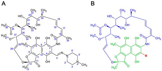
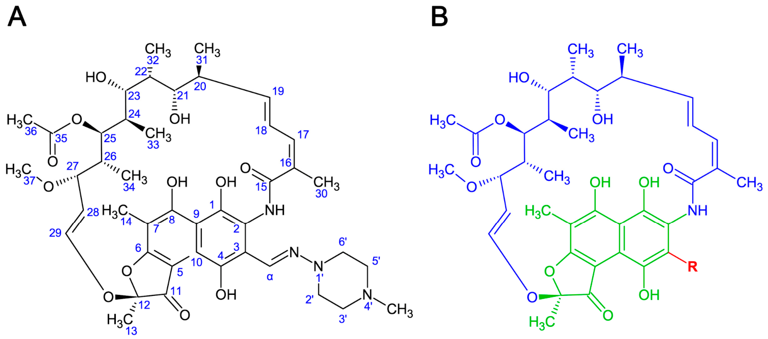
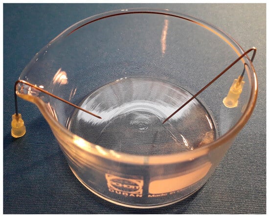
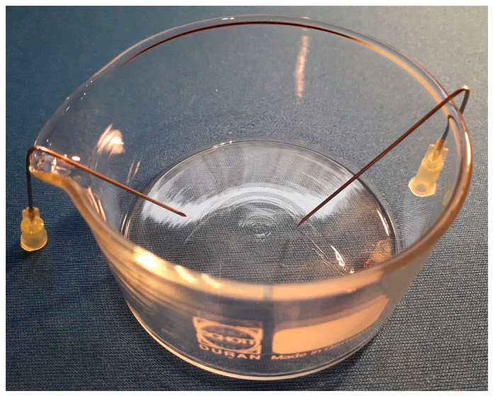
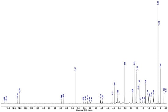
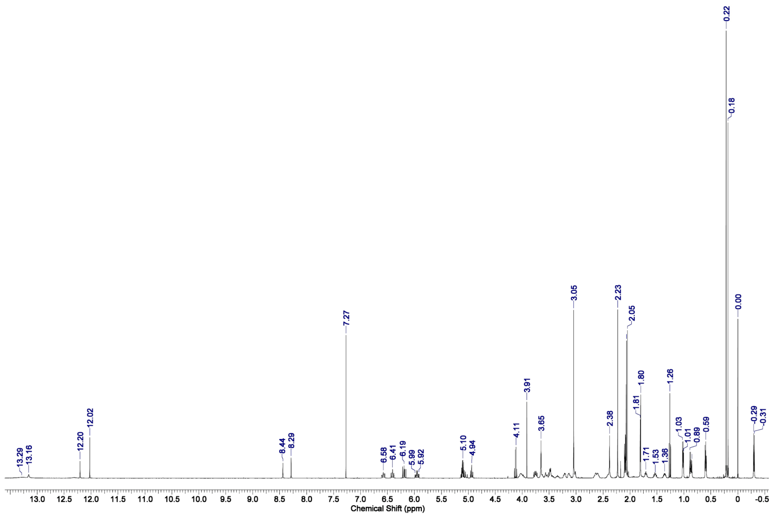
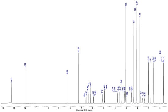
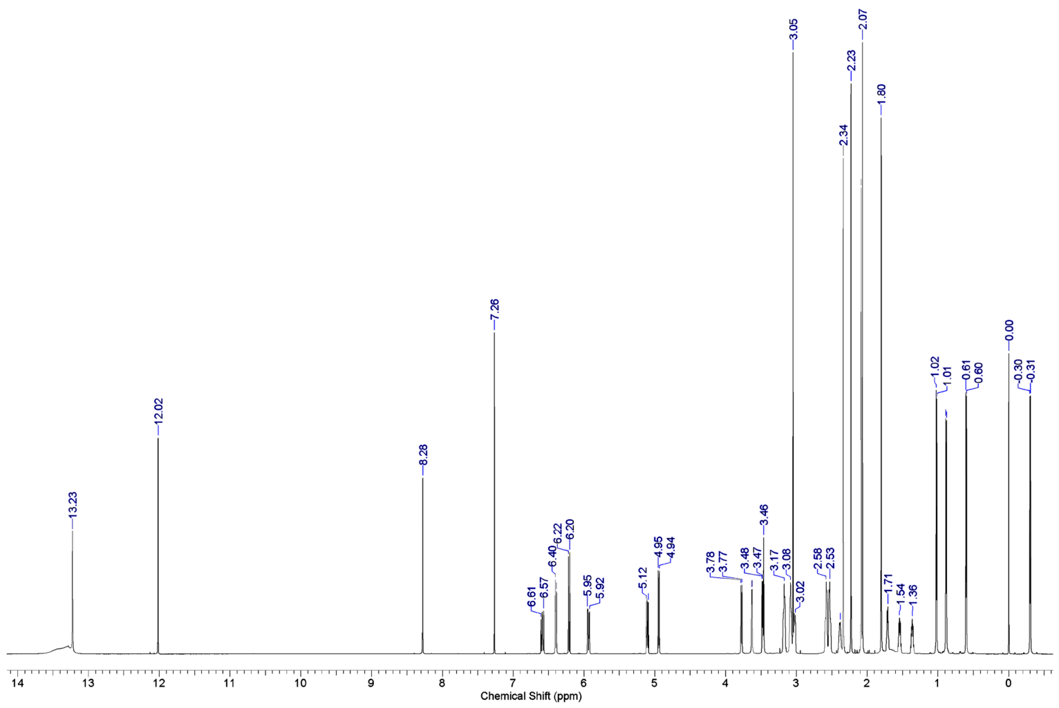
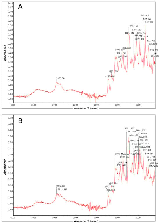
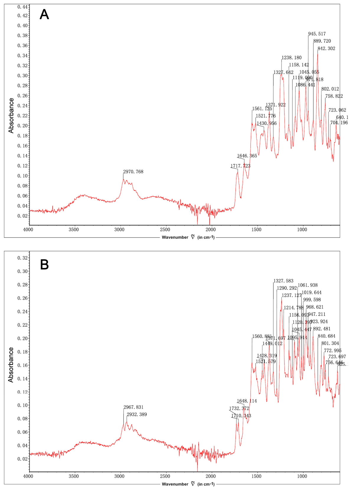
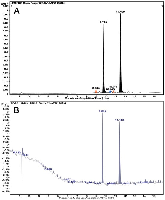
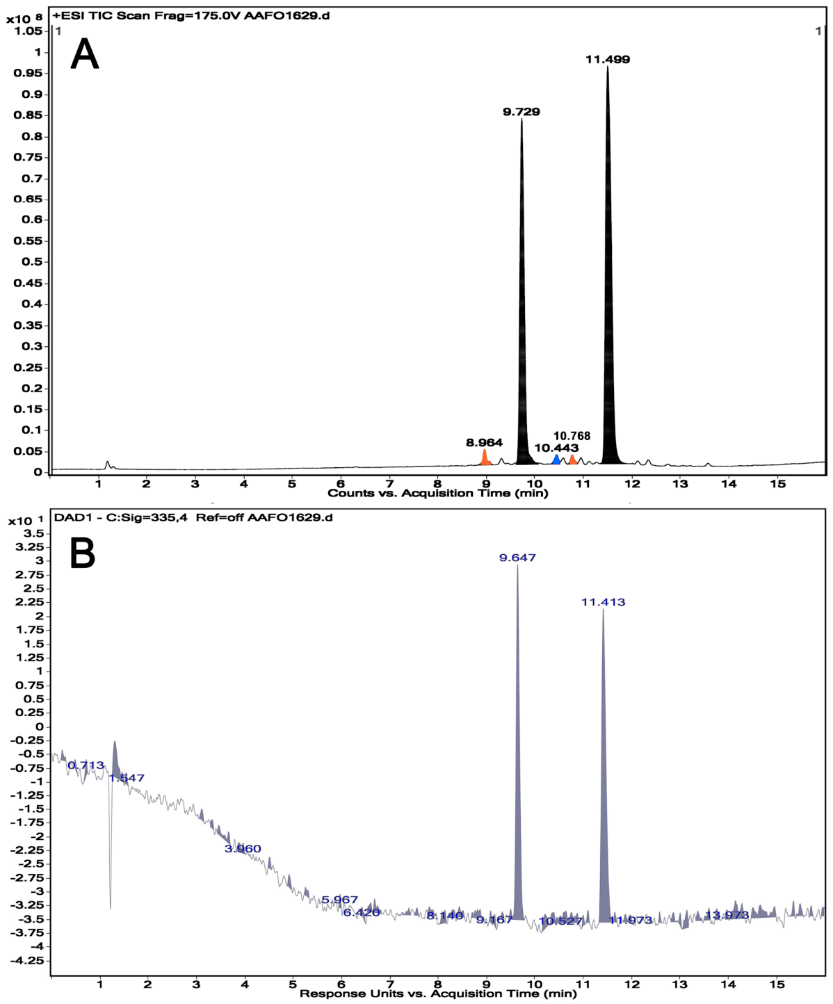
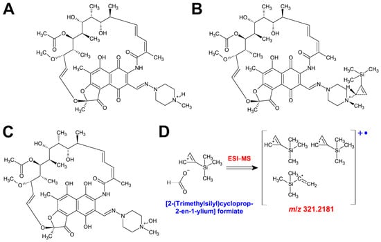
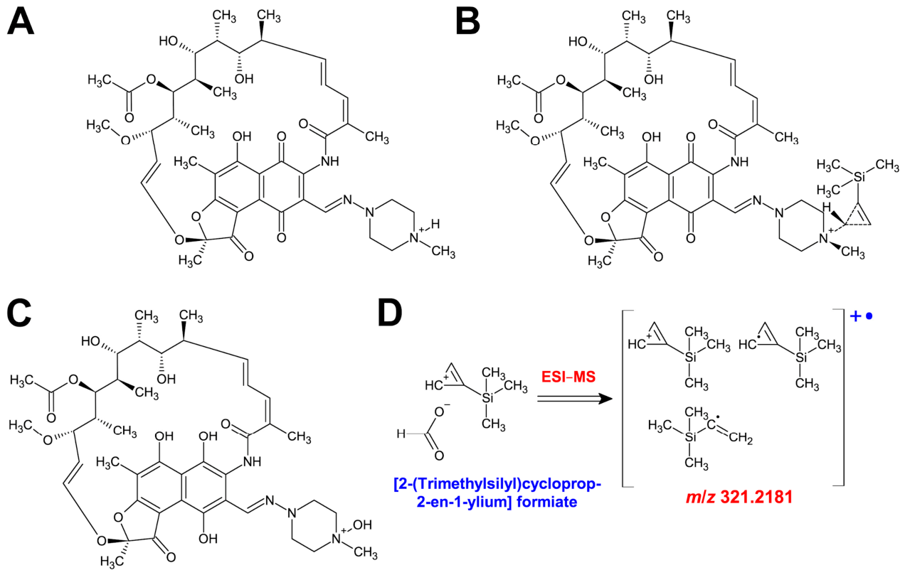
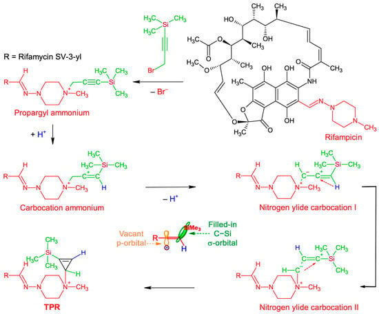
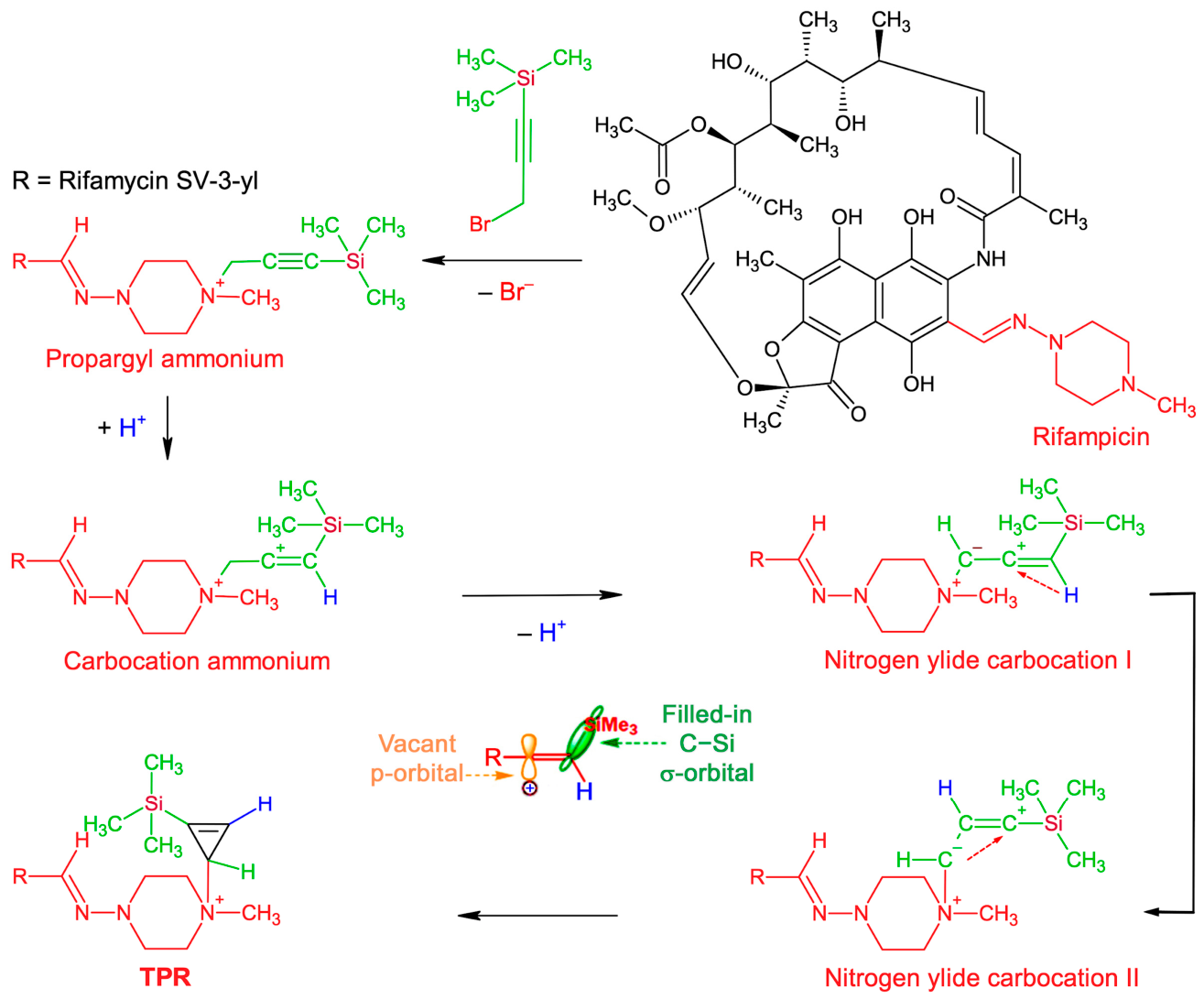
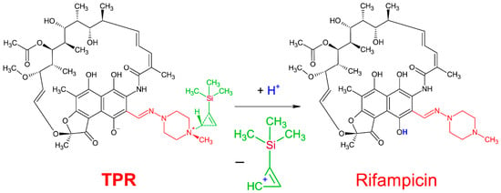
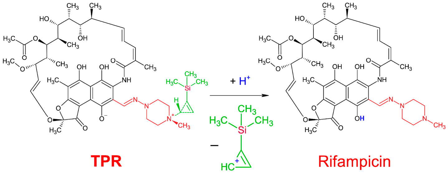
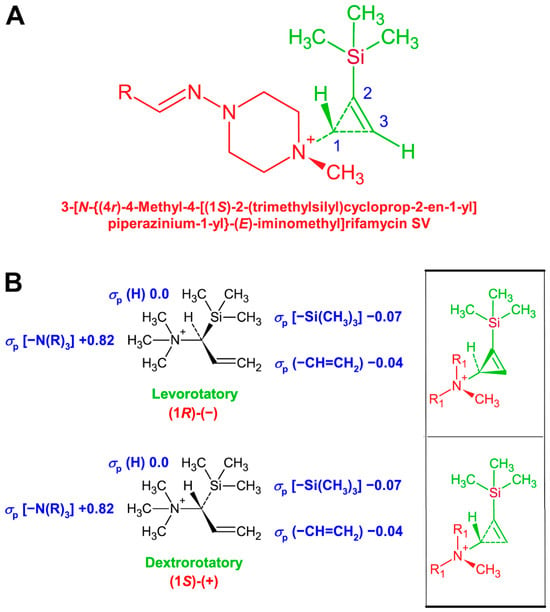
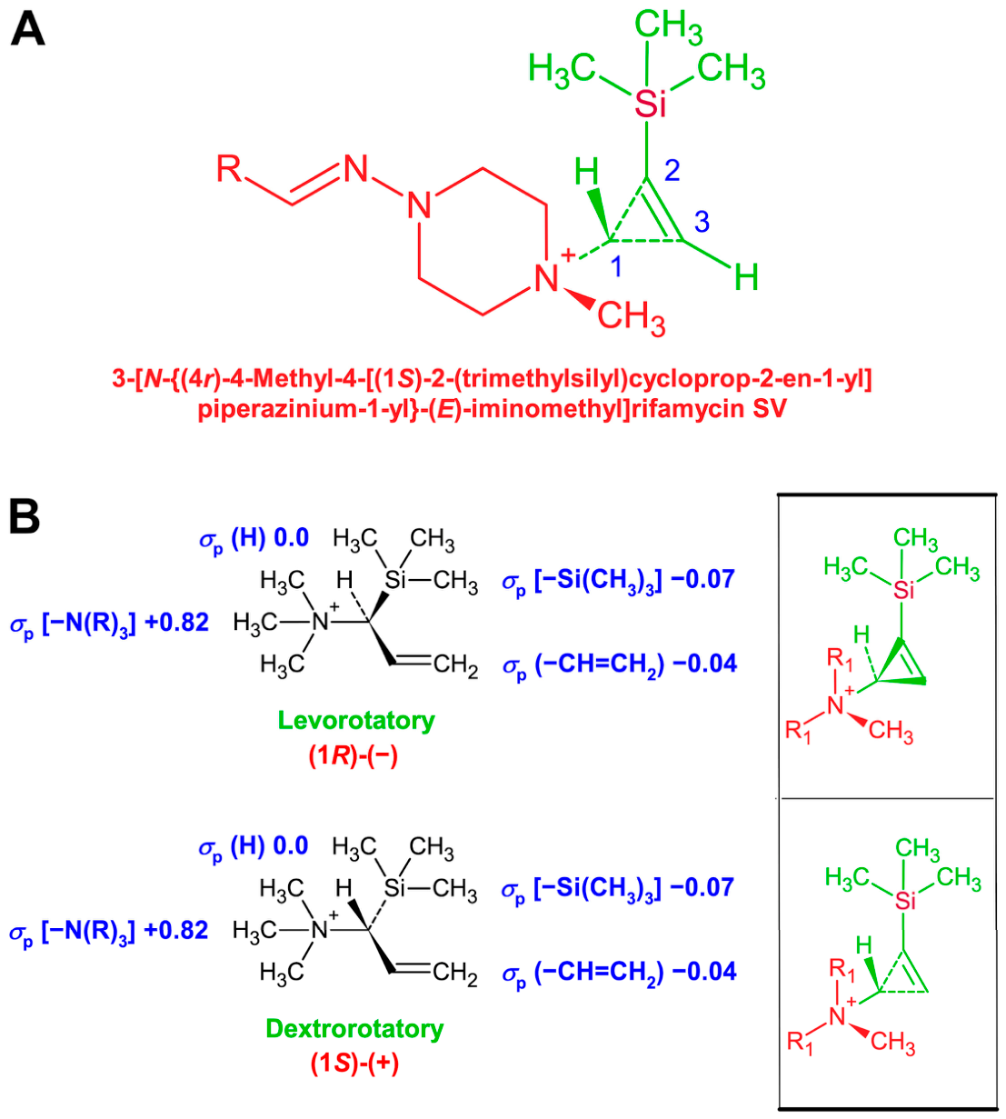
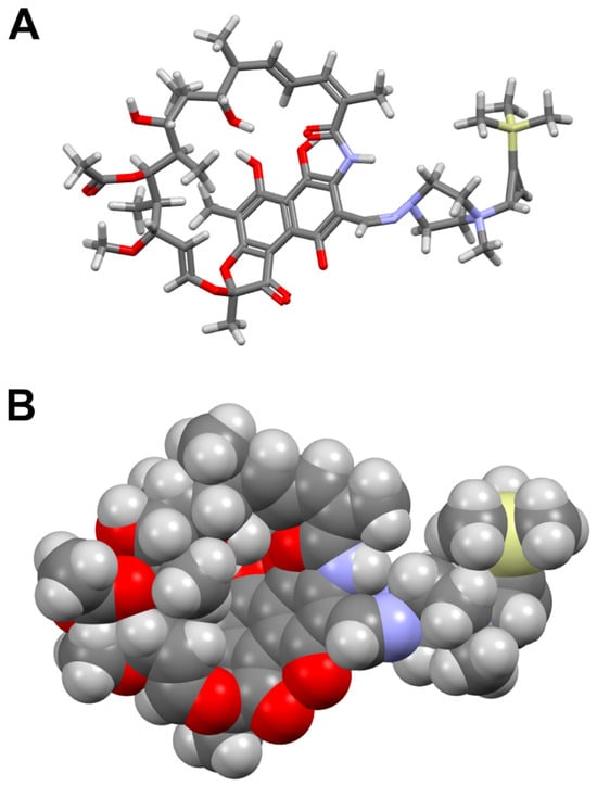
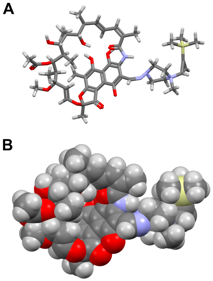
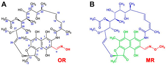
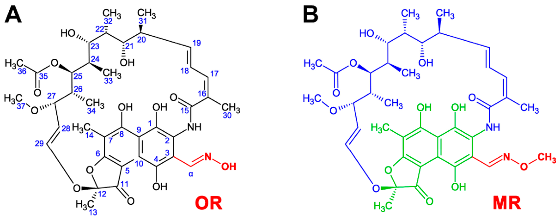
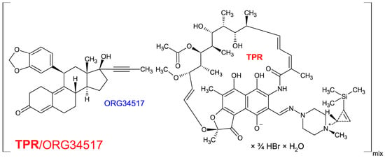
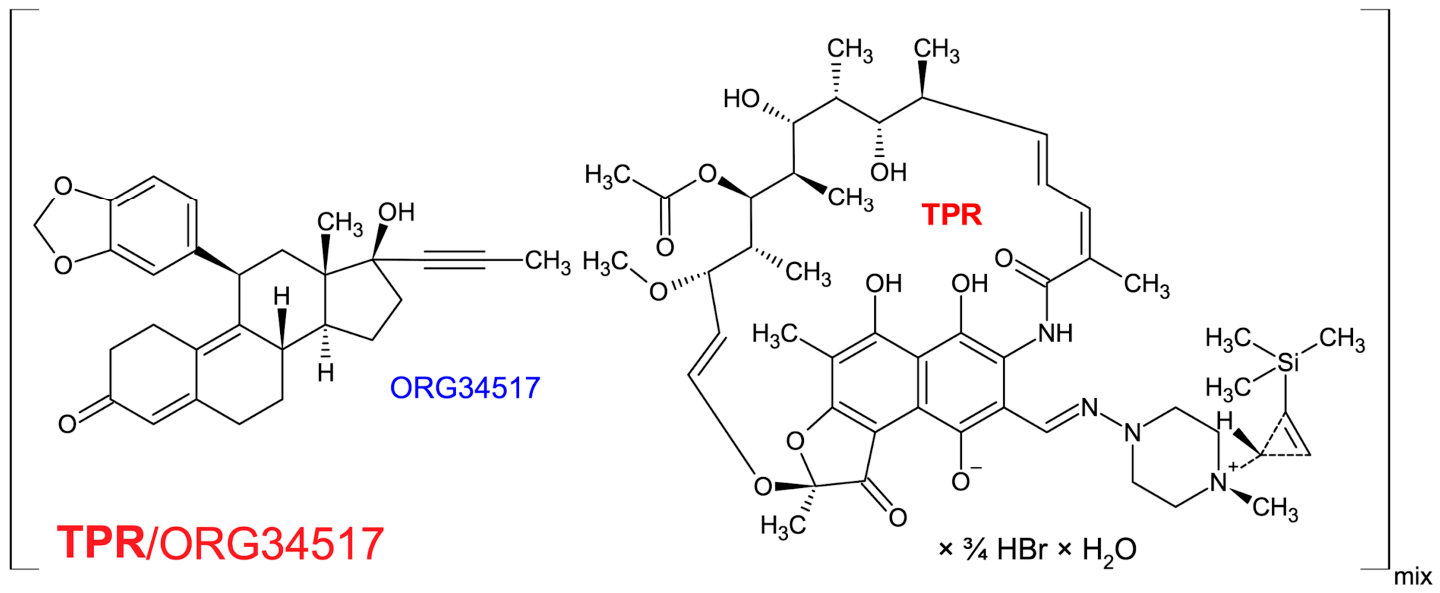
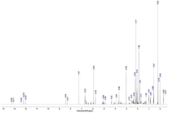
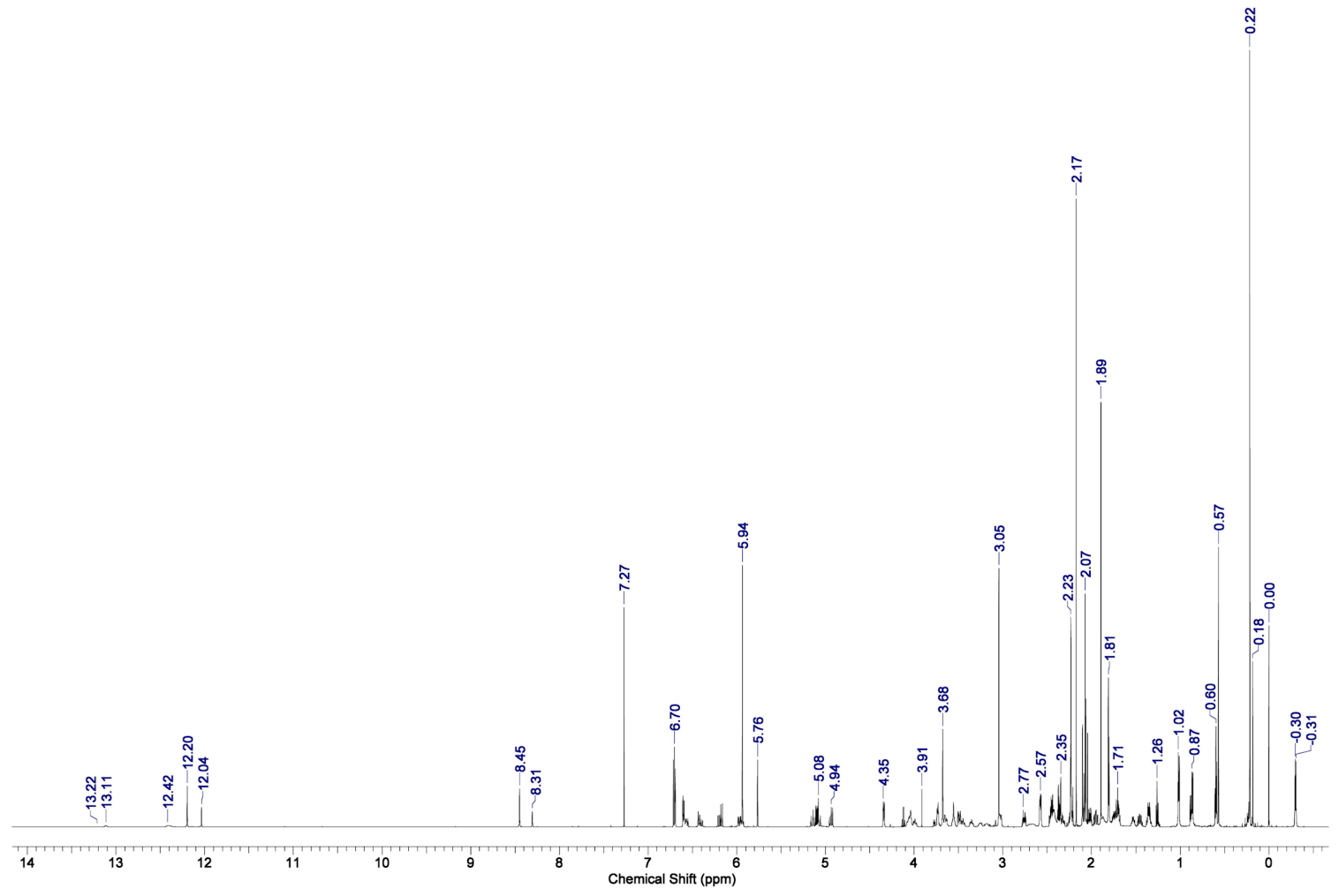
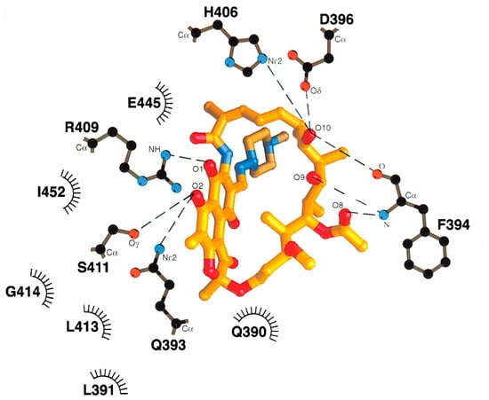
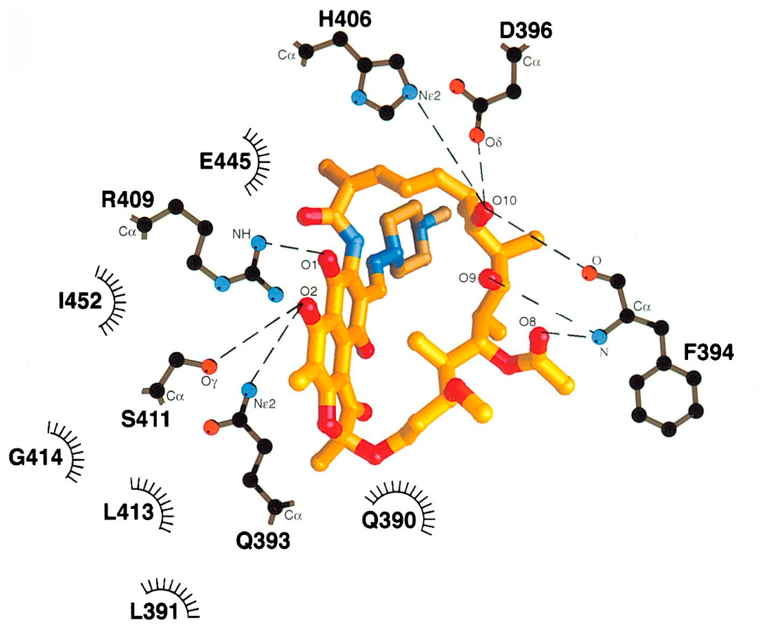
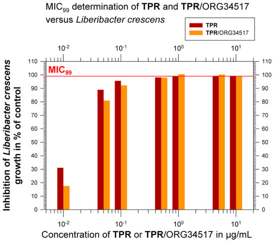
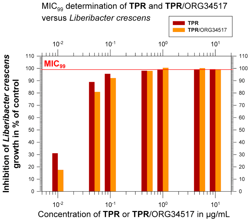
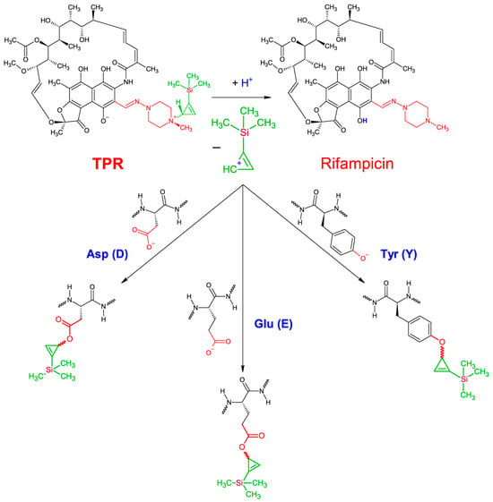
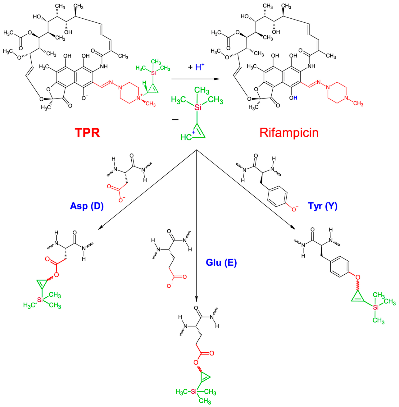

Disclaimer/Publisher’s Note: The statements, opinions and data contained in all publications are solely those of the individual author(s) and contributor(s) and not of MDPI and/or the editor(s). MDPI and/or the editor(s) disclaim responsibility for any injury to people or property resulting from any ideas, methods, instructions or products referred to in the content. |
© 2026 by the authors. Licensee MDPI, Basel, Switzerland. This article is an open access article distributed under the terms and conditions of the Creative Commons Attribution (CC BY) license.