


Abstract
Pinus densiflora extracts contain diverse bioactive components with antioxidant, antimicrobial, and skin-protective properties. A P. densiflora dried extract (PDDE) was prepared by self-steam extraction followed by reverse osmosis (RO) processing to examine its chemical profile, colloidal structure, and preliminary biological properties. Samples collected after two and four RO cycles were labeled PDDE-2C and PDDE-4C, respectively. High-performance liquid chromatography characterized the ultraviolet-detectable profile of PDDE-4C. Acidic pH and delayed thermogravimetric mass loss distinguished the processed extract from water. Liquid chromatography–mass spectrometry revealed signals at nominal m/z 137 with different retention times in the two samples under identical analytical conditions. Small-angle X-ray scattering (SAXS) profiles were described using a core–shell sphere model. PDDE-4C had a fitted core radius of 108.6 ± 4.5 nm and shell thickness of 1.60 ± 0.17 nm, comparable to those of PDDE-2C. Its higher low-q SAXS intensity and fitted scale factor were consistent with colloidal enrichment during RO processing. Small-angle neutron scattering showed differences in the high-q background of the samples. PDDE-4C exhibited a 2,2-diphenyl-1-picrylhydrazyl radical scavenging response corresponding to 86.7 ± 1.7% of the ascorbic acid reference and limited growth inhibition against Cutibacterium acnes at the highest tested concentration, with an 11 mm total inhibition-zone diameter including the 10 mm disc. RAW 264.7 cell viability remained above 96% at concentrations up to 1000 µg/mL. A patch test involving 33 volunteers yielded a primary irritation index of 0.02, classified as non-irritating under the tested conditions. These findings support RO-assisted colloidal enrichment and further evaluation of the processed extract.
1. Introduction
Pinus densiflora is a major coniferous tree species native to East Asia and has long been utilized in traditional medicine, functional foods, and cosmetic formulations [1,2,3]. Various extracts derived from P. densiflora contain biologically active compounds, including terpenes, phenolic compounds, flavonoids, and other secondary metabolites, which have been reported to exhibit antioxidant, antimicrobial, anti-inflammatory, and skin-protective properties [1,2,3,4,5,6]. Owing to these biological activities, pine-derived extracts have attracted increasing attention as renewable natural resources for cosmetic, pharmaceutical, and biomedical applications. To obtain bioactive constituents from plant materials, a variety of extraction techniques have been employed, including solvent extraction, steam distillation, and supercritical fluid extraction [7,8,9]. However, conventional extraction processes often require large volumes of organic solvents or additional purification steps, which may increase process complexity and limit sustainable utilization. Furthermore, plant extracts generally contain chemically diverse components spanning a broad range of molecular sizes and polarities, making it challenging to selectively concentrate desirable constituents while maintaining their biological activity [7,10,11]. Consequently, the development of environmentally friendly processing strategies capable of concentrating bioactive plant-derived components remains an important area of research. Membrane-based separation technologies have emerged as attractive alternatives for the concentration and processing of natural-product extracts [12,13,14]. Among them, reverse osmosis (RO) has been widely used because it enables concentration under relatively mild conditions without the need for thermal evaporation or organic solvents. During RO processing, water preferentially permeates through the membrane, while membrane-retained constituents become progressively concentrated in the retentate [12,15]. As a result, RO processing has been applied to concentrate plant extracts, food products, and biologically derived solutions while preserving sensitive functional components [12,13,14]. Although the concentration performance of RO membranes has been extensively investigated, relatively little attention has been paid to the physicochemical state of the retained organic components after repeated membrane concentration. In particular, it remains unclear whether membrane concentration simply increases the concentration of dissolved constituents or whether retained components become organized into distinct colloidal structures during the enrichment process. Understanding the structural organization of concentrated plant-derived organic components is important because colloidal organization may influence stability, physicochemical behavior, and biological performance [16,17,18]. However, detailed investigations combining chemical characterization, nanoscale structural analysis, and biological evaluation remain limited.
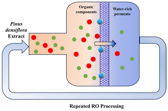
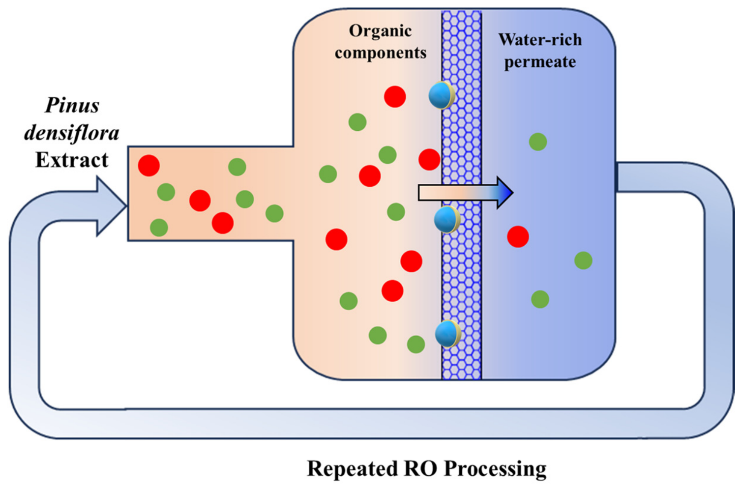
RO offers a potential route for concentrating aqueous plant extracts without an additional thermal evaporation step. However, scale-up would require evaluation of membrane flux, fouling, cleaning requirements, component recovery, and batch-to-batch reproducibility. The energy demand and throughput of the prolonged autoclave extraction step must also be considered. These process variables provide the basis for a subsequent technical and economic assessment of industrial production. In the present study, a P. densiflora dried extract (PDDE) was prepared using a self-steam extraction process followed by repeated RO membrane concentration, and the overall experimental workflow is illustrated in Scheme 1. The objective of this study was to characterize the chemical and colloidal features of PDDE collected after repeated RO processing and to evaluate the preliminary antioxidant activity, antibacterial response, and acute skin irritation potential of the final PDDE sample. The membrane-retained organic components were characterized using high-performance liquid chromatography (HPLC), liquid chromatography–mass spectrometry (LC–MS), pH analysis, thermogravimetric analysis (TGA), small-angle X-ray scattering (SAXS), and small-angle neutron scattering (SANS). In addition, antioxidant activity, antibacterial activity, cytotoxicity, and human skin irritation were evaluated to assess the biological responses and preliminary compatibility of the concentrated extract. This integrated analysis compares the colloidal scattering features of PDDE-2C and PDDE-4C and characterizes the chemical profile and preliminary biological properties of the RO-processed extract. In particular, it examines whether the higher scattering intensity after additional RO cycles is accompanied by a substantial change in the fitted core radius or shell thickness obtained from the core–shell model.
Scheme 1.
Schematics of multi-cycle RO membrane processing for PDDE-2C and PDDE-4C. PDDE-2C and PDDE-4C denote RO-concentrated retentate solutions collected after two and four RO concentration cycles. Arrows indicate the flow paths and repeated RO processing. Colored spheres schematically represent extract components; the colors are illustrative and do not identify specific compounds. The patterned central band represents the RO membrane.
2. Results and Discussion
To investigate the compositional and structural characteristics of RO-processed PDDE, the aqueous solutions collected after the second and fourth RO concentration cycles were designated PDDE-2C and PDDE-4C, respectively. These designations identify successive processing stages rather than isolated fractions or purified compounds. RO was employed as a membrane-assisted concentration step, during which water permeation can increase the abundance of membrane-retained constituents in the retentate. The analysis therefore focused on the chemical features of the processed extracts and on whether further RO processing was accompanied by changes in scattering intensity, core radius, or shell thickness. HPLC, pH measurements, TGA, LC–MS, SAXS, and SANS were used to examine these complementary aspects, followed by biological evaluation of PDDE-4C. This approach connects the physicochemical characteristics of the concentrated extract with its measured biological responses. Two and four RO cycles were selected to compare the chemical profiles and colloidal characteristics of extracts subjected to different extents of membrane processing. This comparison was designed to examine whether additional RO cycles were accompanied by changes in scattering intensity, core radius, and shell thickness. Selection of the cycle number for a particular application would additionally require consideration of processing time, component recovery, and membrane performance. A single cycle would shorten processing time but, for a comparable water recovery per cycle, provide less cumulative water removal. Further cycles may increase retentate concentration, while also increasing processing time and the potential effects of fouling and component loss. These considerations explain the practical trade-off; the two selected stages were used for comparison rather than to establish an optimum cycle number.
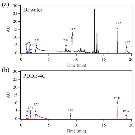
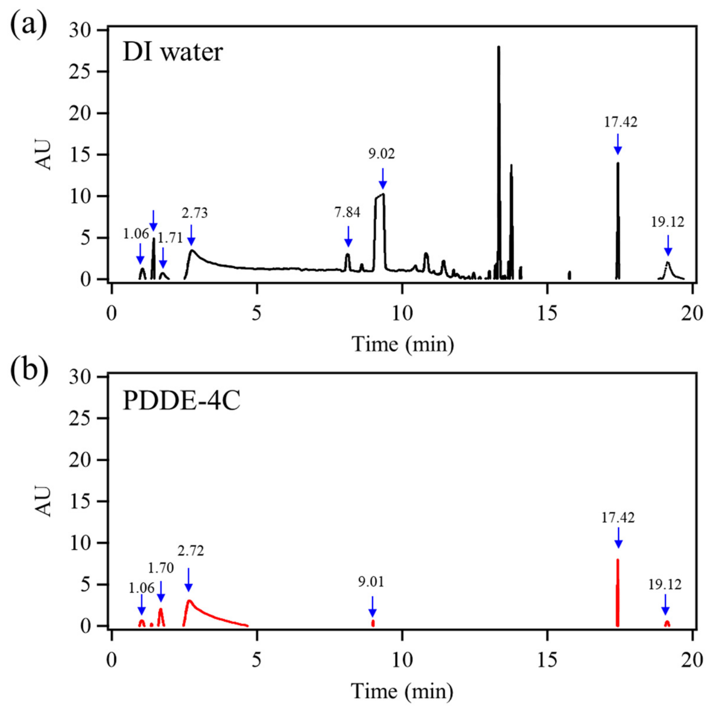
Figure 1 compares the HPLC–diode-array detection chromatograms of deionized (DI) water (Figure 1a) and PDDE-4C obtained after four RO cycles (Figure 1b). DI water, commonly used as an analytical blank, exhibited multiple sharp signals across the chromatographic measurements. The DI water chromatogram provides a reference for considering background contributions associated with the analytical conditions, potentially including the mobile phase, injection system, or carryover. In comparison, PDDE-4C showed fewer prominent sharp peaks, with a broad response near 2.7 min, a dominant peak near 17.4 min, and smaller signals in the early-eluting region and near 19.1 min. Several signals occurred at similar retention times in both traces and were considered when interpreting the ultraviolet (UV)-detectable profile of PDDE-4C. The significance of this relatively simple chromatographic pattern should be considered in relation to the detection principle of HPLC–diode-array analysis. The chromatographic separation distinguishes eluting species according to their retention behavior, while the detector records their absorbance at the selected wavelength. Consequently, the observed peak intensities reflect both the abundance and the UV absorption characteristics of the detected species. Components with different absorption characteristics can produce different responses even at similar concentrations, and weakly absorbing components may contribute little to the recorded chromatogram. The PDDE-4C trace thus describes the UV-detectable signals of the processed solution under the applied analytical conditions. This profile provides a useful starting point for examining how the chemical characteristics of the extract relate to the properties of the aqueous preparation.
Figure 1.
HPLC detection chromatograms of (a) deionized (DI) water and (b) PDDE-4C collected after four RO cycles. Blue arrows mark selected chromatographic signals, and the accompanying numbers indicate retention times in minutes.
To develop this connection, the chromatographic observations were considered together with measurements of the bulk physicochemical properties of the RO-processed solutions. The following pH measurements examine the acidity of the aqueous preparation, while TGA follows its temperature-dependent mass-loss behavior, including the evaporation of water and the subsequent behavior of the remaining material. These complementary measurements extend the characterization from the UV-detectable chromatographic profile to the behavior of the solution as a whole.
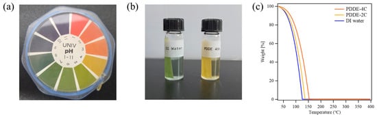
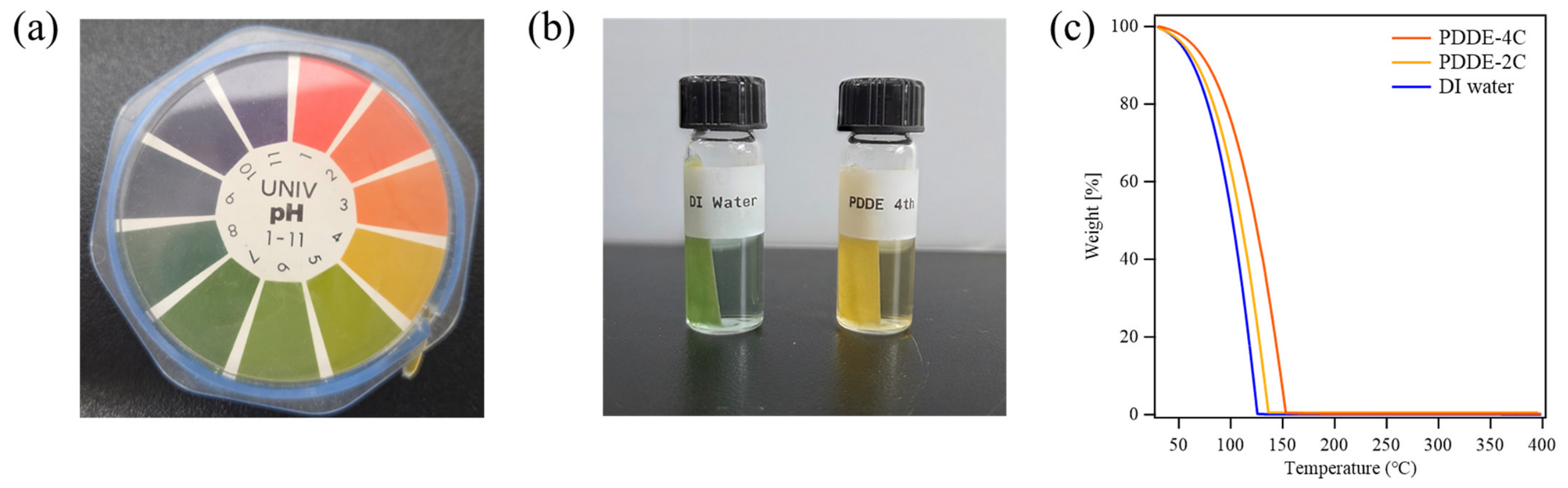
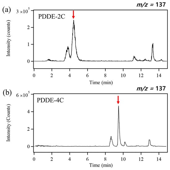
As shown in Figure 2a,b, DI water exhibited a near-neutral pH of 7.0, whereas PDDE-4C showed an acidic pH of 4.0. This difference indicates that the RO-processed extract remained chemically distinct from the DI water reference in addition to the chromatographic observations. TGA provided complementary information on the weight-loss behavior of the aqueous samples (Figure 2c). Here, T95, T50, T10, and T5 denote the temperatures at which 95%, 50%, 10%, and 5% of the initial sample mass remained, respectively. As shown in Figure 2c, DI water showed rapid weight loss during the main evaporation step, with T95, T50, T10, and T5 values of 52.8, 101.7, 121.5, and 123.6 °C, respectively. In contrast, PDDE-2C showed increased T95, T50, T10, and T5 values of 58.3, 112.6, 134.7, and 137.1 °C. Furthermore, PDDE-4C showed delayed weight loss, with corresponding T95, T50, T10, and T5 values of 63.8, 123.5, 147.9, and 150.5 °C, respectively. At 130 °C, DI water almost completely evaporated, retaining only 0.1% of its initial mass, whereas PDDE-4C still retained 40.9%. Even at 150 °C, PDDE-4C retained approximately 6.0% of its initial mass, while DI water exhibited negligible remaining mass. Because the aqueous sample is dominated by water evaporation in the low-temperature region, the TGA profiles were interpreted as solvent-loss behavior rather than direct evidence of thermal decomposition of organic constituents. The delayed weight-loss behavior of PDDE-2C and PDDE-4C is consistent with contributions from extract-derived solutes or colloidal components to the evaporation behavior of the RO-processed solutions. Taken together, the acidic pH and delayed mass loss distinguish the processed extract from the water reference and support the presence of components that modify the properties of the aqueous matrix. These observations complement the chromatographic measurements by probing the bulk behavior of the samples rather than the response of individual UV-active species. LC–MS analysis was subsequently performed to examine the mass-selected chromatographic features of PDDE-2C and PDDE-4C.
Figure 2.
(a) Photograph of a universal pH indicator and (b) pH test results for DI water and PDDE-4C. (c) TGA thermograms of DI water, PDDE-2C and PDDE-4C.
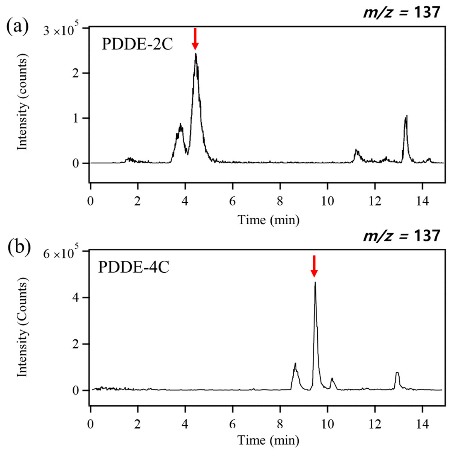
To examine the chemical features of the RO-processed solutions, LC–MS analysis was performed on PDDE-2C and PDDE-4C under identical chromatographic and mass-spectrometric conditions. As shown in Figure 3, both samples exhibited signals in the nominal m/z 137 channel, with dominant peaks at retention times of 4.45 min for PDDE-2C and 9.48 min for PDDE-4C. A nominal m/z of 137 is compatible with protonated monoterpene ions, and monoterpenes have been reported among the volatile constituents of P. densiflora [4,19,20]. This provides a basis for considering a possible monoterpene-related contribution to the observed signals, while retaining the assignment as tentative. The substantial difference in retention time is important because ions sharing a nominal m/z can originate from different compounds, including isomeric species or fragments. Accordingly, the two peaks are treated as distinct chromatographic signals within the same nominal mass channel. Under the common analytical conditions, their different elution times indicate different chromatographic retention behavior among the species contributing to that channel. In reversed-phase chromatography, later elution generally reflects stronger retention under the applied mobile-phase conditions [21]. The comparison therefore reveals differences in the mass-selected profiles at the two RO processing stages and motivates consideration of changes in the relative contributions of retained extract components. This chemical-profile comparison complements the pH and TGA observations. SAXS and SANS were subsequently used to examine the colloidal scattering and aqueous-matrix characteristics of the processed extracts, providing a structural context for the chemical and biological characterization.
Figure 3.
LC–MS extracted ion chromatograms at nominal m/z 137 for (a) PDDE-2C and (b) PDDE-4C. Red arrows indicate the dominant peaks at retention times of 4.45 min for PDDE-2C and 9.48 min for PDDE-4C.
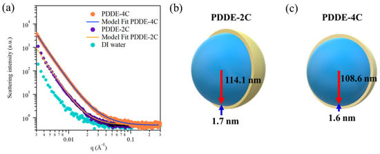
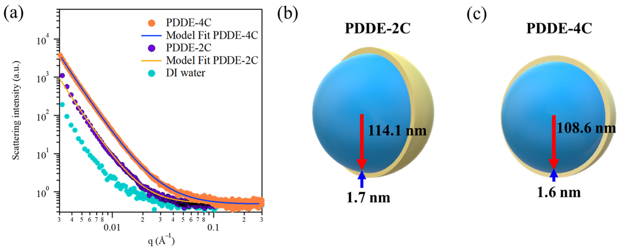
To investigate the nanoscale structural organization of the RO-processed solutions, SAXS measurements were performed on PDDE-2C and PDDE-4C. The measured SAXS intensities and fitted curves are shown in Figure 4a. Both SAXS intensities were analyzed using the same core–shell sphere model [22,23,24]. The fitted core radius and shell thickness were 114.1 ± 5.4 nm and 1.71 ± 0.11 nm for PDDE-2C, compared with 108.6 ± 4.5 nm and 1.60 ± 0.17 nm for PDDE-4C. These comparable values support a similar core–shell description of the colloidal assemblies at the two processing stages. During fitting, the solvent scattering length density (SLD) was fixed at the value for water, 9.41 × 10⁻⁶ Å−2, while the core and shell SLDs were allowed to vary. The fitted core SLDs were 9.38 × 10⁻⁶ Å−2 for PDDE-2C and 9.36 × 10⁻⁶ Å−2 for PDDE-4C. The corresponding shell SLDs were 11.7 × 10⁻⁶ and 11.8 × 10⁻⁶ Å−2. Thus, the fits returned comparable core and shell SLDs in addition to comparable core radii and shell thicknesses. In both samples, the core SLD was close to that of water, whereas the shell SLD was higher. Within the applied core–shell model, the fitted SLD distribution describes a core with near-solvent contrast and a shell with higher contrast.
Figure 4.
SAXS measurements. (a) Measured SAXS intensities and fitted curves for PDDE-2C and PDDE-4C, together with the DI water reference. Schematic representations of the fitted core–shell structures of (b) PDDE-2C and (c) PDDE-4C. In (a), marks represent measured SAXS intensities and solid lines represent the model fits.
PDDE-4C exhibited higher scattering intensity in the low-q region, accompanied by an increase in the fitted scale factor from 0.1381 for PDDE-2C to 0.5864 for PDDE-4C. The scale factor controls the magnitude of the modeled scattering contribution and is interpreted together with the SLD contrasts and structural parameters because these quantities jointly determine the calculated intensity [22,24]. The agreement between the experimental SAXS intensities and fitted curves was assessed using χ2/N, where N is the number of fitted data points. The fit for PDDE-2C yielded a χ2 value of 1377 for 1277 data points, corresponding to χ2/N = 1.08. The corresponding fit for PDDE-4C yielded a χ2 value of 5219 for 4048 data points, corresponding to χ2/N = 1.29. Taken together, the two samples exhibited comparable fitted core radii, shell thicknesses, and core and shell SLDs, while PDDE-4C showed higher low-q SAXS intensity and a larger fitted scale factor. Within the applied core–shell model, this combined comparison supports an interpretation of RO-assisted enrichment of similarly sized colloidal assemblies. The scale factor is interpreted alongside the fitted SLD contrasts and structural parameters, rather than being converted directly into a particle concentration. Water permeation during RO processing provides a physical basis for enrichment of membrane-retained components in the aqueous retentate. SANS measurements were subsequently performed to examine the neutron-scattering characteristics of the processed aqueous samples.
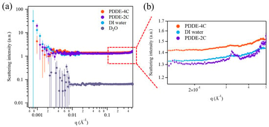
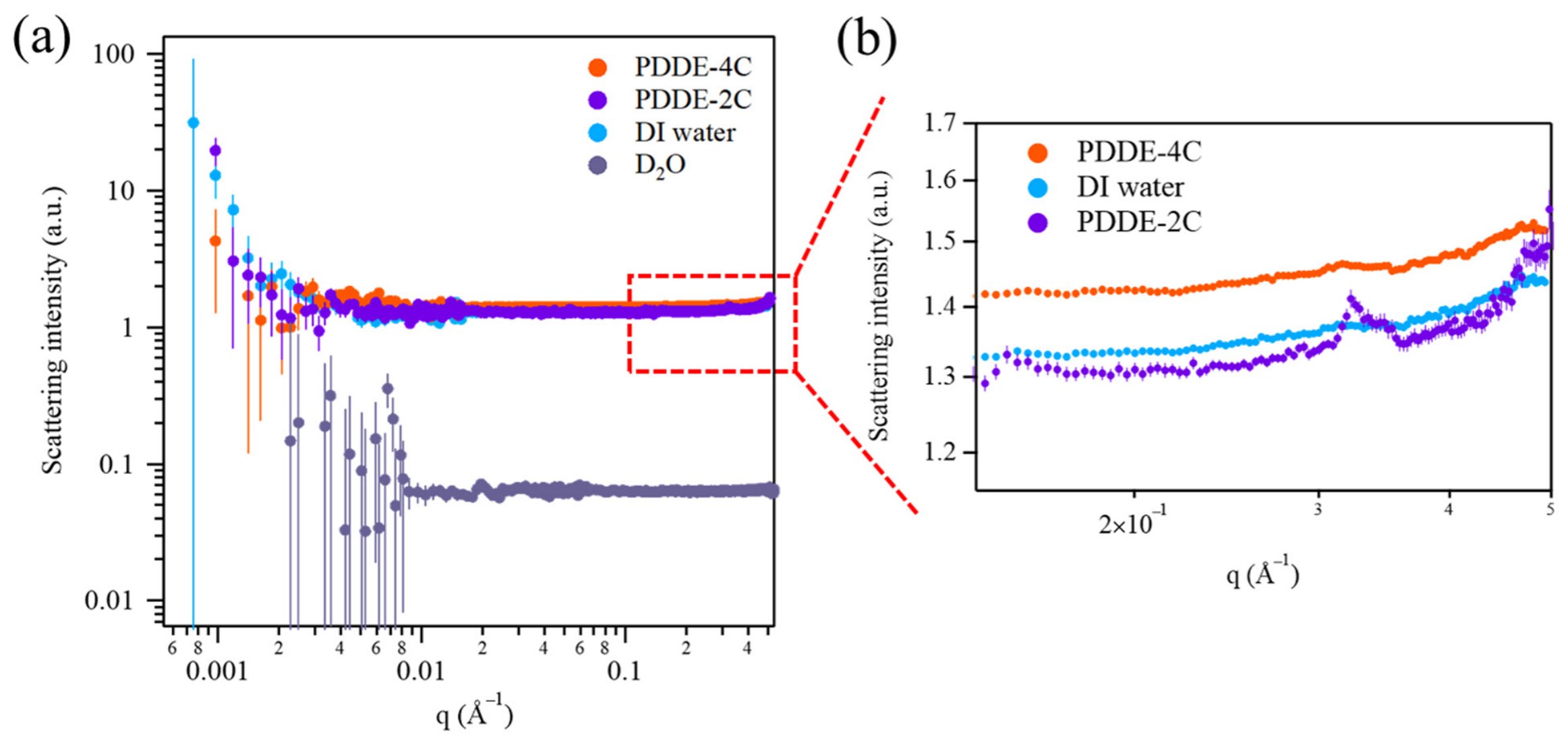
To further examine the aqueous-matrix characteristics of the RO-concentrated solutions, SANS measurements were performed on D2O, DI water, PDDE-2C, and PDDE-4C (Figure 5a). Neutron scattering provides contrast complementary to SAXS, and the large incoherent scattering cross section of hydrogen makes hydrogen-containing media important contributors to the measured background [25,26,27]. As shown in Figure 5b, PDDE-4C exhibited an elevated high-q background compared with DI water and PDDE-2C. D2O showed a substantially lower background (Figure 5a). The contrast between the water references illustrates the sensitivity of the background to isotopic composition. In the extracts, both water and hydrogen-containing organic constituents contribute to this response. Repeated RO processing can change the relative amounts of solvent and retained components, and these changes can influence the background of the resulting aqueous mixture. Sample thickness, neutron transmission, and inelastic or multiple scattering also affect the measured background and must be considered in comparisons between aqueous samples [28]. The elevated background of PDDE-4C is therefore interpreted as a change in the hydrogen-containing sample matrix following additional RO processing, with retained organic components providing one possible contribution. This observation complements the higher low-q SAXS intensity and the mass-selected signals detected by LC–MS. Together, the measurements characterize changes in colloidal scattering and aqueous-matrix properties at the two RO stages, with the principal evidence for colloidal enrichment arising from the SAXS intensity and model comparison.
Figure 5.
SANS measurements of DI water, D2O, PDDE-2C, and PDDE-4C. (a) Scattering profiles over the measured q range. (b) Magnified high-q region showing the higher background of PDDE-4C relative to DI water and PDDE-2C.
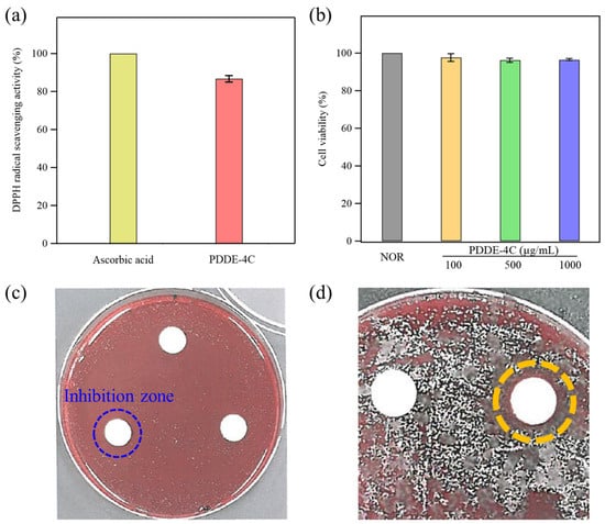
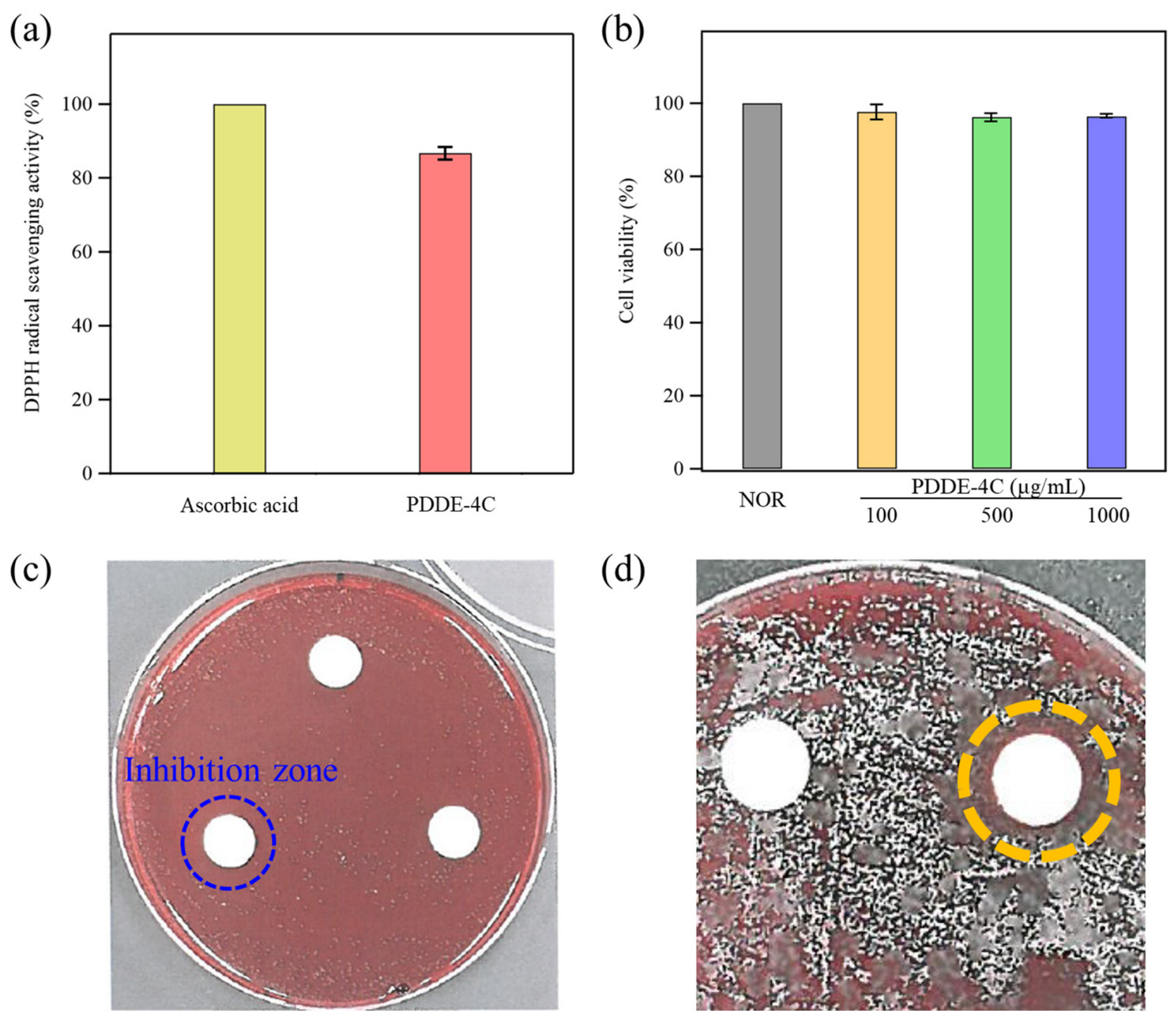
As shown in Figure 6a, PDDE-4C exhibited a 2,2-diphenyl-1-picrylhydrazyl (DPPH) radical scavenging response corresponding to 86.7 ± 1.7% of the ascorbic acid reference under the tested conditions [29,30,31]. This response demonstrates that the final RO-processed extract contained constituents capable of scavenging DPPH radicals. The biological result adds functional information to the chemical and colloidal characterization by showing radical scavenging activity in the processed aqueous preparation. Plant extracts can contain several classes of antioxidant compounds, and their combined contributions may account for the measured response. The present result is therefore discussed at the extract level, alongside the chromatographic features and colloidal assemblies characterized above. DPPH was selected to provide a direct chemical screening response against the ascorbic acid reference.
Figure 6.
(a) DPPH scavenging response of PDDE-4C relative to the ascorbic acid reference, set to 100%. (b) Cell viability of untreated cells (NOR) and cells treated with PDDE-4C at 100, 500, and 1000 µg/mL. (c) Disc diffusion test: 10 µg/mL (top), 100 µg/mL (lower right), and 1000 µg/mL (lower left). (d) Comparison plate of hot-water pine extract (left) and PDDE-4C (right). Discs are 10 mm in diameter, and the medium contains 5% sheep blood. The 11 mm inhibition zone is the total diameter including the disc. The dashed circles indicate the regions of interest rather than the measured boundaries of the inhibition zones.
The activity of the final preparation should also be considered in relation to the extraction conditions. Prolonged treatment at 121 °C may alter heat-sensitive constituents, and the composition of the collected extract may reflect both extraction and thermal transformation. The observed DPPH response demonstrates radical-scavenging activity in the preparation obtained after self-steam extraction and RO processing. This result characterizes the processed extract rather than the thermal stability of individual native compounds.
The cytotoxicity of PDDE-4C was evaluated in RAW 264.7 cells at 100, 500, and 1000 µg/mL. RAW 264.7 murine macrophages were used for the initial cellular screening of PDDE-4C. Macrophage-based studies have also been used to investigate biological responses to pine extracts [2,3]. Relative cell viability was 97.7 ± 2.1%, 96.2 ± 1.2%, and 96.4 ± 0.6%, respectively (Figure 6b). These values indicate high cell viability, as assessed by the 3-(4,5-dimethylthiazol-2-yl)-5-(3-carboxymethoxyphenyl)-2-(4-sulfophenyl)-2H-tetrazolium (MTS) assay, under the tested exposure conditions. Together, the DPPH and cell viability results show a radical-scavenging response and high cell viability in their respective assays. The cell viability assay provided an initial assessment of the effects of PDDE-4C on RAW 264.7 cells. The human patch test evaluated primary skin irritation following topical application.
The antibacterial response of PDDE-4C was evaluated against Cutibacterium acnes using a disc diffusion assay. At the highest concentration of 1000 µg/mL, the total inhibition zone diameter was 11 mm, including the 10 mm paper disc (Figure 6c). This corresponds to an approximately 0.5 mm radial extension beyond the disc edge and is interpreted as limited local growth inhibition under the applied conditions. Growth inhibition was observed at 1000 µg/mL, whereas no measurable inhibition zone was observed at 10 or 100 µg/mL. The images therefore support a qualitative screening observation at the highest tested concentration. Although antimicrobial activity has been reported for plant-derived preparations [4,5,32], the present extract-level observation is not assigned to a specific compound. In the comparison plate, the conventional hot-water pine extract showed no clear inhibition zone, whereas PDDE-4C showed a total inhibition zone diameter of approximately 11 mm, including the 10 mm paper disc (Figure 6d). This comparison concerns two differently prepared extracts and provides preliminary context for their assay responses.
The primary skin irritation potential of PDDE-4C was evaluated by a human patch test involving 33 healthy volunteers. The human primary skin irritation results are summarized in Table 1. Because concentrated plant extracts may contain acidic, phenolic, or terpene-related components that can induce irritation when present as free-form molecules, this test was performed to determine whether the RO-concentrated sample caused acute skin responses under controlled topical exposure conditions [33,34,35]. PDDE-4C was applied to the upper back under occlusive conditions for 24 h, and skin reactions were assessed at 30 min and 24 h after patch removal. Erythema and edema were scored according to the applied clinical evaluation criteria. Among the 33 volunteers, one participant showed erythema at 30 min after patch removal, but this response was not classified as clinically significant according to the scoring criteria. No persistent irritation response was observed at the 24 h evaluation point. As a result, PDDE-4C yielded a primary irritation index of 0.02 and was classified as non-irritant under the tested human patch-test conditions (Supporting Information). These findings indicate a low primary skin irritation response to PDDE-4C under the tested patch test conditions. The primary skin irritation test showed minimal acute irritation following application of PDDE-4C in the enrolled cohort. This observation, together with the DPPH response and high macrophage viability, provides complementary information on the final processed preparation. The results support further evaluation of PDDE-4C for topical formulations, including studies of skin-cell responses and repeated exposure. The observed compatibility is discussed for the tested preparation and exposure conditions, without attributing it to a protective effect of the colloidal structure.
Table 1.
Summary of the human primary skin irritation test for PDDE-4C (detailed clinical information, grading criteria, participant demographics, and individual skin reaction scores are provided in the Supporting Information).
3. Materials and Methods
3.1. Preparation of PDDE
The PDDE was prepared from P. densiflora stems using an autoclave-assisted self-steam extraction process based on endogenous moisture. This process was conducted without the addition of external water or organic solvents, and extraction was achieved solely using the intrinsic moisture naturally present within the tissues. The outer bark was removed to minimize the introduction of unnecessary impurities, and the debarked stems were cut into pieces 3–10 cm in length. The pretreated stems were densely loaded into a heat-resistant container and placed inside an autoclave. The extraction process consisted of two thermal treatment steps. First, the stems were pretreated at 80 °C for 30 min to soften the plant tissue and facilitate internal moisture migration. Subsequently, the main extraction was performed at 121 °C for 24 h under high-temperature and pressurized autoclave conditions. During this process, endogenous moisture in the tissue generated a pressurized steam environment, facilitating the release of volatile and water-soluble extractable components. After extraction, the autoclave was naturally depressurized and sufficiently cooled at room temperature. The condensed liquid extract generated inside the chamber was collected and filtered through qualitative filter paper to remove solid residues.
3.2. RO Membrane Processing of PDDE
The PDDE obtained from the self-steam extraction process was further processed using an RO membrane system equipped with a ULP-2012-100 membrane element (Vontron Technology, Guiyang, China). Prior to RO processing, the extract was filtered through qualitative filter paper to remove residual particulates. The system was operated at a pressure of 220 psi (approximately 1.52 MPa), a feed flow rate of 250 mL/min, and a temperature of 25 °C. The RO-processed retentate samples collected after the second and fourth processing cycles were designated PDDE-2C and PDDE-4C, respectively.
3.3. Chemical and Thermal Analysis
HPLC chromatograms of PDDE-4C and the DI water reference were acquired using a photodiode-array detector on a liquid chromatography system coupled to a Xevo G2-XS mass spectrometer (Waters, Milford, MA, USA) at the Central Research Facility, Chungbuk National University, Republic of Korea. The chromatograms were examined to characterize the UV-detectable profile of the processed extract alongside the water reference.
LC–MS was performed to examine mass-detectable components in PDDE-2C and PDDE-4C using an UltiMate 3000 system coupled to an ISQ EC single-quadrupole mass spectrometer (Thermo Fisher Scientific, San Jose, CA, USA) at the Central Analysis Center, KAIST, Republic of Korea. Both samples were analyzed under identical chromatographic and mass-spectrometric conditions. Extracted ion chromatograms at nominal m/z 137 were used to compare the profiles of the detected features at their respective retention times.
TGA was performed using a Discovery TGA instrument (TA Instruments, New Castle, DE, USA) at the Central Research Facility, Chungbuk National University, Republic of Korea. DI water, PDDE-2C, and PDDE-4C were heated from 25 °C to 400 °C at a ramp rate of 10 °C/min under a nitrogen atmosphere.
3.4. Colloidal Structural Analysis
SAXS measurements were performed to examine the colloidal structure of PDDE-2C and PDDE-4C using a NANOPIX SAXS instrument (Rigaku, Akishima, Japan) at HANARO, Korea Atomic Energy Research Institute (KAERI), Republic of Korea. Sample-to-detector distances of 300 and 1300 mm covered a measured q range of 0.003–0.3 Å−1. An empty sample holder was measured for background correction, and silver behenate (AgBE) was used for sample-to-detector distance calibration. SAXS and SANS data analysis, including SAXS model fitting, was performed using HANARO SANS Macro (version 2.1) in Igor Pro (version 8.0.4.2; WaveMetrics, Lake Oswego, OR, USA). The scattering profiles were analyzed using a core–shell sphere model [22,23,24]. The solvent SLD was fixed at 9.41 × 10⁻⁶ Å−2 for H2O. The core radius, shell thickness, core and shell SLDs, scale factor, and background were allowed to vary during fitting. PDDE-2C was fitted first, and its fitted core radius and shell thickness were used as initial estimates for fitting PDDE-4C. These parameters were subsequently refined against the PDDE-4C scattering profile.
Fit quality was reported as χ2/N, where χ2 is the statistic returned by the fitting software and N is the number of data points included in the fit. The PDDE-2C and PDDE-4C fits included 1277 and 4048 data points, respectively. The fitted core radii, shell thicknesses, and SLDs were compared alongside the measured scattering intensities and fitted scale factors to examine changes associated with additional RO processing.
SANS measurements were performed to examine the scattering profiles and hydrogen-related background contributions of the RO-processed aqueous samples. Measurements were conducted using the 40 m SANS instrument at HANARO, KAERI, Republic of Korea, with a neutron wavelength of 7.49 Å. The measured q range was 0.0006–0.5 Å−1, where q = (4π/λ) sin(θ/2), λ is the neutron wavelength, and θ is the scattering angle. The data were corrected for empty-cell scattering, background, and detector sensitivity. PDDE-2C and PDDE-4C were compared with DI water and D2O to examine the high-q background in the context of the strong incoherent scattering contribution from hydrogen-containing media. The SAXS and SANS measurements were interpreted together to distinguish colloidal scattering features from changes in the background of the aqueous matrix.
3.5. DPPH Radical Scavenging Assay
The DPPH assay was used as an initial chemical screen of the radical scavenging response of PDDE-4C. PDDE-4C and the ascorbic acid reference (Sigma-Aldrich, St. Louis, MO, USA) were prepared at the same nominal concentration of 10,000 µg/mL. A 60 µL aliquot of each solution was mixed with 120 µL of 0.45 mM DPPH solution (Sigma-Aldrich, St. Louis, MO, USA), and absorbance was measured at 517 nm using a microplate reader (Tecan, Männedorf, Switzerland). For these mixing volumes, the calculated final test-material and DPPH concentrations are approximately 3333 µg/mL and 0.30 mM, respectively. For each test material, DPPH scavenging was calculated as scavenging (%) = [1 − As/A0] × 100, where As and A0 are the absorbances at 517 nm of the DPPH reaction mixtures with and without the test material, respectively. The scavenging response of PDDE-4C was then expressed relative to that of the ascorbic acid reference, set to 100%. Measurements were performed in triplicate (n = 3), and results are presented as mean ± standard deviation.
3.6. Antibacterial Activity Test
Antibacterial activity was evaluated against C. acnes (ATCC 6919; ATCC, Manassas, VA, USA) using a disc diffusion assay. The organism was cultured in tryptone soya broth (TSB; Oxoid, Basingstoke, UK) under anaerobic conditions at 37 °C using a gas pack and an anaerobic indicator. The medium was sterilized at 121 °C for 15 min. Cells were harvested by centrifugation at 13,000 rpm for 2 min and washed twice with phosphate-buffered saline (PBS; Sigma-Aldrich, St. Louis, MO, USA). A bacterial suspension of 2.43 × 10⁴ CFU/mL was incorporated into tryptone soya agar (TSA; Oxoid, Basingstoke, UK) supplemented with 5% sheep blood (Synergy Innovation Co., Ltd., Seongnam, Republic of Korea). Extract solutions were passed through a 0.2 µm syringe filter (Advantec, Tokyo, Japan). Each 10 mm paper disc was loaded with 0.1 mL of extract solution at a concentration of 10, 100, or 1000 µg/mL and placed on the agar. Plates were incubated anaerobically at 37 °C. Inhibition-zone diameters include the paper disc. A separate comparison plate contained hot-water-extracted and processed pine samples; other plant extracts on the same plate were separate test materials. The disc-diffusion results are presented as preliminary descriptive observations of local growth inhibition under the specified conditions. They are not used to establish comparative antibacterial potency or an RO-specific enhancement of antibacterial activity.
3.7. Cytotoxicity Assay (MTS Assay)
The cytotoxicity of PDDE-4C was assessed using the MTS assay on RAW 264.7 murine macrophage cells (Korean Cell Line Bank, Seoul, Republic of Korea). Cells were cultured in Dulbecco’s Modified Eagle’s Medium (HyClone, Logan, UT, USA) supplemented with 10% fetal bovine serum (FBS), 100 units/mL penicillin, and 100 µg/mL streptomycin in a humidified incubator at 37 °C with 5% CO2. All cell-culture media and supplements were purchased from HyClone (Logan, UT, USA). For the assay, cells were seeded in 96-well plates (3.0 × 10⁴ cells/well) and stabilized for 1 h. The cells were then treated with PDDE-4C at 100, 500, or 1000 µg/mL for 1 h and subsequently incubated for an additional 24 h without replacing the medium. PDDE-4C remained in the culture medium at the corresponding test concentration throughout this period. Cell viability was determined using the CellTiter 96 AQueous One Solution Cell Proliferation Assay kit (Promega, Madison, WI, USA), which utilizes the reduction of the MTS tetrazolium compound to a colored formazan product through the reducing activity of metabolically active cells [36]. After adding the MTS reagent and incubating for 2 h at 37 °C, the absorbance was measured at 490 nm using an Infinite M200 PRO microplate reader (Tecan, Männedorf, Switzerland). Untreated cells cultured under the same conditions served as the control, and their viability was set to 100%. The MTS assay was performed in three independent experiments (n = 3). Cell viability results are presented as mean ± standard deviation.
3.8. Human Primary Skin Irritation Test
Primary skin irritation was evaluated using a human skin patch test involving 33 healthy adult volunteers (31 women and 2 men) with no history of skin disease [37]. Female volunteers were recruited in larger numbers in view of the intended use of the test material as a skin-care cosmetic ingredient. The study protocol was approved by the Institutional Review Board (IRB) and conducted in accordance with the Declaration of Helsinki and the Ministry of Food and Drug Safety (MFDS) guidelines. A 20 µL dose of PDDE-4C was applied to the upper back of each participant using an IQ Ultimate™ patch (Chemotechnique MB Diagnostics AB, Vellinge, Sweden) for 24 h. The test assessed the primary skin response to the sample; no separate negative-control patch was included. After patch removal, skin reactions were visually evaluated by a dermatologist at 30 min and 24 h. Erythema and edema were scored on a scale of 0 to 4 according to Frosch & Kligman and Cosmetic, Toiletry, and Fragrance Association (CTFA) guidelines [38]. The primary irritation index was calculated from the skin reaction scores, and the irritation potential was classified according to the applied clinical evaluation criteria. Detailed grading criteria, participant information, and individual skin reaction scores are provided in the Supporting Information.
4. Conclusions
In this study, PDDE was prepared through self-steam extraction followed by repeated RO membrane processing. HPLC characterized the UV-detectable profile of the final processed sample alongside the water reference, while the acidic pH and delayed TGA mass loss distinguished the processed extract from DI water. LC–MS revealed features at nominal m/z 137 in both PDDE-2C and PDDE-4C, with different retention times under identical analytical conditions. These observations characterize the chemical and physicochemical properties of the RO-processed solutions and provide a basis for interpreting their colloidal and biological behavior.
SAXS analysis supported a core–shell description of the colloidal assemblies, with a fitted core radius of 108.6 ± 4.5 nm and shell thickness of 1.60 ± 0.17 nm for PDDE-4C. The two samples exhibited comparable fitted core radii and shell thicknesses, whereas PDDE-4C showed higher low-q scattering intensity. The higher SAXS intensity, considered together with the comparable fitted structural parameters and SLDs, supports an interpretation of RO-assisted colloidal enrichment within the applied core–shell model. SANS provided complementary information on changes in the hydrogen-containing aqueous matrix, reflected in the elevated high-q background of PDDE-4C.
Biological evaluation showed a DPPH radical scavenging response corresponding to 86.7 ± 1.7% of the ascorbic acid reference and limited local growth inhibition of C. acnes, with an 11 mm total zone including the 10 mm disc at the highest tested concentration. RAW 264.7 cell viability remained above 96% across the tested concentrations. PDDE-4C yielded a primary irritation index of 0.02 in a human patch test involving 33 volunteers and was classified as non-irritating under the tested conditions. The combination of radical scavenging activity, high cell viability, and low acute skin irritation supports further evaluation of the processed extract for topical use.
Taken together, these findings support an interpretation of colloidal enrichment associated with repeated RO processing of PDDE. The comparison between two and four RO cycles links increased SAXS intensity with comparable core radii and shell thicknesses, while biological evaluation demonstrates radical scavenging activity and a low acute irritation response in the final preparation. These characteristics provide a basis for investigating PDDE-4C as a naturally derived ingredient for cosmetic, personal-care, and topical formulations. Further application studies should examine formulation compatibility, storage stability, batch-to-batch reproducibility, and performance under the intended conditions of use.
Supplementary Materials
The following supporting information can be downloaded at: https://www.mdpi.com/article/10.3390/molecules31224128/s1, Human primary skin irritation study methods; Table S1. Summary of human primary skin irritation test results for PDDE-4C; Table S2. Test product specifications; Table S3. Grading criteria for skin irritation assessment; Table S4. Classification of skin irritation potential based on primary skin irritation index; Table S5. Demographic summary of test participants; Table S6. Summary of clinical skin reaction scores and skin irritation index; Table S7. Individual profiles of test participants; Table S8. Individual skin reaction scores recorded 30 min and 24 h after patch removal.
Author Contributions
J.D.J. served as the corresponding author. Conceptualization, S.-W.J., S.L. and J.D.J.; methodology, S.-W.J., S.L. and J.D.J.; software, S.-W.J., S.L. and J.D.J.; validation, S.-W.J., S.L. and J.D.J.; formal analysis, S.-W.J., S.L. and J.D.J.; investigation, S.-W.J., S.L. and J.D.J.; resources, S.-W.J., S.L. and J.D.J.; data curation, S.-W.J., S.L. and J.D.J.; writing—original draft preparation, S.-W.J., S.L. and J.D.J.; writing—review and editing, S.-W.J., S.L. and J.D.J.; visualization, S.-W.J., S.L. and J.D.J.; supervision, J.D.J.; project administration, J.D.J.; funding acquisition, J.D.J. All authors have read and agreed to the published version of the manuscript.
Funding
This work was supported by the National Research Foundation of Korea (NRF) grant funded by the Ministry of Science and ICT (MSIT) (No. RS-2025-06132971, Development of highly ion-conductive polymer composite materials and safe lithium-ion battery manufacturing technology using electron beam-induced molecular solidification).
Institutional Review Board Statement
The human skin patch study was approved by the Institutional Review Board of OATC Skin Clinical Trial Center, Republic of Korea, on 13 November 2019 (study reference: OATC-191120-DHP-KR0001).
Informed Consent Statement
Informed consent was obtained from all subjects involved in the study.
Data Availability Statement
The data that support the findings of this study are available from the corresponding author upon reasonable request.
Conflicts of Interest
The authors declare no conflicts of interest.
Abbreviations
The following abbreviations are used in this manuscript:
| AgBE | Silver behenate |
| CFU | Colony-forming units |
| CTFA | Cosmetic, Toiletry, and Fragrance Association |
| DI | Deionized |
| DPPH | 2,2-Diphenyl-1-picrylhydrazyl |
| FBS | Fetal bovine serum |
| HPLC | High-performance liquid chromatography |
| IRB | Institutional Review Board |
| KAERI | Korea Atomic Energy Research Institute |
| LC–MS | Liquid chromatography–mass spectrometry |
| MFDS | Ministry of Food and Drug Safety |
| MTS | 3-(4,5-Dimethylthiazol-2-yl)-5-(3-carboxymethoxyphenyl)-2-(4-sulfophenyl)-2H-tetrazolium |
| NOR | Untreated cell control |
| PBS | Phosphate-buffered saline |
| PDDE | Pinus densiflora dried extract |
| PDDE-2C | PDDE collected after two RO concentration cycles |
| PDDE-4C | PDDE collected after four RO concentration cycles |
| RO | Reverse osmosis |
| SANS | Small-angle neutron scattering |
| SAXS | Small-angle X-ray scattering |
| SLD | Scattering length density |
| TGA | Thermogravimetric analysis |
| TSA | Tryptone Soya Agar |
| TSB | Tryptone Soya Broth |
| UV | Ultraviolet |
References
- Lee, J.W.; Wu, Q.; Jang, Y.P.; Choung, S.Y. Pinus densiflora Bark Extract Ameliorates 2,4-Dinitrochlorobenzene-Induced Atopic Dermatitis in NC/Nga Mice by Regulating Th1/Th2 Balance and Skin Barrier Function. Phytother. Res. 2018, 32, 1135–1143. [Google Scholar] [CrossRef] [Scilit] [PubMed]
- Jeong, S.Y.; Choi, W.S.; Kwon, O.S.; Lee, J.S.; Son, S.Y.; Lee, C.H.; Lee, S.; Song, J.Y.; Lee, Y.J.; Lee, J.-Y. Extract of Pinus densiflora Needles Suppresses Acute Inflammation by Regulating Inflammatory Mediators in RAW264.7 Macrophages and Mice. Pharm. Biol. 2022, 60, 1148–1159. [Google Scholar] [CrossRef] [Scilit] [PubMed]
- Venkatesan, T.; Choi, Y.W.; Lee, J.; Kim, Y.K. Pinus densiflora Needle Supercritical Fluid Extract Suppresses the Expression of Pro-Inflammatory Mediators iNOS, IL-6 and IL-1β, and Activation of Inflammatory STAT1 and STAT3 Signaling Proteins in Bacterial Lipopolysaccharide-Challenged Murine Macrophages. DARU J. Pharm. Sci. 2017, 25, 18. [Google Scholar] [CrossRef] [Scilit] [PubMed]
- Park, J.S.; Lee, G.H. Volatile Compounds and Antimicrobial and Antioxidant Activities of the Essential Oils of the Needles of Pinus densiflora and Pinus thunbergii. J. Sci. Food Agric. 2011, 91, 703–709. [Google Scholar] [CrossRef] [Scilit] [PubMed]
- Oh, Y.J.; Kim, Y.S.; Kim, J.W.; Kim, D.W. Antibacterial and Antiviral Properties of Pinus densiflora Essential Oil. Foods 2023, 12, 4279. [Google Scholar] [CrossRef] [Scilit] [PubMed]
- Lee, J.; Kang, H.K.; Cheong, H.; Park, Y. A Novel Antimicrobial Peptides From Pine Needles of Pinus densiflora Sieb. et Zucc. Against Foodborne Bacteria. Front. Microbiol. 2021, 12, 662462. [Google Scholar] [CrossRef] [Scilit] [PubMed]
- Azmir, J.; Zaidul, I.S.M.; Rahman, M.M.; Sharif, K.M.; Mohamed, A.; Sahena, F.; Jahurul, M.H.A.; Ghafoor, K.; Norulaini, N.A.N.; Omar, A.K.M. Techniques for Extraction of Bioactive Compounds From Plant Materials: A Review. J. Food Eng. 2013, 117, 426–436. [Google Scholar] [CrossRef] [Scilit]
- Chemat, F.; Vian, M.A.; Cravotto, G. Green Extraction of Natural Products: Concept and Principles. Int. J. Mol. Sci. 2012, 13, 8615–8627. [Google Scholar] [CrossRef] [Scilit] [PubMed]
- Chemat, F.; Rombaut, N.; Sicaire, A.-G.; Meullemiestre, A.; Fabiano-Tixier, A.-S.; Abert-Vian, M. Ultrasound Assisted Extraction of Food and Natural Products. Mechanisms, Techniques, Combinations, Protocols and Applications. A Review. Ultrason. Sonochem. 2017, 34, 540–560. [Google Scholar] [CrossRef] [Scilit] [PubMed]
- Kähkönen, M.P.; Hopia, A.I.; Vuorela, H.J.; Rauha, J.-P.; Pihlaja, K.; Kujala, T.S.; Heinonen, M. Antioxidant Activity of Plant Extracts Containing Phenolic Compounds. J. Agric. Food Chem. 1999, 47, 3954–3962. [Google Scholar] [CrossRef] [Scilit] [PubMed]
- Amorati, R.; Valgimigli, L. Methods to Measure the Antioxidant Activity of Phytochemicals and Plant Extracts. J. Agric. Food Chem. 2018, 66, 3324–3329. [Google Scholar] [CrossRef] [Scilit] [PubMed]
- Alsobh, A.; Zin, M.M.; Vatai, G.; Bánvölgyi, S. The Application of Membrane Technology in the Concentration and Purification of Plant Extracts: A Review. Period. Polytech. Chem. Eng. 2022, 66, 394–408. [Google Scholar] [CrossRef] [Scilit]
- Zin, M.M.; Márki, E.; Bánvölgyi, S. Evaluation of Reverse Osmosis Membranes in Concentration of Beetroot Peel Extract. Period. Polytech. Chem. Eng. 2020, 64, 340–348. [Google Scholar] [CrossRef] [Scilit]
- Ivić, I.; Kopjar, M.; Buljeta, I.; Pichler, D.; Mesić, J.; Pichler, A. Influence of Reverse Osmosis Process in Different Operating Conditions on Phenolic Profile and Antioxidant Activity of Conventional and Ecological Cabernet Sauvignon Red Wine. Membranes 2022, 12, 76. [Google Scholar] [CrossRef] [Scilit] [PubMed]
- Yang, Z.; Zhou, Y.; Feng, Z.; Rui, X.; Zhang, T.; Zhang, Z. A Review on Reverse Osmosis and Nanofiltration Membranes for Water Purification. Polymers 2019, 11, 1252. [Google Scholar] [CrossRef] [Scilit] [PubMed]
- McClements, D.J. Delivery by Design (DbD): A Standardized Approach to the Development of Efficacious Nanoparticle- and Microparticle-Based Delivery Systems. Compr. Rev. Food Sci. Food Saf. 2018, 17, 200–219. [Google Scholar] [CrossRef] [Scilit] [PubMed]
- Cuomo, F.; Iacovino, S.; Sacco, P.; De Leonardis, A.; Ceglie, A.; Lopez, F. Progress in Colloid Delivery Systems for Protection and Delivery of Phenolic Bioactive Compounds: Two Study Cases—Hydroxytyrosol and Curcumin. Molecules 2022, 27, 921. [Google Scholar] [CrossRef] [Scilit] [PubMed]
- Wang, B.; LvYe, J.; Yang, S.; Shi, Y.; Chen, Q. Critical Review of Food Colloidal Delivery System for Bioactive Compounds: Physical Characterization and Application. Foods 2024, 13, 2596. [Google Scholar] [CrossRef] [Scilit] [PubMed]
- Materić, D.; Lanza, M.; Sulzer, P.; Herbig, J.; Bruhn, D.; Gauci, V.; Mason, N.; Turner, C. Selective Reagent Ion-Time of Flight-Mass Spectrometry Study of Six Common Monoterpenes. Int. J. Mass Spectrom. 2017, 421, 40–50. [Google Scholar] [CrossRef] [Scilit]
- Yasmeen, F.; Szmigielski, R.; Vermeylen, R.; Gómez-González, Y.; Surratt, J.D.; Chan, A.W.H.; Seinfeld, J.H.; Maenhaut, W.; Claeys, M. Mass Spectrometric Characterization of Isomeric Terpenoic Acids From the Oxidation of α-Pinene, β-Pinene, d-Limonene, and Δ3-Carene in Fine Forest Aerosol. J. Mass Spectrom. 2011, 46, 425–442. [Google Scholar] [CrossRef] [Scilit] [PubMed]
- Snyder, L.R.; Kirkland, J.J.; Dolan, J.W. Introduction to Modern Liquid Chromatography, 3rd ed.; John Wiley & Sons: Hoboken, NJ, USA, 2010. [Google Scholar] [CrossRef] [Scilit]
- Feigin, L.A.; Svergun, D.I. Structure Analysis by Small-Angle X-Ray and Neutron Scattering; Taylor, G.W., Ed.; Plenum Press: New York, NY, USA, 1987. [Google Scholar] [CrossRef] [Scilit]
- Roe, R.-J. Methods of X-Ray and Neutron Scattering in Polymer Science; Oxford University Press: New York, NY, USA, 2000. [Google Scholar]
- Pedersen, J.S. Analysis of Small-Angle Scattering Data From Colloids and Polymer Solutions: Modeling and Least-Squares Fitting. Adv. Colloid Interface Sci. 1997, 70, 171–210. [Google Scholar] [CrossRef] [Scilit]
- Wignall, G.D.; Bates, F.S. Absolute Calibration of Small-Angle Neutron Scattering Data. J. Appl. Crystallogr. 1987, 20, 28–40. [Google Scholar] [CrossRef] [Scilit]
- Sears, V.F. Neutron Scattering Lengths and Cross Sections. Neutron News 1992, 3, 26–37. [Google Scholar] [CrossRef] [Scilit]
- Kline, S.R. Reduction and Analysis of SANS and USANS Data Using IGOR Pro. J. Appl. Crystallogr. 2006, 39, 895–900. [Google Scholar] [CrossRef] [Scilit]
- Barker, J.G.; Mildner, D.F.R. Survey of Background Scattering from Materials Found in Small-Angle Neutron Scattering. J. Appl. Crystallogr. 2015, 48, 1055–1071. [Google Scholar] [CrossRef] [Scilit] [PubMed]
- Blois, M.S. Antioxidant Determinations by the Use of a Stable Free Radical. Nature 1958, 181, 1199–1200. [Google Scholar] [CrossRef] [Scilit]
- Brand-Williams, W.; Cuvelier, M.E.; Berset, C. Use of a Free Radical Method to Evaluate Antioxidant Activity. LWT-Food Sci. Technol. 1995, 28, 25–30. [Google Scholar] [CrossRef] [Scilit]
- Koleva, I.I.; van Beek, T.A.; Linssen, J.P.H.; de Groot, A.; Evstatieva, L.N. Screening of Plant Extracts for Antioxidant Activity: A Comparative Study on Three Testing Methods. Phytochem. Anal. 2002, 13, 8–17. [Google Scholar] [CrossRef] [Scilit] [PubMed]
- Chen, J.; Zhang, J.; Zhu, L.; Qian, C.; Tian, H.; Zhao, Z.; Jin, L.; Yang, D. Antibacterial Activity of the Essential Oil From Litsea cubeba Against Cutibacterium acnes and the Investigations of Its Potential Mechanism by Gas Chromatography-Mass Spectrometry Metabolomics. Front. Microbiol. 2022, 13, 823845. [Google Scholar] [CrossRef] [Scilit] [PubMed]
- Hayashi, M.; Nakamura, Y.; Higashi, K.; Kato, H.; Kishida, F.; Kaneko, H. A Quantitative Structure-Activity Relationship Study of the Skin Irritation Potential of Phenols. Toxicol. In Vitro 1999, 13, 915–922. [Google Scholar] [CrossRef] [Scilit] [PubMed]
- Sköld, M.; Börje, A.; Harambasic, E.; Karlberg, A.-T. Contact Allergens Formed on Air Exposure of Linalool: Identification and Quantification of Primary and Secondary Oxidation Products and the Effect on Skin Sensitization. Chem. Res. Toxicol. 2004, 17, 1697–1705. [Google Scholar] [CrossRef] [Scilit] [PubMed]
- Lalko, J.; Api, A.M. Investigation of the Dermal Sensitization Potential of Various Essential Oils in the Local Lymph Node Assay. Food Chem. Toxicol. 2006, 44, 739–746. [Google Scholar] [CrossRef] [Scilit] [PubMed]
- Cory, A.H.; Owen, T.C.; Barltrop, J.A.; Cory, J.G. Use of an Aqueous Soluble Tetrazolium/Formazan Assay for Cell Growth Assays in Culture. Cancer Commun. 1991, 3, 207–212. [Google Scholar] [CrossRef] [Scilit] [PubMed]
- An, S.M.; Ham, H.; Choi, E.J.; Shin, M.K.; An, S.S.; Kim, H.O.; Koh, J.S. Primary Irritation Index and Safety Zone of cosmetics: Retrospective Analysis of Skin Patch Tests in 7440 Korean Women During 12 Years. Int. J. Cosmet. Sci. 2014, 36, 62–67. [Google Scholar] [CrossRef] [Scilit] [PubMed]
- Frosch, P.J.; Kligman, A.M. The Chamber-Scarification Test for Irritancy. Contact Dermat. 1976, 2, 314–324. [Google Scholar] [CrossRef] [Scilit] [PubMed]

Disclaimer/Publisher’s Note: The statements, opinions and data contained in all publications are solely those of the individual author(s) and contributor(s) and not of MDPI and/or the editor(s). MDPI and/or the editor(s) disclaim responsibility for any injury to people or property resulting from any ideas, methods, instructions or products referred to in the content. |
© 2026 by the authors. Licensee MDPI, Basel, Switzerland. This article is an open access article distributed under the terms and conditions of the Creative Commons Attribution (CC BY) license.