


Abstract
Schisandra (Schisandra chinensis) and Astragalus (Astragalus membranaceus) have long been used as medicinal plants in Asia and exhibit various physiological effects, including estrogen-like activities. Recent studies have suggested that plant extracts may also exert estrogen-like effects in the Japanese eel (Anguilla japonica). This study investigated the estrogen-like effects of Schisandra and Astragalus extracts on sex differentiation and gonadal development of the eel. Glass eels were fed a diet (control) or a diet supplemented with Schisandra or Astragalus extract at a low or high concentration. Additionally, a positive control group was fed a diet supplemented with 17β-estradiol (E2). Growth was lower in the Schisandra high-dose group, and survival was decreased in the Astragalus high-dose group during the early stage of the experiment. At the end of the experiment, all fish except those in the E2-treated group had differentiated into males. Histological analysis revealed that testicular development was significantly delayed in all Schisandra and Astragalus treatment groups compared with the control group at 12 months. Further analysis of the relationship between growth and gonadal development suggested that the differences in gonadal development between treatments may have been influenced not only by individual growth rate but also by dietary administration of the plant extracts. Hepatic vtg, erα and erβ expression was significantly increased in the Astragalus high-dose group and in both Schisandra groups, whereas gonadal amh expression was reduced in these groups and gsdf expression was reduced only in the Astragalus high-dose group, compared with the control group. These changes coincided with delayed testicular development in the extract-treated groups, suggesting a possible association between the extracts and the delayed testicular differentiation.
1. Introduction
Phytoestrogens are plant-derived substances that exhibit structures and functions similar to 17β-estradiol (E2), making them useful as estrogen substitutes [1]. They are broadly classified into three categories: isoflavones, coumestans and lignans. Isoflavones are primarily found in soybeans, chickpeas and lentils. Lignans are present in flaxseeds and vegetables, and coumestans are abundant in alfalfa, clover and red clover. These phytoestrogens induce responses similar to endogenous estrogens by binding to estrogen receptors (Erα, Erβ) within cells [2].
Various responses to phytoestrogen treatment have also been observed in fish. In particular, responses in growth [3,4,5,6,7] and sex differentiation [5,8,9,10] have been frequently reported. When phytoestrogens are administered during the early stages of sex differentiation, they significantly affect the direction of gonadal differentiation and the rate of development [9,11]. These responses to phytoestrogens vary depending on fish species, developmental stage, treatment duration, treatment concentration and the specific characteristics of the compound [5,8,12,13]. However, to date, research on the types and effects of phytoestrogens in fish remains very limited.
Schisandra (Schisandra chinensis) and Astragalus (Astragalus membranaceus) have been utilized as medicinal plants and food ingredients in Asia for thousands of years. Various beneficial effects of these plants have been reported, including antioxidant [14,15,16,17], anti-inflammatory [18,19,20] and hepatoprotective [21,22] effects; prevention of osteoporosis [23,24]; activation of estrogen-induced pathways [25,26]; and promotion of steroidogenesis [27]. Schisandra contains lignan-class phytoestrogens, such as gomisin N, gomisin A and schizandrin, while Astragalus contains isoflavone-class phytoestrogens, including formononetin and calycosin. Our recent study demonstrated that extracts of these plants exhibit estrogenic activity by inducing vitellogenin (vtg) expression in Japanese eel hepatocytes [28].
Japanese eels (Anguilla japonica) differentiate mostly into males under aquaculture conditions, whereas the sex ratio in natural habitats is balanced or shows a higher proportion of females [29,30]. The male-skewed sex ratio in aquaculture is assumed to stem from environmental stressors such as high rearing density and water temperature. However, the specific sex determination mechanism has not been clearly identified [31]. In the eel aquaculture industry, female individuals are highly valued economically due to their superior growth rates and meat quality compared to males. Furthermore, securing female broodstock is essential for artificial seed production. Female broodstock is secured either by capturing maturing females near river estuaries or by treating undifferentiated eels with an exogenous hormone (E2) to induce sex differentiation into females [32,33]. Collecting migrating females from the wild can negatively impact natural populations and may, in the long term, lead to a decline in eel resources. Additionally, exogenous E2 discharged during the aquaculture process may cause endocrine disruption in aquatic ecosystems. This poses a risk of adverse effects not only on aquatic organisms but also on humans as the final consumers. Thus, recent studies have been conducted to control sex differentiation and gonadal development in eels using phytoestrogens as alternatives [9,11].
In this study, based on the estrogenic activity of these extracts in eel hepatocytes [28], we investigated whether this activity also appears in vivo by examining the effects of dietary Schisandra and Astragalus extracts, administered during the sex differentiation period, on the direction and speed of gonadal differentiation in the eel. Gonadal development of the eels was histologically observed, and the correlation between growth and sex differentiation was investigated. In addition, changes in the expression of key genes related to sex differentiation [anti-Müllerian hormone (amh), gonadal soma-derived factor (gsdf), cytochrome P450 aromatase (cyp19a1), forkhead box L2a (foxl2a)] and estrogen-sensitive genes [vitellogenin (vtg), estrogen receptor α (erα), estrogen receptor β (erβ)] were analyzed.
2. Results
2.1. Effects on Growth and Survival
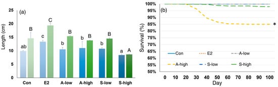
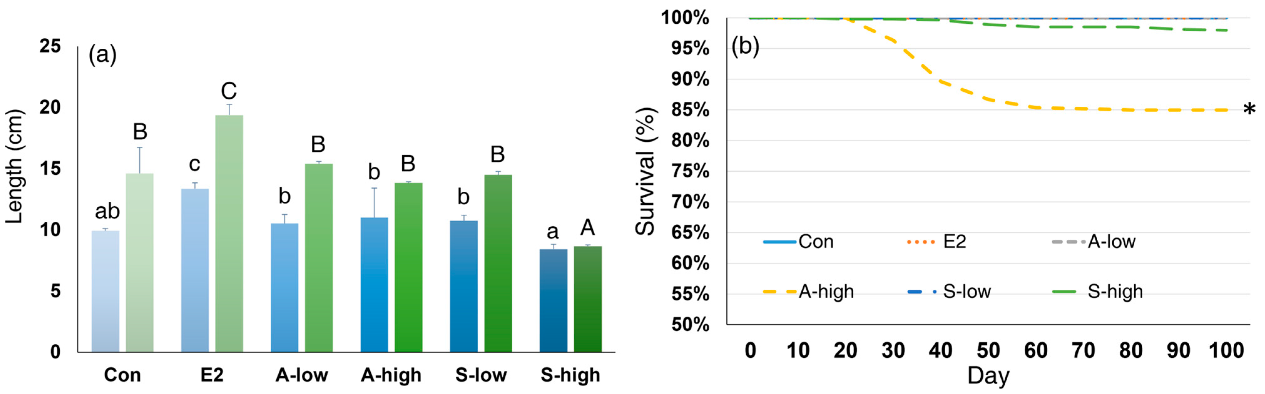
Total length was compared between the treatment groups at 1 and 2 months after the initiation of the experiment (Figure 1a). The E2 group showed a higher total length than the other groups at both time points, whereas the S-high group showed a lower total length than the other groups at 2 months. No significant differences in total length were observed between replicate tanks within any treatment group at 1 or 2 months after Holm–Bonferroni correction (p > 0.05). When each tank was analyzed separately, the same pattern was observed in both tanks: the E2 group showed the highest total length and the S-high group the lowest. At 2 months, the difference between the Con and E2 groups was significant in tank 1 (p < 0.001) and showed the same direction in tank 2, close to the significance level (p = 0.054).
Figure 1.
Effects of Schisandra chinensis and Astragalus membranaceus extracts on growth and survival of Japanese eels. (a) Total length at 1 month (blue bars) and 2 months (green bars) of the treatment; (b) cumulative survival rate during the first 3 months of treatment. In (a), data are presented as mean ± SE (n = 30; 15 fish from each of two replicate tanks); statistical analysis was performed separately for each time point, and different lowercase (1 month) and uppercase (2 months) letters indicate significant differences among groups (one-way ANOVA followed by Tukey’s post hoc test; (p < 0.05)). Experimental groups consist of control (Con), E2, Schisandra chinensis extract at low and high concentrations (S-low, S-high), and Astragalus membranaceus extract at low and high concentrations (A-low, A-high). The asterisk (*) indicates a significant difference in survival compared with the Con group (Fisher’s exact test with Holm–Bonferroni correction, p < 0.001).
Survival was significantly lower in the A-high group (85%) than in the Con group (p < 0.001), and slight mortality was also observed in the S-high group (98.5%) (Figure 1b). No mortality occurred in the other groups. Mortality was confined to the first 3 months of the experiment, and no further mortality occurred in any group during the subsequent rearing period until 12 months.
2.2. Effects on Gonadal Differentiation Direction and Rate
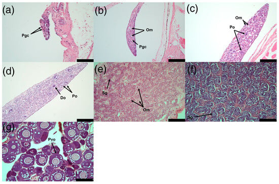
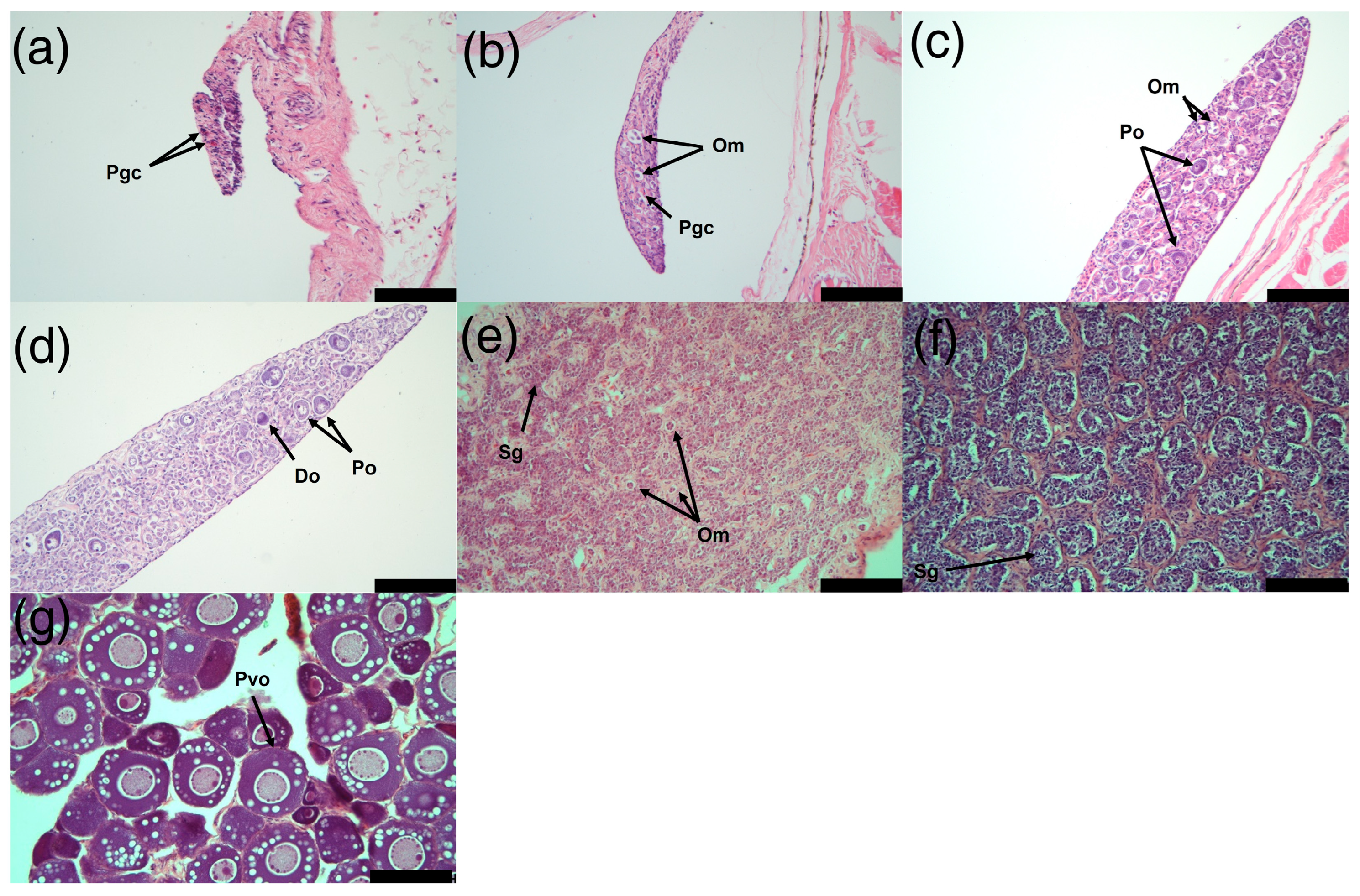
In Japanese eel, males differentiate through gonads containing oocytes, whereas females differentiate directly from undifferentiated gonads [34]. Eels in the Schisandra- and Astragalus-treated groups differentiated into males, whereas E2-treated eels differentiated into females, although the rate of differentiation differed between the treatments (Figure 2). Male gonadal development was classified into an undifferentiated stage (ud) and five distinct stages (S1–S5) by modifying the four-stage criteria described by Jeng et al. [34] to better reflect the morphological and developmental characteristics observed in the present study. Gonads containing only primordial germ cells (Pgc) were classified as the undifferentiated stage (ud). The S1 stage was characterized by the presence of Pgc and oocytes in meiosis (Om). At the S2 stage, Pgc, Om and primary oocytes (Po) were observed. The S3 stage was characterized by the presence of Om, Po and degenerating oocytes (Do). At the S4 stage, spermatogonia (Sg) and Om were observed, whereas the S5 stage was characterized predominantly by Sg, representing the most advanced stage of testicular development.
Figure 2.
Representative histological sections showing the developmental stages of the Japanese eel gonads: (a) undifferentiated (ud); (b) S1; (c) S2; (d) S3; (e) S4; (f) S5; and (g) ovary. Pgc, primordial germ cell; Om, oocyte in meiosis; Po, primary oocyte; Do, degenerating oocyte; Sg, spermatogonium; Pvo, pre-vitellogenic oocyte. Scale bars = 100 µm.
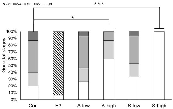
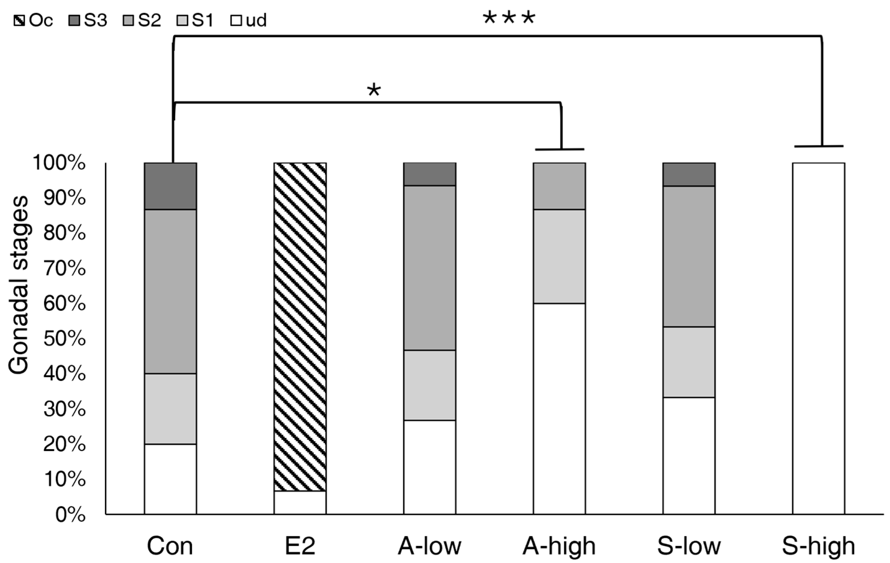
At 3 months (n = 15 per group), undifferentiated individuals were observed in all treatment groups, and the proportion of undifferentiated fish differed among the treatment groups (Figure 3). In the Con group, approximately 20% of the fish remained undifferentiated, whereas the S1, S2, and S3 stages accounted for 20%, 46.7%, and 13.3%, respectively, indicating the fastest rate of male differentiation among the treatment groups. In contrast, most fish (93.3%) in the E2 group developed ovaries and differentiated into females. In the low-dose extract treatment groups, the A-low group showed 26.7% undifferentiated fish, with 20%, 46.7% and 6.7% at the S1, S2 and S3 stages, respectively, showing a distribution similar to that of the Con group. The S-low group also showed a developmental stage distribution similar to that of the A-low group, with 33.3% undifferentiated fish and 20%, 40% and 6.7% at the S1, S2 and S3 stages, respectively. Compared with the low-dose treatment groups, the high-dose treatment groups showed a higher proportion of undifferentiated fish. In the A-high group, 60% of the fish remained undifferentiated, whereas the remaining 40% had reached the S1 and S2 stages. All fish in the S-high group remained undifferentiated. The gonadal stage distribution differed significantly from that of the Con group in the S-high and A-high groups (p < 0.001 and p = 0.027, respectively), whereas no significant differences were observed in the low-dose groups (Figure 3).
Figure 3.
Gonadal stages and sex ratio of Japanese eels in each treatment group after 3 months of treatment. Experimental groups consisted of a control (Con), E2, and low and high concentrations of Schisandra chinensis extract (S-low, S-high) and Astragalus membranaceus extract (A-low, A-high). Data represent the percentages of undifferentiated gonads (ud), differentiated testes (S1–S3) and oocytes (Oc) (n = 15 per group). Asterisks indicate significant differences in gonadal stage distribution from the Con group (Mann–Whitney U test with Holm–Bonferroni correction; * p < 0.05, *** p < 0.001). The E2 group was excluded from the statistical analysis because most fish had differentiated into females.
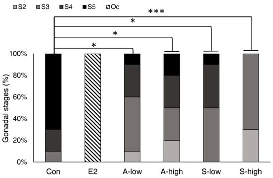
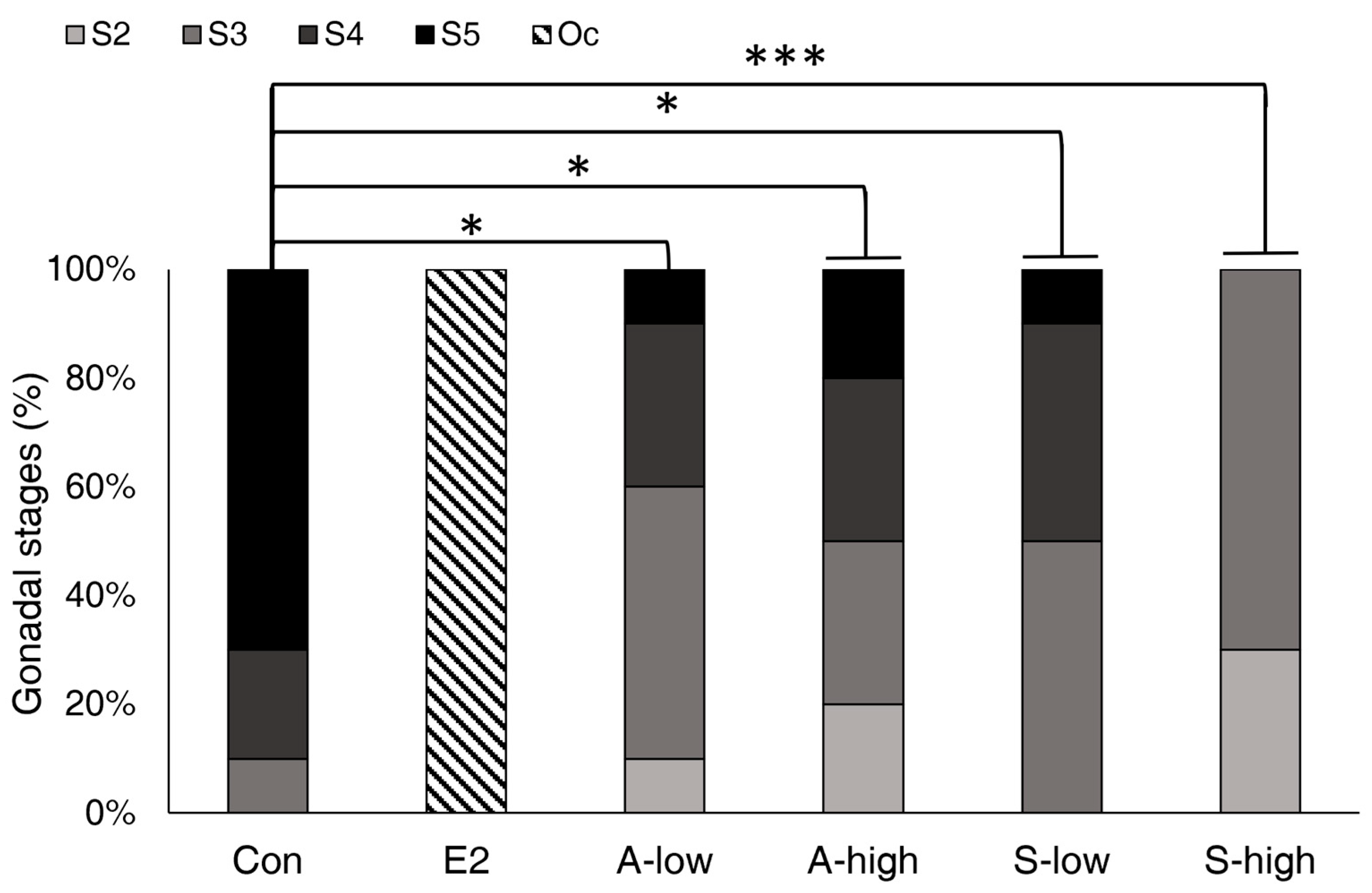
At 12 months (n = 10 per group), all fish in the treatments except the E2 group were identified as males (Figure 4). In the Con group, all fish had differentiated into males, with the S5 stage accounting for the highest proportion (70%). In contrast, all fish (100%) in the E2 group were identified as females with ovarian tissue. In the Astragalus groups, the A-low group showed 50% and 30% at the S3 and S4 stages, respectively, whereas the A-high group showed 30% at each of the S3 and S4 stages, indicating that differentiation had progressed mainly up to the S3 or S4 stages. A similar pattern was observed in the Schisandra groups. The S-low group showed 50% and 40% at the S3 and S4 stages, respectively, whereas the S-high group also showed the highest proportion of fish at the S3 stage. While the highest proportion of individuals in the Con group was observed at the S5 stage, the extract-treated groups (S-low, S-high, A-low, A-high) showed higher proportions at the S3 or S4 stages. All extract-treated groups showed significantly delayed testicular development compared with the Con group (p < 0.05) (Figure 4).
Figure 4.
Gonadal stages (S2–S5) of Japanese eels in each treatment group after 12 months of treatment. Experimental groups consisted of a control (Con), E2, and low and high concentrations of Schisandra chinensis extract (S-low, S-high) and Astragalus membranaceus extract (A-low, A-high). Data represent the percentages of differentiated testes (S2–S5) and oocytes (Oc) (n = 10 per group). Asterisks indicate significant differences in gonadal stage distribution from the Con group (Mann–Whitney U test with Holm–Bonferroni correction; * p < 0.05, *** p < 0.001). The E2 group was excluded from the statistical analysis because all fish had differentiated into females.
2.3. Correlation Between Growth and Gonadal Differentiation
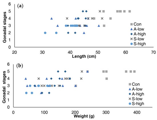
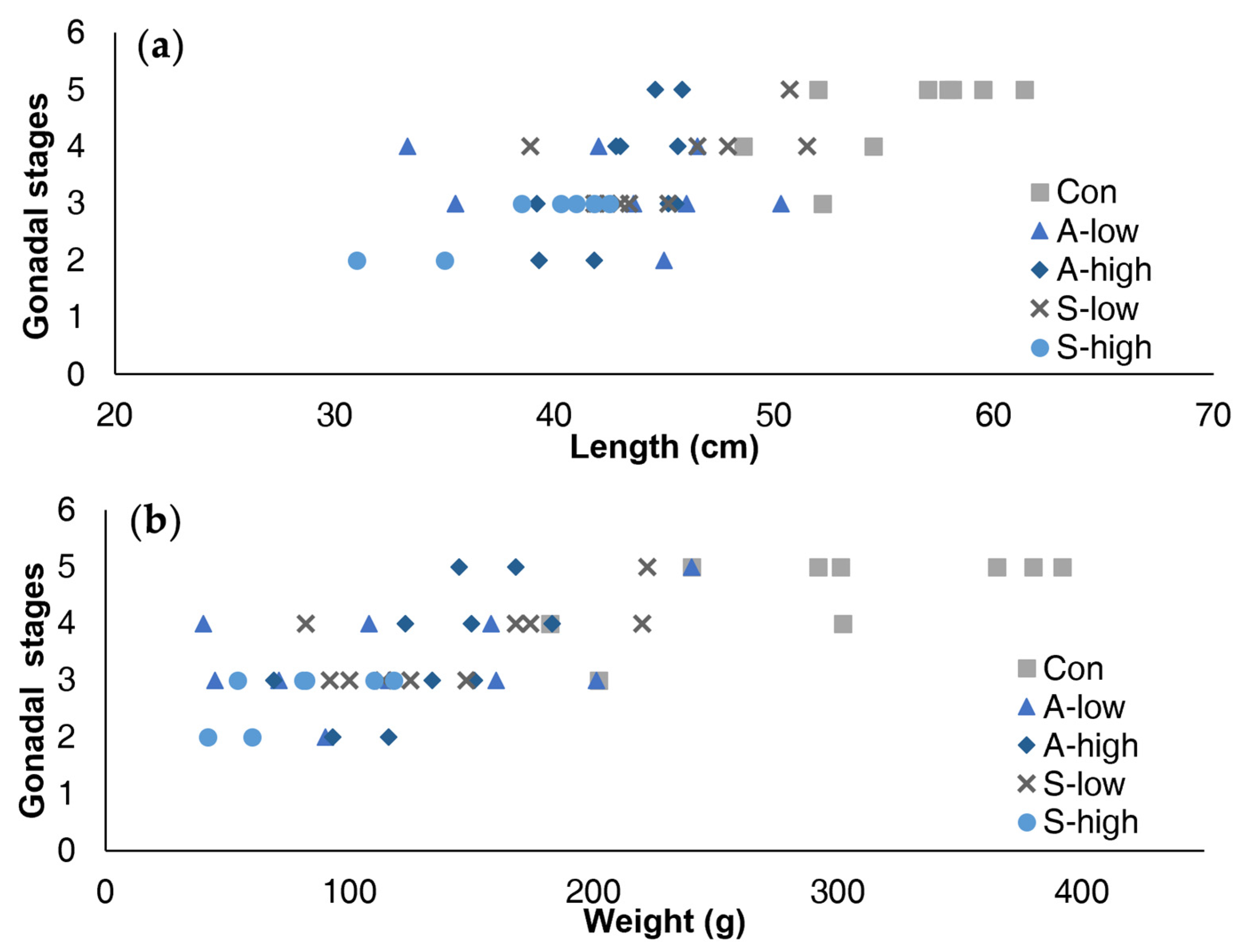
Spearman’s rank correlation analysis was performed to assess the relationship between growth parameters and gonadal developmental stage in males at 12 months, excluding the E2 group (Table 1). Analysis of all groups combined revealed a significant positive association between growth and gonadal stage. In contrast, analysis of each group separately showed that the relationship was significant only for body weight in the A-high group. Notably, individuals of similar size exhibited distinct gonadal stages across treatments (Figure 5), suggesting that these differences may have been influenced not only by growth rate but also by treatment with Schisandra and Astragalus extracts.
Table 1.
Spearman’s rank correlation coefficients (ρ) between gonadal stage and growth parameters in male Japanese eels at 12 months, with the corresponding p-values.
Figure 5.
Scatter plots showing the relationship between gonadal stage and growth parameters in male Japanese eels of each treatment group at 12 months: (a) total length; (b) body weight.
2.4. Effects on the Expression of Sex Differentiation-Related Genes
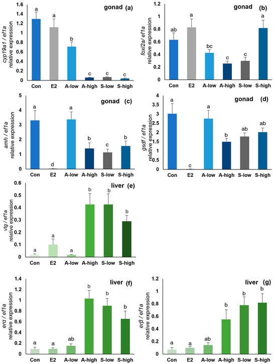
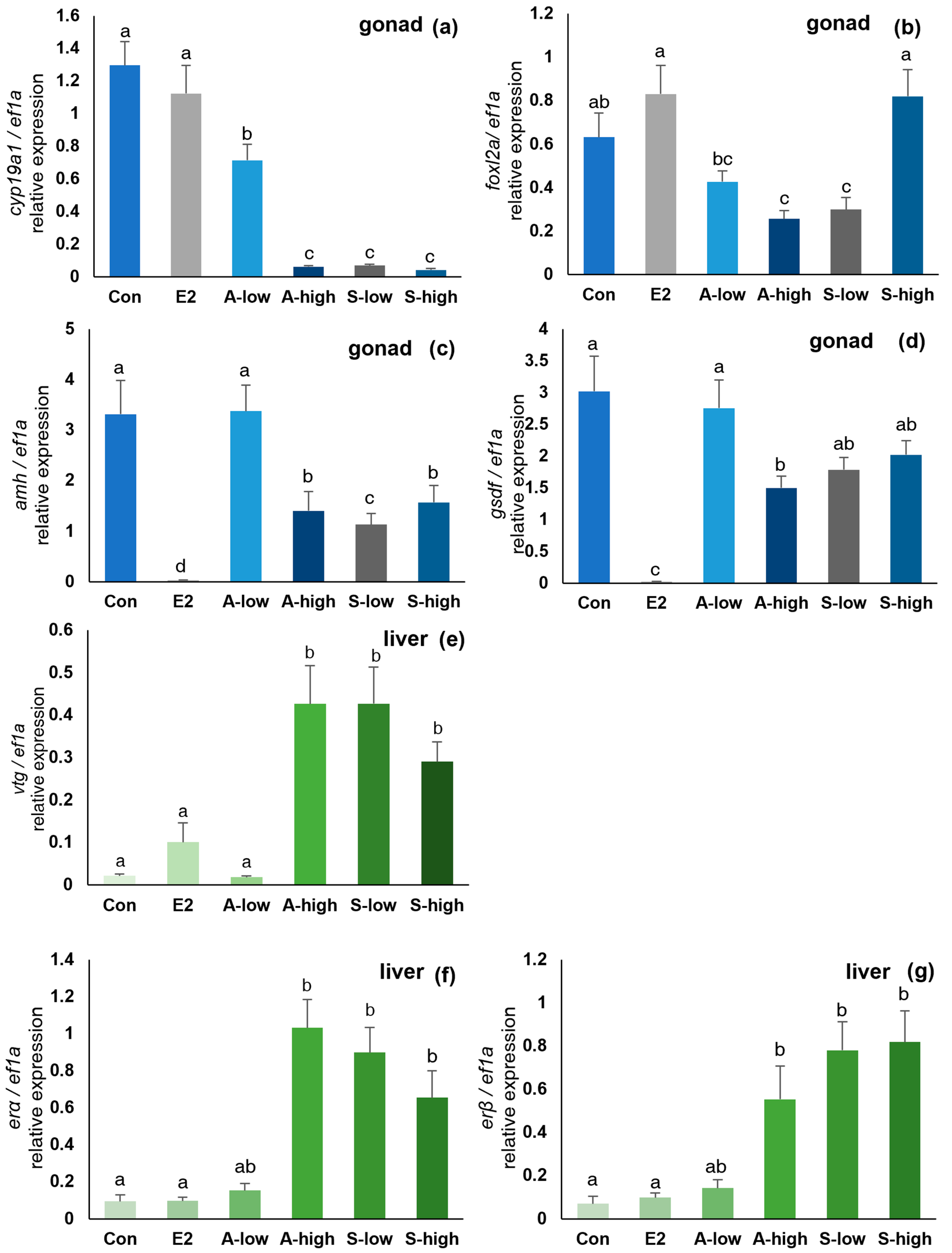
The expression of sex differentiation-related genes (cyp19a1, foxl2a, amh, gsdf) and estrogen-sensitive genes (vtg, erα, erβ) was analyzed at 12 months (Figure 6). No significant difference in cyp19a1 expression was observed between the Con and E2 groups. In contrast, cyp19a1 expression was significantly lower in the extract treatment groups (S-low, S-high, A-low, A-high) than in the Con group. The expression of foxl2a was highest in the E2 and S-high groups, with no significant difference between the two groups. In contrast, relatively lower expression was observed in the A-high and S-low groups than in the Con group. The expression of the male differentiation-related genes, amh and gsdf, was significantly lower in the E2 group than in the Con group. The expression of amh was also significantly decreased in the A-high, S-low and S-high groups compared with the Con group, whereas gsdf expression was significantly decreased only in the A-high group, with no significant difference observed in the S-low and S-high groups compared with the Con group. In the liver, vtg expression was significantly higher in the A-high, S-low and S-high groups than in the Con and E2 groups. Similarly, the expression of erα and erβ was significantly higher in the extract-treated groups (A-high, S-low and S-high) than in the Con and E2 groups, whereas no significant difference was observed between the Con and E2 groups.
Figure 6.
Relative expression of (a) cyp19a1, (b) foxl2a, amh (c) and (d) gsdf in gonadal tissues, and (e) vtg, (f) erα and (g) erβ in liver tissue in each treatment group after 12 months. Expression levels were normalized to ef1α and are presented as mean ± SE (n = 10 per group; 5 fish from each of two replicate tanks). Different letters indicate significant differences between groups by (Welch’s ANOVA followed by the Games–Howell post hoc test (p < 0.05)).
2.5. Phytoestrogen Content of the Extracts
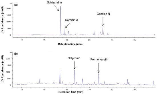
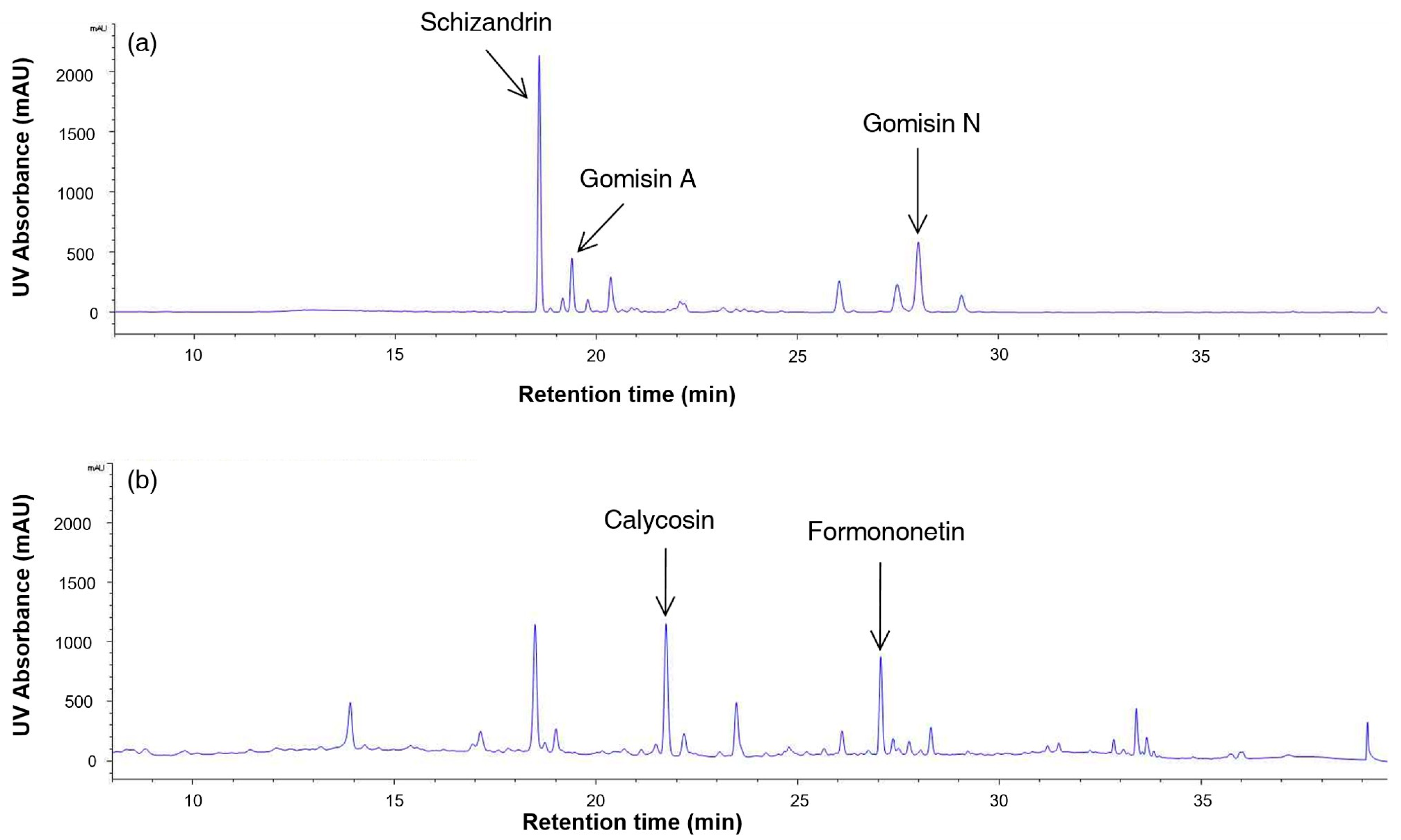
The calibration curves of all compounds showed good linearity (R2 > 0.999). In the Schisandra extract, peaks corresponding to schizandrin, gomisin A and gomisin N were detected at 18.8, 19.4 and 28.2 min, respectively, and in the Astragalus extract, peaks corresponding to calycosin and formononetin were detected at 21.8 and 27.2 min, respectively (Figure 7). These retention times were consistent with those of the corresponding standards analyzed under the same conditions. The Schisandra extract contained schizandrin (5.73 ± 0.01 mg/g), gomisin A (1.72 ± 0.00 mg/g) and gomisin N (3.96 ± 0.01 mg/g), and the Astragalus extract contained calycosin (0.180 ± 0.000 mg/g) and formononetin (0.070 ± 0.001 mg/g).
Figure 7.
HPLC-DAD chromatograms (254 nm) of (a) the Schisandra chinensis extract and (b) the Astragalus membranaceus extract used in the present study. Arrows indicate the peaks of schizandrin, gomisin A, and gomisin N in (a), and of calycosin and formononetin in (b).
3. Discussion
Schisandra and Astragalus extracts are known to contain several phytoestrogen compounds that exhibit estrogen-like activity. Phytoestrogens exert this activity by affecting sex differentiation [5,8,9,10] and sexual maturation [35] in various fish species. In the present study, treatment with Schisandra and Astragalus extracts delayed testicular differentiation and altered the expression of sex differentiation-related genes in the eel. These findings are generally consistent with previous reports. However, the extent of the effects of Schisandra and Astragalus extracts on sex differentiation differed from that reported for other phytoestrogen sources.
3.1. Effects on Growth and Survival Rate
Lower growth in the S-high group and a high proportion of undifferentiated fish in the high-dose treatment groups were observed during the early stage of the experiment. This was presumably due to reduced feed intake resulting from decreased feed palatability caused by the characteristic odor or chemical constituents of the high-dose plant extracts. Previous studies have reported that differences in feed additives and their components affect feed palatability [36,37] and that variation in feed intake among individual fish can negatively affect growth [38]. However, feed intake was not measured in this study.
In addition, during the early period of the experiment, survival was significantly reduced in the A-high group, and slight mortality was observed in the S-high group. A previous study reported that treatment of hepatocytes with the same extracts resulted in a concentration-dependent decrease in cell viability, and some other plant extracts also significantly affected cell viability [28]. This finding suggests that certain unpurified compounds in Schisandra and Astragalus extracts, or the high concentrations of the extracts themselves, may have exerted toxic effects on eels during the early glass eel stage.
3.2. Effects on Gonadal Differentiation Direction and Rate
After 12 months of the experiment, all individuals in the Con group and the Schisandra and Astragalus treatment groups differentiated into males, whereas all individuals in the E2 group had differentiated into females. Compared with the Con group, all Schisandra and Astragalus treatment groups showed significantly delayed testicular differentiation at 12 months, remaining mainly at the S3 and S4 stages, and this delay was already significant in the high-dose groups at 3 months. These results suggest that although the components of the extracts did not alter the direction of sex differentiation in eels, they may act as exogenous factors that delay the rate of gonadal differentiation.
The effects of estrogen as an exogenous hormone have already been reported in various fish species, although studies on the direct mechanisms by which phytoestrogens affect gonadal development in male fish are still limited. Estrogen treatment in male fish inhibits spermatogenesis and germ cell maintenance, resulting in sterility [39] or delayed sex differentiation [40,41]. These findings suggest that hormonal imbalance caused by exogenous estrogen can affect overall gonadal development. In particular, teleost fish, including eels, are highly sensitive to hormone exposure during the early glass eel stage, which has a decisive influence on sex differentiation [42]. Therefore, the delayed testicular development observed in the Schisandra and Astragalus treatment groups may also have been related to the estrogen-like components contained in the extracts, although the present data do not identify a direct action on the gonads.
3.3. Correlation Between Growth and Gonadal Differentiation
Sex differentiation in Japanese eel is more closely associated with body growth than with chronological age, and gonadal differentiation generally begins after fish reach a certain body size [43,44]. In the present study, the S-high group exhibited lower growth during the early stage of the experiment, and the high-dose treatment groups showed a higher proportion of undifferentiated individuals after 3 months, suggesting that delayed growth might initially explain the delay in gonadal differentiation.
However, Spearman’s rank correlation analysis results suggest that growth may not have been the only factor involved in the delayed gonadal differentiation. Although a significant correlation between growth parameters and gonadal differentiation was observed when all groups were combined, this correlation was not consistently observed in each treatment group. Furthermore, when individuals of similar size were compared, gonadal differentiation was slower in the extract-treated groups than in the Con group.
These results suggest that differences in gonadal differentiation may have been influenced not only by growth rate but also by treatment with Schisandra and Astragalus extracts.
3.4. Expression Patterns of Sex Differentiation-Related Genes
The cyp19a1 gene encodes an enzyme that converts androgens into estrogens and plays an important role in testicular development and spermatogenesis in fish [45,46,47]. Previous studies have reported that cyp19a1 expression is higher in females than in males during the early stages of sexual development, whereas no difference is observed between sexes during the later stages due to the increase in cyp19a1 as testicular differentiation proceeds [34]. Similarly, no significant difference in cyp19a1 expression was observed between the Con and E2 treatment groups in this study. In contrast, the extract treatment groups showed lower cyp19a1 expression than the Con group, possibly because testicular development remained at relatively earlier stages. Therefore, the reduced expression observed in the extract treatment groups is consistent with the delay of testicular differentiation.
The highest foxl2a expression was observed in the E2 and S-high groups. No significant difference was observed between the Con and E2 groups. In general, foxl2a expression increases during the early stages of ovarian differentiation and gradually decreases as it progresses [43]. Therefore, the absence of a significant difference between the Con and E2 groups is considered to reflect that the ovaries in the E2 group had already reached the later stages of differentiation. Although the A-high, S-low and S-high groups showed typical characteristics of male testes, the S-high group unexpectedly exhibited foxl2a expression similar to that of the E2 group. However, as there are currently no previous reports explaining this unexpected expression pattern in male-differentiated gonads, definitive conclusions cannot be drawn at this stage, and further study is required to clarify these findings.
The male differentiation-related genes amh and gsdf are known to increase during testicular development and to be strongly suppressed in feminized individuals induced by E2 treatment [44,48]. In this study, the expression of both genes was significantly lower in the E2 group than in the Con group, which is consistent with previous studies. The amh expression was significantly reduced in the A-high, S-low and S-high groups compared with the Con group, whereas gsdf expression was significantly reduced only in the A-high group. Because amh expression remains high during the later stages of testicular development [44], the reduced amh expression in the A-high, S-low and S-high groups, together with the reduced gsdf expression in the A-high group, is consistent with the delayed testicular development observed histologically. These findings suggest that the extracts may also have influenced male differentiation at the gene expression level.
Finally, hepatic vtg, erα and erβ expression was significantly higher in the A-high, S-low and S-high groups than in the Con and E2 groups. Vitellogenin is a sensitive biomarker of exposure to exogenous estrogenic compounds [49,50], mainly expressed in the liver of mature females and found at low levels in males [51]. The induction of vtg and the increased expression of erα and erβ therefore indicate that Schisandra and Astragalus extracts exerted estrogenic effects in the liver. In the E2 group, hepatic expression did not differ significantly from that of the Con group. E2 administration was terminated at 8 months due to the completion of feminization. The E2 group was used as a female reference group; therefore, hepatic expression in this group would be expected to be similar to that in the Con group. In contrast, the extract-treated groups, which were fed the experimental diets until sampling, showed significantly higher hepatic expression of these genes than the Con group, indicating that the extracts exerted estrogenic effects in the liver under continuous dietary administration.
Overall, the histological observations were consistent with the gene expression profiles. Increased hepatic expression of vtg, erα and erβ indicated that Schisandra and Astragalus extracts exhibited estrogenic activity in the liver of treated fish. In contrast, the reduced expression of cyp19a1 and amh in the gonads, together with the significant reduction in gsdf expression in the A-high group, corresponded well with the relatively earlier stages of testicular differentiation identified histologically. These results suggest that Schisandra and Astragalus extracts exhibited estrogenic activity in the liver and were associated with delayed male gonadal differentiation.
4. Materials and Methods
4.1. Animals
Glass eels (total length: 5.6 ± 0.4 cm, body weight: 0.10 ± 0.02 g) used in this experiment were captured on the coast of Jeollanam-do, Korea. These fish were transported to the aquarium facility at Sunmoon University and immersed in 7 psu saltwater to prevent pathogen infection before stocking. No mortality occurred during transportation, and the health status of the fish was confirmed by visual inspection for abnormal swimming behavior and external lesions or wounds on the body surface. The fish were then acclimated to freshwater for 7 days by gradually reducing the salinity, while the water temperature was simultaneously raised from 21 °C to 28 °C at a rate of 1 °C per day to minimize stress [52]. Following acclimation, the eels were randomly assigned to each treatment group. For each treatment group, the eels were reared in two tanks equipped with biological filters. Rearing conditions (DO: 9.9 mg/L, pH: 8.3, water temperature: 28.4 °C) were identical in all treatment groups except for the experimental diet. Total length was measured at 1 and 2 months, and sampling was conducted at 3 and 12 months after the initiation of the experiment. Early gonadal differentiation was examined at 3 months. Because a high proportion of undifferentiated fish was observed, the final sex ratio and gonadal developmental stages were re-examined at 12 months. At each sampling, fish were anesthetized with 200 ppm 2-phenoxyethanol (Daejung, Republic of Korea) before total length and body weight were measured and tissues required for subsequent analyses were collected.
4.2. Groups
The experiment consisted of six treatment groups: control (Con), positive control (E2 treatment, E2), Schisandra extract-treated (Schisandra-low and Schisandra-high, S-low and S-high, respectively) and Astragalus extract-treated (Astragalus-low and Astragalus-high, A-low and A-high, respectively) groups.
4.3. Dietary Administration
Dietary administration began on the fifth day after transportation to the laboratory. The glass eels were initially fed a commercial weaning diet (Nosan Corporation, Kanagawa, Japan) containing 51% crude protein, 3% crude fat, 2% calcium, 2.7% phosphorus, maximum 1.0% crude fiber and 17% crude ash. The feed was gradually transitioned to a commercial eel grow-out diet (KOFEC Corporation, Naju, Republic of Korea) according to the growth stages of the eels. The proximate composition of the grow-out diet was 50% crude protein, 5% crude fat, 1% calcium, 2.7% phosphorus, maximum 2.0% crude fiber and maximum 15% crude ash. The experimental diets were provided when the average total length of the fish reached 7–8 cm, corresponding to the onset of sex differentiation. The daily feeding rate was initially 8% of body weight and was gradually reduced to 4% as the fish grew, because feed intake relative to body weight decreases with increasing body size. Fish were observed daily during feeding, and dead fish were removed and recorded.
The Con group was fed the commercial feed diet (KOFEC Corporation, Naju, Republic of Korea), whereas the E2 treatment group was fed a basal diet supplemented with 10 mg of E2 (Sigma-Aldrich, St. Louis, MO, USA) per kilogram of feed [33]. To compare the estrogenic effects of phytoestrogens derived from Schisandra and Astragalus, each extract was administered at two dietary concentrations: low-dose (S-low, A-low) and high-dose (S-high, A-high). The extracts were mixed with the dough during feed preparation, and the prepared dough feed was provided to the fish. The extracts were added to the basal diet rather than substituted for any dietary ingredient. Each treatment consisted of two replicate tanks containing 200 fish each (400 fish per treatment). The extracts were administered continuously for 12 months. However, E2 administration was terminated at 8 months because feminization had already been confirmed at 3 months, and the fish were subsequently fed the basal diet until the end of the experiment. In all groups, the fish were fed until the day before each sampling.
4.4. Preparation of Plant Extracts
Schisandra and Astragalus used in this experiment were purchased as commercially available powdered products (Garunara Corporation, Seoul, Republic of Korea). Each powder was mixed with 90% ethanol at a ratio of 1:10 (w/v) and extracted for 30 min using an ultrasonic extractor (Hwashin Tech, Seoul, Republic of Korea) at 30 °C under an output power of 580–700 W. The extraction conditions were adopted from a previous study [53] to maximize the extraction efficiency of non-polar aglycone compounds. The extract was filtered through a cell strainer (SPL Life Sciences, Pocheon-si, Republic of Korea) to collect the supernatant. The supernatant was then concentrated using a rotary evaporator (Eyela, Tokyo, Japan) until the final volumes reached 4% (36 mL) of the initial solvent volume for the Schisandra extract and 1.5% (13.6 mL) for the Astragalus extract. Because the viscosity of the Schisandra extract increased markedly during concentration, further concentration was difficult. Although the concentration ratios of the two extracts differed, the amount of concentrate added during feed preparation was adjusted to achieve the intended extract concentration in each experimental diet. The dietary concentrations were determined with reference to a previous study in which dietary phytoestrogens induced sex differentiation in Japanese eel [9]. The high dose was set as the maximum inclusion level of the concentrated extract at which mass mortality did not occur in preliminary feeding trials (9.05% for Schisandra and 5.14% for Astragalus, dry feed weight basis), and the low dose was set at 20% of the corresponding high dose.
4.5. RNA Extraction and cDNA Synthesis
Total RNA was extracted from individual gonadal and liver tissues (without pooling) using TRIzolTM reagent (Thermo Fisher Scientific, Waltham, MA, USA), followed by chloroform extraction, isopropanol precipitation, and ethanol washing according to the manufacturer’s protocol. RNA concentration and purity were determined using a NanoDrop 2000 spectrophotometer (Thermo Fisher Scientific, Waltham, MA, USA). Complementary DNA (cDNA) was synthesized using TOPscriptTM RT DryMIX (Enzynomics, Daejeon, Republic of Korea) according to the manufacturer’s instructions.
4.6. Real-Time PCR
Quantitative real-time PCR (qPCR) was performed using the synthesized cDNA as a template and TOPreal™ qPCR 2X PreMIX SYBR Green (Enzynomics, Daejeon, Republic of Korea). The PCR conditions were as follows: initial denaturation at 95 °C for 10 min, followed by 35 cycles of denaturation at 95 °C for 5 s, annealing at 60 °C for 30 s and extension at 60 °C for 30 s. A melting curve analysis was subsequently performed to confirm the specificity of amplification.
The relative expression levels of sex differentiation-related genes (cyp19a1, foxl2a, amh and gsdf) in the gonads and estrogen-sensitive genes (vtg, erα and erβ) in the liver were analyzed. The primers for ef1α, amh, gsdf, cyp19a1 and foxl2a were adopted from Inaba et al. [54], and those for erβ from Hyeon et al. [55]; those for vtg and erα were designed in this study. The primer information is listed in Table 2. The relative expression levels of all target genes were calculated using the 2−ΔΔCt method with ef1α as the reference gene, which has been used for gene expression analyses in Anguilla japonica [9,54,55].
Table 2.
Primer sets used for qPCR analysis.
Gene expression analysis was conducted at 12 months using randomly selected individuals from each treatment group (Con, E2, A-low, A-high, S-low and S-high; n = 10 per group, five fish from each of the two replicate tanks). All qPCR assays for each biological sample were performed in technical duplicates, and a no-template control was included in every run. Before pooling, differences between the two replicate tanks within each treatment group were assessed using the Mann–Whitney U test, and no significant differences were detected for any gene (p > 0.05).
4.7. Histological Analysis
Gonadal samples were fixed in 10% neutral buffered formalin, automatically processed, and embedded in paraffin blocks. The embedded tissues were sectioned at a thickness of 5 µm using a microtome (Leica Biosystems, Wetzlar, Germany). The sectioned tissues were stained with hematoxylin and eosin (H&E) to observe gonadal developmental stages and morphological characteristics under an optical microscope (Leica Microsystems, Wetzlar, Germany). For histological analysis, 15 fish per group at 3 months (seven from one replicate tank and eight from the other) and 10 fish per group at 12 months (five fish from each tank) were examined. At 12 months, the fish used for histological analysis and those used for gene expression analysis were randomly selected from the same replicate tanks of each treatment group, but the two analyses were not performed on the same individuals.
4.8. HPLC Analysis
For HPLC analysis, the Schisandra and Astragalus extracts were filtered through a 0.22 µm PVDF membrane filter. Schizandrin, gomisin A and gomisin N, the major phytoestrogens of Schisandra, and calycosin and formononetin, the major phytoestrogens of Astragalus, were used as standards; these were the same standards as those used in our previous study [28]. Each standard was dissolved in methanol and serially diluted to construct calibration curves.
Analyses were performed on an Agilent 1260 Infinity II Quat pump system equipped with a diode array detector (DAD WR; Agilent Technologies, Santa Clara, CA, USA) and a YMC-Pack Pro C18 column (4.6 × 250 mm, 5 µm; YMC, Kyoto, Japan) at room temperature. The mobile phase consisted of water containing 0.1% trifluoroacetic acid (A) and acetonitrile containing 0.1% trifluoroacetic acid (B). The injection volume was 10 μL, the flow rate was 1.0 mL/min, and detection was performed at 254 nm. The gradient for the Schisandra extract was as follows: 0–7 min, 20% B; 7–15 min, 20–70% B; 15–25 min, 70–82% B; 25–35 min, 82–100% B; 35–38 min, 100% B; 38–40 min, 100–20% B; and 40–55 min, 20% B. The gradient for the Astragalus extract was as follows: 0–5 min, 15% B; 5–15 min, 15–32% B; 15–20 min, 32–40% B; 20–27 min, 40–65% B; 27–30 min, 65–100% B; 30–35 min, 100% B; 35–38 min, 100–15% B; and 38–52 min, 15% B. Each compound was identified by comparing its retention time with that of the corresponding standard and quantified using the calibration curve.
4.9. Statistical Analysis
Data are presented as the mean ± SE. Differences in total length between groups were analyzed separately at 1 and 2 months by one-way ANOVA followed by Tukey’s post hoc test. Because each treatment consisted of two replicate tanks, differences between tanks within each treatment group were first assessed using the Mann–Whitney U test, with Holm–Bonferroni correction for multiple comparisons. To further confirm that pooling individuals from the two tanks did not affect the results, one-way ANOVA followed by Tukey’s test was also performed separately for each tank. Survival rates were compared between replicate tanks and between each treatment group and the Con group using Fisher’s exact test with Holm–Bonferroni correction. In the A-high group, survival differed between the two tanks (p = 0.042), but was significantly lower than in the Con group in both tanks (p < 0.001).
Gonadal developmental stages were scored as ordinal values (ud = 0, S1 = 1, S2 = 2, S3 = 3, S4 = 4, S5 = 5), and the stage distribution of each extract-treated group was compared with that of the Con group at 3 and 12 months using the Mann–Whitney U test with Holm–Bonferroni correction. The E2 group was excluded from this analysis because the fish had differentiated into females.
For gene expression data, homogeneity of variance was assessed using Levene’s test, which revealed a significant violation of this assumption for all analyzed genes (p < 0.001 for all). Accordingly, differences between experimental groups were analyzed using Welch’s ANOVA, which does not require the assumption of equal variances, followed by the Games–Howell post hoc test for multiple comparisons. Statistical significance was accepted at p < 0.05. Because gonadal developmental stage represents ordinal rather than continuous data, Spearman’s rank correlation analysis was used to evaluate the relationship between body growth and gonadal development in males at 12 months; the E2 group was excluded because the fish had differentiated into females. All statistical analyses were performed using SPSS Statistics version 20.0 (IBM Corporation, Armonk, NY, USA).
5. Conclusions
Dietary Schisandra and Astragalus extracts delayed testicular differentiation in Japanese eel. At the gene expression level, amh expression was reduced in the A-high, S-low and S-high groups, and gsdf expression was reduced in the A-high group. In particular, the increased hepatic expression of vtg and estrogen receptors (erα and erβ) indicates that these extracts exerted estrogen-like activity in vivo. However, in contrast to the clear response in the liver, the effect on the gonads was limited to delayed male differentiation rather than feminization. This may be because the crude extracts contained a mixture of compounds, and the amount of each individual phytoestrogen may not have been sufficient to alter the direction of sex differentiation. Therefore, further studies are needed to examine the dose-dependent responses to Schisandra and Astragalus extracts and the mechanisms of action of the individual phytoestrogens they contain.
Author Contributions
Conceptualization, J.H.Y., J.E.H. and J.Y.K.; methodology, J.H.Y., J.E.H. and J.Y.K.; formal analysis, J.E.H.; investigation, J.H.M., J.H.Y. and B.R.P.; resources, J.H.M., J.H.Y. and B.R.P.; data curation, J.E.H.; writing—original draft preparation, J.E.H.; writing—review and editing, J.Y.K.; visualization, J.E.H.; supervision, J.Y.K.; project administration, J.Y.K.; funding acquisition, J.Y.K. All authors have read and agreed to the published version of the manuscript.
Funding
This work was supported by the National Research Foundation of Korea (NRF) grant funded by the Korea government (MSIT) (IRIS RS-2026-25485246).
Institutional Review Board Statement
The animal study protocol was approved by the Institutional Animal Care and Use Committee (IACUC) of Sunmoon University (protocol code SM-2023-04-01, approved on 15 November 2023).
Informed Consent Statement
Not applicable.
Data Availability Statement
The data presented in this study are available upon request from the corresponding author.
Conflicts of Interest
The authors declare no conflicts of interest. The funders had no role in the design of the study; in the collection, analyses, or interpretation of data; in the writing of the manuscript; or in the decision to publish the results.
Abbreviations
The following abbreviations are used in this manuscript:
| E2 | 17β-estradiol |
| Con | Control |
| S-low, S-high | Schisandra extract low- and high-dose groups |
| A-low, A-high | Astragalus extract low- and high-dose groups |
| ud | Undifferentiated |
| Pgc | Primordial germ cells |
| Om | Oocytes in meiosis |
| Po | Primary oocyte |
| Do | Degenerating oocyte |
| Sg | Spermatogonia |
| Pvo | Pre-vitellogenic oocyte |
| Oc | Oocyte |
| qPCR | Quantitative real-time PCR |
| cyp19a1 | Cytochrome P450 aromatase |
| foxl2a | Forkhead box L2a |
| amh | Anti-Müllerian hormone |
| gsdf | Gonadal soma-derived factor |
| vtg | Vitellogenin |
| erα, erβ | Estrogen receptor α, β |
| ef1α | Elongation factor 1 α |
References
- Chakraborty, S.B.; Horn, P.; Hancz, C. Application of phytochemicals as growth-promoters and endocrine modulators in fish culture. Rev. Aquac. 2014, 6, 1–19. [Google Scholar] [CrossRef] [Scilit]
- Kuiper, G.G.; Lemmen, J.G.; Carlsson, B.O.; Corton, J.C.; Safe, S.H.; van der Saag, P.T.; van der Burg, B.; Gustafsson, J.A. Interaction of estrogenic chemicals and phytoestrogens with estrogen receptor β. Endocrinology 1998, 139, 4252–4263. [Google Scholar] [CrossRef] [PubMed]
- Çek, Ş.; Turan, F.; Atik, E. Masculinization of convict cichlid (Cichlasoma nigrofasciatum) by immersion in Tribulus terrestris extract. Aquac. Int. 2007, 15, 109–119. [Google Scholar] [CrossRef] [Scilit]
- Çek, Ş.; Turan, F.; Atik, E. The effects of Gokshura, Tribulus terrestris, on sex reversal of guppy, Poecilia reticulata. Pak. J. Biol. Sci. 2007, 10, 718–725. [Google Scholar] [CrossRef] [Scilit] [PubMed]
- DiMaggio, M.A.; Kenter, L.W.; Breton, T.S.; Berlinsky, D.L. Effects of dietary genistein administration on growth, survival and sex determination in southern flounder, Paralichthys lethostigma. Aquac. Res. 2016, 47, 82–90. [Google Scholar] [CrossRef] [Scilit]
- Park, I.S.; Oh, H.S.; Koo, J.G. Effect of oral tamoxifen on growth and survival in the bagrid catfish Pseudobagrus fulvidraco. Aquac. Res. 2003, 34, 1471–1474. [Google Scholar] [CrossRef] [Scilit]
- Turan, F.; Akyurt, I. Effects of red clover extract on growth performance and body composition of African catfish Clarias gariepinus. Fish. Sci. 2005, 71, 618–620. [Google Scholar] [CrossRef] [Scilit]
- Fajkowska, M.; Adamek-Urbańska, D.; Ostaszewska, T.; Szczepkowski, M.; Rzepkowska, M. Effect of genistein, daidzein and coumestrol on sex-related genes expression in Russian sturgeon (Acipenser gueldenstaedtii). Aquaculture 2021, 530, 735872. [Google Scholar] [CrossRef] [Scilit]
- Inaba, H.; Iwata, Y.; Suzuki, T.; Horiuchi, M.; Surugaya, R.; Ijiri, S.; Uchiyama, A.; Takano, R.; Hara, S.; Yazawa, T.; et al. Soy isoflavones induce feminization of Japanese eel (Anguilla japonica). Int. J. Mol. Sci. 2023, 24, 396. [Google Scholar] [CrossRef] [Scilit] [PubMed]
- Kiparissis, Y.; Balch, G.C.; Metcalfe, T.L.; Metcalfe, C.D. Effects of the isoflavones genistein and equol on the gonadal development of Japanese medaka Oryzias latipes. Environ. Health Perspect. 2003, 111, 1158–1163. [Google Scholar] [CrossRef] [Scilit] [PubMed]
- Tzchori, I.; Degani, G.; Elisha, R.; Eliyahu, R.; Hurvitz, A.; Vaya, J.; Moav, B. The influence of phytoestrogens and oestradiol-17β on growth and sex determination in the European eel (Anguilla anguilla). Aquac. Res. 2004, 35, 1213–1219. [Google Scholar] [CrossRef] [Scilit]
- Holbech, H.; Schröder, K.D.; Nielsen, M.L.; Brande-Lavridsen, N.; Holbech, B.F.; Bjerregaard, P. Estrogenic effect of the phytoestrogen biochanin A in zebrafish, Danio rerio, and brown trout, Salmo trutta. Aquat. Toxicol. 2013, 144, 19–25. [Google Scholar] [CrossRef] [Scilit] [PubMed]
- Latonnelle, K.; Le Menn, F.; Kaushik, S.J.; Bennetau-Pelissero, C. Effects of dietary phytoestrogens in vivo and in vitro in rainbow trout and Siberian sturgeon: Interests and limits of the in vitro studies of interspecies differences. Gen. Comp. Endocrinol. 2002, 126, 39–51. [Google Scholar] [CrossRef] [Scilit] [PubMed]
- Jung, T.K.; Kim, M.J.; Lim, K.R.; Yoon, K.S. Moisturizing and anti-oxidation effect of Astragalus membranaceus root extract. J. Soc. Cosmet. Sci. Korea 2006, 32, 193–200. [Google Scholar]
- Kim, S.I.; Sim, K.H.; Ju, S.Y.; Han, Y.S. A study of antioxidative and hypoglycemic activities of Omija (Schizandra chinensis Baillon) extract under variable extract conditions. Korean J. Food Nutr. 2009, 22, 41–47. [Google Scholar]
- Kim, J.W.; Kim, S.D.; Youn, K.S. Antioxidant activity of Hwangki and Beni-Koji extracts and mixture. J. Korean Soc. Food Sci. Nutr. 2011, 40, 1–6. [Google Scholar] [CrossRef] [Scilit]
- Ryu, I.H.; Kwon, T.O. The antioxidative effect and ingredients of oil extracted from Schizandra chinensis seed. Korean J. Med. Crop Sci. 2012, 20, 63–71. [Google Scholar] [CrossRef] [Scilit]
- Bae, H.K.; You, S.H. Biological activity study on anti-oxidant, whitening, and anti-inflammatory effects of Astragalus membranaceus ethanol extracts and bioconversion extracts. Asian J. Beauty Cosmetol. 2017, 15, 489–499. [Google Scholar] [CrossRef] [Scilit]
- Kang, C.H.; Kwak, D.Y.; So, J.S. Inhibition of nitric oxide production and hyaluronidase activities from the combined extracts of Platycodon grandiflorum, Astragalus membranaceus, and Schisandra chinensis. J. Korean Soc. Food Sci. Nutr. 2013, 42, 844–850. [Google Scholar] [CrossRef] [Scilit]
- Park, J.H.; Shin, J.Y.; Heo, J.H.; Kim, S.J.; Kang, E.S.; Jeong, S.Y.; Kim, T.J.; Jang, S.I.; Cho, B.O. Antioxidant, anti-inflammatory, anti-collagenase, and anti-elastase effects of fermented Astragalus membranaceus extracts. Korean J. Food Sci. Technol. 2024, 56, 320–327. [Google Scholar] [CrossRef] [Scilit]
- Chen, Q.; Zhan, Q.; Li, Y.; Sun, S.; Zhao, L.; Zhang, H.; Zhang, G. Schisandra lignan extract protects against carbon tetrachloride-induced liver injury in mice by inhibiting oxidative stress and regulating the NF-κB and JNK signaling pathways. Evid. Based Complement. Altern. Med. 2017, 2017, 5140297. [Google Scholar] [CrossRef] [Scilit] [PubMed]
- Chen, C.; Bu, X.; Deng, L.; Xia, J.; Wang, X.; Chen, L.; Li, W.; Huang, J.; Chen, Q.; Wang, C. Astragaloside IV as a promising therapeutic agent for liver diseases: Current landscape and future perspectives. Front. Pharmacol. 2025, 16, 1574154. [Google Scholar] [CrossRef] [Scilit] [PubMed]
- Kang, S.C.; Kim, H.J.; Kim, M.H. Effects of Astragalus membranaceus with supplemental calcium on bone mineral density and bone metabolism in calcium-deficient ovariectomized rats. Biol. Trace Elem. Res. 2013, 151, 68–74. [Google Scholar] [CrossRef] [Scilit] [PubMed]
- Kim, M.H.; Choi, Y.Y.; Han, J.M.; Lee, H.S.; Hong, S.B.; Lee, S.G.; Yang, W.M. Ameliorative effects of Schizandra chinensis on osteoporosis via activation of estrogen receptor (ER)-α/-β. Food Funct. 2014, 5, 1594–1601. [Google Scholar] [CrossRef] [Scilit] [PubMed]
- Lee, D.; Kim, Y.M.; Chin, Y.W.; Kang, K.S. Schisandrol A exhibits estrogenic activity via estrogen receptor α-dependent signaling pathway in estrogen receptor-positive breast cancer cells. Pharmaceutics 2021, 13, 1082. [Google Scholar] [CrossRef] [Scilit] [PubMed]
- Tang, Z.; Tian, X. Astragalus membranaceus: A traditional Chinese medicine with multifaceted impacts on breast cancer treatment. Biomolecules 2024, 14, 1339. [Google Scholar] [CrossRef] [Scilit] [PubMed]
- Jin, J.Y. Gomisin N, a Major Lignan in Schisandra Chinensis, Increases Production of Testosterone and 17β-Estradiol via Stimulating Steroidogenic Enzymes. Master’s Thesis, Seoul National University, Seoul, Republic of Korea, 2018. [Google Scholar]
- Yoon, J.H.; Ha, J.E.; Kwon, J.Y. Estrogenic effect of various plant extracts on eel (Anguilla japonica) hepatocytes. Molecules 2025, 30, 2781. [Google Scholar] [CrossRef] [Scilit] [PubMed]
- Han, Y.S.; Liao, I.C.; Huang, Y.S.; He, J.T.; Chang, C.W.; Tzeng, W.N. Synchronous changes of morphology and gonadal development of silvering Japanese eel Anguilla japonica. Aquaculture 2003, 219, 783–796. [Google Scholar] [CrossRef] [Scilit]
- Hong, Y.K.; Song, M.Y.; Yi, S.E.; Lee, W.O. Change in morphological and biological characteristics by maturation of Japanese eel, Anguilla japonica, collected in Korea waters. Korean J. Ichthyol. 2015, 27, 263–274. [Google Scholar]
- Geffroy, B.; Bardonnet, A. Sex differentiation and sex determination in eels: Consequences for management. Fish. Fish. 2016, 17, 375–398. [Google Scholar] [CrossRef] [Scilit]
- Chiba, H.; Iwatsuki, K.; Hayami, K.; Yamauchi, K. Effects of dietary estradiol-17β on feminization, growth and body composition in the Japanese eel (Anguilla japonica). Comp. Biochem. Physiol. A Physiol. 1993, 106, 367–371. [Google Scholar] [CrossRef] [Scilit]
- Kim, D.J.; Lee, B.I.; Kim, K.K.; Kim, E.O.; Son, M.H.; Seong, K.B. Effects of estradiol-17β on the feminization of Japanese eel, Anguilla japonica. J. Life Sci. 2013, 23, 998–1003. [Google Scholar] [CrossRef] [Scilit]
- Jeng, S.R.; Wu, G.C.; Yueh, W.S.; Kuo, S.F.; Dufour, S.; Chang, C.F. Gonadal development and expression of sex-specific genes during sex differentiation in the Japanese eel. Gen. Comp. Endocrinol. 2018, 257, 74–85. [Google Scholar] [CrossRef] [Scilit] [PubMed]
- Bennetau-Pelissero, C.; Breton, B.; Bennetau, B.; Corraze, G.; Le Menn, F.; Davail-Cuisset, B.; Helou, C.; Kaushik, S.J. Effect of genistein-enriched diets on the endocrine process of gametogenesis and on reproduction efficiency of the rainbow trout Oncorhynchus mykiss. Gen. Comp. Endocrinol. 2001, 121, 173–187. [Google Scholar] [CrossRef] [Scilit] [PubMed]
- Conti, F.; Olivotto, I.; Cattaneo, N.; Pavanello, M.; Şener, İ.; Antonucci, M.; Chemello, G.; Gioacchini, G.; Zarantoniello, M. The promising role of synthetic flavors in advancing fish feeding strategies: A focus on adult female zebrafish (Danio rerio) growth, welfare, appetite, and reproductive performances. Animals 2024, 14, 2588. [Google Scholar] [CrossRef] [Scilit] [PubMed]
- Levina, A.D.; Mikhailova, E.S.; Kasumyan, A.O. Taste preferences and feeding behaviour in the facultative herbivorous fish, Nile tilapia Oreochromis niloticus. J. Fish. Biol. 2021, 98, 1385–1400. [Google Scholar] [CrossRef] [Scilit] [PubMed]
- Heinsbroek, L.T.; Goedegebuur, B.J.; Bloemhof, G.; Flach, R.B.; de Jong, G.D. Gastrointestinal and metabolic effects of feeding schedule on voluntary feed intake and growth of European eel, Anguilla anguilla. Aquac. Int. 2008, 16, 93–108. [Google Scholar] [CrossRef] [Scilit]
- Papoulias, D.M.; Noltie, D.B.; Tillitt, D.E. An in vivo model fish system to test chemical effects on sexual differentiation and development: Exposure to ethinyl estradiol. Aquat. Toxicol. 2000, 48, 37–50. [Google Scholar] [CrossRef] [Scilit]
- Andersen, L.; Holbech, H.; Gessbo, Å.; Norrgren, L.; Petersen, G.I. Effects of exposure to 17α-ethinylestradiol during early development on sexual differentiation and induction of vitellogenin in zebrafish (Danio rerio). Comp. Biochem. Physiol. C Toxicol. Pharmacol. 2003, 134, 365–374. [Google Scholar] [CrossRef] [Scilit] [PubMed]
- Selmoni, O.M.; Maitre, D.; Roux, J.; Wilkins, L.G.E.; Marques da Cunha, L.; Vermeirssen, E.L.M.; Knörr, S.; Robinson-Rechavi, M.; Wedekind, C. Sex-specific changes in gene expression in response to estrogen pollution around the onset of sex differentiation in grayling (Salmonidae). BMC Genom. 2019, 20, 583, Correction in BMC Genom. 2020, 21, 371. https://doi.org/10.1186/s12864-020-06782-w. [Google Scholar] [CrossRef] [Scilit] [PubMed]
- Leet, J.K.; Gall, H.E.; Sepúlveda, M.S. A review of studies on androgen and estrogen exposure in fish early life stages: Effects on gene and hormonal control of sexual differentiation. J. Appl. Toxicol. 2011, 31, 379–398. [Google Scholar] [CrossRef] [Scilit] [PubMed]
- Horiuchi, M.; Hagihara, S.; Kume, M.; Chushi, D.; Hasegawa, Y.; Itakura, H.; Yamashita, Y.; Adachi, S.; Ijiri, S. Morphological and molecular gonadal sex differentiation in the wild Japanese eel Anguilla japonica. Cells 2022, 11, 1554. [Google Scholar] [CrossRef] [Scilit] [PubMed]
- Lin, C.J.; Jeng, S.R.; Lei, Z.Y.; Yueh, W.S.; Dufour, S.; Wu, G.C.; Chang, C.F. Involvement of transforming growth factor beta family genes in gonadal differentiation in Japanese eel, Anguilla japonica, according to sex-related gene expressions. Cells 2021, 10, 3007. [Google Scholar] [CrossRef] [Scilit] [PubMed]
- Piferrer, F. Endocrine sex control strategies for the feminization of teleost fish. Aquaculture 2001, 197, 229–281. [Google Scholar] [CrossRef] [Scilit]
- Simpson, E.R.; Michael, M.D.; Agarwal, V.R.; Hinshelwood, M.M.; Bulun, S.E.; Zhao, Y. Expression of the CYP19 (aromatase) gene: An unusual case of alternative promoter usage. FASEB J. 1997, 11, 29–36. [Google Scholar] [CrossRef] [Scilit] [PubMed]
- Miura, T.; Miura, C.; Ohta, T.; Nader, M.R.; Todo, T.; Yamauchi, K. Estradiol-17β stimulates the renewal of spermatogonial stem cells in males. Biochem. Biophys. Res. Commun. 1999, 264, 230–234. [Google Scholar] [CrossRef] [Scilit] [PubMed]
- Liu, Y.; Zhang, W.; Du, X.; Zhao, J.; Liu, X.; Li, X.; Zhang, Q.; Wang, X. Sexually dimorphic expression in developing and adult gonads shows an important role of gonadal soma-derived factor during sex differentiation in olive flounder (Paralichthys olivaceus). Comp. Biochem. Physiol. B Biochem. Mol. Biol. 2017, 210, 1–8. [Google Scholar] [CrossRef] [Scilit] [PubMed]
- Ryu, B.H. Vitellogenin as a biomarker of endocrine disrupter in the aquatic environment. J. Food Hyg. Saf. 1999, 14, 408–414. [Google Scholar]
- Frelih, M.; Ayobahan, S.U.; Marghany, F.; Essfeld, F.; Eilebrecht, S. Toxicogenomic signatures of estrogen-related modes of action in the zebrafish embryo. Environ. Toxicol. Chem. 2025, 44, 2568–2579. [Google Scholar] [CrossRef] [Scilit] [PubMed]
- Peters, L.D.; Doyotte, A.; Mitchelmore, C.L.; McEvoy, J.; Livingstone, D.R. Seasonal variation and estradiol-dependent elevation of Thames estuary eel Anguilla anguilla plasma vitellogenin levels and comparisons with other United Kingdom estuaries. Sci. Total Environ. 2001, 279, 137–150. [Google Scholar] [CrossRef] [Scilit] [PubMed]
- Bull, H.O. Studies on conditioned responses in fishes. Part VII. Temperature perception in teleosts. J. Mar. Biol. Assoc. UK 1936, 21, 1–27. [Google Scholar] [CrossRef] [Scilit]
- Yu, C.A.; Yang, C.Y. Effect of ultrasound on the extraction of bioactive aglycone isoflavones for the green valorization of black soybean residue (okara). J. Food Process. Preserv. 2019, 43, e13944. [Google Scholar] [CrossRef] [Scilit]
- Inaba, H.; Hara, S.; Horiuchi, M.; Ijiri, S.; Kitano, T. Gonadal expression profiles of sex-specific genes during early sexual differentiation in Japanese eel Anguilla japonica. Fish. Sci. 2021, 87, 203–209. [Google Scholar] [CrossRef] [Scilit]
- Hyeon, J.Y.; Hur, S.P.; Kim, B.H.; Byun, J.H.; Kim, E.S.; Lim, B.S.; Lee, B.I.; Kim, S.K.; Takemura, A.; Kim, S.J. Involvement of estrogen and its receptors in morphological changes in the eyes of the Japanese eel, Anguilla japonica, in the process of artificially-induced maturation. Cells 2019, 8, 310. [Google Scholar] [CrossRef] [Scilit] [PubMed]

Disclaimer/Publisher’s Note: The statements, opinions and data contained in all publications are solely those of the individual author(s) and contributor(s) and not of MDPI and/or the editor(s). MDPI and/or the editor(s) disclaim responsibility for any injury to people or property resulting from any ideas, methods, instructions or products referred to in the content. |
© 2026 by the authors. Licensee MDPI, Basel, Switzerland. This article is an open access article distributed under the terms and conditions of the Creative Commons Attribution (CC BY) license.