


Abstract
Paris polyphylla-derived steroidal saponins have potential immunomodulatory and antitumor activities, but their effects on macrophage phenotypes remain incompletely defined. Here, we examined pennogenin 3-O-β-chacotrioside (P3Oβ) and polyphyllin VI (PPVI) in THP-1-derived macrophages and CaL27 oral squamous cell carcinoma cells. In M0 macrophages, P3Oβ was associated with increased M1-related markers, whereas PPVI produced more limited marker changes. Transcriptomic analysis further identified enrichment of an NF-κB-associated transcriptional program after P3Oβ treatment. In M2 macrophages, both compounds shifted surface and transcriptional marker profiles toward a more M1-like pattern. After compound pretreatment and washout, non-contact co-culture with P3Oβ- or PPVI-pretreated macrophages reduced CaL27 viability and adherent-cell-associated crystal violet staining. These findings indicate that P3Oβ and PPVI modulate macrophage-associated phenotypic profiles and can be associated with reduced CaL27 viability after compound withdrawal, supporting further investigation of their macrophage-modulating effects in oral squamous cell carcinoma.
1. Introduction
Oral squamous cell carcinoma (OSCC) represents one of the most common malignancies of the head and neck, with its high incidence and poor prognosis posing a significant clinical challenge [1,2]. Emerging evidence has established that the tumor immune microenvironment (TIME) is a critical determinant of OSCC initiation, progression, and therapeutic response [3,4,5]. Within this complex ecosystem, tumor-associated macrophages (TAMs) constitute a dominant and highly influential leukocyte population [6,7]. A high density of TAMs in tumor tissues is often correlated with aggressive tumor behavior, metastasis, and resistance to conventional therapies, ultimately leading to unfavorable patient outcomes [8,9,10,11].
TAMs exhibit remarkable plasticity and can be polarized into functionally distinct phenotypes, defined as the anti-tumor M1-like macrophages and the pro-tumor M2-like macrophages. While M1 macrophages can eliminate tumor cells and orchestrate anti-tumor immune response, M2 macrophages promote tumor growth, angiogenesis, invasion, and immunosuppression [9,12,13,14]. In the OSCC microenvironment, the TAM population is predominantly skewed toward this M2-like phenotype, creating a permissive niche for tumor progression [15,16]. Consequently, therapeutic strategies aimed at depleting M2-like TAMs or, more attractively, reprogramming them toward an M1-like state, have emerged as a promising approach to reinvigorate anti-tumor immunity [17,18].
In the pursuit of more effective cancer treatment, there is growing interest in integrating agents derived from traditional medicine with conventional therapies like immunotherapy [19,20,21]. The rationale is that specific natural compounds may act synergistically to enhance anti-tumor responses, particularly by modulating the complex tumor immune microenvironment [22,23]. In this context, Paris polyphylla var. chinensis (Chonglou), a traditional medicine herb, has garnered considerable attention for its anti-cancer properties. Its major bioactive components, steroidal saponins, have been reported to exert direct cytotoxic effects on various cancer cell lines by including apoptosis and inhibiting proliferation [24,25,26,27]. However, while their direct impact on malignant cells is well documented, the capacity of these saponins to modulate the cellular components of the tumor immune microenvironment, particularly their influence on macrophage polarization, remains largely unexplored. This represents a critical knowledge gap in understanding the full spectrum of their anti-neoplastic activity.
Therefore, this study was designed to investigate the immunomodulatory effects of two major steroidal saponins from Paris polyphylla—pennogenin 3-O-β-chacotrioside (P3Oβ) and polyphyllin VI (PPVI)—on macrophage polarization. We hypothesized that these compounds could modulate macrophage polarization-associated phenotypes, shifting them away from an M2-like state toward a more M1-like marker profile. By characterizing the effects of these saponins on macrophage polarization-associated markers and examining whether compound-pretreated macrophages, after compound washout, were associated with reduced OSCC cell viability in non-contact co-culture, this study aims to provide a preclinical rationale for targeting TAM plasticity with Chonglou saponins as a potential strategy to modulate anti-tumor immunity.
2. Results
2.1. P3Oβ and PPVI Exhibit Dose-Dependent Cytotoxicity on THP-1 Monocytes and CaL27 Cells
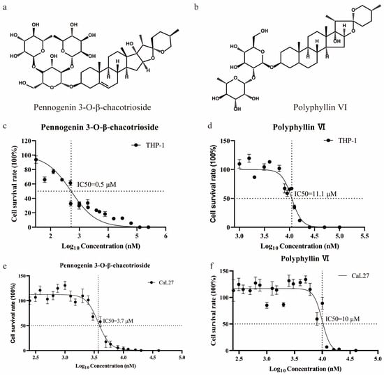
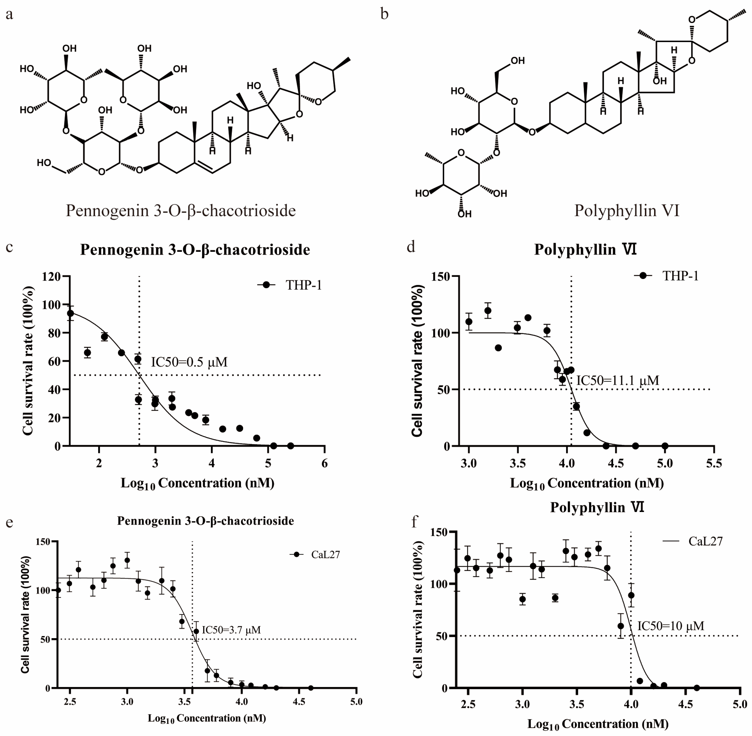
We first characterized the concentration-dependent cytotoxicity of two major steroidal saponins from Paris polyphylla, pennogenin 3-O-β-chacotrioside (P3Oβ) and polyphyllin VI (PPVI) (Figure 1a,b). THP-1 monocytes and oral squamous cell carcinoma (OSCC) CaL27 cells were treated with serial dilutions of each compound for 48 h (Supplementary Figure S1a,b). Cell viability, assessed using the CCK-8 assay, decreased in a concentration-dependent manner in both cell lines (Figure 1c–f). The calculated half-maximal inhibitory concentrations (IC50) for P3Oβ were 0.5 µM in THP-1 monocytes and 3.7 µM in CaL27 cells. The corresponding IC50 values for PPVI were 11.1 µM and 10.0 µM, respectively. Based on these findings, concentrations approximating the IC50 for THP-1 cells (0.5 µM for P3Oβ and 11.1 µM for PPVI) were selected for subsequent macrophage polarization experiments.
Figure 1.
P3Oβ and PPVI exhibit dose-dependent cytotoxicity on THP-1 monocytes and OSCC cells. (a,b) The molecular structures of pennogenin 3-O-β-chacotrioside (P3Oβ, (a)) and polyphyllin VI (PPVI, (b)) from Paris polyphylla. (c–f) Cell survival was determined by CCK-8 assay and IC50 values of THP-1 and CaL27 cells were determined for the cytotoxicity of P3Oβ and PPVI. n = 3 biological replicates. Error bars represent the mean ± SEM. IC50 values were calculated using non-linear regression analysis in GraphPad Prism v [8.4.2] with a sigmoidal dose–response curve fit. Statistical significance was determined using one-way ANOVA, followed by a two-side Dunnett’s test. IC50, half-maximal inhibitory concentration.
2.2. P3Oβ and PPVI Show Limited Effects on M0 and M2 Macrophage Viability at the Concentrations Used for Phenotypic Analyses
Because the concentrations initially selected for subsequent experiments were based on IC50 values determined in undifferentiated THP-1 monocytes, we further evaluated the effects of P3Oβ and PPVI on differentiated M0 macrophages and IL-4/IL-13-polarized M2 macrophages under the corresponding treatment conditions. M0 and M2 macrophages were treated for 48 h with P3Oβ at 0.125, 0.25, 0.5, or 5.0 μM, or with PPVI at 2.8, 5.6, 11.1, or 22.2 μM. Cell viability was assessed using the CCK-8 assay, apoptosis-associated DNA fragmentation was evaluated by TUNEL staining, and absolute viable cell numbers were determined by trypan blue exclusion after cell harvesting.
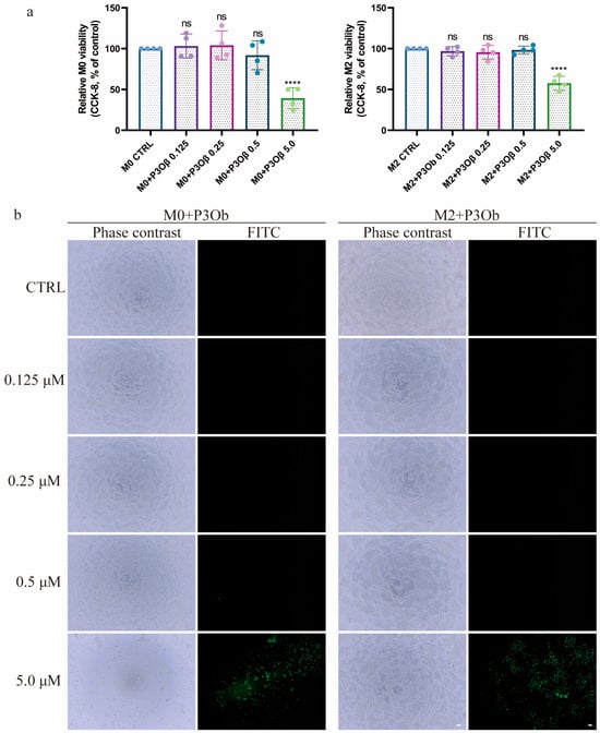
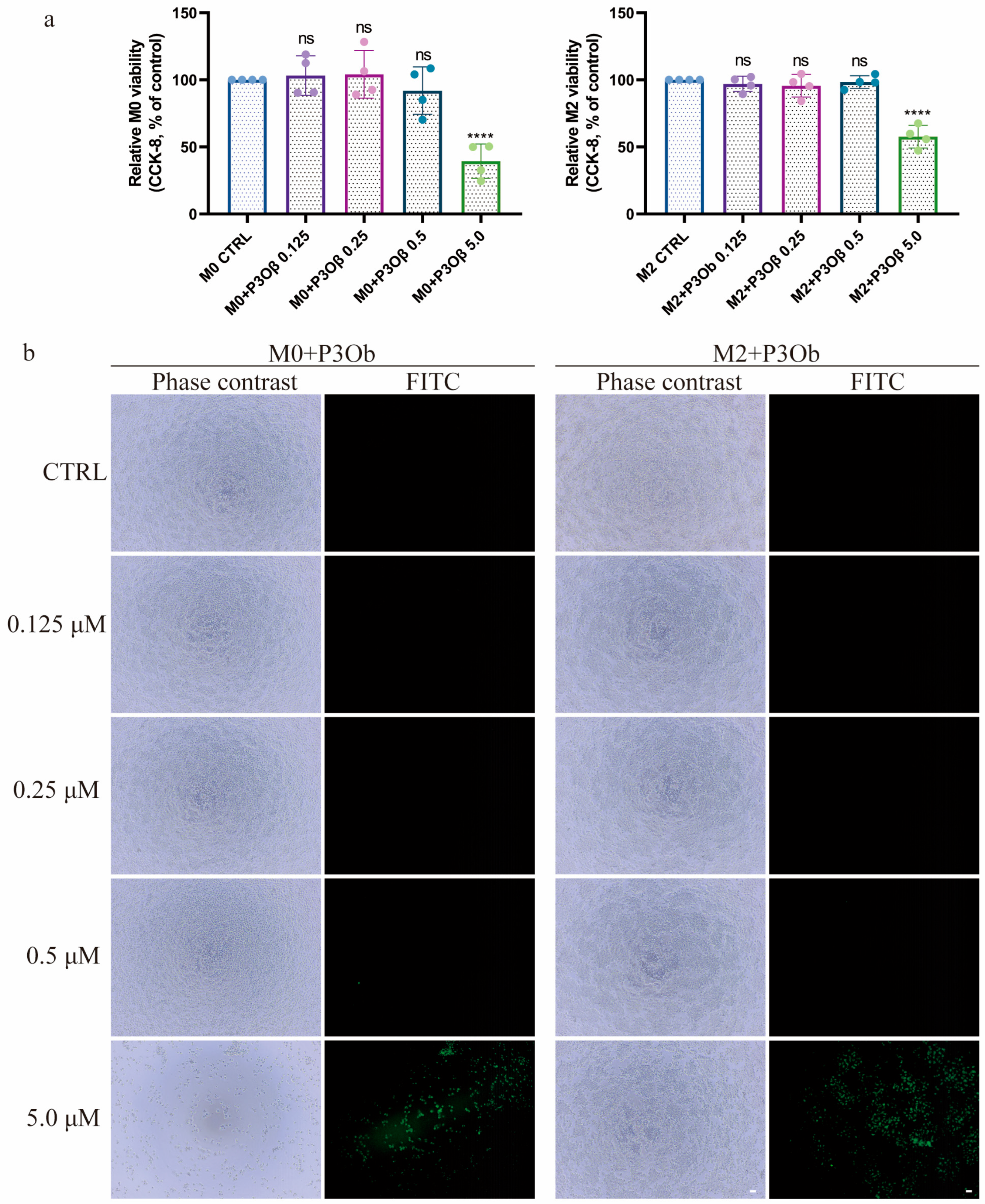
In M0 macrophages, treatment with 5.0 μM P3Oβ significantly reduced CCK-8 viability compared with the vehicle-treated control, whereas 0.125, 0.25, and 0.5 μM P3Oβ did not produce significant changes. None of the tested PPVI concentrations significantly affected M0 macrophage viability (Figure 2a, Supplementary Figure S2a). Consistent with these findings, TUNEL-positive staining was detectable in M0 macrophages treated with 5.0 μM P3Oβ, whereas little or no detectable TUNEL signal was observed at the lower P3Oβ concentrations or following PPVI treatment under the tested conditions (Figure 2b, Supplementary Figure S2b). Trypan blue exclusion-based cell counting showed a corresponding reduction in the absolute number of viable M0 macrophages following treatment with 5.0 μM P3Oβ, while no significant reduction was observed at 0.125–0.5 μM P3Oβ or 2.8–22.2 μM PPVI (Supplementary Figure S2c).
Figure 2.
Effects of P3Oβ and PPVI on the viability and TUNEL staining of M0 and M2 macrophages. M0 macrophages and IL-4/IL-13-polarized M2 macrophages were treated with P3Oβ (0.125, 0.25, 0.5, or 5.0 μM) for 48 h. (a) Cell viability was assessed using the CCK-8 assay and is presented relative to the corresponding vehicle-treated control. (b) Representative fluorescence images of TUNEL staining in M0 and M2 macrophages following treatment with the indicated concentrations of P3Oβ. TUNEL-positive cells show green fluorescence. Scale bar = 100 μm. Data are presented as mean ± SEM from 3 independent experiments. Statistical comparisons were performed using one-way ANOVA, followed by a two-side Dunnett’s test. **** p < 0.0001, ns = not significant, p ≥ 0.05.
A similar concentration-dependent pattern was observed in M2 macrophages. P3Oβ at 5.0 μM significantly reduced M2 macrophage viability, whereas 0.125, 0.25, and 0.5 μM P3Oβ had no significant effect. PPVI at 22.2 μM significantly reduced M2 macrophage viability, whereas 2.8, 5.6, and 11.1 μM PPVI did not significantly alter viability (Figure 2a, Supplementary Figure S2a). TUNEL staining was prominent following treatment with 5.0 μM P3Oβ and was also detectable in M2 macrophages exposed to 22.2 μM PPVI, whereas little or no detectable TUNEL staining was observed at the lower concentrations (Figure 2b, Supplementary Figure S2b). Absolute viable cell counts were consistent with the CCK-8 results, showing reductions following treatment with 5.0 μM P3Oβ and 22.2 μM PPVI, but no significant reduction at the lower concentrations used for subsequent phenotypic analyses (Supplementary Figure S2d).
2.3. P3Oβ Treatment Is Associated with an M1-like Polarization-Associated Marker Profile in M0 Macrophages
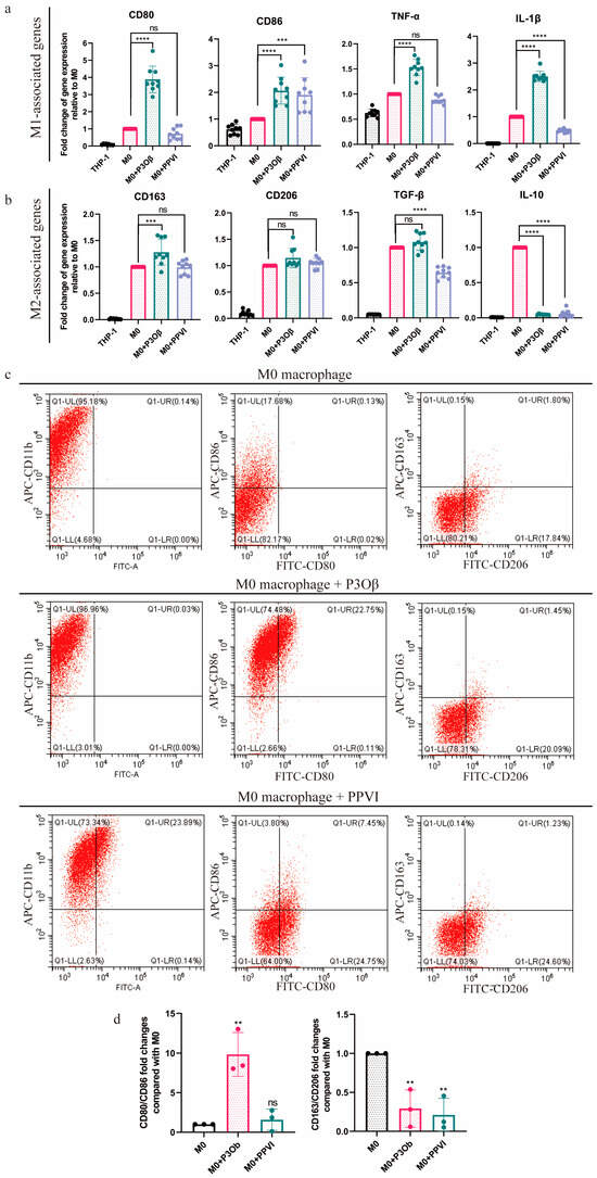
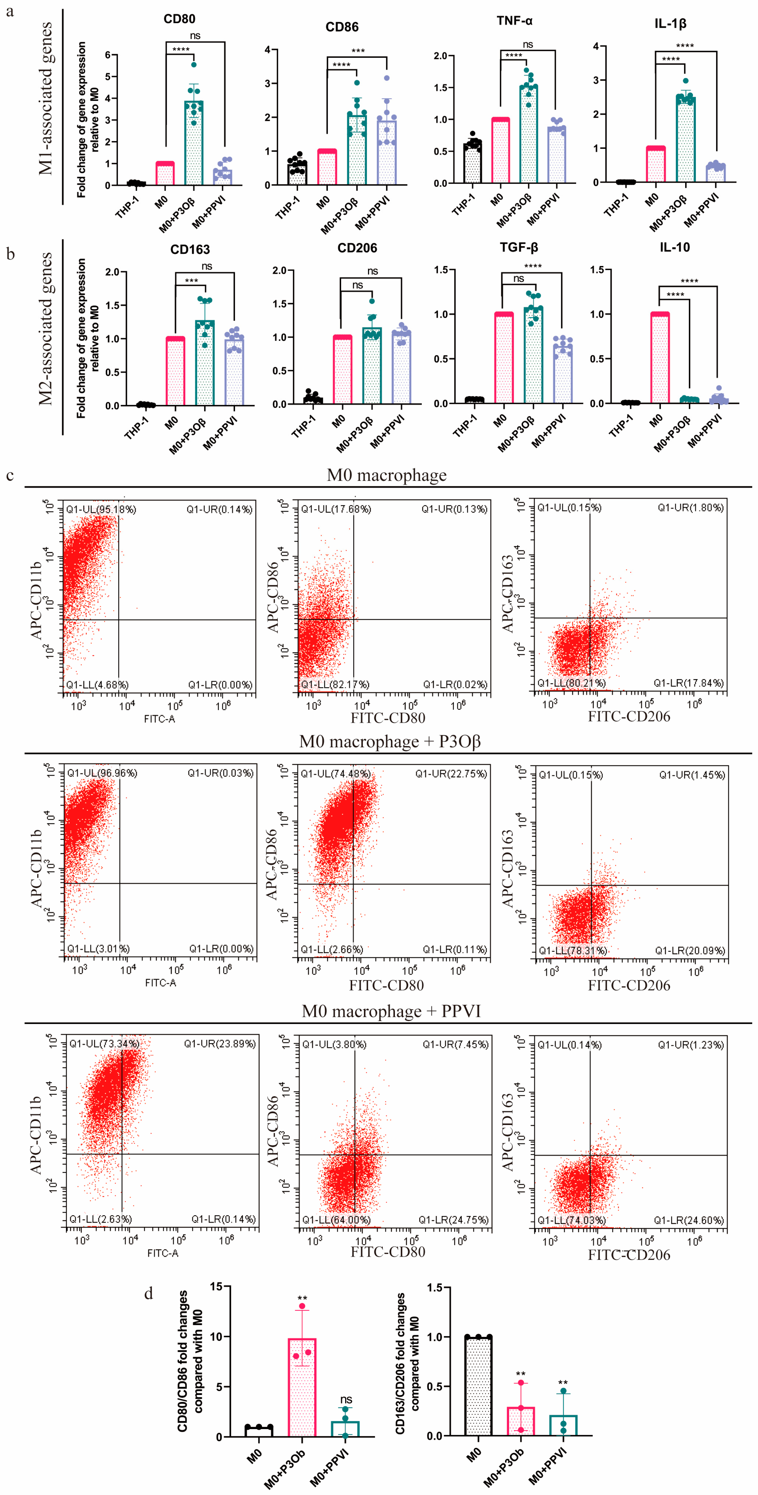
We next investigated the effects of P3Oβ and PPVI on macrophage polarization-associated markers. THP-1 cells were differentiated into M0 macrophages using PMA and subsequently treated with P3Oβ (0.5 µM) or PPVI (11.1 µM) for 48 h (Supplementary Figure S3a,b). Quantitative RT-PCR (qRT-PCR) analysis showed that P3Oβ treatment was associated with increased expression of several M1-associated transcripts, including CD80, CD86, TNF-α, and IL-1β. In comparison, PPVI produced a more restricted and marker-dependent response, including increased CD86 expression (Figure 3a). Among the M2-associated transcripts examined, P3Oβ reduced IL-10 expression, whereas PPVI reduced TGF-β and IL-10 expression. Neither compound produced a broad coordinated reduction in CD163 and CD206 expression under these conditions (Figure 3b).
Figure 3.
P3Oβ, but not PPVI, drives naïve macrophage toward an M1 phenotype. (a) qRT-PCR for the mRNA expression of M1-associated genes in M0-macrophages treated with P3Oβ (0.5 μM) or PPVI (11.1 μM) for 48 h, respectively. n = 3 biological replicates. Error bars represent the mean ± SEM. Significance refers to the comparison of different samples against M0 macrophages, which was determined using a two-sided Student t-test. **** p≤ 0.0001, *** p ≤ 0.001, ns = not significant, p ≥ 0.05. (b) qRT-PCR for the mRNA expression of M2-associated genes in M0 macrophages treated with P3Oβ (0.5 μM) or PPVI (11.1 μM) for 48 h, respectively. n = 3 biological replicates. Error bars represent the mean ± SEM. Significance refers to the comparison of different samples against M0 macrophages, which was determined using a two-sided Student t-test. **** p ≤ 0.0001, *** p ≤ 0.001, ns = not significant, p ≥ 0.05. (c) The flow cytometry analysis examined the M1 macrophages (positive for CD86 and/or CD80) and M2 macrophages (positive for CD163 and/or CD206) in M0 macrophages treated with P3Oβ (0.5 μM) or PPVI (11.1 μM) for 48 h, respectively. M0 macrophage discrimination was based on the expression of CD11b marker. n = 3 biological replicates. (d) Quantification of the flow cytometry results obtained in (c). Error bars represent the mean ± SEM. Significance refers to the comparison of different samples against M0 macrophages, which was determined using a two-sided Student t-test. ** p ≤ 0.01, ns = not significant, p ≥ 0.05. The gating strategy is shown in Supplementary Figure S4.
Flow cytometry further showed that P3Oβ significantly increased the proportion of CD80+CD86+ double-positive cells compared with vehicle-treated controls, whereas PPVI did not produce a comparable change in the M1 surface-marker profile (Figure 3c,d, gating strategy in Supplementary Figure S4). Neither compound increased the CD163+ CD206+ population (Figure 3c,d).
Taken together, the transcript-level and surface-marker data indicate that P3Oβ treatment is associated with an M1-like polarization-associated marker profile in PMA-differentiated THP-1 macrophages. In contrast, PPVI produced a more limited and heterogeneous pattern of marker changes under the same experimental conditions.
2.4. Transcriptomic Analysis Identifies an NF-κB-Associated Transcriptional Response to P3Oβ
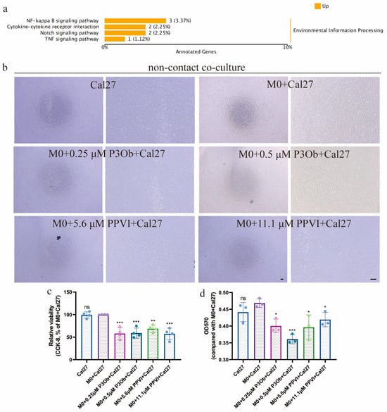
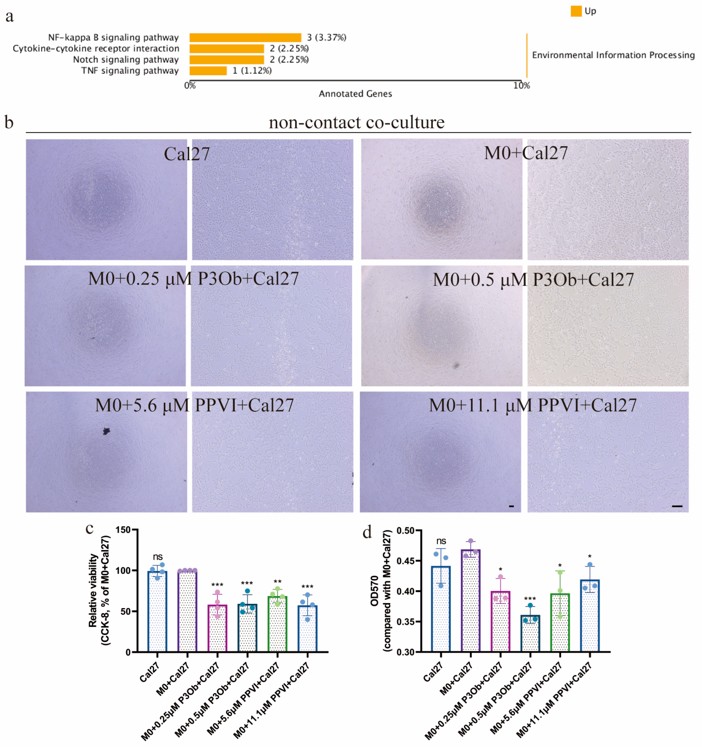
To further investigate the transcriptional changes associated with P3Oβ treatment, we performed whole-transcriptome RNA sequencing on M0 macrophages treated with vehicle or P3Oβ. Differential expression analysis identified a substantial set of genes that were significantly upregulated following P3Oβ treatment. Kyoto Encyclopedia of Genes and Genomes (KEGG) enrichment analysis of these upregulated genes identified multiple pathways related to inflammatory and immune responses. Notably, the NF-κB signaling pathway was among the significantly enriched pathways (Figure 4a).
Figure 4.
P3Oβ-associated transcriptional changes in M0 macrophages and the effects of compound-pretreated macrophages on CaL27 cells in a non-contact co-culture system (a) Kyoto Encyclopedia of Genes and Genomes (KEGG) pathway enrichment analysis of genes upregulated in M0 macrophages following treatment with P3Oβ compared with the vehicle-treated control. (b) Representative bright-field images of adherent CaL27 cells after 24 h of non-contact co-culture with vehicle-pretreated, P3Oβ-pretreated, or PPVI-pretreated M0 macrophages. M0 macrophages were pretreated with P3Oβ or PPVI for 48 h, after which compound-containing medium was removed and macrophages were washed before co-culture with CaL27 cells in compound-free medium. Scale bar = 100 μm. (c) CCK-8 assessment of CaL27 cell viability after 24 h of non-contact co-culture with M0 macrophages pretreated with P3Oβ (0.25 or 0.5 μM). CaL27 cells cultured alone and CaL27 cells co-cultured with vehicle-pretreated M0 macrophages were included as controls. (d) Representative crystal violet staining quantitative analysis of extracted crystal violet staining in adherent CaL27 cells after non-contact co-culture with the indicated macrophage groups. Crystal violet staining reflects the relative amount of adherent cell-associated staining remaining after co-culture. Data are presented as mean ± SEM from 3 independent experiments. Statistical comparisons were performed using one-way ANOVA, followed by a two-side Dunnett test. * p < 0.05, ** p < 0.01, *** p < 0.001, and ns = not significant, p ≥ 0.05.
These transcriptomic findings are consistent with the increased expression of several pro-inflammatory and M1-associated markers observed following P3Oβ treatment and indicate that P3Oβ exposure is associated with an NF-κB-related transcriptional program. Because pathway enrichment does not directly demonstrate biochemical activation of NF-κB signaling, these findings are interpreted as evidence of an NF-κB-associated transcriptional response rather than direct NF-κB activation.
2.5. Co-Culture with P3Oβ- or PPVI-Pretreated Macrophages Reduces CaL27 Viability
To determine whether compound-associated changes in macrophages were accompanied by functional effects on CaL27 cells independently of continuous compound exposure, we performed non-contact co-culture experiments using compound-pretreated macrophages. M0 macrophages were pretreated with P3Oβ or PPVI, after which the compound-containing medium was removed and the macrophages were washed before initiation of co-culture with CaL27 cells in compound-free medium. Concentrations that did not significantly reduce macrophage viability were selected for this experiment, including 0.25 and 0.5 μM P3Oβ and 5.6 and 11.1 μM PPVI. CaL27 cells cultured alone and CaL27 cells co-cultured with vehicle-treated M0 macrophages were included as controls. Microscopic examination showed a reduction in the density of adherent CaL27 cells after co-culture with P3Oβ- or PPVI-pretreated macrophages compared with the control conditions (Figure 4b). Consistent with these observations, CCK-8 analysis showed reduced viability of CaL27 cells following co-culture with macrophages pretreated with either 0.25 or 0.5 μM P3Oβ and with either 5.6 or 11.1 μM PPVI compared with CaL27 cells cultured alone and CaL27 cells co-cultured with vehicle-treated M0 macrophages (Figure 4c).
To provide an additional measurement independent of the CCK-8 assay, the remaining adherent CaL27 cells were assessed by crystal violet staining. The quantitative measurement of the extracted crystal violet staining showed changes consistent with the CCK-8 results, with lower adherent-cell-associated staining in the P3Oβ- and PPVI-pretreated macrophage co-culture groups than in the corresponding control groups (Figure 4d). Because the compounds were removed before initiation of the macrophage–CaL27 co-culture, these findings support an effect associated with compound-pretreated macrophages that persisted after withdrawal of extracellular compound exposure. However, these experiments do not establish a specific macrophage-mediated cytotoxic mechanism or demonstrate direct induction of CaL27 cell death. The findings are therefore interpreted as a macrophage-associated reduction in CaL27 viability under the tested non-contact co-culture conditions.
2.6. P3Oβ and PPVI Are Associated with Shifts in Polarization-Associated Marker Profiles in IL-4/IL-13-Polarized Macrophages
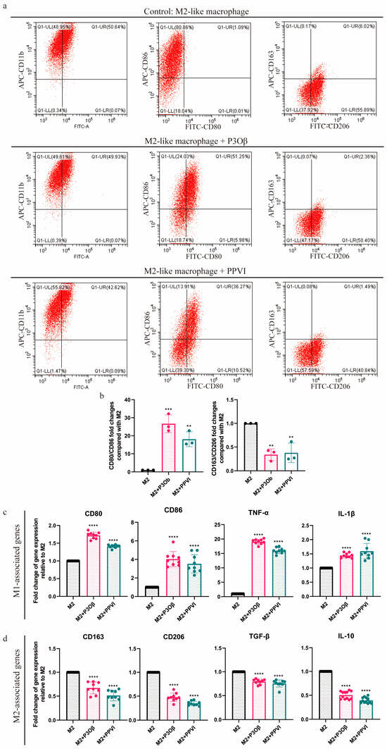
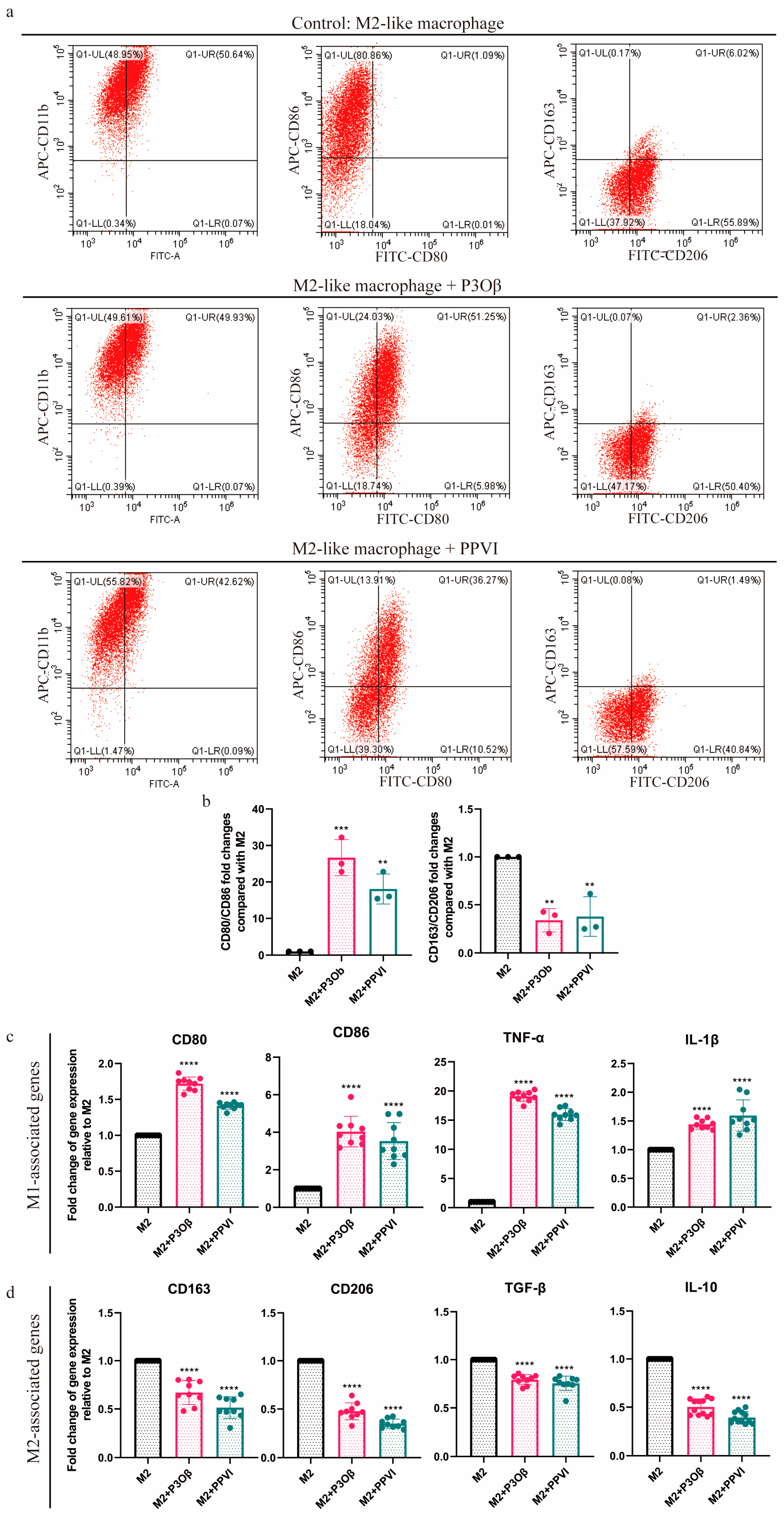
Because tumor-associated macrophages frequently exhibit M2-like characteristics, we next examined whether P3Oβ and PPVI were associated with changes in polarization-associated markers in macrophages that had first been polarized with IL-4 and IL-13. Flow cytometry analysis showed that treatment with either P3Oβ or PPVI increased the CD80+CD86+ population and decreased the CD163+CD206+ population relative to the corresponding polarized control group (Figure 5a,b). These surface-marker changes were accompanied by alterations at the transcript level. qRT-PCR analysis showed increased expression of several M1-associated genes, including CD80, CD86, TNF-α, and IL-1β, together with decreased expression of multiple M2-associated genes, including CD163, CD206, TGF-β, and IL-10 (Figure 5c,d).
Figure 5.
P3Oβ and PPVI are associated with shifts in polarization-associated marker profiles in IL-4/IL-13-polarized macrophages. (a) The flow cytometry analysis examined the M1 macrophages (positive for CD86 and/or CD80) and M2 macrophages (positive for CD163 and/or CD206) in M2 macrophages treated with P3Oβ (0.5 μM) or PPVI (11.1 μM) for 48 h, respectively. n = 3 biological replicates. (b) Quantification of the flow cytometry results obtained in a. Error bars represent the mean ± SEM. Significance refers to the comparison of different samples against M2 macrophages, which was determined using a two-sided Student t-test. *** p ≤ 0.001, ** p ≤ 0.01. The gating strategy was shown in Supplementary Figure S4. (c) qRT-PCR for the mRNA expression of M1-associated genes in M2 macrophages treated with P3Oβ (0.5 μM) or PPVI (11.1 μM) for 48 h, respectively. n = 3 biological replicates. Error bars represent the mean ± SEM. Significance refers to the comparison of different samples against M2-macrophages, which was determined using a two-sided Student t-test. **** p ≤ 0.0001. (d) qRT-PCR for the mRNA expression of M2-associated genes in M2 macrophages treated with P3Oβ (0.5 μM) or PPVI (11.1 μM) for 48 h, respectively. n = 3 biological replicates. Error bars represent the mean ± SEM. Significance refers to the comparison of different samples against M2 macrophages, which was determined using a two-sided Student t-test. **** p ≤ 0.0001.
The concordant changes in surface and transcript-level markers are consistent with a shift from an M2-associated marker profile to a more M1-like phenotype under the tested conditions. Importantly, parallel viability experiments showed that neither 0.5 μM P3Oβ nor 11.1 μM PPVI significantly reduced the CCK-8 signal in M2 macrophages, and neither condition was associated with evident TUNEL-positive staining. These findings support an association between P3Oβ or PPVI exposure and altered polarization-associated marker profiles in IL-4/IL-13-polarized macrophages. Nevertheless, the results are interpreted as phenotypic marker changes rather than evidence of complete macrophage reprogramming or acquisition of a defined antitumor functional state.
3. Discussion
The TIME represents an important obstacle to effective cancer therapy, and TAMs are major cellular components of the OSCC microenvironment [2,11]. Macrophages exhibit substantial phenotypic plasticity in response to environmental signals, which has generated considerable interest in strategies aimed at modifying tumor-supportive macrophage states toward phenotypes associated with inflammatory and antitumor immune responses [10,27]. In the present study, we investigated the effects of two steroidal saponins from Paris polyphylla, P3Oβ and PPVI, on macrophage polarization-associated phenotypes. P3Oβ produced a relatively broad M1-like marker shift in PMA-differentiated THP-1-derived macrophages, whereas PPVI elicited a more restricted response under the same M0 conditions. Both compounds also altered polarization-associated marker profiles in IL-4/IL-13-polarized macrophages. Importantly, these phenotypic changes were observed at concentrations associated with minimal detectable macrophage cytotoxicity. In addition, macrophage pretreatment followed by compound washout was associated with reduced CaL27 viability during subsequent non-contact co-culture.
A substantial body of previous work on Chonglou steroidal saponins has focused on their direct cytotoxic effects on malignant cells [25,28,29,30]. Consistent with these reports, direct exposure to P3Oβ or PPVI reduced the viability of both THP-1 monocytes and CaL27 cells in a concentration-dependent manner. The macrophage experiments, however, revealed additional effects under conditions in which differentiated macrophage viability was largely preserved. In M0 macrophages, P3Oβ increased several M1-associated transcripts, including CD80, CD86, TNF-α, and IL-1β, together with increasing the proportion of CD80+CD86+ cells. PPVI produced a more limited and marker-dependent response, including increased CD86 expression without a comparable increase in the CD80+CD86+ population. Moreover, neither compound induced a uniformly coordinated change across all M1- and M2-associated markers. These findings highlight the multidimensional nature of macrophage phenotypes and support interpretation of the observed effects as shifts in polarization-associated marker profiles rather than conversion into a discrete macrophage state.
The transcriptomic analysis provides additional context for the P3Oβ-associated phenotype. RNA sequencing followed by KEGG pathway enrichment analysis identified several inflammatory and immune-related pathways among genes upregulated after P3Oβ treatment, including the NF-κB signaling pathway. NF-κB is an important regulator of inflammatory gene expression and has been extensively associated with macrophage responses to inflammatory stimuli [31,32,33]. Enrichment of NF-κB-related genes is therefore consistent with the accompanying increase in TNF-α, IL-1β, and other M1-associated markers. Nevertheless, pathway enrichment reflects coordinated transcriptional changes in genes associated with a signaling pathway and does not establish biochemical pathway activation or causal dependence. Because phosphorylation of NF-κB pathway components, nuclear translocation, transcriptional reporter activity, and pharmacological or genetic inhibition were not examined, the present findings are interpreted specifically as evidence of an NF-κB-associated transcriptional response. Whether NF-κB signaling is mechanistically required for the P3Oβ-associated marker changes remains to be determined.
The responses observed in IL-4/IL-13-polarized macrophages further indicate that the effects of P3Oβ and PPVI depend, at least in part, on the pre-existing macrophage state. Treatment with either compound increased the CD80+CD86+ population and decreased the CD163+CD206+ population relative to the polarized control, accompanied by corresponding changes in M1- and M2-associated transcripts [18,34]. The concordance between transcript-level and surface-marker measurements supports a shift away from the IL-4/IL-13-induced M2-associated marker profile toward a more M1-like phenotype. However, macrophage polarization represents a continuum of activation states rather than a simple M1/M2 dichotomy, particularly within tumors. The present observations should therefore not be interpreted as evidence of complete macrophage reprogramming or acquisition of a defined antitumor macrophage state.
An important consideration is whether the observed changes in macrophage markers could be secondary to cytotoxic stress or selective cell loss. Because the concentrations used for phenotypic analyses were initially selected on the basis of IC50 values determined in undifferentiated THP-1 monocytes, we directly evaluated their effects in differentiated M0 and IL-4/IL-13-polarized M2 macrophages. At the concentrations used for the phenotypic experiments (0.5 μM P3Oβ and 11.1 μM PPVI), neither compound significantly reduced macrophage viability or absolute viable cell number, and little or no TUNEL-positive staining was detected. In contrast, cytotoxic effects became evident at higher concentrations, particularly with 5.0 μM P3Oβ and, in M2 macrophages, 22.2 μM PPVI. These findings reduce the likelihood that the polarization-associated marker changes observed at the experimental concentrations primarily reflect overt cytotoxicity or selective survival, although more subtle forms of cellular stress cannot be excluded.
The compound-washout co-culture experiments extend these phenotypic observations by linking macrophage pretreatment to a subsequent change in CaL27 cell viability. This distinction is particularly important because both P3Oβ and PPVI can directly affect CaL27 cells, making continuous compound exposure difficult to distinguish from a macrophage-associated effect. Restricting compound exposure to the macrophage pretreatment phase, followed by washout and non-contact co-culture in compound-free medium, substantially reduces continuous extracellular drug exposure as an explanation for the observed reduction in CaL27 viability. The concordant decreases in CCK-8 signal and crystal violet staining further indicate that the observation was not restricted to a single analytical readout. Because macrophages and CaL27 cells were physically separated during co-culture, the effect is compatible with the involvement of diffusible macrophage-derived signals rather than requiring direct macrophage–tumor cell contact. However, the responsible mediators remain unknown, and the current assays do not distinguish among reduced proliferation, altered metabolic activity, increased detachment, or direct tumor-cell death. Thus, the present findings support a macrophage-associated reduction in CaL27 viability rather than establishing a specific macrophage-mediated cytotoxic mechanism. Analysis of macrophage-conditioned medium, secreted cytokines or other soluble mediators, mediator-neutralization experiments, and direct measurements of tumor-cell proliferation and death will be required to define the underlying mechanism.
The differential responses to P3Oβ and PPVI are also noteworthy. P3Oβ produced a relatively consistent M1-like marker profile in M0 macrophages and was associated with an NF-κB-related transcriptional program, whereas PPVI induced more limited changes in M0 macrophages but more pronounced alterations after IL-4/IL-13 polarization. Despite these differences, pretreatment with either compound was associated with reduced CaL27 viability after washout. This divergence suggests that conventional M1/M2-associated markers alone may not fully predict the functional consequences of macrophage modulation. Macrophage-derived effects on neighboring tumor cells are likely to reflect integrated changes in secreted factors, metabolic state, and signaling networks rather than a single polarization marker or predefined macrophage category. Broader transcriptomic, proteomic, and secretome analyses may therefore help identify the macrophage-associated changes most closely related to the observed CaL27 response.
Several limitations should be considered when interpreting these findings. First, THP-1-derived macrophages and IL-4/IL-13-induced polarization provide experimentally tractable models but do not reproduce the heterogeneity or tissue-specific characteristics of primary human macrophages and TAMs within OSCC. Validation using primary monocyte-derived macrophages and, where feasible, patient-derived OSCC-associated macrophages would therefore strengthen the biological relevance of these observations. Second, although transcriptomic enrichment implicates an NF-κB-associated response to P3Oβ, direct biochemical and functional validation of this pathway remains necessary. Third, the washout co-culture experiments support a macrophage-associated reduction in CaL27 viability but neither identify the responsible macrophage-derived mediators nor establish direct tumor-cell killing. Finally, in vivo studies will be required to determine whether these macrophage-associated effects are maintained within the substantially more complex OSCC immune microenvironment.
Taken together, the present findings indicate that P3Oβ and PPVI modulate macrophage polarization-associated marker profiles under conditions in which overt macrophage cytotoxicity is limited. P3Oβ was associated with a broader M1-like marker response in M0 macrophages and an NF-κB-associated transcriptional program, whereas both P3Oβ and PPVI shifted IL-4/IL-13-polarized macrophages toward a more M1-like marker profile. Furthermore, macrophages pretreated with either compound were associated with reduced CaL27 viability after compound withdrawal in a non-contact co-culture system. These observations support further investigation of P3Oβ and PPVI as macrophage-modulatory steroidal saponins while emphasizing that the molecular mediators, functional mechanisms, and in vivo relevance of these macrophage-associated effects remain to be established.
4. Materials and Methods
4.1. Cell Culture
The human acute monocytic leukemia cell line THP-1 (TIB-202) and human Jurkat T cells (TIB-152) were acquired from American Type Culture Collection (ATCC) and mantained in RPMI 1640 medium (HyClone, Logan, UT, USA) supplemented with 10% fetal bovine serum (FBS, Biological Industries, Kibbutz Beit-Haemek, Israel) at 37 °C in a humidified atmosphere containing 5% CO2. THP-1 cells were differentiated into M0 macrophages by treatment with phorbol 12-myristate 13-acetate (PMA, 100 ng/mL, #1652981, BioGems, Westlake Village, CA, USA) for 48 h. To induce the polarization of M2 macrophages, M0 macrophages were treated with Interleukin-4 (IL-4, 20 ng/mL, # TMPY-01862, TopScience, Shanghai, China) and IL-13 (20 ng/mL, #TMPY-01993, TopScience, China) for 48 h. The human oral squamous carcinoma cell line CaL27 (CRL-2095) was acquired from ATCC and maintained in Dulbecco’s Modified Eagle’s Medium F12 (DMEM/F12, Merck Millipore, Darmstadt, Germany) supplemented with 10% FBS under 37 °C, with 5% CO2. The identities of the cell lines used in this study were verified by short tandem repeat (STR) profiling. All experiments were performed using cells within 10 passages after recovery from cryopreserved stocks to minimize potential phenotypic changes associated with prolonged in vitro culture.
For compound-treatment experiments, Pennogenin 3-O-β-chacotrioside (P3Oβ, #HY-N4180, MedChemExpress, Monmouth Junction, NJ, USA) and Polyphyllin VI (PPVI, #S9302, Selleck, Shanghai, China) were applied for 48 h at the concentrations specified for each experiment.
4.2. Cell Viability and IC50 Assay
Cell viability was determined using Cell Counting Kit-8 (CCK-8, #C0005, TargetMol, Shanghai, China). A total of 1 × 10⁴ cells were seeded per well in a 96-well plate, at least in 3 triplicates. After a 6-h adhesion period, cells were treated with varying concentrations of P3Oβ and PPVI as indicated in the figure. Following a 48-h treatment period, cells were exposed to 10 μL of CCK-8 reagent (100 μL medium per well) for 2 h at 37 °C, 5% CO2. The absorbance at a wavelength of 450 nm was measured by Bio Tek Synergy LX multimode reader at 570 nm (Bio Tek Instruments, Inc. Winooski, VT, USA). IC50 was calculated using GraphPad Prism non-linear regression.
4.3. RNA Isolation and qRT-PCR
Total RNA was extracted using the Super FastPure Cell RNA Isolation Kit (#RC102-01, Vazyme, Nanjing, China). For cDNA synthesis, 2 μg of RNA was utilized with the HiScript III All-in-one RT SuperMix Perfect for qPCR (#R333, Vazyme, China). qRT-PCR was conducted using the LightCycler 480 SYBR-Green I Master (Roche Diagnostics, Basel, Switzerland) as per the manufacturer’s guidelines. The primer sequences for qRT-PCR were as follows: CD80 (forward primer: 5′-TGTCGCCTCTCTGAAGATTACC-3′; reverse primer: 5′-TTCCTGGGTCTCCAAAGGTTG-3′). CD86 (forward primer: 5′-CCTTCCTGCTCTCTGCTAACTT-3′; reverse primer: 5′-ACAAGCTGATGGAAACGTCGT-3′). TNFα (forward primer: 5′-CTCTCTCTAATCAGCCCTCTGG-3′; reverse primer: 5′-AGGACCTGGGAGTAGATGAGG-3′). IL-1β (forward primer: 5′-TCGCCAGTGAAATGATGGCT-3′; reverse primer: 5′-AGATTCGTAGCTGGATGCCG-3′). CD163 (forward primer: 5′-GACGCATTTGGATGGATCATGTT-3′; reverse primer: 5′-CCCACCGTCCTTGGAATTTGA-3′). CD206 (forward primer: 5′-TCCGGGTGCTGTTCTCCTA-3′; reverse primer: 5′-CAGTCCGTTTTTGATGGCACTC-3′). TGF-β (forward primer: 5′-CTTCAATACGTCAGACATTCGGG-3′; reverse primer: 5′-GTAACGCCAGGAATTGTTGCTA-3′). IL-10 (forward primer: 5′-ACTTTAAGGGTTACCTGGGTTGC-3′; reverse primer: 5′-TCACATGCGCCTTGATGTCT-3′).
4.4. Flow Cytometry
Cells were dissociated into a single-cell suspension and permeabilized in 0.1% Triton-100 (#T9284, Sigma-Aldrich, St. Louis, MO, USA) for 10 min. Subsequently, the cells were incubated with antibody at 4 °C for 1 h. The gating strategy is described in Supplementary Information. Flow cytometric analysis was performed on a CytExpert flow cytometer (Beckman Coulter, Inc. Brea, CA, USA) and data analysis was performed using CytExpert 2.3 software. The antibodies employed in this study were as follows: APC anti-human CD11b (#301309, BioLegend, San Diego, CA, USA), FITC anti-human CD80 (#375405, BioLegend), APC anti-human CD86 (#374207, BioLegend), FITC anti-human CD206 (MMR, #321103, BioLegend), and APC anti-human CD163 (#326509, BioLegend).
4.5. Assessment of Apoptosis, and Absolute Viable Cell Number in M0 and M2 Macrophages
To evaluate whether the concentrations of P3Oβ and PPVI used in the phenotypic analyses affected differentiated macrophage viability, dose–response experiments were performed in both M0 and M2 macrophages. THP-1 monocytes were differentiated into M0 macrophages by treatment with 100 ng/mL phorbol 12-myristate 13-acetate (PMA; Sigma-Aldrich, St. Louis, MO, USA) for 48 h. For M2 polarization, PMA-differentiated macrophages were further stimulated with IL-4 and IL-13 as described above. M0 and M2 macrophages were subsequently treated for 48 h with P3Oβ at 0.125, 0.25, 0.5, or 5.0 μM; PPVI at 2.8, 5.6, 11.1, or 22.2 μM; or the corresponding vehicle control.
Apoptosis-associated DNA fragmentation was additionally examined using a One Step TUNEL Apoptosis Assay Kit (green fluorescence; #C1088, Beyotime Biotechnology, Shanghai, China). Following treatment, adherent macrophages were gently washed with PBS and fixed with 4% paraformaldehyde for 30 min at room temperature. Cells were washed with PBS and permeabilized with PBS containing 0.3% Triton X-100 for 5 min at room temperature. The TUNEL reaction mixture was freshly prepared according to the manufacturer’s instructions by combining TdT enzyme with fluorescent labeling solution. Cells were incubated with the TUNEL reaction mixture at 37 °C for 60 min in the dark and subsequently washed with PBS. TUNEL fluorescence was visualized using a fluorescence microscope under identical imaging settings among experimental groups. Representative fields were acquired for comparison of TUNEL-positive staining.
To independently assess changes in viable macrophage number, absolute viable cell counts were determined using the trypan blue exclusion method. M0 and M2 macrophages were cultured in 24-well plates and treated with the indicated concentrations of P3Oβ or PPVI for 48 h. After treatment, cells were harvested from individual wells, resuspended thoroughly to obtain single-cell suspensions, and mixed with 0.4% trypan blue solution. Unstained viable cells and trypan blue-positive non-viable cells were counted using a hemocytometer. The absolute number of viable cells per well was calculated and compared with that of the corresponding vehicle-treated control. Each well was analyzed independently.
4.6. Assessment of CaL27 Viability in a Macrophage–CaL27 Non-Contact Co-Culture System Following Compound Washout
A transwell-based non-contact co-culture system was established to determine whether pretreatment of macrophages with P3Oβ or PPVI altered their subsequent effects on CaL27 cells after removal of the compounds. THP-1 monocytes (5 × 10³ cells per insert) were seeded into the upper chambers of 0.4-μm-pore polyester transwell inserts (Merck Millipore, Germany) and differentiated into M0 macrophages by treatment with 100 ng/mL PMA for 48 h. Following differentiation, non-adherent cells were removed by gentle washing with PBS. The differentiated M0 macrophages were subsequently treated for 48 h with P3Oβ (0.25 or 0.5 μM), PPVI (5.6 or 11.1 μM), or the corresponding vehicle control. At the end of the pretreatment period, compound-containing medium was completely removed, and the macrophages were gently washed three times with PBS to minimize residual compound carryover. Fresh compound-free complete culture medium was then added to the inserts before initiation of the co-culture experiment.
CaL27 cells (1 × 10⁴ cells per well) were seeded into 24-well plates 6 h before initiation of co-culture. The compound-pretreated and washed macrophage-containing transwell inserts were then transferred to the CaL27-containing wells. The experimental conditions included CaL27 cells cultured alone, CaL27 cells co-cultured with vehicle-pretreated M0 macrophages, CaL27 cells co-cultured with M0 macrophages pretreated with 0.25 or 0.5 μM P3Oβ, and CaL27 cells co-cultured with M0 macrophages pretreated with 5.6 or 11.1 μM PPVI. No compound was intentionally added during the co-culture period. After 24 h of non-contact co-culture, representative bright-field images of the adherent CaL27 cells in the lower chamber were acquired using an inverted microscope with a 4× and 10× objective. Imaging conditions were kept constant among experimental groups.
4.7. Crystal Violet Staining and Quantitative Assessment of Adherent CaL27 Cells
Crystal violet staining was performed as an independent assay to assess the relative amount of adherent CaL27 cells remaining after macrophage–CaL27 co-culture. Following the 24-h co-culture period, the transwell inserts were removed and CaL27 cells in the lower chamber were gently washed with PBS to remove non-adherent material. Cells were fixed with 4% paraformaldehyde for 30 min at room temperature and subsequently stained with 2% crystal violet solution for 30 min at room temperature. Excess crystal violet was removed, and the wells were gently washed with distilled water until the background staining was eliminated. For quantitative analysis, the crystal violet retained by adherent cells was solubilized using 30% acetic acid. After complete solubilization, equal volumes of the resulting solution were transferred to a 96-well plate, and absorbance was measured at 570 nm using a microplate reader.
4.8. Statistical Analysis
Comparisons between two groups were made using an unpaired two-sided Student t-test, while comparisons among multiple groups utilized one-way or two-way analysis of variance (ANOVA) with Dunnett’s multiple comparisons tests. These analyses were performed using GraphPad Prism Software, version 9 (GraphPad Software). A p < 0.05 was considered statistically significant. Error bars in the figures represent the standard error of the mean (SEM). All experiments were conducted independently at least three times.
5. Conclusions
In this in vitro study, P3Oβ and PPVI were found to modulate macrophage polarization-associated phenotypes in THP-1-derived macrophages. P3Oβ produced a relatively broad M1-like marker profile in PMA-differentiated M0 macrophages and was associated with an NF-κB-related transcriptional response, whereas PPVI induced a more limited and context-dependent pattern of marker changes. In IL-4/IL-13-polarized macrophages, both compounds shifted polarization-associated surface and transcriptional markers away from an M2-like profile toward a more M1-like phenotype. Furthermore, following compound pretreatment and washout, non-contact co-culture with P3Oβ- or PPVI-pretreated macrophages was associated with reduced CaL27 viability in compound-free medium. These findings support an association between compound-induced macrophage changes and subsequent effects on OSCC cells, while not establishing a specific macrophage-mediated cytotoxic mechanism.
Overall, these results identify P3Oβ and PPVI as potential modulators of macrophage phenotype and macrophage–tumor cell interactions. Further studies using primary macrophage models, mechanistic analysis of macrophage-derived mediators, direct tumor-cell death assays, and appropriate in vivo OSCC models will be required to define the underlying mechanisms and evaluate the translational relevance of these observations.
Supplementary Materials
The following supporting information can be downloaded at https://www.mdpi.com/article/10.3390/molecules31224067/s1: Figure S1. The bright filed images shown the results of THP-1 cells after treatment with different concentrations of P3Oβ for 48 h. n = 3 independent cell cultures. Scale bar = 100 μm. Figure S2. M0 macrophages and IL-4/IL-13-polarized M2 macrophages were treated with PPVI (2.8, 5.6, 11.1, or 22.2 μM) for 48 h. (a) CCK-8 assay of macrophage viability relative to the corresponding vehicle-treated control. (b) Representative TUNEL-stained images of M0 and M2 macrophages following treatment with the indicated concentrations of PPVI. TUNEL-positive cells are shown in green. Scale bar = 100 μm. (c) Absolute viable M0 macrophage numbers determined by trypan blue exclusion after treatment with P3Oβ or PPVI. (d) Absolute viable M2 macrophage numbers determined by trypan blue exclusion after treatment with P3Oβ or PPVI. Data are presented as mean ± SEM from 3 independent experiments. Statistical comparisons were performed using one-way ANOVA, followed by a two-side Dunnett’s test. * p < 0.05, ** p < 0.01, ns = not significant, p ≥ 0.05. Figure S3. (a) The bright filed images shown the results of THP-1 cells after treatment with different concentrations of PPVI for 48 h. n = 3 independent cell cultures. Scale bar = 100 μm. (b) The images shown different macrophage states when cultured in vitro. Undifferentiated THP-1 monocytes grow in suspension, after 48 h of PMA stimulation, THP-1 monocytes differentiated into adherent M0-macrophages. M0-macrophages differentiated into M2-macrophages when treated with IL-4 and IL-13 for 48 h. Scale bar = 100 μm. Representative images showing the cellular morphology of M0 macrophages at 24 and 48 hours after treatment with P3Oβ (0.5 μM) and PPVI (11.1 μM). Scale bar = 100 μm. Figure S4. The gating strategy was used in the FACS data panels of Figure 3c and Figure 5a. Representative dots plots demonstrated gating strategy to assess positive cells by Flow cytometry. THP-1 cells were incubated with the isotype antibody serve as a blank control group (upper panel). Representative flow plots showed THP-1 staining on CD11b+gated cells (left), CD86+CD80+gated cells (middle), CD163+CD206+gated cells (right, middle panel). M0-macrophages were incubated with the isotype antibody serve as a blank control group (lower panel). PMA, phorbol 12-myristate 13-acetate.
Author Contributions
W.L., J.X., and L.C. conceived and designed this study, collected data, performed data analysis and prepared the manuscript, and provided funding for the conduct of this study. S.Y., H.Z., F.L., and J.Z.: formal analysis, validation, and review and editing. All authors have read and agreed to the published version of the manuscript.
Funding
This work was supported by grants from The Educational and Scientific Research Program for Young and Middle-aged Instructor of Fujian province (No. JAT231109, JAT210944), the Natural Science Foundation of Fujian Province (No. 2024J01314, 2024J01892), and Sanming University Research Fund Program for Introducing High-Level Talents (22YG16).
Institutional Review Board Statement
Not applicable.
Informed Consent Statement
Not applicable.
Data Availability Statement
The data that support the findings of this study are available from the corresponding authors upon reasonable request.
Acknowledgments
We thank the laboratory of Yan Zhang at Sun Yat-sen University for their technical support and valuable suggestions in this study.
Conflicts of Interest
The authors declare no conflicts of interest.
References
- Johnson, D.E.; Burtness, B.; Leemans, C.R.; Lui, V.W.Y.; Bauman, J.E.; Grandis, J.R. Head and neck squamous cell carcinoma. Nat. Rev. Dis. Primers 2020, 6, 92. [Google Scholar] [CrossRef] [Scilit] [PubMed]
- Leemans, C.R.; Snijders, P.J.F.; Brakenhoff, R.H. The molecular landscape of head and neck cancer. Nat. Rev. Cancer 2018, 18, 269–282. [Google Scholar] [CrossRef] [Scilit] [PubMed]
- Fu, T.; Dai, L.J.; Wu, S.Y.; Xiao, Y.; Ma, D.; Jiang, Y.Z.; Shao, Z.M. Spatial architecture of the immune microenvironment orchestrates tumor immunity and therapeutic response. J. Hematol. Oncol. 2021, 14, 98. [Google Scholar] [CrossRef] [Scilit] [PubMed]
- Lei, X.; Lei, Y.; Li, J.K.; Du, W.X.; Li, R.G.; Yang, J.; Li, J.; Li, F.; Tan, H.B. Immune cells within the tumor microenvironment: Biological functions and roles in cancer immunotherapy. Cancer Lett. 2020, 470, 126–133. [Google Scholar] [CrossRef] [Scilit] [PubMed]
- Leone, R.D.; Zhao, L.; Englert, J.M.; Sun, I.M.; Oh, M.H.; Sun, I.H.; Arwood, M.L.; Bettencourt, I.A.; Patel, C.H.; Wen, J.; et al. Glutamine blockade induces divergent metabolic programs to overcome tumor immune evasion. Science 2019, 366, 1013–1021. [Google Scholar] [CrossRef] [Scilit] [PubMed]
- Pan, Y.; Yu, Y.; Wang, X.; Zhang, T. Tumor-Associated Macrophages in Tumor Immunity. Front. Immunol. 2020, 11, 583084. [Google Scholar] [CrossRef] [Scilit] [PubMed]
- Chu, X.; Tian, Y.; Lv, C. Decoding the spatiotemporal heterogeneity of tumor-associated macrophages. Mol. Cancer 2024, 23, 150. [Google Scholar] [CrossRef] [Scilit] [PubMed]
- Zhu, Y.; Zhou, Z.; Du, X.; Lin, X.; Liang, Z.M.; Chen, S.; Sun, Y.; Wang, Y.; Na, Z.; Wu, Z.; et al. Cancer cell-derived arginine fuels polyamine biosynthesis in tumor-associated macrophages to promote immune evasion. Cancer Cell 2025, 43, 1045–1060.e7. [Google Scholar] [CrossRef] [Scilit] [PubMed]
- Xiang, X.; Wang, J.; Lu, D.; Xu, X. Targeting tumor-associated macrophages to synergize tumor immunotherapy. Signal Transduct. Target. Ther. 2021, 6, 75. [Google Scholar] [CrossRef] [Scilit] [PubMed]
- Park, J.; Hsueh, P.C.; Li, Z.; Ho, P.C. Microenvironment-driven metabolic adaptations guiding CD8(+) T cell anti-tumor immunity. Immunity 2023, 56, 32–42. [Google Scholar] [CrossRef] [Scilit] [PubMed]
- Li, C.X.; Gong, Z.C.; Yu, J.W. Deliberation concerning the role of M1-type macrophage subset in oral carcinogenesis. J. Exp. Clin. Cancer Res. 2024, 43, 220. [Google Scholar] [CrossRef] [Scilit] [PubMed]
- Shao, N.; Qiu, H.; Liu, J.; Xiao, D.; Zhao, J.; Chen, C.; Wan, J.; Guo, M.; Liang, G.; Zhao, X.; et al. Targeting lipid metabolism of macrophages: A new strategy for tumor therapy. J. Adv. Res. 2025, 68, 99–114. [Google Scholar] [CrossRef] [Scilit] [PubMed]
- Chen, D.; Zhang, X.; Li, Z.; Zhu, B. Metabolic regulatory crosstalk between tumor microenvironment and tumor-associated macrophages. Theranostics 2021, 11, 1016–1030. [Google Scholar] [CrossRef] [Scilit] [PubMed]
- Malla, R.; Padmaraju, V.; Kundrapu, D.B. Tumor-associated macrophages: Potential target of natural compounds for management of breast cancer. Life Sci. 2022, 301, 120572. [Google Scholar] [CrossRef] [Scilit] [PubMed]
- Zeng, J.; Ye, Z.; Shi, S.; Liang, Y.; Meng, Q.; Zhang, Q.; Le, A.D. Targeted inhibition of eIF5A(hpu) suppresses tumor growth and polarization of M2-like tumor-associated macrophages in oral cancer. Cell Death Dis. 2023, 14, 579. [Google Scholar] [CrossRef] [Scilit] [PubMed]
- Di Martile, M.; Farini, V.; Consonni, F.M.; Trisciuoglio, D.; Desideri, M.; Valentini, E.; D’Aguanno, S.; Tupone, M.G.; Buglioni, S.; Ercolani, C.; et al. Melanoma-specific bcl-2 promotes a protumoral M2-like phenotype by tumor-associated macrophages. J. Immunother. Cancer 2020, 8, e000489. [Google Scholar] [CrossRef] [Scilit] [PubMed]
- Chen, J.; Wu, Q.; Berglund, A.E.; Macaulay, R.J.; Mulé, J.J.; Etame, A.B. Tumor-Associated Macrophages in Glioblastoma: Mechanisms of Tumor Progression and Therapeutic Strategies. Cells 2025, 14, 1458. [Google Scholar] [CrossRef] [Scilit] [PubMed]
- Gao, J.; Liang, Y.; Wang, L. Shaping Polarization Of Tumor-Associated Macrophages In Cancer Immunotherapy. Front. Immunol. 2022, 13, 888713. [Google Scholar] [CrossRef] [Scilit] [PubMed]
- Rosenscheg, M.; Pedron, J.; Pedroso, L. Treatment of Cancer Pain: A Systematic Review. Am. J. Clin. Oncol. 2023, 46, 450–458. [Google Scholar] [PubMed]
- Johnson, R.; Fiddler, T.; Pirozek, J.; Gordon, J.; Sodhi, S.; Poirier, J.; Kattini, R.; Kelly, L. Traditional Medicine and Type 2 Diabetes in First Nations Patients. Can. J. Diabetes 2022, 46, 53–59. [Google Scholar] [CrossRef] [Scilit] [PubMed]
- Xu, M.; Wu, R.X.; Li, X.L.; Zeng, Y.S.; Liang, J.Y.; Fu, K.; Liang, Y.; Wang, Z. Traditional medicine in China for ischemic stroke: Bioactive components, pharmacology, and mechanisms. J. Integr. Neurosci. 2022, 21, 26. [Google Scholar] [CrossRef] [Scilit] [PubMed]
- Liao, X.; Bu, Y.; Jia, Q. Traditional Chinese medicine as supportive care for the management of liver cancer: Past, present, and future. Genes. Dis. 2020, 7, 370–379. [Google Scholar] [CrossRef] [Scilit] [PubMed]
- Liauw, W. Western medicine and traditional Chinese medicine: Encouraging the twain to meet. Intern. Med. J. 2021, 51, 833–834. [Google Scholar] [CrossRef] [Scilit] [PubMed]
- Hua, X.; Kou, C.; Wang, F.; Zhang, J.; Yuan, J.; Xue, Z. Steroidal compounds in Paris polyphylla:structure, biological activities, and biosynthesis. Curr. Opin. Plant Biol. 2025, 84, 102695. [Google Scholar] [CrossRef] [Scilit] [PubMed]
- Duyen, N.T.; Vinh, L.B.; Phong, N.V.; Khoi, N.M.; Ha, D.T.; Long, P.Q.; Dung, L.V.; Hien, T.T.; Dat, N.T.; Lee, K.Y. Steroid glycosides isolated from Paris polyphylla var. chinensis aerial parts and paris saponin II induces G1/S-phase MCF-7 cell cycle arrest. Carbohydr. Res. 2022, 519, 108613. [Google Scholar] [CrossRef] [Scilit] [PubMed]
- Zhang, C.; Jia, X.; Bao, J.; Chen, S.; Wang, K.; Zhang, Y.; Li, P.; Wan, J.B.; Su, H.; Wang, Y.; et al. Polyphyllin VII induces apoptosis in HepG2 cells through ROS-mediated mitochondrial dysfunction and MAPK pathways. BMC Complement. Altern. Med. 2016, 16, 58. [Google Scholar] [CrossRef] [Scilit] [PubMed]
- Li, M.; Yang, Y.; Xiong, L.; Jiang, P.; Wang, J.; Li, C. Metabolism, metabolites, and macrophages in cancer. J. Hematol. Oncol. 2023, 16, 80. [Google Scholar] [CrossRef] [Scilit] [PubMed]
- Wang, Z.; Yang, C.; Wu, L.; Sun, J.; Wang, Z.; Wang, Z. Variation of Saponins in Sanguisorba officinalis L. before and after Processing (Paozhi) and Its Effects on Colon Cancer Cells In Vitro. Molecules 2022, 27, 9046. [Google Scholar] [CrossRef] [Scilit] [PubMed]
- Zhou, Y.; Farooqi, A.A.; Xu, B. Comprehensive review on signaling pathways of dietary saponins in cancer cells suppression. Crit. Rev. Food Sci. Nutr. 2023, 63, 4325–4350. [Google Scholar] [CrossRef] [Scilit] [PubMed]
- Chen, T.; Lin, J.; Tang, D.; Zhang, M.; Wen, F.; Xue, D.; Zhang, H. Paris saponin H suppresses human hepatocellular carcinoma (HCC) by inactivation of Wnt/β-catenin pathway in vitro and in vivo. Int. J. Clin. Exp. Pathol. 2019, 12, 2875–2886. [Google Scholar] [PubMed]
- Liu, P.S.; Wang, H.; Li, X.; Chao, T.; Teav, T.; Christen, S.; Di Conza, G.; Cheng, W.C.; Chou, C.H.; Vavakova, M.; et al. α-ketoglutarate orchestrates macrophage activation through metabolic and epigenetic reprogramming. Nat. Immunol. 2017, 18, 985–994. [Google Scholar] [CrossRef] [Scilit] [PubMed]
- Tan, Y.; Sun, R.; Liu, L.; Yang, D.; Xiang, Q.; Li, L.; Tang, J.; Qiu, Z.; Peng, W.; Wang, Y.; et al. Tumor suppressor DRD2 facilitates M1 macrophages and restricts NF-κB signaling to trigger pyroptosis in breast cancer. Theranostics 2021, 11, 5214–5231. [Google Scholar] [CrossRef] [Scilit] [PubMed]
- Cornice, J.; Verzella, D.; Arboretto, P.; Vecchiotti, D.; Capece, D.; Zazzeroni, F.; Franzoso, G. NF-κB: Governing Macrophages in Cancer. Genes 2024, 15, 197. [Google Scholar] [CrossRef] [Scilit] [PubMed]
- Orihuela, R.; McPherson, C.A.; Harry, G.J. Microglial M1/M2 polarization and metabolic states. BR. J. Pharmacol. 2016, 173, 649–665. [Google Scholar] [CrossRef] [Scilit] [PubMed]
Disclaimer/Publisher’s Note: The statements, opinions and data contained in all publications are solely those of the individual author(s) and contributor(s) and not of MDPI and/or the editor(s). MDPI and/or the editor(s) disclaim responsibility for any injury to people or property resulting from any ideas, methods, instructions or products referred to in the content. |
© 2026 by the authors. Licensee MDPI, Basel, Switzerland. This article is an open access article distributed under the terms and conditions of the Creative Commons Attribution (CC BY) license.