


Abstract
Plant-derived essential oils and hydrosols represent valuable sources of volatile compounds with diverse biological properties and potential industrial applications. Nevertheless, comparative information regarding the chemical characteristics and biological activities of essential oil and hydrosol-derived volatile fractions from Evodia rutaecarpa remains limited. Here, essential oil (EO) and volatile constituents recovered from hydrosol (VCH) were comparatively investigated through gas chromatography–mass spectrometry (GC–MS) and evaluated for antibacterial and antifungal activities, cytoprotective effects in RSV-infected Vero cells using a CCK-8 assay, and radical-scavenging activities against DPPH and ABTS radicals. Chemical profiling revealed that EO predominantly contained β-myrcene (21.46 ± 0.31%), D-limonene (18.57 ± 0.51%), and trans-β-ocimene (18.63 ± 0.38%), whereas linalool (14.91 ± 0.63%) and cryptone (11.76 ± 0.69%) were prominent in VCH. For the three bacterial species examined, EO exhibited minimum inhibitory concentrations (MICs) of 1.25–5.00 μL/mL and minimum bactericidal concentrations (MBCs) of 5.00–20.00 μL/mL, compared with 5.00–10.00 μL/mL and 10.00–20.00 μL/mL, respectively, for VCH. Both fractions inhibited the three fungal species at MICs of 0.625–1.25 μL/mL, with minimum fungicidal concentrations (MFCs) of 1.25–2.50 μL/mL for EO and 1.25–5.00 μL/mL for VCH. In RSV-infected Vero cells, VCH exhibited a higher apparent selectivity index (SI) than EO (5.63 versus 2.94) based on the CCK-8 assay. VCH exhibited stronger in vitro radical-scavenging activity than EO in both the DPPH and ABTS assays, with IC50 values of 1.70 and 0.43 mg/mL, respectively, compared with 4.67 and 1.31 mg/mL for EO. Overall, these findings demonstrate marked differences in the chemical composition and biological activities of E. rutaecarpa essential oil and hydrosol-derived volatile constituents, suggesting that E. rutaecarpa, as a natural product with potential applications, warrants further investigation.
1. Introduction
Essential oils are complex mixtures of volatile compounds obtained from plant materials such as leaves, flowers, seeds, roots, and bark. Their major constituents commonly include terpenes (e.g., α-pinene and β-pinene), phenols (e.g., eugenol), and alcohols (e.g., linalool) [1]. Owing to their diverse chemical composition, essential oils exhibit a broad spectrum of biological activities [2], including antioxidant [3], antibacterial [4], anti-inflammatory [5], antiviral [6], insecticidal, and herbicidal effects [7]. Hydrosols are aqueous co-products generated during essential-oil distillation. During hydrodistillation, the condensate separates into an oil phase and an aqueous hydrosol phase. Because volatile constituents differ in polarity and water solubility, a portion of the more hydrophilic compounds partitions into the aqueous phase during prolonged contact with steam [8]. Consequently, essential oils and their corresponding hydrosols often differ markedly in chemical composition and relative abundance. Essential oils are generally rich in monoterpene and sesquiterpene hydrocarbons, whereas hydrosol volatile fractions are often enriched in oxygenated monoterpenes [9], which may lead to differences in biological activity.
Evodia rutaecarpa (Juss.) Benth. belongs to the family Rutaceae. Its dried, nearly mature fruits are widely used in traditional Chinese medicine and are traditionally associated with dispelling cold, relieving pain, reducing counterflow qi, alleviating vomiting, warming yang, and relieving diarrhea [10]. The plant is distributed mainly in regions south of the Qinling Mountains in China [11]. Its reported bioactive constituents include alkaloids, terpenoids, flavonoids, and volatile oils [12], and previous studies have investigated its anticancer, cardioprotective, anti-ulcer, antibacterial, anti-inflammatory, and analgesic activities [13]. In addition, previous studies have shown that the essential oil of E. rutaecarpa exhibits certain insecticidal and repellent activities, whereas other constituents of this plant have demonstrated potential applications in neuroprotection and the management of gastrointestinal disorders [14,15,16]. The major indole alkaloids present in E. rutaecarpa have also been reported to possess potential therapeutic effects against gastrointestinal diseases such as gastritis and colitis. In addition, aqueous extracts of E. rutaecarpa have been shown to alleviate diarrhea symptoms and improve gastrointestinal function in animal models of irritable bowel syndrome [11,17]. In contrast, research on the volatile constituents of E. rutaecarpa remains relatively limited, particularly with respect to systematic comparisons of the chemical composition and biological activities of its essential oil and volatile constituents of hydrosol.
Therefore, this study aimed to compare the essential oil (EO) and hydrosol-derived volatile constituents (VCH) obtained from E. rutaecarpa. EO was obtained by hydrodistillation, whereas VCH was recovered from the hydrosol by dichloromethane extraction. Their chemical compositions were characterized by GC–MS, and their extraction yields, antimicrobial activities, cytotoxicity toward Vero cells, cytoprotective effects in RSV-infected Vero cells assessed using a CCK-8 assay, and in vitro radical-scavenging activities against DPPH and ABTS radicals were evaluated. The findings are expected to provide an experimental basis for the value-added utilization of E. rutaecarpa and its hydrodistillation co-products.
2. Results
2.1. Extraction Yields of EO and VCH
Hydrodistillation of dried E. rutaecarpa fruits yielded 2.15 ± 0.002 mL/100 g of EO, while dichloromethane extraction of the hydrosol yielded 0.27 ± 0.001 g/100 g of VCH.
2.2. GC-MS Analysis of EO and VCH
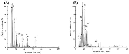
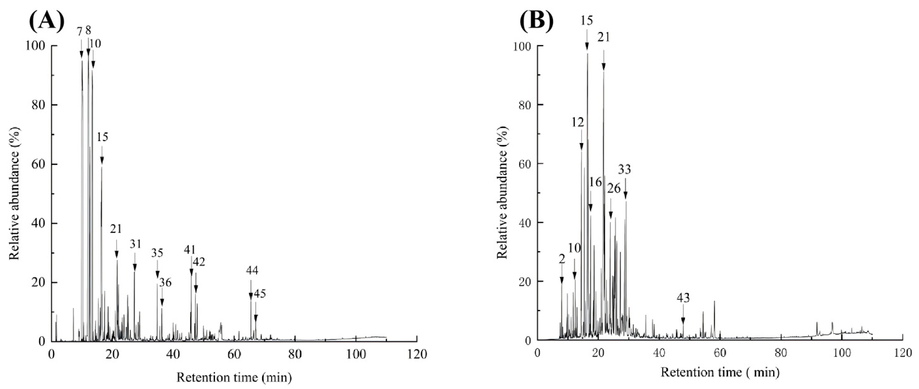
The volatile chemical profiles of EO and VCH were characterized by GC–MS following the analytical procedure outlined in Section 4.3, with their total ion chromatograms (TICs) displayed in Figure 1.
Figure 1.
Total ion chromatograms (TICs) of essential oil (EO, (A)) and hydrosol-derived volatile constituents (VCH, (B)) from E. rutaecarpa. The numbered peaks correspond to the major compounds listed in Table 1.
The two fractions exhibited distinctly different distributions of chemical classes. In EO, hydrocarbons constituted the largest proportion, comprising 16 (Table 1) identified compounds and representing 67.07% (Table 2) of the total chromatographic peak area. Alcohols formed the second-largest group, with 16 (Table 1) compounds contributing 10.72% (Table 2). By comparison, VCH was dominated by alcohols, with 15 compounds collectively contributing 42.41% (Table 2), whereas four ketones accounted for a further 19.43% (Table 2). A complete overview of the identified compounds and their relative abundances in both fractions is provided in Table 1.
Table 1.
Chemical composition and relative contents of EO and VCH from E. rutaecarpa.
Table 2.
Chemical classes and relative contents of EO and VCH from E. rutaecarpa.
As shown in Table 1, EO was dominated by β-myrcene (21.46 ± 0.31%) (Table 1), trans-β-ocimene (18.63 ± 0.38%) (Table 1), limonene (18.57 ± 0.51%) (Table 1), and linalool (4.53 ± 0.22%) (Table 1). In comparison, the relative peak-area percentages of β-myrcene, trans-β-ocimene, and limonene were substantially lower in VCH, at 1.00 ± 0.29% (Table 1), 0.64 ± 0.14% (Table 1), and 0.88 ± 0.21% (Table 1), respectively, whereas linalool was more abundant (14.91 ± 0.63%) (Table 1). Cryptone (11.76 ± 0.69%) (Table 1) and carvone (2.60 ± 0.24%) (Table 1) were also notable constituents of VCH but accounted for only 1.74 ± 0.42% (Table 1) and 0.57 ± 0.06% (Table 1), respectively, in EO.
Clear differences were also evident at the chemical-class level (Table 2). Hydrocarbons accounted for 67.07% of EO, with monoterpene hydrocarbons representing 63.58% (Table 2) and constituting the dominant fraction. In contrast, VCH was enriched in oxygenated compounds, with alcohols and ketones accounting for 42.41% and 19.43%, respectively. Oxygenated monoterpenes represented 67.52% (Table 2) of VCH, with linalool, cryptone, and carvone among the representative constituents.
These compositional differences should be interpreted in the context of the extraction and sample preparation procedures. Both fractions were obtained through hydrodistillation, whereas VCH underwent additional liquid–liquid extraction with dichloromethane followed by a 24 h solvent-recovery process. Therefore, differences in compound partitioning and potential losses or transformations during processing may have influenced the observed volatile profiles. Although solvent removal was performed before biological evaluation, residual dichloromethane levels were not analytically verified. Future studies should assess residual solvent levels and investigate greener extraction approaches, such as ethyl acetate extraction for volatile constituent recovery and HS-SPME for solvent-free chemical profiling.
2.3. Antimicrobial Activity
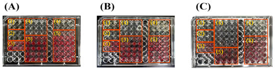
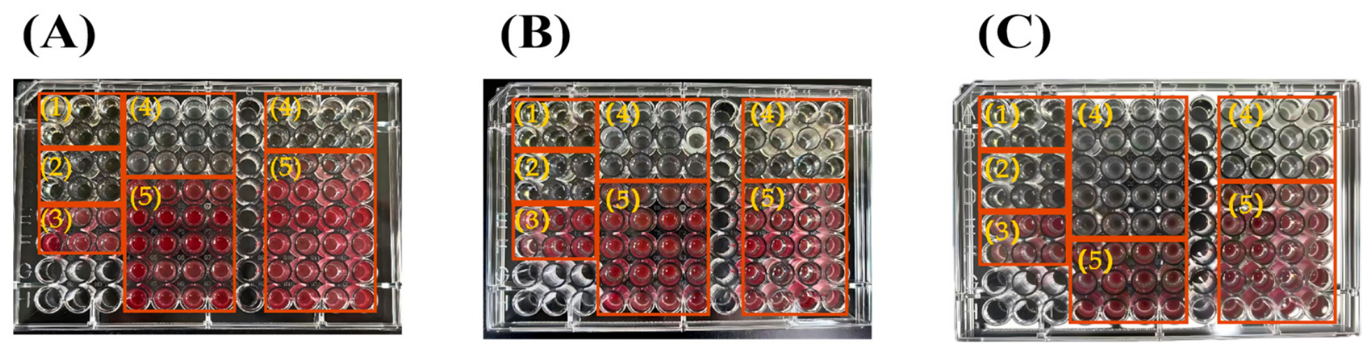
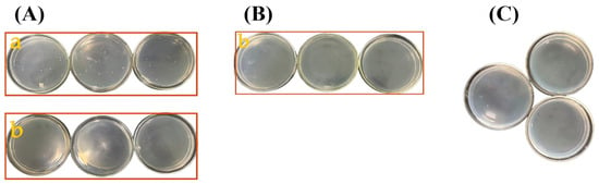
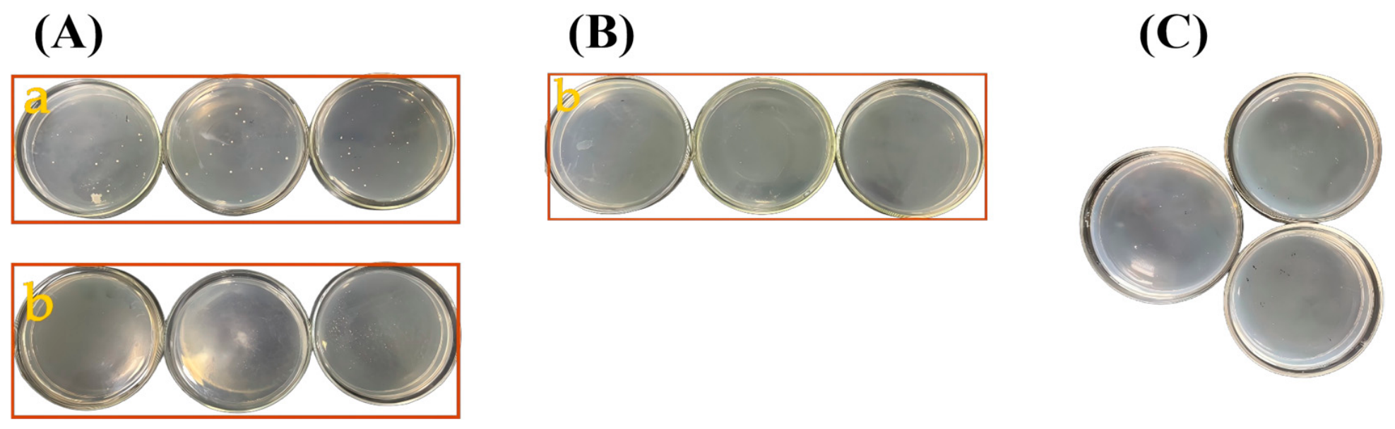
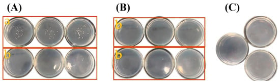
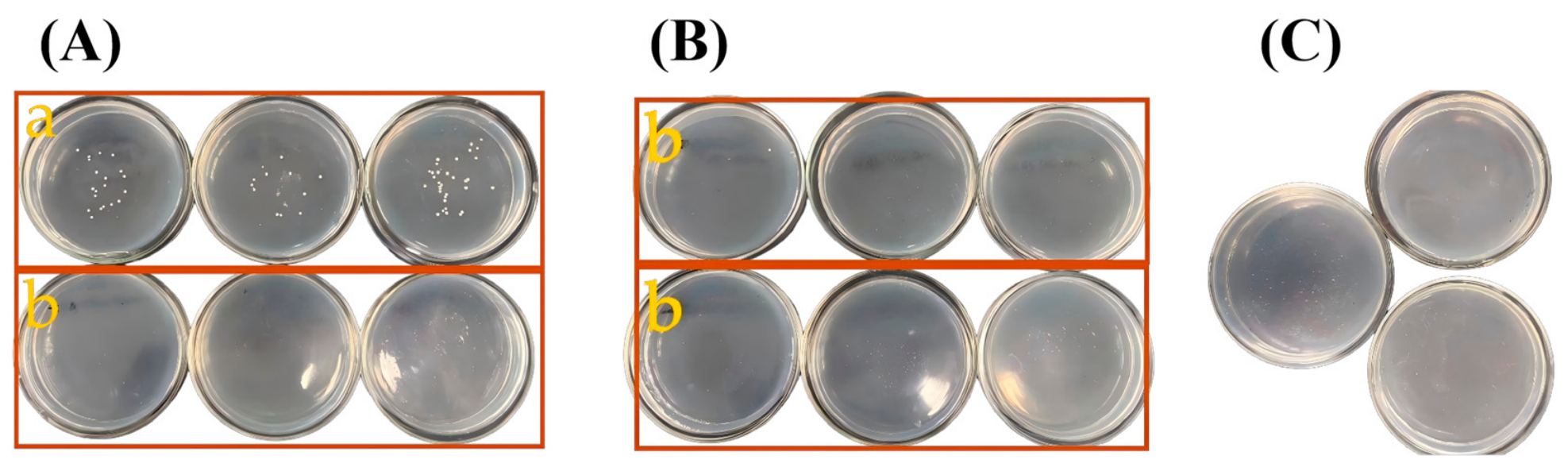
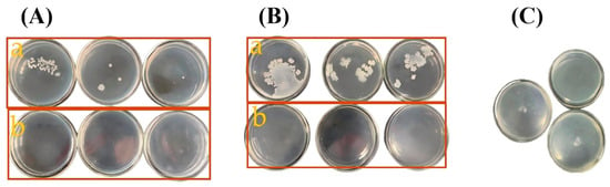
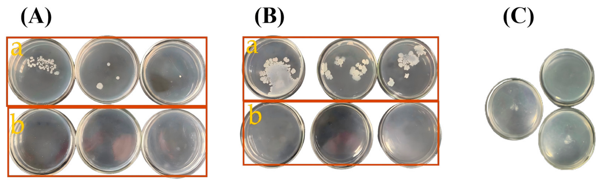
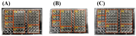
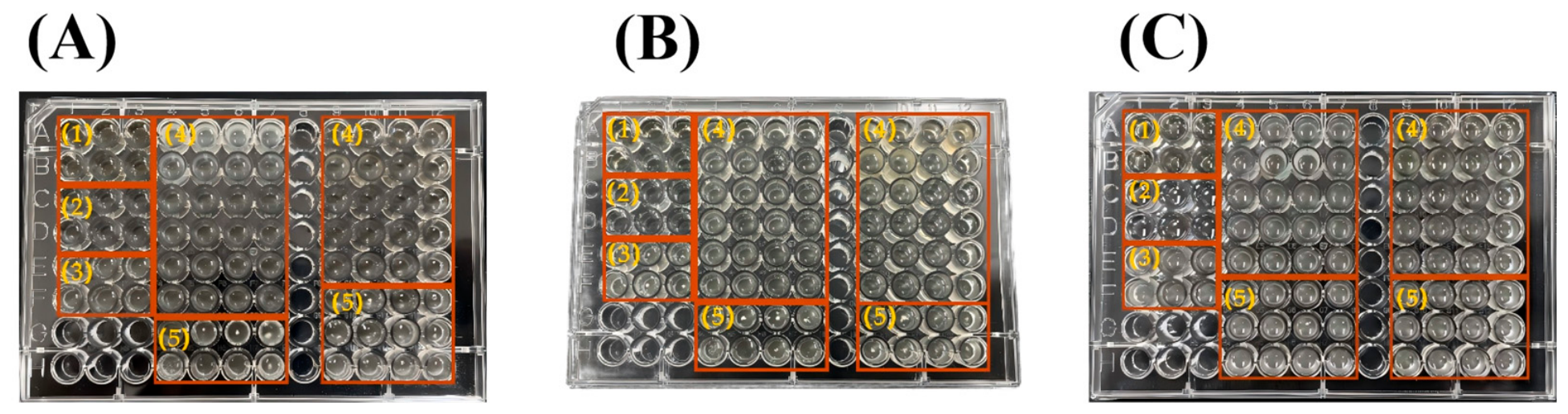
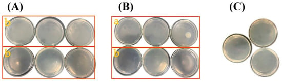
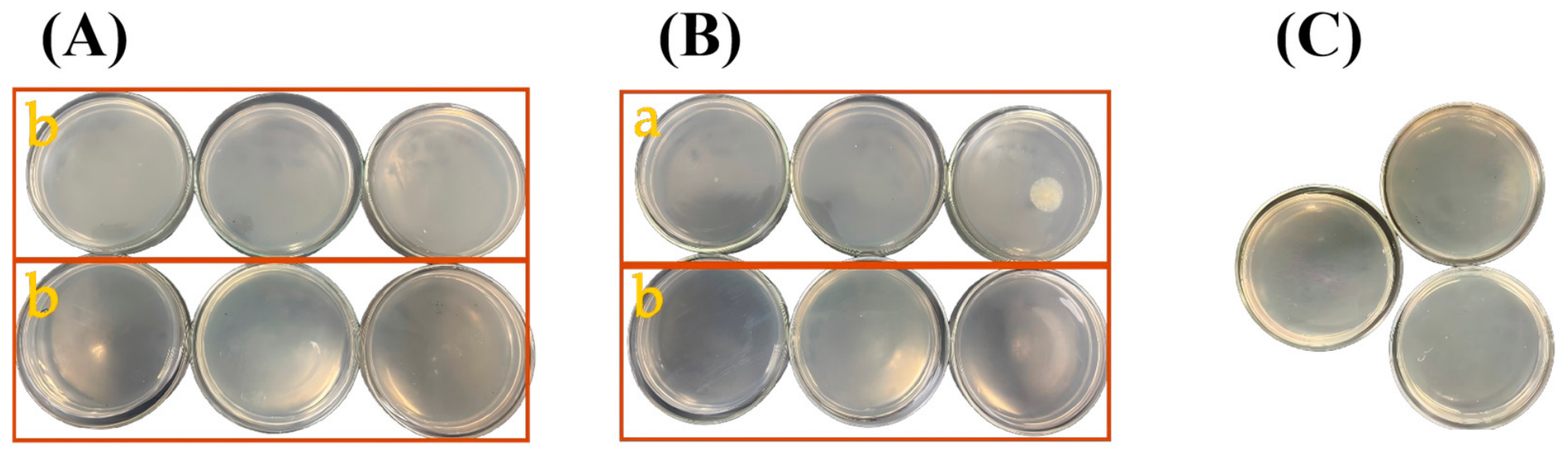
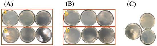
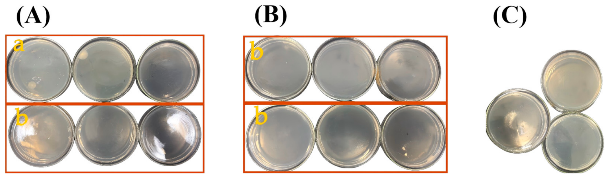
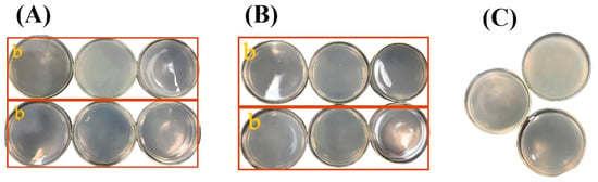
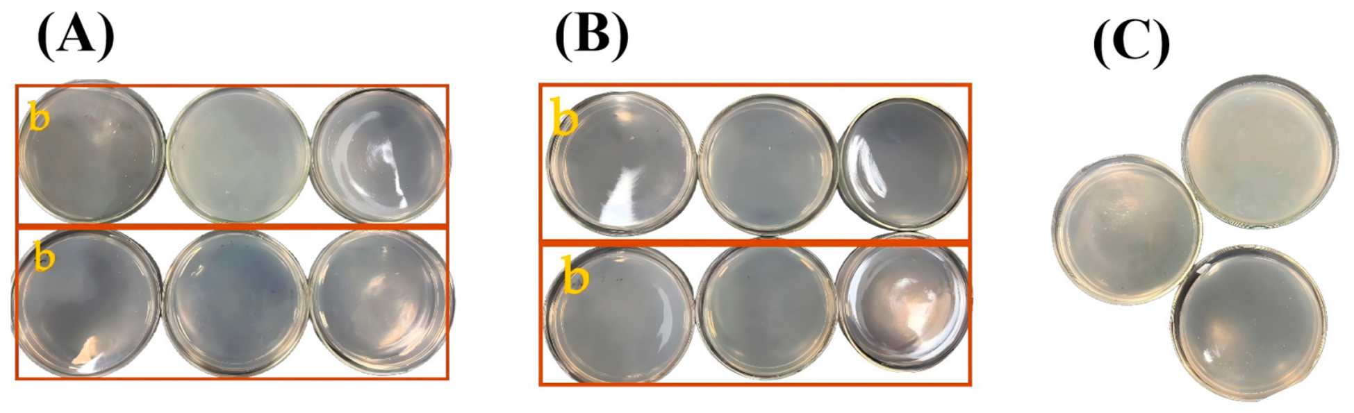
The antibacterial effects of EO and VCH on the three bacterial strains are presented in Table 3 and Figure 2. EO exhibited MICs of 1.25–5.00 μL/mL (Table 3), compared with 5.00–10.00 μL/mL (Table 3) for VCH. Subsequent subculturing at concentrations equal to or exceeding the MICs yielded MBCs of 5.00–20.00 μL/mL for EO and 10.00–20.00 μL/mL for VCH (Figure 3, Figure 4 and Figure 5). Among the tested strains, Bacillus subtilis was the most susceptible to EO, with an MIC of 1.25 μL/mL (Table 3) and an MBC of 5.00 μL/mL (Table 3). For VCH, the lowest MIC (5.00 μL/mL) (Table 3) and MBC (10.00 μL/mL) (Table 3) were observed against Escherichia coli.
Table 3.
MIC and MBC values of EO and VCH from E. rutaecarpa against the tested bacterial strains.
Figure 2.
Representative 96-well microdilution plate images showing the antibacterial effects of essential oil (EO) and hydrosol-derived volatile constituents (VCH) from E. rutaecarpa against (A) S. aureus, (B) E. coli, and (C) B. subtilis. In each plate image, the four columns in the left-hand frame correspond to EO, whereas the four columns in the right-hand frame correspond to VCH. Numbers indicate (1) sterility control (medium and solvent without bacterial inoculum), (2) positive control, (3) solvent control, (4) wells showing no bacterial growth, and (5) wells showing bacterial growth. The same layout and numbering scheme apply to all plate images.
Figure 3.
Bactericidal effects of (A) essential oil (EO), (B) hydrosol-derived volatile constituents (VCH), and (C) streptomycin against S. aureus. Labels a and b indicate two adjacent concentrations at which bacterial growth was observed and not observed, respectively. The same notation applies to the subsequent figures. Note: In panel (B), b corresponds to the highest concentration tested.
Figure 4.
Bactericidal effects of (A) essential oil (EO), (B) hydrosol-derived volatile constituents (VCH), and (C) streptomycin against E. coli. Labels a and b indicate two adjacent concentrations at which bacterial growth was observed and not observed, respectively. When two b labels appear, the first and second boxes correspond to 2 × MIC and 4 × MIC, respectively, and neither shows bacterial growth. The same applies to the following figures.
Figure 5.
Bactericidal effects of (A) EO, (B) VCH, and (C) streptomycin against B. subtilis.
EO and VCH also differed in their inhibitory effects against the tested fungi. As shown in Table 4 and Figure 6, the MIC values of both EO and VCH ranged from 0.625 to 1.25 μL/mL. The MFC values ranged from 1.25 to 2.50 μL/mL for EO and from 1.25 to 5.00 μL/mL for VCH.
Table 4.
MIC and MFC values of EO and VCH from E. rutaecarpa against the tested filamentous fungi.
Figure 6.
Representative 96-well plate images showing the antifungal effects of EO and VCH from E. rutaecarpa against (A) A. niger, (B) A. flavus, and (C) P. citrinum.
Among the tested fungi, as shown in Figure 7, Figure 8 and Figure 9, EO showed its lowest MIC and MFC against Aspergillus niger (0.625 and 1.25 μL/mL (Table 4), respectively). VCH showed its lowest MIC and MFC against Aspergillus flavus (0.625 and 1.25 μL/mL (Table 4), respectively).
Figure 7.
Fungicidal effects of (A) EO, (B) VCH, and (C) fluconazole against A. niger.
Figure 8.
Fungicidal effects of (A) EO, (B) VCH, and (C) fluconazole against A. flavus.
Figure 9.
Fungicidal effects of (A) EO, (B) VCH, and (C) fluconazole against P. citrinum.
2.4. Antiviral Activity
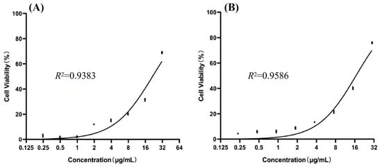
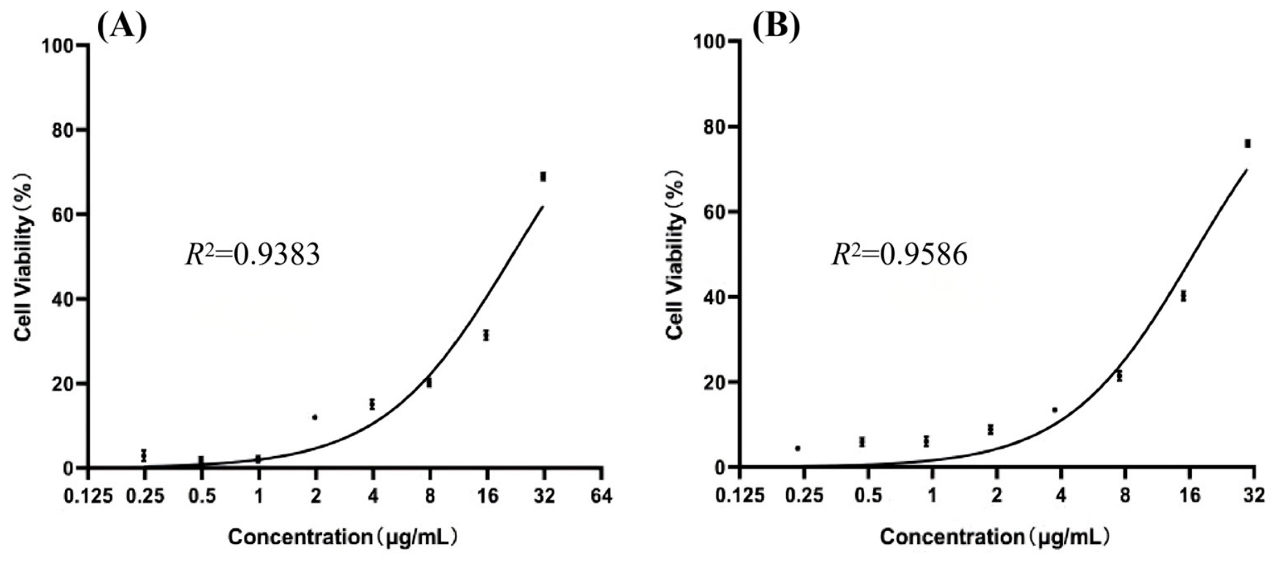
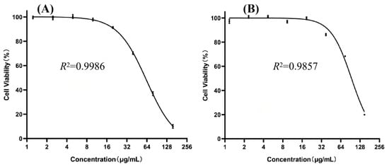
The anti-RSV activity and cytotoxicity of E. rutaecarpa EO and VCH are shown in Figure 10 and Figure 11. In the antiviral assay, RSV-infected Vero-cell viability ranged from 2.87–68.97% (Figure 10) at EO concentrations of 0.25 to 31.55 μg/mL (Figure 10) and from 4.42% to 75.97% (Figure 10) at VCH concentrations of 0.23 to 29.95 μg/mL (Figure 10). Cell viability generally increased with increasing sample concentration, indicating concentration-dependent protection against RSV-induced cytopathic effects under the conditions tested. The EC50 values of EO and VCH were 21.49 (Table 5) and 16.70 μg/mL (Table 5), respectively.
Figure 10.
Protective effects of (A) EO and (B) VCH on RSV-infected Vero cells.
Figure 11.
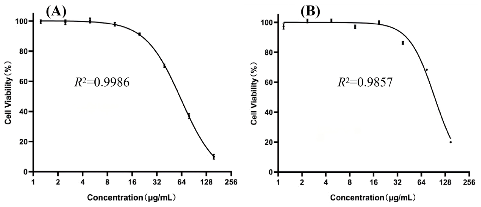
Cytotoxicity of (A) EO and (B) VCH toward Vero cells.
Table 5.
Selectivity indices of EO and VCH against RSV.
In the cytotoxicity assay, Vero-cell viability ranged from 9.97% to 100% (Figure 11) at EO concentrations of 1.23 to 157.77 μg/mL (Figure 11) and from 19.92% to 100% (Figure 11) at VCH concentrations of 1.17 to 149.75 μg/mL (Figure 11). The CC50 values of EO and VCH were 63.28 and 93.99 μg/mL (Table 5), respectively. The higher CC50 of VCH indicates lower cytotoxicity toward Vero cells within the tested concentration range.
The apparent selectivity indices (SIs), calculated as CC50/EC50, were 2.94 (Table 5) for EO and 5.63 (Table 5) for VCH. The higher SI of VCH suggests a more favorable cell-protection-related response relative to its cytotoxicity under the tested conditions. However, these SI values should be interpreted as preliminary in vitro indicators based on the CCK-8 assay rather than definitive measures of antiviral selectivity, because viral replication was not directly quantified.
2.5. Antioxidant Activity
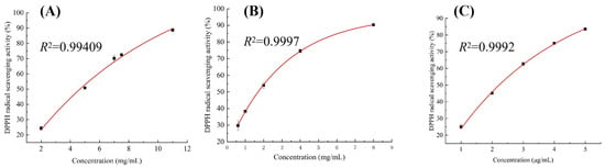
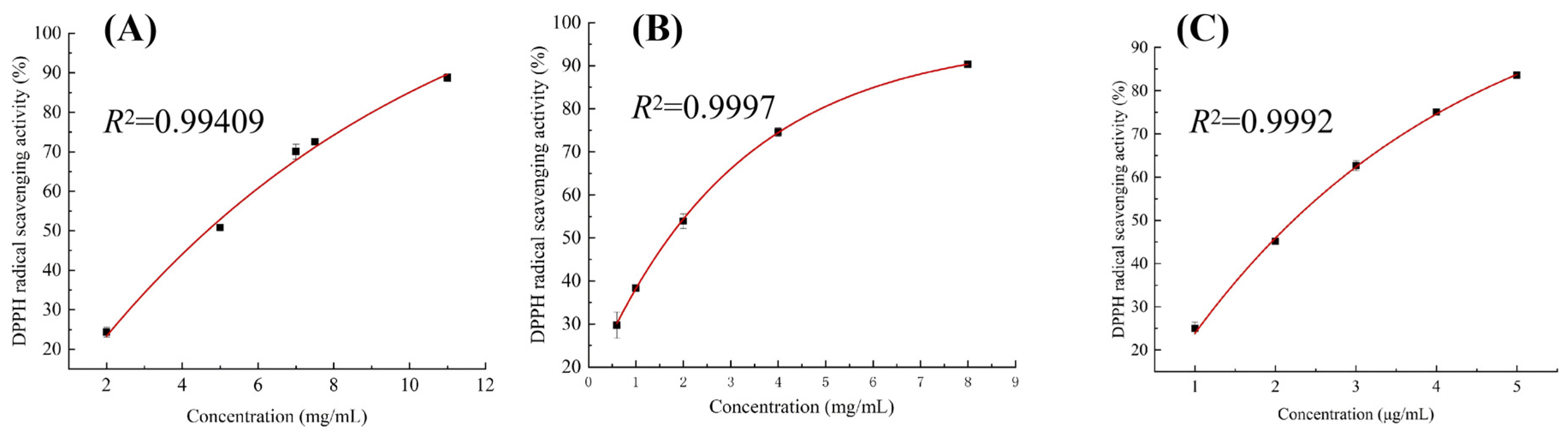
The DPPH· radical-scavenging activities of E. rutaecarpa EO and VCH are shown in Figure 12, and the corresponding IC50 values are presented in Table 6. Scavenging activity increased with sample concentration for both EO and VCH. At 2 mg/mL, EO and VCH showed DPPH· scavenging activities of 24.34% (Figure 12) and 53.90% (Figure 12), respectively, with VCH exhibiting approximately 2.2-fold greater activity.
Figure 12.
DPPH· radical-scavenging activities of (A) EO, (B) VCH, and (C) TBHQ.
Table 6.
Comparison of the antioxidant activities of EO and VCH.
As shown in Table 6, the IC50 values of EO and VCH were 4.67 and 1.70 mg/mL (Table 6), respectively. Because a lower IC50 indicates greater radical-scavenging activity, VCH showed stronger DPPH· scavenging capacity than EO. This difference may reflect differences in chemical composition and relative constituent abundance, together with possible interactions among antioxidant components.
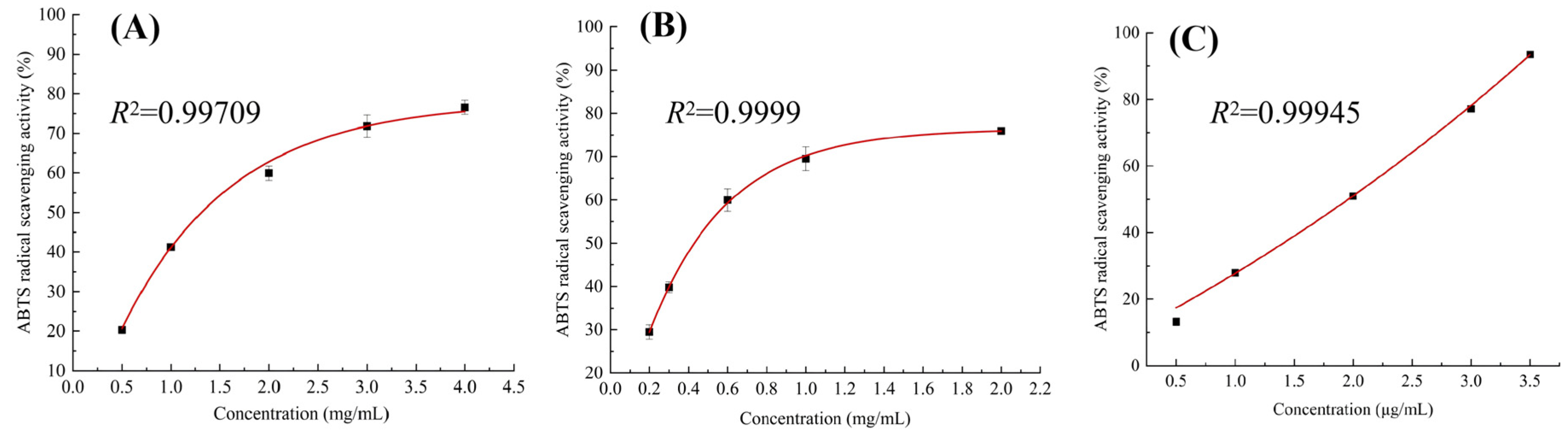
ABTS+· radical-scavenging activity increased with concentration for both E. rutaecarpa EO and VCH (Figure 13), and the corresponding IC50 values are listed in Table 6. At the same concentration, VCH consistently showed greater scavenging activity than EO; at 2 mg/mL, the activities of EO and VCH were 59.94% and 75.94% (Figure 13), respectively.
Figure 13.
ABTS+· radical-scavenging activities of (A) EO, (B) VCH, and (C) TBHQ.
The IC50 values of EO and VCH in the ABTS+· assay were 1.31 and 0.43 mg/mL (Table 6), respectively, further demonstrating the greater radical-scavenging capacity of VCH under the conditions tested.
Compared with the positive control TBHQ, both EO and VCH showed substantially higher IC50 values in the DPPH· and ABTS+· assays. The IC50 values of TBHQ were 2.22 × 10⁻³ and 1.96 × 10⁻³ mg/mL (Table 6), respectively, indicating much stronger radical-scavenging activity. Nevertheless, EO and VCH both exhibited clear concentration-dependent antioxidant activity, and VCH consistently showed lower IC50 values than EO in both assays.
3. Discussion
3.1. Chemical Differentiation Between EO and VCH
The most prominent compositional finding was the clear chemical differentiation between the essential oil (EO) and the volatile constituents recovered from the hydrosol (VCH). EO was dominated by hydrocarbons (67.07%), particularly monoterpene hydrocarbons (63.58%), with β-myrcene, D-limonene, and trans-β-ocimene as the principal constituents. By contrast, VCH contained only 2.60% hydrocarbons but was strongly enriched in oxygenated monoterpenes (67.52%), with linalool and cryptone among its most abundant constituents. Hydrodistillation therefore did not merely generate two fractions containing different concentrations of the same volatile profile; rather, it produced two chemically distinct fractions with markedly different distributions of hydrocarbon and oxygenated constituents.
This divergence is consistent with the phase-partitioning behavior of volatile compounds during hydrodistillation. In general, less polar and more hydrophobic monoterpene hydrocarbons tend to preferentially partition into the oil phase, whereas some oxygenated volatile compounds, owing to their relatively greater affinity for the aqueous phase, are more readily retained in the hydrosol and may consequently occur at higher relative abundances in the volatile fraction obtained by subsequent solvent extraction [8]. In the present study, the markedly lower relative abundances of β-myrcene, D-limonene, and trans-β-ocimene in VCH, together with the increased proportions of linalool, cryptone, and other oxygenated monoterpenes, are broadly consistent with this partitioning behavior.
Similar compositional differentiation between essential oils and hydrosol-derived volatile fractions has also been reported in other plant species. Kumar et al. [18] compared the essential oils and hydrosol volatile fractions of four cultivars of Coriandrum sativum L. and found that, after n-hexane extraction, the hydrosol-derived volatile fractions were dominated by (Z)-3-hexenyl acetate (66.12–77.12%), nonane (0.95–8.06%), and 1-decanol (2.16–4.14%), whereas the corresponding essential oils were mainly characterized by (E)-2-decenal (15.7–25.1%), decanal (14.4–18.8%), and (E)-2-dodecenal (12.6–13.7%). These findings indicate that essential oils and hydrosol-derived volatile fractions obtained from the same plant can exhibit markedly different chemical profiles. Likewise, Preiner et al. [19] reported that linalool (39.66–43.80%) and terpinen-4-ol (15.04–18.36%) were among the predominant volatile constituents of lavender hydrosol following extraction with different organic solvents. By comparison, Russo et al. [20] identified linalool (33.99%) as the major component of lavender essential oil, together with borneol (13.21%), 1,8-cineole (6.29%), limonene (6.12%), and terpinen-4-ol (5.24%). Their results further showed that oxygenated monoterpenes accounted for 73.06% of the lavender essential oil. Collectively, these observations suggest that, even when derived from the same botanical source, essential oils and hydrosol-derived volatile fractions may display distinct compositional patterns because of differential partitioning of volatile compounds during distillation. Such differences may also be influenced by plant species, raw-material characteristics, distillation conditions, and the subsequent solvent-extraction procedure.
It should be noted that prolonged hydrodistillation may induce thermal degradation, hydrolysis, or other chemical transformations of certain volatile constituents. Therefore, the observed compositional differences between EO and VCH may reflect not only differences in the partitioning behavior of volatile compounds but also potential extraction-related transformations. Since the effects of different distillation durations were not investigated, these possibilities cannot be excluded.
3.2. Antimicrobial Activity
Both EO and VCH inhibited the tested bacteria and filamentous fungi, but their activity patterns were microorganism-dependent. Against the bacterial strains, EO generally showed lower MIC values than VCH and was particularly active against Bacillus subtilis, for which the MIC and MBC were 1.25 and 5.00 μL/mL, respectively. VCH showed its lowest bacterial MIC and MBC against Escherichia coli (5.00 and 10.00 μL/mL, respectively). The antifungal results were less uniform: EO showed its lowest MIC and MFC against Aspergillus niger, whereas VCH showed its lowest values against Aspergillus flavus. Thus, neither fraction exhibited a uniform antimicrobial advantage across all tested microorganisms; susceptibility depended on both the sample and the target organism.
The antibacterial activities of essential oils and hydrosols may differ considerably, which may be attributed to multiple factors, including plant origin, chemical composition, treatment concentration, and the susceptibility of different microorganisms [21,22,23]. LeeAnn Nguyen et al. [21] reported that tea tree essential oil exhibited MIC values of 0.5% (v/v) against Escherichia coli MG1655 (Gram-negative) and 1.0% (v/v) against Staphylococcus epidermidis RP62A (Gram-positive), indicating that different bacterial species may show different sensitivities to the same essential oil. Aćimović et al. [22] further compared the antimicrobial activities of Dracocephalum moldavica essential oil and its original hydrosol without organic-solvent extraction. The MIC values of the essential oil against Staphylococcus aureus, Listeria monocytogenes, Escherichia coli, and Salmonella Typhimurium were 0.78%, 1.56%, 3.125%, and 0.78%, respectively. In comparison, the hydrosol exhibited MIC values of 12.5%, 6.25%, and approximately 3.125% against S. aureus, L. monocytogenes, and E. coli, respectively, and showed no obvious inhibitory effect against S. Typhimurium. These findings indicate that essential oils and hydrosols derived from the same plant may nevertheless exhibit markedly different antibacterial activities.
However, individual constituents were not independently tested in the present study, nor were membrane disruption, metabolic interference, or other potential antimicrobial mechanisms directly evaluated. Therefore, the observed MIC, MBC, and MFC values cannot be attributed to any single constituent. Further studies involving activity-guided fractionation, single-compound assays, and combination experiments are needed to clarify the contributions and potential interactions of individual constituents [24,25]. The potential application of EO and VCH in food preservation requires further validation of their efficacy, stability, and safety in actual food systems.
3.3. Anti-RSV Activity and Cytotoxicity
EO and VCH both showed concentration-dependent cytoprotective effects in RSV-infected Vero cells in the CCK-8 assay, but VCH showed a more favorable cell-protection-to-cytotoxicity profile. VCH had a lower EC50 based on the CCK-8-derived cytoprotective response (16.70 μg/mL) than EO (21.49 μg/mL) and a higher CC50 (93.99 μg/mL versus 63.28 μg/mL), resulting in apparent selectivity indices (SI = CC50/EC50) of 5.63 and 2.94, respectively. The SI of VCH was therefore approximately 1.9-fold that of EO. These findings suggest that VCH exhibited a more favorable cell-protection-related response relative to its cytotoxicity under the tested conditions. However, because viral replication was not directly quantified, the observed cytoprotective effects cannot be attributed specifically to inhibition of RSV replication.
The antiviral properties of plant-derived essential oils and hydrosols may vary considerably depending on their botanical sources and volatile constituents. Taglienti et al. [5] investigated essential oils and hydrosols from Origanum vulgare, Thymus vulgaris, and Rosmarinus officinalis against tomato leaf curl New Delhi virus, revealing substantial variation in antiviral effects. Notably, rosemary hydrosol rendered the virus undetectable in plant leaves at certain sampling points, while several treatments decreased viral titers by approximately 100-fold. In a subsequent investigation, the same researchers examined Mentha suaveolens and Foeniculum vulgare against zucchini yellow mosaic virus. The essential oil of M. suaveolens produced an approximately 100-fold reduction in viral titer, with the effect persisting into the later infection stage. By contrast, the essential oil and hydrosol of F. vulgare achieved reductions of approximately 10-fold at the later stage [26]. Collectively, these observations suggest that plant origin and variations in volatile composition may influence the magnitude and persistence of antiviral activity.
In addition to differences among volatile fractions, the same essential oil may also exhibit markedly different activities against different viruses. Li et al. [27] reported that Myrtus communis L. essential oil showed an EC50 of 0.1204 mg/mL and a CC50 of 4.197 mg/mL against HCoV-229E, corresponding to a selectivity index (SI) of approximately 35. In contrast, against HCoV-OC43, the EC50 was 1.405 mg/mL and the SI was only approximately 3 at the same CC50, indicating substantial virus-dependent differences in antiviral potency and selectivity. Vuko et al. [28] further reported that volatile constituents derived from hydrosol exhibited IC50 values of 151.35, 256.45, 298.34, and 677.40 μg/mL against HeLa, HCT116, U2OS, and normal RPE1 cells, respectively, suggesting that the biological effects of complex volatile mixtures may also be strongly dependent on cell type.
In the present study, EO and VCH showed different CCK-8-derived cytoprotective responses in RSV-infected Vero cells, with apparent SI values of 2.94 and 5.63, respectively, with VCH exhibiting the higher value. The proportion of oxygenated monoterpenes was also markedly higher in VCH than in EO, suggesting that differences in volatile composition may be associated with their distinct cytoprotective responses. However, a major limitation of the present study is that the effects on RSV-infected cells were evaluated using a CCK-8-based cell viability assay, without direct quantification of viral replication [29]. Therefore, the observed association between chemical composition and cytoprotective responses remains correlative, and the overall effects of these complex volatile mixtures cannot be directly attributed to one or several specific constituents solely on the basis of compositional data. Further studies using direct virological assays are needed to determine whether EO and VCH inhibit RSV replication and to elucidate the mechanisms underlying their potential cytoprotective effects. Moreover, their potential therapeutic relevance remains hypothetical and requires further validation through mechanistic investigations, appropriate biological models, and comprehensive safety assessments.
3.4. Antioxidant Activity and Integrated Interpretation
Terpenoids have been widely associated with antioxidant properties, with radical-scavenging responses often increasing with concentration [30,31]. According to Table 6, EO and VCH exhibited greater scavenging efficiency against ABTS+· than DPPH·, as reflected by their lower IC50 values. A similar difference between the two assays was reported by Lin et al. [32] for lime peel essential oil. Their results showed that DPPH· inhibition increased from 10.65% to 66.44% at concentrations of 0.08–3.46 mg/mL, while ABTS+· inhibition rose from 16.62% to 51.07% over 0.01–0.27 mg/mL. The respective IC50 values of 2.36 and 0.26 mg/mL further supported this assay-dependent difference. In our study, VCH showed lower IC50 values than EO in both the DPPH· (1.70 versus 4.67 mg/mL) and ABTS+· (0.43 versus 1.31 mg/mL) assays, suggesting greater radical-scavenging capacity of VCH under the tested conditions. However, neither fraction approached the activity of the positive control TBHQ, whose IC50 values were 2.22 × 10⁻³ mg/mL for DPPH· and 1.96 × 10⁻³ mg/mL for ABTS+·.
As shown in Table 1, EO and VCH contained several major volatile constituents that have previously been associated with antioxidant activity, including β-myrcene, D-limonene, trans-β-ocimene, and cryptone [33,34,35,36]. Oxygenated monoterpenes have also been reported to exhibit notable radical-scavenging activity [32]. Therefore, differences in the chemical compositions of EO and VCH may partly contribute to their different scavenging activities against DPPH· and ABTS+·. However, both EO and VCH are complex mixtures composed of multiple volatile compounds, and it remains unclear whether any single compound or group of compounds plays a predominant role in their overall antioxidant activity. Their radical-scavenging capacity may depend not only on the intrinsic activity of individual constituents but also on potential synergistic or antagonistic interactions among different components [37]. Consequently, chemical composition data and previously reported antioxidant activities of individual compounds alone are insufficient to identify the chemical basis of the overall antioxidant effects of EO and VCH. Further studies involving the evaluation of major individual constituents, reconstituted mixtures, and different component ratios are needed to clarify potential synergistic or antagonistic interactions among volatile constituents and their contributions to the overall antioxidant activity.
VCH exhibited stronger in vitro radical-scavenging activity than EO in both the DPPH and ABTS assays, as indicated by its lower IC50 values. However, these results reflect radical-scavenging activity under the experimental conditions of chemical assays and do not directly demonstrate antioxidant efficacy in biological systems.
4. Materials and Methods
4.1. Materials, Chemicals, and Instruments
Dried, nearly mature fruits of E. rutaecarpa. (Juss.) Benth. were collected in March in Guangxi, China, and used as the plant material.
Dichloromethane (≥99.98%), sodium chloride (≥99.98%), and anhydrous sodium sulfate (Na2SO4) were obtained from Sinopharm Chemical Reagent Co., Ltd. (Shanghai, China), Xilong Scientific Co., Ltd. (Guangzhou, China), and Tianjin Damao Chemical Reagent Factory (Tianjin, China), respectively. Potato dextrose broth (PDB), potato dextrose agar (PDA), nutrient broth (NB), and nutrient agar (NA) were supplied by Guangdong Huankai Microbial Sci. & Tech. Co., Ltd. (Guangzhou, China). 2,3,5-Triphenyltetrazolium chloride (TTC) was purchased from Tianjin Kermel Chemical Reagent Co., Ltd. (Tianjin, China).
High-glucose Dulbecco’s modified Eagle’s medium (DMEM), penicillin–streptomycin, and L-glutamine were obtained from Gibco (Thermo Fisher Scientific, Waltham, MA, USA). Fetal bovine serum (FBS) was supplied by PAN-Biotech GmbH (Ortenbert, Germany), while trypsin–EDTA, phosphate-buffered saline (PBS), and Cell Counting Kit-8 (CCK-8) were purchased from Beyotime Biotechnology Co., Ltd. (Shanghai, China). Dimethyl sulfoxide (DMSO) and Triton X-100 were obtained from MedChemExpress (Monmouth Junction, NJ, USA) and Yeasen Biotechnology Co., Ltd. (Shanghai, China), respectively.
The antioxidant assay reagents included 2,2-diphenyl-1-picrylhydrazyl (DPPH; ≥97%; TCI, Shanghai, China), 2,2′-azino-bis(3-ethylbenzothiazoline-6-sulfonic acid) diammonium salt (ABTS; ≥98%; Fuzhou Feijing Biotechnology Co., Ltd., Fuzhou, China), tert-butylhydroquinone (TBHQ; ≥97%; Sigma-Aldrich, Shanghai, China), and potassium persulfate (K2S2O8) (≥99.5%; Xilong Scientific Co., Ltd., Guangzhou, China). All other chemicals were of analytical grade unless otherwise specified.
GC–MS analysis was conducted using a TRACE 1300–ISQ QD system (Thermo Fisher Scientific, S.p.A., Rodano, Milan, Italy) equipped with an HP-5 capillary column (30 m × 0.25 mm × 0.25 μm; Agilent Technologies, Santa Clara, CA, USA). Essential oil extraction was performed using a Model 1788 extractor (Chongqing Jixiang Teaching and Experimental Equipment Co., Ltd., Chongqing, China). A FLUOstar Omega multimode microplate reader (BMG LABTECH, GmbH, Ortenbert, Germany), a CKX53 inverted microscope (Olympus, Corporation, Tokyo, Japan), and a 722S visible spectrophotometer (Shanghai INESA Analytical Instrument Co., Ltd., Shanghai, China) were used for the corresponding biological and spectrophotometric measurements.
Microbial Strains, Cell Line, and Virus
Escherichia coli (GDMCC 1.1917), Staphylococcus aureus (GDMCC 1.2442), Bacillus subtilis (GDMCC 1.3473), Aspergillus flavus (GDMCC 3.626), Aspergillus niger (GDMCC 1.2374), and Penicillium citrinum (GDMCC 3.693) were obtained from the Guangdong Microbial Culture Collection Center (GDMCC, Guangzhou, China). African green monkey kidney (Vero) cells (Cat. No. CL-0242) were purchased from Procell Life Science & Technology Co., Ltd. (Wuhan, China). Respiratory syncytial virus (RSV) was supplied by the laboratory strain collection center of Shanghai Fuda Testing Technology Group.
4.2. Sample Preparation
Extraction of essential oil [38]. A 100 g portion of dried E. rutaecarpa fruits was combined with distilled water at a material-to-water ratio of 1:20 (w/v) and subjected to hydrodistillation for 6 h [39]. Upon completion, the collected distillate was left undisturbed to allow the oil and aqueous layers to separate. The oil layer was then recovered and its volume measured, while the aqueous portion was retained for subsequent recovery of hydrosol-derived volatile constituents.
Extraction of volatile constituents of hydrosol [38]. A 1000 mL portion of hydrosol obtained during distillation was treated with 2 g of NaCl to promote the partitioning of volatile compounds, followed by liquid–liquid extraction with dichloromethane (CH2Cl2) [40] at a volume ratio of 1:5 (CH2Cl2, v/v; 200 mL of CH2Cl2 per 1000 mL of hydrosol). The hydrosol was extracted once. The organic phase was dried over anhydrous Na2SO4 and subsequently subjected to conventional distillation using a water bath set at 55 °C. The entire solvent-recovery process lasted 24 h to remove the extraction solvent before subsequent biological activity assays. The resulting brownish-yellow oily residue was collected as VCH, and its mass was recorded to determine the extraction yield according to Equations (1) and (2).
4.3. GC–MS Analysis
The chemical profiles of EO and VCH were investigated using GC–MS equipped with an HP-5 MS fused-silica capillary column (30 m × 0.25 mm × 0.25 μm). Helium (≥99.999%) was delivered through the column at a flow rate of 1.0 mL/min. For each measurement, 1 μL of sample was introduced with a split ratio of 1:150 for EO or 1:100 for VCH. The injection port and transfer line were both operated at 280 °C. The column oven was maintained at 60 °C for the first 5 min, after which the temperature was raised at 2 °C/min to 260 °C and maintained for another 5 min [41,42]. A 7 min solvent delay was applied exclusively to VCH measurements.
Mass spectral data were collected under electron ionization (EI) at 70 eV, with the electron multiplier operated at 1.5 kV. Full-scan acquisition covered an m/z interval of 33–550 [41,42]. Three independent extractions were performed, and each extract was analyzed by GC–MS.
Volatile compounds were tentatively identified by matching their mass spectra against the NIST 17 mass spectral library. Only compounds with both similarity index (SI) and reverse similarity index (RSI) values greater than 900 were considered for tentative identification and included in the reported compound list. A homologous series of n-alkanes (C7–C40) analyzed under the same chromatographic conditions was used as a reference to calculate the experimental retention indices (RIs), according to Equation (3). The calculated RIs were then compared with the reference retention indices (RIlit) recorded in the database to assist compound identification. The relative contents of the volatile compounds were calculated using the chromatographic peak-area normalization method and expressed as relative peak-area percentages rather than absolute compound concentrations. Peaks that did not meet both spectral matching criteria were excluded from the reported compound list but retained in the total integrated peak area used for normalization.
GC–MS data acquisition and processing were performed using Thermo Scientific Xcalibur software (version 3.0.63; Thermo Fisher Scientific, Waltham, MA, USA). The mass spectra of the detected compounds were compared with those in the NIST/EPA/NIH Mass Spectral Library (NIST 17; National Institute of Standards and Technology, Gaithersburg, MD, USA) for compound identification.
4.4. Antimicrobial Activity
4.4.1. Determination of MIC
The inhibitory effects of EO and VCH against the selected microorganisms were evaluated using a broth microdilution assay. Twofold serial dilutions of the samples were prepared in 96-well plates to achieve final concentrations ranging from 0.15625 to 20 μL/mL, with a final DMSO concentration of 10% (v/v) [43]. Nutrient broth (NB) and potato dextrose broth (PDB) were used for bacterial and fungal cultures, respectively. Each well contained 100 μL of the diluted sample solution and 100 μL of microbial suspension. The initial inoculum concentrations were adjusted to 1 × 10⁶ CFU/mL for bacteria and 1 × 10⁵ CFU/mL for fungi, resulting in final concentrations of 5 × 10⁵ and 5 × 10⁴ CFU/mL, respectively, in a total reaction volume of 200 μL. Streptomycin and fluconazole stock solutions (20 mg/mL) were subjected to twofold serial dilution to determine their respective MIC values. The corresponding MBC and MFC values were subsequently determined using the bactericidal and fungicidal assays, respectively. [44].
For bacterial MIC determination, the inoculated plates were incubated at 37 °C for 24 h. Subsequently, 10 μL of sterile 1% TTC solution was added to each well, followed by an additional 2 h incubation in the dark. The MIC was defined as the lowest tested concentration at which no red color developed. For fungal assays, the plates were incubated at 28 °C for 48 h, and the MIC was recorded as the lowest concentration that completely prevented visible mycelial growth. All experiments were independently repeated at least four times [45,46].
4.4.2. Determination of MBC and MFC
To evaluate the bactericidal and fungicidal effects of EO and VCH, 10 μL of culture suspension was withdrawn from each well exhibiting complete growth inhibition in the MIC assay and transferred onto the appropriate agar medium. Following inoculation, bacterial and fungal plates were maintained at 37 °C for 24 h and 28 °C for 48 h, respectively. The minimum bactericidal concentration (MBC) and minimum fungicidal concentration (MFC) were identified as the lowest concentrations of EO or VCH that yielded no detectable colony formation under the respective culture conditions. Each assay was conducted independently at least three times [44,47].
4.5. Protection of RSV-Infected Vero Cells and Cytotoxicity Assays
4.5.1. Sample Preparation and Vero Cell Culture
The cytotoxicity of EO and VCH in uninfected Vero cells and their cytoprotective effects in RSV-infected Vero cells were assessed using a Cell Counting Kit-8 (CCK-8) assay. Stock solutions of both fractions were prepared by dissolving the samples in dimethyl sulfoxide (DMSO), followed by dilution with double-distilled water to produce working solutions containing 20% (v/v) DMSO. The final DMSO concentration in each well was 10% (v/v). Before the antiviral assays, preliminary experiments were conducted to evaluate the potential cytotoxicity of DMSO toward Vero cells at the final concentration used in the study (10%, v/v). Cell viability was assessed using the CCK-8 assay under the corresponding experimental conditions. The results indicated that this concentration of DMSO did not cause an apparent reduction in Vero-cell viability. Vehicle controls containing the same final concentration of DMSO were included in both the cytotoxicity and RSV-infected-cell assays. Vero cells in the exponential growth phase were detached using trypsin, collected, and resuspended in complete culture medium. The cells were seeded into 96-well plates at a density of 1 × 10⁴ cells per well and incubated for 24 h in complete medium supplemented with serum [48].
4.5.2. Determination of EC50 and CC50
To evaluate the cytoprotective effects of EO and VCH in RSV-infected Vero cells, the culture medium was aspirated from the pre-cultured cells and replaced with 100 μL of the designated EO or VCH dilution. Eight concentrations were prepared through successive twofold dilutions. The working solutions were prepared at twice the intended final concentrations to account for the subsequent addition of an equal volume of RSV inoculum. Each well then received 100 μL of RSV inoculum at a multiplicity of infection (MOI) of 0.01, resulting in final EO concentrations of 0.247–31.55 μg/mL and final VCH concentrations of 0.234–29.95 μg/mL in a total volume of 200 μL per well. The plates were incubated for 48 h at 37 °C under humidified conditions with 5% CO2 [49].
Following the exposure period, 20 μL of CCK-8 reagent was introduced into each well. After gentle mixing, the plates were returned to the incubator for an additional 2 h before absorbance was recorded at 450 nm. Blank wells, virus-infected controls, and ribavirin-treated positive controls were included. Each treatment was tested in triplicate, and the corresponding cell viability was calculated using Equation (3) [49,50].
where (ODS) represents the absorbance of sample-treated, RSV-infected cells, (ODV) represents the absorbance of the virus-infected control, and (ODb) represents the absorbance of the uninfected cell control. The calculated value reflects the relative recovery of cell viability following sample treatment, with the virus-infected control and uninfected cell control serving as the lower and upper reference levels, respectively.
Dose–response relationships were established by plotting cell viability against sample concentration. The EC50 and CC50 values were estimated through nonlinear regression in GraphPad Prism 8.0 (GraphPad Software, San Diego, CA, USA).
Cytotoxicity was examined separately in uninfected Vero cells. After the initial attachment period, the culture medium was replaced with 100 μL of the respective sample dilution. Twofold serial dilutions yielded eight concentration levels of 1.232–157.77 μg/mL for EO and 1.170–149.75 μg/mL for VCH. Following 48 h of sample exposure under the same incubation conditions, 10 μL of CCK-8 solution was dispensed into each well. The cells were incubated for another 2 h, after which the absorbance was determined at 450 nm. Blank wells and Triton X-100-treated cytotoxicity controls were established. All measurements were performed in triplicate, and cell viability was determined according to Equation (4) [51,52].
where (ODS) is the absorbance of the sample-treated group, (ODb) is the absorbance of the blank control without cells, and (ODc) is the absorbance of the untreated, uninfected cell control. Cell viability was expressed as a percentage of the untreated, uninfected cell control after background correction.
The apparent selectivity index (SI) was calculated using Equation (5) [49,53] as the ratio of CC50 to EC50 (SI = CC50/EC50). CC50 was defined as the concentration that reduced the viability of uninfected Vero cells by 50%, whereas EC50 was defined as the concentration required to achieve 50% cell protection against RSV-induced cytotoxicity, as determined by the CCK-8 assay.
4.6. Antioxidant Activity
4.6.1. DPPH Radical-Scavenging Assay
The DPPH assay was conducted using an ethanolic DPPH solution (0.02 g/L). EO and VCH were separately diluted with ethanol from higher to lower concentrations to prepare a series of sample solutions at different concentrations. The sample concentrations were expressed as the final concentrations in the reaction mixtures. For each determination, 2 mL of EO or VCH solution at the designated concentration was combined with an equal volume of DPPH solution. After thorough mixing, the reaction mixtures were maintained at room temperature in the dark, and absorbance (Ai) was monitored at 517 nm every 10 min over a period of 180 min. Two reference mixtures were prepared in parallel: one containing absolute ethanol and DPPH solution (2 mL each; Ac), and the other containing sample solution and absolute ethanol in equal volumes (2 mL each; Aj). tert-Butylhydroquinone (TBHQ) served as the positive control. Measurements were carried out in triplicate, and the absorbance values recorded at 60 min were used to calculate DPPH radical-scavenging activity (RSA, %) according to Equation (6) [54,55,56]. The resulting concentration–response data were used to determine the IC50 values.
The RSA recorded at 60 min was used to establish the concentration-dependent scavenging response. Nonlinear curve fitting was performed in Origin 2024 (OriginLab Corporation, Northampton, MA, USA), from which the concentration corresponding to 50% DPPH radical scavenging was obtained and expressed as IC50.
4.6.2. ABTS Radical-Scavenging Assay
The ABTS radical-cation working solution was prepared by mixing equal volumes of ABTS (7 mmol/L) and potassium persulfate (K2S2O8) (2.45 mmol/L) solutions prepared in distilled water. The mixture was maintained at room temperature in the dark for 24 h to obtain the ABTS radical-cation stock solution, which was subsequently diluted with absolute ethanol until its absorbance reached 0.700 ± 0.002 at 734 nm [55]. EO and VCH were separately diluted with ethanol from higher to lower concentrations to prepare a series of sample solutions at different concentrations. The sample concentrations were expressed as the final concentrations in the reaction mixtures. Equal volumes (2 mL each) of the working solution and EO or VCH at different concentrations were combined and maintained in darkness for 6 min. The absorbance of the resulting mixture (Ai) was subsequently recorded at 734 nm.
A reference mixture containing 2 mL of absolute ethanol and 2 mL of the ABTS radical-cation working solution (A0) was prepared under identical conditions. TBHQ was included as the positive control. Three replicate measurements were obtained for each treatment, and their mean values were used to calculate ABTS radical-scavenging activity (RSA, %) using Equation (7) [54,57].
The IC50 values were determined using the same fitting procedure as that employed for the DPPH assay.
5. Conclusions
This study systematically characterized the chemical composition and evaluated the antimicrobial and potential cytoprotective effects in RSV-infected Vero cells and in vitro radical-scavenging activities of E. rutaecarpa EO and VCH. The extraction yields were 2.15 ± 0.002 mL/100 g for EO and 0.27 ± 0.001 g/100 g for VCH. EO was dominated by hydrocarbons (67.07%), particularly monoterpene hydrocarbons, whereas VCH was enriched in oxygenated monoterpenes (67.52%).
EO showed notable activity against Bacillus subtilis and Aspergillus niger, with MIC values of 1.25 and 0.625 μL/mL, respectively; the corresponding MBC and MFC values were 5.0 and 1.25 μL/mL. VCH showed its lowest antibacterial and antifungal MIC values against Escherichia coli (5.0 μL/mL) and Aspergillus flavus (0.625 μL/mL), with corresponding MBC and MFC values of 10.0 and 1.25 μL/mL, respectively. In the RSV-infected Vero cell model, the CCK-8 assay results suggested potential cytoprotective effects of EO and VCH, with calculated SI values of 2.94 and 5.63, respectively. In the antioxidant assays, the DPPH· IC50 values were 4.67 mg/mL for EO and 1.70 mg/mL for VCH, whereas the ABTS+· IC50 values were 1.31 and 0.43 mg/mL, respectively.
Overall, EO and VCH differed markedly in both chemical composition and their measured activities under the experimental conditions. VCH showed a higher calculated SI in the RSV-infected Vero cell model and stronger radical-scavenging activity in the DPPH and ABTS chemical assays than EO, whereas antimicrobial activity varied with the target microorganism. However, the contributions of individual constituents and their potential interactions remain unclear. Furthermore, the CCK-8-based results do not establish direct inhibition of RSV replication or specific antiviral mechanisms, and the radical-scavenging activities observed in chemical assays cannot be directly extrapolated to antioxidant efficacy in biological systems. Further work should identify the principal bioactive constituents, establish structure–activity relationships, verify potential antiviral activity using virus-specific assays, clarify mechanisms of antimicrobial and potential antiviral action, and evaluate safety and toxicological profiles. The potential applications of EO and VCH in food preservation and therapeutic contexts remain hypothetical and require further validation of their efficacy and safety in relevant experimental models.
Author Contributions
Conceptualization, X.L. and Y.T.; methodology, X.L., N.X. and Y.T.; validation, N.L. and Q.Z.; formal analysis, X.L. and Z.W.; investigation, X.L., N.X., N.L. and Q.Z.; resources, Y.T.; data curation, X.L. and Z.W.; writing—original draft preparation, X.L.; writing—review and editing, X.L. and Y.T.; visualization, X.L. and Z.W.; supervision, Y.T.; project administration, Y.T.; funding acquisition, Y.T. All authors have read and agreed to the published version of the manuscript.
Funding
This research was funded by the National Natural Science Foundation of China (grant number 22268012), the Guangxi Natural Science Foundation (grant number 2021GXNSFAA220067), and Innovation Project of Guangxi Graduate Education (grant number YCSW2026635).
Data Availability Statement
The data supporting the findings of this study are available from the corresponding author upon reasonable request.
Acknowledgments
The authors would like to thank Yuting Lu from the School of Medicine, Guangxi University of Science and Technology, for her assistance with the identification of the plant material. During the preparation of this manuscript, the authors used ChatGPT-6 for English translation, language editing, and refinement of academic expression. The authors have reviewed and edited the output and take full responsibility for the content of this publication.
Conflicts of Interest
The authors declare no conflicts of interest. The funders had no role in the design of the study; in the collection, analyses, or interpretation of data; in the writing of the manuscript; or in the decision to publish the results.
Abbreviations
| Abbreviation | Full term |
| E. rutaecarpa | Evodia rutaecarpa |
| EO | Essential oil |
| VCH | Volatile constituents of hydrosol |
| GC–MS | Gas chromatography–mass spectrometry |
| MIC | Minimum inhibitory concentration |
| MBC | Minimum bactericidal concentration |
| MFC | Minimum fungicidal concentration |
| RSV | Respiratory syncytial virus |
| CCK-8 | Cell Counting Kit-8 |
| DPPH | 2,2-Diphenyl-1-picrylhydrazyl |
| ABTS | 2,2′-Azino-bis(3-ethylbenzothiazoline-6-sulfonic acid) |
| IC50 | Half-maximal inhibitory concentration |
| CC50 | Half-maximal cytotoxic concentration |
| SI | Selectivity index |
| MOI | Multiplicity of infection |
| DMSO | Dimethyl sulfoxide |
| TTC | 2,3,5-Triphenyltetrazolium chloride |
| DMEM | Dulbecco’s Modified Eagle Medium |
| PDB | Potato dextrose broth |
| PDA | Potato dextrose agar |
| NB | Nutrient broth |
| NA | Nutrient agar |
| TBHQ | Tert-butylhydroquinone |
References
- Çolak, H.; Durmuş, M.; Küley, E.; Köşker, A.R.; Sakarya, Y.; Esatbeyoglu, T.; Özogul, F. Application of juniper (Juniperus communis) essential oil nanoemulsions to control spoilage and pathogenic bacteria in fish. Front. Microbiol. 2026, 17, 1758540. [Google Scholar] [CrossRef] [Scilit] [PubMed]
- Ferreira, A.P.; Bezerra, J.J.L.; França, J.F.; de Sousa Fernandes, D.D.; da Luz, M.A.; de Oliveira Alves, J.V.; dos Santos Correia, M.T.; da Silva, M.V.; de Menezes Torres, M.C. Influence of seasonality on the chemical composition and antioxidant activity of essential oils from the leaves of Croton heliotropiifolius Kunth and Croton jacobinensis Baill. (Euphorbiaceae). Biochem. Syst. Ecol. 2026, 130, 105370. [Google Scholar] [CrossRef] [Scilit]
- El Bakkali, M.; Bouchfara, A.; Zerrad, H.; Souhail, B.; Nechar, M.; Bano, R.; Fiser, B. Composition and antioxidant activity of Abies marocana essential oil. Sci. Rep. 2026, 16, 6904. [Google Scholar] [CrossRef] [Scilit] [PubMed]
- Yin, J.; Mo, J.; Zhu, J.; Wu, Y.; Lu, S.; Zeng, J.; Wang, K. Mosla chinensis maxim. essential oil ameliorates DSS-induced colitis and enhances the intestinal barrier via the PI3K-AKT signaling pathway. Front. Nutr. 2026, 13, 1778915. [Google Scholar] [CrossRef] [Scilit] [PubMed]
- Taglienti, A.; Donati, L.; Ferretti, L.; Tomassoli, L.; Sapienza, F.; Sabatino, M.; Di Massimo, G.; Fiorentino, S.; Vecchiarelli, V.; Nota, P.; et al. In vivo antiphytoviral activity of essential oils and hydrosols from Origanum vulgare, Thymus vulgaris, and Rosmarinus officinalis to control zucchini yellow mosaic virus and tomato leaf curl New Delhi virus in Cucurbita pepo L. Front. Microbiol. 2022, 13, 840893. [Google Scholar] [CrossRef] [Scilit] [PubMed]
- Baranová, B.; Kudláčková, B.; Baral, R.P.; Svojanovská, L.; Javed, Q.; Amato, G.; Caputo, L.; De Martino, L.; Francolino, R.; Chaudhary, S.K.; et al. Potential herbicidal and insecticidal (Beneficial) properties of Nepalese lemongrass essential oil. Chem. Biodivers. 2025, 22, e01095. [Google Scholar] [CrossRef] [Scilit] [PubMed]
- Almeida, H.H.; Santamaria-Echart, A.; Amaral, J.S.; Aquino, L.L.; Rodrigues, A.E.; Barreiro, M.F. Chitosan-xanthan gum-based hydrogels loaded with essential oil distillation by-products of Aloysia citrodora Paláu for antimicrobial systems. Int. J. Biol. Macromol. 2026, 342, 150368. [Google Scholar] [CrossRef] [Scilit] [PubMed]
- Packer, C.; Calero-Torres, V.; Abad, A.; Burgos-Moreira, L.; Muzzio, M.; Cevallos, J.M.; Manzano-Santana, P.; Orellana-Manzano, A. Antioxidant, anti-inflammatory, and phytochemical properties of Juniperus osteosperma (Torr.) little hydrosol: An in vitro evaluation. Front. Pharmacol. 2026, 17, 1774954. [Google Scholar] [CrossRef] [Scilit] [PubMed]
- Cichosz, P.; Walasek-Janusz, M.; Grzegorczyk, A.; Papliński, R.; Kiczorowski, P.; Nurzyńska-Wierdak, R. Chemical Composition, and Antioxidant and Antimicrobial Properties of Monarda didyma L.’s Essential Oils and Hydrosols. Molecules 2026, 31, 2252. [Google Scholar] [CrossRef] [Scilit] [PubMed]
- Chinese Pharmacopoeia Commission. Pharmacopoeia of the People’s Republic of China; China Medical Science Press: Beijing, China, 2020; Volume I, p. 178. [Google Scholar]
- He, J.; Zhang, Y.; Yang, C.; Li, K.; Yuan, X.; Zhang, Z.; Gu, C.; Chen, Y. Effects of Alpinia Officinarum and Euodia Rutaecarpa on the expression of TRPA1 and TRPM8 in rats with irritable bowel syndrome. J. Holist. Integr. Pharm. 2023, 4, 1–13. [Google Scholar] [CrossRef] [Scilit]
- Hao, Y.; Qi, J.; Huang, X.; Liu, C.; Liu, Y. Euodiae Fructus: A review of botany, application, processing, phytochemistry, quality control, pharmacology, and toxicology. Front. Pharmacol. 2025, 16, 1509032. [Google Scholar] [CrossRef] [Scilit] [PubMed]
- Lin, L.; Liu, Y.; Tang, R.; Ding, S.; Lin, H.; Li, H. Evodiamine: A Extremely Potential Drug Development Candidate of Alkaloids from Evodia rutaecarpa. Int. J. Nanomed. 2024, 19, 9843–9870. [Google Scholar] [CrossRef] [Scilit] [PubMed]
- Zhang, Y.C.; Liu, A.; Wang, J.Z.; Qi, Y.T.; Du, S.S. Chemical compositions and insecticidal activities of Tetradium ruticarpum essential oils against Lasioderma serricorne. J. Stored Prod. Res. 2025, 111, 102517. [Google Scholar] [CrossRef] [Scilit]
- Zhang, Y.-N.; Yang, Y.-F.; Yang, X.-W. Blood-brain barrier permeability and neuroprotective effects of three main alkaloids from the fruits of Euodia rutaecarpa with MDCK-pHaMDR cell monolayer and PC12 cell line. Biomed. Pharmacother. 2018, 98, 82–87. [Google Scholar] [CrossRef] [Scilit] [PubMed]
- Wang, L.; Gao, F.; Tian, J.; Wang, L.; Zhao, Y.; Liu, S.; Yi, Y.; Li, C.; Liu, C.; Meng, J.; et al. Euodia rutaecarpa (Juss.) Benth. var. officinalis (Dode) Huang and evodiamine ameliorate N-methyl-N′-nitro-N-nitrosoguanidine-induced chronic atrophic gastritis in rats via the phosphatidylinositol 3-kinase/protein kinase B/nuclear factor kappa-B pathway. Phytomedicine 2026, 159, 158515. [Google Scholar] [CrossRef] [Scilit] [PubMed]
- Chen, L.; Hu, Y.; Ye, Z.; Li, L.; Qian, H.; Wu, M.; Qin, K.; Li, N.; Wen, X.; Pan, T.; et al. Major indole alkaloids in evodia rutaecarpa: The latest insights and review of their impact on gastrointestinal diseases. Biomed. Pharmacother. 2023, 167, 115495. [Google Scholar] [CrossRef] [Scilit] [PubMed]
- Kumar, S.; Ahmad, R.; Saeed, S.; Azeem, M.; Mozūraitis, R.; Borg-Karlson, A.K.; Zhu, G. Chemical composition of fresh leaves headspace aroma and essential oils of four Coriander cultivars. Front. Plant Sci. 2022, 13, 820644. [Google Scholar] [CrossRef] [Scilit] [PubMed]
- Preiner, S.; Tarcsay, B.L.; Pethő, D.; Miskolczi, N. Lavender hydrosol analysis using UV spectroscopy data and partial least squares regression. MethodsX 2025, 14, 103304. [Google Scholar] [CrossRef] [Scilit] [PubMed]
- Russo, M.; Martella, N.; Gargano, D.; Fantasma, F.; Marcovecchio, C.; Russo, V.; Oliva, M.A.; Segatto, M.; Saviano, G.; Di Bartolomeo, S.; et al. Lavender essential oil and its terpenic components negatively affect tumor properties in a cell model of glioblastoma. Molecules 2024, 29, 6044. [Google Scholar] [CrossRef] [Scilit] [PubMed]
- Nguyen, L.; DeVico, B.; Mannan, M.; Chang, M.; Rada Santacruz, C.; Siragusa, C.; Everhart, S.; Fazen, C.H. Tea tree essential oil kills Escherichia coli and Staphylococcus epidermidis persisters. Biomolecules 2023, 13, 1404. [Google Scholar] [CrossRef] [Scilit] [PubMed]
- Aćimović, M.; Šovljanski, O.; Šeregelj, V.; Pezo, L.; Zheljazkov, V.D.; Ljujić, J.; Tomić, A.; Ćetković, G.; Čanadanović-Brunet, J.; Miljković, A.; et al. Chemical composition, antioxidant, and antimicrobial activity of Dracocephalum moldavica L. Essential oil and hydrolate. Plants 2022, 11, 941. [Google Scholar] [CrossRef] [Scilit] [PubMed]
- Galgano, M.; Capozza, P.; Pellegrini, F.; Cordisco, M.; Sposato, A.; Sblano, S.; Camero, M.; Lanave, G.; Fracchiolla, G.; Corrente, M.; et al. Antimicrobial activity of essential oils evaluated in vitro against Escherichia coli and Staphylococcus aureus. Antibiotics 2022, 11, 979. [Google Scholar] [CrossRef] [Scilit] [PubMed]
- Zhao, A.; Zhang, Y.; Li, F.; Chen, L.; Huang, X. Analysis of the antibacterial properties of compound essential oil and the main antibacterial components of unilateral essential oils. Molecules 2023, 28, 6304. [Google Scholar] [CrossRef] [Scilit] [PubMed]
- Latorre, R.; Valerii, M.C.; Benati, M.; Lewis, R.E.; Spigarelli, R.; Bernacchi, A.; Lippi, G.; Spisni, E.; Gaibani, P. Lights and shadows of essential oil-derived compounds: Antimicrobial and anti-inflammatory properties of eugenol, thymol, cinnamaldehyde, and carvacrol. Curr. Issues Mol. Biol. 2025, 47, 915. [Google Scholar] [CrossRef] [Scilit] [PubMed]
- Taglienti, A.; Donati, L.; Dragone, I.; Ferretti, L.; Gentili, A.; Araniti, F.; Sapienza, F.; Astolfi, R.; Fiorentino, S.; Vecchiarelli, V.; et al. In vivo antiphytoviral and aphid repellency activity of essential oils and hydrosols from Mentha suaveolens and Foeniculum vulgare to control zucchini yellow mosaic virus and its vector Aphis gossypii. Plants 2023, 12, 1078. [Google Scholar] [CrossRef] [Scilit] [PubMed]
- Li, D.-Y.; Donadu, M.G.; Shue, T.; Dangas, G.; Athanasiadis, A.; Lan, S.; Wen, X.; Battah, B.; Zanetti, S.; Mazzarello, V.; et al. Myrtus communis L. Essential oil exhibits antiviral activity against coronaviruses. Pharmaceuticals 2024, 17, 1189. [Google Scholar] [CrossRef] [Scilit] [PubMed]
- Vuko, E.; Radman, S.; Bočina, I.; Kamenjarin, J.; Bezmalinović, I.; Fredotović, Ž. Cell-type specific localization and biological activity of the volatiles from the endemic species Chaerophyllum coloratum L. Planta 2024, 260, 95. [Google Scholar] [CrossRef] [Scilit] [PubMed]
- Wang, J.; Wang, S.; Fang, K.; Yuan, Y.; Fan, S.; Wang, Q. Quercetin exhibits antiviral activity against duck Tembusu virus in vitro and in vivo. Poult. Sci. 2026, 105, 107795. [Google Scholar] [CrossRef] [Scilit] [PubMed]
- Duan, M.; Feng, J.; Gu, N.; Tao, Q.; Wang, X.; Li, J.; Liu, Z.; Duan, X.; Wan, T.; Rao, M.J. Variation in terpenoids and antioxidant activity of loquat flowers during post-harvest processing: A metabolomics study. Front. Plant Sci. 2026, 16, 1728193. [Google Scholar] [CrossRef] [Scilit] [PubMed]
- Duc, D.X.; Tran-Trung, H.; Khoa, N.D.; Chanh, N.M.; Quang, L.D.; Thi, H.L.; Trung, H.V.; Thuan, V.T.; Giang, L.D. Chemical Analysis and Biological Activities of the Essential Oils of Beilschmiedia bidoupensis and Beilschmiedia tsangii Growing Wild in Vietnam. ACS Omega 2026, 11, 39689–39699. [Google Scholar] [CrossRef] [Scilit] [PubMed]
- Lin, L.Y.; Chuang, C.H.; Chen, H.C.; Yang, K.M. Lime (Citrus aurantifolia (Christm.) Swingle) essential oils: Volatile compounds, antioxidant capacity, and hypolipidemic effect. Foods 2019, 8, 398. [Google Scholar] [CrossRef] [Scilit] [PubMed]
- Almarzooqi, S.; Venkataraman, B.; Raj, V.; Alkuwaiti, S.A.A.; Das, K.M.; Collin, P.D.; Adrian, T.E.; Subramanya, S.B. β-Myrcene mitigates colon inflammation by inhibiting MAP kinase and NF-κB signaling pathways. Molecules 2022, 27, 8744. [Google Scholar] [CrossRef] [Scilit] [PubMed]
- AlBairmani, R.J.H.; Shihab, E.M.; Ridha-Salman, H.; Kadhim, H.M.; Mohammed, R.M.; Shahooth, S.S.; Abdalah, M.E.; Hameed, Z.E.; Hajwal, S.K.; Albasri, O.W.; et al. D-limonene attenuates D-galactose-induced skin aging mouse model. J. Mol. Histol. 2025, 56, 364. [Google Scholar] [CrossRef] [Scilit] [PubMed]
- Badalamenti, N.; Maresca, V.; Di Napoli, M.; Bruno, M.; Basile, A.; Zanfardino, A. Chemical composition and biological activities of Prangos ferulacea essential oils. Molecules 2022, 27, 7430. [Google Scholar] [CrossRef] [Scilit] [PubMed]
- Terado, T.; Kim, C.J.; Ushio, A.; Minami, K.; Tambe, Y.; Kageyama, S.; Kawauchi, A.; Tsunoda, T.; Shirasawa, S.; Tanaka, H.; et al. Cryptotanshinone suppresses tumorigenesis by inhibiting lipogenesis and promoting reactive oxygen species production in KRAS-activated pancreatic cancer cells. Int. J. Oncol. 2022, 61, 108. [Google Scholar] [CrossRef] [Scilit] [PubMed]
- Nguyen, M.M.; Karboune, S. Combinatorial interactions of essential oils enriched with individual polyphenols, polyphenol mixes, and plant extracts: Multi-antioxidant systems. Antioxidants 2023, 12, 486. [Google Scholar] [CrossRef] [Scilit] [PubMed]
- Kunc, N.; Frlan, A.; Baričevič, D.; Kočevar Glavač, N.; Kokalj Ladan, M. Essential oil and hydrosol composition of immortelle (Helichrysum italicum). Plants 2022, 11, 2573. [Google Scholar] [CrossRef] [Scilit] [PubMed]
- Liu, Z.L.; Du, S.S. Fumigant components from the essential oil of Evodia rutaecarpa Hort unripe fruits. E-J. Chem. 2011, 8, 1937–1943. [Google Scholar] [CrossRef] [Scilit]
- Komnenić, A.; Lazarević, Z.; Jovović, Z.; Šavikin, K.; Pljevljakušić, D. Multiple liquid-liquid extraction of dissolved compounds in immortelle hydrosol with four different solvents. Chem. Biodivers. 2024, 21, e202301528. [Google Scholar] [CrossRef] [Scilit] [PubMed]
- Bsharat, O.; Alkowni, R.; Al-Hajj, N.; Elsaid, E.; Janem, A.; Al-Maharik, N. Chemical composition, antioxidant properties, antibacterial, and antifungal activities of essential oils from three basil cultivars in Jordan Valley. Ind. Crops Prod. 2026, 244, 123221. [Google Scholar] [CrossRef] [Scilit]
- Danna, C.; Malaspina, P.; Cornara, L.; Smeriglio, A.; Trombetta, D.; De Feo, V.; Vanin, S. Eucalyptus essential oils in pest control: A review of chemical composition and applications against insects and mites. Crop Prot. 2024, 176, 106319. [Google Scholar] [CrossRef] [Scilit]
- Moukhles, A.; Ellaghdach, A.; Driss, A.B.; El Amrani, M.A.; Aghmiz, A.; Mansour, A.I. Chemical profile and in vitro antibacterial potential of essential oils and hydrolat extracts from aerial parts of three wild species of Moroccan Thymus. Sci. Afr. 2022, 18, e01434. [Google Scholar] [CrossRef] [Scilit]
- Guedmioui, A.; El Faqer, O.; Benchama, Z.; Ouadghiri, Z.; El Ouaddari, A.; Rais, S.; El Amrani, A.; Mtairag, E.M. Chemical Diversity and Bioactivity of Dysphania ambrosioides Essential Oil from Morocco: A New α-pinene Chemotype with Potent Antimicrobial and Antioxidant Properties. Sci. Afr. 2026, 33, e03594. [Google Scholar] [CrossRef] [Scilit]
- Ghabbour, I.; Bentouhami, N.E.; El Aboui, F.Z.; Ghabbour, N.; Khabbach, A.; Asehraou, A.; Louahlia, S.; Hammani, K. Inclusive Comparative Study of the Phytochemical Composition, Antioxidant Potential, and Antimicrobial Activities of Salvia rosmarinus Spenn. Essential Oils from Seven Sites in Taza Province (Morocco). Sci. Afr. 2026, 33, e03558. [Google Scholar] [CrossRef] [Scilit]
- El Mouzazi, I.; Laghmari, M.; Iraqi, O.; Ghabbour, I.; Benali, T.; Hammani, K.; Bour, A.; Khal-Layoun, S. Phytochemical content, in vitro antioxidants and antimicrobial activities of Daphne gnidium L. leaves. Sci. Afr. 2025, 30, e03000, Corrigendum in Sci. Afr. 2025, 30, e03021. [Google Scholar] [CrossRef] [Scilit]
- Değirmenci, H.; Erkurt, H. Relationship between volatile components, antimicrobial and antioxidant properties of the essential oil, hydrosol and extracts of Citrus aurantium L. flowers. J. Infect. Public Health 2020, 13, 58–67. [Google Scholar] [CrossRef] [Scilit] [PubMed]
- Sena, J.D.S.; Rodrigues, S.A.; Sakumoto, K.; Inumaro, R.S.; González-Maldonado, P.; Mendez-Scolari, E.; Piau, R., Jr.; Gonçalves, D.D.; Mandim, F.; Vaz, J.; et al. Antioxidant activity, antiproliferative activity, antiviral activity, NO production inhibition, and chemical composition of essential oils and crude extracts of leaves, flower buds, and stems of Tetradenia riparia. Pharmaceuticals 2024, 17, 888. [Google Scholar] [CrossRef] [Scilit] [PubMed]
- Uwamariya, C.; Mollel, J.T.; Rad, F.M.; Remmerswaal, W.A.; Said, J.; Hannoun, C.; Miah, M.; Kärmander, A.; Umereweneza, D.; Muhizi, T.; et al. Ethnopharmacological survey of Rwandese medicinal plants used for respiratory infections. Effect of Senecio maranguensis extract against respiratory syncytial virus. Phytomed. Plus 2025, 5, 100842. [Google Scholar] [CrossRef] [Scilit]
- Nikolova, I.; Paunova-Krasteva, T.; Petrova, Z.; Grozdanov, P.; Nikolova, N.; Tsonev, G.; Triantafyllidis, A.; Andreev, S.; Trepechova, M.; Milkova, V.; et al. Bulgarian medicinal extracts as natural inhibitors with antiviral and antibacterial activity. Plants 2022, 11, 1666. [Google Scholar] [CrossRef] [Scilit] [PubMed]
- Haemmerli, P.; Hubert, M.; Gaudry, A.; Quiros-Guerrero, L.-M.; Mehl, F.; Pagni, M.; Marcourt, L.; Kirchhoffer, O.A.; Grondin, A.; Constant, S.; et al. An innovative approach for anti-respiratory syncytial virus compound discovery from plant extracts, combining high-throughput screening and metabolomics. Biomed. Pharmacother. 2026, 194, 118917. [Google Scholar] [CrossRef] [Scilit] [PubMed]
- Jana, S.; Dyna, A.L.; Pal, S.; Mukherjee, S.; Bissochi, I.M.T.; Yamada-Ogatta, S.F.; Darido, M.L.G.; Oliveira, D.B.L.; Durigon, E.L.; Ray, B.; et al. Anti-respiratory syncytial virus and anti-herpes simplex virus activity of chemically engineered sulfated fucans from Cystoseira indica. Carbohydr. Polym. 2024, 337, 122157. [Google Scholar] [CrossRef] [Scilit] [PubMed]
- Mollel, J.T.; Said, J.S.; Masalu, R.J.; Hannoun, C.; Mbunde, M.V.N.; Nondo, R.S.O.; Bergström, T.; Trybala, E. Anti-respiratory syncytial virus and anti-herpes simplex virus activity of six Tanzanian medicinal plants with extended studies of Erythrina abyssinica stem bark. J. Ethnopharmacol. 2022, 292, 115204. [Google Scholar] [CrossRef] [Scilit] [PubMed]
- Abdel-Haleem, M.; Abozeid, S.; Elhawary, E.A.; Gad, M.E.; Gad, M.; Awad, M.A.; Ibrahim, M.; Gattan, H.S.; Alruhaili, M.H.; Selim, A.M.; et al. Chemical composition and bioactivities of Rosmarinus officinalis and Jasminum officinale essential oils against Culex pipiens and Aedes aegypti: Larvicidal, antibacterial, and antioxidant assessment. Exp. Parasitol. 2026, 289, 109208. [Google Scholar] [CrossRef] [Scilit] [PubMed]
- Paulus, D.; León, L.D.V.T.; Gonçalves, J.E.; de Oliveira, G.S. Harvest month and time of day impact the content, chemical composition, and antioxidant activity of the essential oil of Tetradenia riparia (Hochst.) Codd. J. Appl. Res. Med. Aromat. Plants 2026, 54, 100737. [Google Scholar] [CrossRef] [Scilit]
- Kamal, F.Z.; Lefter, R.; Mihai, C.T.; Farah, H.; Ciobica, A.; Ali, A.; Radu, I.; Mavroudis, I.; Ech-Chahad, A. Chemical composition, antioxidant and antiproliferative activities of Taraxacum officinale essential oil. Molecules 2022, 27, 6477. [Google Scholar] [CrossRef] [Scilit] [PubMed]
- Zhu, X.; Liu, X. Chemical Composition, Antioxidant, and Enzyme Inhibitory Activities of Artemisia schmidtiana Maxim. Essential Oil. Biomolecules 2025, 15, 736. [Google Scholar] [CrossRef] [Scilit] [PubMed]

Disclaimer/Publisher’s Note: The statements, opinions and data contained in all publications are solely those of the individual author(s) and contributor(s) and not of MDPI and/or the editor(s). MDPI and/or the editor(s) disclaim responsibility for any injury to people or property resulting from any ideas, methods, instructions or products referred to in the content. |
© 2026 by the authors. Licensee MDPI, Basel, Switzerland. This article is an open access article distributed under the terms and conditions of the Creative Commons Attribution (CC BY) license.