


Abstract
Alzheimer’s disease (AD) is a multifactorial neurodegenerative disorder in which declining acetylcholinesterase activity increases the relative importance of butyrylcholinesterase (BChE) in cholinergic regulation. Compound 4u, a donepezil–melatonin hybrid, was previously reported to inhibit human BChE with a half-maximal inhibitory concentration of 56 nM. We investigated its BChE-bound dynamics using three independently propagated 200 ns molecular dynamics replicas and a 100 ns simulation of crystallographically bound tacrine, totaling 700 ns. Compound 4u remained within the binding gorge in all three replicas, with persistent critical-residue contacts and replica-dependent hydrogen bonding. Clustering, supported by an alternative distance-based clustering method, revealed two persistent bound-state ensembles with replica-dependent populations: one ensemble accounted for 74.5%, 16.5%, and 100.0% of snapshots sampled at 1 ns intervals from replicas 1–3, respectively. Equilibrium state populations and kinetic metastability were not established. Despite distinct dominant conformations, the replicas produced complete-trajectory molecular mechanics/generalized Born surface area (MM/GBSA) estimates (−47.11, −45.66, and −48.66 kcal/mol for replicas 1–3, respectively) and shared a stabilizing core involving Trp82, Leu286, Phe329, and Tyr332. Tacrine retained its crystallographic anchoring environment. These results indicate that compound 4u combines persistent BChE engagement with conformational adaptability, providing testable structural hypotheses for future analogue design and experimental validation.
1. Introduction
Current symptomatic pharmacotherapy for Alzheimer’s disease (AD) includes cholinesterase inhibitors and the N-methyl-D-aspartate (NMDA) receptor antagonist memantine [1]. Alongside these symptomatic therapies, the recent US Food and Drug Administration approvals of the anti-amyloid-β monoclonal antibodies lecanemab and donanemab mark an important transition toward disease-modifying treatment in early AD after decades of iterative refinement in amyloid-targeted clinical trials [2]. However, these advances do not diminish the continuing clinical relevance of cholinergic dysfunction and cholinesterase-based symptomatic treatment [1,2]. AD remains a major therapeutic challenge because of its multifactorial pathobiology, in which progressive cognitive impairment is accompanied by interacting cholinergic, amyloidogenic, oxidative, and neuroinflammatory mechanisms [1,2,3]. Although acetylcholinesterase (AChE) is the predominant enzyme responsible for acetylcholine hydrolysis in the healthy brain, the relative contribution of butyrylcholinesterase (BChE) increases during AD progression as AChE activity declines and BChE activity is preserved or elevated. This enzymatic redistribution makes BChE a particularly relevant target for sustaining cholinergic signaling, especially during the more advanced stages of the disease [4,5,6,7]. Rivastigmine, a clinically used cholinesterase inhibitor, targets both AChE and BChE, providing clinical pharmacological precedent for BChE engagement. Preclinical evidence further indicates that selective BChE inhibition can elevate brain acetylcholine, improve learning, and reduce amyloid-β levels, supporting the therapeutic relevance of BChE-directed strategies in AD [8].
A recent translational perspective emphasizes that the biological heterogeneity of AD may require therapeutic strategies that address interconnected pathways rather than isolated molecular lesions [9]. Microglial dysregulation further illustrates this mechanistic interdependence by linking chronic neuroinflammation with impaired clearance of pathological aggregates and neuronal injury in AD [3]. In this context, multi-target-directed ligands have emerged as a promising approach for simultaneously addressing cholinergic, amyloidogenic, oxidative, and metal-associated mechanisms [10,11,12,13,14]. Compound 4u was developed through molecular hybridization of a donepezil-derived N-benzylpiperidine moiety with a melatonin-related indole-amide region. The incorporation of a melatonin-related pharmacophore is conceptually consistent with the broader interest in melatonin-associated pathways in neurodegenerative drug discovery [15]. A previous medicinal chemistry study established nanomolar inhibition of human BChE (IC50 = 56 nM) together with broader AD-relevant biological activities [16]. These findings position compound 4u as an experimentally characterized multifunctional compound rather than a candidate derived solely from virtual screening. Nevertheless, the structural basis of its BChE engagement—particularly whether binding is sustained by a single dominant pose or multiple persistent conformational states—remains unresolved.
The elongated BChE binding gorge contains chemically distinct recognition regions capable of supporting aromatic, hydrophobic, and polar interactions during ligand reorientation. Consequently, a static docking pose cannot establish whether compound 4u persistently occupies the gorge, redistributes its residue contacts, or transitions between alternative bound-state ensembles. Molecular dynamics (MD) simulations can resolve time-dependent protein–ligand behavior [17]. Complementary molecular-modeling approaches have also been applied to other AD-relevant targets [18]. Integrated computational and experimental workflows combining molecular docking, replicate MD simulations, molecular mechanics/generalized Born surface area (MM/GBSA) calculations, residue-level analyses, and biological validation have been used to evaluate multi-target candidates acting on amyloidogenic and neuroinflammatory pathways in AD [19]. More generally, reliance on a single trajectory may overrepresent one region of the accessible conformational landscape; independent replicas are therefore important for distinguishing conserved binding features from replica-dependent occupancy patterns [20,21]. In parallel, the crystal structure of human BChE complexed with tacrine (PDB ID: 4BDS) provides an experimentally defined reference for assessing whether the simulation workflow preserves an established inhibitor-binding environment [22,23]. Preservation of the tacrine complex does not validate the docking-derived pose of compound 4u.
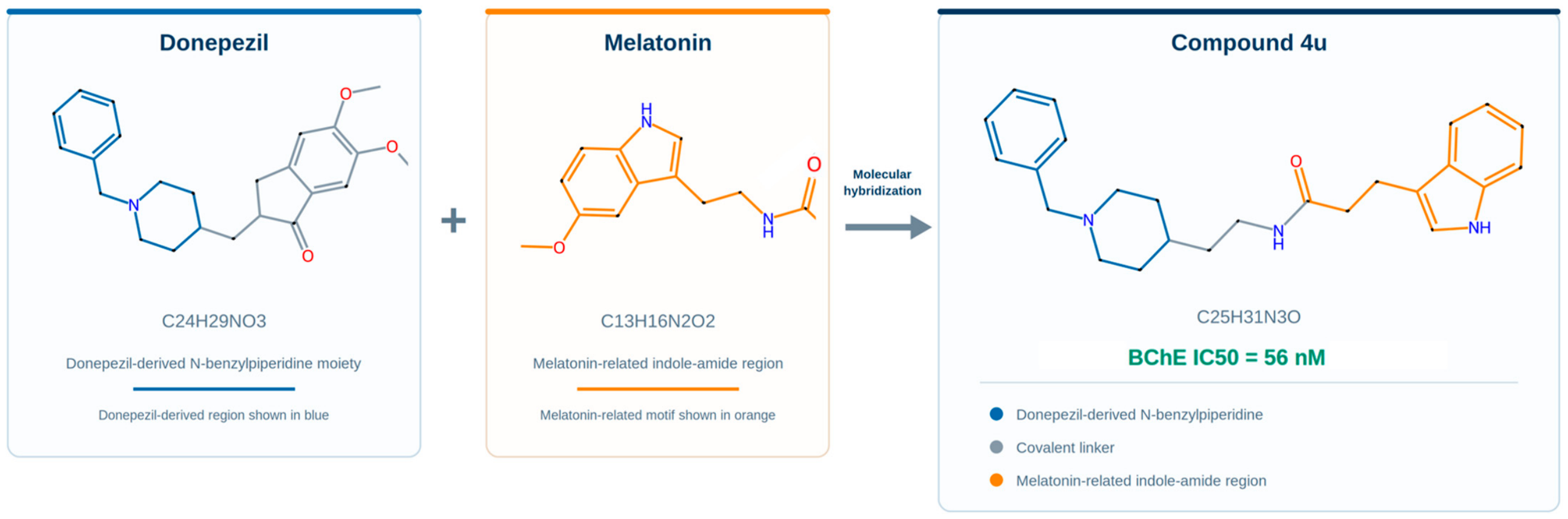
Building on this experimental foundation, the present study was designed to characterize the BChE-bound conformational landscape of compound 4u using a replica-resolved MD framework and a crystal-bound tacrine reference. The central hypothesis was that compound 4u could remain within the BChE binding gorge while adopting alternative bound conformations. Three independently propagated 200 ns replicas initiated from the same equilibrated docking-derived BChE–4u complex were therefore used to distinguish conserved molecular features from replica-dependent binding patterns, while a 100 ns simulation of crystal-bound tacrine served as a crystallographically defined structural reference. By integrating structural, interaction, clustering, and energetic analyses, this study extends the previous experimental characterization by examining conserved and conformation-dependent contacts, fragment reorientation, and state-specific endpoint energetic profiles, providing testable hypotheses for analogue design and experimental validation. The donepezil–melatonin hybrid design of compound 4u is illustrated in Figure 1.
Figure 1.
Donepezil–melatonin hybrid design of compound 4u. The donepezil-derived N-benzylpiperidine moiety and the melatonin-related indole-amide region were integrated through a covalent linker to generate compound 4u (C25H31N3O). Compound 4u was previously reported as a potent human BChE inhibitor (IC50 = 56 nM) [16]. Blue, orange, and gray indicate the donepezil-derived region, melatonin-related region, and covalent linker, respectively.
2. Results
2.1. Simulation Completion and Quality Control
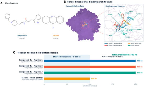
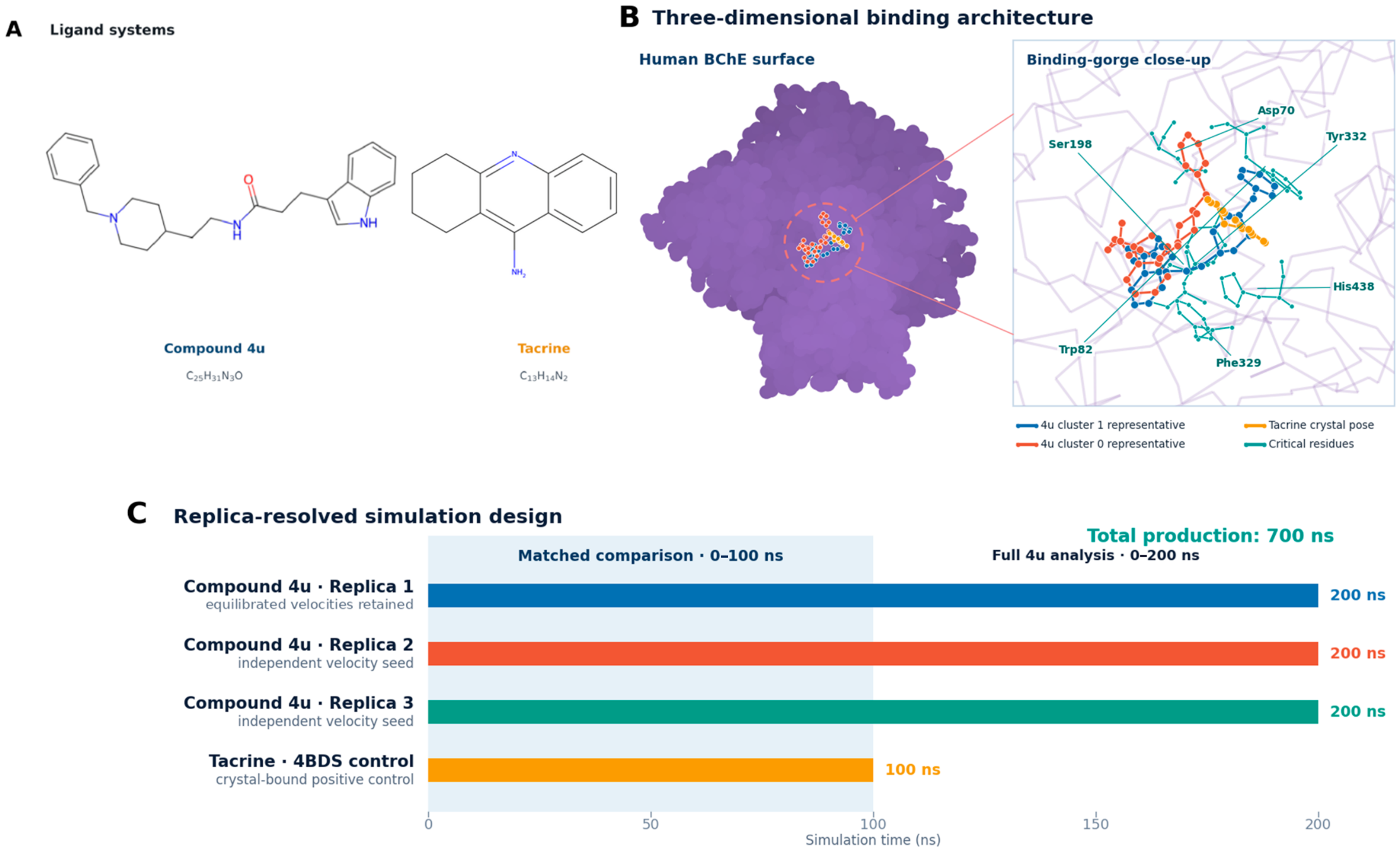
All simulations reached their predefined production durations. The three independently propagated compound 4u replicas were completed to 200 ns each, and the crystallographically bound tacrine control was completed to 100 ns, yielding 700 ns of cumulative production sampling. The molecular systems, three-dimensional binding architecture, and replica-resolved simulation design are summarized in Figure 2. Trajectory–topology consistency and temporal continuity checks of the prepared trajectories detected no missing intervals, duplicate time points, particle-count inconsistencies, or non-finite coordinates.
Figure 2.
Molecular systems and replica-resolved simulation design. (A) Chemical structures of compound 4u and tacrine. (B) Three-dimensional BChE binding architecture showing the representative dominant states of compound 4u, crystallographic tacrine pose, and selected binding-gorge residues. (C) Three independent 200 ns compound 4u replicas and one 100 ns tacrine control. Direct four-system comparisons used the matched 0–100 ns interval, whereas complete compound 4u trajectories were analyzed over 0–200 ns.
Temperature and density profiles showed stable fluctuations throughout production, with no evidence of sustained drift. All four trajectories passed the trajectory-integrity checks and were retained for subsequent structural, interaction, clustering, and energetic analyses. The corresponding temperature and density profiles are provided in Figure S2.
2.2. Global Stability of the BChE Systems
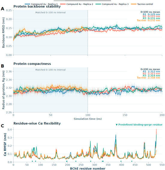
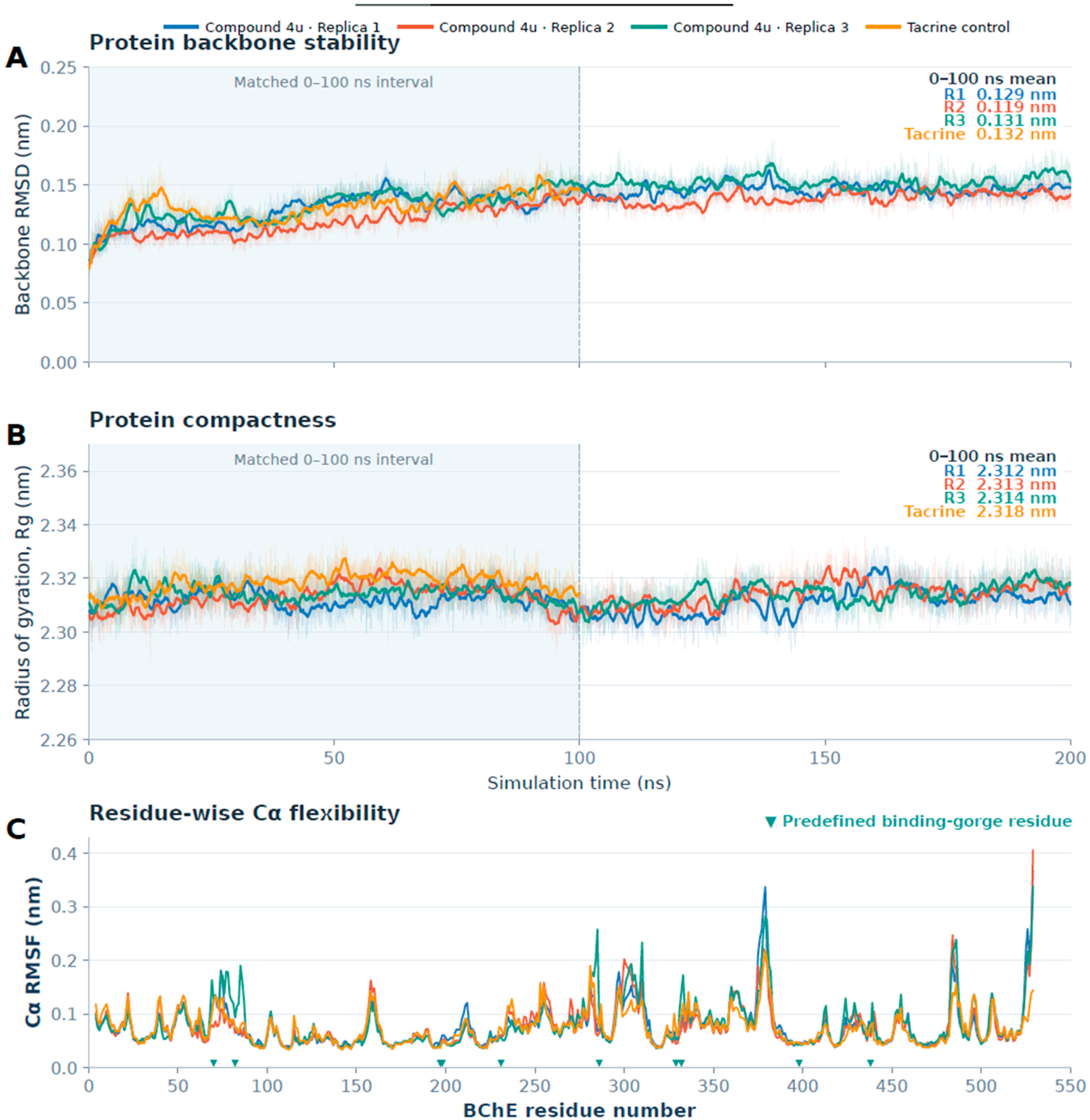
Human BChE remained structurally stable throughout all three 200 ns compound 4u replicas. For direct quantitative comparison, protein stability metrics were evaluated over the matched 0–100 ns interval, yielding mean backbone RMSD values of 0.129, 0.119, and 0.131 nm for replicas 1–3, respectively. The complete trajectories showed limited backbone deviations, consistent with preservation of the global BChE fold.
Over the matched interval, the mean radii of gyration were 2.312, 2.313, and 2.314 nm for replicas 1–3, respectively. The 100 ns tacrine-bound system showed a mean protein backbone RMSD of 0.132 nm and a mean radius of gyration of 2.318 nm. The closely comparable RMSD and radius-of-gyration profiles were consistent with preservation of the overall BChE fold and structural compactness across all four systems (Figure 3A,B).
Figure 3.
Global structural stability and residue-wise flexibility of the BChE systems. (A) Protein backbone RMSD and (B) radius of gyration (Rg) for compound 4u replicas 1–3 and the crystallographically bound tacrine control. Light traces show raw values, whereas bold lines show 1 ns moving averages. The shaded region marks the matched 0–100 ns comparison interval, and the displayed means were calculated over this common interval. (C) Residue-wise Cα RMSF profiles calculated over the complete 0–200 ns compound 4u trajectories and the 0–100 ns tacrine trajectory. Downward triangles indicate the ten predefined binding-gorge residues: Asp70, Trp82, Glu197, Ser198, Trp231, Leu286, Phe329, Tyr332, Phe398, and His438. The closely comparable global stability profiles and broadly overlapping residue-level fluctuation patterns support preservation of the BChE fold across all four systems.
Residue-wise Cα RMSF profiles were also broadly comparable across the four systems. The predefined binding-gorge residues generally showed limited fluctuations, whereas the largest RMSF peaks were restricted to localized flexible regions of the protein. These findings further indicated that the replica-dependent ligand behavior was not associated with widespread changes in BChE residue-level flexibility (Figure 3C).
Within-system Cα superposition of the 0.02 and 50 ns conformations further supported preservation of the global BChE fold, yielding RMSD values of 0.136, 0.126, 0.147, and 0.130 nm for compound 4u replicas 1–3 and the tacrine control, respectively (Figure S1).
2.3. Crystal-Bound Tacrine as a Positive-Control Benchmark
Tacrine remained within the human BChE binding gorge throughout the 100 ns simulation, without evidence of pocket departure or sustained disruption of its crystallographic anchoring environment (Figure 2B). The ligand showed a mean RMSD of 0.183 nm and a 95th-percentile value of 0.268 nm, supporting limited rearrangement within the bound state rather than displacement from the active-site gorge.
Using the primary 4.0 Å contact cutoff, Trp82 and His438 remained in contact with tacrine in 100% of the analyzed frames, while Glu197 showed 96.3% contact occupancy. At least one direct protein–ligand hydrogen bond was present in 83.0% of snapshots sampled at 1 ns intervals. Together, these findings showed that the simulation workflow preserved the pocket retention and principal anchoring interactions of an experimentally resolved BChE–inhibitor complex. This control does not by itself validate the docking-derived binding pose of compound 4u.
2.4. Replica-Resolved Ligand-Dynamic Profiles of Compound 4u
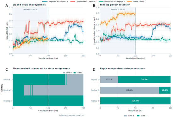
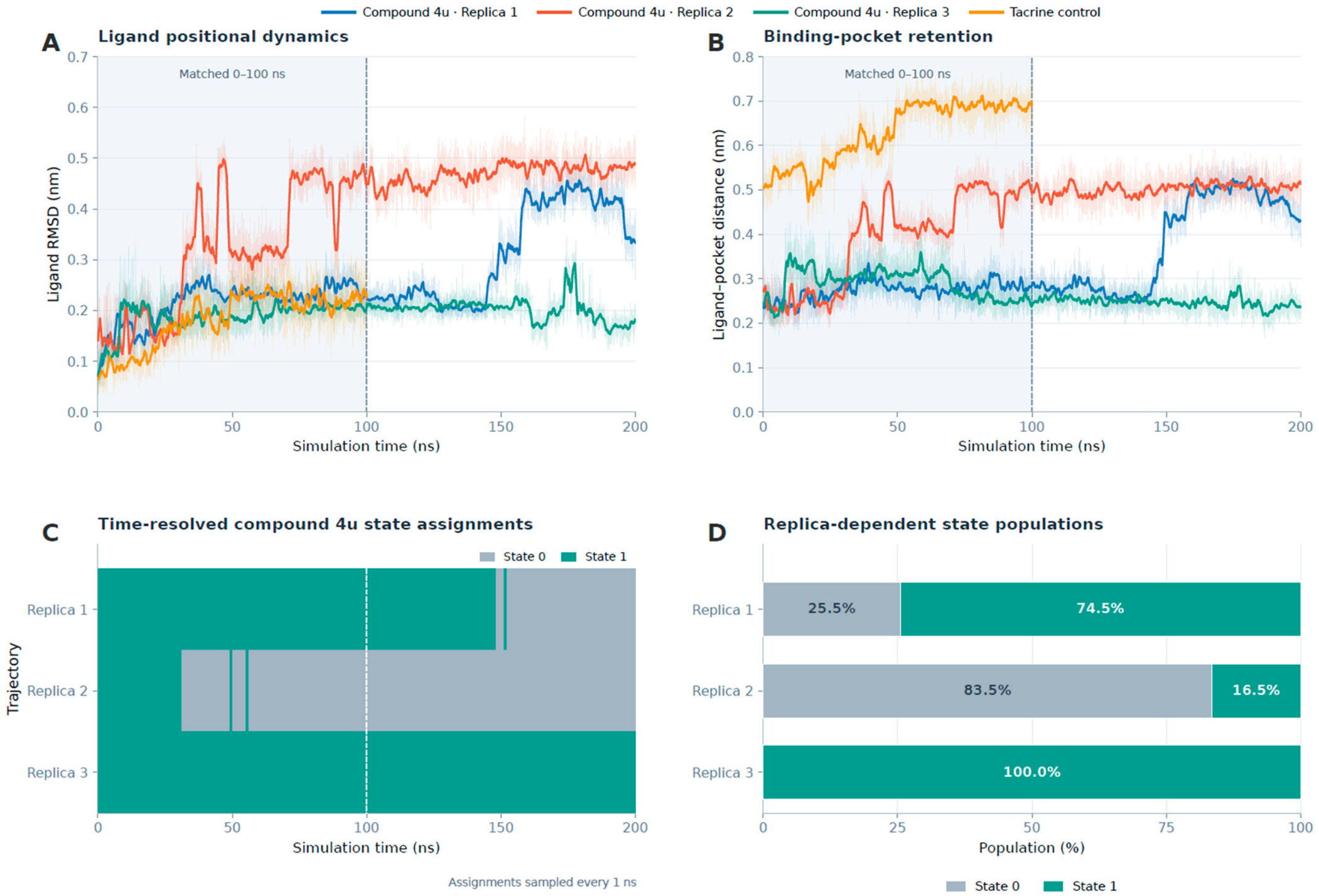
Compound 4u exhibited replica-dependent conformational dynamics across the three complete 200 ns trajectories. Over the matched 0–100 ns interval, the mean ligand heavy-atom RMSD was 0.215, 0.316, and 0.189 nm for replicas 1–3, respectively (Table 1). A descriptive analysis of the final 25 ns of this common interval yielded corresponding mean values of 0.240, 0.451, and 0.205 nm for replicas 1–3, respectively, further illustrating the more pronounced late-window reorientation observed in replica 2 (Figure 4A).
Table 1.
Matched-window structural and energetic metrics for the compound 4u replicas and tacrine control.
Figure 4.
Replica-resolved ligand dynamics and persistent bound-state ensembles. (A) Ligand heavy-atom RMSD and (B) distance between the geometric centers of the ligand heavy atoms and the predefined binding-pocket heavy atoms for compound 4u replicas 1–3 and the tacrine control. The compound 4u trajectories extend to 200 ns, whereas tacrine was simulated for 100 ns; shading indicates the matched 0–100 ns comparison interval. (C) Time-resolved assignments of compound 4u conformations to the two selected clusters and (D) cluster populations calculated separately for each complete 0–200 ns replica. Tacrine was excluded from the clustering analysis because of its distinct chemotype and atom count.
The mean distances between the geometric centers of the ligand and the predefined pocket were 0.274, 0.389, and 0.290 nm for replicas 1–3, respectively, over the matched interval. Despite the greater positional displacement observed in replica 2, analysis of the complete 0–200 ns trajectories showed continued binding-gorge retention without sustained ligand departure in any of the three replicas. Together with the stable protein backbone profiles, these findings indicated that the replica differences reflected alternative ligand arrangements within the BChE gorge rather than ligand dissociation or global complex destabilization.
2.5. Replica-Resolved Persistent Bound-State Ensembles of Compound 4u
According to the clustering procedure described in Section 4.4, joint conformational clustering of all three replicas identified two principal persistent bound-state ensembles for compound 4u. Among the candidate solutions evaluated for (k = 2–6), the (k = 2) solution produced the highest silhouette score (0.543; Figure S3). RMSD-based hierarchical clustering supported the two-group partition (adjusted Rand index, 0.967), which remained consistent across the tested sampling intervals and clustering parameters. The ensembles showed replica-dependent populations: cluster 1 accounted for 74.5%, 16.5%, and 100.0% of snapshots sampled at 1 ns intervals from replicas 1–3, respectively, whereas cluster 0 accounted for 25.5%, 83.5%, and 0.0%. Time-resolved cluster assignments showed that the dominant states represented extended occupancy rather than isolated conformational excursions (Figure 4C,D). Projection of the pooled conformations onto the first two principal components provided an additional visualization of the conformational-state distribution (Figure S4). Replica-resolved state populations over consecutive 50 ns intervals are provided in Table S4.
The two persistent ensembles also exhibited state-dependent contact signatures. At the 4.0 Å cutoff, analysis of snapshots sampled at 1 ns intervals showed Asp70 contact occupancies of 98.0% and 97.6% for cluster 0 in replicas 1 and 2, respectively, compared with 10.7%, 9.1%, and 8.5% for cluster 1 in replicas 1–3. Conversely, His438 contact occupancies were 25.5% and 18.6% for cluster 0 in replicas 1 and 2, compared with 55.7%, 90.9%, and 98.0% for cluster 1 in replicas 1–3. These contact differences supported a structural distinction between the two ensembles. Cluster 0 was characterized by greater engagement of the peripheral Asp70 region, whereas cluster 1 favored deeper interaction with the His438-containing region of the gorge. Neither state represented ligand dissociation, and both retained the shared aromatic and hydrophobic interaction network. Complete replica- and state-specific contact occupancies are reported in Table S5.
Core-based assignments identified one cluster 1-to-0 transition in each of replicas 1 and 2 and none in replica 3, with no reverse core-to-core transition under either core definition. Replicas 1 and 2 ended in cluster 0, whereas replica 3 ended in cluster 1. A 0.04 ns nearest-center excursion in replica 3 did not meet either core-based transition criterion. Terminal core-assigned spans were approximately 42.6, 163.4, and 197.5–197.8 ns for replicas 1–3, respectively. These trajectory-censored observations do not establish equilibrium residence times, equilibrium populations, or kinetic metastability.
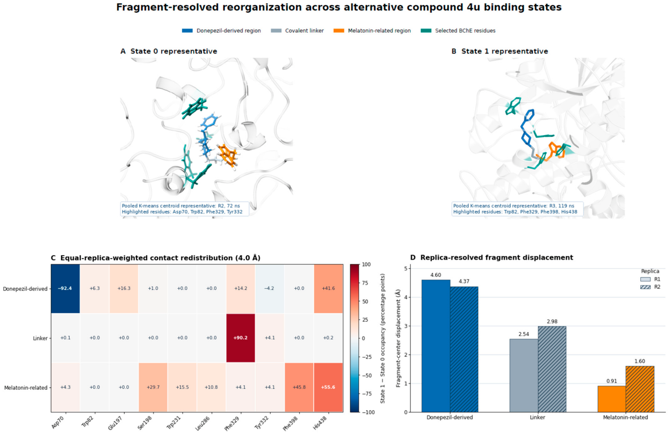
Fragment-resolved analysis linked the alternative bound-state ensembles to differential motion of the hybrid scaffold (Figure 5). The donepezil-derived N-benzylpiperidine region showed the largest state-associated center displacement in replicas 1 and 2 (4.60 and 4.37 Å, respectively), whereas the corresponding shifts of the covalent linker were 2.54 and 2.98 Å and those of the melatonin-related indole-amide region were 0.91 and 1.60 Å, respectively. A within-replica comparison between the two ensembles was unavailable for replica 3 because only cluster 1 was represented in its 1 ns snapshots. Equal-replica-weighted contact differences further showed that state 1 was characterized by loss of the donepezil-derived Asp70 contact (−92.4 percentage points) and enrichment of linker–Phe329 (+90.2 percentage points), melatonin-related–Phe398 (+45.8 percentage points), and melatonin-related–His438 (+55.6 percentage points) contacts relative to state 0. State-specific averages included three replicas for state 1 and two for state 0. Replica-separated maps showed the shared loss of the donepezil-derived Asp70 contact and enrichment of the linker–Phe329 contact in replicas 1 and 2 (Figure S5). Thus, the two ensembles differed primarily through reorientation of the donepezil-derived region and redistribution of fragment-specific contacts within the gorge, while the melatonin-related region underwent a comparatively smaller positional displacement.
Figure 5.
Fragment-resolved reorganization across alternative compound 4u binding states. (A,B) Representative structures selected as the real sampled conformations closest to the pooled K-means ligand-coordinate centroids of states 0 and 1 after protein-backbone alignment. The state 0 and state 1 representatives correspond to frames sampled in replica 2 at 72 ns and replica 3 at 119 ns, respectively. The donepezil-derived N-benzylpiperidine region, covalent linker, and melatonin-related indole-amide region are shown in blue, gray, and orange, respectively; selected BChE residues are highlighted in teal. (C) Equal-replica-weighted difference in fragment-specific contact occupancy between state 1 and state 0 using a 4.0 Å heavy-atom cutoff. Positive values indicate enrichment in state 1, whereas negative values indicate enrichment in state 0. State-specific occupancies were calculated from snapshots sampled at 1 ns intervals and averaged equally across replicas sampling that state: three replicas for state 1 and two for state 0. (D) State-associated displacement of the geometric center of each compound 4u region, shown separately for replicas 1 and 2. Fragment-center displacement is a descriptive protein-aligned geometrical measure and does not represent a kinetic transition coordinate. Replica 3 was excluded from this within-replica comparison because state 0 was absent from its snapshots sampled at 1 ns intervals.
2.6. Critical-Residue Interaction Networks
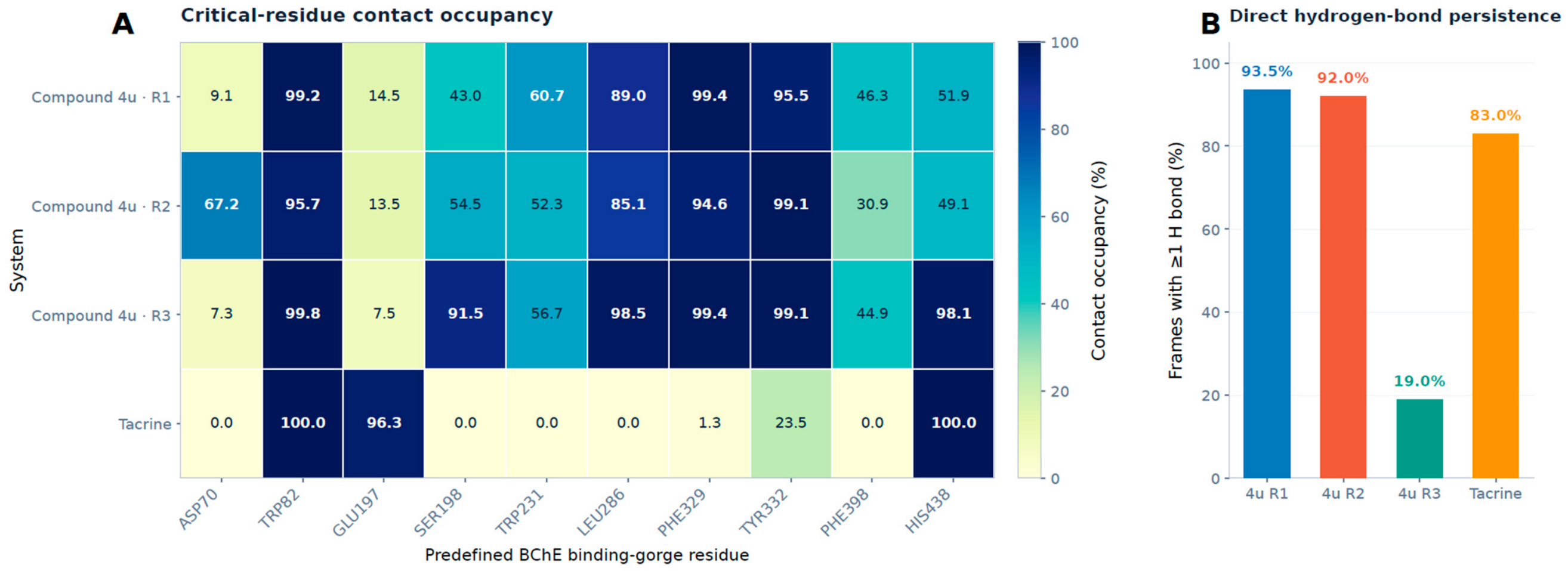
Across the matched 0–100 ns interval, contact analysis was performed for ten predefined BChE binding-gorge residues: Asp70, Trp82, Glu197, Ser198, Trp231, Leu286, Phe329, Tyr332, Phe398, and His438. Using a 4 Å contact cutoff, nine, ten, and eight residues reached an occupancy of at least 10% in replicas 1–3, respectively. Asp70 was the only residue below the threshold in replica 1, with occupancies of 9.1% and 67.2% in replicas 1 and 2, respectively. In replica 3, Asp70 and Glu197 were below this threshold, with occupancies of 7.3% and 7.5%, respectively. Core aromatic and hydrophobic contacts remained highly occupied in all three replicas, including Trp82 (99.2%, 95.7%, and 99.8%), Leu286 (89.0%, 85.1%, and 98.5%), Phe329 (99.4%, 94.6%, and 99.4%), and Tyr332 (95.5%, 99.1%, and 99.1%) for replicas 1–3, respectively (Figure 6A; Table S1). Complete contact-occupancy sensitivity results obtained using the 3.5, 4.0, and 5.0 Å thresholds are provided in Table S6.
Over the complete 0–200 ns trajectories, at least one direct protein–ligand hydrogen bond was present in 93.5%, 92.0%, and 19.0% of snapshots sampled at 1 ns intervals from replicas 1–3, respectively (Figure 6B). Residue-resolved analysis identified hydrogen bonds to the Pro285 backbone carbonyl oxygen and His438 Nε2. Pro285 hydrogen-bond occupancies were 100.0% in cluster 0 for replicas 1 and 2, compared with 79.9%, 36.4%, and 8.0% in cluster 1 for replicas 1–3, respectively. His438 hydrogen-bond occupancies in cluster 1 were 14.1%, 21.2%, and 13.0%, respectively, with none observed in cluster 0 in replicas 1 and 2. Occupancies were calculated within each replica and cluster; cluster 0 was absent from the replica 3 snapshots sampled at 1 ns intervals. Matched 0–100 ns and complete-trajectory hydrogen-bond results, together with residue- and state-specific occupancies, are provided in Table S7. Contacts with Ser198 and His438 were also retained in all three replicas, while the marked difference at Asp70 reflected state-dependent redistribution within the binding gorge. Thus, the three replicas preserved a common aromatic and hydrophobic contact core despite differences in ligand dynamics and direct hydrogen-bond occupancy.
Figure 6.
Critical-residue interactions in the BChE systems. (A) Contact occupancies of the ten predefined binding-gorge residues for compound 4u replicas 1–3 and the tacrine control. Contact occupancy was calculated as the percentage of frames saved at 20 ps intervals in which the minimum residue–ligand heavy-atom distance was below 4.0 Å. (B) Percentage of snapshots sampled at 1 ns intervals containing at least one direct protein–ligand hydrogen bond. Panel (A) reports contact occupancies over the matched 0–100 ns interval. Panel (B) reports hydrogen-bond persistence over the complete available trajectory of each system: 0–200 ns for the compound 4u replicas and 0–100 ns for tacrine.
2.7. MM/GBSA Binding Energetics, Temporal Robustness, and Per-Residue Context
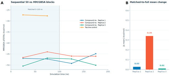
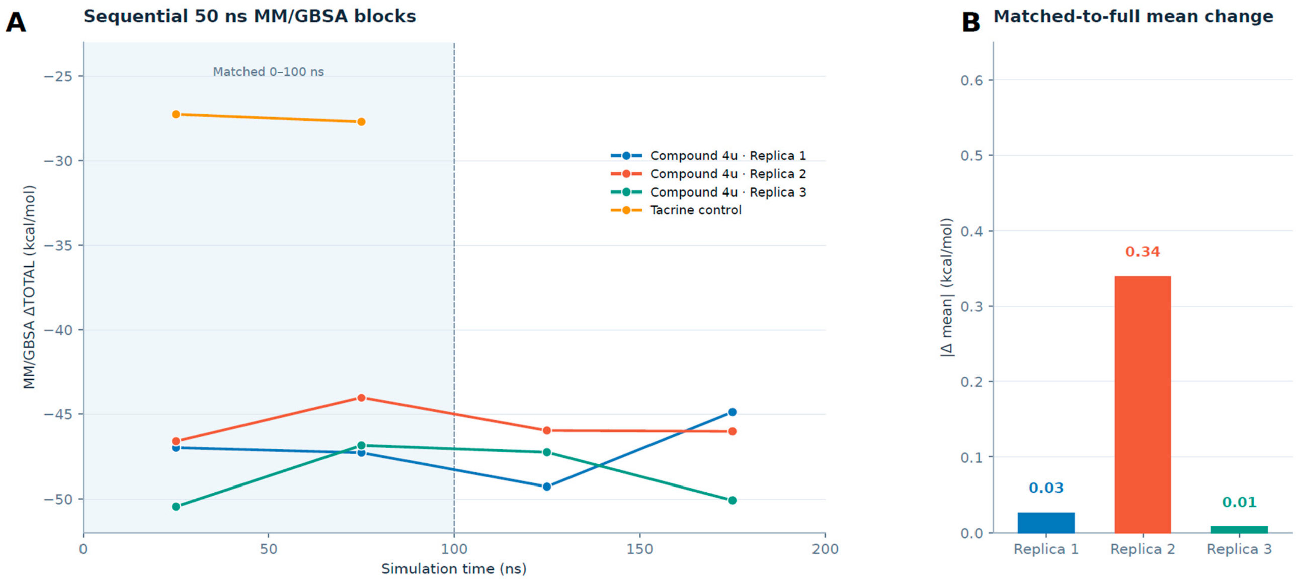
MM/GBSA analysis was performed for all three compound 4u replicas using snapshots sampled at 1 ns intervals. Over the matched 0–100 ns interval, the mean estimates were −47.14, −45.32, and −48.65 kcal/mol for replicas 1–3, respectively. Analysis of the complete 0–200 ns trajectories produced corresponding values of −47.11, −45.66, and −48.66 kcal/mol. The corresponding 95% confidence intervals were −48.24 to −45.94, −46.33 to −44.58, and −49.63 to −47.48 kcal/mol, respectively, estimated using moving-block bootstrap resampling with 10 ns blocks. These intervals describe uncertainty conditional on the sampled trajectories. Extending the analysis from 100 to 200 ns changed the mean estimates by 0.03, 0.34, and 0.01 kcal/mol for replicas 1–3, respectively. These small changes in cumulative means do not establish convergence of conformational-state populations (Figure 7; Table S8).
Figure 7.
Temporal robustness of the MM/GBSA estimates. (A) Mean MM/GBSA binding-energy estimates calculated over sequential 50 ns blocks for compound 4u replicas 1–3 and the tacrine control. Tacrine values are shown for the available 0–100 ns interval. Each 50 ns block contained 50 snapshots sampled at 1 ns intervals. (B) Absolute differences between the matched 0–100 ns and complete 0–200 ns means for compound 4u replicas 1–3. The blocks represent descriptive trajectory intervals rather than statistically independent replicates. Small changes in cumulative mean energies do not establish convergence of conformational-state populations.
Cluster-specific MM/GBSA means were −47.87, −48.26, and −48.66 kcal/mol for cluster 1 in replicas 1–3, respectively, compared with −44.91 and −45.15 kcal/mol for cluster 0 in replicas 1 and 2. The cluster 1-minus-cluster 0 differences were −2.96 kcal/mol (95% confidence interval: −5.33 to −0.85) in replica 1 and −3.11 kcal/mol (−4.32 to +2.35) in replica 2, using 10 ns moving-block bootstrap resampling of energies and state labels together. The replica 2 interval included zero, and its contrast was sensitive to bootstrap block length. A within-replica contrast was unavailable for replica 3 because cluster 0 was absent from its sampled energy frames. These endpoint estimates omit entropy and do not establish an equilibrium free-energy ranking of the ensembles (Table S8).
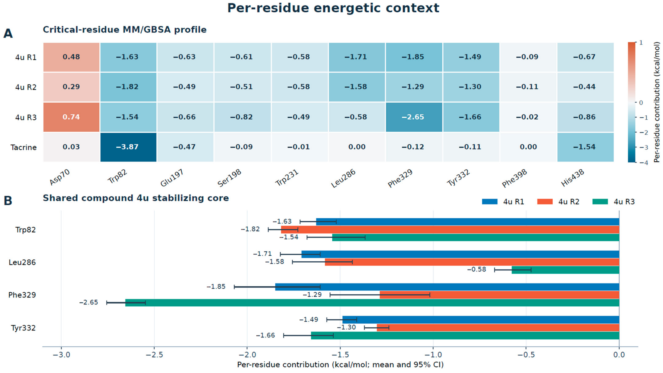
Across the three replicas, the complete-trajectory mean estimates spanned a range of 3.00 kcal/mol. Per-residue decomposition identified a shared stabilizing core comprising Trp82, Leu286, Phe329, and Tyr332. Their mean contributions were −1.63, −1.71, −1.85, and −1.49 kcal/mol in replica 1; −1.82, −1.58, −1.29, and −1.30 kcal/mol in replica 2; and −1.54, −0.58, −2.65, and −1.66 kcal/mol in replica 3, respectively. These results indicated that the three replicas preserved a common favorable energetic environment despite their different dominant ligand orientations (Figure 8; Table S2).
Figure 8.
Per-residue energetic context of the BChE systems. (A) Mean MM/GBSA contributions of the ten predefined binding-gorge residues for compound 4u replicas 1–3 and the tacrine control. (B) Shared stabilizing energetic core of compound 4u comprising Trp82, Leu286, Phe329, and Tyr332. Negative values indicate favorable contributions. In panel B, error bars represent 95% percentile confidence intervals estimated using moving-block bootstrap resampling with 10 ns blocks. These intervals describe uncertainty conditional on the sampled trajectories. Compound 4u and tacrine decompositions cover 0–200 and 0–100 ns, respectively.
Topology audits confirmed that compound 4u was protonated at the piperidine nitrogen (N3–H21), with ligand partial charges summing to +1.00 e in all three replicas. Asp70 showed favorable electrostatic contributions of −20.09, −25.41, and −17.45 kcal/mol, offset by polar-solvation contributions of +21.54, +28.04, and +18.55 kcal/mol for replicas 1–3, respectively. After inclusion of all decomposition terms, its net contributions were +0.48, +0.29, and +0.74 kcal/mol, respectively. Thus, the positive net contribution reflected the balance of interaction and solvation terms despite favorable electrostatics.
The tacrine control produced a mean MM/GBSA estimate of −27.47 kcal/mol. Its critical-residue profile was dominated by favorable contributions from Trp82 (−3.87 kcal/mol) and His438 (−1.54 kcal/mol), consistent with retention of its crystallographic anchoring environment. Because compound 4u and tacrine differ in chemotype and were analyzed over different trajectory durations, their total and residue-level estimates were treated as internal energetic descriptors rather than direct measures of relative experimental affinity or potency. Overall, the structural and energetic results supported persistent binding-gorge retention and a shared favorable energetic core across the three compound 4u replicas, despite their different conformational-state populations. The principal structural, interaction, and energetic metrics are summarized in Table 1 and Table 2.
Table 2.
Replica-resolved bound-state, interaction, and energetic characteristics of compound 4u.
3. Discussion
The present study showed that compound 4u remained within the human BChE binding gorge across three independent 200 ns MD simulations while sampling distinct bound-state ensembles. The principal difference between the replicas was therefore not ligand retention but the relative population of alternative conformational states. Reliance on a single trajectory could have presented either the compact or reoriented arrangement as the predominant binding mode and obscured the underlying conformational heterogeneity [20,21]. The contribution of this study is a replica-resolved structural interpretation of BChE recognition by an experimentally characterized inhibitor, identifying a shared interaction and energetic core alongside conformation-dependent contacts.
Replica 1 preferentially sampled a more compact ligand arrangement, whereas replica 2 favored a more displaced but non-dissociated orientation. Replica 3 predominantly sampled cluster 1. All three replicas preserved extensive contacts with the BChE binding gorge, whereas direct hydrogen-bond occupancy was substantially lower in replica 3 (19.0%) than in replicas 1 and 2 (93.5% and 92.0%, respectively). The redistribution of individual contacts, including the replica-dependent involvement of Asp70, suggests that compound 4u accommodated conformational reorientation without losing its principal aromatic and hydrophobic anchoring environment. Trp82, Leu286, Phe329, and Tyr332 formed a conserved interaction and energetic core across all three trajectories. These residues span the choline-binding region, acyl-binding pocket, and surrounding aromatic gorge environment, providing a distributed structural basis for sustained compound 4u retention across all three replicas [7,23,24]. Recent experimental and computational studies of structurally diverse cholinesterase inhibitors have similarly used binding-gorge contacts and MD-derived interaction profiles to rationalize inhibitory binding [25,26,27,28,29]. Multi-target computational investigations have further demonstrated the value of residue-level interaction analysis for evaluating cholinesterase-directed scaffolds [30,31,32].
The replica-dependent cluster populations support structural heterogeneity within the sampled bound-state ensembles. Agreement between clustering methods supports the structural partition but does not establish kinetic metastability. Replicas 1 and 2 ended in cluster 0, whereas replica 3 ended in cluster 1; no core-to-core transition from cluster 0 to cluster 1 was observed. The terminal states and trajectory-censored occupancy spans do not establish equilibrium populations, equilibrium residence times, or a thermodynamic preference for cluster 0. Accordingly, the groups are interpreted as persistent alternative bound-state ensembles, with rigorous kinetic characterization requiring further sampling and validation [33,34].
The energetic analyses were consistent with the structural findings. Complete-trajectory MM/GBSA means spanned 3.00 kcal/mol across the three replicas. Cluster 1 had more negative state-specific point estimates than cluster 0 in replicas 1 and 2, but the replica 2 contrast was sensitive to bootstrap block length and its 10 ns-block confidence interval included zero. These endpoint estimates omit entropy and do not establish an equilibrium free-energy ranking of the ensembles. Per-residue decomposition further indicated that stabilization was distributed across several binding-gorge residues rather than controlled by a single interaction.
The crystallographically bound tacrine simulation provided an internal positive-control benchmark. Tacrine remained within its experimentally resolved binding environment and retained its principal anchoring contacts throughout the 100 ns trajectory, indicating that the workflow preserved a structurally established BChE–inhibitor complex [22,23]. Within this framework, the tacrine MM/GBSA estimate complemented the structural stability and contact-retention results by providing an energetic descriptor of the crystallographically anchored control complex. Preservation of the crystallographic tacrine complex does not by itself validate the docking-derived pose of compound 4u. The endpoint energetic estimates were interpreted as internal computational descriptors rather than direct measures of relative experimental affinity or potency [35,36,37,38].
Several limitations should be considered. Three independent replicas revealed substantial state-population variability but cannot establish fully converged equilibrium populations or binding kinetics. The 200 ns duration may also exclude slower conformational transitions, which could require additional replicas or enhanced-sampling methods [20,21,39]. All three compound 4u replicas originated from the same equilibrated docking-derived complex, so the present design does not establish independence from the selected starting pose or convergence from alternative docking orientations. Force-field selection, ligand parameterization, protonation states, and the implicit-solvent and entropic approximations of MM/GBSA may influence the calculated values [35,36,37,38,39,40]. The results are also conditional on the fixed GLH197/HID438 protonation model; alternative protonation states were not simulated. Experimental structural studies, mutational analysis, enzyme kinetics, and additional independent simulations would therefore be valuable for testing the predicted binding modes and residue-level contributions.
Overall, the combined structural, interaction, clustering, and energetic evidence identifies compound 4u as a persistently engaged and conformationally adaptable BChE-binding scaffold. Together with its previously reported nanomolar BChE inhibition and multifunctional biological profile, these findings provide testable structural hypotheses for analogue design and experimental validation of BChE recognition in the context of Alzheimer’s disease research [7,16]. Conformational restriction near the N-benzylpiperidine–linker junction is hypothesized to narrow the range of bound orientations while preserving the shared aromatic and hydrophobic contact core. Amide N-methylation is hypothesized to preferentially weaken the Pro285-associated cluster 0 arrangement relative to cluster 1. Matched analogue synthesis, BChE inhibition assays, and structural characterization could test these predictions.
4. Materials and Methods
4.1. Study Design and Molecular Systems
The study comprised three independently propagated MD replicas of human BChE complexed with compound 4u and one simulation of crystallographically bound tacrine as a positive control. Compound 4u was simulated for 200 ns in each replica starting from the same equilibrated docking-derived complex, with independent stochastic propagation. Tacrine was simulated for 100 ns from its experimentally resolved binding pose in the human BChE–tacrine crystal structure (PDB ID: 4BDS) [22]. The cumulative production sampling was therefore 700 ns.
Replica-resolved analyses of compound 4u were performed over the complete 0–200 ns trajectories. Direct quantitative comparisons involving all four systems were performed over the common 0–100 ns interval. Analyses intended to characterize the complete trajectories used 0–200 ns for compound 4u and 0–100 ns for tacrine, with cross-system comparisons interpreted qualitatively when the analyzed durations differed. The compound 4u trajectories were treated as independent dynamical realizations for evaluating reproducible binding features and replica-dependent conformational populations, rather than as independent biological observations [20,21].
4.2. System Preparation and Molecular Dynamics Protocol
Human BChE was prepared from chain A of the 4BDS crystal structure [22]. Protein protonation states were assigned for a target pH of 7.4 using PROPKA 3.5.1 predictions followed by manual review of catalytic-site residues. Initial receptor hydrogen placement used OpenMM Modeller (version 8.4.0.dev-4768436) with ff14SB templates and explicitly specified residue variants. The selected pH 7.4 protonation-state model, used consistently across all four systems, contained HID77, HID126, HID207, HID372, and HID438; HIE214 and HIE423; and protonated Glu197 (GLH, with HE2 on OE2). No histidine residues were assigned the doubly protonated HIP state. Glu197 was assigned the neutral GLH state, consistent with its predicted pKa of 8.27. His438 was assigned the neutral HID tautomer to represent a resting catalytic-site arrangement with Nδ1 protonated and Nε2 available as a hydrogen-bond acceptor toward Ser198. This was an explicit modeling choice that overrode the predicted His438 pKa of 8.75, which favored a cationic histidine at pH 7.4. The same assignments were retained across all simulations; alternative protonation states were not simulated. The N terminus was acetyl-capped (ACE), whereas Val529 was retained as a standard anionic C terminus containing OXT. The three native disulfide bonds, Cys65–Cys92, Cys252–Cys263, and Cys400–Cys519, were retained. The prepared receptor contained no crystallographic waters, glycans, ions, or other nonprotein hetero-residues before ligand placement and explicit resolvation. The internal 4BDS gap comprising Asp378 and Asp379 and the missing heavy atoms of Gln486 were reconstructed and structurally validated before protonation-state assignment; during AMBER system assembly, LEaP rebuilt protein hydrogen atoms from the heavy-atom receptor while retaining the assigned protonation-state residue names. System compositions and residue-state definitions are provided in Table S3.
The selected compound 4u microstate, designated QP1, corresponded to protonation of the piperidine nitrogen and carried a net charge of +1. This microstate was used in all three compound 4u replicas. The piperidinium N3–H21 connectivity and ligand partial-charge sum of +1.00 e were verified in the production topologies of all three replicas. The initial BChE–4u complex was generated using AutoDock Vina 1.2.7 [41]. Docking was performed within a 22 × 22 × 22 Å grid centered at 133.076, 116.113, and 41.335 Å, encompassing the crystallographic BChE binding gorge. An exhaustiveness value of 24 was used, and 20 poses were generated in each of two runs using seeds 20260711 and 20260712. The five highest-ranked QP1 poses from each run were pooled and clustered using a ligand heavy-atom RMSD threshold of 2.0 Å. Pose assessment employed protein-contact, polar-contact, and aromatic-contact cutoffs of 4.5, 3.5, and 5.0 Å, respectively, with a minimum requirement of three contacting protein residues. Configurations with a minimum protein–ligand heavy-atom distance below 1.2 Å were classified as containing severe steric clashes. The selected cluster was represented in both docking runs, preserved contacts across the BChE gorge, and contained no severe clash; its highest-ranked representative from the run using seed 20260711 was selected as the MD starting pose. As a protocol control, tacrine redocking recovered second-ranked poses with heavy-atom RMSDs of 0.37 and 0.36 Å from the crystallographic pose for seeds 20260711 and 20260712, respectively. The top-ranked poses had RMSDs of approximately 4.79 Å in both runs, indicating near-native pose recovery with ranking ambiguity. Nevertheless, protonated tacrine was retained in its experimentally resolved 4BDS pose for the positive-control simulation.
The protein was described using the AMBER ff14SB force field [42]. Compound 4u and tacrine were parameterized using GAFF2 [43,44] with AM1-BCC partial charges [45]. Each complex was placed in a truncated-octahedral TIP3P water box extending 12 Å from the solute [46]. Na+ and Cl− ions were added to neutralize the systems and establish a target NaCl concentration of 0.15 M.
The initial BChE–4u and BChE–tacrine complexes underwent four consecutive energy-minimization stages. Protein and ligand heavy atoms were restrained at 10.0 kcal mol−1 Å−2 during the first stage, followed by restraint constants of 5.0, 1.0, and 0.25 kcal mol−1 Å−2 applied to the protein backbone and ligand heavy atoms during the subsequent stages. The corresponding minimization tolerances were 100, 50, 20, and 10 kJ mol−1 nm−1. Staged NVT heating and stabilization totaled 200 ps, during which the temperature was increased from 25 to 300 K, the positional restraints were progressively reduced, and the integration time step was increased from 0.5 to 1 fs. Three consecutive NPT stages of 250, 500, and 1000 ps were subsequently performed at 300 K and 1 bar using restraint constants of 1.0, 0.25, and 0 kcal mol−1 Å−2, respectively, and a final integration time step of 2 fs. The complete stage-wise minimization and equilibration protocol is provided in Table S3. Throughout equilibration and production, long-range electrostatic interactions were treated using the particle-mesh Ewald method with a real-space cutoff of 1.0 nm [47]. Bonds involving hydrogen atoms were constrained, and water molecules were treated as rigid. Temperature, density, particle number, and coordinate integrity were verified before production.
Production simulations were performed with OpenMM 8.2 under periodic NPT conditions [48]. Temperature was maintained at 300 K using the LangevinMiddleIntegrator with a friction coefficient of 1.0 ps−1 and an integration time step of 2 fs. Pressure was maintained at 1 bar using a MonteCarloBarostat, with volume-change attempts performed every 50 integration steps. Calculations were executed on the CUDA platform using mixed precision. Coordinates were recorded every 20 ps, and restartable checkpoints were generated at 1 ns intervals. The three compound 4u replicas originated from the same equilibrated coordinates and were propagated as independent stochastic realizations. Replica 1 retained the velocities of the final equilibrated state. Replica 2 was initiated after independent velocity reassignment at 300 K using seed 2026082503, with barostat and integrator seeds 2026082504 and 2026082505, respectively. Replica 3 was initiated by independent velocity reassignment at 300 K using seed 2026102303, with initial barostat and integrator seeds 2026102304 and 2026102305, respectively. Each compound 4u replica was simulated for 200 ns. The crystallographic tacrine complex was simulated for 100 ns using initial barostat and integrator seeds 2026082903 and 2026082904, respectively. The documented software versions and stochastic seeds are listed in Table S3.
4.3. Trajectory Processing and Structural Analyses
Trajectories were processed using MDTraj (version 1.11.1.post2) after periodic-boundary correction and reconstruction of intact protein and ligand molecules [49]. Protein-backbone least-squares alignment used each system’s first saved production frame (0.02 ns) as the initial reference for backbone and ligand RMSD analyses [50]. Pooled compound 4u analyses and Cα RMSF used a common backbone reference from replica 1 at 0.02 ns; tacrine Cα RMSF used its own first saved frame. For the within-system structural superpositions shown in Figure S1, each 50 ns conformation was separately least-squares fitted to its corresponding first saved production conformation at 0.02 ns using the 526 matched protein Cα atoms, and the post-fit Cα RMSD was calculated. Protein stability was evaluated using backbone and Cα RMSD, Cα RMSF, and radius of gyration calculated with equal weighting of protein atoms. Cα RMSD used Cα-only least-squares alignment to each system’s first saved frame. Complete trajectories were used for within-system structural assessments, whereas matched 0–100 ns windows were used for direct quantitative comparisons among the four systems. Residue-wise Cα RMSF profiles were calculated over the complete 0–200 ns compound 4u trajectories and the 0–100 ns tacrine trajectory and were interpreted as descriptive fluctuation patterns.
Ligand dynamics were evaluated using heavy-atom RMSD after protein alignment, with each system’s first saved production frame (0.02 ns) and the 50 ns conformation used as separate references. Displacement of the ligand heavy-atom geometric center relative to the 50 ns reference, the ligand–pocket geometric-center distance, and minimum ligand–protein heavy-atom distance were also calculated. The pocket center was defined as the geometric center of the combined heavy atoms of Asp70, Trp82, Glu197, Ser198, Trp231, Leu286, Phe329, Tyr332, Phe398, and His438, selected to represent the principal functional regions of the BChE binding gorge [7,22,23]. Ligand–pocket geometric-center distance was used as a system-specific descriptor of ligand positioning within the binding gorge.
Residue contacts were defined using the minimum heavy-atom distance between each residue and the ligand. A distance below 4.0 Å was used as the primary criterion, while 3.5 and 5.0 Å thresholds were retained for sensitivity assessment. Primary contact occupancies (Table S1) were calculated as the proportion of frames saved at 20 ps intervals satisfying the specified threshold. State-specific contact occupancies (Table S5) and contact-cutoff sensitivity results (Table S6) were calculated using snapshots sampled at 1 ns intervals. Direct protein–ligand hydrogen bonds were evaluated at 1 ns intervals over the complete available trajectory of each system using the Baker–Hubbard geometrical definition: a hydrogen–acceptor distance below 2.5 Å and a donor–hydrogen–acceptor angle above 120° [51]. Donors and acceptors were restricted to chemically eligible nitrogen and oxygen atoms using topology-defined bonds, protonation states, and ligand chemistry. The indole, amide, and protonated piperidine nitrogens of compound 4u were excluded as acceptors. Hydrogen-bond analysis was restricted to direct protein–ligand interactions in the solute-only archived trajectories. Individual hydrogen-bond and residue-level occupancies were calculated for each replica and separately for each conformational cluster, using all snapshots in the relevant window or cluster, including those without hydrogen bonds, as the denominator. Residue-level occupancy represented the fraction of snapshots with at least one hydrogen bond to that residue; occupancies for clusters absent from a replica were reported as unavailable.
4.4. Conformational Clustering and Bound-State Characterization
One frame per nanosecond was extracted from each 200 ns compound 4u trajectory, yielding 200 conformers per replica and 600 conformers in total. Following periodic-boundary correction and protein-backbone alignment, ligand heavy atoms were paired using a bijective topology-based atom map, and the coordinate vectors from all three replicas were pooled without coordinate scaling or ligand self-fitting. K-means clustering was performed for k = 2–6 using 30 initializations and a fixed random seed of 20260828 [52]. The solution with the highest silhouette score was selected [53]. Silhouette scores were reported for all tested values of k. The partition was assessed using average-linkage hierarchical clustering based on pairwise ligand heavy-atom RMSD in the common protein-aligned frame, without ligand self-fitting. Sensitivity analyses separately varied the sampling interval (0.2, 0.5, 1, and 2 ns), random seed, and number of initializations (10, 20, and 30). Partition agreement was quantified using the adjusted Rand index on the common 1 ns sampling grid, and replica-specific populations were compared after label matching. Cluster populations were calculated separately for each replica, and representative structures were defined as the conformations closest to the corresponding centroids. For continuity of presentation, joint-cluster labels were matched to the original two-replica labels by one-to-one centroid matching that minimized the total ligand-coordinate RMSD. All reported populations and assignments were obtained from the new three-replica model. Time-resolved assignments were summarized over consecutive 50 ns intervals, and principal component analysis was used to visualize the pooled conformational space [54].
To relate the hybrid architecture of compound 4u to its replica-resolved binding behavior, its 29 heavy atoms were partitioned into three non-overlapping structural regions: the donepezil-derived N-benzylpiperidine region (13 atoms), the covalent linker (3 atoms), and the melatonin-related indole-amide region (13 atoms). The partition was defined using the validated compound 4u atom and bond mapping and was identical in all three replicas. For each 1 ns sampled conformation, the minimum heavy-atom distance between each ligand region and each of the ten predefined BChE binding-gorge residues was calculated. State-specific contact occupancies were determined separately for each replica using 3.5, 4.0, and 5.0 Å distance thresholds. Equal-replica-weighted contact occupancies were calculated as the arithmetic mean of the corresponding replica-specific occupancies, including only replicas in which the state was sampled. For each replica sampling both states, state-associated fragment displacement was calculated as the Euclidean distance between the mean geometric centers of the fragment in states 0 and 1 after protein-backbone alignment. The structures displayed in Figure 5A,B were real sampled conformations closest to the corresponding pooled K-means centroids. These analyses were performed using periodic-boundary-corrected, protein-aligned trajectories. Fragment-center displacement was used as a descriptive geometrical measure of state-associated ligand reorientation.
The clusters were interpreted as persistent alternative bound-state ensembles rather than kinetically established metastable states [33,34]. Nearest-centroid assignments were evaluated at 20 ps intervals. Two core definitions used the 50th and 75th percentiles of within-cluster ligand heavy-atom RMSD to the centroid, calculated from the pooled 1 ns snapshots. A frame entered a core when its distance to the nearest centroid did not exceed the corresponding threshold. The last visited core assignment was retained until a different core was reached, while frames preceding the first core visit remained unassigned. Transitions and observed occupancy spans were evaluated separately for each replica; the first and last episodes were treated as left- and right-censored, respectively. These observations were not interpreted as equilibrium residence times or converged equilibrium populations. Joint coordinate-space clustering was restricted to compound 4u because tacrine has a distinct chemotype and atom count.
4.5. MM/GBSA Binding-Energy Estimation, Temporal Analysis, and Per-Residue Decomposition
Binding-energy estimates were calculated using a consistent single-trajectory MM/GBSA protocol with the sander backend [55,56]. Receptor and ligand coordinates were extracted from the corresponding complex snapshots after removal of explicit solvent molecules and ions. All complex, receptor, and ligand topologies used the mbondi2 radius set. Calculations employed the generalized Born model igb = 5 and a salt concentration of 0.150 M. The reported estimates comprised molecular-mechanics van der Waals and electrostatic terms together with polar and nonpolar solvation contributions. MM/GBSA values were reported as comparative binding-energy estimates derived from molecular-mechanics and implicit-solvation contributions, consistent with the endpoint protocol applied here [35,36,37,38,55,56]. No explicit conformational entropy term was included.
For each compound 4u replica, 200 snapshots sampled at 1 ns intervals were analyzed over the complete 0–200 ns trajectory. Temporal robustness was evaluated using four consecutive 50 ns blocks containing 50 snapshots each, and the matched 0–100 ns estimates were calculated from the first two blocks. Tacrine was evaluated using 100 snapshots sampled at 1 ns intervals over its complete 0–100 ns trajectory and two consecutive 50 ns blocks containing 50 snapshots each. Block-wise estimates were treated as descriptive temporal intervals rather than statistically independent replicates. Cluster-specific MM/GBSA means were calculated separately for each compound 4u replica by matching energy snapshots to the nearest-centroid assignments from the joint clustering at the same 1 ns sampling times. Within-replica contrasts were calculated as the cluster 1 mean minus the cluster 0 mean; contrasts were unavailable when either cluster was absent.
Per-residue decomposition (idecomp = 2) was performed using the same snapshots sampled at 1 ns intervals as the total-energy calculations: 200 snapshots for each compound 4u replica and 100 for tacrine. Particular attention was given to Asp70, Trp82, Glu197, Ser198, Trp231, Leu286, Phe329, Tyr332, Phe398, and His438. Overall nonpolar solvation used the LCPO surface term, whereas residue-level nonpolar contributions were calculated from SANDER per-residue solvent-accessible areas using a coefficient of 0.0072 kcal mol−1 Å−2. These decomposition terms were not rescaled to reproduce the overall nonpolar term. For Asp70, electrostatic and polar-solvation contributions were examined separately alongside the net decomposition contribution.
Total and residue-level estimates were interpreted as system-specific endpoint energetic descriptors, without inferring relative experimental affinity or potency. Uncertainty in total and residue-level means was quantified using 95% percentile confidence intervals from a noncircular moving-block bootstrap with 2000 attempted resamples. The primary block length was 10 ns, with 5 and 20 ns blocks used for sensitivity analysis. Overlapping chronological blocks were sampled with replacement separately within each system and analysis window. For cluster-specific means and contrasts, energies and cluster labels were jointly resampled from each complete chronological trajectory. Resamples lacking the required cluster or clusters were excluded, and valid resample counts were recorded. Confidence intervals were not reported when temporal support or the number of valid resamples was insufficient. These intervals describe uncertainty conditional on the sampled trajectories and do not establish equilibrium sampling or convergence.
5. Conclusions
This replica-resolved study demonstrated that compound 4u remained persistently engaged within the human BChE binding gorge across three independent 200 ns simulations while preferentially sampling distinct bound-state ensembles. Replica-dependent populations supported persistent alternative bound-state ensembles; equilibrium populations and kinetic metastability were not established. Despite this conformational heterogeneity, all three replicas preserved a common interaction and energetic core, and their complete-trajectory MM/GBSA means spanned 3.00 kcal/mol. The third replica reproduced this conserved core despite substantially lower direct hydrogen-bond occupancy, strengthening the distinction between shared anchoring features and replica-dependent interactions. The crystallographically bound tacrine control retained its established anchoring environment throughout 100 ns, further supporting the structural reliability of the simulation framework.
Together with the previously reported nanomolar inhibition of human BChE and multifunctional biological profile of compound 4u, these findings provide testable structural hypotheses for analogue design and experimental validation of BChE recognition in Alzheimer’s disease research. The conserved contributions of Trp82, Leu286, Phe329, and Tyr332, together with the state-dependent involvement of Asp70, provide specific targets for biochemical, kinetic, and residue-directed experimental validation. Further experimental studies and expanded conformational sampling are needed to test these structural hypotheses and establish their relevance to BChE inhibition.
Supplementary Materials
The following supporting information can be downloaded at: https://www.mdpi.com/article/10.3390/molecules31224051/s1, Figure S1, Within-system structural superposition of human BChE at 0.02 and 50 ns; Figure S2, Thermodynamic quality-control profiles for compound 4u replicas 1–3 and the tacrine control; Figure S3, Selection and robustness of the joint three-replica ligand conformational-clustering solution; Figure S4, Principal-component analysis of pooled compound 4u conformations; Figure S5, Replica-resolved fragment-specific compound 4u contacts. Table S1, Critical-residue contact occupancies over the matched 0–100 ns analysis interval; Table S2, Per-residue MM/GBSA decomposition using snapshots sampled at 1 ns intervals; Table S3A, System composition and residue-state definitions; Table S3B, Staged minimization, equilibration, and production protocol; Table S3C, Software versions and stochastic seeds; Table S3D, Tacrine redocking rank comparison; Table S4A, Joint-cluster populations within consecutive 50 ns intervals; Table S4B, Clustering selection and sensitivity to method, sampling interval, seed, and initialization count; Table S4C, Core-based transition counts and terminal observed occupancy spans; Table S5, State-specific residue-contact profiles from the joint three-replica clustering; Table S6, Contact-cutoff sensitivity analysis using snapshots sampled at 1 ns intervals; Table S7A, Protein–ligand hydrogen-bond results over matched and complete trajectories; Table S7B, Residue-resolved hydrogen-bond occupancies by window and conformational cluster; Table S7C, Individual donor–hydrogen–acceptor hydrogen-bond occupancies; Table S8A, MM/GBSA components and temporal-interval estimates; Table S8B, Cluster-specific MM/GBSA components and conditional uncertainty; Table S8C, Moving-block bootstrap sensitivity and state-energy contrasts.
Author Contributions
Conceptualization, İ.C.H. and Z.G.Ö.; methodology, İ.C.H. and Z.G.Ö.; writing—original draft preparation, İ.C.H.; writing—review and editing, İ.C.H. and Z.G.Ö.; supervision, Z.G.Ö. All authors have read and agreed to the published version of the manuscript.
Funding
This research received no external funding.
Data Availability Statement
The processed numerical data supporting the findings of this study are provided in the Supplementary Materials. Additional reproducibility materials, including input structures, topology and parameter files, analysis scripts, representative structures, methodological audit records, software-version information, and random-seed records, are available from the corresponding author upon reasonable request and will be deposited in a public repository upon publication.
Conflicts of Interest
The authors declare no conflicts of interest.
Abbreviations
| AChE | Acetylcholinesterase |
| AD | Alzheimer’s disease |
| AM1-BCC | Austin Model 1 bond charge corrections |
| AMBER | Assisted Model Building with Energy Refinement |
| BChE | Butyrylcholinesterase |
| COM | Center of mass |
| ff14SB | AMBER force field 14SB |
| GAFF2 | General AMBER Force Field 2 |
| IC50 | Half-maximal inhibitory concentration |
| MD | Molecular dynamics |
| MM/GBSA | Molecular mechanics/generalized Born surface area |
| NPT | Constant number of particles, pressure, and temperature ensemble |
| NVT | Constant number of particles, volume, and temperature ensemble |
| PAMPA-BBB | Parallel artificial membrane permeability assay for the blood–brain barrier |
| PCA | Principal component analysis |
| PDB | Protein Data Bank |
| PME | Particle-mesh Ewald |
| Rg | Radius of gyration |
| RMSD | Root-mean-square deviation |
| RMSF | Root-mean-square fluctuation |
| SEM | Standard error of the mean |
| TIP3P | Three-point transferable intermolecular potential water model |
References
- Wu, C.-K.; Fuh, J.-L. A 2025 Update on Treatment Strategies for the Alzheimer’s Disease Spectrum. J. Chin. Med. Assoc. 2025, 88, 495–502. [Google Scholar] [CrossRef] [Scilit] [PubMed]
- Courade, J.-P.; Zetterberg, H.; Höglinger, G.U.; Dewachter, I. The evolving landscape of Alzheimer’s disease therapy: From Aβ to tau. Cell 2025, 188, 7337–7354. [Google Scholar] [CrossRef] [Scilit] [PubMed]
- Erdag, E.; Haskologlu, I.C. Microglial Neuroinflammation in Alzheimer’s Disease: Mechanisms and Therapies. J. Dement. Alzheimer’s Dis. 2025, 2, 29. [Google Scholar] [CrossRef] [Scilit]
- Jasiecki, J.; Targońska, M.; Wasąg, B. The Role of Butyrylcholinesterase and Iron in the Regulation of Cholinergic Network and Cognitive Dysfunction in Alzheimer’s Disease Pathogenesis. Int. J. Mol. Sci. 2021, 22, 2033. [Google Scholar] [CrossRef] [Scilit] [PubMed]
- Bekdash, R.A. The Cholinergic System, the Adrenergic System and the Neuropathology of Alzheimer’s Disease. Int. J. Mol. Sci. 2021, 22, 1273. [Google Scholar] [CrossRef] [Scilit] [PubMed]
- Zhou, S.; Huang, G. The Biological Activities of Butyrylcholinesterase Inhibitors. Biomed. Pharmacother. 2022, 146, 112556. [Google Scholar] [CrossRef] [Scilit] [PubMed]
- Gambardella, M.D.; Wang, Y.; Pang, J. The Selectivity of Butyrylcholinesterase Inhibitors Revisited. Molecules 2025, 30, 4201. [Google Scholar] [CrossRef] [Scilit] [PubMed]
- Greig, N.H.; Utsuki, T.; Ingram, D.K.; Wang, Y.; Pepeu, G.; Scali, C.; Yu, Q.-S.; Mamczarz, J.; Holloway, H.W.; Giordano, T.; et al. Selective Butyrylcholinesterase Inhibition Elevates Brain Acetylcholine, Augments Learning and Lowers Alzheimer β-Amyloid Peptide in Rodent. Proc. Natl. Acad. Sci. USA 2005, 102, 17213–17218. [Google Scholar] [CrossRef] [Scilit] [PubMed]
- Das, V.; Hajdúch, M. Oncology Drug Repurposing as a Blueprint for Alzheimer’s Therapy. Alzheimer’s Dement. Transl. Res. Clin. Interv. 2026, 12, e70231. [Google Scholar] [CrossRef] [Scilit] [PubMed]
- Rossi, M.; Freschi, M.; de Camargo Nascente, L.; Salerno, A.; de Melo Viana Teixeira, S.; Nachon, F.; Chantegreil, F.; Soukup, O.; Prchal, L.; Malaguti, M.; et al. Sustainable Drug Discovery of Multi-Target-Directed Ligands for Alzheimer’s Disease. J. Med. Chem. 2021, 64, 4972–4990. [Google Scholar] [CrossRef] [Scilit] [PubMed]
- Pathak, C.; Kabra, U.D. A Comprehensive Review of Multi-Target Directed Ligands in the Treatment of Alzheimer’s Disease. Bioorg. Chem. 2024, 144, 107152. [Google Scholar] [CrossRef] [Scilit] [PubMed]
- Pravin, N.; Jozwiak, K. Effects of Linkers and Substitutions on Multitarget Directed Ligands for Alzheimer’s Diseases: Emerging Paradigms and Strategies. Int. J. Mol. Sci. 2022, 23, 6085. [Google Scholar] [CrossRef] [Scilit] [PubMed]
- Angelova, V.T.; Stoyanov, B.P.; Simeonova, R. New Insights into the Development of Donepezil-Based Hybrid and Natural Molecules as Multi-Target Drug Agents for Alzheimer’s Disease Treatment. Molecules 2024, 29, 5314. [Google Scholar] [CrossRef] [Scilit] [PubMed]
- Queda, F.; Calò, S.; Gwizdala, K.; Magalhães, J.D.; Cardoso, S.M.; Chaves, S.; Piemontese, L.; Santos, M.A. Novel Donepezil–Arylsulfonamide Hybrids as Multitarget-Directed Ligands for Potential Treatment of Alzheimer’s Disease. Molecules 2021, 26, 1658. [Google Scholar] [CrossRef] [Scilit] [PubMed]
- Baser, K.H.C.; Haskologlu, I.C.; Erdag, E. Molecular Links Between Circadian Rhythm Disruption, Melatonin, and Neurodegenerative Diseases: An Updated Review. Molecules 2025, 30, 1888. [Google Scholar] [CrossRef] [Scilit] [PubMed]
- Wang, J.; Wang, Z.-M.; Li, X.-M.; Li, F.; Wu, J.-J.; Kong, L.-Y.; Wang, X.-B. Synthesis and Evaluation of Multi-Target-Directed Ligands for the Treatment of Alzheimer’s Disease Based on the Fusion of Donepezil and Melatonin. Bioorg. Med. Chem. 2016, 24, 4324–4338. [Google Scholar] [CrossRef] [Scilit] [PubMed]
- Sinha, S.; Tam, B.; Wang, S.M. Applications of Molecular Dynamics Simulation in Protein Study. Membranes 2022, 12, 844. [Google Scholar] [CrossRef] [Scilit] [PubMed]
- Haskologlu, I.C.; Erdag, E.; Sehirli, A.O.; Uludag, O.; Abacioglu, N. Beyond Conventional Therapies: Molecular Dynamics of Alzheimer’s Treatment through CLOCK/BMAL1 Interactions. Curr. Alzheimer Res. 2023, 20, 862–874. [Google Scholar] [CrossRef] [Scilit] [PubMed]
- Bhogal, I.; Frydrych, I.; Modi, A.; Pankaj, V.; Kumar, A.; Khairnar, A.; Das, V.; Roy, S. Naringenin as a Dual-Target Modulator of RAGE–NF-κB Signaling and Aβ Fibrillization in Alzheimer’s Disease: Integrated Computational and Experimental Analyses. Front. Pharmacol. 2026, 17, 1901236. [Google Scholar] [CrossRef] [Scilit] [PubMed]
- Wan, S.; Bhati, A.P.; Wade, A.D.; Coveney, P.V. Ensemble-Based Approaches Ensure Reliability and Reproducibility. J. Chem. Inf. Model. 2023, 63, 6959–6963. [Google Scholar] [CrossRef] [Scilit] [PubMed]
- Knapp, B.; Ospina, L.; Deane, C.M. Avoiding False Positive Conclusions in Molecular Simulation: The Importance of Replicas. J. Chem. Theory Comput. 2018, 14, 6127–6138. [Google Scholar] [CrossRef] [Scilit] [PubMed]
- Nachon, F.; Carletti, E.; Ronco, C.; Trovaslet, M.; Nicolet, Y.; Jean, L.; Renard, P.-Y. Crystal Structures of Human Cholinesterases in Complex with Huprine W and Tacrine: Elements of Specificity for Anti-Alzheimer’s Drugs Targeting Acetyl- and Butyryl-Cholinesterase. Biochem. J. 2013, 453, 393–399. [Google Scholar] [CrossRef] [Scilit] [PubMed]
- Gambardella, M.D.; Wang, Y.; Pang, J. The Cholinergic Selectivity of FDA-Approved and Metabolite Compounds Examined with Molecular-Docking-Based Virtual Screening. Molecules 2024, 29, 2333. [Google Scholar] [CrossRef] [Scilit] [PubMed]
- Arsić, B.; Petrović, S.; Ilić, B.S.; Vrecl, M.; Trobec, T.; Sepčić, K.; Frangež, R.; Glišić, S.M.; Milićević, J.S. Inhibitory Potential of Boscalid and Abamectin Towards Acetylcholinesterase and Butyrylcholinesterase: Computational and In Vitro Studies. Int. J. Mol. Sci. 2025, 26, 2865. [Google Scholar] [CrossRef] [Scilit] [PubMed]
- Salamanova, E.; Atanasova, M.; Doytchinova, I. A Novel Galantamine–Curcumin Hybrid Inhibits Butyrylcholinesterase: A Molecular Dynamics Study. Chemistry 2024, 6, 1645–1657. [Google Scholar] [CrossRef] [Scilit]
- de Almeida, R.B.M.; Barbosa, D.B.; do Bomfim, M.R.; Amparo, J.A.O.; Andrade, B.S.; Costa, S.L.; Campos, J.M.; Cruz, J.N.; Santos, C.B.R.; Leite, F.H.A.; et al. Identification of a Novel Dual Inhibitor of Acetylcholinesterase and Butyrylcholinesterase: In Vitro and In Silico Studies. Pharmaceuticals 2023, 16, 95. [Google Scholar] [CrossRef] [Scilit] [PubMed]
- Kuzu, B.; Alagöz, M.A.; Demir, Y.; Gülçin, İ.; Burmaoğlu, S.; Algül, Ö. Structure-Based Inhibition of Acetylcholinesterase and Butyrylcholinesterase with 2-Aryl-6-Carboxamide Benzoxazole Derivatives: Synthesis, Enzymatic Assay, and In Silico Studies. Mol. Divers. 2025, 29, 671–693. [Google Scholar] [CrossRef] [Scilit] [PubMed]
- Kumar, S.; Manoharan, A.; Jayalakshmi, J.; Abdelgawad, M.A.; Mahdi, W.A.; Alshehri, S.; Ghoneim, M.M.; Pappachen, L.K.; Zachariah, S.M.; Aneesh, T.P.; et al. Exploiting Butyrylcholinesterase Inhibitors through a Combined 3-D Pharmacophore Modeling, QSAR, Molecular Docking, and Molecular Dynamics Investigation. RSC Adv. 2023, 13, 9513–9529. [Google Scholar] [CrossRef] [Scilit] [PubMed]
- Haskologlu, I.C.; Erdag, E.; Uludag, O.; Abacioglu, N. A Chronobiological Approach: The Potential of Photoswitchable Drug Derivatives in the Treatment of Alzheimer’s Disease. Chronobiol. Med. 2024, 6, 194–204. [Google Scholar] [CrossRef] [Scilit]
- Mendes, G.O.; Pita, S.S.d.R.; Carvalho, P.B.d.; Silva, M.P.d.; Taranto, A.G.; Leite, F.H.A. Molecular Multi-Target Approach for Human Acetylcholinesterase, Butyrylcholinesterase and β-Secretase 1: Next Generation for Alzheimer’s Disease Treatment. Pharmaceuticals 2023, 16, 880. [Google Scholar] [CrossRef] [Scilit] [PubMed]
- Barbosa, D.B.; do Bomfim, M.R.; de Oliveira, T.A.; da Silva, A.M.; Taranto, A.G.; Cruz, J.N.; de Carvalho, P.B.; Campos, J.M.; Santos, C.B.R.; Leite, F.H.A. Development of Potential Multi-Target Inhibitors for Human Cholinesterases and Beta-Secretase 1: A Computational Approach. Pharmaceuticals 2023, 16, 1657. [Google Scholar] [CrossRef] [Scilit] [PubMed]
- Sadeghian, S.; Razmi, R.; Khabnadideh, S.; Khoshneviszadeh, M.; Mardaneh, P.; Talashan, A.; Pirouti, A.; Khebre, F.; Zahmatkesh, Z.; Rezaei, Z. Synthesis, Biological Evaluation, Molecular Docking, and MD Simulation of Novel 2,4-Disubstituted Quinazoline Derivatives as Selective Butyrylcholinesterase Inhibitors and Antioxidant Agents. Sci. Rep. 2024, 14, 15577. [Google Scholar] [CrossRef] [Scilit] [PubMed]
- Suárez, E.; Wiewiora, R.P.; Wehmeyer, C.; Noé, F.; Chodera, J.D.; Zuckerman, D.M. What Markov State Models Can and Cannot Do: Correlation versus Path-Based Observables in Protein-Folding Models. J. Chem. Theory Comput. 2021, 17, 3119–3133. [Google Scholar] [CrossRef] [Scilit] [PubMed]
- Arbon, R.E.; Zhu, Y.; Mey, A.S.J.S. Markov State Models: To Optimize or Not to Optimize. J. Chem. Theory Comput. 2024, 20, 977–988. [Google Scholar] [CrossRef] [Scilit] [PubMed]
- Chang, Y.; Hawkins, B.A.; Du, J.J.; Groundwater, P.W.; Hibbs, D.E.; Lai, F. A Guide to In Silico Drug Design. Pharmaceutics 2023, 15, 49. [Google Scholar] [CrossRef] [Scilit] [PubMed]
- Liu, X.; Zheng, L.; Qin, C.; Cong, Y.; Zhang, J.Z.H.; Sun, Z. Comprehensive Evaluation of End-Point Free Energy Techniques in Carboxylated-Pillar[6]arene Host–Guest Binding: III. Force-Field Comparison, Three-Trajectory Realization and Further Dielectric Augmentation. Molecules 2023, 28, 2767. [Google Scholar] [CrossRef] [Scilit] [PubMed]
- Forouzesh, N.; Mishra, N. An Effective MM/GBSA Protocol for Absolute Binding Free Energy Calculations: A Case Study on SARS-CoV-2 Spike Protein and the Human ACE2 Receptor. Molecules 2021, 26, 2383. [Google Scholar] [CrossRef] [Scilit] [PubMed]
- Bansal, N.; Wang, Y.; Sciabola, S. Machine Learning Methods as a Cost-Effective Alternative to Physics-Based Binding Free Energy Calculations. Molecules 2024, 29, 830. [Google Scholar] [CrossRef] [Scilit] [PubMed]
- Hénin, J.; Lelièvre, T.; Shirts, M.R.; Valsson, O.; Delemotte, L. Enhanced Sampling Methods for Molecular Dynamics Simulations [Article v1.0]. Living J. Comput. Mol. Sci. 2022, 4, 1583. [Google Scholar] [CrossRef] [Scilit]
- Soares, T.A.; Cournia, Z.; Naidoo, K.; Amaro, R.; Wahab, H.; Merz, K.M., Jr. Guidelines for Reporting Molecular Dynamics Simulations in JCIM Publications. J. Chem. Inf. Model. 2023, 63, 3227–3229. [Google Scholar] [CrossRef] [Scilit] [PubMed]
- Eberhardt, J.; Santos-Martins, D.; Tillack, A.F.; Forli, S. AutoDock Vina 1.2.0: New Docking Methods, Expanded Force Field, and Python Bindings. J. Chem. Inf. Model. 2021, 61, 3891–3898. [Google Scholar] [CrossRef] [Scilit] [PubMed]
- Maier, J.A.; Martinez, C.; Kasavajhala, K.; Wickstrom, L.; Hauser, K.E.; Simmerling, C. ff14SB: Improving the Accuracy of Protein Side Chain and Backbone Parameters from ff99SB. J. Chem. Theory Comput. 2015, 11, 3696–3713. [Google Scholar] [CrossRef] [Scilit] [PubMed]
- Wang, J.; Wolf, R.M.; Caldwell, J.W.; Kollman, P.A.; Case, D.A. Development and Testing of a General AMBER Force Field. J. Comput. Chem. 2004, 25, 1157–1174. [Google Scholar] [CrossRef] [Scilit] [PubMed]
- He, X.; Man, V.H.; Yang, W.; Lee, T.-S.; Wang, J. A Fast and High-Quality Charge Model for the Next Generation General AMBER Force Field. J. Chem. Phys. 2020, 153, 114502. [Google Scholar] [CrossRef] [Scilit] [PubMed]
- Jakalian, A.; Jack, D.B.; Bayly, C.I. Fast, Efficient Generation of High-Quality Atomic Charges. AM1-BCC Model: II. Parameterization and Validation. J. Comput. Chem. 2002, 23, 1623–1641. [Google Scholar] [CrossRef] [Scilit] [PubMed]
- Jorgensen, W.L.; Chandrasekhar, J.; Madura, J.D.; Impey, R.W.; Klein, M.L. Comparison of Simple Potential Functions for Simulating Liquid Water. J. Chem. Phys. 1983, 79, 926–935. [Google Scholar] [CrossRef] [Scilit]
- Essmann, U.; Perera, L.; Berkowitz, M.L.; Darden, T.; Lee, H.; Pedersen, L.G. A Smooth Particle Mesh Ewald Method. J. Chem. Phys. 1995, 103, 8577–8593. [Google Scholar] [CrossRef] [Scilit]
- Eastman, P.; Galvelis, R.; Peláez, R.P.; Abreu, C.R.A.; Farr, S.E.; Gallicchio, E.; Gorenko, A.; Henry, M.M.; Hu, F.; Huang, J.; et al. OpenMM 8: Molecular Dynamics Simulation with Machine Learning Potentials. J. Phys. Chem. B 2024, 128, 109–116. [Google Scholar] [CrossRef] [Scilit] [PubMed]
- McGibbon, R.T.; Beauchamp, K.A.; Harrigan, M.P.; Klein, C.; Swails, J.M.; Hernández, C.X.; Schwantes, C.R.; Wang, L.-P.; Lane, T.J.; Pande, V.S. MDTraj: A Modern Open Library for the Analysis of Molecular Dynamics Trajectories. Biophys. J. 2015, 109, 1528–1532. [Google Scholar] [CrossRef] [Scilit] [PubMed]
- Kabsch, W. A Solution for the Best Rotation to Relate Two Sets of Vectors. Acta Crystallogr. A 1976, 32, 922–923. [Google Scholar] [CrossRef] [Scilit]
- Baker, E.N.; Hubbard, R.E. Hydrogen Bonding in Globular Proteins. Prog. Biophys. Mol. Biol. 1984, 44, 97–179. [Google Scholar] [CrossRef] [Scilit] [PubMed]
- Pedregosa, F.; Varoquaux, G.; Gramfort, A.; Michel, V.; Thirion, B.; Grisel, O.; Blondel, M.; Prettenhofer, P.; Weiss, R.; Dubourg, V.; et al. Scikit-learn: Machine Learning in Python. J. Mach. Learn. Res. 2011, 12, 2825–2830. Available online: https://jmlr.org/papers/v12/pedregosa11a.html (accessed on 15 September 2026).
- Rousseeuw, P.J. Silhouettes: A Graphical Aid to the Interpretation and Validation of Cluster Analysis. J. Comput. Appl. Math. 1987, 20, 53–65. [Google Scholar] [CrossRef] [Scilit]
- Jolliffe, I.T.; Cadima, J. Principal Component Analysis: A Review and Recent Developments. Philos. Trans. R. Soc. A 2016, 374, 20150202. [Google Scholar] [CrossRef] [Scilit] [PubMed]
- Genheden, S.; Ryde, U. The MM/PBSA and MM/GBSA Methods to Estimate Ligand-Binding Affinities. Expert Opin. Drug Discov. 2015, 10, 449–461. [Google Scholar] [CrossRef] [Scilit] [PubMed]
- Wang, E.; Sun, H.; Wang, J.; Wang, Z.; Liu, H.; Zhang, J.Z.H.; Hou, T. End-Point Binding Free Energy Calculation with MM/PBSA and MM/GBSA: Strategies and Applications in Drug Design. Chem. Rev. 2019, 119, 9478–9508. [Google Scholar] [CrossRef] [Scilit] [PubMed]

Disclaimer/Publisher’s Note: The statements, opinions and data contained in all publications are solely those of the individual author(s) and contributor(s) and not of MDPI and/or the editor(s). MDPI and/or the editor(s) disclaim responsibility for any injury to people or property resulting from any ideas, methods, instructions or products referred to in the content. |
© 2026 by the authors. Licensee MDPI, Basel, Switzerland. This article is an open access article distributed under the terms and conditions of the Creative Commons Attribution (CC BY) license.