


Abstract
Plant-derived secondary metabolites are a promising source of new anti-infective agents owing to their remarkable chemical diversity and broad spectrum of biological activities. Among them, halophytes are particularly promising because adaptation to saline environments promotes the accumulation of structurally diverse secondary metabolites. In the present study, organic extracts from four Tunisian halophytic species collected across different seasons were evaluated for their activity against Leishmania infantum axenic amastigotes, the etiological agent of visceral leishmaniasis in the Mediterranean Basin. Among the species tested, Suaeda vera was the only one to exhibit significant antileishmanial activity. Mass spectrometry-based phytochemical profiling of the bioactive extracts resulted in the putative annotation of 53 secondary metabolites and revealed qualitative differences in their annotated chemical composition among the seasonal samples. Three compounds putatively annotated in the active extracts—demethylzeylasteral, caffeic acid, and protocatechuic aldehyde—were subsequently evaluated individually using commercially available reference compounds and exhibited EC50 values of 0.20, 1.09, and 14.56 μM, respectively. Caffeic acid displayed the most favorable overall profile, combining potent antileishmanial activity with a high selectivity index (SI > 91.7) in mouse intestinal organoids. In vitro assays revealed that none of the three compounds inhibited trypanothione reductase-associated activity. Furthermore, in silico drug-likeness and ADMET analyses suggested that caffeic acid may have a comparatively favorable predicted pharmacokinetic and toxicity profile among the three compounds evaluated. Collectively, these findings highlight S. vera as a promising source of antileishmanial metabolites and establish caffeic acid as the most promising hit compound for further investigation.
1. Introduction
Plants represent one of the richest sources of structurally diverse bioactive molecules owing to their remarkable metabolic diversity and their ability to synthesize a wide range of secondary metabolites [1]. These compounds play key roles in plant defense and adaptation to environmental stress and exhibit a broad range of biological activities, including antioxidant, antimicrobial, anti-inflammatory, and anticarcinogenic effects [2]. Consequently, plant-derived bioactive compounds have attracted considerable interest in the food, pharmaceutical, and cosmetic industries, as well as in the development of functional ingredients and natural products with potential health benefits [3]. Despite the remarkable success of natural products in drug discovery, most plant metabolites remain unexplored, highlighting the enormous potential of plants as a reservoir of novel bioactive scaffolds.
Among the immense diversity of plant species, halophytes have emerged as particularly attractive sources of bioactive natural products. In the southern Mediterranean region, halophytic plants have traditionally been used as forage resources in extensive livestock production systems, particularly for sheep, goats and camels [4,5,6]. These plants have evolved to survive in highly saline environments by developing complex physiological and biochemical adaptations, including the enhanced biosynthesis and accumulation of secondary metabolites that protect against oxidative, osmotic, and environmental stress [7]. Consequently, halophytes are often enriched in structurally diverse phenolic compounds, flavonoids, terpenoids, alkaloids, coumarins, tannins, saponins, and other specialized metabolites with documented antioxidant, antimicrobial, anti-inflammatory, and antiproliferative properties [8,9]. Furthermore, the production and relative abundance of these metabolites are strongly influenced by environmental conditions, including salinity, water availability, temperature, and solar radiation, resulting in seasonal variations in the phytochemical composition and biological activity of halophytic species [10,11]. These characteristics make halophytes a particularly attractive source of structurally diverse natural products for drug discovery and other pharmaceutical applications [12,13].
Natural products, including those derived from halophytic species, have been extensively investigated as sources of novel antimicrobial and antiparasitic agents [14,15]. Among the diseases that would particularly benefit from the discovery of new bioactive compounds is leishmaniasis, a group of neglected tropical diseases caused by protozoan parasites of the genus Leishmania and transmitted to humans through the bite of infected phlebotomine sandflies [16]. More than 20 Leishmania species are pathogenic to humans. Leishmania donovani and Leishmania infantum are the principal causative agents of visceral leishmaniasis, the most severe clinical form of the disease, whereas species such as L. major, L. tropica, L. mexicana, and L. braziliensis are mainly responsible for cutaneous and mucocutaneous leishmaniasis [17,18]. According to the World Health Organization (WHO), between 50,000 and 90,000 new cases of visceral leishmaniasis occur annually, although only 25–45% are officially reported. Without treatment, the disease is fatal in more than 95% of affected patients [16].
The current treatment of leishmaniasis relies on a limited number of outdated drugs, including pentavalent antimonials, amphotericin B, miltefosine, and paromomycin. However, their clinical use is constrained by important drawbacks such as toxicity, prolonged treatment regimens, high cost, parenteral administration, variable efficacy depending on the infecting species and geographical region, and the emergence of drug-resistant parasites [19,20,21]. Moreover, climate change and increasing human mobility are expected to facilitate the expansion of phlebotomine sandfly vectors into previously non-endemic regions, potentially increasing the global burden of leishmaniasis in the coming decades [18]. Consequently, the identification of new, safe, and effective antileishmanial compounds remains a high priority [21].
Among the molecular targets currently explored for antileishmanial drug discovery, trypanothione reductase (TryR) has attracted considerable attention because it plays a central role in maintaining the unique trypanothione-dependent redox metabolism of Leishmania. This enzyme, which is absent in mammalian cells, is essential for protecting the parasite against oxidative stress by reducing oxidized trypanothione (TS2) to its active dithiol state in the presence of NADPH, making it an attractive target for the development of selective antileishmanial agents [22,23,24]. Nevertheless, despite the identification of numerous natural products with antileishmanial activity, relatively few studies have attempted to identify their molecular targets, limiting the translation of these compounds into drug discovery programs.
Based on these considerations, the present study aimed to evaluate the antileishmanial activity of extracts obtained from halophytic plants collected from Kerkennah Islands (eastern coast of Tunisia). The chemical composition of the bioactive extracts was characterized by mass spectrometry, resulting in the putative annotation of secondary metabolites. Selected compounds were subsequently evaluated for their antileishmanial activity and cytotoxicity in a 3D (three-dimensional) mouse intestinal organoid model, and their potential effects on TryR-associated activity were explored in a preliminary assay. Predicted drug-likeness, pharmacokinetic, and toxicity profiles were also assessed to identify promising candidates for further antileishmanial drug development.
2. Results
2.1. Antileishmanial Activity of Plant Extracts
Halophytes, typically utilized for local extensive small ruminant grazing, were harvested from the Kerkennah Islands (Tunisia) and botanically identified before processing. A total of nine samples (TN1–TN9) were obtained by drying and milling plant material from four species collected across multiple seasons (Table 1).
Table 1.
Samples analyzed in this work.
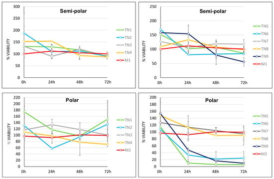
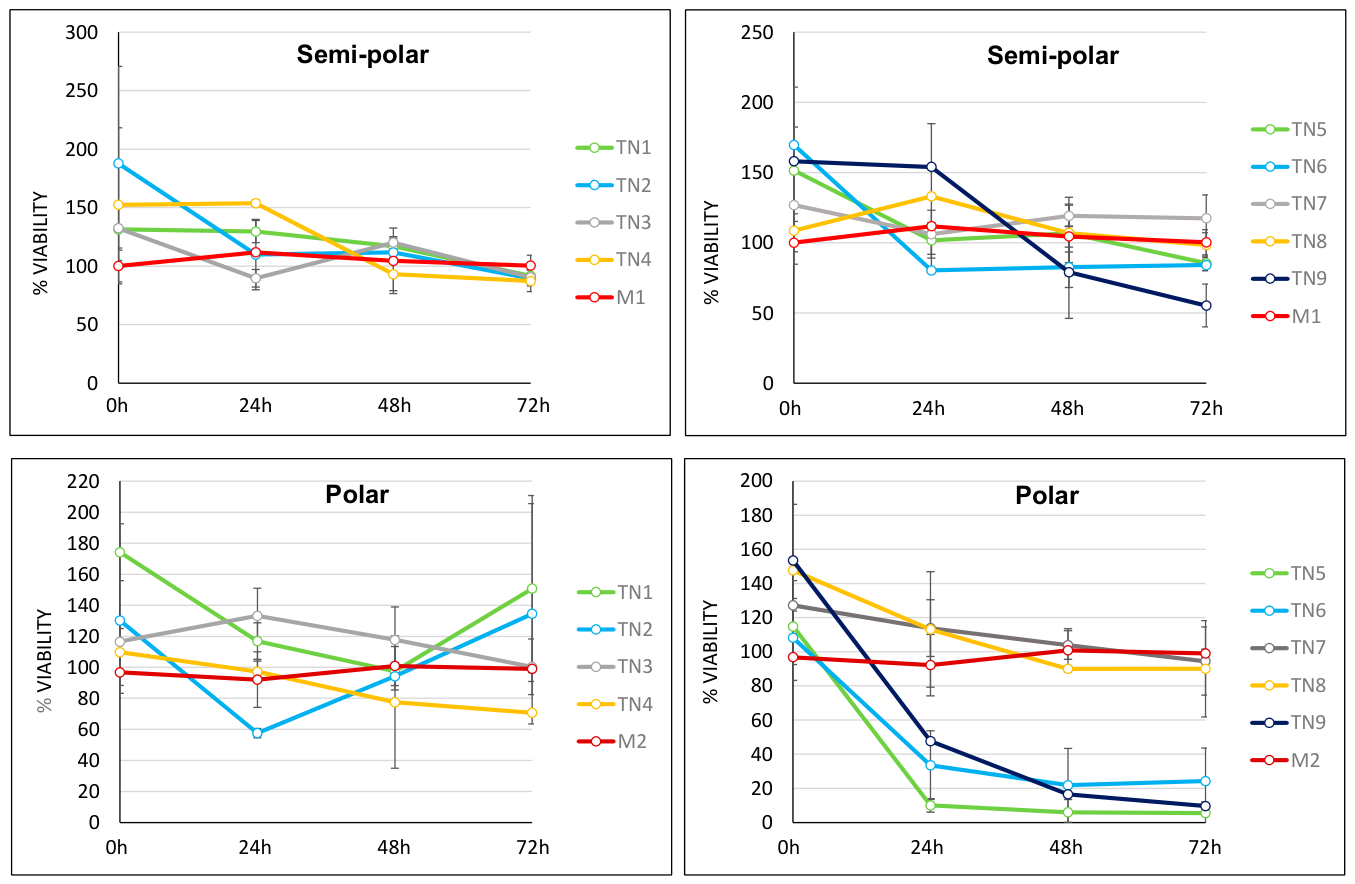
Polar and semi-polar extracts were prepared from TN1-TN9 samples as indicated in Section 4. Activity against L. infantum-iRFP axenic amastigotes, expressing infrared fluorescent protein (iRFP), was assessed for up to 72 h by means of reduction in fluorescence emitted by the parasites, which was used as a proxy for parasite viability (Figure 1).
Figure 1.
Efficacy of semi-polar (upper) panels and polar (lower) panels extracts from samples TN1–TN9 against L. infantum-iRFP axenic amastigotes. M1 (methyl tert-butyl ether:methanol 3:1, v/v) and M2 (water:methanol 3:1, v/v) correspond to the extraction solvent controls. Parasite viability (%) was measured at 0, 24, 48, and 72 h. Results show the mean ± SD of three independent experiments with three technical replicates each.
The semi-polar extracts exhibited lower antiparasitic activity than the polar extracts. Among the semi-polar extracts, only those obtained from S. vera collected in winter (TN9) achieved a significant reduction in parasite viability, reaching 55.30% after 72 h of incubation. The polar extracts of L. guyonianum (TN1, TN2), S. europaea (TN3, TN4), and H. portulacoides (TN7, TN8) generally exhibited weak or no antiparasitic activity, with viability values remaining above 75% at most of the evaluated time points. Although sample TN2 (L. guyonianum, spring) produced a marked decrease in viability at 24 h, this effect was not sustained over time, reverting at 48 and 72 h to values even higher than those observed for the M2 (water:methanol 3:1, v/v) solvent control. Likewise, sample TN4 (S. fruticosa, spring) exerted a mild antileishmanial effect, reducing viability to 70.73% after 72 h. In contrast, the polar extracts of S. vera (TN5, TN6, and TN9) exhibited the highest antiparasitic activity among all the extracts tested. Sample TN5 (summer) reduced amastigote viability to 9.98% at 24 h, 5.97% at 48 h, and 5.53% at 72 h. Samples TN6 (spring) and TN9 (winter) also decreased parasite viability to 24.20% and 9.62%, respectively, after 72 h of incubation (Figure 1).
2.2. Cytotoxicity of Antileishmanial Plant Extracts in Intestinal Organoids
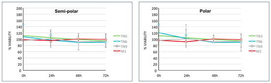
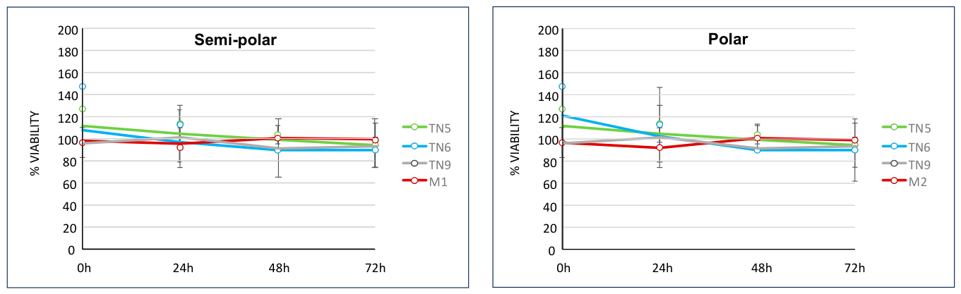
The safety of the plant extracts that showed efficacy against L. infantum-iRFP axenic amastigotes (S. vera TN5, TN6, and TN9) was evaluated for up to 72 h in murine intestinal organoids obtained as described in Section 4. Cell viability was determined using the alamarBlue assay. As shown in Figure 2, none of the extracts tested caused a significant decrease in cell viability, indicating that they are not cytotoxic in this 3D culture model.
Figure 2.
Cytotoxicity of the semi-polar and polar extracts of S. vera (TN5, TN6, and TN9 samples). Solvent controls M1 (methyl tert-butyl ether:methanol 3:1, v/v) and M2 (water:methanol 3:1, v/v) are shown in red. Cell viability (%) was assessed at 0, 24, 48, and 72 h. Results show the mean ± SD of three independent experiments with three technical replicates each.
2.3. Mass Spectrometric Characterization of Bioactive and Non-Toxic S. vera Extracts
Mass spectrometry-based phytochemical profiling of S. vera extracts TN5, TN6, and TN9 revealed differences in their chemical composition depending on both the polarity of the extraction solvent and the sampling time. Candidate metabolites detected in positive and negative ionization modes with a similarity score above 70% to compounds in the reference databases were selected for further analysis. This relatively permissive threshold was adopted due to the exploratory nature of the study and the limited representation of halophyte metabolites in available databases. After discarding primary metabolites, the putatively annotated secondary metabolites present in the polar and semi-polar extracts are listed in Supplementary Tables S1 and S2, respectively.
A total of 53 distinct secondary metabolites were putatively annotated in the extracts of S. vera. These annotations encompassed several chemical categories including flavonoids, terpenoids, alkaloids, phenylpropanoids, phenolic compounds, coumarins, chromones, lipids, and natural peptides. Based on the putative annotations, polyphenols and terpenoids were the most represented chemical groups, mainly comprising flavonoids, triterpenoids, and triterpenoid saponins (Table 2).
Table 2.
Putatively annotated secondary metabolites in the bioactive and non-cytotoxic S. vera extracts. Compounds are listed in alphabetical order, indicating the main chemical class and subclass.
Most compounds were detected in both polar and semi-polar extracts, although some metabolites were found exclusively in a specific extract type. For example, asiatic acid and thymol were only detected in the polar and semi-polar extracts, respectively (Supplementary Tables S1 and S2).
In the polar extracts (Supplementary Table S1), three compounds were consistently putatively annotated in all samples: protocatechuic aldehyde, a phenolic aromatic aldehyde, and two flavonoids, isorhamnetin, and narcissoside. Similarly, protocatechuic aldehyde was putatively annotated in all semi-polar extracts (Supplementary Table S2), suggesting that this phenolic aromatic aldehyde was consistently annotated across the S. vera samples analyzed. The polar extracts of the spring and summer samples exhibited similar chemical profiles, with most of the annotated metabolites corresponding to phenolic acids and their derivatives or flavonoids. In contrast, the winter sample showed a greater number of putatively annotated metabolites, including not only flavonoids but also alkaloids, such as isorhynchophylline, and terpenoids, including asiatic acid, betulonic acid, and ursolic acid (Supplementary Table S1). Likewise, the semi-polar extracts (Supplementary Table S2) displayed higher chemical diversity in the winter sample than in the spring and summer samples. Spring and summer extracts contained compounds that were absent from their corresponding polar extracts, such as demethylzeylasteral and nodakenin, whereas the winter extract included additional annotations, including peiminine and thymol.
Overall, the winter samples showed the highest numbers of putatively annotated metabolites (n = 24 and n = 22 in the polar and semi-polar extracts, respectively). In contrast, spring and summer extracts showed lower and comparable numbers of annotations, with 11 and 8 putatively annotated metabolites in the polar extracts and 13 in the semi-polar extracts for both seasons (Supplementary Tables S1 and S2).
2.4. In Vitro Efficacy and Safety of the Compounds Putatively Annotated in S. vera Extracts
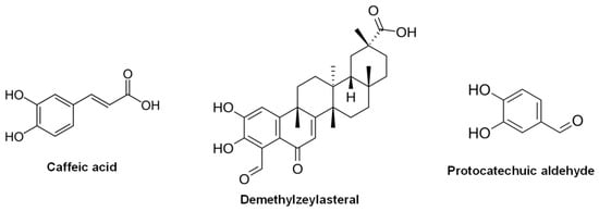
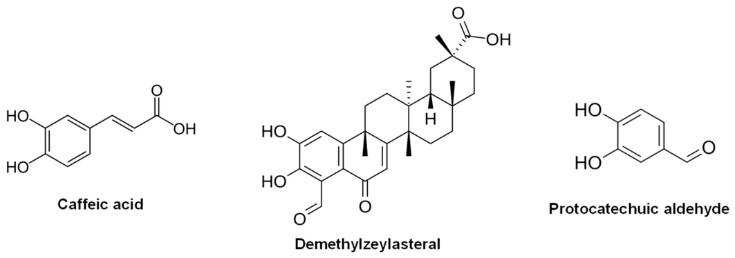
Following the putative annotation of candidate active compounds in the S. vera extracts, pure reference standards were purchased from MedChemExpress (Sollentuna, Sweden) and Merck (Darmstadt, Germany). Their leishmanicidal activity was subsequently evaluated against axenic amastigotes of L. infantum-iRFP in a single-concentration screen at 20 μM and 1 μM. In this initial screen, only three compounds exceeded the pre-established threshold of a >70% reduction in parasite viability at 20 μM: caffeic acid (phenolic compound), protocatechuic aldehyde (3,4-dihydroxybenzaldehyde) (phenolic compound), and demethylzeylasteral (terpene) (Figure 3). Caffeic acid and demethylzeylasteral also yielded similar results at 1 μM.
Figure 3.
Chemical structure of caffeic acid, demethylzeylasteral, and protocatechuic aldehyde.
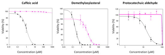
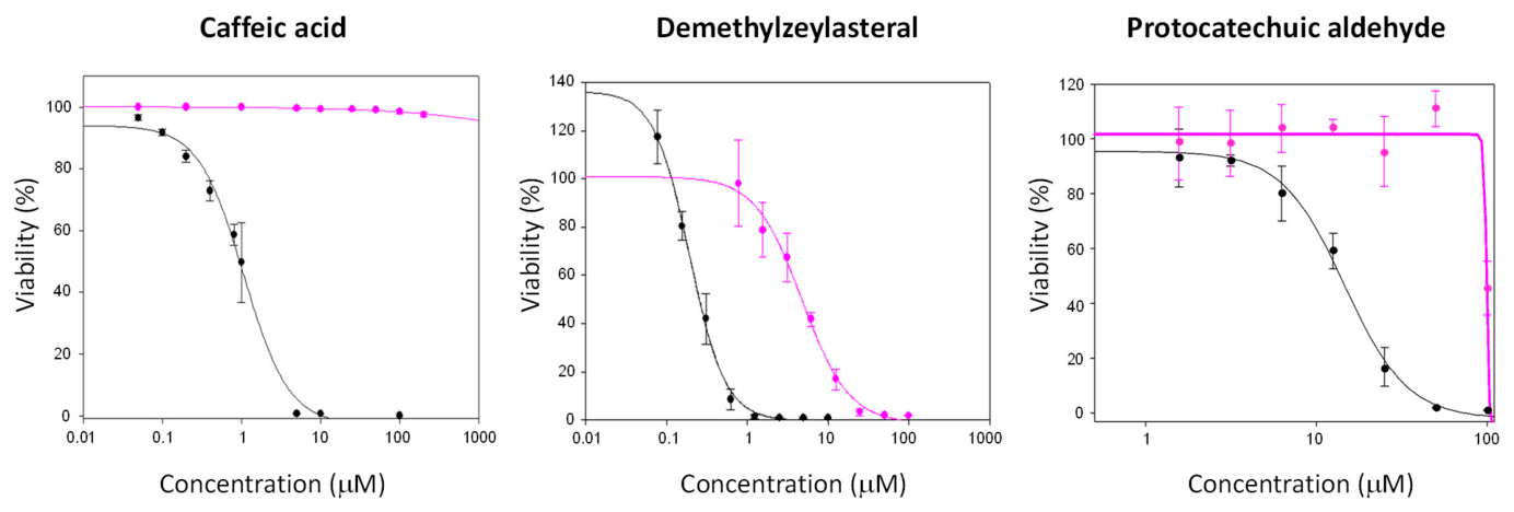
Dose–response curves were prepared for these three compounds using a range of increasing concentrations after testing their bioactivity against axenic amastigotes of L. infantum-iRFP (efficacy) and their cytotoxicity in murine intestinal organoids (safety) (Figure 4). Parasite cell viability was determined by measuring infrared fluorescence emission, while organoid viability was determined using the alamarBlue assay, as described in Section 4.
Figure 4.
Dose–response curves for caffeic acid, demethylzeylasteral, and protocatechuic aldehyde against L. infantum-iRFP amastigotes (black lines) and murine intestinal organoids (pink lines). Percentage viability (Y-axis) was plotted against compound concentration (μM) (X-axis in logarithmic scale). The curves were generated using SigmaPlot software (version 10.0) based on data obtained after 72 h of incubation in the presence of each compound. Data are presented as the mean ± SD from three different experiments, each carried out in triplicate.
Based on the dose–response curves shown in Figure 4, EC50 (half maximal effective concentration) values for L. infantum-iRFP (efficacy) and CC50 values (half maximal cytotoxic concentration) for mouse intestinal organoids (safety) were determined and allowed for the calculation of the selectivity index (SI) using the CC50/EC50 ratio (Table 3).
Table 3.
In vitro efficacy against L. infantum-iRFP amastigotes (EC50) and safety (CC50) in intestinal organoids of caffeic acid, demethylzeylasteral, and protocatechuic aldehyde. SI values are indicated.
Demethylzeylasteral was the most potent compound, followed by caffeic acid and protocatechuic aldehyde. However, potency and selectivity differed substantially among the compounds. Although demethylzeylasteral exhibited the lowest EC50, its lower CC50 (4.78 ± 0.68 μM) resulted in a SI of 23.9. In contrast, caffeic acid showed a CC50 > 100 μM and an SI > 91.74, indicating a more favorable selectivity profile. Protocatechuic aldehyde also showed a CC50 > 100 μM, but its lower potency resulted in an SI > 6.86. According to these data and considering the criteria proposed for visceral leishmaniasis (Hit: EC50 ≤ 10 μM and SI ≥ 10; Lead: EC50 ≤ 1 μM and SI ≥ 50) [25,26], caffeic acid and demethylzeylasteral were considered as hit compounds.
Since TryR is a validated molecular target involved in the maintenance of parasite redox homeostasis [24,27], as a preliminary assessment of a possible mechanism of action, caffeic acid, demethylzeylasteral, and protocatechuic aldehyde were evaluated for their effects on TryR-associated activity using L. infantum-iRFP protein extracts. None of the compounds inhibited the measured activity at the concentrations tested (25–100 μM; Supplementary Figure S1). However, as the assay was performed using protein extracts rather than purified recombinant TryR, these results do not rule out direct inhibition of the enzyme.
2.5. Predictive Drug-likeness and ADMET of the Selected Compounds
Predictive ADMET of caffeic acid, demethylzeylasteral, and protocatechuic aldehyde, was analyzed in silico using SwissADME and pkCSM (Table 4).
Table 4.
Predictive drug-likeness and ADMET of caffeic acid, demethylzeylasteral, and protocatechuic aldehyde.
The ADMET properties of caffeic acid, demethylzeylasteral, and protocatechuic aldehyde were evaluated in silico using SwissADME and pkCSM (Table 4). All three compounds complied with Lipinski’s rule of five [28]. Caffeic acid and protocatechuic aldehyde were predicted to have high human intestinal absorption and not to be P-glycoprotein substrates, whereas demethylzeylasteral was predicted to have low intestinal absorption and to be a P-glycoprotein substrate. Regarding metabolism, caffeic acid was predicted to inhibit CYP1A2 and protocatechuic aldehyde to inhibit CYP3A4, while demethylzeylasteral was not predicted to inhibit any of the five CYP450 isoforms evaluated. A catechol_A PAINS alert was reported for all three compounds. None were predicted to be hepatotoxic, although protocatechuic aldehyde was predicted to be Ames-positive. The predicted acute oral toxicity values were of a similar order of magnitude for the three compounds (Table 4). These findings are computational predictions and require experimental validation; they should not be interpreted as confirmed pharmacokinetic or toxicological outcomes.
3. Discussion
The growing emergence of antimicrobial resistance is recognized by the WHO as one of the major public health threats of the coming decades, as it compromises the effectiveness of previously successful therapies and hinders the control of infectious diseases. In parallel, climate change is driving human population movements and facilitating the geographical expansion of vectors of neglected tropical diseases into regions where they are currently absent, which is expected to increase the global incidence of these diseases in the coming years. In this context, the identification of novel bioactive compounds with the potential to serve as therapeutic or prophylactic agents against diseases such as leishmaniasis has become an urgent priority [18].
Halophytes constitute a promising source of antimicrobials—including antileishmanial agents—as their adaptation to saline environments is frequently associated with the accumulation of a structurally diverse array of secondary metabolites absent in non-halophytic species. Nevertheless, the antileishmanial potential of this group of plants remains largely unexplored. In line with this hypothesis, extracts from the halophyte Atriplex halimus—a European fodder shrub of the Chenopodiaceae family—have previously shown modest activity against Leishmania [29].
In the present study, extracts from four halophytic species harvested in the Tunisian Kerkennah Islands were evaluated, and S. vera was the only species displaying significant antileishmanial activity. To the best of our knowledge, this is the first report describing the antileishmanial properties of this plant. Species of the genus Suaeda—Mediterranean halophytes belonging to the Amaranthaceae family that are adapted to saline soils and serve as a vital forage resource for small ruminants in North Africa—remain unexplored against Leishmania, despite reported antimicrobial activity in congeneric species such as S. monoica, S. vermiculata, and S. pruinosa [30,31,32]. Furthermore, S. vera has long been used in the traditional medicine of arid and semi-arid regions and is known to contain phenolic compounds, flavonoids, terpenoids, and saponins with antioxidant, anti-inflammatory, and antimicrobial properties [33]. The present study confirmed the antileishmanial activity of the polar extracts obtained from S. vera collected during spring (TN6), summer (TN5), and winter (TN9). Phytochemical profiling revealed differences in the qualitative composition of the putatively annotated metabolites among the S. vera samples collected in spring, summer, and winter, with the winter extract showing the highest number of annotations. Such seasonal fluctuations have previously been described in halophytic species and are thought to reflect adaptive responses to environmental stress that activate defense pathways and stimulate secondary metabolite biosynthesis [34,35]. Interestingly, despite the higher number of metabolites putatively annotated in the winter extract, the strongest antileishmanial activity was observed for the summer extract. This observation indicates that the number of metabolite annotations alone does not explain the differences in biological activity among the extracts. However, the contribution of individual metabolites to these differences and their concentrations remain to be established. Factors such as temperature, light availability, water availability, and salinity can influence the biosynthesis and accumulation of specialized metabolites, including phenolic compounds and flavonoids. In fact, the combined effects of intense solar radiation, water deficit, and increased soil salinity during summer are known to stimulate the biosynthesis and accumulation of secondary metabolites involved in stress adaptation [10,36], which may account for the enhanced biological activity observed in the summer extract. As these environmental variables were not directly measured in the present study, their contribution to the observed seasonal differences cannot be established. Since all three active polar extracts shared antileishmanial activity, one or more metabolites common to these extracts may contribute to the observed effect. However, the present qualitative profiling data do not allow us to determine whether differences in the concentrations of individual metabolites account for the differences in antileishmanial activity. Targeted quantitative metabolomic analyses will be required to test this hypothesis and identify the compounds primarily responsible for the biological activity. These observations are specific to the S. vera samples analyzed from spring, summer, and winter and should not be interpreted as establishing a general seasonal pattern for the species.
A total of 53 secondary metabolites were putatively annotated in the S. vera extracts. Among the three selected compounds evaluated individually, demethylzeylasteral exhibited the highest antileishmanial activity. Interestingly, although the semipolar spring extract (TN6), in which this compound was putatively annotated, showed only weak activity against Leishmania, the commercially obtained compound displayed a remarkably low EC50 value against axenic amastigotes. This discrepancy may reflect the complexity of the crude extract, including possible interactions with other metabolites that could attenuate or modify its biological activity. However, the contribution of these factors cannot be established from the present data. Conversely, synergistic interactions among compounds present in the more active extracts cannot be excluded, highlighting the complexity of phytochemical mixtures and the need for future bioactivity-guided fractionation studies. Demethylzeylasteral is a pentacyclic 6-oxophenolic triterpenoid previously reported to possess anti-inflammatory and antimicrobial activities, particularly against Gram-positive bacteria such as Bacillus subtilis [37,38,39]. To the best of our knowledge, this is the first study demonstrating its activity against Leishmania. Its antimicrobial activity has been associated with inhibition of N-acetyl-D-glucosamine incorporation, leading to disruption of bacterial cytoplasmic membrane integrity [37]. The antibacterial activity of demethylzeylasteral suggests that membrane-associated mechanisms may contribute to its biological activity. Although the plasma membrane of Leishmania differs substantially from that of bacteria, its unique lipid composition and abundance of surface glycoconjugates [40] also make it a potential target for membrane-active compounds [41]. Therefore, membrane perturbation cannot be excluded as a contributing mechanism underlying the antileishmanial activity of demethylzeylasteral. In addition to its direct leishmanicidal activity, demethylzeylasteral may exert beneficial host-directed effects. Previous studies have shown that this compound modulates signaling pathways such as NF-κB and TGF-β, both of which are manipulated by Leishmania to evade host immune responses [39]. Although the precise molecular targets of demethylzeylasteral remain to be elucidated, its combined antiparasitic and immunomodulatory properties make it an attractive scaffold for the development of new antileishmanial agents.
Caffeic acid, putatively annotated in the winter polar extract of S. vera (TN9), emerged as the most promising lead compound in this study. Although its antileishmanial potency was lower than that of demethylzeylasteral, its excellent SI value (>91.7), together with its favorable predicted pharmacokinetic profile and previous evidence of in vivo efficacy, make it a particularly attractive candidate for further development. It should be noted that the antileishmanial activity reported in this study was evaluated against axenic amastigotes. Further validation using intracellular amastigote models is therefore required to confirm the activity of caffeic acid under more physiologically relevant conditions and to better assess its therapeutic potential. Previous studies have identified L. infantum arginase as a molecular target of caffeic acid [42]. L. infantum arginase catalyzes the hydrolysis of L-arginine to L-ornithine, the first committed step in polyamine biosynthesis. This pathway is essential for parasite proliferation, differentiation, oxidative stress defense through trypanothione biosynthesis, and infectivity [43,44,45]. Caffeic acid inhibited approximately 57% of arginase activity at a concentration of 100 μM [42], while molecular docking analyses confirmed a stable interaction between the compound and the enzyme active site [46]. The antileishmanial activity of caffeic acid has been previously demonstrated against several Leishmania species [42,47], although with substantially higher EC50 values than those obtained in the present study. These differences are most likely explained by variations in the experimental models employed, including parasite species and developmental stage, as well as the use of intracellular versus axenic amastigotes. Moreover, the therapeutic potential of caffeic acid has already been supported by in vivo studies showing a significant reduction in lesion development in BALB/c mice infected with L. amazonensis [47]. Beyond its direct effects on parasite metabolism, caffeic acid has also been reported to enhance host antimicrobial responses. It increases nitric oxide production by activated macrophages, counteracting one of the immune evasion strategies employed by Leishmania; enhances macrophage iron sequestration, thereby restricting parasite access to this essential micronutrient; and promotes the generation of reactive oxygen species (ROS), contributing to oxidative stress-mediated parasite killing [48,49]. Collectively, these findings suggest that the antileishmanial activity of caffeic acid is likely multifactorial, involving both direct inhibition of parasite metabolic pathways and modulation of host immune defenses.
Finally, protocatechuic aldehyde exhibited moderate antileishmanial activity and relatively low SI. Although its activity against Leishmania has not previously been reported, its structural analog, protocatechuic acid, has been shown to display modest activity against L. infantum (IC50 = 32 μM) [50], suggesting that this class of phenolic compounds possesses intrinsic antileishmanial potential. Importantly, protocatechuic aldehyde was putatively annotated in all three S. vera extracts that exhibited antileishmanial activity and also demonstrated activity when tested as a purified compound. Together, these findings support the hypothesis that protocatechuic aldehyde contributes, at least in part, to the biological activity of the extracts. However, because plant extracts are chemically complex mixtures, the observed activity cannot be attributed exclusively to this metabolite, and synergistic or antagonistic interactions with other constituents are likely to influence the overall leishmanicidal effect.
To gain further insight into the mechanism underlying the antileishmanial activity of the three selected compounds, their ability to inhibit TryR was evaluated. TryR is one of the best-characterized molecular targets for antileishmanial drug discovery [24] because of its essential role in maintaining intracellular redox homeostasis. Given that caffeic acid has been reported to promote ROS generation and oxidative stress, inhibition of TryR was considered a plausible mechanism underlying its antileishmanial activity. However, none of the three compounds produced clear inhibition of TryR-associated activity in L. infantum protein extracts under the experimental conditions tested. However, because purified recombinant TryR was not used, these results do not conclusively establish whether the compounds directly inhibit the enzyme. Further biochemical studies with purified TryR will be required to clarify its potential involvement in their antileishmanial activity. Therefore, the oxidative stress associated with caffeic acid cannot be attributed to direct TryR inhibition on the basis of the present results. ROS accumulation may instead result from indirect effects on parasite metabolism or from modulation of host cell antimicrobial responses, including enhanced ROS, nitric oxide, and TNF-α production by infected macrophages, all of which have previously been reported for caffeic acid [42,49]. Similarly, although no clear inhibition of TryR-associated activity was observed for demethylzeylasteral or protocatechuic aldehyde, the molecular mechanisms underlying their antileishmanial activity remain to be elucidated.
The in silico evaluation provided additional insights into the potential developability and limitations of the three active compounds. All three complied with Lipinski’s rule of five, although this does not establish their suitability as oral drug candidates. Caffeic acid and protocatechuic aldehyde were predicted to have high human intestinal absorption and not to be P-glycoprotein substrates, whereas demethylzeylasteral was predicted to have low intestinal absorption and to be a P-glycoprotein substrate, suggesting that its absorption and exposure may require further investigation. The predicted inhibition of CYP1A2 by caffeic acid and CYP3A4 by protocatechuic aldehyde also warrants experimental assessment. In addition, protocatechuic aldehyde was predicted to be Ames-positive, highlighting a potential mutagenicity concern that requires further evaluation. Although none of the compounds was predicted to be hepatotoxic, these computational results do not establish their safety in vivo. SwissADME also identified a catechol_A PAINS alert for all three compounds. Such alerts indicate a potential risk of assay interference but do not, by themselves, demonstrate nonspecific activity or invalidate the observed antileishmanial effects. Nevertheless, orthogonal assays will be needed to assess the specificity of these effects, particularly for caffeic acid. Overall, the three compounds exhibited distinct predicted ADMET profiles and potential liabilities that require experimental validation before their further development can be assessed.
4. Materials and Methods
4.1. Plant Samples
Four halophyte plant species (L. guyonianum, S. fruticosa, S. vera, and H. portulacoides) were collected on the Kerkennah Islands (34°42′ N, 11°11′ E; Gulf of Gabès, Tunisia) in spring, summer, or winter of 2023. The aerial parts were shade-dried at 40 °C and then ground in a mill until a fine powder was obtained, thus giving rise to nine different samples (TN1-TN9) (see Table 1 for details).
4.2. Plant Extracts
The “single-step extraction method for the rapid preparation of polar and semi-polar metabolites from plant samples”, based on a protocol previously described [51], was used. Briefly, a 25 mg aliquot of each sample was placed in microcentrifuge tubes. A fixed volume (1 mL) of extraction solvent M1 (methyl tert-butyl ether:methanol 3:1, v/v) was added to the homogenized tissues. After adding the extraction solvent, the tubes were vortexed for 1 min and then incubated on an orbital shaker for 45 min at 4 °C, followed by a 15 min sonication step. For phase separation, a volume of 650 μL of solvent M2 (water:methanol 3:1, v/v) was added to each vial/tube and the samples were vortexed again for 1 min. The samples were then centrifuged at 20,000× g for 10 min at 4 °C. After centrifugation, two phases were obtained: a polar phase and a semi-polar phase, separated by a thin interphase. The polar and semi-polar phases were separated by pipetting into independent microcentrifuge tubes and stored at −20 °C to prevent evaporation.
4.3. Mass Spectrometry-Based Putative Annotation of Metabolites in Plant Extracts and Acquisition of Reference Compounds
Metabolites in the plant extracts were profiled by ultra-high-resolution mass spectrometry (hybrid Q-TOF with trapped ion mobility spectrometry, TIMS-TOF FLEX, Bruker Daltonics, Bremen, Germany), using both positive and negative ionization modes. Data were acquired in full-scan mode, without MS/MS fragmentation. Candidate compounds were evaluated based on accurate-mass measurements (mass error < 5 ppm) and matching against two natural product libraries: the NIST Research Library (National Institute of Standards and Technology, USA) and the Natural Products Library (Centre of Excellence for Innovative Medicines Research, Andalusia, Spain). The analysis was carried out at the University Institute of Materials Science (ICMUV), Universidad de Valencia, Spain. The available analytical data supporting the putative annotation of the three selected compounds (caffeic acid, demethylzeylasteral, and protocatechuic aldehyde), including retention times, mass-related information, and library-match results, are provided in Supplementary Table S3.
Following the phytochemical analysis, compounds showing a similarity score below 70% in the natural products library were excluded. Primary metabolites were then discarded to prioritize secondary metabolites. No MS/MS fragmentation analysis or confirmation using authentic reference standards was performed. Therefore, the reported metabolite identities should be regarded as tentative MS-based annotations rather than confirmed identifications. In addition, compounds reported in the literature as toxic and/or synthetic were excluded, and only naturally occurring compounds were considered.
After phytochemical analysis, the selected compounds were purchased from MedChemExpress (Sollentuna, Sweden) and Merck (Darmstadt, Germany) for further studies. They were dissolved in DMSO to prepare stock solutions at initial concentrations of 100 mM. Stock solutions were aliquoted and stored at −20 °C until use to avoid repeated freeze–thaw cycles.
4.4. Parasites and Experimental Infections
For the efficacy assays, the L. infantum-iRFP strain was employed. This parasite line was generated from the L. infantum BCN150 isolate (MCAN/ES/96/BCN150) and was previously developed in our laboratory to constitutively express the iRFP. Stable iRFP expression enables the detection and quantification of viable parasites by near-infrared fluorescence imaging, as previously described [52]. Promastigotes were routinely cultured at 26 °C under continuous shaking in Schneider’s insect medium (Sigma-Aldrich, Merck, Darmstadt, Germany) supplemented with 20% fetal bovine serum (FBS), 100 U/mL penicillin, and 100 μg/mL streptomycin [53].
For the generation of axenic amastigotes, eight-week-old female BALB/c mice were infected intraperitoneally with 2.5 × 10⁹ metacyclic promastigotes of the L. infantum-iRFP strain. Eight weeks post-infection, animals were humanely euthanized, and the femurs and tibias from both hind limbs were aseptically collected. Bone marrow cells recovered from the medullary cavities were processed to obtain L. infantum-iRFP axenic amastigotes according to the protocol previously described [54]. Briefly, cells were incubated at 37 °C in a humidified atmosphere containing 5% CO2 in amastigote culture medium composed of 15 mM KCl, 136 mM KH2PO4, 10 mM K2HPO4·3H2O, 0.5 mM MgSO4·7H2O, 24 mM NaHCO3, 22 mM glucose, 1 mM glutamine, RPMI 1640 vitamin mixture (1×; Sigma-Aldrich, Merck, Darmstadt, Germany), 10 μM folic acid, 100 μM adenosine, RPMI amino acid mixture (1×; Sigma-Aldrich, Merck, Darmstadt, Germany), 5 μg/mL hemin, and antibiotic cocktail, 25 mM MES, and 20% FBS, allowing their differentiation into axenic amastigotes.
4.5. Efficacy Assays Against L. infantum-iRFP Axenic Amastigotes
A total of 3 × 10⁴ iRFP-expressing L. infantum axenic amastigotes were seeded per well in 40 μL of amastigote culture medium in 384-well microplates. Amphotericin B (10 μM) was used as the positive control, whereas 0.1% (v/v) DMSO served as the negative control.
To test the bioactivity of plant extracts, polar and semi-polar extracts were added in a final volume of 10 μL, and the volume in each well was adjusted to 80 μL with amastigote culture medium. Additional control wells containing the corresponding solvent mixtures (M1 and M2, see Section 4.2) were also included.
The antileishmanial activity of the purified compounds was initially assessed by single-concentration screening at 20 and 1 μM. Compounds producing a significant reduction in parasite viability (>70%) were subsequently selected to determine their EC50 values using a range of increasing concentrations up to 10 μM (demethylzeylasteral) or 100 μM (caffeic acid and protocatechuic aldehyde) dissolved in the amastigote culture medium and adjusting the final volume to 80 μL.
Plates were incubated for 72 h at 37 °C in a humidified atmosphere containing 5% CO2. Parasite viability was determined by measuring the fluorescence emitted by the iRFP-expressing parasites using an Odyssey Infrared Imaging System (LI-COR Biosciences, Lincoln, NE, USA). Fluorescence intensity at 700 nm was recorded at 0, 24, 48, and 72 h, with the plates returned to the incubator between measurements. Parasite viability was expressed relative to the fluorescence of the controls, assigning 100% viability to parasites treated with 0.1% (v/v) DMSO and 0% viability to those exposed to 10 μM amphotericin B. Three independent biological replicates carried out in duplicate were performed.
4.6. Cell Cytotoxicity
The cytotoxicity of the plant extracts and chemical compounds was evaluated in fully developed mouse intestinal organoid cultures (day 4 of growth), which were obtained, cultured and adapted to a 384-well format as previously described [55]. Aliquots (8 μL) were dispensed into each well, followed by matrix polymerization and addition of 32 μL of culture medium. Then, they were treated with 10 μL of plant extract fractions or with 10 μL of serial dilutions of the compounds of interest dissolved in organoid culture medium, and the volume in each well was adjusted to 80 μL with organoid culture medium. For compound testing, 1:2 serial dilutions were prepared, ranging from 100 μM to 1.56 μM (demethylzeylasteral and protocatechuic aldehyde) or from 200 μM to 0.05 μM (caffeic acid) to calculate the CC50 values.
Cultures were incubated for 72 h at 37 °C in a humidified atmosphere containing 5% CO2. Hydrogen peroxide (0.15% v/v) was used as the positive control, whereas 0.2% (v/v) DMSO served as the negative control. After the incubation period, cell viability was determined using the alamarBlue assay (Thermo Fisher Scientific, Waltham, MA, USA). A volume equivalent to 10% of the well content was added to each well, and plates were incubated for an additional 4 h at 37 °C and 5% CO2. Fluorescence, which is proportional to cell viability, was measured using a Varioskan Lux plate reader (Thermo Fisher Scientific) with excitation and emission wavelengths of 560 and 590 nm, respectively. Results were normalized to the corresponding controls, assigning 100% viability to organoids treated with 0.2% (v/v) DMSO and 0% viability to those exposed to hydrogen peroxide and expressed as percentage cell viability. Three independent biological replicates carried out in duplicate were performed.
4.7. Dose–Response Analysis and SI Determination
Dose–response curves were generated by plotting cell viability (parasites and organoids) as a function of compound concentration. EC50 (efficacy) and CC50 (cytotoxicity) values were calculated by non-linear regression analysis using SigmaPlot version 10.1. The SI was calculated as the ratio of the CC50 to the EC50 values.
4.8. TryR Colorimetric Inhibition Assay
TryR activity was determined using a colorimetric assay based on the reduction of 5,5′-dithiobis-(2-nitrobenzoic acid) (DTNB; Alfa Aesar, Fisher Scientific, Inc., Waltham, MA, USA), as previously described [56]. Enzyme extracts were prepared from 1 × 10¹⁰ L. infantum-iRFP promastigotes harvested at the late logarithmic growth phase. Briefly, 25 μL of enzyme extract was dispensed into each well, followed by the addition of 1 μL of the test compound at different concentrations. Wells containing 2.5% (v/v) DMSO served as the negative control, whereas 100 μM thioridazine (MedChemExpress), a well-known potent inhibitor of TryR [57], was used as the positive control. The negative control showed a specific enzymatic activity of 1.83 × 10⁻² μmol/mg · min. (ΔA/t = ɛ · d · c; ɛ = 14.150 M−1 cm−1; d = 0.34 cm).
The reaction was initiated by adding 14 μL of reaction mixture containing 200 μM NADPH, 75 µM TS2, 75 µM DTNB, and 50 mM Tris-HCl buffer (pH 7.5). Absorbance at 412 nm was recorded every 5 min for 2 h at 27 °C using a Varioskan LUX microplate reader (Thermo Fisher Scientific).
4.9. Predictive Drug-likeness and ADMET
Physicochemical properties, drug-likeness, gastrointestinal absorption, blood–brain barrier permeability, CYP450 inhibition, and medicinal chemistry parameters were predicted using SwissADME [58]. Toxicological endpoints, including Ames mutagenicity, hepatotoxicity, and oral rat acute toxicity (LD50), were predicted using pkCSM [59].
5. Conclusions
The present study highlights S. vera as a promising source of antileishmanial metabolites and reports the putative annotation of secondary metabolites in its extracts. Caffeic acid, demethylzeylasteral, and protocatechuic aldehyde, selected based on the phytochemical profiling, exhibited antileishmanial activity when evaluated individually, although their contribution to the activity of the original extracts remains to be established. None of these compounds inhibited TryR-associated activity in the assay performed using parasite protein extracts, although direct inhibition of the purified enzyme cannot be excluded. In silico analyses revealed distinct predicted pharmacokinetic and toxicity profiles, with caffeic acid showing a promising overall profile despite potential liabilities requiring further investigation. These findings support further studies to clarify the molecular mechanisms underlying the antileishmanial activity of these compounds and to assess their therapeutic potential in vivo.
Supplementary Materials
The following supporting information can be downloaded at https://www.mdpi.com/article/10.3390/molecules31213967/s1, Table S1: Putatively annotated secondary metabolites in the polar extracts of S. vera collected during the spring (TN6), summer (TN5), and winter (TN9) seasons; Table S2: Putatively annotated secondary metabolites in the semi-polar extracts of S. vera collected during the spring (TN6), summer (TN5), and winter (TN9) seasons; Table S3: Analytical data supporting the putative annotation of selected compounds in S. vera extracts; Figure S1: Time-dependent in vitro inhibition of TryR by different concentrations (25 μM to 100 μM) of caffeic acid (left panel), demethylzeylasteral (middle panel), and protocatechuic aldehyde (right panel).
Author Contributions
Conceptualization, R.B.-F., C.G.-E., L.M.-M. and E.S.; methodology, E.S.-M., M.-C.G.-M., J.A.-R., E.M.-F., M.R., M.F. and C.S.-K.; software, C.S.-K., E.S.-M.; validation, C.G.-E., R.B.-F., Y.P.-P., R.M.R., L.M.-M. and E.S.; formal analysis, R.B.-F., C.G.-E. and E.S.-M.; investigation, E.S.-M., M.-C.G.-M., J.A.-R., E.M.-F., M.R., M.F. and C.S.-K.; resources, R.B.-F., C.G.-E., R.M.R. and L.M.-M.; data curation, R.B.-F. and C.G.-E.; writing—original draft preparation, E.S.-M. and C.G.-E.; writing—review and editing, C.G.-E., R.B.-F., L.M.-M. and E.S.; visualization, E.S.-M., J.A.-R., E.M.-F. and C.S.-K.; supervision, C.G.-E., R.B.-F., R.M.R., L.M.-M. and E.S.; project administration, R.B.-F., C.G.-E., E.S. and L.M.-M.; funding acquisition, R.B.-F. and L.M.-M. All authors have read and agreed to the published version of the manuscript.
Funding
This research belongs to project PCI2022-132925 funded by the Agencia Estatal de Investigación through the PCI (2022) call and is part of the HaloSheep (Agroecological sheep/goat production system based on the valorization of halophytes of saline areas in the Mediterranean basin) project granted by the PRIMA Programme (European Union, NextGenerationEU). M.-C.G.-M. received a PhD fellowship by Universidad de León. J.A.-R. received a fellowship from Junta de Castilla y León (ORDEN EDU/48/2026, 22 de enero).
Institutional Review Board Statement
The animal study protocol was approved by the Ethics Committee of the University of León, protocol code OEBA-ULE-010-2023, in compliance with Spanish and EU legislation (RD 53/2013 and 2010/63/EU, approved on 14 April 2023).
Informed Consent Statement
Not applicable.
Data Availability Statement
Data supporting reported results can be found in the Zenodo repository at https://doi.org/10.5281/zenodo.23269656.
Acknowledgments
The authors wish to thank Irene Pilar Martínez Torres and Guillermo Peñalba González for their excellent technical assistance. The expert help of Yelko Rodríguez (Universidad de Valencia) with the mass spectrometry analysis is acknowledged. During the preparation of this work the authors used ChatGPT (OpenAI, GPT-6) in order to improve readability and language of the work to ensure correct grammar and spelling. The authors have reviewed and edited the output and take full responsibility for the content of this publication.
Conflicts of Interest
The authors declare no conflicts of interest.
Abbreviations
The following abbreviations are used in this manuscript:
| 3D | three-dimensional |
| CC50 | half maximal cytotoxic concentration |
| DTNB | 5,5′-dithiobis-(2-nitrobenzoic acid) |
| EC50 | half maximal effective concentration |
| FBS | fetal bovine serum |
| iRFP | infrared fluorescent protein |
| ROS | reactive oxygen species |
| SI | selectivity index |
| TryR | trypanothione reductase |
| TS2 | oxidized trypanothione |
References
- Zagoskina, N.V.; Zubova, M.Y.; Nechaeva, T.L.; Kazantseva, V.V.; Goncharuk, E.A.; Katanskaya, V.M.; Baranova, E.N.; Aksenova, M.A. Polyphenols in Plants: Structure, Biosynthesis, Abiotic Stress Regulation, and Practical Applications (Review). Int. J. Mol. Sci. 2023, 24, 13874. [Google Scholar] [CrossRef] [Scilit] [PubMed]
- Wink, M. Modes of Action of Herbal Medicines and Plant Secondary Metabolites. Medicines 2015, 2, 251–286. [Google Scholar] [CrossRef] [Scilit] [PubMed]
- El-Saadony, M.T.; Saad, A.M.; Mohammed, D.M.; Alkafaas, S.S.; Abd El-Mageed, T.A.; Fahmy, M.A.; Ahmed, A.E.; Algopishi, U.B.; Abu-Elsaoud, A.M.; Mosa, W.F.A.; et al. Plant Bioactive Compounds: Extraction, Biological Activities, Immunological, Nutritional Aspects, Food Application, and Human Health Benefits—A Comprehensive Review. Front. Nutr. 2025, 12, 1659743. [Google Scholar] [CrossRef] [Scilit] [PubMed]
- Laudadio, V.; Tufarelli, V.; Dario, M.; Hammadi, M.; Seddik, M.M.; Lacalandra, G.M.; Dario, C. A Survey of Chemical and Nutritional Characteristics of Halophytes Plants Used by Camels in Southern Tunisia. Trop. Anim. Health Prod. 2009, 41, 209–215. [Google Scholar] [CrossRef] [Scilit] [PubMed]
- Abd El-Hack, M.E.; Samak, D.H.; Noreldin, A.E.; Arif, M.; Yaqoob, H.S.; Swelum, A.A. Towards Saving Freshwater: Halophytes as Unconventional Feedstuffs in Livestock Feed: A Review. Environ. Sci. Pollut. Res. 2018, 25, 14397–14406. [Google Scholar] [CrossRef] [Scilit] [PubMed]
- Oliveira, M.; Hoste, H.; Custódio, L. A Systematic Review on the Ethnoveterinary Uses of Mediterranean Salt-Tolerant Plants: Exploring Its Potential Use as Fodder, Nutraceuticals or Phytotherapeutics in Ruminant Production. J. Ethnopharmacol. 2021, 267, 113464. [Google Scholar] [CrossRef] [Scilit] [PubMed]
- Gupta, B.; Huang, B. Mechanism of Salinity Tolerance in Plants: Physiological, Biochemical, and Molecular Characterization. Int. J. Genom. 2014, 2014, 701596. [Google Scholar] [CrossRef] [Scilit] [PubMed]
- Stanković, M.; Jakovljević, D.; Stojadinov, M.; Dajić-Stevanović, D. Halophyte Species as a Source of Secondary Metabolites with Antioxidant Activity. In Ecophysiology, Abiotic Stress Responses and Utilization of Halophytes; Hasanuzzaman, M., Nahar, K., Öztürk, M., Eds.; Springer: Singapore, 2019; pp. 289–312. [Google Scholar]
- Attia-Ismail, S.A. Plant Secondary Metabolites of Halophytes and Salt Tolerant Plants. In Halophytic and Salt-Tolerant Feedstuffs: Impacts on Nutrition, Physiology and Reproduction of Livestock; El Shaer, H.M., Squires, V.R., Eds.; CRC Press: Boca Raton, FL, USA, 2016; pp. 348–357. [Google Scholar]
- Yang, L.; Wen, K.S.; Ruan, X.; Zhao, Y.X.; Wei, F.; Wang, Q. Response of Plant Secondary Metabolites to Environmental Factors. Molecules 2018, 23, 762. [Google Scholar] [CrossRef] [Scilit] [PubMed]
- Lopes, M.; Sanches-Silva, A.; Castilho, M.; Cavaleiro, C.; Ramos, F. Halophytes as Source of Bioactive Phenolic Compounds and Their Potential Applications. Crit. Rev. Food Sci. Nutr. 2023, 63, 1078–1101. [Google Scholar] [CrossRef] [Scilit] [PubMed]
- Buhmann, A.; Papenbrock, J. An Economic Point of View of Secondary Compounds in Halophytes. Funct. Plant Biol. 2013, 40, 952–967. [Google Scholar] [CrossRef] [Scilit] [PubMed]
- Custodio, L.; Garcia-Caparros, P.; Pereira, C.G.; Castelo-Branco, P. Halophyte Plants as Potential Sources of Anticancer Agents: A Comprehensive Review. Pharmaceutics 2022, 14, 2406. [Google Scholar] [CrossRef] [Scilit] [PubMed]
- Giordano, R.; Saii, Z.; Fredsgaard, M.; Hulkko, L.S.S.; Poulsen, T.B.G.; Thomsen, M.E.; Henneberg, N.; Zucolotto, S.M.; Arendt-Nielsen, L.; Papenbrock, J.; et al. Pharmacological Insights into Halophyte Bioactive Extract Action on Anti-Inflammatory, Pain Relief and Antibiotics-Type Mechanisms. Molecules 2021, 26, 3140. [Google Scholar] [CrossRef] [Scilit] [PubMed]
- Oryan, A. Plant-Derived Compounds in Treatment of Leishmaniasis. Iran. J. Vet. Res. 2015, 16, 629863. [Google Scholar] [CrossRef] [Scilit] [PubMed]
- World Health Organization Leishmaniasis. Available online: https://www.who.int/news-room/fact-sheets/detail/leishmaniasis (accessed on 11 August 2026).
- Burza, S.; Croft, S.L.; Boelaert, M. Leishmaniasis. Lancet 2018, 392, 951–970. [Google Scholar] [CrossRef] [Scilit] [PubMed]
- Pareyn, M.; Alves, F.; Burza, S.; Chakravarty, J.; Alvar, J.; Diro, E.; Kaye, P.M.; van Griensven, J. Leishmaniasis. Nat. Rev. Dis. Primers 2025, 11, 81. [Google Scholar] [CrossRef] [Scilit]
- Reguera, R.M.; Pérez-Pertejo, Y.; Gutiérrez-Corbo, C.; Domínguez-Asenjo, B.; Ordóñez, C.; García-Estrada, C.; Martínez-Valladares, M.; Balaña-Fouce, R. Current and Promising Novel Drug Candidates against Visceral Leishmaniasis. Pure Appl. Chem. 2019, 91, 1385–1404. [Google Scholar] [CrossRef] [Scilit]
- van Griensven, J.; Diro, E. Visceral Leishmaniasis: Recent Advances in Diagnostics and Treatment Regimens. Infect. Dis. Clin. N. Am. 2019, 33, 79–99. [Google Scholar] [PubMed]
- Zhang, H.; Yan, R.; Liu, Y.; Yu, M.; He, Z.; Xiao, J.; Li, K.; Liu, G.; Ning, Q.; Li, Y. Progress in Antileishmanial Drugs: Mechanisms, Challenges, and Prospects. PLoS Negl. Trop. Dis. 2025, 19, e0012735. [Google Scholar] [CrossRef] [Scilit] [PubMed]
- Krauth-Siegel, R.L.; Comini, M.A. Redox Control in Trypanosomatids, Parasitic Protozoa with Trypanothione-Based Thiol Metabolism. Biochim. Biophys. Acta Gen. Subj. 2008, 1780, 1236–1248. [Google Scholar] [CrossRef] [Scilit] [PubMed]
- Ilari, A.; Baiocco, P.; Messori, L.; Fiorillo, A.; Boffi, A.; Gramiccia, M.; Di Muccio, T.; Colotti, G. A Gold-Containing Drug against Parasitic Polyamine Metabolism: The X-ray Structure of Trypanothione Reductase from Leishmania infantum in Complex with Auranofin Reveals a Dual Mechanism of Enzyme Inhibition. Amino Acids 2012, 42, 803–811. [Google Scholar] [CrossRef] [Scilit] [PubMed]
- González-Montero, M.C.; Andrés-Rodríguez, J.; García-Fernández, N.; Pérez-Pertejo, Y.; Reguera, R.M.; Balaña-Fouce, R.; García-Estrada, C. Targeting Trypanothione Metabolism in Trypanosomatids. Molecules 2024, 29, 2214. [Google Scholar] [CrossRef] [Scilit] [PubMed]
- Don, R.; Ioset, J.R. Screening Strategies to Identify New Chemical Diversity for Drug Development to Treat Kinetoplastid Infections. Parasitology 2014, 141, 140–146. [Google Scholar] [CrossRef] [Scilit] [PubMed]
- Katsuno, K.; Burrows, J.N.; Duncan, K.; Hooft van Huijsduijnen, R.; Kaneko, T.; Kita, K.; Mowbray, C.E.; Schmatz, D.; Warner, P.; Slingsby, B.T. Hit and lead criteria in drug discovery for infectious diseases of the developing world. Nat. Rev. Drug Discov. 2015, 14, 751–758. [Google Scholar] [CrossRef] [Scilit] [PubMed]
- Galarreta, B.C.; Sifuentes, R.; Carrillo, A.K.; Sanchez, L.; Del Rosario I Amado, M.; Maruenda, H. The Use of Natural Product Scaffolds as Leads in the Search for Trypanothione Reductase Inhibitors. Bioorg. Med. Chem. 2008, 16, 6689–6695. [Google Scholar] [CrossRef] [Scilit] [PubMed]
- Lipinski, C.A.; Lombardo, F.; Dominy, B.W.; Feeney, P.J. Experimental and Computational Approaches to Estimate Solubility and Permeability in Drug Discovery and Development Settings. Adv. Drug Deliv. Rev. 2001, 46, 3–26. [Google Scholar] [CrossRef] [Scilit] [PubMed]
- Kabbash, A.; Shoeib, N. Chemical and Biological Investigation of Some Secondary Metabolites in Atriplex halimus Growing in Egypt. Nat. Prod. Commun. 2012, 7, 1465–1468. [Google Scholar] [CrossRef] [Scilit]
- Al-Tohamy, R.; Ali, S.S.; Saad-Allah, K.; Fareed, M.; Ali, A.; El-Badry, A.; El-Zawawy, N.A.; Wu, J.; Sun, J.; Mao, G.-H.; et al. Phytochemical Analysis and Assessment of Antioxidant and Antimicrobial Activities of Some Medicinal Plant Species from Egyptian Flora. J. Appl. Biomed. 2018, 16, 289–300. [Google Scholar] [CrossRef] [Scilit]
- Shakthi, A.; Linoj, J.; Suresh, V.; Manikandan, S.; Mohite, P. Evaluation of Antibacterial and Antioxidant Activities of Suaeda monoica Extract for Its Potential Application. Cureus 2024, 16, e53091. [Google Scholar] [CrossRef] [Scilit] [PubMed]
- Abd-ElGawad, A.M.; Elghonemy, M.M.; Khalaf, D.D.; Son, N.T.; Ha, N.X.; El Gendy, A.E.N.G.; Elshamy, A.I. Distinctive Chemical Profile and Bioactivity of Suaeda pruinosa var. pruinosa Essential Oil: Evidence from GC–MS, Antimicrobial, Antioxidant, and in silico Analyses. J. Essent. Oil Bear. Plants 2026, 29, 189–200. [Google Scholar] [CrossRef] [Scilit]
- Wang, X.; Shao, X.; Zhang, W.; Sun, T.; Ding, Y.; Lin, Z.; Li, Y. Genus Suaeda: Advances in Phytology, Chemistry, Pharmacology and Clinical Application (1895–2021). Pharmacol. Res. 2022, 179, 106203. [Google Scholar] [CrossRef] [Scilit] [PubMed]
- Petropoulos, S.; Karkanis, A.; Martins, N.; Ferreira, I.C.F.R. Edible Halophytes of the Mediterranean Basin: Potential Candidates for Novel Food Products. Trends Food Sci. Technol. 2018, 74, 69–84. [Google Scholar] [CrossRef] [Scilit]
- Ferreira, M.J.; Pinto, D.C.G.A.; Cunha, Â.; Silva, H. Halophytes as Medicinal Plants against Human Infectious Diseases. Appl. Sci. 2022, 12, 7493. [Google Scholar] [CrossRef] [Scilit]
- Ramakrishna, A.; Ravishankar, G.A. Influence of Abiotic Stress Signals on Secondary Metabolites in Plants. Plant Signal. Behav. 2011, 6, 1720–1731. [Google Scholar] [CrossRef] [Scilit] [PubMed]
- de León, L.; Beltrán, B.; Moujir, L. Antimicrobial Activity of 6-Oxophenolic Triterpenoids: Mode of Action against Bacillus subtilis. Planta Med. 2005, 71, 313–319. [Google Scholar] [CrossRef] [Scilit] [PubMed]
- Moujir, L. Screening Natural Products for Drug Discovery: The Experience of Fundación MEDINA. Appl. Sci. 2019, 9, 2957. [Google Scholar]
- Sun, X.; Shen, B.; Yu, H.; Wu, W.; Sheng, R.; Fang, Y.; Guo, R. Therapeutic Potential of Demethylzeylasteral, a Triterpenoid of the Genus Tripterygium wilfordii. Fitoterapia 2022, 163, 105333. [Google Scholar] [CrossRef] [Scilit] [PubMed]
- McConville, M.J.; Ferguson, M.A. The Structure, Biosynthesis and Function of Glycosylated Phosphatidylinositols in the Parasitic Protozoa and Higher Eukaryotes. Biochem. J. 1993, 294, 305–324. [Google Scholar] [CrossRef] [Scilit] [PubMed]
- de Souza, W.; Rodrigues, J.C. Sterol Biosynthesis Pathway as Target for Anti-Trypanosomatid Drugs. Interdiscip. Perspect. Infect. Dis. 2009, 2009, 642502. [Google Scholar] [CrossRef] [Scilit] [PubMed]
- Garcia, A.R.; Oliveira, D.M.P.; Amaral, A.C.F.; Jesus, J.B.; Sodero, A.C.R.; Souza, A.M.T.; Supuran, C.T.; Vermelho, A.B.; Rodrigues, I.A.; Pinheiro, A.S. Leishmania infantum Arginase: Biochemical Characterization and Inhibition by Naturally Occurring Phenolic Substances. J. Enzym. Inhib. Med. Chem. 2019, 34, 1100–1109. [Google Scholar] [CrossRef] [Scilit] [PubMed]
- Reguera, R.M.; Balaña-Fouce, R.; Showalter, M.; Hickerson, S.; Beverley, S.M. Leishmania major Lacking Arginase (ARG) Are Auxotrophic for Polyamines but Retain Infectivity to Susceptible BALB/c Mice. Mol. Biochem. Parasitol. 2009, 165, 48–56. [Google Scholar] [CrossRef] [Scilit] [PubMed]
- Colotti, G.; Ilari, A. Polyamine Metabolism in Leishmania: From Arginine to Trypanothione. Amino Acids 2011, 40, 269–285. [Google Scholar] [CrossRef] [Scilit] [PubMed]
- Balaña-Fouce, R.; Calvo-Álvarez, E.; Álvarez-Velilla, R.; Prada, C.F.; Pérez-Pertejo, Y.; Reguera, R.M. Role of Trypanosomatid’s Arginase in Polyamine Biosynthesis and Pathogenesis. Mol. Biochem. Parasitol. 2012, 181, 85–93. [Google Scholar] [CrossRef] [Scilit] [PubMed]
- da Silva, E.R.; Brogi, S.; Grillo, A.; Campiani, G.; Gemma, S.; Vieira, P.C.; Maquiaveli, C.D.C. Cinnamic Acids Derived Compounds with Antileishmanial Activity Target Leishmania amazonensis Arginase. Chem. Biol. Drug Des. 2019, 93, 139–146. [Google Scholar] [CrossRef] [Scilit] [PubMed]
- Montrieux, E.; Perera, W.H.; García, M.; Maes, L.; Cos, P.; Monzote, L. In Vitro and In Vivo Activity of Major Constituents from Pluchea carolinensis against Leishmania amazonensis. Parasitol. Res. 2014, 113, 2925–2932. [Google Scholar] [CrossRef] [Scilit] [PubMed]
- Belkhelfa-Slimani, R.; Djerdjouri, B. Caffeic Acid and Quercetin Exert Caspase-Independent Apoptotic Effects on Leishmania major Promastigotes, and Reactivate the Death of Infected Phagocytes Derived from BALB/c Mice. Asian Pac. J. Trop. Biomed. 2017, 7, 321–331. [Google Scholar] [CrossRef] [Scilit]
- Bortoleti, B.T.d.S.; Tomiotto-Pellissier, F.; Gonçalves, M.D.; Miranda-Sapla, M.M.; Assolini, J.P.; Carloto, A.C.; Lima, D.M.; Silveira, G.F.; Almeida, R.S.; Costa, I.N.; et al. Caffeic Acid Has Antipromastigote Activity by Apoptosis-Like Process and Anti-Amastigote Activity by TNF-α/ROS/NO Production and Decreased Iron Availability. Phytomedicine 2019, 57, 262–270. [Google Scholar] [CrossRef] [Scilit] [PubMed]
- Okba, M.M.; El Awdan, S.A.; Yousif, M.F.; El Deeb, K.S.; Soliman, F.M. Entada rheedii Phaseoloidin, Protocatechuic Acid and Entadamide A against Protozoal Diseases: Trypanosomiasis and Leishmaniasis. Jordan J. Pharm. Sci. 2020, 13, 283–290. [Google Scholar]
- Salem, M.A.; Jüppner, J.; Bajdzienko, K.; Giavalisco, P. Protocol: A Fast, Comprehensive and Reproducible One-Step Extraction Method for the Rapid Preparation of Polar and Semi-Polar Metabolites, Lipids, Proteins, Starch and Cell Wall Polymers from a Single Sample. Plant Methods 2016, 12, 45. [Google Scholar] [CrossRef] [Scilit] [PubMed]
- Calvo-Álvarez, E.; Stamatakis, K.; Punzón, C.; Álvarez-Velilla, R.; Tejería, A.; Escudero-Martínez, J.M.; Pérez-Pertejo, Y.; Fresno, M.; Balaña-Fouce, R.; Reguera, R.M. Infrared Fluorescent Imaging as a Potent Tool for In Vitro, Ex Vivo and In Vivo Models of Visceral Leishmaniasis. PLoS Negl. Trop. Dis. 2015, 9, e0003666. [Google Scholar] [CrossRef] [Scilit] [PubMed]
- Reguera, R.M.; Fouce, R.B.; Cubría, J.C.; Bujidos, M.L.; Ordóñez, D. Fluorinated Analogues of L-Ornithine Are Powerful Inhibitors of Ornithine Decarboxylase and Cell Growth of Leishmania infantum Promastigotes. Life Sci. 1995, 56, 223–230. [Google Scholar] [CrossRef] [Scilit] [PubMed]
- Melcon-Fernandez, E.; Galli, G.; García-Estrada, C.; Balaña-Fouce, R.; Reguera, R.M.; Pérez-Pertejo, Y. Miltefosine and Nifuratel Combination: A Promising Therapy for the Treatment of Leishmania donovani Visceral Leishmaniasis. Int. J. Mol. Sci. 2023, 24, 1635. [Google Scholar] [CrossRef] [Scilit] [PubMed]
- Galli, G.; Melcón-Fernández, E.; de Garnica García, M.G.; Martínez-Fernández, B.; Dehnavi, M.; Andrés, S.; Pérez-Pertejo, Y.; Reguera, R.M.; García-Estrada, C.; Martínez-Valladares, M.; et al. Development of Sheep Duodenum Intestinal Organoids and Implementation of a High-Throughput Screening Platform for Veterinary Applications. Int. J. Mol. Sci. 2025, 26, 3452. [Google Scholar] [CrossRef] [Scilit] [PubMed]
- Andrés-Rodríguez, J.; González-Montero, M.C.; García-Fernández, N.; Galano-Frutos, J.J.; Rosa, M.C.; Ferreira, P.; Pérez-Pertejo, M.Y.; Reguera, R.M.; Balaña-Fouce, R.; García-Estrada, C. Mechanistic, In Silico and In Vitro Studies with Nitrofurans Reveal Potent Leishmanicidal Activity and Inhibition of Trypanothione Reductase. Int. J. Parasitol. Drugs Drug Resist. 2025, 28, 100605. [Google Scholar] [CrossRef] [Scilit] [PubMed]
- Lo Presti, M.S.; Bazán, P.C.; Strauss, M.; Báez, A.L.; Rivarola, H.W.; Paglini-Oliva, P.A. Trypanothione reductase inhibitors: Overview of the action of thioridazine in different stages of chagas disease. Acta Trop. 2015, 145, 79–87. [Google Scholar] [CrossRef] [Scilit] [PubMed]
- Daina, A.; Michielin, O.; Zoete, V. SwissADME: A Free Web Tool to Evaluate Pharmacokinetics, Drug-Likeness and Medicinal Chemistry Friendliness of Small Molecules. Sci. Rep. 2017, 7, 42717. [Google Scholar] [CrossRef] [Scilit] [PubMed]
- Pires, D.E.; Blundell, T.L.; Ascher, D.B. pkCSM: Predicting Small-Molecule Pharmacokinetic and Toxicity Properties Using Graph-Based Signatures. J. Med. Chem. 2015, 58, 4066–4072. [Google Scholar] [CrossRef] [Scilit] [PubMed]

Disclaimer/Publisher’s Note: The statements, opinions and data contained in all publications are solely those of the individual author(s) and contributor(s) and not of MDPI and/or the editor(s). MDPI and/or the editor(s) disclaim responsibility for any injury to people or property resulting from any ideas, methods, instructions or products referred to in the content. |
© 2026 by the authors. Licensee MDPI, Basel, Switzerland. This article is an open access article distributed under the terms and conditions of the Creative Commons Attribution (CC BY) license.