


Abstract
Plant alkaloids have been of great interest in drug development research. Alkaloid lupinine is of great interest because of its availability and biological activity. While several lupinine derivatives have been identified with specific biological activities, the ongoing challenge lies in the design and synthesis of novel derivatives with high specificity for additional pharmacological targets. This paper describes the synthesis of lupinine derivatives as acetylcholinesterase (AChE) inhibitors. Two approaches were proposed and implemented for the synthesis of new compounds (yields 71–90%): the CuAAC reaction of lupinyl azide with a series of prop-2-ynyl- and ethynyl-substituted alkaloid derivatives, and a copper-catalyzed one-pot three-component reaction (A3-coupling) involving 1-((prop-2-ynyloxy)methyl)octahydro-1H-quinolizine, formaldehyde, and secondary amines. The biological activity of the new compounds was evaluated using the Ellman method; the results showed that most of the synthesized substances demonstrated good or moderate inhibitory activity against acetylcholinesterase in vitro. Hybrid compounds containing lupinine and xanthine fragments (theophylline, caffeine) 11, 12, or colchicine 15, as well as the bis-lupinyl derivative 16, exhibited the highest inhibitory activity against AChE with IC50 values of 0.47, 0.035, 1.3, and 1.4 μM, respectively. The binding conformation and simultaneous interaction modes were further clarified by molecular docking studies.
1. Introduction
Alkaloids represent a promising source for the discovery of new therapeutic agents, due to the diversity of their chemical structures and pharmacological activities. The neuroprotective properties of alkaloids in Alzheimer’s disease (AD) are attributed to their ability to regulate processes such as beta-amyloid aggregation, oxidative stress, synaptic dysfunction, tau protein hyperphosphorylation, neuroinflammation, and, also, cholinesterase inhibition. The antiacetylcholinesterase activity is a significant and medically relevant biological effect. This activity is particularly important because it has the potential to inhibit the degradation of acetylcholine, a neurotransmitter released in the synaptic clefts.
Previous studies have shown that quinolizidine (octahydro-2H-quinolizine) alkaloids, characterized by a structural feature of two fused six-membered rings that share nitrogen, possess the ability to inhibit acetylcholinesterase [1,2]. Within this class, the readily available bicyclic alkaloid (−)-L-lupinine ([(1R,9aR)-octahydro-2H-quinolizin- 1-yl]methanol) 1 is of particular interest, due to its low toxicity and genotoxicity [3,4,5]. Significant attention has been devoted to the isolation of this alkaloid from plants of the Lupinus and Anabasis genera, as well as to the study of its biological activity [2,5,6,7]. Lupinine 1 provides a skeletal structure that can be easily modified for further optimization. Alkaloid 1, along with epilupinine and lupinylamine, has been described as a ligand for 5-HT3 and 5-HT4 serotonin receptors [8]. The ability of 2-(4R-phenoxy/phenylthio) alkanoic esters of L-lupinine to displace quinuclidinyl benzilate ([3H]QNB) from rat brain preparation was investigated, and almost all the studied esters showed good affinity to muscarinic receptors [9]. Isomeric derivatives of lupinine and epilupinine exhibited differential cholinesterase inhibition, and the variation in activity was attributed to substituent at C-10 position of the molecules, and also to the source of enzymes [10,11,12,13,14,15]. The aryltriazole derivatives of L-lupinine exhibited potent AChE inhibitory activity (IC50 = 7.18 μM), and kinetic analysis demonstrated that these compounds belong to the mixed-type AChE inhibitors [15].
Given the critical role of acetylcholinesterase inhibition in the development of agents for AD treatment, recent research has focused on the design of multifunctional cholinesterase inhibitors. These agents combine enzymatic inhibition with antioxidant, anti-inflammatory, and neuroprotective activities, thereby addressing the multifactorial nature of neurodegenerative processes [16]. We hypothesized that molecules with improved or synergistic biological properties could exert such an effect. This study aimed to design, synthesize, and characterize a novel series of lupinine-based hybrids incorporating well-known Plant alkaloid cores–anabisine, (−)-cytisine, xantines (theophyline, caffeine, and theobromine) or (−)-colchicine (Figure 1). Alkaloid anabazine (3-[(2S)-piperidin-2-yl]pyridine) was used as a chiral scaffold in various synthesis [17,18]. Anabasine and its derivatives have been characterized as cholinesterase inhibitors, as well as agonists of nicotinic acetylcholine receptors, which are currently considered as important targets in the drug discovery efforts aimed at treating neurological disorders [18,19]. Since the early pharmacological studies, cytisine derivatives were characterized as reversible inhibitors of cholinesterases. Of interest, nitrogen substitution modifies the activity of cytisine on neuronal nicotinic receptor subtypes, suggesting that the most relevant targets of these compounds are cholinergic nicotinic acetylcholine receptors (AChRs) [20,21]. The natural methylxanthines caffeine, theobromine, and theophylline are widely used in traditional medicines, and also in the field of biological and medicinal chemistry [22]. In the past years, the impact of methylxanthines in neurodegenerative diseases has been extensively studied, and several new aspects have been elucidated [23,24,25], which show that synthesis and pharmacological analysis of such xanthine derivatives could be a promising approach in therapies of neurodegenerative diseases. (−)-Colchicine, a toxic alkaloid derived from Colchicum autumnale, is being used in rheumatology, dermatology, cardiology, and neurology. Beyond being used for treatment of gout and FMF, colchicine is also routinely used in other rheumatic diseases The use of (−)-colchicine for the treatment of arrhythmia and ischemic heart disease is currently being investigated through clinical trials [26,27,28]. Synthetic transformation of (−)-colchicine have been employed to modify physicochemical and bioactivity profiles [29]. Recent studies on colchicines-aryl/alkyl amine hybrids have shown their dual action in inhibiting cholinesterase activity and suppressing NLRP3 inflammasome activation, making them promising candidates for addressing both cholinergic dysfunction and neuroinflammation in AD [30].
Figure 1.
Design of novel AChE inhibitors based on quinolizidine scaffold.
We employed a 1,2,3-triazole pharmacofore as a linker to connect two alkaloid moieties within the structure. The 1,2,3-triazole structural fragment has emerged as a favored choice because of its properties, and could play a major role in the common mechanisms associated with various disorders like cancer, inflammation and neurodegeneration [31,32,33]. We also considered propargylamine as a pharmacophore, given its successful use in developing CNS-targeted multi-target directed ligands for neurodegenerative disorders [33,34,35,36,37]. As represented by such modified preparation as ladostigil, donepezil-based hybrid ASS234, memantine-propargylamine, tacrine-selegiline, and chromene-propargylamine hybrids [38,39,40,41,42], the propargylamine fragment has been identified as being essential for the beneficial effects of these compounds. We hypothesized that the modification of the octahydro-1H-quinolizine core via conjugation with nitrogen-containing pharmacophoric fragments through a rigid acetylene linker could be a promising strategy for the development of potent cholinesterase inhibitors.
In this context, the present work has focused on the study of the copper-catalyzed 1,3-dipolar cycloaddition reaction of lupinyl azide 2 with a diversity of alkynyl-substituted plant alkaloids and the three-component one-pot copper-catalyzed Mannich reaction of 10-O-(prop-2-ynyl)lupinine 3, formaldehyde, and secondary amines (dialkylamines, piperidine, azepane). We have synthesized six triazole-linked hybrid compounds containing methyloctahydro-1H-quinolizine and plant alkaloid fragments, one dimeric compound with two lupinine residues, and four N-substituted 4-(aminobut-2-ynyloxy)methyloctahydro-1H-quinolizine derivatives, and evaluated them for AChE inhibitory activity. This screening resulted in the identification of some novel AChE inhibitors, with the most potent being compound 12. To better understand the key structural requirements for effective receptor–ligand interactions, in parallel with the biological evaluation, docking studies have also been performed.
2. Results
2.1. Chemical Synthesis
The reaction of lupinyl azide 2 [43] with N-(prop-2-ynyl)anabazine 4 (1 equiv.) in DMF in the presence of sodium ascorbate (15 mol%) and copper(II) sulfate (5 mol%) (the conditions used in our previous studies [15,43,44]) gave the 1-(4-(((S)-2-(pyridine-3-yl)piperidin-1-yl)methyl)-1H-1,2,3-triazol-1-yl)methyl)octahydro-1H-quinolizine 5 (yield 86%) (Scheme 1). Reaction of azide 2 with 3-(prop-2-ynyl)cytisine 6 produced an 88% yield of the octahydroquinolizine 7, containing a (cytisinylmethyl)triazole substituent. We found that the reaction of azide 2 with alkynylxanthines (7-(prop-2-yn-1-yl)theophylline 8, 8-ethynylcaffeine 9, and 1-(prop-2-yn-1-yl)-3,7-dimet-hylxanthine 10 (from theobromine) proceeded well, and led to hybrid compound 11–13 isolated in 85–90% yield. The copper-mediated reaction of azide 2 with 10-propargylamino-substituted colchicine 14 afforded the targeted octahydro-1H-quinolizine-colchicine hybrid compound 15 in 72% isolated yield.
Scheme 1.
Synthesis of triazole-linked hybrid compounds 6, 7, 11–13, 15 by CuAAC reaction of lupinylazide 2 with 8-ethynylcaffeine 9 or N-(prop-2-ynyl) derivatives of plant alkaloids (4, 5, 8, 10, 14). Reagents and conditions: (a) CuSO4·5H2O, sodium ascorbate, DMF, 75 °C, 5–8 h.
Given the valuable pharmacological properties of bisquinolizidine alkaloids [1,45,46], we have developed a method for the synthesis of bis-octahydro-1H-quinolizine derivatives linked by a methyltriazolylmethoxymethyl bridge (compound 16) (Scheme 2). To this end, azide 2 was subjected to an azide–alkyne cycloaddition reaction with a novel alkyne, 10-O-(prop-2-ynyl)lupinine 3. Compound 3 was synthesized by the reaction of (−)-lupinine 1 with propargyl bromide 17 in the presence of sodium hydride (isolated yield 46%). The 1,3-dipolar reaction of lupinyl azide 2 with alkyne 3 was realized in DMF medium in the presence of sodium ascorbate (15 mol%) and copper(II) sulfate (5 mol%) by heating to 75 °C (TLC control). Compound 16 was isolated from the reaction mixture ( yield 71%).
Scheme 2.
Synthesis of methyltriazolyloxymethyl-linked bis-octahydro-1H-quinolizine 16 and N-substituted (4-(amino)but-2-ynyloxy)methyl)octahydroqiunolizines 20, 21, 24, 25. Reagents and conditions: (a) CuSO4·5H2O, sodium ascorbate, DMF, 75 °C, 6 h; (b) NaH (60% in oil), DMSO, 10 → 20 °C, 10 h; (c) Cu(OAc)2·H2O, 1,4-dioxane, 65–70 °C, 5–8 h.
(1R,9aR)-1-((Prop-2-ynyloxy)methyl)octahydro-1H-quinolizine 3 was also utilized as an alkyne component in the A3-coupling reaction. The interaction of compound 3 with an aq. formaldehyde solution (4 equiv.) and diisopropylamine 18 or dibutylamine 19 (1.8 equiv.) in the presence of copper(II) acetate (5% mol) in 1,4-dioxane proceeded smoothly, and, by heating at 70 °C for 5 h, the alkyne 3 was almost completely consumed (TLC control). Following work-up and column chromatography, the target 9-((4-(dialkylamino)but-2-ynyloxy)methyl)octahydro-1H-quinolizines 20, 21 were isolated in good yields (71–77%) (Scheme 2). The reaction of alkyne 3 with aq. formaldehyde and cyclic secondary amines (piperidine 22 and azepane 23) required a longer duration, and resulted in the formation of 10-(4-(piperidin-1-yl)- but-2-ynyloxy)octahydro-1H-quinolizine 24 or 10-(4-(azepan-1-yl)- but-2-ynyloxy)octahydro-1H-quinolizine 25 (yields 81–84%) (Scheme 2).
The structures of the novel compounds were confirmed by 1H and 13C NMR, IR and HR-MS data (Supplementary Materials, Figures S1–S39). The 1H and 13C NMR spectra of novel lupinine derivatives (recorded in CDCl3) were in good agreement with their structure, and contained one set of characteristic signals of the lupinine core, the corresponding substituents, and the triazole or butynyloxy linker group. In particular, the 1H NMR spectra of hybrid compounds 5, 7, 11–13, and 15 contain the resonance signals of the 10-CH2 groups, which are represented as dd of two protons at the range of δ 4.42–4.70 and δ 4.33–4.60 ppm and a singlet signal of the 1,2,3-triazole H-5 proton (δ 6.94–8.11 ppm). The IR spectrum of final compounds 21, 24, and 25 showed C≡C stretching vibration around 2110–2180 cm−1 (as broad absorption bands). In the 13C NMR spectra, the signals of the triple-bond carbon atoms appear at δC 79.29–81.05 (C-13) and δC 81.02–85.14 (C-14) ppm. These chemical-shift values are characteristic of di-substituted alkynes. The ESI mass spectrometric data of all the synthesized compounds showed peaks at relevant [M]+ m/z, which complemented the IR and NMR spectra regarding the confirmation of expected structures of all the compounds. Additionally, the structure of compound 5 was confirmed by X-ray single-crystal diffraction study (Figure 2). The bond lengths, valence angles, and torsion angles in the quinolizidine framework are standard, and consistent with those found in the structure of lupinine hydrochloride [47] and the molecules previously studied by us: (1S,9aR)-1-({4-[4-(benzyloxy)-3-methoxyphenyl]-1H-1,2,3-triazol-1-yl}methyl)octahydro-2H-quinolizine [48] and (1S,9aR)-1-((4-(4-methoxyphenyl)-1H-1,2,3-triazol-1-yl)methyl)-octahydro-1H-quinolizine [43]. The conformations of the six-membered rings N5,C6…N9a (5) are similar to those in the crystal structure of the molecule. The orientation of the 1,2,3-triazole ring relative to the octahydroquinolizine framework is pseudo-equatorial, with the C9a-C1-N1′-N2′ torsion angle being −108.9°. The 1,2,3-triazole ring is planar within 0.07 Å. The piperidine ring in molecule 5 is oriented axially, relative to the triazole ring (torsion angle C4′C6′N1″C6″ = 69.0°). The piperidine ring N1″C2″C3″C4″C5″C6″ in molecule 5 adopts a nearly ideal chair conformation (asymmetry parameters ΔC2 2″,3″ = 0.51, ΔCs 2″ = 0.76).
Figure 2.
X-ray crystal structure of (1S,9aR)-1-((4-(((S)-2-(pyridin-3-yl)piperidin-1-yl)methyl)-1H-1,2,3-triazol-1-yl)methyl)octahydro-1H-quinolizine (5) (CCDC 2562170).
2.2. Enzyme Inhibition
The synthesized 10-O-(prop-2-ynyl)lupinine 3, triazole-linked hybrid compounds 5, 7, 11–13, 15, 16, substituted at the 4-nitrogen atom 4-(R-amino)but-2-ynyloxy)methyl)octahydro-1H-qiunolizines 21, 24, 25, and the parent compounds (−)-lupinine 1, (−)-colchicine 14 were evaluated for their ability to inhibit AChE from electric eel (EeAChE), using Ellman’s method [49]. The enzymatic activity was quantified indirectly by spectrophotometric determination of the 5-thio-2-nitrobenzoate concentration formed in the reaction between the reagent 5,5′-dithio-bis(2-nitrobenzoic acid) and thiocholine, a product of acetylthiocholine hydrolysis catalyzed by cholinesterases. The efficacy of inhibitors is expressed as IC50, i.e., the concentration required for 50% inhibition of the enzyme activity obtained from three independent experiments. Results are listed in Table 1. Galantamine, a clinically used drug acting as a balanced inhibitor of AChE, was used as a standard for comparing IC50 values. It is evident that the substituent at the C-10 position of lupinine has a significant effect on AChE inhibition. It should be noted that four new triazole-linked bis-alkaloids (11, 12, 15, and 16) are more potent AChE inhibitors than galantamine, and the activity of 10-(4-(piperidin-1-yl)but-2-ynyloxy-24 and 10-(4-(azepan-1-yl)but-2-ynyloxy)octahydro-1H-quinolizine 25 was comparable to a galantamine positive control. The obtained data indicated that the caffeine-octahydro-1H-quinolizine hybride 12 represented the most potent suppression activity (Table 1).
Table 1.
AChE inhibitory activity of 10-O-(prop-2-ynyl)lupinine 3, N-(prop-2-ynyl)anabazine 4, triazole-linked hybrids 5, 7, 11–13, 15, 16, N-substituted (4-(amino)but-2-ynyloxy)methyl)octa-hydro-1H-quinolizines 21, 24, 25, the parent compounds (−)-lupinine 1, and the reference compounds Galantamine and Donepezil.
3. Discussion
In recent years, numerous attempts have been made to modify multifunctional plant alkaloids under mild reaction conditions using simple and selective methodologies, including one-pot synthesis strategies. In the present work, we investigated the transformations of previously obtained 10-azidolupinine 2 [43], as well as the novel lupinine derivatives (1R,9aR)-1-((prop-2-ynyloxy)methyl)octahydro-1H-quinolizine 3. We employed two synthetic approaches: copper-catalyzed azide–alkyne 1,3-dipolar cycloaddition and the copper catalyzed three-component A3-coupling reaction of an alkyne, aldehyde, and amine.
As shown in Scheme 1, our strategy using the readily available lupinyl azide 2 is based on the possibility of regioselective synthesis of octahydro-1H-quinolizines containing fragments of natural alkaloids: anabasine, cytisine, methylxanthine, and colchicine. When performing the 1,3-dipolar cycloaddition of compound 2 with acetylenic derivatives of alkaloids (4, 6, 8–10, 14), it proved beneficial to use a catalytic system of sodium ascorbate (15 mol%) and copper(II) sulfate (5 mol%), with an increased content of sodium ascorbate to suppress side reactions. As a result of the reaction of azide 2 with the above specified alkynes under heating in DMF to 75 °C for 5–8 h (TLC-control), the target hybrid compounds 5, 7, 11–13, 15 were obtained in good-to-excellent yields (Scheme 1). Of interest also, was the preparation of a bis(octahydro-1H-quinolizine) compound 16 by the CuAAC reaction of lupinyl azide 2 with 1-((prop-2-ynyloxy)methyl)octahydro-1H-quinolizine 3 (yield 71%).
The synthesis of octahydro-1H-quinolizine derivatives having several nitrogen-substituted pharmacophores linked by the but-2-ynyloxymethyl substituent at the C-1 position was carried out by the three-component one-pot copper-catalyzed Mannich reaction of 1-(prop-2-ynyloxymethyl)octahydro-1H-quinolizine 3 with aq. formaldehyde and secondary amines. As can be seen, the target compounds 20, 21, 24, and 25 are formed in high yields (71–84%) (Scheme 2). Thus, the reaction involving alkyne 2 with formaldehyde and various secondary amines (including the alkaloid compound) opens up the possibility for functionalization of alkaloid lupinine at the hydroxyl function.
The AChE inhibitory activity of the synthesized compounds was evaluated in comparison to the standard drug galantamine, and the results are shown in Table 1 as IC50 (μM) values. All tested octahydro-1H-quinolizine derivatives inhibited AChE at lower concentrations than the parent compound (–)-lupinine 1. The IC50 values for the triazole-linked bis-alkaloids (5, 7, 11–13, 15, and 16) ranged from 0.035 to 47.0 μM. Theophylline–octahydro-1H-quinolizine 11 and caffeine–octahydro-1H-quinolizine 12 are identified as the most active inhibitors (IC50 = 0.47 and 0.035 μM, respectively). The activity of compound 12 was comparable with that of donepezil—a highly active and specific AChE inhibitor [50]. Compound 12, bearing an 8-substituted caffeine moiety, is more potent than lupinine derivatives 11 and 13 derived from 7-substituted theophylline and 1-substituted theobromine. These data reveal that the direct attachment of a triazolylmethyloctahydro-1H-quinolizine moiety to the dimethylxanthine core could establish favorable interactions with the amino acid residues from the gorge of the AChE. In addition, attachment of the lupinine fragment at the C-1 position in analogue 13 (theobromine derivatives) decreases the enzymatic activity, in comparison with compound 11 (IC50 5.8 and 0.47 μM, respectively). These data point out that the correct orientation of the xanthine improves inhibition by 11 orders of magnitude. Recently, compounds derived by chemical modifications of theophylline, theobromine, or caffeine, have been identified as inhibitors of the acetylcholinesterase enzyme [51,52,53,54,55], and all these results give valuable information about the necessary pharmacophoric chemical properties of AChE molecular targets.
Of particular interest is the inhibitory activity of the colchicine–octahydro-1H-quinolizine hybrid 15 against AChE (IC50 = 1.3 μM). Colchicine is well-known for its use in the treatment of gout. Recent studies on 10-(2-(1-benzylpiperidin-4-yl)ethylamino)colchicine have demonstrated its dual action: inhibition of cholinesterase activity and suppression of NLRP3 inflammasome activation [30]. These findings, along with our results, suggest that modification of colchicine at the C10 position is a promising strategy for the development of potential agents for the treatment of AD.
Among the synthesized bis-alkaloid hybrids, compounds 5 and 7 displayed the weakest AChE inhibitory activity (Table 1). Notably, N-(prop-2-ynyl)anabasine 4, which served as the precursor for hybrid 5, was found to be inactive (IC50 = 260.0 μM).
The obtained results indicate significant inhibition of acetylcholinesterase by the bis-octahydro-1H-quinolizine derivative 16 (IC50 = 1.4 μM). These findings are comparable to those reported in [12], where a group of reversible acetylcholinesterase inhibitors—specifically, bis-lupinine derivatives featuring dicarboxylic acid ester linkers—was studied.
An observable increase in activity was detected by the two 10-(4-aminobut-2-yn-1-yloxy)lupinine derivatives 24 and 25 (IC50 = 3.4–4.0 μM) compared to the starting alkyne compound, 10-O-(prop-2-ynyl)lupinine 3 (IC50 = 15.0 μM). A visual inspection of the ester lupinine derivatives showed that a active compound should contain a sufficiently bulky R group for it to exhibit AChE inhibitory activity [15]. The inhibitory activity of compounds 21, 24, and 25 depends on the amino substituent. The presence of a piperidine or azepane cyclic moiety in the structure enhances the inhibitory activity. These results are in agreement with previously reported data indicating that the introduction of a 4-(amino)but-2-yn-1-yl fragment into the methylxanthine structure leads to an increase in acetylcholinesterase inhibitory activity, with the variation of the amino substituent having a significant impact on the activity [50]. Based on these data, molecular docking of the seven lupinine derivatives 11, 12, 13, 15, 16, 24, and 25 into the active site of human acetylcholinesterase (AChE; PDB ID: 4EY7, co-crystallized with donepezil) was undertaken, to interpret the measured inhibitory potencies at the atomic level, continuing our previous computational work on lupinine-based AChE inhibitors [15]. In the best-scored poses, all compounds adopted extended conformations spanning the active-site gorge (Figure 3). The lupinine fragment was invariably oriented toward the bottom of the gorge, within the subpocket bounded by Trp86, Tyr133, His447, Glu202, Ser203, Gly448, Gly120, Gly121, Ser125 and Tyr124, whereas the structurally variable opposite end of each molecule projected into the upper region framed by Ser293, Val294, Trp286, Tyr72 and Tyr341. The lower subpocket encompasses the catalytic residues Ser203 and His447, the oxyanion-domain residue Gly121 and the anionic-subsite residues Trp86, Tyr337 and Phe338, while Trp286 and Tyr124 in the upper region belong to the peripheral anionic site (PAS) at the gorge entrance [15]. Hence, analogously to donepezil, the present hybrids behave as dual-site ligands, simultaneously engaging the catalytic and peripheral subsites, reproducing the binding mode of the previously reported lupinine–triazole lead [15]. The Rosetta Interface Scores (IScores), the measured IC50 values, and the principal ligand–receptor interactions are presented in Table 2.
Figure 3.
Superimposed docking poses of the lupinine hybrids 11 (light-blue), 12 (gray), 13 (dark-green), 15 (red), 16 (blue), 24 (green), and 25 (yellow) in the active site of human AChE (PDB ID: 4EY7). Amino acid residues located within 3 Å of the co-crystallized ligand donepezil (pink sticks) are shown.
Table 2.
AChE inhibitory activities, Rosetta Interface Scores (IScores) and key ligand–receptor interactions of the docked lupinine hybrids (PDB ID: 4EY7).
Lupinine–xanthine hybrids. The point of attachment of the triazole linker to the xanthine core strongly influenced binding. Compound 12, in which the caffeine unit is connected through its C8 position directly to the triazole ring, displayed the most favorable IScore (−20.945) and, consistently, the highest potency of the series (IC50 = 0.035 μM). Its docked pose (Figure 4) is stabilized by hydrogen bonds between the triazole and Tyr337/Tyr124, between a xanthine carbonyl oxygen and Arg296 and, weakly, Phe295, supplemented by van der Waals contacts with Tyr341, Trp286, Trp86, Phe338, His447, Gly121, Val294, and Phe297. The theophylline hybrid 11 preserved hydrogen bonds to Phe295 (xanthine nitrogen) and Tyr124 (triazole), whereas the theobromine hybrid 13 relied on hydrogen bonds from both xanthine carbonyls (Tyr124, Phe295) and on π–π stacking of the triazole with Tyr341 and of the xanthine with Trp286. Thus, although 13 exhibits the richest stacking pattern, the rigid, directly C8-linked caffeine architecture of 12 provides the optimal binding geometry: it is ~165-fold more potent than its N1-linked theobromine counterpart. Inhibitor 12 approaches the hAChE potency of donepezil (IC50 = 0.04 μM), and is about 200-fold more potent than caffeine itself (IC50 = 7.25 μM), a modest catalytic-site inhibitor whose enlargement in order to reach both subsites was shown to improve inhibition [56]; similar potency gains were reported upon grafting triazole-bearing arms onto caffeine (from 129 to 0.49 μM; [57] and for methylxanthine derivatives obtained by A3-coupling chemistry (IC50, down to 0.089 μM; [50]). The orientation of the xanthine toward the PAS observed here also agrees with the binding modes of caffeine-based hybrids, in which the xanthine core occupies the PAS, while a second pharmacophore anchors in the catalytic site [57,58].
Figure 4.
Docking pose of 12 in the AChE binding site (PDB ID: 4EY7). Amino acid residues located within 3 Å of the docked pose are shown. Hydrogen bonds are indicated by blue dashed lines.
Bis-lupinine and lupinine-colchicine hybrids. Compound 16 (IScore −17.865; IC50 = 1.4 μM) is held mainly by van der Waals contacts, with a single triazole–Tyr124 hydrogen bond; one quinolizidine ring occupies the deep anionic subpocket (Trp86, His447), while the second is accommodated in the upper region (Trp286, Tyr341, Tyr337, Phe338). The bis-lupinine hybrid 16 can thus be regarded as a triazole-based analogue of the bis-alkaloid derivatives of dicarboxylic acids built on lupinine, which are potent reversible cholinesterase inhibitors [59]. The colchicine hybrid 15 (IScore −18.755; IC50 = 1.3 μM) showed a better score in the series: its acetamide NH forms a hydrogen bond with Asp74, anchoring the colchicine end at the rim of the upper gorge, while the trimethoxyphenyl–tropolone system establishes multiple aromatic contacts (Trp286, Phe338, Tyr341, Tyr337) and the lupinine end contacts Trp86, His447 and Gly121. This arrangement agrees with the docking of colchicine–aryl/alkyl amine hybrids, in which the hydrophobic trimethoxyphenyl framework of colchicine preferentially occupies the PAS [30]; fusion with the lupinine anchor evidently improves AChE complementarity, relative to colchicine alkaloids alone.
Acetylene-type amine hybrids. Compounds 24 (IScore −16.696; IC50 = 4.2) and 25 (IScore −15.817; IC50 = 3.4 ± 0.1 μM), which carry a small saturated piperidine or azocane ring instead of a heteroaromatic terminus, and which lack the triazole, exhibited the least favorable scores and the weakest activities of the series. Compound 24 forms a single hydrogen bond (Tyr124 with the ether oxygen), whereas 25 is bound exclusively through van der Waals contacts. Because these heterocyclic rings cannot engage the aromatic residues of the upper gorge as effectively as the planar xanthine or colchicine systems, these results indirectly highlight the contribution of the triazole and of the heteroaromatic termini to binding [33].
Correlation between docking scores and activity. The pIC50 values correlated satisfactorily with the IScores (r = −0.82, n = 7, F = 10.37, p = 0.023): more favorable (more negative) scores corresponded to higher potency, and the best-scoring compound 12 was, indeed, the most active one (Table 2). A score–activity correlation of comparable strength (r = 0.800 for an external test set) was reported for galantamine derivatives docked into AChE [60], supporting the quantitative plausibility of the present model. At the same time, the Interface Score reflects the complementarity of a single best pose, and does not capture desolvation, conformational entropy, protonation equilibria of the basic nitrogen atoms, or assay-medium effects; consequently, minor inversions—such as that between 24 and 25—are to be expected, and the score should be regarded as a within-series ranking tool, rather than an absolute affinity predictor.
All the obtained data revealed that the lupinine derivatives reported here could be used for further biological evaluation in cell culture and with in vivo models.
4. Materials and Methods
4.1. General Information
Melting points were determined on the Mettler Toledo FP900 thermosystem (Columbus, OH, USA). NMR spectra were acquired on Bruker Avance-400 (1H: 400.13 MHz, 13C: 100.62 MHz) and Bruker DRX 500 (1H: 500.13 MHz, 13C: 125.76 MHz) spectrometers (Bruker Corporation, Karlsruhe, Germany). Deuterochloroform (CDCl3) was used as a solvent and the residual CHCl3 (δH = 7.26 ppm) and CDCl3 (δC = 77.0 ppm) were employed as internal standards. NMR signal assignments were carried out with the aid of a combination of 1D and 2D NMR techniques that included 1H, 13C, COSY, HSQC, and HMBC spectra. In the description of the 1H and 13C-NMR spectra for all compounds, the atom numbering system given in Scheme 1 and Scheme 2 was used. IR absorption spectra were recorded on a Bruker Vector 22 FT-IR spectrometer (Bruker Corporation, Karlsruhe, Germany) in KBr pellets. Optical rotation values ([α]Dt) were registered on PolAAr 3005 (Optical Activity Ltd., Canbridgeshire, UK). Mass spectra were recorded on a DFS Thermo Scientific high-resolution mass spectrometer (evaporator temperature 120–280 °C, electron impact ionization at 70 eV) (Thermo Fisher Scientific, Waltham, MA, USA). The X-ray diffraction experiments for compound 5 were carried out at 296(2) K on a Bruker KAPPA APEX II CCD diffractometer (graphite-monochromated Mo Kα radiation). Reflection intensities were corrected for absorption by the multi-scan method the SAINT [61] and SADABS [62] programs included in the APEX2 package [63]. The structure of compound 5 was solved by direct methods using SHELXS-2014/5 [64] and refined by the anisotropic (isotropic for all H atoms) full-matrix least-squares method, against F2 of all reflections, by SHELXL2018/3 [65]. Crystallographic data for the structure have been deposited at the Cambridge Crystallographic Data Centre as as Supplementary Publication no. CCDC 2562170. These data can be obtained free of charge from The Cambridge Crystallographic Data Centre via www.ccdc.cam.ac.uk/data_request/cif (accessed on 15 June 2026).
The reaction progress was controlled by TLC on Sorbfil UV-254 plates using CHCl3-EtOH, 10:1, or petroleum ether-EtOAc, 3:1; detection was under UV light or by treatment with iodine vapor. Column chromatography was performed using silica gel 60 (0.063–0.200 mm, Merck KGaA, Darmstadt, Germany).
The chemicals used were as follows: (–)-Lupinine (1) (m.p. 69–71 °C (EtOH), [α]D25 = −30.5° (c 0.41, MeOH); (literature data: mp 68–69 °C (EtOH), [α]D25 = −23.52° (EtOH) [66], [α]D26 = −25.93° (c 3.18, H2O [67]) was isolated from the Anabasis aphyla L., as described in [67]. Lupinine azide (2) [43] N-(prop-2-ynyl)cytisine (6) [68], 8-ethynylcaffeine (9) [50,69], 7-(prop-2-yn-1-yl)-1,3-dimethyl-3,7-dihydro-1H-purine-2,6-dione (8) [50,70], 1-(prop-2-yn-1-yl)-3,7-dimethylxanthine (10) [50,70] and 10-(prop-2ynylamino)-10-nor-colchicine (14) [71] were obtained in accordance with methods described earlier. Anabasine (for preparation of the compound 4) was isolated from the plant Anabasis aphylla L., according to a known procedure [72], [α]D23 = −72.3 (c 5.2, CHCl3). The chemicals usedwere propargyl bromide (80% solution in toluene) 17, formalin (30% formaldehyde in aq. solution), NaH (60% in mineral oil), diisopropylamine 18, dibutylamine 19, piperidine 22, azepane 23, Cu(OAc)2·H2O, CuSO4·5H2O, and sodium ascorbate, Ellman’s reagents, were purchased from Aldrich (St. Louis, MO, USA) or Alfa Aesar (GmbH, Karlsruhe, Germany). Solvents (DMF, DMSO, 1,4-dioxane, CHCl3, CH2Cl2) were purified by standard methods.
4.2. Synthetic Methods
Synthesis of(1R,9aR)-1-((prop-2-ynyloxy)methyl)octahydro-1H-quinolizine (3). To a stirred solution of lupinine 1 (1.69 g, 10 mmol) in DMSO (20 mL) cooled to 10 °C, sodium hydride (60% dispersion in mineral oil, 0.8 g, 20 mmol) was added portion-wise, under an argon atmosphere. After 30 min. of stirring at 10 °C, an 80% solution of propargyl bromide in toluene 17 (2 g, 14 mmol) was added portion-wise. The mixture was stirred at room temperature for 10 h, diluted with saturated aq. sodium chloride, and extracted with EtOAc (3 × 30 mL). The combined extracts were washed with brine, dried over MgSO4, evaporated, and purified by silica-gel column chromatography (eluent: chloroform). Yellowish oil; Yield 45% (0.95 g); [α]D20 = −22.8 (c 1.04, CHCl3). IR (KBr) ν: 3310 (C≡CH), 2933, 2858, 2759 (quinolizine), 2113 (C≡C), 1097 (C-O) cm–1; 1H NMR (CDCl3, 500 MHz) δ: 1.15–1.26 (1H, m, H-2a), 1.33 (1H, dd, J = 13.0, 4.2 Hz, H-9a), 1.38–1.57 (5H, m, H-3a,7a, 8a,7e,8e), 1.62–1.75 (2H, m, H-2e,9e), 1.81 (1H, m, H-3e), 1.84–2.04 (4H, m, H-1a,9a,4a,6a), 2.38 (1H, t, J = 2.4 Hz, H-13), 2.74–2.83 (2H, m, H-4e,6e), 3.59 (1H, dd, J = 10.0, 8.5 Hz, H-10), 3.72 (1H, dd, J = 10.0, 5.4 Hz, H-10), 4.09 (1H, d, J = 2.4 Hz, H-12) ppm; 13C NMR (CDCl3, 125 MHz) δ: 21.06 (C-3), 24.95 (C-8), 25.32 (C-7), 26.92 (C-2), 29.75 (C-9), 38.49 (C-1), 56.94 (C-4(6)), 57.32 (C-6(4)), 58.12 (C-11), 64.52 (C-9a), 69.05 (C-10), 74.6 (C-13), 79.98 (C-12) ppm; HR-MS, m/z (Irel, %): 207 (4), 206 (11, [M]+), 168 (33), 153(29), 152 (80), 138 (20), 124 (100), 110 (29), 98 (38), 97 (25), 96 (33), 83 (41); Calcd for: C13H20NO: 206.1539; found, m/z: 206.1538 [M]+.
Synthesis of (S)-3-(1-(prop-2-ynyl)piperidin-2-yl)pyridine [(S)-N-(prop-2-ynyl)anabasine] (4). To a stirred solution of anabasine (2.0 g, 12.33 mmol) in acetone (50 mL) was added K2CO3 (5.11 g, 36.98 mmol), followed by a solution of propargyl bromide 17 (1.87 mL, 24.66 mmol) in acetone (5 mL) at 5–10 °C. The reaction mixture was stirred at room temperature for 8 h, diluted with brine (100 mL), and extracted with chloroform (3 × 70 mL). The combined organic layers were washed with brine (50 mL), dried over magnesium sulfate, and filtered. The solvent was removed under reduced pressure, and the residue was purified by column chromatography (eluting with a chloroform–ethanol mixture, 100:1 → 20:1) to afford compound 4 as white crystals. Yield 78% (1.56g); mp 84–85 °C; [α]D21 = −296.6 (c 0.6, CHCl3); 1H NMR (CDCl3, 400 MHz) δ: 1.28–1.34 (1H, m, H-5a), 1.38–1.44 (1H, m, H-4a), 1.49–1.65 (3H, m, H-3a,4e,5e), 1.70–1.80 (1H, m, H-3e), 2.20 (1H, s, H-9), 2.68 (1H, dd, J = 12.4, 7.9 Hz, H-6a), 2.93 (1H, dd, J = 12.4, 2.8 Hz, H-6e), 3.03 (1H, d, J = 15.9 Hz, H-7), 3.14 (1H, d, J = 15.9 Hz, H-7), 3.31 (1H, dd, J = 11.9, 2.9 Hz, H-2a), 7.23 (1H, dd, J = 6.8, 7.6 Hz, H-5′), 7.68 (1H, dd, J = 7.6, 1.4 Hz, H-4′), 8.49 (1H, d, J = 6.8 Hz, H-6′), 8.66 (1H, d, J = 1.4 Hz H-2′) ppm; 13C NMR (CDCl3, 101 MHz) δ: 24.46 (C-4), 25.66 (C-3), 35.46 (C-5), 44.04 (C-7), 52.86 (C-6), 62.87 (C-2), 73.64 (C-9), 77.74 (C-10), 123.57 (C-5′), 134.31 (C-4′), 138.75 (C-3′), 148.74 (C-2′), 149.42 (C-6′) ppm; HR-MS, m/z (Irel, %): 201 (6), 200 (35, [M]+), 122 (100), 99 (29); Calcd for C13H16N2: 200.1308; found m/z 200.1307 [M]+.
4.2.1. General Procedure for Synthesis of Triazol-Linked Bis-Alkaloid Derivatives 5, 7, 11–13, 15 and 16
A mixture of lupinine azide (2) (0.29 g, 1.5 mmol), substituted alkynes [N-(prop-2-ynyl)anabasine (4), N-(prop-2-ynyl)cytisine (6), 7-(prop-2-yn-1-yl)-1,3-dimethyl-3,7-dihydro-1H-purine-2,6-dione (8), 8-ethynylcaffeine (9), 1-(prop-2-yn-1-yl)-3,7-dimethylxanthine (10), 10-(prop-2-ynylamino)-10-nor-colchicine (14), and 1-((prop-2-ynyloxy)methyl)octahydro-1H-quinolizine (3)] (1.35 mmol), CuSO4·5H2O (0.017 g, 0.0675 mmol) and sodium ascorbate (0.039 g, 0.2025 mmol) in DMF (20 mL) was stirred at 75 °C for 5–8 h, using TLC monitoring. After cooling, the residue was filtered, the precipitate was washed with CHCl3 (50 mL), the combined organic extract was evaporated, and the residue was purified by column chromatography (eluting with a chloroform–ethanol mixture, 100:1 → 10:1) to afford compounds 5, 7, 11–13, 15, and 16.
(1S,9aR)-1-((4-(((S)-2-(pyridin-3-yl)piperidin-1-yl)methyl)-1H-1,2,3-triazol-1-yl)methyl)octahydro-1H-quinolizine (5). Yellowish crystals; yield 86%; mp 109–112 °C, [α]D20 = −65.9 (c 0.7, CHCl3); IR (KBr) ν: 2933, 2854, 2750 (quinolizine), 1625 (C=C), 1589, 1577 (C=N) cm–1; 1H NMR (CDCl3, 400 MHz) δ: 1.15–1.37 (4H, m, H-2a,9a,5′,6′); 1.38–1.48 (2H, m, H-3a,7a), 1.49–1.63 (5H, m, H-2e,7e,8e,8a,3e), 1.64–1.88 (5H, M, H-9e,3′,5′,4′,3′), 1.89–2.12 (3H, m, H-1,4a,6a), 2.13–2.26 (2H, m, H-9a,6′), 2.79–2.95 (2H, m, H-4e,6e), 3.02–3.11 (m, 1H, H-6′), 3.16 (1H, dd, J = 11, 2.4 Hz, H-2′), 3.32 (1H, d, J = 14.3 Hz, CH2), 3.67 (1H, d, J = 14.3 Hz, CH2), 4.49 (1H, dd, J = 14.1, 5.5 Hz, H-10), 4.58 (1H, dd, J = 14.1, 10.3 Hz, H-10), 7.27 (1H, s, H-5, triazole), 7.30 (1H, dd, J = 6.8, 7.9 Hz, H-5″), 7.77 (1H, dd, J = 7.9, 1.6 Hz, H-6″), 8.51 (1H, dd, J = 6.8, 1.6 Hz, H-4″), 8.61 (1H, d, J = 1.6 Hz, H-2″) ppm; 13C NMR (CDCl3, 125 MHz) δ: 20.39 (C-3), 24.65 (C-4′), 24.70 (C-8), 25.38 (C-7), 25.63 (C-5′), 26.11 (C-2), 29.65 (C-9), 36.67 (C-3′), 39.05 (C-1), 48.18 (C-10), 49.90 (C-14), 53.11 (C-13), 56.91 (C-4(6)), 57.14 (C-6(4)), 64.16 (C-9a), 65.16 (C-2′), 122.65 (C-5-triazole), 123.57 (C-5′’), 135.03 (C-6″), 140.20 (C-4-triazole), 144.26 (C-3″), 148.48 (C4″), 149.19 (C2″) ppm; HR-MS, m/z (Irel, %): 396 (0.4), 395 (1.4), 394 (1.6, [M]+), 234 (21), 206 (12), 162 (24), 161 (100), 152 (46), 151 (24), 150 (19), 133 (18), 106 (18), 105 (23), 84 (38); Calcd for C23H34N6: 394.2840; found m/z 394.2836 [M]+.
Crystal data 5: C23H34N6, M = 394.56, crystal size 0.680 × 0.180 × 0.060 mm3; orthorhombic, space group P 21 21 21, a = 17.5198(5), b = 27.842(3), c = 5.5235(4) Å, α = 90 (3), β = 90 (2), γ = 90 (2)°, V = 2694.2(4) Å3, Z = 4, dcalc = 0.973 g·cm−3, µ = 0.060 mm−1, a total of 36,707 (θ range for data collection 3.149 to 25.088°; completeness θmax = 20.273° – 99.2%), 4770 unique (Rint = 0.0787), 4770 [I > 2σ(I)], 262 parameters. GooF = 2.515, R1 = 0.1005, wR2 = 0.2556 [I > 2σ(I)], R1 = 0.0852, wR2 = 0.2437 (all data), max/min diff. peak 2.408 and −0.756 e·Å−3; absolute structure parameter: −1.7(9).
(1R,5S)-3-(1-(((1S,9aR)-octahydro-1H-quinolizin-1-yl)methyl)-1H-1,2,3-triazol-4-yl)methyl-1,2,3,4,5,6-hexahydro-8H-1,5-methanopyrido[1,2-a][1,5]diazocine-8-one (7). White amorphous solid; yield 88%; [α]D22 = −106.0 (c 0.5, CHCl3); IR (KBr) ν: 2935, 2854, 2777 (quinolizine), 1645 (C=O), 1618, 1548 (C=C, C=N) cm–1; 1H NMR (CDCl3, 400 MHz) δ: 1.03–1.12 (1H, m, H-2a), 1.23–1.33 (2H, m, H-8a,9a), 1.40–1.61 (5H, m, H-2e,3e,7e,3a,7a), 1.70–1.88 (4H, M, 4a,6a,8e,9e), 2.00–2.05 (m, 1H, H-12′), 2.06–2.19 (2H, m, H-1,9a), 2.32–2.44 (3H, m, H-1′,5′,12′), 2.75–2.93 (6H, m, H-4e,6e,2H-2′,2H-4′), 3.53 (2H, c, CH2), 3.80 (1H, dd, J = 15.4, 7.0 Hz, H-6′), 4.00 (1H, d, J = 15.4 Hz, H-6′), 4.41 (1H, dd, J = 14.2, 5.0 Hz, H-10), 4.48 (1H, dd, J =14.2, 10.8 Hz, H-10), 5.88 (1H, dd, J = 7.2, 1.4 Hz, H-11′), 6.41 (1H, dd, J = 8.6, 1.4 Hz, H-9′), 6.94 (1H, c, H-5-triazole), 7.23 (1H, dd, J = 8.6, 7.2, H-10′) ppm; 13C NMR (CDCl3, 125 MHz) δ: 20.12 (C-3), 24.45 (C-8), 24.98 (C-7), 25.54 (C-12′), 25.72 (C-2), 27.82 (C-5′), 29.41 (C-9), 35.24 (C-1′), 38.89 (C-1), 38.45 (C-13), 48.40 (C-10), 49.88 (C-6′); 52.97 (C-6′), 56.58, 57.00 (C-4,2′), 59.72, 59.78 (C-6,4′), 64.13 (C-9a), 104.48 (C-11′), 116.39 (C-9), 122.57 (C-5-triazole), 138.64 (C-10′), 144.95 (C-4-triazole), 151.46 (C-11a′), 163.49 (C-8′) ppm; HR-MS, m/z (Irel, %): 425 (3), 424 (13), 423 (4), 422 (8, [M]+), 152 (30), 151 (100), 150 (15), 138 (14), 136 (12); Calcd for C24H34N6O: 422.2789; found m/z 422.2790 [M]+.
1,3-Dimethyl-7-((1-(((1S,9aR)-octahydro-1H-quinolizin-1-yl)methyl)-1H-1,2,3-triazol-4-yl)methyl)-1H-purine-2,6(3H,7H)-dione (11). White solid; yield 87%; mp 162–164 °C; [α]D20 = −17.5 (c 0.7, CHCl3). IR (KBr) ν: 2935, 2863, 2802 (quinolizine), 1700 (C=O), 1645 (C=O), 1604, 1550 (C=C, C=N) cm–1; 1H NMR (CDCl3, 500 MHz) δ: 1.11–1.36 (3H, m, H-2e,2a,8a), 1.39–1.64 (5H, m, H-7e,8e,3a,7a,9a), 1.71–1.88 (2H, M, H-3e,9e), 1.89–2.05 (2H, m, H4a,6a), 2.05–2.13 (1H, m, H-1), 2.15–2.26 (1H, m, H-9a), 2.80–2.93 (2H, m, H-4e,6e), 3.37 (3H, s, CH3-N-3′), 3.53 (3H, s, CH3-N-1′), 4.49 (1H, dd, J = 14.0, 5.2 Hz, H-10), 4.59 (1H, dd, J = 14.0, 10.8 Hz, C-10), 5.54 (2H, s, CH2), 7.77 (1H, s, H-8′), 7.78 (1H, c, H-5-triazole) ppm; 13C NMR (CDCl3, 125 MHz) δ: 20.22 (C-3), 24.53 (C-8), 25.22 (C-2), 25.95 (C-7), 27.86 (CH3), 29.56 (C-9), 29.68 (CH3), 38.88 (C-1), 41.42 (CH2), 48.51 (C-10), 56.69 (C-4), 57.01 (C-6), 64.11 (C-9a), 106.31 (C-5′), 124.11 (C-5-triazole), 141.16 (C-8′), 141.70 (C-4-triazole), 148.82 (C-6′), 151.47 (C-2′), 155.25 (C-4′) ppm; HR-MS, m/z (Irel, %): 413 (5), 412 (16, [M]+), 411 (2), 180 (12), 152 (41), 151 (100), 150 (60), 136 (48), 122 (19), 111 (17), 110 (20), 98 (19), 96 (23), 83 (36); calcd for C20H28N8O2: 412.2330; found m/z 412.2334 [M]+.
1,3,7-Trimethyl-8-(1-(((1S,9aR)-octahydro-1H-quinolizin-1-yl)methyl)-1H-1,2,3-triazol-4-yl)-1H-purine-2,6(3H,7H)-dione (12). White solid; yield 85%; mp 238–239 °C; [α]D20 = −22.2 (c 0.4, CHCl3). IR (KBr) ν: 2939, 2860, 2804 (quinolizine), 1703 (C=O), 1662 (C=O), 1608, 1545 (C=C, C=N) cm–1; 1H NMR (CDCl3, 400 MHz) δ: 1.18–1.32 (2H, m, 2a,8a), 1.37 (1H, dd, J = 13.2, 11.2 Hz, H-9a), 1.43–1.64 (5H, m, H-2e,7a,8e,3a,7a), 1.73–1.81 (1H, m, H-3e), 1.87 (1H, dd, J = 13.2, 4.0 Hz, H-9e), 1.91–2.04 (2H, m, H-4a,6a), 2.06–2.14 (1H, m, H-1), 2.21–2.30 (1H, m, H-9a), 2.80–2.90 (2H, m, H-4e,6e), 3.37 (3H, s, CH3-N-1′), 3.54 (3H, s, CH3-N-3’), 4.41 (3H, s, CH3-N-7′), 4.60 (1H, dd, J = 14.1, 5.2 Hz, H-10), 4.70 (1H, dd, J =14.1, 11.2 Hz, H-10), 8.11 (1H, s, H-5-triazole) ppm; 13C NMR (CDCl3, 125 MHz) δ: 20.25 (C-3), 24.58 (C-8), 25.29 (C-7), 26.05 (C-2), 27.83 (CH3-N-7′), 29.57 (C-1), 29.71 (C-9), 34.24 (CH3-N-1′), 39.05 (CH3-N-3′), 48.70 (C-10), 56.80 (C-4), 57.06 (C-6), 64.07 (C-9a), 108.22 (C-5′), 125.39 (C-5-triazole), 139.18 (C-8′), 142.86 (C-4-triazole), 148.07 (C-4′), 151.58 (C-2′), 155.21 (C-6′) ppm; HR-MS, m/z (Irel, %): 414 (3), 413 (25), 412 (95, [M]+), 278 (13), 277 (17), 152 (25), 151 (100), 150 (49), 136 (14); calcd for C20H28N8O2: 412.2330; found m/z 412.2329 [M]+.
3,7-Dimethyl-1-((1-(((1S,9aR)-octahydro-1H-quinolizin-1-yl)methyl)-1H-1,2,3-triazol-4-yl)methyl)-1H-purine-2,6(3H,7H)-dione (13). White solid; yield 90%; mp 138–140 °C; [α]D20 = −12.2 (c 0.5, CHCl3); IR (KBr) ν: 2935, 2856, 2802 (quinolizine), 1709 (C=O), 1658 (C=O), 1604, 1548 (C=C, C=N) cm–1; 1H NMR (CDCl3, 400 MHz) δ: 1.05–1.23 (3H, m, H-2e,8e,2a), 1.25–1.49 (5H, m, H-7e,8e,3a,7a,9a), 1.58–1.66 (1H, m, H-3e), 1.72 (1H, dd, J = 13.2, 4.2 Hz, H-9e), 1.80–1.92 (2H, m, H-1,9a), 1.93–2.09 (2H, m, H-4a,6a), 2.67–2.75 (2H, m, 4e,6e), 3.39 (3H, s, CH3-N-3′), 3.84 (3H, s, CH3-N-7′), 4.33 (1H, dd, J = 14.2, 5.2 Hz, H-10), 4.42 (1H, dd, J = 14.2, 10.4 Hz, H-10), 5.13 (2H, m, CH2), 7.44 (1H, s, H-5-triazole), 7.46 (1H, s, H-8′) ppm; 13C NMR (CDCl3, 125 MHz) δ: 19.93 (C-3), 24.23 (C-7), 24.77 (C-8), 25.57 (C-2), 28.99 (C-9), 29.28 (CH3-N-3′), 33.19 (C-1), 35.54 (CH2), 38.62 (CH3-N-7′), 47.90 (C-10), 56.30 (C-4(6)), 56.70 (C-6(4)), 63.72 (C-9a), 107.07 (C-5′), 123.38 (C-5-triazole), 141.45 (C-8′), 142.86 (C-4-triazole), 148.4 (C-4′), 150.82 (C-2′), 154.27 (C-6′) ppm; HR-MS, m/z (Irel, %): 413 (3), 412 (13, [M]+), 261 (13), 152 (26), 151 (100), 150 (36), 136 (26), 111 (15), 83 (21); calcd for C20H28N8O2: 412.2330; found 412.2326 [M]+.
(1R,5S)-10-(1-(((1S,9aR)-octahydro-1H-quinolizin-1-yl)methyl)-1H-1,2,3-triazol-4-yl)methylamino-N-[(7S)-acetylamino-1,2,3,-trimethoxy-6,7-dihydro-5H-benzo[a]heptalen-9-one (15). White solid; yield 72%; mp 208–210 °C; [α]D20 = −87.2 (c 1.2, CHCl3); IR (KBr) ν: 2935, 2866, 2802 (quinolizine), 1700 (C=O), 1666 (C=O), 1604, 1548 (C=C, C=N) cm–1; 1H NMR (CDCl3, 400 MHz) δ: 1.23–1.42 (4H, m, H-2a,3a,2e,8a), 1.47–1.65 (5H, m, H-3e,7a,8e,7e,9a), 1.73–1.90 (4H, M, H-4a,6a,9e,6′), 1.95 (c, 3H, CH3C=O), 2.03–2.12 (1H, m, H-1), 2.19–2.37 (3H, m, H-9a,6′,5′), 2.44 (1H, dd, J = 12.8, 5.9 Hz, H-7′), 2.86–3.00 (2H, m, H-4e,6e), 3.57 (3H, s, OCH3-C-2′), 3.86 (3H, s, OCH3-C-3′), 3.90 (3H, s, OCH3-C-1′), 4.53– 4.67 (3H, m, H-10,7′,10), 4.69 (2H, d, J = 5.6 Hz, CH2NH), 6.49 (1H, s, H-4′), 6.66 (1H, d, J = 11.2 Hz, H-11′), 7.37 (1H, d, J = 11.2 Hz, H-12′), 7.45 (1H, s, H-5-triazole), 7.51 (1H, s, H-8′), 7.49–7.54 (1H, m, NH-CH2), 7.63 (1H, d, J = 5.8 Hz, NH-Ac) ppm; 13C NMR (CDCl3, 125 MHz) δ: 19.75 (C-3), 22.61 (CH3-Ac), 23.83 (C-8), 24.29 (C-7), 25.36 (C-2), 28.80 (C-9), 29.78 (C-5′), 36.72 (C-6′), 38.31 (C-1), 38.45 (CH2), 48.40 (C-10), 55.87 (OCH3-C-2′), 56.65 (C-4,6), 61.11 (OCH3), 61.19 (OCH3), 64.25 (C-9a), 106.86 (C-4′), 108.65 (C-11′), 122.66 (C-5-triazole), 123.56 (C-8′), 126.39 (C-12b′), 131.08 (C-4a′), 134.37 (C-12a′), 138.84 (C-12′), 141.11 (C-7a′), 143.32 (C-4-triazole), 150.74 (C-2′), 151.43 (C-10′), 152.65 (C-3′), 153.32 C-1′), 169.82 (C=O-Ac), 175.14 (C-9′) ppm; HR-MS, m/z (Irel, %): 618 (6), 617 (8), 616 (31, [M]+), 385 (25), 384 (30), 309 (24), 282 (20), 263 (21), 262 (100), 203 (75), 190 (37), 152 (33), 151 (65), 150 (25), 136 (37), 133 (42), 119 (31); calcd for C34H44N6O5: 616.3368; found m/z: 616.3367 [M]+.
(1S,9aR)-1-((4-((((1S,9aS)-octahydro-1H-quinolizin-1-yl)methoxy)methyl)-1H-1,2,3-triazol-1-yl)methyl)octahydro-1H-quinolizine (16). Yellowish oil; yield 71%; [α]D20 = −18.6 (c 0.9, CHCl3); IR (KBr) ν: 2933, 2858, 2800, 2761 (quinolizine), 1604, 1548 (C=C, C=N), 1110, 1103, 1051 (C-O-C) cm–1; 1H NMR (CDCl3, 300 MHz) δ: 1.15–1.33 (4H, m, H-2a,2′a,9a,9′a), 1.34–1.65 (11H, m, 3a,3′a,7a,7′a,8a,8′a,3e,3′e,7e,7′e,8e), 1.66–1.90 (6H, m, 1′,2e,2′e,8′e,9e,9′e); 1.91–2.42 (7H, m, H-1,9a,9a′,4a,4′a,6a,6′a), 2.77–3.13 (4H, m, 4e,4′e,6e,6′e), 3.62 (br s, 2H, H-10), 4.59 (1H, d, J = 10.2 Hz, 1H, H-10′), 4.53 (2H, c, CH2O), 4.59 (1H, d, H-10′), 7.57 (1H, c, H-5-triazole) ppm; 13C NMR (CDCl3, 125 MHz) δ: 20.24 (C-3,3’), 23.91 (C-8,8’), 24.49 (C-7(7′)), 25.11 (C-2,2′), 25.86 (C-7′(7)), 29.49 (C-9,9′), 37.78 (C-1(1′)), 38.86 (C1′(1)), 48.23 (C-10), 56.69 (C-4,4′), 57.02 (C-6,6′), 64.07 (C-9a,9′a), 64.23 (C-10′), 68.98 (CH2-O), 123.25 (C-11), 144.60 (C-12) ppm; HR-MS, m/z (Irel, %): 402 (3), 401 (12.6, [M]+), 169 (13), 168 (100), 153 (80), 152 (69), 151 (18), 138 (21), 85 (21), 83 (38), 45(26); calcd for C23H39N5O: 401.3149; found m/z 401.3150 [M]+.
4.2.2. General Procedure for A3-Coupling Reaction; Synthesis of Lupinine Derivatives 20, 21, 24, and 25
Secondary amines diisopropylamine 18, dibutylamine 19, piperidine 22, or azepane 23 (5.2 mmol), aq. formaldehyde (60 mg, 20 mmol), and Cu(OAc)2·H2O (0.75 mL, 0.5 mmol) were successively added in argon to the solution of 1-(prop-2-ynyloxymethyl)octahydro-1H-quinolizine 3 (0.61g, 5 mmol) in dioxane (10 mL), and the reaction mixture was stirred at 65–70 °C for a period of 5 to 8 h (TLC-control). After the consumption of the starting materials, the reaction mixture was cooled, and evaporated in vacuo. The residue was dissolved in EtOAc (150–200 mL), washed with brine, dried over MgSO4, filtered, and evaporated. The oily residue was dissolved in a minimum amount of CHCl3 and separated by silica-gel column chromatography (eluent CHCl3–EtOH, gradient 100:0.5 → 10:1).
N,N-Diisopropyl-4-(((1R,9aR)-octahydro-1H-quinolizin-1-yl)methoxy)but-2-yn-1-amine (20). Yellowish oil; yield 77%; 1H NMR (CDCl3, 300 MHz) δ: 1.06 (12H, d, J = 6.5 Hz, 4 × CH3-i-Pr), 1.12–1.24 (1H, m, H-2a) 1.25–1.35 (1H, m, H-9a), 1.36–1.57 (5H, m, 3a,7a,8a,7e,8e), 1.59–1.81 (3H, m, 2e,3e,9e), 1.82–2.04 (4H, m, 1,9a,4a,6a), 2.73–2.83 (2H, m, H-2e,6e), 3.08–3.24 (2H, m, 2-CH-i-Pr), 3.41 (2H, m, H-15), 3.56 (1H, dd, J = 10.6, 8.3 Hz, H-10), 3.72 (1H, dd, J = 10.6, 5.2 Hz, H-10), 4.09 (2H, m, H-12) ppm; 13C NMR (CDCl3, 125 MHz) δ: 20.01 (4-CH3-i-Pr), 21.19 (C-3), 24.98 (C-8), 25.42 (C-7), 27.09 (C-2), 29.55 (C-9), 32.61 (2-CH-i-Pr), 34.21 (C-15), 38.48 (C-1), 56.81 (C-4), 57.39 (C-6), 58.57 (C-12), 64.55 (C-9a), 68.78 (C-10), 79.29 (C-13), 85.14 (C-14) ppm; HR-MS, m/z (Irel., %): 321 (2), 320.3 (4.5, [M]+), 305 (4), 277 (19), 221 (18), 168 (17), 153 (59), 152 (36), 151 (100), 150 (21), 136 (45), 124 (15); calcd for C20H36N2O: 320.2822; found m/z 320.2819 [M]+.
N,N-Dibutyl-4-(((1R,9aR)-octahydro-1H-quinolizin-1-yl)methoxy)but-2-yn-1-amine (21). Yellowish oil; yield 71%; [α]D22 = −4.2 (c 0.9, CHCl3); IR (KBr) ν: 2953, 2931, 2860, 2761 (quinolizine), 2110 (C≡C), 1103, 1086 (C-O-C) cm–1; 1H NMR (CDCl3, 300 MHz) δ: 0.85 (6H, t, J = 7.4 Hz, 2-CH3-20,24), 1.19 (1H, m, H-2a), 1.21–1.33 (5H, m, H-9a, 2-CH2-19,23), 1.31–1.50 (8H, m, H-3a,7a,8a,7e,2-CH2-18,22), 1.54–1.71 (4H, m, H-2e,3e,8e,9e), 1.72–1.92 (4H, m, H-1,9a,4a,6a), 2.39 (4H, t, J = 7.8 Hz, 2-CH2-17,21), 2.99–3.20 (2H, m, H-4e,6e), 3.39 (2H, br. s, H-15), 3.43-3.78 (2H, m, H-12), 4.10 (1H, dd, J = 10.2, 8.6 Hz, H-10) 4.16 (1H, dd, J = 10.2, 5.0 Hz, H-10) ppm; 13C NMR (CDCl3, 125 MHz) δ: 13.92 (C-20,24), 20.09 (C-3), 20.49 (C-19,23), 23.60 (C-7,8), 26.56 (C-2), 29.38 (C-18,22), 29.54 (C-9), 37.52 (C-1), 41.87 (C-15), 53.31 (C-17,21), 56.67 (C-4,6), 58.38 (C-12), 64.94 (C-9a), 68.47 (C-10). 80.55 (C-13), 81.02 (C-14) ppm; HR-MS, m/z (Irel, %): 349 (3), 348 (9.7), 347 (5.8, [M]+), 305 (38), 222 (24), 221 (100), 220 (32), 181 (36), 180 (18), 179 (47), 168 (54), 153 (32), 152 (85), 151 (24), 150 (79), 138 (29), 127 (26), 136 (33), 124 (51), 86 (39); calcd for C22H39N2O: 347.3057; found m/z 347.3055.
(1R,9aR)-1-((4-(Piperidin-1-yl)but-2-ynyloxy)methyl)octahydro-1H-quinolizine (24). Yellowish oil; yield 84%; [α]D23 = −11.3 (c 1.0, CHCl3); IR (KBr) ν: 2946, 2854, 2798, 2765 (quinolizine), 2160 (C≡C), 1111, 1082 (C-O-C) cm–1; 1H NMR (CDCl3, 300 MHz) δ: 1.11– 1.34 (2H, m, H-2a,9a), 1.34–1.48 (5H, m, H-3a,7a,8a,19,19), 1.48–1.64 (6H, m, H-7e,8e,18,20, 18,20), 1.64–1.83 (3H, m, H-2e,3e,9e), 1.83–2.08 (4H, m, H-1,9a,4a,6a), 2.36–2.55 (4H, m H-17,21,17,21), 2.70–2.92 (2H, m, 4e,6e), 3.26 (2H, br. s, H-15), 3.58 (1H, dd, J = 10.6, 8.4 Hz, H-10), 3.73 (1H, dd, J = 10.6, 5.2 Hz, H-10), 4.12 (2H, m, H-12) ppm; 13C NMR (CDCl3, 100 MHz) δ: 21.18 (C-3), 23.80 (C-19), 24.99 (C-8), 25.47 (C-7), 25.85 (C-18,20), 27.05 (C-9), 29.62 (C-2), 38.54 (C-1), 47.94 (C-15), 53.19 (C-17,21), 56.91 (C-4(6)), 57.39 (C-6(4)), 58.44 (C-12), 64.64 (C-9a), 68.89 (C-10), 81.05 (C-13), 81.48 (C-14) ppm; HR-MS, m/z (Irel, %): 305 (4), 304 (17.4, [M]+), 303 (5), 222 (18), 221 (97), 220 (25), 168 (53), 166 (23), 153 (37), 152 (100), 150 (28), 137 (35), 136 (37), 135 (62), 134 (26), 124 (71), 122 (22), 98 (40), 97 (21), 96 (27), 84 (42), 83 (36), 41 (33); calcd for C19H32N2O 304.2509; found m/z: 304.2507 [M]+.
(1R,9aR)-1-((4-(Azepan-1-yl)but-2-ynyloxy)methyl)octahydro-1H-quinolizine (25). Yellowish oil; yield 81%; [α]D20 = −9.4 (c 0.7, CHCl3); IR (KBr) ν: 2923, 2854, 2802, 2757 (quinolizine), 2180 (C≡C), 1103, 1061 (C-O-C) cm–1; 1H NMR (CDCl3, 300 MHz) δ: 1.15–1.27 (1H, m, H-2a), 1.33 (1H, dd, J = 12.9, 4.2 Hz, H-9a), 1.40–1.48 (3H, m, H-3a,7a,8a), 1.49–1.60 (13H, m, H-3e,7e,8e,20,21,18,19,18,19,20,21), 1.63–1.76 (2H, m, 2e,9e), 1.76–1.90 (2H, m, 1,9a), 1.92–2.08 (2H, m, 4a,6a), 2.55–2.59 (4H, m, H-17,22,17,22), 2.78–2.86 (2H, m, 4e,6e), 3.33 (2H, br. s, H-15), 3.56 (1H, dd, J = 10.3, 8.6 Hz, H-10), 3.71 (1H, dd, J = 10.3, 5.2 Hz, H-10), 4.10 (2H, m, H-12) ppm; 13C NMR (CDCl3, 100 MHz) δ: 21.10 (C-3), 24.73 (C-7), 25.08 (C-8), 25.85 (C-19,20), 26.61 (C-2), 27.34, 27.48 (C-18,21), 29.34 (C-9), 38.34 (C-1), 47.76 (C-15), 53.06 (C-17,22), 56.50 (C-4(6)), 57.08 (C-6(4)), 58.38 (C-12), 64.33 (C-9a), 68.72 (C-10), 79.53 (C-13), 83.01 (C-14) ppm; HR-MS, m/z (Irel, %): 333 (2), 332 (7.4, [M]+), 331(4), 221 (97), 168 (50), 165 (28), 164(24), 163 (100), 153 (29), 152 (99), 150 (33), 138 (26), 124 (49), 98 (22), 84 (24); calcd for C21H36N2O: 332.2822; found: 332.2816 [M]+.
4.3. Biochemical Method
AChE activity inhibition was evaluated by the assay described by Ellman et al. [49]. 5,5′-dithiobis(2-nitrobenzoic acid) (DTNB), AChE (AChE, E.C.3.1.1.7, Type V-S, lyophilized powder, from electric eel, 1000 units), and acetylthiocholine iodide were obtained from Sigma. The lyophilized enzyme was prepared in buffer to obtain 1130 U/mL stock solution. The enzyme stock solution was kept at −80 °C. We performed the Ellmann assay in 96-well microtiter plates in a final reaction volume of 200 mL. 1E-2 units (1 μL) of acetylcholinesterase was added to a solution of 500 μM DTNB in 0.05 M Tris-HCl pH7.5, into polystyrene wells. Then, 2 μL of solutions of tested compounds (10−4–10−8 M) in DMSO were added. The control was supplemented with 2 µL of DMSO without inhibitors. Incubation was performed at room temperature for 15 min., then 5 min., at 4 °C, on a planetary shaker. After that, 10 µL of a cooled solution containing 1 mM Tris-HCl pH7.5, 0.5 mM DTNB, and 1 μM acetylthiocholine iodide, prepared immediately before addition, were added to each well. The wells were placed in a reader (PerkinElmer 2103 Multilabel Reader, Waltham, MA, USA), and the reaction was monitored at room temperature at a wavelength of 405 nm. Each inhibitor was applied at three concentrations in half-log increments (e.g., 10, 30, and 100 μM or 1, 3, 10 μM, for active compounds) in triplicate. The measurements were carried out at a wavelength of 405 nm every 2 min. The 10th measurement was used for calculations (18 min. from the beginning of the reaction). For each well, the first measurement (background) was subtracted from the indicator of the last measurement, and the resulting numbers were used for further calculations.
Next, activity curves were built, where the point without inhibitor was taken as 100%. The points were used to build a trend line from which the inhibitor concentration where the activity was at 50% was calculated. All constructions and determination of the mean and standard deviation were made in Excel 2007 and using online services for solving equations. The curves for AChE inhibition by compounds 3, 4, 5, 7, 11, 12, 13, 15, 16, 21, 24, and 25 are given in the Supplementary Materials (Figure S40A–L).
4.4. Docking Methodology
Molecular docking was performed against the crystal structure of human AChE in complex with donepezil (PDB ID: 4EY7), the AChE structure most frequently used in docking studies [73], using the ligand docking application of the ROSIE server [74]. The docking region was centered on the geometric center of the co-crystallized donepezil. For each compound, up to 2000 conformers were generated with the BCL algorithm [75] to adequately sample their conformational flexibility (including up to 8 internal rotational degrees of freedom and mobile rings). The number of intermediate docking poses evaluated during the protocol was set to 2000 (the maximum limit allowed in the ROSIE interface); however, only the top 10 final docking poses were requested and saved for each ligand. All other parameters were defaults of the ROSIE ligand docking protocol, which implements the RosettaLigand approach with full backbone and side-chain flexibility of residues near the docking region [76], and whose scoring framework has been independently validated in the CASF-2016 benchmark [77]. The poses with the most favorable Rosetta Interface Scores (IScores) were downloaded and imported into Molegro Virtual Docker 6.0 software, where binding modes and hydrogen-bond, π–π stacking and van der Waals contacts were analyzed with the built-in “Pose Organizer” tool. The IScores were compared with the experimental pIC50 values by Pearson correlation analysis.
To validate the docking protocol, a self-docking of the co-crystallized ligand (donepezil) back into the 4EY7 structure was performed, using the ROSIE server. The root-mean-square deviation (RMSD) between the generated docked pose and the original crystal conformation was 0.685 Å for heavy atoms, which is well within the acceptable limit (<2.0 Å), confirming the reliability of the computational setup.
5. Conclusions
In summary, we have proposed copper-catalyzed azide–alkyne cycloaddition (CuAAC) and A3-coupling methods to conjugate the lupinine scaffold with various alkaloids (anabasine, cytisine, methylxanthines, and colchicine) and nitrogen-containing substituents, such as 10-(4-aminobut-2-ynyloxy), to generate novel candidate molecules as acetylcholinesterase (AChE) inhibitors. Six of the new compounds exhibited AChE inhibitory activity comparable to that of galantamine, with 1,3,7-trimethyl-8-(1-(((1S,9aR)-octahydro-1H-quinolizin-1-yl)methyl)-1H-1,2,3-triazol-4-yl)-1H-purine-2,6(3H,7H)-dione (12) demonstrating inhibitory activity against AChE in the nanomolar range. The inhibitory activity of compound 12 was comparable to that of donepezil. The binding mode of the most active molecules with AChE was analyzed, using molecular docking methods. Most of the active compounds interacted with amino acid residues Tyr341, Phe338, Trp86, and Phe297, which are key components of the AChE binding pocket. This indicates that all ligands are well-accommodated within the catalytic center, which may explain the observed in vitro activity.
Overall, some of the described lupinine derivatives have demonstrated promising properties for further investigation. Furthermore, due to the simplicity and efficiency of the proposed CuAAC and A3-coupling methods, it is highly likely that this new modification of the lupinine core will find application in the development of compounds that can serve as scaffolds for creating other libraries of biologically active substances.
Supplementary Materials
The following supporting information can be downloaded at: https://www.mdpi.com/article/10.3390/molecules31213961/s1, Copies of 1H, 13C NMR spectra and HR-MS spectra of all new compounds (Figures S1–S39); The curves for AChE inhibition by compounds 3, 4, 5, 7, 11, 12, 13, 15, 16, 21, 24, and 25 (Figure S40A–L).
Author Contributions
Conceptualization, Z.S.N. and E.E.S.; methodology, I.D.I., Z.S.N. and V.A.S.; software, A.I.K.; validation, E.E.S.; formal analysis, D.M.T.; investigation, I.D.I., Z.S.N., A.M.K. and A.I.K.; resources, Z.M.M. and E.E.S.; data curation, O.A.N. and S.D.F.; writing—original draft preparation, E.E.S., A.I.K. and Z.S.N.; writing—review and editing, E.E.S., A.I.K. and Z.S.N.; visualization, O.A.N.; supervision, D.M.T.; project administration, S.D.F.; funding acquisition, Z.S.N. All authors have read and agreed to the published version of the manuscript.
Funding
This research was funded by the Science Committee of the Ministry of Science and Higher Education of the Republic of Kazakhstan (Grant No. AP23487712). The authors would like to acknowledge the Multi-Access Chemical Research Center SB RAS for spectral and analytical measurements, grant number 23-73-00077.
Institutional Review Board Statement
Not applicable.
Informed Consent Statement
Not applicable.
Data Availability Statement
Data are contained within the article.
Acknowledgments
The work was carried out within the framework of a memorandum of cooperation between Institute of Organic Synthesis and Coal Chemistry (Karaganda, Kazakhstan) and N.N. Vorozhtsov Novosibirsk Institute of Organic Chemistry of the Siberian Branch of the Russian Academy of Sciences.
Conflicts of Interest
The authors declare no conflicts of interest. The funders had no role in the design of the study; in the collection, analyses, or interpretation of data; in the writing of the manuscript; or in the decision to publish the results.
Abbreviations
The following abbreviations are used in this manuscript:
| AD | Alzheimer’s disease |
| CuAAC | Copper-catalyzed azide–alkyne cycloaddition |
| AChE | Acetylcholinesterase |
| MAO | Monoamine oxidase |
References
- Cely-Veloza, W.; Kato, M.J.; Coy-Barrera, E. Quinolizidine-type alkaloids: Chemodiversity, occurrence, and bioactivity. ACS Omega 2023, 8, 27862–27893. [Google Scholar] [CrossRef] [Scilit] [PubMed]
- Bekisheva, P.Z.; Kozyra, M.; Koval, M.; Itzhanova, K.I.; Nurmaganbetov, Z.S.; Kukula-Koch, W. Lupinine as an acetylcholinesterase inhibitor from Anabasis salsa (C.A. Mey.) Benth. ex Volkens—Isolation by centrifugal partition chromatography. Molecules 2026, 31, 2452. [Google Scholar] [CrossRef] [Scilit] [PubMed]
- Schreiber, U.; Schulte, M.; Khlopushina, A.; Esselen, M. Hazard identification of food-relevant lupine alkaloids focusing on in vitro genotoxicity and mutagenicity. ACS Food Sci. Technol. 2025, 5, 726. [Google Scholar] [CrossRef] [Scilit]
- Ammar, H.B.; Pipan, B.; Sinkovič, L. From bitter toxins to bioactive assets: Redefining quinolizidine alkaloids in Lupinus spp. Food Res. Int. 2026, 231, 118750. [Google Scholar] [CrossRef] [Scilit] [PubMed]
- Jakobina, M.; Galek, R.; Preisner, M. Chemical composition, biological activities and application of Lupinus angustifolius in cosmetics and food industry. Molecules 2026, 31, 2918. [Google Scholar] [CrossRef] [Scilit] [PubMed]
- Du, H.; Wang, Y.; Yan, C.; Zhou, L.G.; Hao, X.J. Alkaloids from Anabasis aphylla L. J. Asian Nat. Prod. Res. 2008, 10, 1093–1095. [Google Scholar] [CrossRef] [Scilit] [PubMed]
- Romeo, F.V.; Fabroni, S.; Ballistreri, G.; Muccilli, S.; Spina, A.; Rapisarda, P. Characterization and antimicrobial activity of alkaloid extracts from seeds of different genotypes of Lupinus spp. Sustainability 2018, 10, 788. [Google Scholar] [CrossRef] [Scilit]
- Sparatore, A.; Cagnotto, A.; Sparatore, F. Quinolizidinyl derivatives of 2,3-dihydro-2-oxo-1H-benzimidazole-1-carboxylic acid and 1-homolupinanoyl benzimidazolones as ligands for 5-HT3 and 5-HT4 receptors. Farmaco 1999, 54, 248–254. [Google Scholar] [CrossRef] [Scilit] [PubMed]
- Sparatore, A.; Sparatore, F. 2-(4-R-Phenoxy/phenylthio) alkanoic esters of l-lupinine. II Farm. 2001, 56, 169–174. [Google Scholar] [CrossRef] [Scilit] [PubMed]
- Abduvakhabov, A.A.; Tlegenov, R.; Khaitbaev, K.K.; Vaizburg, G.I.; Dalimov, D.N.; Uteniyazov, K.U. Synthesis of lupinine esters and their interaction with cholinesterases. Chem. Nat. Compd. 1990, 26, 60–63. [Google Scholar] [CrossRef] [Scilit]
- Tlegenov, R.T.; Dalimov, D.N.; Khaitbaev, K.K.; Abduvakhabov, A.A.; Uteniyazov, K.U. Synthesis and anticholinesterase activites of a number of derivatives of the alkaloid lupinine. Chem. Nat. Compd. 1990, 26, 434–436. [Google Scholar] [CrossRef] [Scilit]
- Rozengart, E.V. Bisalkaloid derivatives of dicarboxylic acids on the basis of lupinine, anabasine, and cytisine as reversible cholinesterase inhibitors. Dokl. Biochem. Biophys. 2003, 388, 39–42. [Google Scholar] [CrossRef] [Scilit] [PubMed]
- Tasso, B.; Catto, M.; Nicolotti, O.; Novelli, F.; Tonelli, M.; Giangreco, I.; Pisani, L.; Sparatore, A.; Boido, V.; Carotti, A.; et al. Quinolizidinyl derivatives of bi- and tricyclic systems as potent inhibitors of acetyl- and butyrylcholinesterase with potential in Alzheimer’s disease. Eur. J. Med. Chem. 2011, 46, 2170–2184. [Google Scholar] [CrossRef] [Scilit] [PubMed]
- Tonelli, M.; Catto, M.; Tasso, B.; Novelli, F.; Canu, C.; Iusco, G.; Pisani, L.; Stradis, A.D.; Denora, N.; Sparatore, A.; et al. Multitarget therapeutic leads for Alzheimer’s disease: Quinolizidinyl derivatives of bi- and tricyclic systems as dual inhibitors of cholinesterases and β-amyloid (Aβ) aggregation. ChemMedChem 2015, 10, 1040–1053. [Google Scholar] [CrossRef] [Scilit] [PubMed]
- Schepetkin, I.A.; Nurmaganbetov, Z.S.; Fazylov, S.D.; Nurkenov, O.A.; Khlebnikov, A.I.; Seilkhanov, T.M.; Kishkentaeva, A.S.; Shults, E.E.; Quinn, M.T. Inhibition of acetylcholinesterase by novel lupinine derivatives. Molecules 2023, 28, 3357. [Google Scholar] [CrossRef] [Scilit] [PubMed]
- Mella, J.; Vega-Muñoz, A.; Soto, M.; Moraga, D.; Campanini-Salinas, J.; Sandoval-Obando, E.; Contreras-Barraza, N.; Salazar-Sepúlveda, G.; Salas-Guzmán, N.; Carabantes-Silva, R.; et al. Evolution of multitarget strategies for Alzheimer’s disease: From cholinergic inhibition to network-oriented therapeutic design (2006–2025). Pharmaceuticals 2026, 19, 1024. [Google Scholar] [CrossRef] [Scilit] [PubMed]
- Musina, L.A.; Shults, E.E.; Bagryanskaya, I.Y.; Gatilov, Y.V.; Ahakirov, M.M.; Tolstikov, G.A.; Adekenov, S.M. Synthesis based on anabasine. Russ. Chem. Bull. 2009, 57, 140–150. [Google Scholar] [CrossRef] [Scilit]
- Bhatti, B.S.; Strachan, J.-P.; Breining, S.R.; Miller, C.H.; Tahiri, P.; Crooks, P.A.; Deo, N.; Day, C.S.; Caldwell, W.S. Synthesis of 2-(pyridin-3-yl)-1-azabicyclo[3.2.2]nonane, 2-(pyridin-3-yl)-1-azabicyclo[2.2.2]octane, and 2-(pyridin-3-yl)-1-azabicyclo [3.2.1]octane, a class of potent nicotinic acetylcholine receptor–ligands. J. Org. Chem. 2008, 73, 3497–3507. [Google Scholar] [CrossRef] [Scilit] [PubMed]
- Romanelli, M.; Gratteri, P.; Guandalini, L.; Martini, E.; Bonaccini, C.; Gualtieri, F. Central Nicotinic Receptors: Structure, Function, Ligands, and Therapeutic Potential. ChemMedChem 2007, 2, 746–767. [Google Scholar] [CrossRef] [Scilit] [PubMed]
- Carbonnelle, E.; Sparatore, F.; Canu-Boido, C.; Salvagno, C.; Baldani-Guerra, B.; Terstappen, G.; Zwart, R.; Vijverberg, H.; Clementi, F.; Gotti, C. Nitrogen substitution modifies the activity of cytisine on neuronal nicotinic receptor subtypes. Eur. J. Pharmacol. 2003, 471, 85–96. [Google Scholar] [CrossRef] [Scilit] [PubMed]
- Wang, X.; Yang, J.; Huang, P.; Wang, D.; Zhang, Z.; Zhou, Z.; Liang, L.; Yao, R.; Yang, L. Cytisine: State of the art in pharmacological activities and pharmacokinetics. Biomed. Pharmacother. 2024, 171, 116210. [Google Scholar] [CrossRef] [Scilit] [PubMed]
- Janitschke, D.; Lauer, A.A.; Bachmann, C.M.; Grimm, H.S.; Hartmann, T.; Grimm, M.O.W. Methylxanthines and neurodegenerative diseases: An update. Nutrients 2021, 13, 803. [Google Scholar] [CrossRef] [Scilit] [PubMed]
- Temirak, A.; Schlegel, J.G.; Voss, J.H.; Vaaßen, V.J.; Vielmuth, C.; Claff, T.; Müller, C.E. Irreversible antagonists for the adenosine A2B receptor. Molecules 2022, 27, 3792. [Google Scholar] [CrossRef] [Scilit] [PubMed]
- Bhat, J.A.; Gupta, S.; Kumar, M. Neuroprotective effects of theobromine in transient global cerebral ischemia-reperfusion rat model. Biochem. Biophys. Res. Commun. 2021, 571, 74–80. [Google Scholar] [CrossRef] [Scilit] [PubMed]
- Goyal, M.; Utreja, D.; Saini, A.; Sharma, S.; Kaur, P.; Priya, F.A. Xanthine and its derivatives: A comprehensive review of synthetic methodologies and biological activities. ChemistrySelect 2026, 11, e73490. [Google Scholar] [CrossRef] [Scilit]
- Amario, D.; Cappetta, D.; Cappannoli, L.; Princi, G.; Migliaro, S.; Diana, G.; Chouchane, K.; Borovac, J.A.; Restivo, A.; Arcudi, A.; et al. Colchicine in ischemic heart disease: The good, the bad and the ugly. Clin. Res. Cardiol. 2021, 110, 1531–1542. [Google Scholar] [CrossRef] [Scilit] [PubMed]
- Corbali, O.; Nahab, F. Colchicine in stroke prevention: Potential benefits and limitations of an anti-inflammatory therapy. Front. Neurol. 2025, 16, 1529458. [Google Scholar] [CrossRef] [Scilit] [PubMed]
- Sumi, S.A.; Saha, S.; Sultana, N.; Islam, T.; Mohiuddin, G.G.; Ali, S.; Hussain, M.S. Colchicine’s multifaceted therapeutic potentials beyond gout: Mechanism, applications, and future perspectives. ChemistrySelect 2026, 11, e74418. [Google Scholar] [CrossRef] [Scilit]
- Ghawanmeh, A.A.; Al-Bajalan, H.M.; Mackeen, M.M.; Alali, F.Q.; Chong, K.F. Recent developments on (-)-colchicine derivatives: Synthesis and structure-activity relationship. Eur. J. Med. Chem. 2020, 185, 111788. [Google Scholar] [CrossRef] [Scilit] [PubMed]
- Reddy, C.N.; Nuthakki, V.K.; Sharma, A.; Malik, S.; Tabassum, M.; Kumar, R.; Choudhary, S.; Iqbal, F.; Tufail, Z.; Mondhe, D.M.; et al. Synthesis and biological evaluation of colchicine–aryl/alkyl amine hybrids as potential noncytotoxic cholinesterase inhibitors: Identification of SBN-284 as a dual inhibitor of cholinesterases and NLRP3 inflammasome. ACS Chem. Neurosci. 2024, 15, 2779–2794. [Google Scholar] [CrossRef] [Scilit] [PubMed]
- Khan, S.A.; Akhtar, M.J.; Gogoi, U.; Meenakshi, D.U.; Das, A. An Overview of 1,2,3-triazole-Containing Hybrids and Their Potential Anticholinesterase Activities. Pharmaceuticals 2023, 16, 179. [Google Scholar] [CrossRef] [Scilit] [PubMed]
- Arya, D.; Shafaq, F.; Singh, S.; Khan, G.A.; Upadhyay, A.; Sharma, M.L.; Dubey, S.S.; Bisht, H.; Srivastava, V.P.; Pathak, P.; et al. Recent Developments in Pharmacological Outcomes of Synthetic Triazoles: A Comprehensive Review. Arch. Pharm. 2025, 358, e70103. [Google Scholar] [CrossRef] [Scilit] [PubMed]
- Gulati, H.K.; Kumar, N.; Sharma, A.; Jyoti Khanna, A.; Sharma, S.; Salwan, R.; Bedi, P.M.S. A comprehensive review on triazole based conjugates as acetylcholinesterase inhibitors: Design strategies, synthesis, biological activity, structure activity relationships, molecular docking studies. J. Mol. Struct. 2023, 1284, 135354. [Google Scholar] [CrossRef] [Scilit]
- Bolea, I.; Gella, A.; Unzeta, M. Propargylamine-derived multitarget-directed ligands: Fighting Alzheimer’s disease with monoamine oxidase inhibitors. J. Neural Transm. 2013, 120, 893–902. [Google Scholar] [CrossRef] [Scilit] [PubMed]
- Bautista-Aguilera, O.M.; Samadi, A.; Chioua, M.; Nikolic, K.; Filipic, S.; Agbaba, D.; Soriano, E.; de Andrés, L.; Rodríguez-Franco, M.I.; Alcaro, S.; et al. N-Methyl-N-((1-methyl-5-(3-(1-(2-methyl benzyl)piperidin-4-yl)propoxy)-1H-indol-2-yl)methyl)-prop-2-yn-1-amine, a new cholinesterase and monoamine oxidase dual inhibitor. J. Med. Chem. 2014, 57, 10455–10463. [Google Scholar] [CrossRef] [Scilit] [PubMed]
- Krátky, M.; Vu, Q.A.; Štepánková, S.; Maruca, A.; Silva, T.B.; Ambroz, M.; Pflégr, V.; Rocca, R.; Svrčcková, K.; Alcaro, S.; et al. Novel propargylamine-based inhibitors of cholinesterases and monoamine oxidases: Synthesis, biological evaluation and docking study. Bioorg. Chem. 2021, 116, 105301. [Google Scholar] [CrossRef] [Scilit] [PubMed]
- Kumar, B.; Dwivedi, A.R.; Arora, T.; Raj, K.; Prashar, V.; Kumar, V.; Singh, S.; Prakash, J.; Kumar, V. Design, synthesis, and pharmacological evaluation of N-propargylated diphenylpyrimidines as multitarget directed ligands for the treatment of Alzheimer’s disease. ACS Chem. Neurosci. 2022, 13, 2122–2139. [Google Scholar] [CrossRef] [Scilit] [PubMed]
- Bolea, I.; Juárez-Jiménez, J.; de los Ríos, C.; Chioua, M.; Pouplana, R.; Luque, F.J.; Unzeta, M.; Marco-Contelles, J.; Samadi, A. Synthesis, biological evaluation and molecular modeling of donepezil and N-[(5-(benzyloxy)-1-methyl-1H-indol-2-yl)methyl]- N-methylprop-2-yn-1-amine hybrids as new multipotent cholinesterase/monoamine oxidase inhibitors for the treatment of Alzheimer’s disease. J. Med. Chem. 2011, 54, 8251–8270. [Google Scholar] [CrossRef] [Scilit] [PubMed]
- Bautista-Aguilera, O.M.; Esteban, G.; Bolea, I.; Nikolic, K.; Agbaba, D.; Moraleda, I.; Iriepa, I.; Samadi, A.; Soriano, E.; Unzeta, M.; et al. Design, synthesis, pharmacological evaluation, QSAR analysis, molecular modeling and ADMET of novel donepezil-indolyl hybrids as multipotent cholinesterase/monoamine oxidase inhibitors for the potential treatment of Alzheimer’s disease. Eur. J. Med. Chem. 2014, 75, 82–95. [Google Scholar] [CrossRef] [Scilit] [PubMed]
- Wang, L.; Esteban, G.; Ojima, M.; Bautista-Aguilera, O.M.; Inokuchi, T.; Moraleda, I.; Iriepa, I.; Samadi, A.; Youdim, M.B.H.; Romero, A.; et al. Donepezil+propargylamine+8-hydroxyquinoline hybrids as new multifunctional metal-chelators, ChE and MAO inhibitors for the potential treatment of Alzheimer’s disease. Eur. J. Med. Chem. 2014, 80, 543–561. [Google Scholar] [CrossRef] [Scilit] [PubMed]
- Marco-Contelles, J.; Unzeta, M.; Bolea, I.; Esteban, G.; Ramsay, R.R.; Romero, A.; Martínez-Murillo, R.; Carreiras, M.C.; Ismaili, L. ASS234, as a new multi-target directed propargylamine for Alzheimer’s disease therapy. Front. Neurosci. 2016, 10, 294. [Google Scholar] [CrossRef] [Scilit] [PubMed]
- Guieu, B.; Lecoutey, C.; Legay, R.; Davis, A.; Sopkova de Oliveira Santos, J.; Altomare, C.D.; Catto, M.; Rochais, C.; Dallemagne, P. First synthesis of racemic trans propargylamino donepezil, a pleiotrope agent able to both inhibit AChE and MAOB, with potential interest against Alzheimer’s disease. Molecules 2021, 26, 80. [Google Scholar] [CrossRef] [Scilit]
- Nurmaganbetov, Z.S.; Savelyev, V.A.; Gatilov, Y.V.; Nurkenov, O.A.; Seidakhmetova, R.B.; Shulgau, Z.T.; Mukusheva, G.K.; Fazylov, S.D.; Shults, E.E. Synthesis and analgesic activity of 1-[(1,2,3-triazol-1-yl)methyl]quinolizines based on the alkaloid lupinine. Chem. Heterocycl. Compd. 2021, 57, 911–919. [Google Scholar] [CrossRef] [Scilit]
- Nurmaganbetov, Z.S.; Nurkenov, O.A.; Khlebnikov, A.I.; Fazylov, S.D.; Seidakhmetova, R.B.; Tukhmetova, Z.K.; Takibayeva, A.T.; Khabdolda, G.; Rakhimberlinova, Z.B.; Kaldybayeva, A.K.; et al. Antiviral Activity of (1S,9aR)-1-[(1,2,3-Triazol-1-yl)methyl]octahydro-1H-quinolizines from the Alkaloid Lupinine. Molecules 2024, 29, 5742. [Google Scholar] [CrossRef] [Scilit] [PubMed]
- Zhang, Y.B.; Yang, L.; Luo, D.; Chen, N.H.; Wu, Z.N.; Ye, W.C.; Li, Y.L.; Wang, G.C. Sophalines E-I, Five quinolizidine-based alkaloids with antiviral activities against the hepatitis B virus from the seeds of Sophora alopecuroides. Org. Lett. 2018, 20, 5942–5946. [Google Scholar] [CrossRef] [Scilit] [PubMed]
- Chen, B.; Huan, X.-J.; Miao, Z.-H.; Voogd, N.J.; Gu, Y.-C.; Wang, C.-Y.; Guo, Y.-W.; Li, X.-W. Uncommon bis-quinolizidine alkaloids from the hainan sponge Neopetrosia chaliniformis. Chin. J. Chem. 2021, 39, 1838–1842. [Google Scholar] [CrossRef] [Scilit]
- Koziol, A.E.; Gdaniec, M.; Kosturkiewicz, Z. Structure of (-)-lupinine. Acta Crystallogr. 1980, 36, 980–981. [Google Scholar] [CrossRef] [Scilit]
- Turdybekov, D.; Nurmaganbetov, Z.; Makhmutova, A.; Baev, D.; Gatilov, Y.; Pankin, D.; Smirnov, M.; Bekisheva, P.; Kopbalina, K. Study of structural, vibrational, and molecular docking properties of (1S,9aR)-1-({4-[4-(benzyloxy)-3-methoxyphenyl]-1H-1,2,3-triazol-1-yl}methyl)octahydro-2H-quinolizine. Molecules 2026, 31, 218. [Google Scholar] [CrossRef] [Scilit] [PubMed]
- Ellman, G.L.; Courtney, K.D.; Andres, V., Jr.; Feather-Stone, R.M. A new and rapid colorimetric determination of acetylcholinesterase activity. Biochem. Pharmacol. 1961, 7, 88–95. [Google Scholar] [CrossRef] [Scilit]
- Sugimoto, H.; Tsuchiya, Y.; Sugumi, H.; Higurashi, K.; Karibe, N.; Iimura, Y.; Sasaki, A.; Araki, S.; Yamanishi, Y.; Yamatsu, K. Synthesis and structure-activity relationships of acetylcholinesterase inhibitors: 1-benzyl-4-[(5,6-dimethoxy-1-oxoindan-2-yl)methyl]piperidine hydrochloride and related compounds. J. Med. Chem. 1992, 35, 4542–4548. [Google Scholar] [CrossRef] [Scilit] [PubMed]
- Reshetnikov, D.V.; Ivanov, I.D.; Baev, D.S.; Rybalova, T.V.; Mozhaitsev, E.S.; Patrushev, S.S.; Vavilin, V.A.; Tolstikova, T.G.; Shults, E.E. Design, synthesis and assay of novel methylxanthine–alkynylmethylamine derivatives as acetylcholinesterase inhibitors. Molecules 2022, 27, 8787. [Google Scholar] [CrossRef] [Scilit] [PubMed]
- Elgazar, A.A.; El-Domany, R.A.; Eldehna, W.M.; Badria, F.A. Theophylline-based hybrids as acetylcholinesterase inhibitors endowed with anti-inflammatory activity: Synthesis, bioevaluation, in silico and preliminary kinetic studies. RSC Adv. 2023, 13, 25616. [Google Scholar] [CrossRef] [Scilit] [PubMed]
- Munafó, J.P.; Biscussi, B.; Obiol, D.; Costabel, M.; Bouzat, C.; Murray, A.P.; Antollini, S. New Multitarget Molecules Derived from Caffeine as Potentiators of the Cholinergic System. ACS Chem. Neurosci. 2024, 15, 994–1009. [Google Scholar] [CrossRef] [Scilit] [PubMed]
- Hung, N.V.; Tien, L.Q.; Linh, V.N.H.; Tran, H.; Nguyen, T.K.; Pham, D.-V.; Hoang, V.-H.; Hien, T.T.T.; Nguyen, T.H.; Thai, Q.M.; et al. Discovery of novel theophylline derivatives bearing tetrazole scaffold for the treatment of Alzheimer’s disease. RSC Adv. 2025, 15, 6994–7003. [Google Scholar] [CrossRef] [Scilit] [PubMed]
- Shaaban, A.E.; Ali, A.R.; Ayyad, S.N.; Badria, F.A. Novel purine-based natural products as inhibitors of cholinesterases and monoamine oxidases presenting potential multitarget therapeutics tackling Alzheimer’s disease. Arch. Pharm. 2025, 358, e70035. [Google Scholar] [CrossRef] [Scilit] [PubMed]
- Mohamed, T.; Osman, W.; Tin, G.; Rao, P.P.N. Selective inhibition of human acetylcholinesterase by xanthine derivatives: In vitro inhibition and molecular modeling investigations. Bioorg. Med. Chem. Lett. 2013, 23, 4336–4341. [Google Scholar] [CrossRef] [Scilit] [PubMed]
- Sharma, M.; Sharma, A.; Nuthakki, V.K.; Bhatt, S.; Nandi, U.; Bharate, S.B. Design, synthesis, and structure–activity relationship of caffeine-based triazoles as dual AChE and BACE-1 inhibitors. Drug Dev. Res. 2022, 83, 1803–1821. [Google Scholar] [CrossRef] [Scilit] [PubMed]
- Biscussi, B.; Cueto-Díaz, E.J.; Pérez, C.; Rodríguez-Franco, M.I.; Murray, A.P. Novel potent and selective dual acetylcholinesterase inhibitors: N-substituted theobromine and theophylline derivatives. RSC Med. Chem. 2025, 16, 2098–2116. [Google Scholar] [CrossRef] [Scilit] [PubMed]
- Rozengart, E.V.; Basova, N.E. Use of bis-alkaloid derivatives of dicarboxylic acids on the basis of lupinine, anabasine and cytisine as reversible inhibitors of cholinesterases of different origin. J. Evol. Biochem. Physiol. 2004, 40, 384–391. [Google Scholar] [CrossRef] [Scilit]
- Atanasova, M.; Yordanov, N.; Dimitrov, I.; Berkov, S.; Doytchinova, I. Molecular docking study on galantamine derivatives as cholinesterase inhibitors. Mol. Inform. 2015, 34, 394–403. [Google Scholar] [CrossRef] [Scilit] [PubMed]
- SMART and SAINT, Area Detector Control and Integration Software; Bruker AXS Inc.: Madison, WI, USA, 2012.
- Sheldrick, G.M. SADABS; Bruker AXS Inc.: Madison, WI, USA, 2012. [Google Scholar]
- APEX2. Software Suite for Crystallographic Programs; Bruker AXS: Madison, WI, USA, 2009. [Google Scholar]
- Sheldrick, G.M. A short history of SHELX. Acta Crystallogr. Sect. A Found. Crystallogr. 2008, 64, 112–122. [Google Scholar] [CrossRef] [Scilit] [PubMed]
- Sheldrick, G.M. Crystal structure refinement with SHELXL. Acta Crystallogr. Sect. C Struct. Chem. 2015, 714, 3–8. [Google Scholar] [CrossRef] [Scilit] [PubMed]
- Winterfeld, K.; Holschneider, F.W. Über die Konstitution des Lupinins (I. Mitteil). Chem. Ber. Recl. 1931, 64, 137–150. [Google Scholar] [CrossRef] [Scilit]
- Couch, J.F. Lupinine studies. VIII. The alkaloids of Lupinus palmeri S Wats. J. Am. Chem. Soc. 1934, 56, 2434–2436. [Google Scholar] [CrossRef] [Scilit]
- Nurkenov, O.A.; Baikenova, G.G.; Turdybekov, D.M.; Ibraev, M.K.; Gasaliev, A.M.; Turdybekov, K.M. Synthesis, structure, and transformations of 3-(N-cytisinyl)propyne. Russ. J. Gen. Chem. 2006, 76, 129–132. [Google Scholar] [CrossRef] [Scilit]
- Rybár, A.; Pfleiderer, W. 7-Alkyl-8-ethinyltheophylline. Collect. Czech. Chem. Commun. 1987, 52, 2730–2734. [Google Scholar] [CrossRef] [Scilit]
- Daly, J.W.; Padgel, W.L.; Shamim, M.T. Analogs of caffeine and theophylline: Effect of structural alterations on affinity at adenosine receptors. J. Med. Chem. 1986, 29, 1305–1308. [Google Scholar] [CrossRef] [Scilit] [PubMed]
- Huczyński, A.; Rutkowski, J.; Popiel, K.; Maj, E.; Wietrzyk, J.; Stefańska, J.; Majcher, U.; Bartl, F. Synthesis, antiproliferative and antibacterial evaluation of C-ring modified colchicine analogues. Eur. J. Med. Chem. 2015, 90, 236–301. [Google Scholar] [CrossRef] [Scilit] [PubMed]
- Tilyabaev, Z.; Abduvakhabov, A.A. Alkaloids of Anabasis aphylla and their cholinergic activities. Chem. Nat. Compd. 1998, 34, 295–297. [Google Scholar] [CrossRef] [Scilit]
- Reynoso-García, M.F.; Nicolás-Álvarez, D.E.; Tenorio-Barajas, A.Y.; Reyes-Chaparro, A. Structural bioinformatics applied to acetylcholinesterase enzyme inhibition. Int. J. Mol. Sci. 2025, 26, 3781. [Google Scholar] [CrossRef] [Scilit] [PubMed]
- Lyskov, S.; Chou, F.-C.; Conchúir, S.Ó.; Der, B.S.; Drew, K.; Kuroda, D.; Xu, J.; Weitzner, B.D.; Renfrew, P.D.; Sripakdeevong, P.; et al. Serverification of molecular modeling applications: The Rosetta Online Server that Includes Everyone (ROSIE). PLoS ONE 2013, 8, e63906. [Google Scholar] [CrossRef] [Scilit] [PubMed]
- Kothiwale, S.; Mendenhall, J.L.; Meiler, J. BCL::Conf: Small molecule conformational sampling using a knowledge based rotamer library. J. Cheminform. 2015, 7, 47. [Google Scholar] [CrossRef] [Scilit] [PubMed]
- DeLuca, S.; Khar, K.; Meiler, J. Fully flexible docking of medium sized ligand libraries with RosettaLigand. PLoS ONE 2015, 10, e0132508. [Google Scholar] [CrossRef] [Scilit] [PubMed]
- Smith, S.T.; Meiler, J. Assessing multiple score functions in Rosetta for drug discovery. PLoS ONE 2020, 15, e0240450. [Google Scholar] [CrossRef] [Scilit] [PubMed]
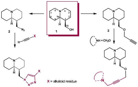
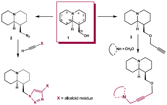
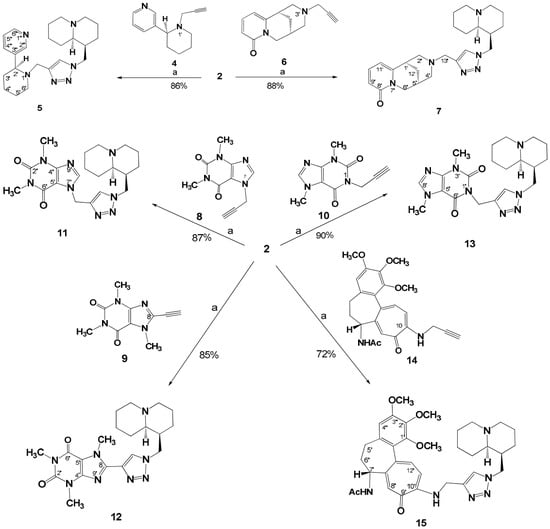
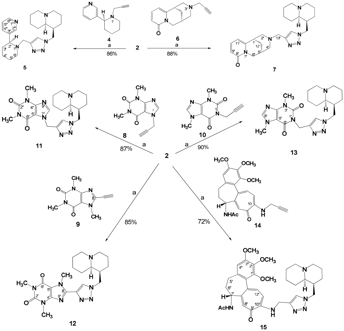
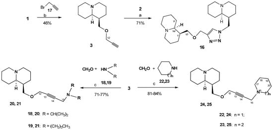
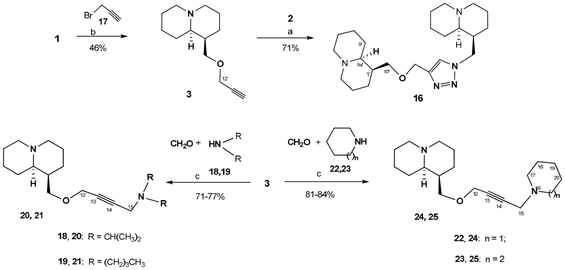
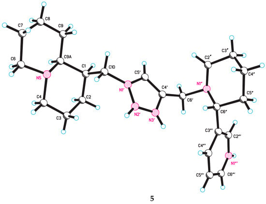
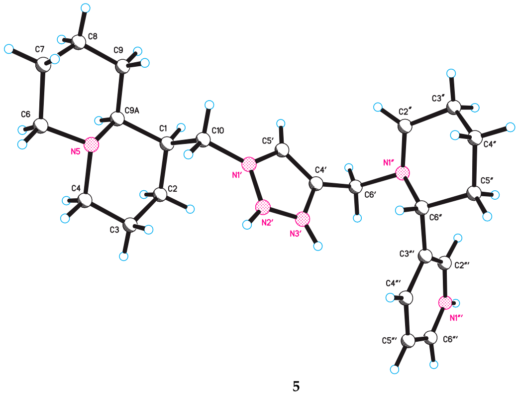
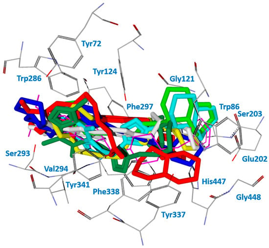
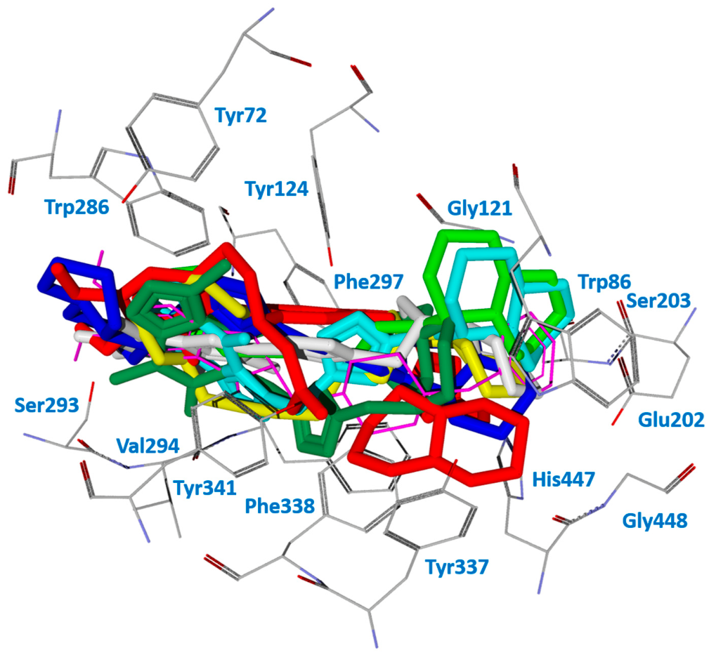
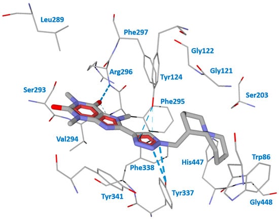
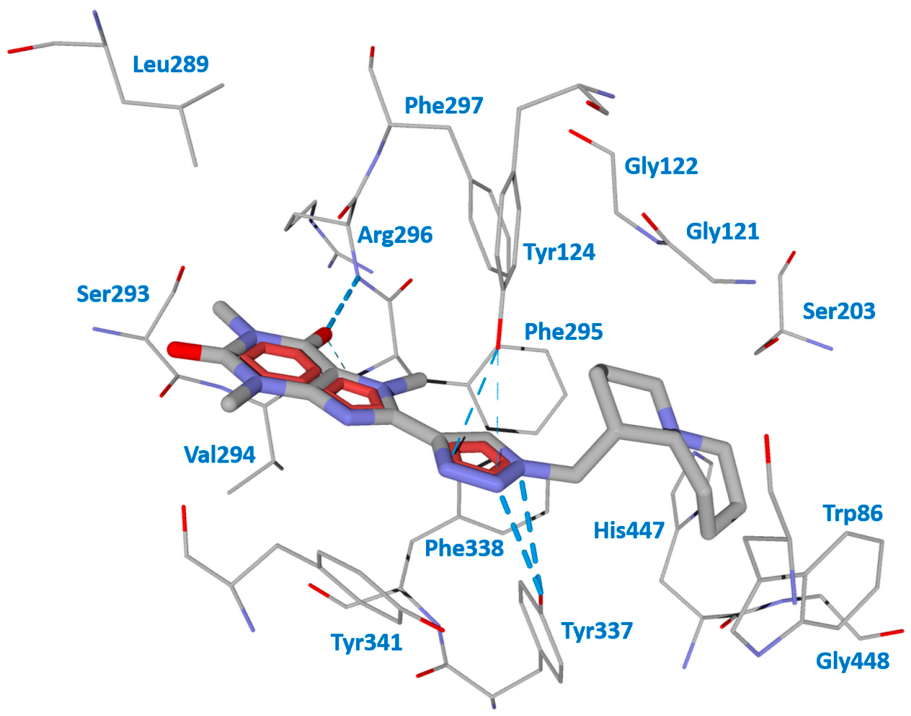

Disclaimer/Publisher’s Note: The statements, opinions and data contained in all publications are solely those of the individual author(s) and contributor(s) and not of MDPI and/or the editor(s). MDPI and/or the editor(s) disclaim responsibility for any injury to people or property resulting from any ideas, methods, instructions or products referred to in the content. |
© 2026 by the authors. Licensee MDPI, Basel, Switzerland. This article is an open access article distributed under the terms and conditions of the Creative Commons Attribution (CC BY) license.