


Abstract
Autism spectrum disorder (ASD) is a systemic multi-organ condition characterized by profound biological heterogeneity. Clinical management currently faces a bottleneck due to reliance on subjective behavioral phenotyping rather than objective molecular markers. This review synthesizes breakthroughs in ASD diagnostics, moving from a reductionist “one-size-fits-all” approach toward an integrative systems biology framework. We evaluate how genetic variants and epigenetic modifications converge on shared pathways, such as synaptic homeostasis and chromatin remodeling. Furthermore, we highlight the diagnostic potential of proteomic and metabolomic signatures, which capture functional states more proximally to the clinical phenotype, including sex-specific metabotypes. The roles of neuroinflammation, oxidative stress, and the gut–microbiota–brain axis are delineated as key contributing factors of pathophysiology. Finally, we propose a roadmap for integrating multi-omics data via machine learning to facilitate early screening and precision therapeutic stratification. By bridging molecular footprints with clinical phenotyping, this synthesis provides a foundation for personalized medicine in neurodevelopmental disorders.
1. Introduction
Autism spectrum disorder (ASD) represents a cluster of heterogeneous neurodevelopmental condition defined by persistent deficits in social communication alongside restricted, repetitive patterns of behaviors [1]. Beyond these core neurobehavioral symptoms, ASD is increasingly conceptualized as a systemic multi-organ disorder. Clinical evidence suggests that the pathophysiology of ASD extends far beyond the central nervous system, frequently involving complex immunological dysregulation, gastrointestinal disturbances, and metabolic imbalances, all of which contribute to a staggering global socioeconomic burden [2,3].
The prevalence of ASD was estimated at 1 in 31 children in the United States and approximately 1 in 143 in China [4,5], attracting widespread societal attention. Currently, the “gold standard” for ASD diagnosis remains rooted in behavioral observations and caregiver reports, such as the Modified Checklist for Autism in Toddlers (M-CHAT), and Ages and Stages Questionnaire (ASQ) as screening tools, Autism Diagnostic Interview-Revised (ADI-R) and Autism Diagnostic Observation Schedule (ADOS) as diagnostic instruments, and Children Autism Rating Scale (CARS) as severity assessment tool [6]. While these psychometric instruments are indispensable and will remain foundational to clinical evaluation, they are inherently subjective and often fail to detect symptoms during the pre-symptomatic developmental period. Rather than serving as replacements for these established behavioral frameworks, emerging molecular biomarkers are intended to complement them by providing earlier pre-symptomatic risk stratification, objective biological subtyping, and real-time monitoring of therapeutic responses. This developmental window, typically before the age of two, represents a “golden period” of peak neural plasticity where intensive behavioral intervention has the maximum potential to normalize neurodevelopmental trajectories [7,8].
The profound biological heterogeneity of ASD, often referred to as “the Autisms”, suggests that a unified behavioral diagnosis masks a multitude of distinct molecular etiologies [9]. Therefore, it is critical to identify presymptomatic biomarkers that could complement the comprehensive clinical assessment of ASD and enable early intervention. The emergence of advanced molecular diagnostic approaches, including high-throughput Next-Generation Sequencing (NGS), single-cell transcriptomics, and deep-phenotyping metabolomics, has begun to unravel these convergent and divergent pathways [10].
In this review, we synthesize recent breakthroughs in the search for ASD biomarkers across genetic, epigenetic, proteomic, metabolic, immune, and microbiome domains. We critically evaluate how these molecular footprints can facilitate early screening and, crucially, act as therapeutic anchors for precision intervention. By bridging the gap between molecular diagnostics and clinical phenotyping, we aim to provide a strategic roadmap for the transition toward personalized medicine in neurodevelopmental disorders.
2. Methods
This article was prepared as a narrative review. The relevant literature was identified through searches of Pubmed (MEDLINE), Scopus, IEEE Xplore, Web of Sciences, and Google Scholar up to August 2026. Search terms included combinations of “autism spectrum disorder”, “genetic”, “epigenetic”, “Non-coding RNA”, “microRNAs”, “proteomic”, “metabolomic”, “immune dysregulation”, “neuroinflammation”, “oxidative stress”, “redox”, and “gut microbiota”. Additional articles were identified through citation tracking of relevant publications. Priority was given to peer-reviewed studies, clinical trials, and recent reviews relevant to molecular diagnostic biomarkers in ASD.
3. The Genetic and Epigenetic Landscape: From Genomic Architecture to Environmental Programming
ASD arises from a complex interplay between inherited or de novo genetic variation and dynamic epigenetic regulation. While high heritability estimates, often exceeding 80%, highlight a substantial genetic contribution to ASD susceptibility [11,12], the pronounced phenotypic heterogeneity observed across individuals suggests that genetic architecture alone cannot fully explain disease manifestation. Epigenetic mechanisms, which integrate environmental exposures with genomic regulation, have therefore emerged as critical modulators of neurodevelopmental trajectories [13,14]. Importantly, both genomic variants and epigenetic signatures are increasingly recognized as potential molecular diagnostic markers capable of identifying biologically distinct ASD subtypes (Figure 1).
Figure 1.
The Multidimensional Molecular Landscape of ASD: From Genomic Architecture to Post-Transcriptional Regulation. This schematic illustrates the hierarchical interplay between diverse molecular layers contributing to the pathophysiology of autism spectrum disorder (ASD). The Genomic Architecture layer represents the spectrum of genetic risk, ranging from rare, highly penetrant syndromic loci to common polygenic variants, which frequently converge on shared pathways like synaptic function and chromatin remodeling to drive excitatory–inhibitory (E/I) imbalance. Intersecting with this is the Epigenetic Mediators layer, where DNA methylation and histone modifications serve as “molecular memories” that integrate prenatal environmental exposures, such as maternal metabolic stress or hormonal influences, into lasting neurodevelopmental shifts, often leading to the silencing of master regulator genes. Finally, the non-coding RNA networks operate as molecular “rheostats,” where microRNAs (miRNAs) and long non-coding RNAs (lncRNAs) fine-tune gene expression post-transcriptionally. These molecules not only serve as dynamic biomarkers for liquid biopsy and symptom monitoring but also offer specialized avenues for RNA-based precision therapeutics to rescue protein haploinsufficiency. Collectively, these integrated molecular signatures transform ASD from a behavioral diagnosis into a molecularly stratified disorder. Note: The schematic illustrations in this figure were created with the assistance of AI tools and edited by the authors.
3.1. Genomic Landscapes: From Monogenic Syndromes to Convergent Pathways
The genetic architecture of ASD encompasses a wide spectrum of variation, ranging from rare highly penetrant de novo mutations to the cumulative effects of common polygenic risk variants [15,16]. Early insights into ASD genetics were largely derived from syndromic forms associated with single-gene mutations, including FMR1 (Fragile X syndrome), TSC1/TSC2 (tuberous sclerosis complex), MECP2 (Rett syndrome), and SHANK3 (Phelan-McDermid syndrome) [17,18,19,20]. However, large-scale sequencing studies, particularly whole-exome and whole-genome sequencing, have substantially expanded the catalog of ASD-associated loci. These efforts have revealed that diverse genetic variants frequently converge on shared neurodevelopmental pathways, including synaptic function, chromatin remodeling, and translational regulation.
More than 1200 genes have been implicated in ASD risk according to the SFARI Gene database, yet these loci rarely operate in isolation. Instead, integrative transcriptomic analyses demonstrate that many ASD-associated genes cluster within convergent functional modules governing neuronal differentiation, synaptic development, and chromatin regulation [21,22,23,24,25,26]. Single-cell transcriptomic studies further indicate that mutations in genes such as SUV420H1, ARID1B, and CHD8 disproportionately affect specific neuronal populations during cortical development. These convergent disruptions are widely hypothesized to contribute to alterations in the balance between excitatory and inhibitory neural signaling, a mechanistic framework frequently referred to as the excitation–inhibition (E/I) imbalance model of ASD [24].
Importantly, emerging evidence suggests that therapeutic responsiveness may depend strongly on the underlying genetic context. For example, dietary zinc supplementation has been shown to rescue synaptic deficits in certain ASD-related genetic models, including Shank3-deficient mice, while similar interventions show limited effects in other genetic backgrounds such as Shank2 deficiency [27,28,29]. These findings indicated that Shank3 or Shank2 deficiency may serve as a predictive biomarker for the response to dietary zinc supplementation in ASD-related genetic models.
3.2. Epigenetic Mechanisms Linking Environmental Exposures to Gene Regulation
Epigenetic modifications, including DNA methylation and histone modifications, provide potential mechanisms through which prenatal environmental exposures may influence gene regulation and neurodevelopment. Maternal metabolic disorders, particularly pregestational and gestational diabetes, have been associated with an increased risk of ASD in offspring [30]. Epigenetic alterations have been proposed as one possible link between maternal metabolic disturbances and neurodevelopmental outcomes. For example, altered DNA methylation at the OR2L13 promoter has been implicated in ASD-related molecular changes, although its functional consequences for early neurogenesis require further investigation [31].
The “Extreme Male Brain” (EMB) theory has also prompted investigations into the contribution of prenatal hormonal exposure to ASD susceptibility [32]. Some studies have linked fetal steroid hormone levels to epigenetic regulation of RORA, including increased H3K9 trimethylation at its promoter and reduced transcriptional activity, although the evidence remains limited [33,34]. The explanatory scope of the EMB theory is also debated, as sex differences in ASD may reflect a combination of biological factors, diagnostic disparities, and behavioral camouflaging. Prenatal hormonal and epigenetic mechanisms may therefore contribute to ASD susceptibility in particular subgroups, but their role in explaining the male-biased prevalence of ASD remains unresolved.
3.3. Non-Coding RNA Networks: The Rheostats of Neurodevelopment
Beyond genetic and epigenetic variation, non-coding RNAs (ncRNAs), including microRNAs (miRNAs) and long non-coding RNAs (lncRNAs), represent an additional regulatory layer that modulates gene expression at the post-transcriptional level. These molecules fine-tune protein translation and cellular signaling pathways during neurodevelopment and in response to environmental stressors. While lncRNAs such as NEAT1, TUG1, and AQP4-AS1 have demonstrated robust potential as peripheral diagnostic signatures [35,36], their functional significance is increasingly centered on their role as molecular effectors. For instance, the natural antisense lncRNA RAB1B-AS1 interacts with the high-risk gene CHD8 to modulate vesicle trafficking pathways, highlighting the intricate lncRNA-chromatin crosstalk inherent in ASD pathology [37].This mechanistic insight has paved the way for “RNA-based precision therapy,” exemplified by the development of synthetic SINEUPs-engineered ncRNAs designed to specifically boost endogenous protein levels, such as CHD8, to rescue haploinsufficiency-driven phenotypes [38].
Simultaneously, the inherent stability of miRNAs in various biofluids (serum, saliva, and feces) renders them promising candidate markers for non-invasive liquid biopsy [39]. However, the diagnostic utility of miRNAs is intrinsically linked to their highly dynamic expression profiles; evidence of opposing age-dependent correlations in toddlers versus older children underscores the necessity of considering temporal windows in biomarker validation (Table 1). Furthermore, the downregulation of miR-484 not only facilitates the differentiation of ASD cohorts but also exhibits a significant inverse correlation with symptom severity [40]. This observation raises the possibility that miRNAs could act as a “molecular gauge”, offering a promising exploratory avenue for early screening research as well as real-time monitoring of disease progression and therapeutic response. These markers, however, remain candidate biomarkers and require extensive independent validation before clinical translation, and correlation does not confirm causal links between miRNA expression and ASD behavioral features.
Table 1.
Alterations of miRNA expression in different biological samples of ASD patients.
Collectively, advances in genomic sequencing, epigenetic profiling, and RNA-based biomarker discovery hold potential to gradually shift ASD research beyond a purely behavioral diagnostic framework toward a model of molecular stratification. These insights provide a conceptual foundation for exploring the future integration of multi-omics candidate biomarkers into potential diagnostic frameworks, although such clinical implementation remains preliminary and unvalidated.
4. Targeted Proteomics and Metabolomics Diagnostic Markers
Proteomic and metabolomic profiling have emerged as central components of molecular diagnostic research in ASD, as they capture functional biological states that lie downstream of genetic and epigenetic regulation [66,67]. Unlike genomic variants, which primarily reflect susceptibility, protein and metabolite signatures provide dynamic readouts of cellular activity and physiological adaptation. Advances in high-resolution mass spectrometry and biofluid-based omics technologies have therefore enabled the identification of candidate molecular signatures detectable in peripheral samples, offering promising avenues for minimally invasive diagnostic development [68,69].
4.1. Proteomic Signatures: From Enzymes to Neural Homeostasis
Proteomic studies of ASD consistently identify alterations across several interconnected biological domains, particularly synaptic signaling [70], immune regulation [71], and cellular metabolism [72]. Early investigations relying on brain tissue analyses revealed dysregulation of synaptic scaffolding and neurotransmission-related proteins [73]; however, recent research has increasingly focused on peripheral biofluids, including plasma and serum, to identify clinically accessible biomarkers [67]. These studies report reproducible alterations in inflammatory mediators, complement cascade components, and oxidative stress-related proteins, suggesting systemic biological involvement beyond the central nervous system.
Importantly, only a subset of reported proteomic alterations demonstrates characteristics required for diagnostic translation. Several studies employing machine-learning-assisted proteomic panels have achieved moderate classification accuracy when distinguishing ASD from typically developing controls, highlighting the potential of multi-protein signatures rather than single biomarkers [68]. Nevertheless, variability across cohorts remains substantial, reflecting differences in age, comorbidities, analytical platforms, and sample processing protocols. Standardization and independent validation therefore represent critical prerequisites for clinical implementation.
4.2. Metabolomics and the “Sex-Bias” Paradigm
Metabolomics provides a direct snapshot of biochemical activity and has therefore become a particularly promising strategy for ASD molecular diagnostics [74]. Because metabolites integrate genetic predisposition, environmental exposure, diet, microbiome activity, and immune signaling, metabolic profiles may capture disease-relevant biology more proximally to phenotype than genomic markers alone. A particularly compelling frontier in metabolomics is the elucidation of sex-specific molecular signatures, which may explain the long-observed male bias in ASD prevalence [32]. Longitudinal analyses have revealed that female offspring with ASD often exhibit a disruption in the ether/non-ether phospholipid balance within maternal mid-gestation plasma [69]. In contrast, ASD-affected males tend to show a higher density of dysregulated chemical clusters in cord blood, suggesting that the “biological hits” leading to ASD may manifest through different metabolic pathways depending on biological sex [69]. This divergence supports the Female Protective Model, suggesting that different metabolic thresholds or compensatory mechanisms may exist across sexes, which is a critical consideration for developing sex-stratified diagnostic tools.
4.3. The Shift Toward Integrated Multi-Biomarker Panels
While individual biomarkers often exhibit variability across disparate cohorts due to the inherent heterogeneity of ASD, the field is rapidly pivoting toward multi-biomarker panels. Integrated models that synthesize multiple protein and metabolite signatures have consistently demonstrated superior diagnostic efficacy, often reaching high AUC values, compared to single-molecule approaches [67,75,76,77]. This transition from isolated markers to integrated metabolic profiles is essential for capturing the multifaceted nature of ASD. Moreover, these panels facilitate the identification of biologically meaningful metabotypes, paving the way for personalized metabolic interventions, such as targeted nutritional supplementation [78] or enzyme replacement therapies [79], tailored to the individual’s specific biochemical deficit. Looking forward, establishing robust and clinically actionable molecular diagnostic models will fundamentally rely on integrating these multi-biomarker frameworks with standardized clinical phenotyping and rigorous machine learning-based validation methodologies.
5. Immune Dysregulation and Neuroinflammation: Systemic Signatures and CNS Crosstalk
Accumulating evidence underscores that immune dysfunction is not merely a comorbid feature but play an important role in the pathophysiology of ASD [80]. This neuroinflammatory state is characterized by a persistent, low-grade systemic imbalance in the cytokine network, reflecting a broader immunological “priming”. Such priming suggests that the immune system of an individual with ASD is in a state of chronic readiness, where minor triggers can elicit exaggerated inflammatory responses that correlate with behavioral severity [81,82,83].
5.1. Systemic Cytokine Profiles: Molecular Correlates of Behavior
A growing body of evidence suggests that immune dysregulation contributes to ASD pathophysiology. Both central and peripheral immune alterations have been reported, including microglial activation within the developing brain and abnormal cytokine profiles in peripheral blood. Elevated levels of pro-inflammatory mediators such as interleukin-6 (IL-6), tumor necrosis factor-α (TNF-α), and interleukin-1β (IL-1β) have been detected in several independent cohorts of ASD individuals, suggesting persistent low-grade inflammatory activity [81,83].
Crucially, these molecular alterations are not mere epiphenomena; they serve as functional correlates of clinical symptoms. Specifically, elevated levels of IL-8 and MCP-1 (CCL2), chemokines involved in leukocyte trafficking, have been linked to the severity of social communication deficits and impaired cognitive flexibility. In contrast, high concentrations of TNF-α and IL-6 are frequently observed in subsets of patients exhibiting prominent irritability, stereotypic behaviors, and aggression [84,85]. This suggests that specific cytokine profiles could eventually serve as “biotypes” to predict behavioral trajectories and tailor behavioral interventions.
5.2. The IL-38/IL-36R Axis: A Failure of Homeostatic Buffering
A significant advance in understanding the systemic-CNS inflammatory dialogue is the discovery of the IL-38/IL-36R signaling axis. IL-38, an anti-inflammatory antagonist of the IL-1 family, represents a candidate biomarker for both diagnostic screening and therapeutic monitoring, though this potential remains preliminary and requires independent replication. A striking paradox has been observed: while children with ASD exhibit compensatory increases in serum IL-38, there is a substantial deficit of IL-38 and its cognate receptor, IL-36R, within the amygdala, a critical neural hub for emotional processing and fear responses [86].
This localized impairment in anti-inflammatory signaling within the brain suggests a “failure of homeostasis,” where the CNS is unable to utilize systemic anti-inflammatory signals to curb local neuroinflammation. Furthermore, the integration of advanced molecular imaging, such as PET tracers for the 18 kDa translocator protein (TSPO) to visualize microglial activation, provides an in vivo methodology to validate these findings. This non-invasive “liquid-to-imaging” correlation offers a robust biological gauge for assessing neuroinflammatory status and monitoring the brain’s response to systemic immunomodulatory therapies [87].
5.3. Immunomodulatory Paradigms: From “Dampening” to Precision
The recognition of neuroinflammation as a targetable driver has catalyzed investigations into various immunomodulatory interventions. Clinical trials have found that pharmacological agents, including celecoxib (a selective COX-2 inhibitor) and prednisolone, as well as nutritional strategies like omega-3 fatty acid supplementation, have shown preliminary efficacy in attenuating irritability and hyperactivity [88,89,90,91].
The therapeutic mechanism is increasingly thought to involve the reprogramming of microglial phenotypes from a pro-inflammatory (M1-like) to a neuroprotective (M2-like) state, alongside the restoration of the T-cell (Treg/Th17) balance [92]. However, the field must move beyond “dampening” general inflammation. The future of ASD immunotherapy lies in identifying individuals with specific immune biotypes who are most likely to respond to targeted cytokine inhibitors, thereby truly reshaping the neurodevelopmental trajectory through precision immuno-psychiatry.
Collectively, immune and microbiome research highlights the importance of systemic biological processes in ASD beyond the central nervous system. Although immune mediators and microbiome-derived metabolites show promise as peripheral biomarkers, current findings remain heterogeneous and require validation in larger, well-characterized cohorts. Integrating immune, microbiome, and multi-omics data may ultimately provide more robust molecular diagnostic signatures for ASD.
6. Oxidative Stress and Redox Imbalance: Molecular Fingerprints of Cytotoxicity
A growing body of evidence suggests oxidative stress not merely as a downstream consequence of ASD pathology but as a important integrative mechanism linking genetic susceptibility, mitochondrial dysfunction, immune activation, and altered neurodevelopmental trajectories [93] (Figure 2). Oxidative stress, defined as an imbalance between reactive oxygen species (ROS) production and antioxidant defense capacity, exerts disproportionate effects on the developing brain due to its high metabolic demand and limited antioxidant reserves. Emerging studies suggest that redox dysregulation acts as a developmental amplifier, destabilizing mitochondrial bioenergetics, reshaping epigenetic regulation, and impairing synaptic plasticity during critical windows of neural maturation [94]. Consequently, oxidative stress represents a mechanistic convergence point through which diverse etiological pathways translate into shared neurobiological phenotypes in ASD [94,95].
Figure 2.
Oxidative Stress as a Mechanistic Convergence Point in ASD Pathophysiology. This schematic delineates the central role of redox imbalance in bridging diverse etiological factors with neurodevelopmental impairment in ASD. The diagram illustrates how genetic susceptibility, mitochondrial dysfunction, and environmental stressors (shown as upstream drivers) collectively trigger an oxidative surge. This systemic imbalance, characterized by the overproduction of reactive oxygen species (ROS) and a compromised antioxidant capacity, acts as a “developmental amplifier.” It leads to a cascade of cellular consequences, including the destabilization of mitochondrial bioenergetics and the manifestation of cytotoxic signatures within the neural environment. These oxidative insults eventually converge to impair synaptic plasticity and neurogenesis, serving as a critical interface through which disparate biological signals translate into the shared neurobiological and behavioral phenotypes characteristic of ASD. Note: The schematic illustrations in this figure were created with the assistance of AI tools and edited by the authors.
6.1. Multidimensional Cascades of Oxidative and Nitrative Damage
The accumulation of oxidative insults in ASD is evidenced by a coordinated elevation of biomarkers across multiple molecular strata, reflecting a systemic failure to contain reactive species. These molecular signatures should not be interpreted as isolated biomarkers but rather as interconnected layers of a systemic redox cascade, reflecting progressive cellular injury from membrane instability to genomic vulnerability. Lipid peroxidation represents an early and pervasive manifestation of this damage; elevated concentrations of malondialdehyde (MDA) and 4-hydroxynonenal (4-HNE) in the blood and urine of individuals with ASD indicate compromised cell membrane integrity and altered permeability, which are critical for proper neuronal signaling [96,97]. Furthermore, the consistent upregulation of 8-iso-prostaglandin F2α (8-iso-PGF2α), the recognized gold standard for lipid oxidative stress, signals a widespread systemic burden [98].
This lipid-level disruption is compounded by protein oxidation and nitrative stress, as evidenced by increased levels of protein carbonyls and 3-nitrotyrosine (3-NT), particularly within the cerebellum [95,99,100]. Such nitrative stress is frequently driven by excessive nitric oxide (NO) and peroxynitrite, which act as potent inhibitors of the mitochondrial electron transport chain, thereby impairing ATP production and fueling a vicious cycle of mitochondrial dysfunction and further ROS generation [101]. Ultimately, this oxidative siege extends to the genome; the elevation of 8-hydroxy-2′-deoxyguanosine (8-OHdG) in brain tissue and biofluids serves as a definitive indicator of chronic DNA oxidation [102,103,104]. Notably, the robust positive correlation between 8-OHdG levels and ASD severity positions this marker as a potentially invaluable molecular gauge for clinical staging and prognostic assessment, bridging the gap between metabolic disturbance and behavioral phenotype [105].
6.2. Compromised Antioxidant Defense and Redox Homeostasis
The severity of oxidative damage in ASD is exacerbated by a concurrent failure of the antioxidant defense system. Glutathione (GSH), the primary radical scavenger in the CNS, is significantly depleted in postmortem ASD brain samples [102,106]. A reduced GSH/GSSG ratio not only indicates poor antioxidant capacity but also correlates with the degree of language impairment and social deficits [107]. Notably, impaired antioxidant capacity in ASD appears to arise not solely from environmental exposure but from intrinsic metabolic vulnerability, including mitochondrial inefficiency and disrupted one-carbon metabolism. This suggests that redox imbalance represents a biologically embedded trait rather than a transient physiological state.
Furthermore, dysregulations in the methionine–homocysteine cycle, evidenced by reduced levels of vitamins B9, B12, and Sadenosylmethionine (SAM), suggest a systemic deficit in methylation capacity and thiol-based redox buffering [96]. While some studies report compensatory increases in erythrocyte superoxide dismutase (SOD) and catalase, the general trend toward reduced serum antioxidant levels reflects a tissue-specific failure to maintain redox homeostasis, leaving the developing brain susceptible to oxidative-induced neurotoxicity [108,109].
7. The Gut–Microbiota–Brain Axis: A Microbial–Metabolic Signaling Hub
Increasing evidence suggests that the gut–microbiota–brain axis represents a biologically plausible interface through which environmental exposures interact with host molecular systems during neurodevelopment [110]. Rather than acting solely through neural signaling pathways, gut microbiota exerts systemic effects via microbial metabolites, immune modulation, and redox regulation, thereby influencing mitochondrial function, epigenetic programming, and synaptic maturation (Figure 3). In ASD, microbial dysbiosis is increasingly interpreted not merely as a gastrointestinal comorbidity but as a potential regulator of metabolic and inflammatory pathways converging on neurodevelopmental vulnerability [111].
Figure 3.
The Gut–Microbiota–Brain Axis: A Microbial–Metabolic Signaling Network in ASD. This schematic illustrates the systemic influence of the gut–microbiota–brain axis on neurodevelopmental trajectories in ASD. The diagram depicts how gut microbiota and their associated microbial metabolites, such as short-chain fatty acids (SCFAs) and trimethylamine N-oxide (TMAO), act as primary signaling molecules that bridge the gastrointestinal environment with the central nervous system. These metabolites exert their effects through multiple interconnected pathways, including the modulation of mitochondrial bioenergetics, immune system activation, and epigenetic programming. A central feature of this axis is the regulation of redox homeostasis; an imbalance between reactive oxygen species (ROS) and antioxidant defenses serves as a critical driver of neurodevelopmental disruption. Furthermore, microbial signaling influences the levels of key neurotransmitters and neurotrophic factors, such as GABA and BDNF, which are essential for synaptic maturation and cognitive function. Collectively, the gut-microbiota–brain axis functions as a biologically plausible interface where microbial dysbiosis translates into systemic metabolic and inflammatory signatures that converge on ASD vulnerability. Note: The schematic illustrations in this figure were created with the assistance of AI tools and edited by the authors.
7.1. Microbial Dysbiosis and Targeted Metabolomics
Fecal metagenomic analyses consistently reveal significant alterations in the microbial landscape of children with ASD, typically characterized by an overgrowth of pathogenic taxa, such as Clostridium species, and a concurrent depletion of beneficial commensals including Bifidobacterium and Bacteroidetes [112,113] (Table 2). It is worth noting the marked inconsistency across studies, with some taxa reported as increased in ASD children in one investigation but decreased in another. Such discrepancies may stem from heterogeneity in participant geography, diet, age, gastrointestinal comorbidities, and prior medication exposure such as antibiotics or probiotics [114,115,116,117]. In addition, differences in stool handling, sequencing platforms, bioinformatic workflows and taxonomic resolution may contribute to divergent results. ASD heterogeneity itself may also lead to distinct microbial subtypes, which partially accounts for opposing abundance patterns [117].
Table 2.
Summary of studies on gut microbiota alterations in children with ASD over the past 5 years.
However, taxonomic alterations alone provide limited mechanistic insight, as functional metabolic outputs, rather than microbial identity per se, are increasingly considered the principal mediators of microbiota–host neurobiological interactions. In this context, the diagnostic and mechanistic relevance of gut dysbiosis is increasingly attributed to microbial-derived metabolites that exert systemic biological effects.
Alterations in short-chain fatty acids (SCFAs), crucial regulators of blood–brain barrier (BBB) integrity, have been implicated in inducing behavioral deficits by modulating neuroinflammation [142,143,144]. In particular, SCFAs such as butyrate are capable of influencing host gene expression via epigenetic mechanisms, suggesting a potential molecular link between microbial metabolism and neurodevelopmental regulation. Furthermore, elevated levels of microbial metabolites including p-cresol and 4-ethylphenol in biofluids not only distinguish ASD cohorts but also correlate with symptom severity, supporting their potential utility as candidate metabolomic biomarkers reflecting gut ecosystem disruption [145,146]. Complementary evidence indicates dysregulation of bile acid and tryptophan metabolic pathways, highlighting a broader metabolic interface through which intestinal dysbiosis may contribute to systemic biochemical imbalance and altered neurodevelopmental signaling.
7.2. Intestinal Permeability and Microbial-Metabolic Crosstalk
Altered intestinal barrier integrity has frequently been reported in individuals with ASD, with increasing evidence suggesting that enhanced intestinal permeability, often referred to as a “leaky gut”, may contribute to systemic inflammatory signaling. Molecular profiling of duodenal biopsies has revealed downregulation of key barrier-forming tight junction proteins (e.g., CLDN1, OCLN) accompanied by increased expression of pore-forming claudins, a shift that may compromise epithelial integrity and facilitate translocation of microbial products [147,148]. These alterations have been partially attributed to reductions in butyrate-producing bacterial populations, which play a critical role in maintaining epithelial homeostasis and anti-inflammatory signaling within the intestinal mucosa.
Beyond bacterial compositional shifts alone, emerging research underscores that ASD-associated microbial alterations are intimately linked to functional metabolic and biochemical interfaces. Advanced metagenomic and metabolomic profiling have identified integrated biomarker panels comprising specific bacterial signatures and downstream metabolic products that demonstrate high discriminative performance for ASD across independent cohorts [149]. The reproducibility of these microbial–metabolic signatures across different populations suggests that functional gut ecosystem readouts may provide robust biological insights. Integrating such multi-layer microbial profiles with host-derived molecular markers may therefore represent a promising direction for developing robust and biologically informed diagnostic frameworks for ASD.
8. Methodological and Translational Hurdles from Multi-Omics Discovery to Clinical Implementation
A critical challenge underlying peripheral ASD biomarkers is their modest diagnostic specificity. Alterations in circulating cytokines, systemic redox states, mitochondrial metabolites, and gut microbiota profiles are non-specific pathophysiological signatures shared across various neurodevelopmental, psychiatric, and metabolic conditions, including schizophrenia, major depressive disorder, epilepsy, and inflammatory bowel diseases. Consequently, individual markers in these pathways often reflect broad biological stress or systemic dysregulation rather than ASD-exclusive etiology, underscoring why multi-domain biomarker integration and inclusion of non-ASD clinical controls are essential to isolate disease-specific profiles.
Translating multi-omics findings into clinically applicable diagnostic tools for ASD remains challenging. A critical conceptual distinction that must be emphasized is the boundary between a statistically significant molecular difference identified in group-level comparisons and a clinically useful diagnostic biomarker. While observational cohorts frequently report molecular features with statistically significant differences between ASD and control groups, statistical significance alone does not demonstrate clinical diagnostic utility. Clinical implementation additionally demands rigorous analytical validity, predefined decision thresholds, clinical validity, and demonstrated clinical utility. A major limitation is the high dimensionality of omics data relative to the sample sizes of most clinical cohorts, which increases the risk of overfitting and limits model generalizability [150,151]. Data leakage during feature selection or hyperparameter tuning can further inflate predictive performance. Nested cross-validation, prespecified preprocessing pipelines, and independent external validation are therefore essential for obtaining reliable performance estimates [151,152]. Technical heterogeneity introduces additional complexity. Differences in sample handling, laboratory protocols, and analytical platforms can produce batch effects that obscure disease-associated signals. Although harmonization methods can mitigate some of this variation, their suitability depends on the data type and study design. For example, ComBat-seq was developed for RNA-sequencing count data and is not directly applicable to all omics modalities [153]. Moreover, when diagnostic groups are confounded with processing batches, technical and biological effects may be difficult to disentangle. Appropriate quality control and validation across analytical settings are therefore necessary to establish the reproducibility of identified biomarkers.
Class imbalance and inadequate model calibration also complicate clinical translation. Unequal representation of ASD cases and neurotypical controls may bias classifiers toward the majority class, while resampling and cost-sensitive learning do not necessarily resolve all performance limitations [154]. Furthermore, high classification accuracy does not guarantee reliable individual-level predictions. Model evaluation should therefore consider discrimination, sensitivity, specificity, predictive values, and calibration, using measures such as the Brier score and calibration plots [155]. Performance must also be interpreted in relation to the target population, as predictive values and estimated risks may differ substantially between case–control studies and routine clinical settings. Generalizability across populations remains a further concern. Biomarker signatures derived from single-center or relatively homogeneous cohorts may reflect differences in ancestry, age, sex, developmental stage, or environmental exposure rather than ASD-related biology alone [156]. Given the clinical and etiological heterogeneity of ASD, validation in independent, geographically and ancestrally diverse cohorts is essential. Subgroup-specific performance should also be examined to identify potential disparities in diagnostic accuracy and determine whether model recalibration or updating is required [152].
Finally, clinical adoption depends on both interpretability and demonstrated utility. Explainable artificial intelligence methods, including SHapley Additive exPlanations (SHAP) and Local Interpretable Model-agnostic Explanations (LIME), can help characterize how individual molecular features contribute to model predictions [157]. However, feature attribution does not establish biological causality, and apparent associations may arise from confounding or correlated features. Integrating computational predictions with biological evidence and independent replication is therefore crucial. Ultimately, clinical translation will require reproducible performance, demonstrable incremental value over existing diagnostic assessments, and prospective evaluation in real-world settings [152,158].
9. Conclusions and Future Perspectives
The expanding body of evidence reviewed herein highlights that neurodevelopmental conditions such as ASD cannot be adequately explained by molecular perturbations within a solitary biological layer. Instead, ASD arises from dynamic, bidirectional interactions across genetic, epigenetic, metabolic, mitochondrial, immune, and microbial networks, which collectively shape atypical neurodevelopmental trajectories. This systems-level paradigm challenges traditional reductionist diagnostics and underscores the necessity of integrated molecular frameworks [Supplementary Table S1].
Genomic studies have successfully mapped numerous risk loci associated with neurodevelopmental vulnerability; however, their limited predictive power at the individual level demonstrates that genetic predisposition alone fails to capture full phenotypic heterogeneity. Epigenetic regulation serves as a critical interface through which environmental exposures, metabolic states, and cellular stress responses modulate gene expression during vulnerable developmental windows. In parallel, mitochondrial dysfunction and systemic redox imbalance have emerged as convergent pathophysiological nodes, bridging metabolic disturbances with aberrant neuronal signaling, impaired synaptic plasticity, and chronic neuroimmune activation. Furthermore, microbial and metabolic derivatives act as systemic modulators influencing blood–brain barrier permeability, neuroinflammation, and host metabolic homeostasis. The convergence of these interconnected pathways indicates that ASD pathophysiology is fundamentally driven by network-level dysregulation rather than isolated molecular anomalies. Consequently, single-biomarker diagnostic models are inherently inadequate for capturing this multidimensional biology.
Looking forward, clinical diagnostics must pivot toward multi-omics synthesis, combining genetic risk scores, epigenetic signatures, metabolic profiles, mitochondrial functional markers, and microbial signatures (Figure 4). Although advanced machine learning and network-based algorithms provide robust analytical tools to decipher these heterogeneous datasets, a realistic appraisal of translational readiness is essential. While peripheral immune profiles, select microbial metabolites, and specific metabolic ratios exhibit emerging consistency across independent cohorts, the vast majority of candidate biomarkers remain strictly in the exploratory discovery phase. They continue to be constrained by modest sample sizes, batch effects, cross-platform technical variance, and a paucity of longitudinal validation. Accordingly, integrated multi-omics diagnostics are not yet mature for routine clinical deployment.
Figure 4.
Future Framework for ASD Precision Medicine: Multi-Omics Integration and Systems-Based Stratification. This schematic presents a conceptual framework for the next generation of molecular diagnostics and personalized intervention in ASD. The framework initiates with the systematic acquisition of high-dimensional datasets (left), encompassing genomic susceptibility markers, epigenetic signatures, and non-coding RNA profiles, alongside functional readouts from proteomics. At the heart of this model is a centralized Data Integration hub (center), which serves to synthesize these heterogeneous biological streams, including immune signaling, metabolic pathways, and microbiome features, to decode the complex, interconnected networks underlying the disorder. This systems-level integration facilitates a multi-faceted clinical transition (right), moving beyond traditional behavioral assessments toward early biomarker detection, biologically driven patient stratification, and the development of personalized therapeutic targets and precision medicine strategies. Ultimately, this integrated approach establishes a robust molecular foundation for improving long-term clinical outcomes and treatment monitoring in ASD. Note: The schematic illustrations in this figure were created with the assistance of AI tools and edited by the authors.
In summary, shifting the paradigm from single-domain discovery to systems-driven biomarker panels holds profound promise for precision psychiatry. Realizing this clinical potential, however, will depend on rigorous, multi-center replication, standardized analytical pipelines, and longitudinal tracking, ultimately paving the way for objective, biology-informed stratification and early intervention in ASD.
Supplementary Materials
The following supporting information can be downloaded at: https://www.mdpi.com/article/10.3390/molecules31213952/s1, Table S1: Summary of major candidate biomarkers for autism spectrum disorder.
Author Contributions
Y.Z.: Writing—original draft, Methodology, Investigation, Formal analysis, Data curation, Conceptualization, Funding acquisition. M.Z.: Writing—review and editing, Validation, Supervision, Resources, Funding acquisition. All authors have read and agreed to the published version of the manuscript.
Funding
This research was supported by the Postdoctoral Fellowship Program of CPSF (GZC20251559), China Postdoctoral Science Foundation (2025M772421), and National Natural Science Foundation of China (82301709).
Institutional Review Board Statement
Not applicable.
Informed Consent Statement
Not applicable.
Data Availability Statement
No new data were created or analyzed in this study.
Acknowledgments
During the preparation of this manuscript/study, the authors used ChatGPT (version: GPT-4) for language polishing, and generating and refining the schematic illustrations. The authors have reviewed and edited the output and take full responsibility for the content of this publication.
Conflicts of Interest
The authors declare no conflicts of interest.
References
- Lord, C.; Elsabbagh, M.; Baird, G.; Veenstra-Vanderweele, J. Autism Spectrum Disorder. Lancet 2018, 392, 508–520. [Google Scholar] [CrossRef] [Scilit] [PubMed]
- Wang, Y.; Lv, R.; Jiang, Z.; Liu, Y.; Zhang, J.; Zhao, Z.; Liu, Y.; Zheng, Y.; Liu, B.; Clauw, D.; et al. Autism Spectrum Disorder across the Lifespan: Dynamic Symptom Trajectories and Multidimensional Support Framework. Mol. Psychiatry 2026, 31, 4124–4143. [Google Scholar] [CrossRef] [Scilit] [PubMed]
- Global Burden of Disease Study 2021 Autism Spectrum Collaborators. The Global Epidemiology and Health Burden of the Autism Spectrum: Findings from the Global Burden of Disease Study 2021. Lancet Psychiatry 2025, 12, 111–121. [Google Scholar] [CrossRef] [Scilit] [PubMed]
- Maenner, M.J.; Warren, Z.; Williams, A.R.; Amoakohene, E.; Bakian, A.V.; Bilder, D.A.; Durkin, M.S.; Fitzgerald, R.T.; Furnier, S.M.; Hughes, M.M.; et al. Prevalence and Characteristics of Autism Spectrum Disorder among Children Aged 8 Years—Autism and Developmental Disabilities Monitoring Network, 11 Sites, United States, 2020. Morb. Mortal. Wkly. Rep. 2023, 72, 1–14. [Google Scholar] [CrossRef] [Scilit] [PubMed]
- Zhou, H.; Xu, X.; Yan, W.; Zou, X.; Wu, L.; Luo, X.; Li, T.; Huang, Y.; Guan, H.; Chen, X.; et al. Prevalence of Autism Spectrum Disorder in China: A Nationwide Multi-Center Population-Based Study among Children Aged 6 to 12 Years. Neurosci. Bull. 2020, 36, 961–971. [Google Scholar] [CrossRef] [Scilit] [PubMed]
- Hirota, T.; King, B.H. Autism Spectrum Disorder: A Review. JAMA 2023, 329, 157–168. [Google Scholar] [CrossRef] [Scilit] [PubMed]
- Gabbay-Dizdar, N.; Ilan, M.; Meiri, G.; Faroy, M.; Michaelovski, A.; Flusser, H.; Menashe, I.; Koller, J.; Zachor, D.A.; Dinstein, I. Early Diagnosis of Autism in the Community Is Associated with Marked Improvement in Social Symptoms within 1-2 Years. Autism Int. J. Res. Pract. 2022, 26, 1353–1363. [Google Scholar] [CrossRef] [Scilit] [PubMed]
- Dawson, G.; Jones, E.J.; Merkle, K.; Venema, K.; Lowy, R.; Faja, S.; Kamara, D.; Murias, M.; Greenson, J.; Winter, J.; et al. Early Behavioral Intervention Is Associated with Normalized Brain Activity in Young Children with Autism. J. Am. Acad. Child Adolesc. Psychiatry 2012, 51, 1150–1159. [Google Scholar] [CrossRef] [Scilit] [PubMed]
- Geschwind, D.H. Autism: Many Genes, Common Pathways? Cell 2008, 135, 391–395. [Google Scholar] [CrossRef] [Scilit] [PubMed]
- Hasin, Y.; Seldin, M.; Lusis, A. Multi-Omics Approaches to Disease. Genome Biol. 2017, 18, 83. [Google Scholar] [CrossRef] [Scilit] [PubMed]
- Sandin, S.; Lichtenstein, P.; Kuja-Halkola, R.; Hultman, C.; Larsson, H.; Reichenberg, A. The Heritability of Autism Spectrum Disorder. JAMA 2017, 318, 1182–1184. [Google Scholar] [CrossRef] [Scilit] [PubMed]
- Tick, B.; Bolton, P.; Happé, F.; Rutter, M.; Rijsdijk, F. Heritability of Autism Spectrum Disorders: A Meta-Analysis of Twin Studies. J. Child Psychol. Psychiatry 2016, 57, 585–595. [Google Scholar] [CrossRef] [Scilit] [PubMed]
- LaSalle, J.M. Epigenomic Strategies at the Interface of Genetic and Environmental Risk Factors for Autism. J. Hum. Genet. 2013, 58, 396–401. [Google Scholar] [CrossRef] [Scilit] [PubMed]
- LaSalle, J.M. Epigenomic Signatures Reveal Mechanistic Clues and Predictive Markers for Autism Spectrum Disorder. Mol. Psychiatry 2023, 28, 1890–1901. [Google Scholar] [CrossRef] [Scilit] [PubMed]
- Devlin, B.; Scherer, S.W. Genetic Architecture in Autism Spectrum Disorder. Curr. Opin. Genet. Dev. 2012, 22, 229–237. [Google Scholar] [CrossRef] [Scilit] [PubMed]
- Vorstman, J.A.S.; Parr, J.R.; Moreno-De-Luca, D.; Anney, R.J.L.; Nurnberger, J.I., Jr.; Hallmayer, J.F. Autism Genetics: Opportunities and Challenges for Clinical Translation. Nat. Rev. Genet. 2017, 18, 362–376. [Google Scholar] [CrossRef] [Scilit] [PubMed]
- Deng, P.Y.; Klyachko, V.A. Channelopathies in Fragile X Syndrome. Nat. Rev. Neurosci. 2021, 22, 275–289. [Google Scholar] [CrossRef] [Scilit] [PubMed]
- Davis, P.E.; Peters, J.M.; Krueger, D.A.; Sahin, M. Tuberous Sclerosis: A New Frontier in Targeted Treatment of Autism. Neurotherapeutics 2015, 12, 572–583. [Google Scholar] [CrossRef] [Scilit] [PubMed]
- Plummer, J.T.; Evgrafov, O.V.; Bergman, M.Y.; Friez, M.; Haiman, C.A.; Levitt, P.; Aldinger, K.A. Transcriptional Regulation of the MET Receptor Tyrosine Kinase Gene by MeCP2 and Sex-Specific Expression in Autism and Rett Syndrome. Transl. Psychiatry 2013, 3, e316. [Google Scholar] [CrossRef] [Scilit] [PubMed]
- Gergoudis, K.; Weinberg, A.; Templin, J.; Farmer, C.; Durkin, A.; Weissman, J.; Siper, P.; Foss-Feig, J.; Del Pilar Trelles, M.; Bernstein, J.A.; et al. Psychometric Study of the Social Responsiveness Scale in Phelan-McDermid Syndrome. Autism Res. 2020, 13, 1383–1396. [Google Scholar] [CrossRef] [Scilit] [PubMed]
- Fazel Darbandi, S.; An, J.Y.; Lim, K.; Page, N.F.; Liang, L.; Young, D.M.; Ypsilanti, A.R.; State, M.W.; Nord, A.S.; Sanders, S.J.; et al. Five Autism-Associated Transcriptional Regulators Target Shared Loci Proximal to Brain-Expressed Genes. Cell Rep. 2024, 43, 114329. [Google Scholar] [CrossRef] [Scilit] [PubMed]
- Liao, C.; Moyses-Oliveira, M.; De Esch, C.E.F.; Bhavsar, R.; Nuttle, X.; Li, A.; Yu, A.; Burt, N.D.; Erdin, S.; Fu, J.M.; et al. Convergent Coexpression of Autism-Associated Genes Suggests Some Novel Risk Genes May Not Be Detectable in Large-Scale Genetic Studies. Cell Genom. 2023, 3, 100277. [Google Scholar] [CrossRef] [Scilit] [PubMed]
- Gandal, M.J.; Haney, J.R.; Wamsley, B.; Yap, C.X.; Parhami, S.; Emani, P.S.; Chang, N.; Chen, G.T.; Hoftman, G.D.; de Alba, D.; et al. Broad Transcriptomic Dysregulation Occurs across the Cerebral Cortex in ASD. Nature 2022, 611, 532–539. [Google Scholar] [CrossRef] [Scilit] [PubMed]
- Paulsen, B.; Velasco, S.; Kedaigle, A.J.; Pigoni, M.; Quadrato, G.; Deo, A.J.; Adiconis, X.; Uzquiano, A.; Sartore, R.; Yang, S.M.; et al. Autism Genes Converge on Asynchronous Development of Shared Neuron Classes. Nature 2022, 602, 268–273. [Google Scholar] [CrossRef] [Scilit] [PubMed]
- Willsey, H.R.; Willsey, A.J.; Wang, B.; State, M.W. Genomics, Convergent Neuroscience and Progress in Understanding Autism Spectrum Disorder. Nat. Rev. Neurosci. 2022, 23, 323–341. [Google Scholar] [CrossRef] [Scilit] [PubMed]
- Jin, X.; Simmons, S.K.; Guo, A.; Shetty, A.S.; Ko, M.; Nguyen, L.; Jokhi, V.; Robinson, E.; Oyler, P.; Curry, N.; et al. In vivo Perturb-Seq Reveals Neuronal and Glial Abnormalities Associated with Autism Risk Genes. Science 2020, 370, eaaz6063. [Google Scholar] [CrossRef] [Scilit] [PubMed]
- Lee, K.; Jung, Y.; Vyas, Y.; Mills, Z.; McNamara, L.; Montgomery, J.M. Differential Effectiveness of Dietary Zinc Supplementation with Autism-Related Behaviours in Shank2 Knockout Mice. Philos. Trans. R. Soc. B Biol. Sci. 2024, 379, 20230230. [Google Scholar] [CrossRef] [Scilit] [PubMed]
- Lee, K.; Jung, Y.; Vyas, Y.; Skelton, I.; Abraham, W.C.; Hsueh, Y.P.; Montgomery, J.M. Dietary Zinc Supplementation Rescues Fear-Based Learning and Synaptic Function in the Tbr1(+/−) Mouse Model of Autism Spectrum Disorders. Mol. Autism 2022, 13, 13. [Google Scholar] [CrossRef] [Scilit] [PubMed]
- Vyas, Y.; Lee, K.; Jung, Y.; Montgomery, J.M. Influence of Maternal Zinc Supplementation on the Development of Autism-Associated Behavioural and Synaptic Deficits in Offspring Shank3-Knockout Mice. Mol. Brain 2020, 13, 110. [Google Scholar] [CrossRef] [Scilit] [PubMed]
- Feig, D.S.; Artani, A.; Asaf, A.; Li, P.; Booth, G.L.; Shah, B.R. Long-term Neurobehavioral and Metabolic Outcomes in Offspring of Mothers with Diabetes During Pregnancy: A Large, Population-Based Cohort Study in Ontario, Canada. Diabetes Care 2024, 47, 1568–1575. [Google Scholar] [CrossRef] [Scilit] [PubMed]
- Howe, C.G.; Cox, B.; Fore, R.; Jungius, J.; Kvist, T.; Lent, S.; Miles, H.E.; Salas, L.A.; Rifas-Shiman, S.; Starling, A.P.; et al. Maternal Gestational Diabetes Mellitus and Newborn DNA Methylation: Findings from the Pregnancy and Childhood Epigenetics Consortium. Diabetes Care 2020, 43, 98–105. [Google Scholar] [CrossRef] [Scilit] [PubMed]
- van Eijk, L.; Zietsch, B.P. Testing the Extreme Male Brain Hypothesis: Is Autism Spectrum Disorder Associated with a More Male-Typical Brain? Autism Res. 2021, 14, 1597–1608. [Google Scholar] [CrossRef] [Scilit] [PubMed]
- Kosidou, K.; Karlsson, H.; Arver, S.; Bhasin, S.; Dalman, C.; Gardner, R.M. Maternal Steroid Hormone Levels in Early Pregnancy and Autism in the Offspring: A Population-Based, Nested Case-Control Study. Biol. Psychiatry 2024, 96, 147–158. [Google Scholar] [CrossRef] [Scilit] [PubMed]
- Jia, Q.; Li, H.; Wang, M.; Wei, C.; Xu, L.; Ye, L.; Wang, C.; Ke, S.; Li, L.; Yao, P. Transcript Levels of 4 Genes in Umbilical Cord Blood Are Predictive of Later Autism Development: A Longitudinal Follow-up Study. J. Psychiatry Neurosci. 2023, 48, E334–E344. [Google Scholar] [CrossRef] [Scilit] [PubMed]
- Sayad, A.; Omrani, M.D.; Fallah, H.; Taheri, M.; Ghafouri-Fard, S. Aberrant Expression of Long Non-Coding RNAs in Peripheral Blood of Autistic Patients. J. Mol. Neurosci. 2019, 67, 276–281. [Google Scholar] [CrossRef] [Scilit] [PubMed]
- Cheng, W.; Zhou, S.; Zhou, J.; Wang, X. Identification of a Robust Non-Coding RNA Signature in Diagnosing Autism Spectrum Disorder by Cross-Validation of Microarray Data from Peripheral Blood Samples. Medicine 2020, 99, e19484. [Google Scholar] [CrossRef] [Scilit] [PubMed]
- Zarantonello, G.; Arnoldi, M.; Filosi, M.; Tebaldi, T.; Spirito, G.; Barbieri, A.; Gustincich, S.; Sanges, R.; Domenici, E.; Di Leva, F.; et al. Natural SINEUP RNAs in Autism Spectrum Disorders: RAB11B-AS1 Dysregulation in a Neuronal CHD8 Suppression Model Leads to RAB11B Protein Increase. Front. Genet. 2021, 12, 745229. [Google Scholar] [CrossRef] [Scilit] [PubMed]
- Di Leva, F.; Arnoldi, M.; Santarelli, S.; Massonot, M.; Lemée, M.V.; Bon, C.; Pellegrini, M.; Castellini, M.E.; Zarantonello, G.; Messina, A.; et al. SINEUP RNA Rescues Molecular Phenotypes Associated with CHD8 Suppression in Autism Spectrum Disorder Model Systems. Mol. Ther. 2025, 33, 1180–1196. [Google Scholar] [CrossRef] [Scilit] [PubMed]
- Bartel, D.P. Metazoan MicroRNAs. Cell 2018, 173, 20–51. [Google Scholar] [CrossRef] [Scilit] [PubMed]
- Li, Y.; Liu, C.; Jin, Q.; Yu, H.; Long, H. H19/miR-484 Axis Serves as a Candidate Biomarker Correlated with Autism Spectrum Disorder. Int. J. Dev. Neurosci. 2025, 85, e10403. [Google Scholar] [CrossRef] [Scilit] [PubMed]
- Hirsch, M.M.; Deckmann, I.; Fontes-Dutra, M.; Bauer-Negrini, G.; Della-Flora Nunes, G.; Nunes, W.; Rabelo, B.; Riesgo, R.; Margis, R.; Bambini-Junior, V.; et al. Behavioral Alterations in Autism Model Induced by Valproic Acid and Translational Analysis of Circulating microRNA. Food Chem. Toxicol. 2018, 115, 336–343. [Google Scholar] [CrossRef] [Scilit] [PubMed]
- Vaccaro, T.D.S.; Sorrentino, J.M.; Salvador, S.; Veit, T.; Souza, D.O.; de Almeida, R.F. Alterations in the MicroRNA of the Blood of Autism Spectrum Disorder Patients: Effects on Epigenetic Regulation and Potential Biomarkers. Behav. Sci. 2018, 8, 75. [Google Scholar] [CrossRef] [Scilit] [PubMed]
- Hosokawa, R.; Yoshino, Y.; Funahashi, Y.; Horiuchi, F.; Iga, J.I.; Ueno, S.I. MiR-15b-5p Expression in the Peripheral Blood: A Potential Diagnostic Biomarker of Autism Spectrum Disorder. Brain Sci. 2022, 13, 27. [Google Scholar] [CrossRef] [Scilit] [PubMed]
- Nakata, M.; Kimura, R.; Funabiki, Y.; Awaya, T.; Murai, T.; Hagiwara, M. MicroRNA Profiling in Adults with High-Functioning Autism Spectrum Disorder. Mol. Brain 2019, 12, 82. [Google Scholar] [CrossRef] [Scilit] [PubMed]
- Kichukova, T.M.; Popov, N.T.; Ivanov, I.S.; Vachev, T.I. Profiling of Circulating Serum MicroRNAs in Children with Autism Spectrum Disorder Using Stem-loop qRT-PCR Assay. Folia Medica 2017, 59, 43–52. [Google Scholar] [CrossRef] [Scilit] [PubMed]
- Kichukova, T.; Petrov, V.; Popov, N.; Minchev, D.; Naimov, S.; Minkov, I.; Vachev, T. Identification of Serum microRNA Signatures Associated with Autism Spectrum Disorder as Promising Candidate Biomarkers. Heliyon 2021, 7, e07462. [Google Scholar] [CrossRef] [Scilit] [PubMed]
- Mehmetbeyoglu, E.; Duman, A.; Taheri, S.; Ozkul, Y.; Rassoulzadegan, M. From Data to Insights: Machine Learning Empowers Prognostic Biomarker Prediction in Autism. J. Pers. Med. 2023, 13, 1713. [Google Scholar] [CrossRef] [Scilit] [PubMed]
- Ivanov, Y.D.; Malsagova, K.A.; Goldaeva, K.V.; Pleshakova, T.O.; Shumov, I.D.; Galiullin, R.A.; Kapustina, S.I.; Iourov, I.Y.; Vorsanova, S.G.; Ryabtsev, S.V.; et al. “Silicon-on-Insulator”-Based Nanosensor for the Revelation of MicroRNA Markers of Autism. Genes 2022, 13, 199. [Google Scholar] [CrossRef] [Scilit] [PubMed]
- Salloum-Asfar, S.; Ltaief, S.M.; Taha, R.Z.; Nour-Eldine, W.; Abdulla, S.A.; Al-Shammari, A.R. MicroRNA Profiling Identifies Age-Associated MicroRNAs and Potential Biomarkers for Early Diagnosis of Autism. Int. J. Mol. Sci. 2025, 26, 2044. [Google Scholar] [CrossRef] [Scilit] [PubMed]
- Jyonouchi, H.; Geng, L.; Toruner, G.A.; Rose, S.; Bennuri, S.C.; Frye, R.E. Serum microRNAs in ASD: Association with Monocyte Cytokine Profiles and Mitochondrial Respiration. Front. Psychiatry 2019, 10, 614. [Google Scholar] [CrossRef] [Scilit] [PubMed]
- Jiang, M.; Chen, G. Investigation of LncRNA PVT1 and MiR-21-5p Expression as Promising Novel Biomarkers for Autism Spectrum Disorder. J. Mol. Neurosci. 2023, 73, 865–873. [Google Scholar] [CrossRef] [Scilit] [PubMed]
- Ozkul, Y.; Taheri, S.; Bayram, K.K.; Sener, E.F.; Mehmetbeyoglu, E.; Öztop, D.B.; Aybuga, F.; Tufan, E.; Bayram, A.; Dolu, N.; et al. A Heritable Profile of Six miRNAs in Autistic Patients and Mouse Models. Sci. Rep. 2020, 10, 9011. [Google Scholar] [CrossRef] [Scilit] [PubMed]
- Gao, J.; Hou, Y.; Mao, J.; Gao, F. Expression of miR-195-5p in the Serum of Children with Autism Spectrum Disorder and Its Correlation with the Severity of the Disease. Psychiatr. Genet. 2025, 35, 107–113. [Google Scholar] [CrossRef] [Scilit] [PubMed]
- Ren, J.; Bai, Y.; Gao, J.; Hou, Y.; Mao, J.; Gao, F.; Wang, J. Diagnostic Value of Serum miR-499a-5p in Chinese Children with Autism Spectrum Disorders. J. Mol. Neurosci. 2025, 75, 8. [Google Scholar] [CrossRef] [Scilit] [PubMed]
- Nt, P.; Ds, M.; Mm, N.; In, M.; Ti, V. Investigation of Circulating Serum MicroRNA-328-3p and MicroRNA-3135a Expression as Promising Novel Biomarkers for Autism Spectrum Disorder. Balk. J. Med. Genet. 2018, 21, 5–12. [Google Scholar] [CrossRef] [Scilit] [PubMed]
- Hicks, S.D.; Carpenter, R.L.; Wagner, K.E.; Pauley, R.; Barros, M.; Tierney-Aves, C.; Barns, S.; Greene, C.D.; Middleton, F.A. Saliva MicroRNA Differentiates Children with Autism from Peers with Typical and Atypical Development. J. Am. Acad. Child Adolesc. Psychiatry 2020, 59, 296–308. [Google Scholar] [CrossRef] [Scilit] [PubMed]
- Kalemaj, Z.; Marino, M.M.; Santini, A.C.; Tomaselli, G.; Auti, A.; Cagetti, M.G.; Borsello, T.; Costantino, A.; Inchingolo, F.; Boccellino, M.; et al. Salivary microRNA Profiling Dysregulation in Autism Spectrum Disorder: A Pilot Study. Front. Neurosci. 2022, 16, 945278. [Google Scholar] [CrossRef] [Scilit] [PubMed]
- Sehovic, E.; Spahic, L.; Smajlovic-Skenderagic, L.; Pistoljevic, N.; Dzanko, E.; Hajdarpasic, A. Identification of Developmental Disorders Including Autism Spectrum Disorder Using Salivary miRNAs in Children from Bosnia and Herzegovina. PLoS ONE 2020, 15, e0232351. [Google Scholar] [CrossRef] [Scilit] [PubMed]
- Ragusa, M.; Santagati, M.; Mirabella, F.; Lauretta, G.; Cirnigliaro, M.; Brex, D.; Barbagallo, C.; Domini, C.N.; Gulisano, M.; Barone, R.; et al. Potential Associations among Alteration of Salivary miRNAs, Saliva Microbiome Structure, and Cognitive Impairments in Autistic Children. Int. J. Mol. Sci. 2020, 21, 6203. [Google Scholar] [CrossRef] [Scilit] [PubMed]
- Hicks, S.D.; Ignacio, C.; Gentile, K.; Middleton, F.A. Salivary miRNA Profiles Identify Children with Autism Spectrum Disorder, Correlate with Adaptive Behavior, and Implicate ASD Candidate Genes Involved in Neurodevelopment. BMC Pediatr. 2016, 16, 52. [Google Scholar] [CrossRef] [Scilit] [PubMed]
- Wu, Y.E.; Parikshak, N.N.; Belgard, T.G.; Geschwind, D.H. Genome-Wide, Integrative Analysis Implicates microRNA Dysregulation in Autism Spectrum Disorder. Nat. Neurosci. 2016, 19, 1463–1476. [Google Scholar] [CrossRef] [Scilit] [PubMed]
- Mor, M.; Nardone, S.; Sams, D.S.; Elliott, E. Hypomethylation of miR-142 Promoter and Upregulation of microRNAs that Target the Oxytocin Receptor Gene in the Autism Prefrontal Cortex. Mol. Autism 2015, 6, 46. [Google Scholar] [CrossRef] [Scilit] [PubMed]
- Nguyen, L.S.; Fregeac, J.; Bole-Feysot, C.; Cagnard, N.; Iyer, A.; Anink, J.; Aronica, E.; Alibeu, O.; Nitschke, P.; Colleaux, L. Role of miR-146a in Neural Stem Cell Differentiation and Neural Lineage Determination: Relevance for Neurodevelopmental Disorders. Mol. Autism 2018, 9, 38. [Google Scholar] [CrossRef] [Scilit] [PubMed]
- Ander, B.P.; Barger, N.; Stamova, B.; Sharp, F.R.; Schumann, C.M. Atypical miRNA Expression in Temporal Cortex Associated with Dysregulation of Immune, Cell Cycle, and Other Pathways in Autism Spectrum Disorders. Mol. Autism 2015, 6, 37. [Google Scholar] [CrossRef] [Scilit] [PubMed]
- Stamova, B.; Ander, B.P.; Barger, N.; Sharp, F.R.; Schumann, C.M. Specific Regional and Age-Related Small Noncoding RNA Expression Patterns within Superior Temporal Gyrus of Typical Human Brains Are Less Distinct in Autism Brains. J. Child Neurol. 2015, 30, 1930–1946. [Google Scholar] [CrossRef] [Scilit] [PubMed]
- Gevi, F.; Zolla, L.; Gabriele, S.; Persico, A.M. Urinary Metabolomics of Young Italian Autistic Children Supports Abnormal Tryptophan and Purine Metabolism. Mol. Autism 2016, 7, 47. [Google Scholar] [CrossRef] [Scilit] [PubMed]
- Mesleh, A.; Ehtewish, H.; de la Fuente, A.; Al-Shamari, H.; Ghazal, I.; Al-Faraj, F.; Al-Shaban, F.; Abdesselem, H.B.; Emara, M.; Alajez, N.M.; et al. Blood Proteomics Analysis Reveals Potential Biomarkers and Convergent Dysregulated Pathways in Autism Spectrum Disorder: A Pilot Study. Int. J. Mol. Sci. 2023, 24, 7443. [Google Scholar] [CrossRef] [Scilit] [PubMed]
- Zhang, H.; Tang, X.; Feng, C.; Gao, Y.; Hong, Q.; Zhang, J.; Zhang, X.; Zheng, Q.; Lin, J.; Liu, X.; et al. The Use of Data Independent Acquisition Based Proteomic Analysis and Machine Learning to Reveal Potential Biomarkers for Autism Spectrum Disorder. J. Proteom. 2023, 278, 104872. [Google Scholar] [CrossRef] [Scilit] [PubMed]
- Che, X.; Roy, A.; Bresnahan, M.; Mjaaland, S.; Reichborn-Kjennerud, T.; Magnus, P.; Stoltenberg, C.; Shang, Y.; Zhang, K.; Susser, E.; et al. Metabolomic Analysis of Maternal Mid-Gestation Plasma and Cord Blood in Autism Spectrum Disorders. Mol. Psychiatry 2023, 28, 2355–2369. [Google Scholar] [CrossRef] [Scilit] [PubMed]
- Ilieva, M.; Aldana, B.I.; Vinten, K.T.; Hohmann, S.; Woofenden, T.W.; Lukjanska, R.; Waagepetersen, H.S.; Michel, T.M. Proteomic Phenotype of Cerebral Organoids Derived from Autism Spectrum Disorder Patients Reveal Disrupted Energy Metabolism, Cellular Components, and Biological Processes. Mol. Psychiatry 2022, 27, 3749–3759. [Google Scholar] [CrossRef] [Scilit] [PubMed]
- Shen, L.; Feng, C.; Zhang, K.; Chen, Y.; Gao, Y.; Ke, J.; Chen, X.; Lin, J.; Li, C.; Iqbal, J.; et al. Proteomics Study of Peripheral Blood Mononuclear Cells (PBMCs) in Autistic Children. Front. Cell. Neurosci. 2019, 13, 105. [Google Scholar] [CrossRef] [Scilit] [PubMed]
- Ristori, M.V.; Mortera, S.L.; Marzano, V.; Guerrera, S.; Vernocchi, P.; Ianiro, G.; Gardini, S.; Torre, G.; Valeri, G.; Vicari, S.; et al. Proteomics and Metabolomics Approaches Towards a Functional Insight onto Autism Spectrum Disorders: Phenotype Stratification and Biomarker Discovery. Int. J. Mol. Sci. 2020, 21, 6274. [Google Scholar] [CrossRef] [Scilit] [PubMed]
- Fatemi, S.H.; Snow, A.V.; Stary, J.M.; Araghi-Niknam, M.; Reutiman, T.J.; Lee, S.; Brooks, A.I.; Pearce, D.A. Reelin Signaling Is Impaired in Autism. Biol. Psychiatry 2005, 57, 777–787. [Google Scholar] [CrossRef] [Scilit] [PubMed]
- Likhitweerawong, N.; Thonusin, C.; Boonchooduang, N.; Louthrenoo, O.; Nookaew, I.; Chattipakorn, N.; Chattipakorn, S.C. Profiles of Urine and Blood Metabolomics in Autism Spectrum Disorders. Metab. Brain Dis. 2021, 36, 1641–1671. [Google Scholar] [CrossRef] [Scilit] [PubMed]
- Cao, X.; Tang, X.; Feng, C.; Lin, J.; Zhang, H.; Liu, Q.; Zheng, Q.; Zhuang, H.; Liu, X.; Li, H.; et al. A Systematic Investigation of Complement and Coagulation-Related Protein in Autism Spectrum Disorder Using Multiple Reaction Monitoring Technology. Neurosci. Bull. 2023, 39, 1623–1637. [Google Scholar] [CrossRef] [Scilit] [PubMed]
- Tang, X.; Feng, C.; Zhao, Y.; Zhang, H.; Gao, Y.; Cao, X.; Hong, Q.; Lin, J.; Zhuang, H.; Feng, Y.; et al. A Study of Genetic Heterogeneity in Autism Spectrum Disorders Based on Plasma Proteomic and Metabolomic Analysis: Multiomics Study of Autism Heterogeneity. MedComm 2023, 4, e380. [Google Scholar] [CrossRef] [Scilit] [PubMed]
- Al-Saei, A.; Nour-Eldine, W.; Rajpoot, K.; Arshad, N.; Al-Shammari, A.R.; Kamal, M.; Akil, A.A.; Fakhro, K.A.; Thornalley, P.J.; Rabbani, N. Validation of Plasma Protein Glycation and Oxidation Biomarkers for the Diagnosis of Autism. Mol. Psychiatry 2024, 29, 653–659. [Google Scholar] [CrossRef] [Scilit] [PubMed]
- Li, Y.J.; Li, Y.M.; Xiang, D.X. Supplement Intervention Associated with Nutritional Deficiencies in Autism Spectrum Disorders: A Systematic Review. Eur. J. Nutr. 2018, 57, 2571–2582. [Google Scholar] [CrossRef] [Scilit] [PubMed]
- Brudnak, M.A.; Rimland, B.; Kerry, R.E.; Dailey, M.; Taylor, R.; Stayton, B.; Waickman, F.; Waickman, M.; Pangborn, J.; Buchholz, I. Enzyme-Based Therapy for Autism Spectrum Disorders—Is It Worth Another look? Med. Hypotheses 2002, 58, 422–428. [Google Scholar] [CrossRef] [Scilit] [PubMed]
- Meltzer, A.; Van de Water, J. The Role of the Immune System in Autism Spectrum Disorder. Neuropsychopharmacology 2017, 42, 284–298. [Google Scholar] [CrossRef] [Scilit] [PubMed]
- Saghazadeh, A.; Ataeinia, B.; Keynejad, K.; Abdolalizadeh, A.; Hirbod-Mobarakeh, A.; Rezaei, N. Anti-Inflammatory Cytokines in Autism Spectrum Disorders: A Systematic Review and Meta-Analysis. Cytokine 2019, 123, 154740. [Google Scholar] [CrossRef] [Scilit] [PubMed]
- Zhao, H.; Zhang, H.; Liu, S.; Luo, W.; Jiang, Y.; Gao, J. Association of Peripheral Blood Levels of Cytokines with Autism Spectrum Disorder: A Meta-Analysis. Front. Psychiatry 2021, 12, 670200. [Google Scholar] [CrossRef] [Scilit] [PubMed]
- Saghazadeh, A.; Ataeinia, B.; Keynejad, K.; Abdolalizadeh, A.; Hirbod-Mobarakeh, A.; Rezaei, N. A Meta-Analysis of Pro-Inflammatory Cytokines in Autism Spectrum Disorders: Effects of Age, Gender, and Latitude. J. Psychiatr. Res. 2019, 115, 90–102. [Google Scholar] [CrossRef] [Scilit] [PubMed]
- Shen, Y.; Li, Y.; Shi, L.; Liu, M.; Wu, R.; Xia, K.; Zhang, F.; Ou, J.; Zhao, J. Autism Spectrum Disorder and Severe Social Impairment Associated with Elevated Plasma Interleukin-8. Pediatr. Res. 2021, 89, 591–597. [Google Scholar] [CrossRef] [Scilit] [PubMed]
- Campêlo Borba de Morais, R.M.; Gouveia, F.V.; Sato, F.; Germann, J.; Pinheiro, S.; Barros, N.; Kuroki, M.A.; Fongaro, C.; Zaffarani, L.; da Rocha, M.M.; et al. Multimodal Associations between Brain Morphology, Immune-Inflammatory Markers, Spatial Transcriptomics, and Behavioural Symptoms in Autism Spectrum Disorder. Brain Behav. Immun. 2025, 129, 1000–1013. [Google Scholar] [CrossRef] [Scilit] [PubMed]
- Tsilioni, I.; Pantazopoulos, H.; Conti, P.; Leeman, S.E.; Theoharides, T.C. IL-38 Inhibits Microglial Inflammatory Mediators and Is Decreased in Amygdala of Children with Autism Spectrum Disorder. Proc. Natl. Acad. Sci. USA 2020, 117, 16475–16480. [Google Scholar] [CrossRef] [Scilit] [PubMed]
- Kaur, P.; Kaur, A. Review of Progress in Diagnostic Studies of Autism Spectrum Disorder Using Neuroimaging. Interdiscip. Sci. Comput. Life Sci. 2023, 15, 111–130. [Google Scholar] [CrossRef] [Scilit] [PubMed]
- Malek, M.; Ashraf-Ganjouei, A.; Moradi, K.; Bagheri, S.; Mohammadi, M.R.; Akhondzadeh, S. Prednisolone as Adjunctive Treatment to Risperidone in Children with Regressive Type of Autism Spectrum Disorder: A Randomized, Placebo-Controlled Trial. Clin. Neuropharmacol. 2020, 43, 39–45. [Google Scholar] [CrossRef] [Scilit] [PubMed]
- Ayatollahi, A.; Bagheri, S.; Ashraf-Ganjouei, A.; Moradi, K.; Mohammadi, M.R.; Akhondzadeh, S. Does Pregnenolone Adjunct to Risperidone Ameliorate Irritable Behavior in Adolescents with Autism Spectrum Disorder: A Randomized, Double-Blind, Placebo-Controlled Clinical Trial? Clin. Neuropharmacol. 2020, 43, 139–145. [Google Scholar] [CrossRef] [Scilit] [PubMed]
- Asadabadi, M.; Mohammadi, M.R.; Ghanizadeh, A.; Modabbernia, A.; Ashrafi, M.; Hassanzadeh, E.; Forghani, S.; Akhondzadeh, S. Celecoxib as Adjunctive Treatment to Risperidone in Children with Autistic Disorder: A Randomized, Double-Blind, Placebo-Controlled Trial. Psychopharmacology 2013, 225, 51–59. [Google Scholar] [CrossRef] [Scilit] [PubMed]
- Bent, S.; Hendren, R.L.; Zandi, T.; Law, K.; Choi, J.E.; Widjaja, F.; Kalb, L.; Nestle, J.; Law, P. Internet-Based, Randomized, Controlled Trial of Omega-3 Fatty Acids for Hyperactivity in Autism. J. Am. Acad. Child Adolesc. Psychiatry 2014, 53, 658–666. [Google Scholar] [CrossRef] [Scilit] [PubMed]
- Estes, M.L.; McAllister, A.K. Immune Mediators in the Brain and Peripheral Tissues in Autism Spectrum Disorder. Nat. Rev. Neurosci. 2015, 16, 469–486. [Google Scholar] [CrossRef] [Scilit] [PubMed]
- Kuźniar-Pałka, A. The Role of Oxidative Stress in Autism Spectrum Disorder Pathophysiology, Diagnosis and Treatment. Biomedicines 2025, 13, 388. [Google Scholar] [CrossRef] [Scilit] [PubMed]
- Frye, R.E.; Rincon, N.; McCarty, P.J.; Brister, D.; Scheck, A.C.; Rossignol, D.A. Biomarkers of Mitochondrial Dysfunction in Autism Spectrum Disorder: A Systematic Review and Meta-Analysis. Neurobiol. Dis. 2024, 197, 106520. [Google Scholar] [CrossRef] [Scilit] [PubMed]
- Bjørklund, G.; Meguid, N.A.; El-Bana, M.A.; Tinkov, A.A.; Saad, K.; Dadar, M.; Hemimi, M.; Skalny, A.V.; Hosnedlová, B.; Kizek, R.; et al. Oxidative Stress in Autism Spectrum Disorder. Mol. Neurobiol. 2020, 57, 2314–2332. [Google Scholar] [CrossRef] [Scilit] [PubMed]
- Chen, L.; Shi, X.J.; Liu, H.; Mao, X.; Gui, L.N.; Wang, H.; Cheng, Y. Oxidative Stress Marker Aberrations in Children with Autism Spectrum Disorder: A Systematic Review and Meta-Analysis of 87 Studies (N = 9109). Transl. Psychiatry 2021, 11, 15. [Google Scholar] [CrossRef] [Scilit] [PubMed]
- Damodaran, L.P.; Arumugam, G. Urinary Oxidative Stress Markers in Children with Autism. Redox Rep. 2011, 16, 216–222. [Google Scholar] [CrossRef] [Scilit] [PubMed]
- Pop, B.; Niculae, A.; Pop, T.L.; Răchișan, A.L. Individuals with Autism Have Higher 8-Iso-PGF2α Levels than Controls, but No Correlation with Quantitative Assay of Paraoxonase 1 Serum Levels. Metab. Brain Dis. 2017, 32, 1943–1950. [Google Scholar] [CrossRef] [Scilit] [PubMed]
- Feng, C.; Chen, Y.; Pan, J.; Yang, A.; Niu, L.; Min, J.; Meng, X.; Liao, L.; Zhang, K.; Shen, L. Redox Proteomic Identification of Carbonylated Proteins in Autism Plasma: Insight into Oxidative Stress and Its Related Biomarkers in Autism. Clin. Proteom. 2017, 14, 2. [Google Scholar] [CrossRef] [Scilit] [PubMed]
- Sajdel-Sulkowska, E.M.; Xu, M.; Koibuchi, N. Increase in Cerebellar Neurotrophin-3 and Oxidative Stress Markers in Autism. Cerebellum 2009, 8, 366–372. [Google Scholar] [CrossRef] [Scilit] [PubMed]
- Frye, R.E.; Rose, S.; Voinsky, I.; Gurwitz, D. Nitrosative Stress in Autism: Supportive Evidence and Implications for Mitochondrial Dysfunction. Adv. Sci. 2024, 11, e2304439. [Google Scholar] [CrossRef] [Scilit] [PubMed]
- Rose, S.; Melnyk, S.; Pavliv, O.; Bai, S.; Nick, T.G.; Frye, R.E.; James, S.J. Evidence of Oxidative Damage and Inflammation Associated with Low Glutathione Redox Status in the Autism Brain. Transl. Psychiatry 2012, 2, e134. [Google Scholar] [CrossRef] [Scilit] [PubMed]
- Osredkar, J.; Gosar, D.; Maček, J.; Kumer, K.; Fabjan, T.; Finderle, P.; Šterpin, S.; Zupan, M.; Jekovec Vrhovšek, M. Urinary Markers of Oxidative Stress in Children with Autism Spectrum Disorder (ASD). Antioxidants 2019, 8, 187. [Google Scholar] [CrossRef] [Scilit] [PubMed]
- Yui, K.; Sato, A.; Imataka, G. Mitochondrial Dysfunction and Its Relationship with mTOR Signaling and Oxidative Damage in Autism Spectrum Disorders. Mini Rev. Med. Chem. 2015, 15, 373–389. [Google Scholar] [CrossRef] [Scilit] [PubMed]
- Ismael, H.M.; Ismail, P.A. Investigating Oxidative Stress and Impaired DNA Repair Capacity as Diagnostic Biomarkers in Autism Spectrum Disorder. J. Mol. Neurosci. 2025, 75, 93. [Google Scholar] [CrossRef] [Scilit] [PubMed]
- Aoyama, K. Glutathione in the Brain. Int. J. Mol. Sci. 2021, 22, 5010. [Google Scholar] [CrossRef] [Scilit] [PubMed]
- Adams, J.B.; Audhya, T.; McDonough-Means, S.; Rubin, R.A.; Quig, D.; Geis, E.; Gehn, E.; Loresto, M.; Mitchell, J.; Atwood, S.; et al. Nutritional and Metabolic Status of Children with Autism vs. Neurotypical Children, and the Association with Autism Severity. Nutr. Metab. 2011, 8, 34. [Google Scholar] [CrossRef] [Scilit] [PubMed]
- Altun, H.; Şahin, N.; Kurutaş, E.B.; Karaaslan, U.; Sevgen, F.H.; Fındıklı, E. Assessment of Malondialdehyde Levels, Superoxide Dismutase, and Catalase Activity in Children with Autism Spectrum Disorders. Psychiatry Clin. Psychopharmacol. 2021, 28, 408–415. [Google Scholar] [CrossRef] [Scilit]
- Wang, L.; Jia, J.; Zhang, J.; Li, K. Serum Levels of SOD and Risk of Autism Spectrum Disorder: A Case-Control study. Int. J. Dev. Neurosci. 2016, 51, 12–16. [Google Scholar] [CrossRef] [Scilit] [PubMed]
- Młynarska, E.; Barszcz, E.; Budny, E.; Gajewska, A.; Kopeć, K.; Wasiak, J.; Rysz, J.; Franczyk, B. The Gut-Brain-Microbiota Connection and Its Role in Autism Spectrum Disorders. Nutrients 2025, 17, 1135. [Google Scholar] [CrossRef] [Scilit] [PubMed]
- Strati, F.; Cavalieri, D.; Albanese, D.; De Felice, C.; Donati, C.; Hayek, J.; Jousson, O.; Leoncini, S.; Renzi, D.; Calabrò, A.; et al. New Evidences on the Altered Gut Microbiota in Autism Spectrum Disorders. Microbiome 2017, 5, 24. [Google Scholar] [CrossRef] [Scilit] [PubMed]
- Settanni, C.R.; Bibbò, S.; Ianiro, G.; Rinninella, E.; Cintoni, M.; Mele, M.C.; Cammarota, G.; Gasbarrini, A. Gastrointestinal Involvement of Autism Spectrum Disorder: Focus on Gut Microbiota. Expert Rev. Gastroenterol. Hepatol. 2021, 15, 599–622. [Google Scholar] [CrossRef] [Scilit] [PubMed]
- Zhao, Y.; Wang, Y.; Meng, F.; Chen, X.; Chang, T.; Huang, H.; He, F.; Zheng, Y. Altered Gut Microbiota as Potential Biomarkers for Autism Spectrum Disorder in Early Childhood. Neuroscience 2023, 523, 118–131. [Google Scholar] [CrossRef] [Scilit] [PubMed]
- Tomaszek, N.; Urbaniak, A.D.; Bałdyga, D.; Chwesiuk, K.; Modzelewski, S.; Waszkiewicz, N. Unraveling the Connections: Eating Issues, Microbiome, and Gastrointestinal Symptoms in Autism Spectrum Disorder. Nutrients 2025, 17, 486. [Google Scholar] [CrossRef] [Scilit] [PubMed]
- Zhang, Z.; Kang, W.; Mi, Y.; Zhong, X.; He, Y. The Microbiota-Gut-Brain Axis in Autism: Associations, Causal Inference, and Interventions-A Narrative Review. Pathogens 2025, 14, 1145. [Google Scholar] [CrossRef] [Scilit] [PubMed]
- Takyi, E.; Nirmalkar, K.; Adams, J.; Krajmalnik-Brown, R. Interventions Targeting the Gut Microbiota and Their Possible Effect on Gastrointestinal and Neurobehavioral Symptoms in Autism Spectrum Disorder. Gut Microbes 2025, 17, 2499580. [Google Scholar] [CrossRef] [Scilit] [PubMed]
- West, K.A.; Yin, X.; Rutherford, E.M.; Wee, B.; Choi, J.; Chrisman, B.S.; Dunlap, K.L.; Hannibal, R.L.; Hartono, W.; Lin, M.; et al. Multi-Angle Meta-Analysis of the Gut Microbiome in Autism Spectrum Disorder: A Step Toward Understanding Patient Subgroups. Sci. Rep. 2022, 12, 17034. [Google Scholar] [CrossRef] [Scilit] [PubMed]
- Novau-Ferré, N.; Papandreou, C.; Rojo-Marticella, M.; Canals-Sans, J.; Bulló, M. Gut Microbiome Differences in Children with Attention Deficit Hyperactivity Disorder and Autism Spectrum Disorder and Effects of Probiotic Supplementation: A Randomized Controlled Trial. Res. Dev. Disabil. 2025, 161, 105003. [Google Scholar] [CrossRef] [Scilit] [PubMed]
- Dan, Z.; Mao, X.; Liu, Q.; Guo, M.; Zhuang, Y.; Liu, Z.; Chen, K.; Chen, J.; Xu, R.; Tang, J.; et al. Altered Gut Microbial Profile is Associated with Abnormal Metabolism Activity of Autism Spectrum Disorder. Gut Microbes 2020, 11, 1246–1267. [Google Scholar] [CrossRef] [Scilit] [PubMed]
- Ha, S.; Oh, D.; Lee, S.; Park, J.; Ahn, J.; Choi, S.; Cheon, K.A. Altered Gut Microbiota in Korean Children with Autism Spectrum Disorders. Nutrients 2021, 13, 3300. [Google Scholar] [CrossRef] [Scilit] [PubMed]
- Hrnciarova, J.; Kubelkova, K.; Bostik, V.; Rychlik, I.; Karasova, D.; Babak, V.; Datkova, M.; Simackova, K.; Macela, A. Modulation of Gut Microbiome and Autism Symptoms of ASD Children Supplemented with Biological Response Modifier: A Randomized, Double-Blinded, Placebo-Controlled Pilot Study. Nutrients 2024, 16, 1988. [Google Scholar] [CrossRef] [Scilit] [PubMed]
- Xie, X.; Li, L.; Wu, X.; Hou, F.; Chen, Y.; Shi, L.; Liu, Q.; Zhu, K.; Jiang, Q.; Feng, Y.; et al. Alteration of the Fecal Microbiota in Chinese Children with Autism Spectrum Disorder. Autism Res. 2022, 15, 996–1007. [Google Scholar] [CrossRef] [Scilit] [PubMed]
- Kadiyska, T.; Vassilev, D.; Tourtourikov, I.; Ciurinskiene, S.; Madzharova, D.; Savcheva, M.; Stoynev, N.; Mileva-Popova, R.; Tafradjiiska-Hadjiolova, R.; Mitev, V. Age-Dependent Gut Microbiome Dysbiosis in Autism Spectrum Disorder and the Role of Key Bacterial Ratios. Nutrients 2025, 17, 1775. [Google Scholar] [CrossRef] [Scilit] [PubMed]
- Ding, H.; Yi, X.; Zhang, X.; Wang, H.; Liu, H.; Mou, W.W. Imbalance in the Gut Microbiota of Children with Autism Spectrum Disorders. Front. Cell. Infect. Microbiol. 2021, 11, 572752. [Google Scholar] [CrossRef] [Scilit] [PubMed]
- Zou, R.; Xu, F.; Wang, Y.; Duan, M.; Guo, M.; Zhang, Q.; Zhao, H.; Zheng, H. Changes in the Gut Microbiota of Children with Autism Spectrum Disorder. Autism Res. 2020, 13, 1614–1625. [Google Scholar] [CrossRef] [Scilit] [PubMed]
- Chen, Y.; Fang, H.; Li, C.; Wu, G.; Xu, T.; Yang, X.; Zhao, L.; Ke, X.; Zhang, C. Gut Bacteria Shared by Children and Their Mothers Associate with Developmental Level and Social Deficits in Autism Spectrum Disorder. mSphere 2020, 5, 1–12. [Google Scholar] [CrossRef] [Scilit] [PubMed]
- Abuljadayel, D.; Alotibi, A.; Algothmi, K.; Basingab, F.; Alhazmi, S.; Almuhammadi, A.; Alharthi, A.; Alyoubi, R.; Bahieldin, A. Gut Microbiota of Children with Autism Spectrum Disorder and Healthy Siblings: A Comparative Study. Exp. Ther. Med. 2024, 28, 430. [Google Scholar] [CrossRef] [Scilit] [PubMed]
- Ahmed, S.A.; Elhefnawy, A.M.; Azouz, H.G.; Roshdy, Y.S.; Ashry, M.H.; Ibrahim, A.E.; Meheissen, M.A. Study of the gut Microbiome Profile in Children with Autism Spectrum Disorder: A Single Tertiary Hospital Experience. J. Mol. Neurosci. 2020, 70, 887–896. [Google Scholar] [CrossRef] [Scilit] [PubMed]
- Ding, X.; Xu, Y.; Zhang, X.; Zhang, L.; Duan, G.; Song, C.; Li, Z.; Yang, Y.; Wang, Y.; Wang, X.; et al. Gut Microbiota Changes in Patients with Autism Spectrum Disorders. J. Psychiatr. Res. 2020, 129, 149–159. [Google Scholar] [CrossRef] [Scilit] [PubMed]
- Huang, M.; Liu, K.; Wei, Z.; Feng, Z.; Chen, J.; Yang, J.; Zhong, Q.; Wan, G.; Kong, X.J. Serum Oxytocin Level Correlates with Gut Microbiome Dysbiosis in Children with Autism Spectrum Disorder. Front. Neurosci. 2021, 15, 721884. [Google Scholar] [CrossRef] [Scilit] [PubMed]
- Huang, M.; Liu, J.; Liu, K.; Chen, J.; Wei, Z.; Feng, Z.; Wu, Y.; Fong, M.; Tian, R.; Wang, B.; et al. Microbiome-Specific Statistical Modeling Identifies Interplay between Gastrointestinal Microbiome and Neurobehavioral Outcomes in Patients with Autism: A Case Control Study. Front. Psychiatry 2021, 12, 682454. [Google Scholar] [CrossRef] [Scilit] [PubMed]
- Chen, Z.; Shi, K.; Liu, X.; Dai, Y.; Liu, Y.; Zhang, L.; Du, X.; Zhu, T.; Yu, J.; Fang, S.; et al. Gut Microbial Profile Is Associated with the Severity of Social Impairment and IQ Performance in Children with Autism Spectrum Disorder. Front. Psychiatry 2021, 12, 789864. [Google Scholar] [CrossRef] [Scilit] [PubMed]
- Chang, X.; Zhang, Y.; Chen, X.; Li, S.; Mei, H.; Xiao, H.; Ma, X.; Liu, Z.; Li, R. Gut Microbiome and Serum Amino Acid Metabolome Alterations in Autism Spectrum Disorder. Sci. Rep. 2024, 14, 4037. [Google Scholar] [CrossRef] [Scilit] [PubMed]
- Li, Y.; Hu, W.; Lin, B.; Ma, T.; Zhang, Z.; Hu, W.; Zhou, R.; Kwok, L.Y.; Sun, Z.; Zhu, C.; et al. Omic Characterizing and Targeting Gut Dysbiosis in Children with Autism Spectrum Disorder: Symptom Alleviation through Combined Probiotic and Medium-Carbohydrate Diet Intervention—A pilot study. Gut Microbes 2024, 16, 2434675. [Google Scholar] [CrossRef] [Scilit] [PubMed]
- Osama, A.; Anwar, A.M.; Ezzeldin, S.; Ahmed, E.A.; Mahgoub, S.; Ibrahim, O.; Ibrahim, S.A.; Abdelhamid, I.A.; Bakry, U.; Diab, A.A.; et al. Integrative Multi-Omics Analysis of Autism Spectrum Disorder Reveals Unique Microbial Macromolecules Interactions. J. Adv. Res. 2025, 77, 265–279. [Google Scholar] [CrossRef] [Scilit] [PubMed]
- Huang, Z.; Wei, A.; Yuan, H.; Huang, S.; Chen, X.; Han, Y.; Li, X. Gut Microbiota and Urine Metabolomics Signature in Autism Spectrum Disorder Children from Southern China. BMC Pediatr. 2025, 25, 621. [Google Scholar] [CrossRef] [Scilit] [PubMed]
- David, M.M.; Tataru, C.; Daniels, J.; Schwartz, J.; Keating, J.; Hampton-Marcell, J.; Gottel, N.; Gilbert, J.A.; Wall, D.P. Children with Autism and Their Typically Developing Siblings Differ in Amplicon Sequence Variants and Predicted Functions of Stool-Associated Microbes. mSystems 2021, 6, 1–18. [Google Scholar] [CrossRef] [Scilit] [PubMed]
- Wan, Y.; Zuo, T.; Xu, Z.; Zhang, F.; Zhan, H.; Chan, D.; Leung, T.F.; Yeoh, Y.K.; Chan, F.K.L.; Chan, R.; et al. Underdevelopment of the Gut Microbiota and Bacteria Species as Non-Invasive Markers of Prediction in Children with Autism Spectrum Disorder. Gut 2022, 71, 910–918. [Google Scholar] [CrossRef] [Scilit] [PubMed]
- Zheng, R.; Huang, S.; Feng, P.; Liu, S.; Jiang, M.; Li, H.; Zheng, P.; Mi, Y.; Li, E. Comprehensive Analysis of Gut Microbiota and Fecal Metabolites in Patients with Autism Spectrum Disorder. Front. Microbiol. 2025, 16, 1557174. [Google Scholar] [CrossRef] [Scilit] [PubMed]
- Tomova, A.; Soltys, K.; Repiska, G.; Palkova, L.; Filcikova, D.; Minarik, G.; Turna, J.; Prochotska, K.; Babinska, K.; Ostatnikova, D. Specificity of Gut Microbiota in Children with Autism Spectrum Disorder in Slovakia and Its Correlation with Astrocytes Activity Marker and Specific Behavioural Patterns. Physiol. Behav. 2020, 214, 112745. [Google Scholar] [CrossRef] [Scilit] [PubMed]
- Tomova, A.; Soltys, K.; Kemenyova, P.; Karhanek, M.; Babinska, K. The Influence of Food Intake Specificity in Children with Autism on Gut Microbiota. Int. J. Mol. Sci. 2020, 21, 2797. [Google Scholar] [CrossRef] [Scilit] [PubMed]
- Wang, L.; Christophersen, C.T.; Sorich, M.J.; Gerber, J.P.; Angley, M.T.; Conlon, M.A. Elevated Fecal Short Chain Fatty Acid and Ammonia Concentrations in Children with Autism Spectrum Disorder. Dig. Dis. Sci. 2012, 57, 2096–2102. [Google Scholar] [CrossRef] [Scilit] [PubMed]
- Lagod, P.P.; Naser, S.A. The Role of Short-Chain Fatty Acids and Altered Microbiota Composition in Autism Spectrum Disorder: A Comprehensive Literature Review. Int. J. Mol. Sci. 2023, 24, 17432. [Google Scholar] [CrossRef] [Scilit] [PubMed]
- Xiao, L.; Yan, J.; Yang, T.; Zhu, J.; Li, T.; Wei, H.; Chen, J. Fecal Microbiome Transplantation from Children with Autism Spectrum Disorder Modulates Tryptophan and Serotonergic Synapse Metabolism and Induces Altered Behaviors in Germ-Free Mice. mSystems 2021, 6, 1–17. [Google Scholar] [CrossRef] [Scilit] [PubMed]
- Kang, D.W.; Adams, J.B.; Vargason, T.; Santiago, M.; Hahn, J.; Krajmalnik-Brown, R. Distinct Fecal and Plasma Metabolites in Children with Autism Spectrum Disorders and Their Modulation after Microbiota Transfer Therapy. mSphere 2020, 5, 1–17. [Google Scholar] [CrossRef] [Scilit] [PubMed]
- Golubeva, A.V.; Joyce, S.A.; Moloney, G.; Burokas, A.; Sherwin, E.; Arboleya, S.; Flynn, I.; Khochanskiy, D.; Moya-Pérez, A.; Peterson, V.; et al. Microbiota-Related Changes in Bile Acid & Tryptophan Metabolism Are Associated with Gastrointestinal Dysfunction in a Mouse Model of Autism. EBioMedicine 2017, 24, 166–178. [Google Scholar] [CrossRef] [Scilit] [PubMed]
- Fiorentino, M.; Sapone, A.; Senger, S.; Camhi, S.S.; Kadzielski, S.M.; Buie, T.M.; Kelly, D.L.; Cascella, N.; Fasano, A. Blood-Brain Barrier and Intestinal Epithelial Barrier Alterations in Autism Spectrum Disorders. Mol. Autism 2016, 7, 49. [Google Scholar] [CrossRef] [Scilit] [PubMed]
- Iglesias-Vázquez, L.; Van Ginkel Riba, G.; Arija, V.; Canals, J. Composition of Gut Microbiota in Children with Autism Spectrum Disorder: A Systematic Review and Meta-Analysis. Nutrients 2020, 12, 792. [Google Scholar] [CrossRef] [Scilit] [PubMed]
- Su, Q.; Wong, O.W.H.; Lu, W.; Wan, Y.; Zhang, L.; Xu, W.; Li, M.K.T.; Liu, C.; Cheung, C.P.; Ching, J.Y.L.; et al. Multikingdom and Functional Gut Microbiota Markers for Autism Spectrum Disorder. Nat. Microbiol. 2024, 9, 2344–2355. [Google Scholar] [CrossRef] [Scilit] [PubMed]
- Ambroise, C.; McLachlan, G.J. Selection Bias in Gene Extraction on the Basis of Microarray Gene-Expression Data. Proc. Natl. Acad. Sci. USA 2002, 99, 6562–6566. [Google Scholar] [CrossRef] [Scilit] [PubMed]
- Varma, S.; Simon, R. Bias in Error Estimation When Using Cross-Validation for Model Selection. BMC Bioinform. 2006, 7, 91. [Google Scholar] [CrossRef] [Scilit] [PubMed]
- Riley, R.D.; Archer, L.; Snell, K.I.E.; Ensor, J.; Dhiman, P.; Martin, G.P.; Bonnett, L.J.; Collins, G.S. Evaluation of Clinical Prediction Models (Part 2): How to Undertake An External Validation Study. BMJ 2024, 384, e074820. [Google Scholar] [CrossRef] [Scilit] [PubMed]
- Zhang, Y.; Parmigiani, G.; Johnson, W.E. ComBat-seq: Batch Effect Adjustment for RNA-seq Count Data. NAR Genom. Bioinform. 2020, 2, lqaa078. [Google Scholar] [CrossRef] [Scilit] [PubMed]
- Das, B.; Krishnan, N.C.; Cook, D.J. RACOG and wRACOG: Two Probabilistic Oversampling Techniques. IEEE Trans. Knowl. Data Eng. 2015, 27, 222–234. [Google Scholar] [CrossRef] [Scilit] [PubMed]
- Van Calster, B.; McLernon, D.J.; van Smeden, M.; Wynants, L.; Steyerberg, E.W. Calibration: The Achilles Heel of Predictive Analytics. BMC Med. 2019, 17, 230. [Google Scholar] [CrossRef] [Scilit] [PubMed]
- Mesleh, A.G.; Abdulla, S.A.; El-Agnaf, O. Paving the Way toward Personalized Medicine: Current Advances and Challenges in Multi-OMICS Approach in Autism Spectrum Disorder for Biomarkers Discovery and Patient Stratification. J. Pers. Med. 2021, 11, 41. [Google Scholar] [CrossRef] [Scilit] [PubMed]
- Quinn, K.N.; Wilber, H.; Townsend, A.; Sethna, J.P. Chebyshev Approximation and the Global Geometry of Model Predictions. Phys. Rev. Lett. 2019, 122, 158302. [Google Scholar] [CrossRef] [Scilit] [PubMed]
- Vickers, A.J.; Elkin, E.B. Decision Curve Analysis: A Novel Method for Evaluating Prediction Models. Med. Decis. Mak. 2006, 26, 565–574. [Google Scholar] [CrossRef] [Scilit] [PubMed]
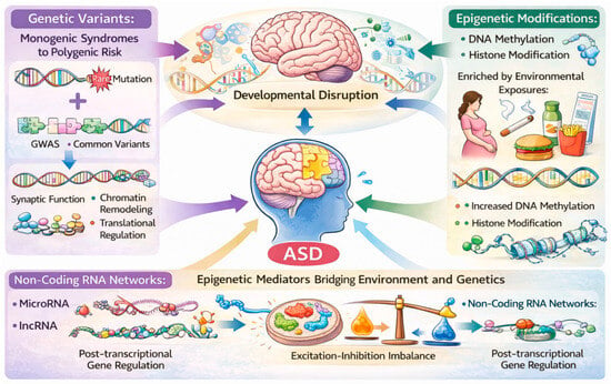
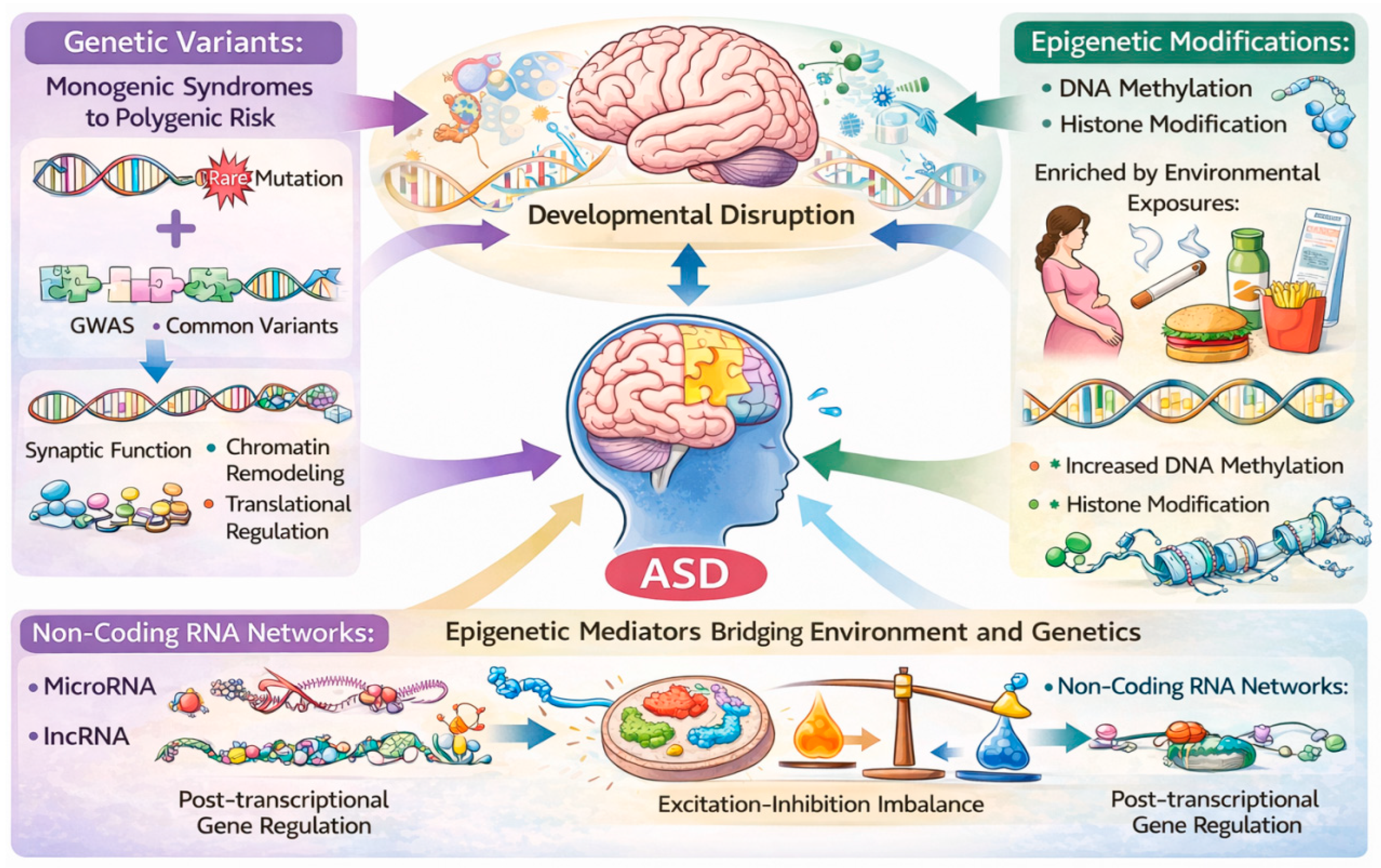
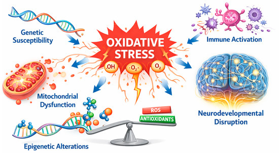
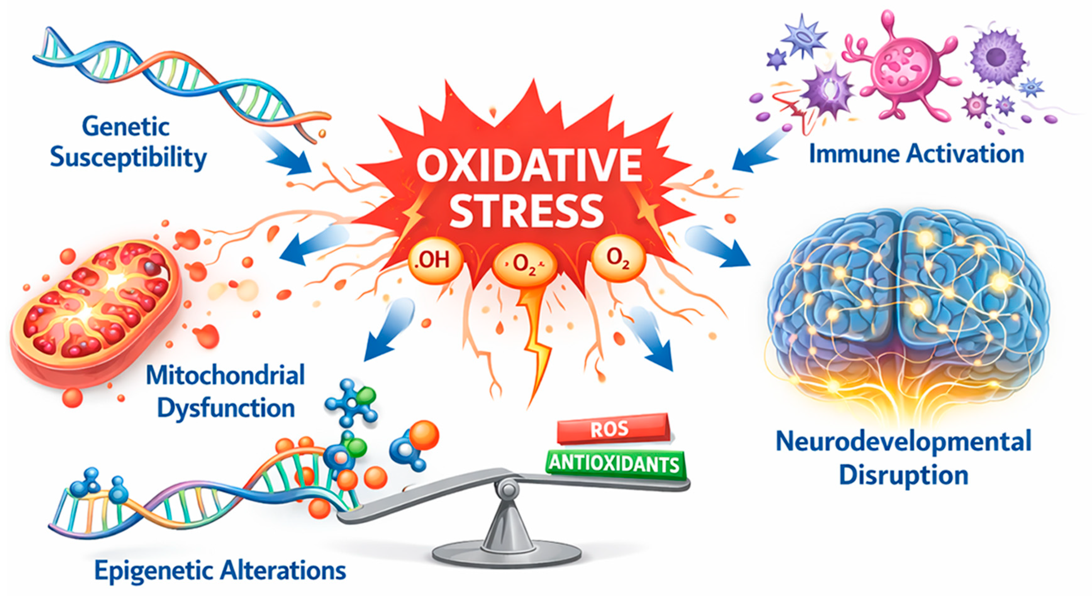
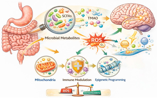
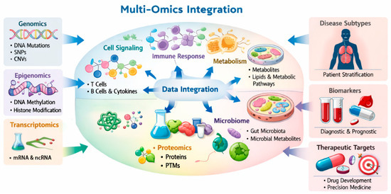
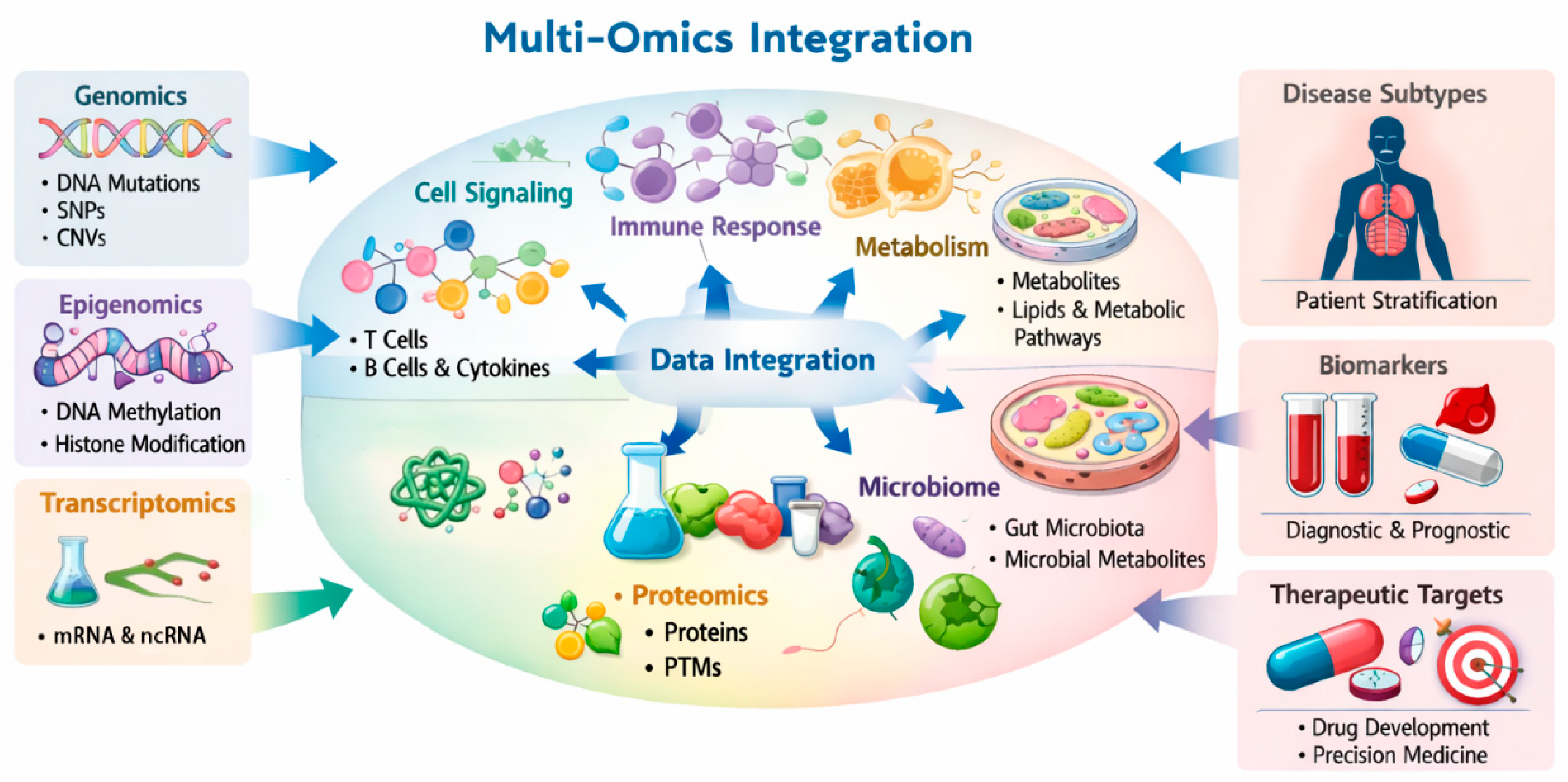

Disclaimer/Publisher’s Note: The statements, opinions and data contained in all publications are solely those of the individual author(s) and contributor(s) and not of MDPI and/or the editor(s). MDPI and/or the editor(s) disclaim responsibility for any injury to people or property resulting from any ideas, methods, instructions or products referred to in the content. |
© 2026 by the authors. Licensee MDPI, Basel, Switzerland. This article is an open access article distributed under the terms and conditions of the Creative Commons Attribution (CC BY) license.