


Abstract
Alzheimer’s disease (AD) is now widely accepted as a complex disorder involving multiple interconnected pathological processes. Increasing evidence suggest that neuroinflammation and immune system dysregulation actively contribute to neurodegeneration, extending beyond the traditional view that the disease is driven solely by amyloid-β (Aβ) and tau pathology. With the advent of studies showing anti-amyloid antibodies to have only modest effects with significant toxicity, there has been a growing interest in investigating non-amyloid non-tau (NANT) treatment approaches. This review provided a general review of small molecules for neuroinflammation-based NANT therapies in AD. We reviewed several agents that affect the brain/immune axis, namely, kinase inhibitors (nelflamaimod and masitinib), NLRP3 inflammasome inhibitors (MCC950, selnoflast, and dapansutrile), TREM2 activators (VG-3927), gingipain inhibitors, PPARγ activators, KCa3.1 inhibitors, and repurposed drugs such as ambroxol and cromolyn. Some common drawbacks were identified for the drugs that failed to provide their anticipated effects. It was found that they all suffered from late-stage treatment, patient heterogeneity, inadequate central nervous system penetration, and poorly designed preclinical studies. Additional translational challenges included sex-specific differences in neuroimmune responses, APOE ε4-associated immune dysfunction, and the need for biomarker-guided patient selection. Although immune-targeted therapies remain strongly supported by biological evidence in AD, meaningful clinical progress will likely require earlier intervention strategies guided by biomarkers and therapies capable of targeting multiple disease pathways simultaneously.
1. Introduction
Alzheimer’s disease (AD) continues to be a major global health concern, affecting millions of older adults and creating growing challenges for healthcare systems, caregivers, and society. According to recent statistics, more than 7.4 million Americans aged 65 years and older are living with Alzheimer’s dementia, while the annual cost of care is expected to reach nearly $409 billion [1]. Unfortunately, after decades of extensive research in this field, effective treatments that would be capable of changing the clinical course of AD are still scarce. The initial approaches to AD research were based on the amyloid cascade hypothesis, which suggested that the aggregation of β-amyloid peptides initiated all subsequent stages of the pathophysiological process, including the appearance of pathological alterations in tau proteins, synapses, and neurons [2]. Recent findings indicated that inflammation and immune dysregulation might occur earlier in the brain, possibly at the very first stages of development preceding cognitive impairment and actively participating in AD pathogenesis [3,4]. Current knowledge on the pathology of AD indicates that the disease is formed by several mechanisms acting together. Apart from amyloid-β and tau, a range of other factors like oxidative stress, mitochondrial abnormalities, damage to the blood–brain barrier, and alteration in signaling pathways involving immune system molecules might contribute to the AD progression [5,6,7,8]. The limited efficiency of interventions targeting only amyloid or tau proteins prompted researchers to investigate other aspects of disease etiology and progression mechanisms. Current Food and Drug Administration (FDA)-approved treatments, including acetylcholinesterase inhibitors (donepezil, rivastigmine, galantamine) and the NMDA receptor antagonist memantine, mainly provide symptomatic relief. They do not stop disease progression [9]. More recently, anti-amyloid antibodies such as lecanemab and donanemab showed modest clinical benefits, but they also carry notable side effects [10,11]. Even with these therapies, cognitive decline was only moderately slowed (around 27–35%), suggesting that Aβ is not the sole driver of AD pathology [12,13,14,15,16]. Adding to this complexity, only about 10% of patients exhibit “pure” AD, while the vast majority have mixed pathologies [17]. The disconnect between amyloid removal and meaningful clinical improvement showed AD complexity and suggested that no single-pathway model could fully explain its progression. In addition to AD, Aβ has been implicated in many other diseases such as type 2 diabetes, heart disease, retinal neurodegeneration, traumatic brain injury, and Parkinson’s disease [18,19,20], which again showed the complexity involved in Aβ biology and the inadequacy of target-based therapies.
Due to these shortcomings, neuroinflammation and immune dysregulation has become an area of growing interest within the NANT approach. Activation of microglia and astrocytes, oxidative stress, the destruction of the BBB, and changes in the peripheral immune system all have an impact on the development of neuronal injury in the case of AD [21,22]. Over time, these processes result in a chronic inflammatory process that affects synaptic transmission and reduces neuroplasticity [23]. The importance of immunological components for the development of AD was evidenced by results obtained during genetic studies. There were multiple risk genes associated with the immune system, such as TREM2, cluster of differentiation 33 (CD33), complement receptor 1 (CR1), and APOE [24,25]. Dementia in elderly patients is always accompanied by various types of diseases, such as vascular injury, TDP-43 accumulation, metabolic changes, and chronic inflammation. As a result, treatments targeted exclusively to amyloid and tau pathology are unlikely to provide desired results due to multiple coexisting pathogenic processes [26,27,28]. Due to this complexity, small molecules to target neuroinflammation gained increasing attention as potential NANT therapeutics. These compounds are attractive because of their low molecular weight (<900 Da), oral bioavailability, BBB penetration, and affordability. They also offer the potential to alter multiple interconnected disease pathways concurrently and thus might provide broader neuroprotective effects. Their structures could further be improved to enhance efficacy and reduce toxicity.
In this review, the focus was on small-molecule neuroinflammatory inhibitors within the context of the NANT theory, particularly in terms of the mode of action and the treatment potential of these compounds. In addition, the ability of these compounds to tackle the multifaceted nature of the pathogenesis associated with AD was also explored. The compounds discussed were selected to provide a representative overview of small-molecule strategies targeting different components of the brain–immune axis. Accordingly, both clinically advanced candidates and selected preclinical compounds were included. Attention was also given to translational challenges that may influence therapeutic success, including patient heterogeneity, apolipoprotein E (APOE ε4)-associated immune dysfunction, sex-specific differences in neuroimmune responses, biomarker-guided patient selection, and the potential role of combination therapies. While clinical-stage compounds offer important translational insights, early-stage compounds were incorporated because they target emerging neuroimmune pathways and provide mechanistic evidence that may inform future therapeutic development.
2. Methods
This article was prepared as a narrative review. The relevant literature was identified through searches of PubMed, Scopus, Web of Science, and ClinicalTrials.gov up to June 2026. Search terms included combinations of “Alzheimer’s disease”, “neuroinflammation”, “immune dysregulation”, “brain–immune axis”, “microglia”, “inflammasome”, “NLRP3”, “TREM2”, “small molecules”, “non-amyloid non-tau”, “NANT”, “kinase inhibitors”, “PPARγ agonists”, “gingipain inhibitors”, “KCa3.1”, and “Alzheimer’s clinical trials”. Additional articles were identified through citation tracking of relevant publications. Priority was given to peer-reviewed studies, clinical trials, and recent reviews relevant to neuroinflammation-targeted therapeutic strategies in AD.
3. The Brain–Immune Axis in AD
The brain was considered an immunologically privileged organ for a long time since it was relatively safe due to its location behind the BBB, which restricted the entry of peripheral immune cells. While immune surveillance was thought to rely primarily on resident cells within the central nervous system, this understanding has changed considerably over the past decade. It is now established that the brain actively communicates with the peripheral immune system via multiple pathways, such as meningeal lymphatic vessels, perivascular spaces, and circumventricular organs [29,30]. Collectively, these anatomical sites have come to be known as the brain–immune axis [31].
In the healthy brain, microglia constantly sense the environment for any signs of distress, while astrocytes help maintain synaptic function and regulate inflammatory signaling [32]. Under normal circumstances, these immune responses are tightly controlled and get resolved once the threat is removed [33]. In AD, however, there is sustained activation of microglia due to long-term exposure to misfolded proteins, neuronal trauma, and metabolic stress, which results in an overproduction of inflammatory cytokines like interleukin-1 beta (IL-1β), interleukin-6 (IL-6), and tumor necrosis factor alpha (TNF-α), causing synaptic dysfunction and neurodegeneration [34]. This inflammatory response is partly regulated by the TREM2 signaling pathway, as its dysfunction impairs these responses, resulting in reduced clearance of toxic proteins and worsening chronic neuroinflammation [35]. As AD is marked by BBB dysfunction, peripheral immune cells such as monocytes, CD8+ T cells, and natural killer cells migrate to the brain and exacerbate neuroinflammation [36]. Additionally, the malfunctioning of the meningeal lymphatic system hinders the removal of inflammatory molecules and protein aggregates from the brain, leading to their accumulation [37]. This causes a feedback loop involving chronic inflammation, impaired waste clearance, and progressive neurodegeneration. Additionally, systemic inflammation also impacts neuroinflammation in AD [38]. The peripheral immune cells, which get activated due to the systemic inflammation, attain a more pro-inflammatory state when entering the brain tissue, thus causing even more damage to neurons [39,40]. Thus, further research into brain–immune communication is essential for developing new AD therapies. In AD, neuroinflammation develops through continuous interaction between activated microglia, reactive astrocytes, and infiltrating immune cells. Together, these processes maintain a chronic inflammatory state that eventually harms synapses and neurons [23]. These mechanisms are closely linked, so targeting only amyloid or tau is unlikely to stop disease progression. This shifted the focus toward inflammatory pathways within the NANT approach. Small molecules offered one possible strategy. They could modulate microglial activity, stabilize mast cells, and inhibit kinase signaling. Importantly, they do this without directly targeting amyloid or tau pathology.
The brain–immune axis comprises a complex network of interactions between resident central nervous system cells, peripheral immune components, and systemic regulatory pathways that collectively influence brain homeostasis and disease progression. In AD, dysregulation of this network contributes to chronic neuroinflammation and neurodegeneration. While microglial activation has received considerable attention, growing evidence indicate that additional mechanisms, including complement signaling, gut–brain communication, and adaptive immune responses, also play important roles in shaping the neuroimmune landscape of AD [41,42,43,44].
3.1. Complement-Mediated Immune Dysregulation in AD
The complement system has emerged as an important contributor to neuroinflammation and immune dysregulation in AD. Beyond its classical role in innate immunity, complement signaling participates in synaptic remodeling and clearance of cellular debris within the central nervous system [45]. However, excessive or chronic activation of complement pathways may promote pathological synapse elimination and neurodegeneration. Genetic studies identified complement receptor 1 (CR1) as a risk factor for late-onset AD, supporting complement dysregulation in disease susceptibility [46]. In addition, clusterin (CLU, apolipoprotein J), another major AD risk gene, regulates complement activation and may influence amyloid-β aggregation and clearance [47,48]. Increasing evidence suggested that aberrant complement activation contributed to microglial-mediated synaptic loss and sustained neuroinflammatory responses, highlighting the complement cascade as an important component of the brain–immune axis in AD [45,49].
3.2. Gut–Brain Axis and Neuroinflammation
Growing evidence indicate that alterations in the gut microbiota could influence AD pathogenesis through bidirectional communication between the gastrointestinal tract and the central nervous system [50,51]. Gut dysbiosis promotes systemic inflammation, alters the production of microbial metabolites, and affects immune signaling pathways that regulate microglial function. Increased intestinal permeability further facilitates the translocation of inflammatory mediators into the circulation, thereby amplifying neuroinflammatory responses [52,53]. Associations between microbial imbalance and cognitive decline have been observed, suggesting a potential role of the gut–brain axis in AD progression [54,55,56,57]. Although the precise mechanisms remained under investigation, modulation of the gut microbiome emerged as a promising strategy for regulating neuroinflammation and restoring immune homeostasis in AD [58].
3.3. Adaptive Immunity and CD8+ T-Cell Infiltration
Although AD has traditionally been viewed as a disorder driven primarily by innate immune mechanisms, increasing evidence has supported the involvement of adaptive immune responses [59,60]. Infiltrating CD8+ T cells were identified within the brains of patients with AD, particularly in regions associated with neurodegeneration [61,62]. These cells may have contributed to disease progression through the release of pro-inflammatory cytokines and cytotoxic mediators that exacerbate neuronal injury [63]. Furthermore, interactions between infiltrating T cells, microglia, and other immune cells may have amplified neuroinflammatory signaling within the brain [64,65]. The recognition of adaptive immune involvement expanded the concept of the brain–immune axis beyond microglial activation alone and suggested that both innate and adaptive immune pathways contributed to AD pathogenesis [66].
Collectively, these findings emphasized the multifaceted nature of immune dysregulation in AD. Therefore, effective therapeutic strategies might need to target multiple interconnected pathways rather than a single inflammatory mechanism.
4. Neuroinflammation-Targeting Small Molecules
Early attention focused on the potential role of non-steroidal anti-inflammatory drugs (NSAIDs) in AD prevention. Long-term use of NSAIDs was repeatedly linked to decreased AD risk, suggesting a link between cyclooxygenase (COX)-dependent neuroinflammation and neurodegeneration [67,68]. Mechanistically, this association is attributed to the role of COX enzymes in regulating prostaglandin-mediated neuroinflammation [69]. COX-2 is induced in neurons, astrocytes, and activated microglia in response to Aβ and inflammatory stimuli, resulting in increased prostaglandin production that promotes microglial activation, inflammatory cytokine release, and neuronal injury. These inflammatory mediators also impair microglial Aβ clearance, thereby contributing to disease progression [70]. However, because conventional NSAIDs non-selectively inhibited both COX isoforms, they broadly suppressed prostaglandin signaling rather than selectively targeting the detrimental inflammatory pathways [71]. Consequently, the promising epidemiological findings were not translated into clinical benefit. The AD Anti-inflammatory Prevention Trial (ADAPT), together with clinical trials of ibuprofen, rofecoxib, and indomethacin, failed to demonstrate meaningful cognitive benefit in symptomatic patients [72,73]. This lack of efficacy suggested that broad COX inhibition alone was insufficient to address the complex neuroimmune mechanisms underlying AD and highlighted the need for more selective NANT approaches targeting the brain–immune axis [73]. Some of the important small molecules targeting neuroinflammation are discussed below (Figure 1).
Figure 1.
Some important small molecules targeting neuroinflammation. [Chemical structures were prepared using ChemDraw Pro 12.0 based on compound structures obtained from the PubChem database].
4.1. Kinase Inhibitors
Kinase inhibitors are one of the most advanced small-molecule drugs targeting neuroinflammation in AD. They disrupt intracellular signaling pathways that lead to microglial activation and cytokine secretion. Neflamapimod (VX-745) selectively inhibits p38α mitogen-activated protein kinase (p38α MAPK) with a 9 nM IC50 (50% inhibitory concentration) value. p38α MAPK is the crucial enzyme in the inflammatory signaling pathways of microglia and neurons [74,75]. Excessive activation of p38α MAPK in AD increases TNF-α and other pro-inflammatory cytokines, contributing to neuroinflammation, synaptic dysfunction, and impaired endosomal recycling and long-term potentiation [76]. By p38α MAPK inhibition, neflamapimod promoted microglial homeostasis and synaptic preservation in preclinical and randomized clinical evaluations [77]. In a completed Phase 2b study in patients with Lewy body dementia (LBD), a disease characterized by neuroinflammation similar to that of AD, neflamapimod led to robust cognitive and neurodegeneration biomarker benefits [78], supporting the validity of p38α MAPK as a drug target until further clinical testing in AD.
In contrast, masitinib adopts a broader anti-inflammatory approach by inhibiting several tyrosine kinases, including the receptor tyrosine kinases c-Kit (CD117) and platelet-derived growth factor receptor (PDGFR), as well as the Src-family non-receptor tyrosine kinase Lyn, in both microglia and mast cells [79,80]. c-Kit regulates mast cell development, survival, and activation, whereas Lyn mediates downstream signaling that triggers mast cell degranulation and the release of pro-inflammatory mediators. By simultaneously inhibiting c-Kit and Lyn, masitinib suppresses both mast cell activation and the subsequent inflammatory cascade, providing broader immunomodulatory effects than targeting a single downstream mediator [79]. Inflammation in the brain is triggered by mast cells present in the meninges and perivascular areas, which releases factors leading to activation of microglia and destruction of the BBB [81]. Thus, by stabilizing both mast cells and microglia simultaneously, masitinib offers a dual-acting anti-neuroinflammatory profile. Masitinib broadly addresses the neuroinflammatory relevance of tyrosine kinase inhibition across neurodegenerative conditions like multiple sclerosis (MS) [82] and amyotrophic lateral sclerosis (ALS) [83].
Masitinib has been evaluated as an add-on to standard therapies, including cholinesterase inhibitors and/or memantine, in patients with mild-to-moderate AD. In a Phase 3 clinical trial, significant improvements in cognition and daily functioning, assessed using the Alzheimer’s Disease Assessment Scale-Cognitive Subscale (ADAS-cog), were observed in a pre-specified subgroup of patients, whereas the primary endpoint was not met in the overall intention-to-treat population [84,85]. Subsequently, the U.S. Food and Drug Administration (FDA) Peripheral and Central Nervous System Drugs Advisory Committee voted against approval in 2023 because of concerns regarding the statistical analysis and subgroup selection. Yet, AB Science continued to evaluate masitinib as an add-on therapy in patients with mild AD [86]. Although masitinib demonstrated encouraging findings in selected patients, its clinical efficacy requires further confirmation in adequately designed studies [85].
4.2. NLRP3 Inflammasome Inhibitors
The NLRP3 inflammasome is one of the more discussed inflammasome pathways in AD research [87]. Under physiological conditions, it helps immune cells respond to stress and cellular damage. In AD, this balance is disturbed. Aβ accumulation, mitochondrial dysfunction, and lysosomal stress all seem to push the system into a persistent activated state. Once activated, NLRP3 promotes the release of pro-inflammatory cytokines such as IL-1β and IL-18, which aggravates neuronal injury [87]. Importantly, abnormal NLRP3 activation was also detected in peripheral immune cells such as monocytes and macrophages, indicating that inflammation outside the brain may further influence neuroinflammatory processes in AD [40,88].
These findings heightened interest in NLRP3 as a therapeutic target. Among the available inhibitors, MCC950 was the first compound to show strong preclinical efficacy in streptozotocin (STZ)-treated SHSY-5Y cells and rat models with AD [89]. It selectively blocks NLRP3 activation by binding to the Walker B motif of the NLRP3-NACHT domain. This blocks ATP hydrolysis and prevents inflammasome assembly [90]. In transgenic AD mice, MCC950 reduced amyloid burden, enhanced microglial phagocytosis, restored autophagy, and improved cognitive performance [89,91,92,93]. Despite these promising findings, MCC950 did not advance into clinical trials for AD. Safety concerns, particularly dose-dependent hepatotoxicity observed in preclinical trials, represented a major limitation [94]. Even so, the compound became a reference point for the field. New inhibitors were designed using its scaffold and binding behavior as a template. CY-09 is one example. It showed selectivity, BBB penetration, and anti-inflammatory effects in preclinical systems [95,96,97]. Another NLRP3 inhibitor, emlenoflast (inzomelid; IZD174) had reached early clinical evaluation. A Phase 1 single ascending dose/multiple ascending dose (SAD/MAD) trial involving 80 healthy volunteers and an open-label preliminary study in patients with cryopyrin-associated periodic syndromes (CAPS) reported acceptable safety and tolerability [98]. Dose-dependent target engagement was also suggested, although full results were not published.
Selnoflast is currently one of the most advanced purpose-designed NLRP3 inhibitors in clinical development. A Phase 1b trial of selnoflast in early PD enrolled 60 participants across 17 centers in Europe and the United States and evaluated 28 days of treatment versus placebo. The drug was generally well tolerated, with headache and nausea being the most common adverse events, and no serious adverse events were reported. Importantly, selnoflast demonstrated target engagement by reducing IL-1β production in peripheral blood cells and IL-18 levels in the cerebrospinal fluid (CSF) by approximately 30%. Although exploratory clinical outcomes showed slight improvement in treated patients compared with the placebo, the differences were not statistically significant [99]. Roche is currently evaluating selnoflast for reducing vascular inflammation associated with atherosclerosis [100].
OLT1177 (dapansutrile), a β-sulfonyl nitrile compound, inhibits ATPase activity, thereby preventing NLRP3 assembly and reducing production of downstream pro-inflammatory cytokines [101]. In APP/PS1 AD mouse models, it reduced hippocampal IL-1β, attenuated microglial activation, and improved spatial memory through a mechanism independent of amyloid or tau [102]. It also improved motor functions and reduced α-synuclein toxicity and inflammation in synucleinopathy with nigral neurodegeneration PD models [103]. OLT1177 has completed Phase 2 trials in gout and heart failure, establishing a human safety record [104]. Currently, it is being evaluated as an anti-inflammatory therapy in PD [105].
Tranilast (TR), originally developed as an antiallergic drug, was found to directly inhibit NLRP3 by blocking its oligomerization through interaction with the NACHT domain [106,107]. Its established clinical safety profile makes it an attractive repurposed candidate for neuroinflammatory disorders. TR reduced NLRP3-dependent IL-1β release and exerted anti-inflammatory effects in preclinical experiments [106,108,109,110]. TR can modulate adaptive immune responses by suppressing B- and T-cell proliferation, inducing T-cell cycle arrest, and regulating the T helper cells (Th1/Th2) balance [108]. It also reduced the release of inflammatory mediators such as interferon gamma (IFN-γ), IL-2α, and chemokine (CXC motif) ligand 9/10 (CXCL9/10) and suppressed pathogenic T-cell responses in autoimmune disease models, including rheumatoid arthritis (RA) and MS [111,112,113]. Still, despite modest CNS penetration, oral bioavailability, and a suitable safety profile [107], tranilast did not enter AD clinical trials, and its relatively lower potency compared with newer NLRP3 inhibitors suggested that further optimization may be needed to achieve sufficient brain target engagement.
Oridonin (ori), a natural diterpenoid from Rabdosia rubescens, covalently binds cysteine 279 on NLRP3, blocks NEK7 recruitment and prevents apoptosis-associated speck-like protein containing a CARD (ASC) adaptor assembly [114]. Ori was found to exert multiple neuroprotective activities on an Aβ1–42-induced mouse model of AD. It reduced Aβ1–42-induced synaptic loss both in vivo and in vitro. Ori also enhanced the formation of dendrites and spines in hippocampal neurons in the same AD model. In addition, Ori promoted the expression of synaptic proteins and activated the brain-derived neurotrophic factor/tyrosine receptor kinase b/cyclic AMP (cAMP) response element-binding (BDNF/TrkB/CREB) signaling pathway, important for synaptic plasticity and memory. The molecular effects observed included enhanced cognitive functions [115]. In 5× FAD transgenic mice models, ori attenuated the production of pro-inflammatory cytokines in microglia by inhibition of the receptor-interacting protein kinase 1-extracellular signal-regulated kinase 1/2-nuclear factor kappa B (RIPK1-ERK1/2-NF-κB) signaling pathway, which plays a key role in regulating inflammation and cell survival [116]. Furthermore, ori improved spatial learning and memory, reduced Aβ plaque deposition, and attenuated inflammatory and necroptotic markers through suppression of RIPK1-RIPK3-MLKL-mediated signaling in 5× FAD mice [116]. Moreover, ori also reduced neuronal loss in ischemic stroke models by inhibition of RIPK3-mediated excessive mitophagy, further supporting its neuroprotective potential [117]. Even though there was strong evidence of neuroprotective activity, no clinical trial of Ori was registered for any neurological disorder. The clinical development of oridonin was limited by its poor bioavailability and unfavorable pharmacokinetic properties, highlighting the need for improved delivery systems.
JC-171 is a structure-based small-molecule NLRP3 inhibitor that targets the leucine-rich repeat domain of NLRP3, giving it a distinct selectivity profile compared with many other inhibitors in this class [118]. JC-171 delayed disease progression and reduced severity in experimental autoimmune encephalomyelitis (EAE), a mouse model of MS, in both preventive and therapeutic settings. These effects were associated with reduced IL-1β production and suppression of pathogenic Th17 immune responses [118].
NBC6 is an oxanorbornene molecule developed as a selective NLRP3 inhibitor identified through high-throughput screening [119]. In cellular neuroinflammation models, NBC6 reduced ASC speck formation, caspase-1 activation, and IL-1β maturation [119,120]. Although mechanistically promising, in vivo evidence in neurodegenerative models remain limited.
4.3. TREM2 Agonists
Interest in TREM2 grew after rare TREM2 variants were linked to a higher risk of late-onset AD, highlighting the important role of immune-cell dysfunction in disease pathogenesis [121,122,123]. TREM2 is mainly expressed by microglia in the brain and modulates cell survival, phagocytosis, metabolism, and the transformation of cells into beneficial disease-associated microglia responsible for amyloid removal and synapse support [124,125]. Defective TREM2 signaling leads to inefficient amyloid removal, abnormal inflammatory modulation, poor cell survival, and inadequate protective mechanisms near amyloid plaques [124]. Consequently, the brain’s immune response becomes increasingly inefficient, leading to chronic neuroinflammation and neurodegeneration.
Small-molecule TREM2 agonists aimed to restore microglial phagocytosis, survival, and anti-inflammatory signaling through pathways such as phosphoinositide 3-kinase/protein kinase B (PI3K/AKT) and DNAX activation protein 12/spleen tyrosine kinase (DAP12/SYK) [126,127]. These compounds were able to overcome some limitations associated with antibody-based therapies, particularly poor brain penetration, and were associated with improved microglial signaling, enhanced phagocytosis, and synaptic protection in cellular studies [126,128]. VG-3927 is currently the most clinically advanced small-molecule TREM2 agonist, having recently completed a Phase 1 clinical trial with positive results that demonstrated acceptable safety, tolerability, and preliminary target engagement [129,130]. This compound is advancing into Phase 2 development [130]. Preclinical compounds such as C1 and S9 showed the ability to activate TREM2 signaling and enhanced microglial phagocytosis through different mechanisms, supporting their potential for further development [126,128,131].
Other than VG-3927, C1, and S9, research on the small-molecule TREM2 agonist is still at an early stage. Researchers are increasingly applying structure-based virtual screening and AI-assisted drug discovery to identify compounds targeting the TREM2 ligand-binding domain, but only a limited number of molecules have been characterized so far in AD-relevant models. In addition, the field still lacks reliable pharmacodynamic biomarkers to confirm whether TREM2 agonists are producing sufficient microglial activation in the human brain, making dose optimization and clinical trial design particularly challenging [132].
4.4. Gingipain Inhibitors
An emerging hypothesis on AD suggests that chronic oral infection with Porphyromonas gingivalis might contribute to neuroinflammation and neurodegeneration through bacterial proteases known as gingipains. Gingipains were detected at higher levels in postmortem AD brains and were shown to damage neuronal proteins, activate complement pathways, stimulate microglial inflammation, and impair the clearance of neurotoxic proteins [133,134]. Thus, chronic peripheral infection could lead to sustained activation of the immune system and neuroinflammation via BBB disruption and microglial priming, making gingipain inhibition an effective NANT therapy [135].
Atuzaginstat (COR388) was developed as a selective small-molecule inhibitor of P. gingivalis gingipain proteases and showed promising preclinical effects, including reduced neuroinflammation, microglial activation, and bacterial burden [136]. However, the Phase 2/3 GAIN trial failed to meet its primary cognitive endpoints and showed hepatotoxicity at higher doses [137]. A second-generation compound, COR588 (later developed clinically as LHP588), was subsequently developed with improved metabolic stability and reduced hepatic exposure and is currently recruiting for Phase 2 evaluation in AD [138] after successful Phase 1 results [139].
4.5. PPARγ Agonists
Peroxisome proliferator-activated receptor gamma (PPARγ) is a nuclear receptor involved in both metabolic regulation and immune signaling, making it an important target in AD [140,141]. In microglia and peripheral macrophages, PPARγ activation promotes an anti-inflammatory state by reducing NF-κB-driven cytokine production, enhancing phagocytic clearance, and supporting BBB integrity [141]. PPARγ is also linked to brain insulin signaling, which is frequently impaired in AD and contributes to neuroinflammation, synaptic dysfunction, and reduced neuronal energy metabolism [142,143].
Pioglitazone and rosiglitazone, two thiazolidinedione-class PPARγ agonists, were originally approved for type 2 diabetes (T2D). They were later investigated for AD treatment because of their anti-inflammatory and insulin-sensitizing properties. Reduced neuroinflammation, improved mitochondrial function, and neuroprotective effects were seen in preclinical models [144,145]. However, these benefits were not clearly reproduced in clinical studies, which showed little or no significant improvement in cognition or overall function in patients with AD [146,147]. One major limitation was poor effective brain exposure. Pioglitazone was actively transported out of the brain by P-glycoprotein (P-gp) efflux transporters at the BBB, whereas rosiglitazone showed even lower brain penetration [148].
Leriglitazone (MIN-102) was developed to address the poor brain penetration observed with earlier PPARγ agonists [149]. Its chemical structure was modified to reduce P-gp-mediated efflux, enabling improved BBB penetration in human adrenoleukodystrophy (ALD) and Friedreich’s ataxia [149,150,151]. In the preclinical setup, leriglitazone reduced microglial activation, decreased pro-inflammatory cytokine release, and improved mitochondrial function through PPARγ-dependent pathways [152]. Leriglitazone-mediated PPARγ activation might support axonal myelination, protect neurons and astrocytes, and help preserve BBB integrity [153,154,155]. Although no AD-specific clinical trial was reported, leriglitazone remains the only PPARγ agonist with confirmed human brain penetration, making it a promising candidate for future neurodegenerative disease studies [154].
Lobeglitazone is a thiazolidinedione approved for T2D in South Korea [156] and had shown a cardioprotective effect in T2D patients with ischemic stroke [157]. Lobeglitazone also has a favorable long-term safety profile, effective glucose-lowering activity, and durable glycemic control in real-world clinical settings [158,159]. It had shown potential neuroprotective effects in intracerebral hemorrhage (ICH) models by reducing brain edema and neuroinflammation through inhibition of the IL-1β-ERK-COX-2 signaling pathway [160]. In addition, lobeglitazone partially improved motor defficits and reduced inflammatory markers such as TNF-α and NF-κB in a diabetic rat model of PD [161], further supporting its anti-inflammatory and neuroprotective potential.
However, no neurological clinical trials have evaluated lobeglitazone in humans.
4.6. KCa3.1 Channel Inhibitors
Senicapoc is an extremely potent small-molecule inhibitor of the calcium-activated potassium channel known as KCa3.1 (or Gardos channel and KCNN4) [162]. KCa3.1 is expressed on microglia, T cells, mast cells, and peripheral macrophages and plays an important role in immune-cell migration and inflammatory signaling [163]. Mechanistically, KCa3.1-mediated K+ efflux maintains the membrane potential required for sustained Ca2+ influx during immune-cell activation [164]. The resulting increase in intracellular Ca2+ activates downstream inflammatory signaling pathways that promote the production of reactive oxygen species, nitric oxide, and pro-inflammatory cytokines in microglia [165]. Therefore, pharmacological inhibition of KCa3.1 interrupts this calcium-dependent inflammatory cascade while largely preserving normal immune-cell viability and function [163]. In ischemic stroke models, senicapoc reduced infarct size, improved neurological recovery, and suppressed microglial activation, T-cell infiltration, and inflammatory mediators, including inducible nitric oxide synthase (iNOS), COX-2, and NLRP3 [166,167]. The compound also demonstrated sufficient brain target engagement and did not interfere with tissue plasminogen activator (tPA) activity, supporting its potential as an adjunctive neuroprotective therapy [167]. Across multiple clinical studies involving hundreds of patients across the United States, senicapoc was found to be orally available, metabolically stable, safe, and well tolerated [162]. In 5x FAD mice models, senicapoc reduced microglial activation, neuroinflammation, and cerebral amyloid burden while improving hippocampal synaptic plasticity. KCa3.1 activity was also increased in postmortem AD brains and 5x FAD mice, further supporting this channel as a potential therapeutic target in AD [168]. At present, the drug is in a Phase 2 clinical trial in patients with mild or prodromal AD [169].
4.7. Emerging and Repurposed Neuroimmune Modulators
4.7.1. MAPK/ERK Signaling Modulators
Bezisterim (NE3107) is a novel agent with a unique structure that functions as a metabolic and anti-inflammatory modulator without producing any of the side effects caused by full PPARγ agonists, such as fluid retention and fat production. It mainly serves as an ERK signaling inhibitor, resulting in a reduction in the production of pro-inflammatory mediators like TNF-α and IL-6 and improved insulin signaling [170]. Mechanistically, chronic ERK activation impairs insulin receptor substrate-1 (IRS-1) signaling, contributing to both neuronal insulin resistance and neuroinflammation [171]. By inhibiting ERK, NE3107 restores insulin signaling while reducing NF-κB-mediated inflammatory cytokine production, thereby targeting two interconnected pathological processes implicated in AD [172]. In a Phase 2 clinical study for AD, NE3107 exhibited promising results [170,173], and its combined metabolic and anti-inflammatory actions have also generated interest in the treatment of other neurodegenerative disorders, including PD [174].
4.7.2. Lysosomal Dysfunction/GCase Enhancement
Ambroxol (ABX), widely used in respiratory medicine, has recently gained attention in neurodegeneration research because of its ability to enhance glucocerebrosidase (GCase) activity [175]. This is particularly relevant in AD, where impaired lysosomal function and reduced GCase activity in microglia can promote NLRP3 inflammasome activation, defective autophagy, and chronic neuroinflammation [176,177,178]. By improving lysosomal homeostasis, ABX reduces upstream drivers of microglial dysfunction and toxic protein accumulation [179,180,181]. ABX enhances GCase activity and promotes autophagy-lysosomal clearance of α-synuclein, supporting its potential disease-modifying effects [182]. Based on these findings, the ABX in new and early Dementia with Lewy Bodies (ANeED) trial was launched to evaluate the safety, tolerability, and potential therapeutic benefits in patients with DLB [183]. In the AiM-PD Phase 2 trial, ABX showed good CNS penetration, increased CSF GCase activity, and was well tolerated during long-term treatment. [184,185]. At present, it is gearing up for its Phase 3 trial [186].
4.7.3. Microglial Modulation and Aβ Clearance
Another repurposing strategy focused on targeting peripheral immune signaling and mast cell-mediated neuroinflammation. Cromolyn sodium was originally developed as an antiallergic drug for asthma. It was later investigated for AD treatment because of its ability to modulate peripheral immune responses and neuroinflammation [187,188]. In preclinical models, cromolyn reduced Aβ42 burden by promoting non-inflammatory microglial clearance of Aβ. It also lowered soluble Aβ40 and Aβ42 levels and improved neuroprotection in transgenic AD mice [189,190,191]. The compound reduced the release of inflammatory cytokines and chemokines, including IL-1β, IL-6, IL-8, IFN-γ, CCL2, and CXCL10, in activated human microglial cells [187]. Modified derivatives such as fluorinated cromolyn were also developed to improve BBB penetration while retaining anti-inflammatory activity [192]. Based on these findings, cromolyn entered a randomized 1/2 Phase study of the ALZT-OP1 combination therapy in AD [193]. It demonstrated good safety and tolerability but failed to meet its primary cognitive endpoint in the overall study population.
4.7.4. p38α MAPK Inhibition
MW150 (MW01-18-150SRM) is a brain-penetrant and highly selective p38α mitogen-activated protein kinase (MAPK) inhibitor developed from the earlier lead compound MW01-2-069A-SRM [194]. Unlike amyloid- or tau-directed therapies, this drug discovery program targeted p38α MAPK, a key regulator of glial activation and pro-inflammatory cytokine production. In preclinical studies, MW01-2-069A-SRM suppressed Aβ-induced upregulation of IL-1β and TNF-α in the hippocampus and attenuated associated synaptic dysfunction and behavioral deficits [195]. The optimized compound, MW150, reduced glial release of pro-inflammatory cytokines and prevented or reversed cognitive deficits in APP/PS1 and APP knock-in mouse models without altering amyloid plaque burden [196], supporting a mechanism independent of direct amyloid targeting. Following favorable safety and tolerability findings in Phase 1 studies, MW150 advanced to Phase 2a clinical evaluation in patients with mild-to-moderate AD [197].
A summary of the mechanisms, developmental progress, and neuroinflammatory relevance of these small molecules is presented in Table 1.
Table 1.
Small-molecule therapeutics in neuroinflammation: mechanisms of action and clinical development.
An overview of the neuroinflammatory cascade and the NANT small-molecule classes acting at each node is provided in Figure 2.
Figure 2.
Neuroinflammatory cascade in Alzheimer’s disease and the non-amyloid, non-tau (NANT) small-molecule classes targeting the brain–immune axis. Upstream triggers: misfolded protein accumulation (Aβ, tau) and lysosomal stress, peripheral and systemic immune signals including Porphyromonas gingivalis infection, and metabolic stress arising from insulin resistance converge on chronic microglial activation, the central hub of the cascade. Activated microglia drive three interconnected effector arms: assembly of the NLRP3 inflammasome with release of IL-1β and IL-18; pro-inflammatory kinase signaling (p38α MAPK, receptor tyrosine kinases, and ERK); and astrocyte and mast-cell amplification. These converge on a sustained pro-inflammatory cytokine milieu (TNF-α, IL-6, IL-1β) and blood–brain barrier breakdown, culminating in synaptic loss, neurodegeneration, and cognitive decline. A self-perpetuating feedback loop (dashed arrow) returns from the downstream inflammatory output to microglial activation, sustaining chronic neuroinflammation and underlying the limited efficacy of late, single-target intervention. Small-molecule classes are positioned at their primary site of action: gingipain inhibitors and lysosomal modulators act on upstream triggers; TREM2 agonists and KCa3.1 channel blockers act at the microglial hub; NLRP3 inhibitors, kinase inhibitors, and PPARγ agonists act on the effector arms, with PPARγ agonists additionally relieving metabolic stress. Collectively, these interconnected processes represent key manifestations of immune dysregulation in AD. Arrowheads denote activating or restorative actions (TREM2 and PPARγ agonists); flat-ended connectors (⊣) denote inhibitory or blocking actions, the green arrows indicate activation. Abbreviations: Aβ, amyloid-β; BBB, blood–brain barrier; ERK, extracellular signal-regulated kinase; IL, interleukin; KCa3.1, intermediate-conductance calcium-activated potassium channel; MAPK, mitogen-activated protein kinase; NLRP3, NOD-, LRR- and pyrin domain-containing protein 3; PPARγ, peroxisome proliferator-activated receptor gamma; TNF-α, tumor necrosis factor-alpha; TREM2, triggering receptor expressed on myeloid cells 2. Figure created in Biorender.com.
5. Translational Challenges and Future Directions
Despite major advances in understanding the neuroimmune mechanisms underlying AD, the clinical translation of neuroinflammation-targeting small molecules remains inconsistent. Importantly, however, the many failures in this field do not necessarily invalidate the biological targets themselves but instead feature broader challenges related to the timing of therapeutic intervention, patient heterogeneity, CNS exposure, and translational model design. Additional considerations, including sex differences, APOE ε4 status, biomarker-guided patient selection, and the need for combination therapies, can further influence the successful translation of these approaches into clinical practice.
5.1. Timing of Therapeutic Intervention
Neuroinflammatory alterations begin many years before the onset of clinical symptoms, and by the time patients are enrolled in clinical trials, substantial synaptic and neuronal loss may already be irreversible. This issue was illustrated by early NSAID research, in which long-term NSAID use was associated with a lower risk of AD in epidemiological studies, whereas clinical trials in symptomatic patients failed to demonstrate meaningful therapeutic benefit [204,205]. This discrepancy was largely explained by a timing paradox (prevention vs. treatment) and reflects the fact that long-term NSAID users often begin treatment earlier in life, before AD-related pathological processes have fully developed [206]. Moreover, as the COX expression changes in the different stages of AD pathology [207], NSAIDs may benefit only during the earliest phases of AD, when initial Aβ deposition and microglial activation begins but lose efficacy once chronic neuroinflammation and amyloid pathology become established. At later stages, prolonged inhibition of activated microglia may even interfere with Aβ clearance mechanisms [208]. In addition, early observational findings were likely influenced by methodological biases, including healthier baseline characteristics in NSAID users and confounding related to disease stage-dependent prescribing patterns [206,209]. Similar limitations may have also contributed to the disappointing outcomes observed with pioglitazone and rosiglitazone [145]. The COR388 trial failed to meet its primary cognitive and functional endpoints and was additionally associated with hepatotoxicity [136]. Current evidence suggests that therapies targeting neuroinflammation may be more effective when introduced in the early or preclinical stages of AD, before significant neuronal loss occurs. This has increased interest in preventive and early-intervention strategies for high-risk groups, including cognitively normal APOE ε4 carriers.
5.2. Translational Limitations of Preclinical Models
One persistent challenge in AD research is the frequent failure of promising preclinical findings translating into successful clinical outcomes. Commonly used animal models, especially transgenic mice, reproduced only limited components of the disease and could not fully mimic the complexity of human immune and glial responses in the brain [210]. Because of these shortcomings, researchers started using alternative systems that might better reflect human biology. Models based on human induced pluripotent stem cells (iPSCs), including microglial cultures and brain organoids, are increasingly explored as tools for studying disease mechanisms and evaluating potential therapies [211,212]. Still, animal models remain the main platform for preclinical testing.
5.3. Patient Heterogeneity and the Need for Multi-Pathway Targeting
The inflammatory response observed in AD is highly complex and involves several overlapping biological processes rather than a single abnormal pathway. Activation of microglia, dysfunction of astrocytes, altered peripheral immune signaling, vascular injury, and metabolic abnormalities all contribute to disease progression and influence one another [4]. Since these mechanisms are interconnected, interventions aimed at only one pathway may have limited therapeutic impact. For this reason, future treatment strategies will likely need to target multiple components of the neuroimmune system simultaneously to achieve more meaningful clinical outcomes.
5.4. Sex Differences in Neuroinflammation and Treatment Response
Sex influences both neuroinflammatory responses and therapeutic outcomes in AD. Women account for approximately two-thirds of AD cases and exhibit differences in microglial activation, inflammatory signaling, and disease progression compared with men [213,214,215]. Hormonal changes, particularly the decline in estrogen during menopause, may further affect immune regulation in the brain [213,216]. Despite these observations, most NANT clinical trials had not reported sex-specific outcomes. Future studies are expected to incorporate sex-stratified analyses to determine whether treatment efficacy, safety, or dosing differ between men and women.
5.5. APOE ε4 Genotype and Innate Immune Dysfunction
The APOE ε4 allele is the strongest genetic risk factor for late-onset AD and plays an important role in neuroimmune regulation [217]. APOE ε4 is associated with impaired microglial function, reduced amyloid clearance, and increased inflammatory signaling [218,219]. As a result, APOE ε4 carriers may respond differently to neuroinflammation-targeting therapies than non-carriers [217]. Incorporating the APOE genotype into clinical trial design may improve patient stratification and help identify individuals most likely to benefit from specific NANT-based interventions.
5.6. Biomarker Strategies for Patient Selection and Trial Design
Biomarkers have become increasingly important for the development of neuroinflammation-targeting therapies. Fluid biomarkers such as GFAP, YKL-40, sTREM2, and inflammatory cytokines could provide insights into astrocyte and microglial activity, while imaging biomarkers might help assess disease burden and treatment response [220,221]. Biomarker-guided patient selection may improve trial efficiency by identifying individuals with prominent neuroinflammatory pathology. In addition, biomarkers could provide evidence of target engagement and support the evaluation of therapeutic efficacy.
5.7. Rationale for Combination Therapy
As AD is driven by multiple interconnected pathological processes, interventions targeting a single pathway often produce limited therapeutic benefits. Combination approaches involving neuroinflammation-targeting agents together with amyloid-, tau-, metabolic-, or vascular-directed therapies may offer a more comprehensive strategy by addressing multiple disease mechanisms simultaneously. Several combination approaches have already entered clinical evaluation. For example, the ALZT-OP1 program combined cromolyn with low-dose ibuprofen to target both Aβ pathology and neuroinflammation, while the DIAN-TU platform evaluated the combination of the anti-amyloid antibody lecanemab with the anti-tau antibody E2814, reflecting increasing interest in multi-target therapeutic strategies. These studies highlight the growing recognition that modifying multiple disease pathways simultaneously may provide greater therapeutic benefit than targeting a single mechanism alone. Such approaches are increasingly recognized as a promising direction for AD treatment, although challenges related to safety, trial design, and patient selection remain to be resolved.
5.8. Shift Toward Precision Neuroimmunology in Clinical Translation
Clinical translation remains another bottleneck. Many therapies that showed promise in preclinical studies failed to replicate their effects in patients. This stressed the need for better trial design. Earlier intervention, careful biomarker-based patient selection, and direct evidence of target engagement in the brain may improve future outcomes. The clinical implementation of combination therapies also presents several practical challenges. Combining neuroinflammation-targeting agents with anti-amyloid or anti-tau therapies may increase the risk of additive adverse effects and drug–drug interactions while complicating the interpretation of treatment efficacy. Furthermore, combination trials generally require larger sample sizes and more complex study designs to distinguish the contribution of each therapeutic component. Biomarker-guided patient selection and evidence of target engagement will therefore be essential for identifying patients most likely to benefit from specific therapeutic combinations and for optimizing future precision medicine approaches in AD.
Despite these challenges, continued advances in biomarker discovery, patient stratification, and the understanding of brain–immune interactions provide a strong foundation for the future development of neuroinflammation-targeting therapies. Together, these advances support a shift toward precision neuroimmunology, in which treatments can be tailored to individual disease mechanisms and combined with complementary therapeutic approaches to improve clinical outcomes in AD.
6. Conclusions
Growing evidence indicate that neuroinflammation is central to AD, broadening therapeutic strategies beyond the traditional focus on amyloid and tau. The small molecules described in this review interact with various aspects of the neuroinflammatory process, including microglia activity, inflammasome pathways, impaired lysosomes, interactions with the peripheral immune system, metabolism disorders, and BBB impairment. Importantly, it should be noted that such therapies were intended to restore the balance in the neuroimmune system rather than suppress inflammation. Although no small molecule demonstrated definitive disease-modifying effects in AD through neuroinflammation targeting, major advances were made in neuroimmunology and biomarker discovery. Significant progress was achieved in CNS-directed drug development and precision-based clinical trial design. Future progress in this field will likely depend on the integration of precision medicine approaches. Consideration of factors such as APOE ε4 genotype, sex-specific differences in neuroimmune responses, and biomarker-guided patient stratification may improve the identification of individuals most likely to benefit from neuroinflammation-targeting therapies. In addition, combination strategies that address multiple pathological pathways simultaneously may offer greater therapeutic potential than single-target approaches. These advances support further exploration of neuroinflammatory modulation in AD. Eventually, small molecules targeting neuroinflammation might serve as an important addition to amyloid- and tau-based therapies within a more comprehensive treatment approach for AD. As knowledge of brain–immune interactions continue to expand, these therapies hold promise for advancing AD care beyond symptomatic relief toward disease-modifying outcomes.
Author Contributions
Conceptualization, N.S. and S.S.A.A.; writing—original draft preparation, N.S.; writing—review and editing, N.S. and S.S.A.A.; funding acquisition, S.S.A.A. All authors have read and agreed to the published version of the manuscript.
Funding
This research was supported by the Basic Science Research Program through the National Research Foundation of Korea (NRF), funded by the Ministry of Education (Grant number RS-2021-NR060117), and by the Korea Institute of Marine Science & Technology Promotion (KIMST), funded by the Ministry of Oceans and Fisheries (Grant number RS-2025-02292973).
Institutional Review Board Statement
Not applicable.
Informed Consent Statement
Not applicable.
Data Availability Statement
No new data were created or analyzed in this study. Data sharing is not applicable to this article.
Acknowledgments
Grammarly (https://www.grammarly.com/) was used for language editing to improve readability. The authors acknowledge the use of BioRender.com for preparing schematic figures. Chemical structures were redrawn using ChemDraw Pro 12.0 based on structures retrieved from the PubChem database.
Conflicts of Interest
The authors declare no conflicts of interest.
References
- Alzheimer’s Association. 2026 Alzheimer’s disease facts and figures. Alzheimer’s Dement. 2026, 22, e71345. [Google Scholar] [CrossRef] [Scilit]
- Hardy, J.A.; Higgins, G.A. Alzheimer’s disease: The amyloid cascade hypothesis. Science 1992, 256, 184–185. [Google Scholar] [CrossRef] [Scilit] [PubMed]
- Heneka, M.T.; Carson, M.J.; El Khoury, J.; Landreth, G.E.; Brosseron, F.; Feinstein, D.L.; Jacobs, A.H.; Wyss-Coray, T.; Vitorica, J.; Ransohoff, R.M.; et al. Neuroinflammation in Alzheimer’s disease. Lancet Neurol. 2015, 14, 388–405. [Google Scholar] [CrossRef] [Scilit] [PubMed]
- Kamila, P.; Kar, K.; Chowdhury, S.; Chakraborty, P.; Dutta, R.; Sowmiya, S.; Singh, S.A.; Prajapati, B.G. Effect of neuroinflammation on the progression of Alzheimer’s disease and its significant ramifications for novel anti-inflammatory treatments. IBRO Neurosci. Rep. 2025, 18, 771–782. [Google Scholar] [CrossRef] [Scilit] [PubMed]
- Wong, J.H.; Lai, K.O.; Barron, A.M. Unlocking therapies for neurodegeneration: Targeting mitochondrial dysfunction and oxidative stress in Alzheimer’s disease. Curr. Opin. Physiol. 2026, 47, 100874. [Google Scholar] [CrossRef] [Scilit]
- Raza, A.; Saleem, S.; Imran, S.; Rahman, S.; Haroon, M.; Razzaq, A.; Hussain, A.; Iqbal, J.; Sathian, B. From metabolic dysregulation to neurodegenerative pathology: The role of hyperglycemia, oxidative stress, and blood-brain barrier breakdown in T2D-driven Alzheimer’s disease. Metab. Brain Dis. 2025, 40, 276. [Google Scholar] [CrossRef] [Scilit] [PubMed]
- Tijms, B.M.; Vromen, E.M.; Mjaavatten, O.; Holstege, H.; Reus, L.M.; van der Lee, S.; Wesenhagen, K.E.J.; Lorenzini, L.; Vermunt, L.; Venkatraghavan, V.; et al. Cerebrospinal fluid proteomics in patients with Alzheimer’s disease reveals five molecular subtypes with distinct genetic risk profiles. Nat. Aging 2024, 4, 33–47. [Google Scholar] [CrossRef] [Scilit] [PubMed]
- Lo Vecchio, F.; la Torre, A.; Gravina, C.; D’Onofrio, G.; Greco, A. Molecular Mechanisms Underlying Alzheimer’s Disease Pathogenesis: Comprehensive Overview. Int. J. Mol. Sci. 2026, 27, 4651. [Google Scholar] [CrossRef] [Scilit] [PubMed]
- Miculas, D.C.; Negru, P.A.; Bungau, S.G.; Behl, T.; Hassan, S.S.U.; Tit, D.M. Pharmacotherapy Evolution in Alzheimer’s Disease: Current Framework and Relevant Directions. Cells 2022, 12, 131. [Google Scholar] [CrossRef] [Scilit] [PubMed]
- Smith, E.E.; Phillips, N.A.; Feldman, H.H.; Borrie, M.; Ganesh, A.; Henri-Bhargava, A.; Desmarais, P.; Frank, A.; Badhwar, A.; Barlow, L.; et al. Use of lecanemab and donanemab in the Canadian healthcare system: Evidence, challenges, and areas for future research. J. Prev. Alzheimer’s Dis. 2025, 12, 100068. [Google Scholar] [CrossRef] [Scilit] [PubMed]
- Hampel, H.; Elhage, A.; Cho, M.; Apostolova, L.G.; Nicoll, J.A.R.; Atri, A. Amyloid-related imaging abnormalities (ARIA): Radiological, biological and clinical characteristics. Brain 2023, 146, 4414–4424. [Google Scholar] [CrossRef] [Scilit] [PubMed]
- ClinicTrials. A Study of Donanemab (LY3002813) in Participants with Early Alzheimer’s Disease (TRAILBLAZER-ALZ 2). 2025. Available online: https://clinicaltrials.gov/study/NCT04437511 (accessed on 5 July 2026).
- Van Dyck, C.H.; Swanson, C.J.; Aisen, P.; Bateman, R.J.; Chen, C.; Gee, M.; Iwatsubo, T. Lecanemab in early Alzheimer’s disease. N. Engl. J. Med. 2023, 388, 9–21. [Google Scholar] [CrossRef] [Scilit] [PubMed]
- Laura Joszt, M. Biogen abandons aducanumab, pivots focus to Lecanemab for alzheimer disease. Am. J. Manag. Care 2024, 31. [Google Scholar]
- Alkhalifa, A.E.; Al Mokhlf, A.; Ali, H.; Al-Ghraiybah, N.F.; Syropoulou, V. Anti-Amyloid Monoclonal Antibodies for Alzheimer’s Disease: Evidence, ARIA Risk, and Precision Patient Selection. J. Pers. Med. 2025, 15, 437. [Google Scholar] [CrossRef] [Scilit] [PubMed]
- Behl, C. In 2024, the amyloid-cascade-hypothesis still remains a working hypothesis, no less but certainly no more. Front. Aging Neurosci. 2024, 16, 1459224. [Google Scholar] [CrossRef] [Scilit] [PubMed]
- Coskun, E.P.; Barber, J.M.; Abner, E.L.; Schmitt, F.A.; Goldstein, L.B.; Van Eldik, L.J.; Nelson, P.T.; Jicha, G.A. Drug Development. Alzheimer’s Dement. J. Alzheimer’s Assoc. 2024, 20, e090032. [Google Scholar] [CrossRef] [Scilit]
- Stakos, D.A.; Stamatelopoulos, K.; Bampatsias, D.; Sachse, M.; Zormpas, E.; Vlachogiannis, N.I.; Tual-Chalot, S.; Stellos, K. The Alzheimer’s Disease Amyloid-Beta Hypothesis in Cardiovascular Aging and Disease. J. Am. Coll. Cardiol. 2020, 75, 952–967. [Google Scholar] [CrossRef] [Scilit] [PubMed]
- Ge, W.Y.; Deng, X.; Shi, W.P.; Lin, W.J.; Chen, L.L.; Liang, H.; Wang, X.T.; Zhang, T.D.; Zhao, F.Z.; Guo, W.H.; et al. Amyloid Protein Cross-Seeding Provides a New Perspective on Multiple Diseases In Vivo. Biomacromolecules 2023, 24, 1–18. [Google Scholar] [CrossRef] [Scilit] [PubMed]
- Abyadeh, M.; Gupta, V.; Paulo, J.A.; Mahmoudabad, A.G.; Shadfar, S.; Mirshahvaladi, S.; Gupta, V.; Nguyen, C.T.O.; Finkelstein, D.I.; You, Y.; et al. Amyloid-beta and tau protein beyond Alzheimer’s disease. Neural Regen. Res. 2024, 19, 1262–1276. [Google Scholar] [CrossRef] [Scilit] [PubMed]
- Luo, L.; Qiao, S. Neuroinflammation and blood-brain barrier dysfunction in cerebral small vessel disease: Mechanisms, biomarkers, and therapeutic implications. Eur. J. Med. Res. 2026, 31, 307. [Google Scholar] [CrossRef] [Scilit] [PubMed]
- Zafar, S. Chief Role of Neuroinflammation and Oxidative Stress in Brain Disorders. In The Role of Natural Antioxidants in Brain Disorders; Imran, A., Hussain, G., Eds.; Springer International Publishing: Cham, Switzerland, 2023; pp. 89–109. [Google Scholar]
- Fakorede, S.; Lateef, O.M.; Garuba, W.A.; Akosile, P.O.; Okon, D.A.; Aborode, A.T. Dual impact of neuroinflammation on cognitive and motor impairments in Alzheimer’s disease. J. Alzheimer’s Dis. Rep. 2025, 9, 25424823251341870. [Google Scholar] [CrossRef] [Scilit] [PubMed]
- Malik, M.; Parikh, I.; Vasquez, J.B.; Smith, C.; Tai, L.; Bu, G.; LaDu, M.J.; Fardo, D.W.; Rebeck, G.W.; Estus, S. Genetics ignite focus on microglial inflammation in Alzheimer’s disease. Mol. Neurodegener. 2015, 10, 52. [Google Scholar] [CrossRef] [Scilit] [PubMed]
- Liang, X.; Wu, H.; Colt, M.; Guo, X.; Pluimer, B.; Zeng, J.; Dong, S.; Zhao, Z. Microglia and its Genetics in Alzheimer’s Disease. Curr. Alzheimer Res. 2021, 18, 676–688. [Google Scholar] [CrossRef] [Scilit] [PubMed]
- Siebner, A.S.; Uversky, V.N. Amyloid-β, Tau Protein, α-Synuclein, TDP-43, and FUS in Mixed Pathology: And Intrinsic Disorder to Rule Them All. Int. J. Mol. Sci. 2026, 27, 3669. [Google Scholar] [CrossRef] [Scilit] [PubMed]
- Sharma, N.; Kim, D.; Sharma, H.; Kim, M.I.; Lee, H.; Kim, M.; Ryoo, N.; Kang, M.J.; Pyun, J.M.; Park, Y.H.; et al. Bridging the barrier: Insights into blood biomarkers and therapeutic strategies targeting choroid plexus and BBB dysfunction in alzheimer’s disease. Biomark. Res. 2025, 13, 116. [Google Scholar] [CrossRef] [Scilit] [PubMed]
- Meneses, A.; Koga, S.; O’Leary, J.; Dickson, D.W.; Bu, G.; Zhao, N. TDP-43 Pathology in Alzheimer’s Disease. Mol. Neurodegener. 2021, 16, 84. [Google Scholar] [CrossRef] [Scilit] [PubMed]
- Kobeissy, F.; Salzet, M. Beyond immune privilege: The brain as a dynamic immunological interface. Cell Death Dis. 2026, 17, 408. [Google Scholar] [CrossRef] [Scilit] [PubMed]
- Wu, D.; Chen, Q.; Chen, X.; Han, F.; Chen, Z.; Wang, Y. The blood-brain barrier: Structure, regulation, and drug delivery. Signal Transduct. Target. Ther. 2023, 8, 217. [Google Scholar] [CrossRef] [Scilit] [PubMed]
- Heyner, M.; Schreier, S.; Kröger, A. The brain-immune cells axis controls tissue specific immunopathology. Cell. Mol. Immunol. 2019, 16, 101–103. [Google Scholar] [CrossRef] [Scilit] [PubMed]
- Vainchtein, I.D.; Molofsky, A.V. Astrocytes and Microglia: In Sickness and in Health. Trends Neurosci. 2020, 43, 144–154. [Google Scholar] [CrossRef] [Scilit] [PubMed]
- Di Pietro, A.A.; Powley, S.; Perciballi, E.; Stewart, M.; Therrien, M.; Scott-Hewitt, N. Microglia in systemic neuroimmune communication: Functions beyond phagocytosis. Trends Immunol. 2026, 47, P495–P510. [Google Scholar] [CrossRef] [Scilit] [PubMed]
- Han, J.; Zhang, Z.; Zhang, P.; Yu, Q.; Cheng, Q.; Lu, Z.; Zong, S. The roles of microglia and astrocytes in neuroinflammation of Alzheimer’s disease. Front. Neurosci. 2025, 19, 1575453. [Google Scholar] [CrossRef] [Scilit] [PubMed]
- Wang, T.; Liu, X.; Wang, X.; Hua, F.; Yan, L. TREM2 and microglial immunity in Alzheimer’s disease: Mechanisms, genetics, and therapeutic opportunities. Front. Immunol. 2026, 17, 1739875. [Google Scholar] [CrossRef] [Scilit] [PubMed]
- Zhang, S.; Gao, Y.; Zhao, Y.; Huang, T.Y.; Zheng, Q.; Wang, X. Peripheral and central neuroimmune mechanisms in Alzheimer’s disease pathogenesis. Mol. Neurodegener. 2025, 20, 22. [Google Scholar] [CrossRef] [Scilit] [PubMed]
- Guo, X.; Zhang, G.; Peng, Q.; Huang, L.; Zhang, Z.; Zhang, Z. Emerging Roles of Meningeal Lymphatic Vessels in Alzheimer’s Disease. J. Alzheimer’s Dis. 2023, 94, S355–S366. [Google Scholar] [CrossRef] [Scilit] [PubMed]
- Bayraktaroglu, I.; Ortí-Casañ, N.; Van Dam, D.; De Deyn, P.P.; Eisel, U.L.M. Systemic inflammation as a central player in the initiation and development of Alzheimer’s disease. Immun. Ageing 2025, 22, 33. [Google Scholar] [CrossRef] [Scilit] [PubMed]
- Zang, X.; Chen, S.; Zhu, J.; Ma, J.; Zhai, Y. The Emerging Role of Central and Peripheral Immune Systems in Neurodegenerative Diseases. Front. Aging Neurosci. 2022, 14, 872134. [Google Scholar] [CrossRef] [Scilit] [PubMed]
- Cao, Y.; Tang, K.; Ma, P.; Zhang, R.; Yang, Y.; Li, T.; Zhang, Y.; Peng, X. The role of peripheral innate immune cells in Alzheimer’s disease progression. Front. Immunol. 2025, 16, 1616939. [Google Scholar] [CrossRef] [Scilit] [PubMed]
- Johnson, A.M.; Lukens, J.R. Emerging roles for innate and adaptive immunity in tauopathies. Cell Rep. 2025, 44, 116232. [Google Scholar] [CrossRef] [Scilit] [PubMed]
- Crehan, H.; Hardy, J.; Pocock, J. Microglia, Alzheimer’s disease, and complement. Int. J. Alzheimer’s Dis. 2012, 2012, 983640. [Google Scholar] [CrossRef] [Scilit] [PubMed]
- Müller, L.; Di Benedetto, S. Bridging the brain and gut: Neuroimmune mechanisms of neuroinflammation and therapeutic insights. Front. Cell. Neurosci. 2025, 19, 1590002. [Google Scholar] [CrossRef] [Scilit] [PubMed]
- Richards, T.; Perron, J.C.; Patel, K.; Wurpel, J.; Reznik, S.E.; Schanne, F. Therapeutic Intervention of Neuroinflammatory Alzheimer Disease Model by Inhibition of Classical Complement Pathway with the Use of Anti-C1r Loaded Exosomes. Res. Sq. 2023. [Google Scholar] [CrossRef] [Scilit] [PubMed]
- Papavergi, M.-T.; Bathini, P.; Singh, B.; Lemere, C.A. The complement cascade in Alzheimer’s disease: Modern implications of an ancient immune protagonist. Mol. Neurodegener. 2025, 21, 5. [Google Scholar] [CrossRef] [Scilit] [PubMed]
- Daskoulidou, N.; Shaw, B.; Zelek, W.M.; Morgan, B.P. The Alzheimer’s disease-associated complement receptor 1 variant confers risk by impacting glial phagocytosis. Alzheimer’s Dement. 2025, 21, e70458. [Google Scholar] [CrossRef] [Scilit] [PubMed]
- Milinkeviciute, G.; Green, K.N. Clusterin/apolipoprotein J, its isoforms and Alzheimer’s disease. Front. Aging Neurosci. 2023, 15, 1167886. [Google Scholar] [CrossRef] [Scilit] [PubMed]
- Yu, Y.; Wang, C.; Wang, B.; Wang, X.; Zhao, Q.; Yan, Y.; Liu, X. Clusterin Regulates the Mechanisms of Neuroinflammation and Neuronal Circuit Impairment in Alzheimer’s Disease. Int. J. Mol. Sci. 2025, 26, 7271. [Google Scholar] [CrossRef] [Scilit] [PubMed]
- Tillmon, H.; Soteros, B.M.; Shen, L.; Cong, Q.; Wollet, M.; General, J.; Chin, H.; Lee, J.B.; Carreno, F.R.; Morilak, D.A.; et al. Complement and microglia activation mediate stress-induced synapse loss in layer 2/3 of the medial prefrontal cortex in male mice. Nat. Commun. 2024, 15, 9803. [Google Scholar] [CrossRef] [Scilit] [PubMed]
- Giau, V.V.; Wu, S.Y.; Jamerlan, A.; An, S.S.A.; Kim, S.Y.; Hulme, J. Gut Microbiota and Their Neuroinflammatory Implications in Alzheimer’s Disease. Nutrients 2018, 10, 1765. [Google Scholar] [CrossRef] [Scilit] [PubMed]
- Chakrabarti, S.K.; Chattopadhyay, D. The Gut–brain–immune Triad in Neurodegeneration: An Integrated Perspective. J. Transl. Gastroenterol. 2025, 3, 136–154. [Google Scholar] [CrossRef] [Scilit]
- Jamerlan, A.M.; An, S.S.A.; Hulme, J.P. Microbial diversity and fitness in the gut–brain axis: Influences on developmental risk for Alzheimer’s disease. Gut Microbes 2025, 17, 2486518. [Google Scholar] [CrossRef] [Scilit] [PubMed]
- Kumari, S.; Desai, V.; Sivakumar, S.; Sikri, K.; Kumar, M. Association between gut microbial dysbiosis and Alzheimer’s disease: An umbrella review. npj Dement. 2025, 1, 41. [Google Scholar] [CrossRef] [Scilit]
- Libriani, S.; Facchinetti, G.; Marti, F.; Tolentino Diaz, M.Y.; Sandri, E. The association between gut microbiota and cognitive decline: A systematic review of the literature. Nutr. Res. 2026, 147, 16–31. [Google Scholar] [CrossRef] [Scilit] [PubMed]
- Kim, E.J.; Kim, J.S.; Park, S.E.; Seo, S.H.; Cho, K.M.; Kwon, S.J.; Lee, M.H.; Kim, J.H.; Son, H.S. Association between Mild Cognitive Impairment and Gut Microbiota in Elderly Korean Patients. J. Microbiol. Biotechnol. 2023, 33, 1376–1383. [Google Scholar] [CrossRef] [Scilit] [PubMed]
- Jimenez-García, A.M.; Villarino, M.; Arias, N. A systematic review and meta-analysis of basal microbiota and cognitive function in Alzheimer’s disease: A potential target for treatment or a contributor to disease progression? Alzheimer’s Dement. Diagn. Assess. Dis. Monit. 2024, 16, e70057. [Google Scholar] [CrossRef] [Scilit]
- Cipriano, G.L.; Grimaldi, A.; Marra, A.; Quartarone, A.; Maresca, G. Gut microbiota and cognitive decline: A scoping review of microbial mechanisms and adaptive responses in dementia. Front. Aging Neurosci. 2026, 18, 1782720. [Google Scholar] [CrossRef] [Scilit] [PubMed]
- Xu, J.; Lu, Y. The microbiota-gut-brain axis and central nervous system diseases: From mechanisms of pathogenesis to therapeutic strategies. Front. Microbiol. 2025, 16, 1583562. [Google Scholar] [CrossRef] [Scilit] [PubMed]
- Liu, H.; Chen, Y.; Zhang, J.; Chen, X. Adaptive immunity in the neuroinflammation of Alzheimer’s disease. Chin. Med. J. 2025, 138, 2116–2129. [Google Scholar] [CrossRef] [Scilit] [PubMed]
- Wang, R.; Lan, C.; Benlagha, K.; Camara, N.O.S.; Miller, H.; Kubo, M.; Heegaard, S.; Lee, P.; Yang, L.; Forsman, H.; et al. The interaction of innate immune and adaptive immune system. MedComm 2024, 5, e714. [Google Scholar] [CrossRef] [Scilit] [PubMed]
- Ohyagi, M.; Ito, M.; Iizuka-Koga, M.; Mise-Omata, S.; Yoshimura, A. Stage-specific roles of clonally expanded CD8+ T cells in regulating amyloid pathology in Alzheimer’s disease models. Nat. Commun. 2025, 16, 9458. [Google Scholar] [CrossRef] [Scilit] [PubMed]
- Jorfi, M.; Park, J.; Hall, C.K.; Lin, C.-C.J.; Chen, M.; von Maydell, D.; Kruskop, J.M.; Kang, B.; Choi, Y.; Prokopenko, D. Infiltrating CD8+ T cells exacerbate Alzheimer’s disease pathology in a 3D human neuroimmune axis model. Nat. Neurosci. 2023, 26, 1489–1504. [Google Scholar] [CrossRef] [Scilit] [PubMed]
- Zhang, Z.; Duan, Z.; Cui, Y. CD8+ T cells in brain injury and neurodegeneration. Front. Cell. Neurosci. 2023, 17, 1281763. [Google Scholar] [CrossRef] [Scilit] [PubMed]
- Asamu, M.O.; Oladipo, O.O.; Abayomi, O.A.; Adebayo, A.A. Alzheimer’s disease: The role of T lymphocytes in neuroinflammation and neurodegeneration. Brain Res. 2023, 1821, 148589. [Google Scholar] [CrossRef] [Scilit] [PubMed]
- Berchtold, D.; Priller, J.; Meisel, C.; Meisel, A. Interaction of microglia with infiltrating immune cells in the different phases of stroke. Brain Pathol. 2020, 30, 1208–1218. [Google Scholar] [CrossRef] [Scilit] [PubMed]
- Huang, Y.; Zhang, G.; Li, S.; Feng, J.; Zhang, Z. Innate and adaptive immunity in neurodegenerative disease. Cell. Mol. Life Sci. 2025, 82, 68. [Google Scholar] [CrossRef] [Scilit] [PubMed]
- Vlad, S.C.; Miller, D.R.; Kowall, N.W.; Felson, D.T. Protective effects of NSAIDs on the development of Alzheimer disease. Neurology 2008, 70, 1672–1677. [Google Scholar] [CrossRef] [Scilit] [PubMed]
- Moussa, N.; Dayoub, N. Exploring the role of COX-2 in Alzheimer’s disease: Potential therapeutic implications of COX-2 inhibitors. Saudi Pharm. J. 2023, 31, 101729. [Google Scholar] [CrossRef] [Scilit] [PubMed]
- Kulesza, A.; Paczek, L.; Burdzinska, A. The Role of COX-2 and PGE2 in the Regulation of Immunomodulation and Other Functions of Mesenchymal Stromal Cells. Biomedicines 2023, 11, 445. [Google Scholar] [CrossRef] [Scilit] [PubMed]
- Guan, P.-P.; Liang, Y.-Y.; Cao, L.-L.; Yu, X.; Wang, P. Cyclooxygenase-2 Induced the β-Amyloid Protein Deposition and Neuronal Apoptosis Via Upregulating the Synthesis of Prostaglandin E2 and 15-Deoxy-Δ12,14-prostaglandin J2. Neurotherapeutics 2019, 16, 1255–1268. [Google Scholar] [CrossRef] [Scilit] [PubMed]
- Shamsudin, Y.; Gutiérrez-de-Terán, H.; Åqvist, J. Molecular Mechanisms in the Selectivity of Nonsteroidal Anti-Inflammatory Drugs. Biochemistry 2018, 57, 1236–1248. [Google Scholar] [CrossRef] [Scilit] [PubMed]
- The Alzheimer’s Disease Anti-inflammatory Prevention Trial Research Group. Results of a follow-up study to the randomized Alzheimer’s Disease Anti-inflammatory Prevention Trial (ADAPT). Alzheimer’s Dement. 2013, 9, 714–723. [Google Scholar] [CrossRef] [Scilit] [PubMed]
- Thal, L.J.; Ferris, S.H.; Kirby, L.; Block, G.A.; Lines, C.R.; Yuen, E.; Assaid, C.; Nessly, M.L.; Norman, B.A.; Baranak, C.C.; et al. A Randomized, Double-Blind, Study of Rofecoxib in Patients with Mild Cognitive Impairment. Neuropsychopharmacology 2005, 30, 1204–1215. [Google Scholar] [CrossRef] [Scilit] [PubMed]
- Germann, U.A.; Alam, J.J. P38α MAPK signaling—A robust therapeutic target for Rab5-mediated neurodegenerative disease. Int. J. Mol. Sci. 2020, 21, 5485. [Google Scholar] [CrossRef] [Scilit] [PubMed]
- Alam, J.J.; Nixon, R.A. Drug development targeting degeneration of the basal forebrain cholinergic system: Its time has come. Mol. Neurodegener. 2023, 18, 74. [Google Scholar] [CrossRef] [Scilit] [PubMed]
- Falcicchia, C.; Tozzi, F.; Arancio, O.; Watterson, D.M.; Origlia, N. Involvement of p38 MAPK in Synaptic Function and Dysfunction. Int. J. Mol. Sci. 2020, 21, 5624. [Google Scholar] [CrossRef] [Scilit] [PubMed]
- Jiang, Y.; Alam, J.J.; Gomperts, S.N.; Maruff, P.; Lemstra, A.W.; Germann, U.A.; Stavrides, P.H.; Darji, S.; Malampati, S.; Peddy, J.; et al. Preclinical and randomized clinical evaluation of the p38α kinase inhibitor neflamapimod for basal forebrain cholinergic degeneration. Nat. Commun. 2022, 13, 5308. [Google Scholar] [CrossRef] [Scilit] [PubMed]
- ClinicalTrials. A Clinical Study of Neflamapimod in Patients with Dementia with Lewy Bodies. Available online: https://clinicaltrials.gov/study/NCT06815965 (accessed on 11 June 2026).
- Dubreuil, P.; Letard, S.; Ciufolini, M.; Gros, L.; Humbert, M.; Castéran, N.; Borge, L.; Hajem, B.; Lermet, A.; Sippl, W.; et al. Masitinib (AB1010), a potent and selective tyrosine kinase inhibitor targeting KIT. PLoS ONE 2009, 4, e7258. [Google Scholar] [CrossRef] [Scilit] [PubMed]
- Klose, K.; Packeiser, E.M.; Granados-Soler, J.L.; Hewicker-Trautwein, M.; Murua Escobar, H.; Nolte, I. Evaluation of the therapeutic potential of masitinib and expression of its specific targets c-Kit, PDGFR-α, PDGFR-β, and Lyn in canine prostate cancer cell lines. Vet. Comp. Oncol. 2022, 20, 641–652. [Google Scholar] [CrossRef] [Scilit] [PubMed]
- Conti, P.; Lauritano, D.; Caraffa, A.; Gallenga, C.E.; Kritas, S.K.; Ronconi, G.; Martinotti, S. Microglia and mast cells generate proinflammatory cytokines in the brain and worsen inflammatory state: Suppressor effect of IL-37. Eur. J. Pharmacol. 2020, 875, 173035. [Google Scholar] [CrossRef] [Scilit] [PubMed]
- Vermersch, P.; Brieva-Ruiz, L.; Fox, R.J.; Paul, F.; Ramio-Torrenta, L.; Schwab, M.; Moussy, A.; Mansfield, C.; Hermine, O.; Maciejowski, M. Efficacy and Safety of Masitinib in Progressive Forms of Multiple Sclerosis: A Randomized, Phase 3, Clinical Trial. Neurol. Neuroimmunol. Neuroinflamm. 2022, 9, e1148. [Google Scholar] [CrossRef] [Scilit] [PubMed]
- ClinicalTrials. Efficacy and Safety of Masitinib in Combination with SoC Versus Placebo in the Treatment of ALS Patients. Available online: https://clinicaltrials.gov/study/NCT07174492 (accessed on 25 May 2026).
- Piette, F.; Belmin, J.; Vincent, H.; Schmidt, N.; Pariel, S.; Verny, M.; Marquis, C.; Mely, J.; Hugonot-Diener, L.; Kinet, J.P.; et al. Masitinib as an adjunct therapy for mild-to-moderate Alzheimer’s disease: A randomised, placebo-controlled phase 2 trial. Alzheimer’s Res. Ther. 2011, 3, 16. [Google Scholar] [CrossRef] [Scilit] [PubMed]
- Dubois, B.; López-Arrieta, J.; Lipschitz, S.; Doskas, T.; Spiru, L.; Moroz, S.; Venger, O.; Vermersch, P.; Moussy, A.; Mansfield, C.D.; et al. Masitinib for mild-to-moderate Alzheimer’s disease: Results from a randomized, placebo-controlled, phase 3, clinical trial. Alzheimer’s Res. Ther. 2023, 15, 39. [Google Scholar] [CrossRef] [Scilit] [PubMed]
- ClinicalTrials. Masitinib in Patients with Mild Alzheimer’s Disease. Available online: https://clinicaltrials.gov/study/NCT05564169 (accessed on 24 May 2026).
- McManus, R.M.; Latz, E. NLRP3 inflammasome signalling in Alzheimer’s disease. Neuropharmacology 2024, 252, 109941. [Google Scholar] [CrossRef] [Scilit] [PubMed]
- Xu, W.; Huang, Y.; Zhou, R. NLRP3 inflammasome in neuroinflammation and central nervous system diseases. Cell. Mol. Immunol. 2025, 22, 341–355. [Google Scholar] [CrossRef] [Scilit] [PubMed]
- Naeem, A.; Prakash, R.; Kumari, N.; Ali Khan, M.; Quaiyoom Khan, A.; Uddin, S.; Verma, S.; Ab Robertson, A.; Boltze, J.; Shadab Raza, S. MCC950 reduces autophagy and improves cognitive function by inhibiting NLRP3-dependent neuroinflammation in a rat model of Alzheimer’s disease. Brain Behav. Immun. 2024, 116, 70–84. [Google Scholar] [CrossRef] [Scilit] [PubMed]
- Coll, R.C.; Hill, J.R.; Day, C.J.; Zamoshnikova, A.; Boucher, D.; Massey, N.L.; Chitty, J.L.; Fraser, J.A.; Jennings, M.P.; Robertson, A.A.B.; et al. MCC950 directly targets the NLRP3 ATP-hydrolysis motif for inflammasome inhibition. Nat. Chem. Biol. 2019, 15, 556–559. [Google Scholar] [CrossRef] [Scilit] [PubMed]
- Dempsey, C.; Rubio Araiz, A.; Bryson, K.J.; Finucane, O.; Larkin, C.; Mills, E.L.; Robertson, A.A.B.; Cooper, M.A.; O’Neill, L.A.J.; Lynch, M.A. Inhibiting the NLRP3 inflammasome with MCC950 promotes non-phlogistic clearance of amyloid-β and cognitive function in APP/PS1 mice. Brain Behav. Immun. 2017, 61, 306–316. [Google Scholar] [CrossRef] [Scilit] [PubMed]
- Naeem, A.; Waseem, A.; Khan, M.A.; Robertson, A.A.B.; Raza, S.S. Therapeutic Potential of MCC950 in Restoring Autophagy and Cognitive Function in STZ-Induced Rat Model of Alzheimer’s Disease. Mol. Neurobiol. 2025, 62, 6041–6058. [Google Scholar] [CrossRef] [Scilit] [PubMed]
- Singh, S.; Shaikh, I.A.; More, S.S.; Mahnashi, M.H.; Almohaimeed, H.M.; El-Sherbiny, M.; Ghoneim, M.M.; Umar, A.; Soni, H.K.; Agrawal, H.; et al. Blockage of KHSRP-NLRP3 by MCC950 Can Reverse the Effect of Manganese-Induced Neuroinflammation in N2a Cells and Rat Brain. Int. J. Mol. Sci. 2022, 23, 13224. [Google Scholar] [CrossRef] [Scilit] [PubMed]
- Li, H.; Guan, Y.; Liang, B.; Ding, P.; Hou, X.; Wei, W.; Ma, Y. Therapeutic potential of MCC950, a specific inhibitor of NLRP3 inflammasome. Eur. J. Pharmacol. 2022, 928, 175091. [Google Scholar] [CrossRef] [Scilit] [PubMed]
- Han, S.; He, Z.; Hu, X.; Li, X.; Zheng, K.; Huang, Y.; Xiao, P.; Xie, Q.; Ni, J.; Liu, Q. Inhibiting NLRP3 Inflammasome Activation by CY-09 Helps to Restore Cerebral Glucose Metabolism in 3×Tg-AD Mice. Antioxidants 2023, 12, 722. [Google Scholar] [CrossRef] [Scilit] [PubMed]
- Jiang, H.; He, H.; Chen, Y.; Huang, W.; Cheng, J.; Ye, J.; Wang, A.; Tao, J.; Wang, C.; Liu, Q.; et al. Identification of a selective and direct NLRP3 inhibitor to treat inflammatory disorders. J. Exp. Med. 2017, 214, 3219–3238. [Google Scholar] [CrossRef] [Scilit] [PubMed]
- Wang, Y.; Liu, Y.-J.; Zhang, M.-M.; Zhou, H.; Gao, Y.-H.; Cheng, W.-J.; Ye, Z.-W.; Yuan, Z.-Y.; Xu, G.-H.; Li, C.-F.; et al. CY-09 Alleviates the Depression-like Behaviors via Inhibiting NLRP3 Inflammasome-Mediated Neuroinflammation in Lipopolysaccharide-Induced Mice. ACS Chem. Neurosci. 2022, 13, 3291–3302. [Google Scholar] [CrossRef] [Scilit] [PubMed]
- ClinicalTrials. Safety and Tolerability, Pharmacokinetic and Pharmacodynamic Study with Inzomelid. Available online: https://clinicaltrials.gov/study/NCT04015076 (accessed on 5 May 2006).
- ClinicalTrials. A Study to Investigate The Safety, Tolerability, Pharmacokinetics and Pharmacodynamics of RO7486967 in Participants with Early Idiopathic Parkinson’s Disease. Available online: https://clinicaltrials.gov/study/NCT05924243 (accessed on 21 May 2026).
- ClinicalTrials. A Study to Evaluate the Efficacy, Safety, Pharmacodynamics (PD), and Pharmacokinetics (PK) of Selnoflast in Reducing Vascular Inflammation in Participants with Atherosclerosis at Risk for Major Adverse Cardiac Events (RIVULET). Available online: https://clinicaltrials.gov/study/NCT07448038?intr=selnoflast&viewType=Card&rank=1 (accessed on 21 May 2026).
- El-Sharkawy, L.Y.; Brough, D.; Freeman, S. Inhibiting the NLRP3 Inflammasome. Molecules 2020, 25, 5533. [Google Scholar] [CrossRef] [Scilit] [PubMed]
- Lonnemann, N.; Hosseini, S.; Marchetti, C.; Skouras, D.B.; Stefanoni, D.; D’Alessandro, A.; Dinarello, C.A.; Korte, M. The NLRP3 inflammasome inhibitor OLT1177 rescues cognitive impairment in a mouse model of Alzheimer’s disease. Proc. Natl. Acad. Sci. USA 2020, 117, 32145–32154. [Google Scholar] [CrossRef] [Scilit] [PubMed]
- Luque, M.; Matic, M.; Heras-Garvin, A.; Amo-Aparicio, J.; Luk, K.C.; Haindl, M.T.; Khalil, M.; Skouras, D.B.; Dinarello, C.A.; Stefanova, N. Clinically advanced NLRP3 inhibitor modulates microglial transcriptome and alleviates α-synuclein-induced progression of parkinsonism. J. Neuroinflammation 2026, 23, 76. [Google Scholar] [CrossRef] [Scilit] [PubMed]
- Jansen, T.; Klück, V.; Janssen, M.; Comarniceanu, A.; Efdé, M.; Scribner, C.; Barrow, R.; Skouras, D.; Dinarello, C.; Joosten, L. P160 The first phase 2A proof-of-concept study of a selective NLRP3 inflammasome inhibitor, dapansutrile™ (OLT1177™), in acute gout. Ann. Rheum. Dis. 2019, 78, A70–A71. [Google Scholar] [CrossRef] [Scilit]
- ClinicalTrials. A Trial to Test the Use of Dapansutrile, an Anti-Inflammatory Medication, in People with Parkinson’s Disease (DAPA-PD). Available online: https://clinicaltrials.gov/study/NCT07157735 (accessed on 20 May 2026).
- Huang, Y.; Jiang, H.; Chen, Y.; Wang, X.; Yang, Y.; Tao, J.; Deng, X.; Liang, G.; Zhang, H.; Jiang, W.; et al. Tranilast directly targets NLRP3 to treat inflammasome-driven diseases. EMBO Mol. Med. 2018, 10, EMMM201708689. [Google Scholar] [CrossRef] [Scilit] [PubMed]
- Darakhshan, S.; Pour, A.B. Tranilast: A review of its therapeutic applications. Pharmacol. Res. 2015, 91, 15–28. [Google Scholar] [CrossRef] [Scilit] [PubMed]
- Platten, M.; Ho, P.P.; Youssef, S.; Fontoura, P.; Garren, H.; Hur, E.M.; Gupta, R.; Lee, L.Y.; Kidd, B.A.; Robinson, W.H. Treatment of autoimmune neuroinflammation with a synthetic tryptophan metabolite. Science 2005, 310, 850–855. [Google Scholar] [CrossRef] [Scilit] [PubMed]
- Chen, S.; Wang, Y.; Pan, Y.; Liu, Y.; Zheng, S.; Ding, K.; Mu, K.; Yuan, Y.; Li, Z.; Song, H.; et al. Novel Role for Tranilast in Regulating NLRP3 Ubiquitination, Vascular Inflammation, and Atherosclerosis. J. Am. Heart Assoc. 2020, 9, e015513. [Google Scholar] [CrossRef] [Scilit] [PubMed]
- Liu, H.; Tian, K.; Fu, W.; Qin, L.; Tian, R.; Wei, P.; Zou, J.; Alam, N.; Malik, F.; Li, K.; et al. Tranilast ameliorates experimental abdominal aortic aneurysm by inhibiting the NLRP3 inflammasome pathway. J. Pharm. Anal. 2026, 16, 101453. [Google Scholar] [CrossRef] [Scilit] [PubMed]
- Kuan, W.P.; Tam, L.-S.; Wong, C.-K.; Ko, F.W.S.; Li, T.; Zhu, T.; Li, E.K. CXCL 9 and CXCL 10 as Sensitive Markers of Disease Activity in Patients with Rheumatoid Arthritis. J. Rheumatol. 2010, 37, 257–264. [Google Scholar] [CrossRef] [Scilit] [PubMed]
- Hertenstein, A.; Schumacher, T.; Litzenburger, U.; Opitz, C.A.; Falk, C.S.; Serafini, T.; Wick, W.; Platten, M. Suppression of human CD4+ T cell activation by 3, 4-dimethoxycinnamonyl-anthranilic acid (tranilast) is mediated by CXCL9 and CXCL10. Biochem. Pharmacol. 2011, 82, 632–641. [Google Scholar] [CrossRef] [Scilit] [PubMed][Green Version]
- Inglis, J.; Criado, G.; Andrews, M.; Feldmann, M.; Williams, R.; Selley, M. The anti-allergic drug, N-(3′, 4′-dimethoxycinnamonyl) anthranilic acid, exhibits potent anti-inflammatory and analgesic properties in arthritis. Rheumatology 2007, 46, 1428–1432. [Google Scholar] [CrossRef] [Scilit] [PubMed]
- He, H.; Jiang, H.; Chen, Y.; Ye, J.; Wang, A.; Wang, C.; Liu, Q.; Liang, G.; Deng, X.; Jiang, W.; et al. Oridonin is a covalent NLRP3 inhibitor with strong anti-inflammasome activity. Nat. Commun. 2018, 9, 2550. [Google Scholar] [CrossRef] [Scilit] [PubMed]
- Wang, S.; Yu, L.; Yang, H.; Li, C.; Hui, Z.; Xu, Y.; Zhu, X. Oridonin Attenuates Synaptic Loss and Cognitive Deficits in an Aβ1–42-Induced Mouse Model of Alzheimer’s Disease. PLoS ONE 2016, 11, e0151397. [Google Scholar] [CrossRef] [Scilit] [PubMed]
- Li, Y.; Cheng, Q.; Tian, S.; Li, X.; Chen, Y.; Guo, Y.; Jiang, Y.; Tao, Y.; Niu, X.; Hu, H.; et al. Oridonin Ameliorates Alzheimer’s Disease-Like Pathology in Male Mice Through Inhibition of Receptor-Interacting Protein Kinase 1. Phytother. Res. 2026, 40, 2270–2287. [Google Scholar] [CrossRef] [Scilit] [PubMed]
- Li, L.; Song, J.J.; Zhang, M.X.; Zhang, H.W.; Zhu, H.Y.; Guo, W.; Pan, C.L.; Liu, X.; Xu, L.; Zhang, Z.Y. Oridonin ameliorates caspase-9-mediated brain neuronal apoptosis in mouse with ischemic stroke by inhibiting RIPK3-mediated mitophagy. Acta Pharmacol. Sin. 2023, 44, 726–740. [Google Scholar] [CrossRef] [Scilit] [PubMed]
- Guo, C.; Fulp, J.W.; Jiang, Y.; Li, X.; Chojnacki, J.E.; Wu, J.; Wang, X.-Y.; Zhang, S. Development and Characterization of a Hydroxyl-Sulfonamide Analogue, 5-Chloro-N-[2-(4-hydroxysulfamoyl-phenyl)-ethyl]-2-methoxy-benzamide, as a Novel NLRP3 Inflammasome Inhibitor for Potential Treatment of Multiple Sclerosis. ACS Chem. Neurosci. 2017, 8, 2194–2201. [Google Scholar] [CrossRef] [Scilit] [PubMed]
- Baldwin, A.G.; Rivers-Auty, J.; Daniels, M.J.D.; White, C.S.; Schwalbe, C.H.; Schilling, T.; Hammadi, H.; Jaiyong, P.; Spencer, N.G.; England, H.; et al. Boron-Based Inhibitors of the NLRP3 Inflammasome. Cell Chem. Biol. 2017, 24, 1321–1335.E5. [Google Scholar] [CrossRef] [Scilit] [PubMed]
- Haque, I.; Thapa, P.; Burns, D.M.; Zhou, J.; Sharma, M.; Sharma, R.; Singh, V. NLRP3 Inflammasome Inhibitors for Antiepileptogenic Drug Discovery and Development. Int. J. Mol. Sci. 2024, 25, 6078. [Google Scholar] [CrossRef] [Scilit] [PubMed]
- Jain, N.; Holtzman, D.M. Insights from new in vivo models of TREM2 variants. Mol. Neurodegener. 2023, 18, 21. [Google Scholar] [CrossRef] [Scilit] [PubMed]
- Tsui, J.S.M.; Au, D.M.; Uhm, H.; Wong, W.-w.; Lo, R.M.N.; Jiang, Y.; Ip, F.C.F.; Mok, K.Y.; Wong, H.Y.; Kwok, T.C.Y.; et al. The TREM2 H157Y variant is associated with more severe neurodegeneration in Alzheimer’s disease and altered immune-related processes. Alzheimer’s Dement. 2025, 21, e70586. [Google Scholar] [CrossRef] [Scilit] [PubMed]
- Guerreiro, R.; Wojtas, A.; Bras, J.; Carrasquillo, M.; Rogaeva, E.; Majounie, E.; Cruchaga, C.; Sassi, C.; Kauwe, J.S.; Younkin, S.; et al. TREM2 variants in Alzheimer’s disease. N. Engl. J. Med. 2013, 368, 117–127. [Google Scholar] [CrossRef] [Scilit] [PubMed]
- Ulland, T.K.; Colonna, M. TREM2—A key player in microglial biology and Alzheimer disease. Nat. Rev. Neurol. 2018, 14, 667–675. [Google Scholar] [CrossRef] [Scilit] [PubMed]
- Yang, H.; Kim, D.; Yang, Y.; Bagyinszky, E.; An, S.S.A. TREM2 in Neurodegenerative Disorders: Mutation Spectrum, Pathophysiology, and Therapeutic Targeting. Int. J. Mol. Sci. 2025, 26, 7057. [Google Scholar] [CrossRef] [Scilit] [PubMed]
- Nada, H.; Yuan, S.; Gaamouch, F.E.; Cho, S.; Gabr, M.T. Small Molecule Agonists of TREM2 Reprogram Microglia and Protect Synapses in Human Alzheimer’s Models. bioRxiv 2026. [Google Scholar] [CrossRef] [Scilit] [PubMed]
- Lin, M.; Yu, J.X.; Zhang, W.X.; Lao, F.X.; Huang, H.-C. Roles of TREM2 in the Pathological Mechanism and the Therapeutic Strategies of Alzheimer’s Disease. J. Prev. Alzheimer’s Dis. 2024, 11, 1682–1695. [Google Scholar] [CrossRef] [Scilit] [PubMed]
- Yuan, S.; El Gaamouch, F.; Cho, S.; Kuncewicz, K.; Nada, H.; Gabr, M.T. Discovery of a Small Molecule TREM2 Agonist with Improved In Vitro Pharmacokinetic Profile and Validated Target Engagement. ACS Med. Chem. Lett. 2025, 16, 1634–1640. [Google Scholar] [CrossRef] [Scilit] [PubMed]
- ClinicalTrials. A Phase 1 Study of VG-3927 in Healthy Adults and Patients with Alzheimer’s Disease. Available online: https://clinicaltrials.gov/study/NCT06343636?intr=VG-3927&viewType=Card&rank=1 (accessed on 23 May 2026).
- Mirescu, C.; Kaufmann, P. Vigil Neuroscience Presents Data on its Small Molecule TREM2 Agonist VG-3927 in Two Oral Presentations. In Proceedings of the AD/PD™ 2025 International Conference on Alzheimer’s and Parkinson’s Diseases, Vienna, Austria, 1–5 April 2025. [Google Scholar]
- Zhang, J.; St. Pierre Schneider, B.; Muguerza, E.; Chung, E.; Hsu, C.G. Synergistic potential of TREM2 agonists and exercise training in Alzheimer’s disease. Am. J. Physiol.-Endocrinol. Metab. 2026, 330, E298–E308. [Google Scholar] [CrossRef] [Scilit] [PubMed]
- Ma, Y.N.; Hu, X.; Karako, K.; Song, P.; Tang, W.; Xia, Y. The potential and challenges of TREM2-targeted therapy in Alzheimer’s disease: Insights from the INVOKE-2 study. Front. Aging Neurosci. 2025, 17, 1576020. [Google Scholar] [CrossRef] [Scilit] [PubMed]
- Jin, S.; Wu, X.; Zhang, Y.; Xu, F.; Zhen, W.; Sun, Y.; Pan, Y.; Lu, Y.; Yang, Y.; Sun, W.; et al. Porphyromonas gingivalis links Alzheimer’s disease and periodontal disease: Neuroinflammatory mechanisms and therapeutic implications. hLife 2026, 4, 267–288. [Google Scholar] [CrossRef] [Scilit]
- Shawkatova, I.; Durmanova, V.; Javor, J. Alzheimer’s Disease and Porphyromonas gingivalis: Exploring the Links. Life 2025, 15, 96. [Google Scholar] [CrossRef] [Scilit] [PubMed]
- Dominy, S.S.; Lynch, C.; Ermini, F.; Benedyk, M.; Marczyk, A.; Konradi, A.; Nguyen, M.; Haditsch, U.; Raha, D.; Griffin, C.; et al. Porphyromonas gingivalis in Alzheimer’s disease brains: Evidence for disease causation and treatment with small-molecule inhibitors. Sci. Adv. 2019, 5, eaau3333. [Google Scholar] [CrossRef] [Scilit] [PubMed]
- Sabbagh, M.N.; Decourt, B. COR388 (atuzaginstat): An investigational gingipain inhibitor for the treatment of Alzheimer disease. Expert Opin. Investig. Drugs 2022, 31, 987–993. [Google Scholar] [CrossRef] [Scilit] [PubMed]
- ClinicalTrials. GAIN Trial: Phase 2/3 Study of COR388 in Subjects with Alzheimer’s Disease. Available online: https://clinicaltrials.gov/study/NCT03823404?intr=cor388&viewType=Card&rank=3&tab=results (accessed on 12 May 2026).
- ClinicalTrials. Study of LHP588 in Subjects with P. Gingivalis-Positive Alzheimer’s Disease. Available online: https://clinicaltrials.gov/study/NCT06847321 (accessed on 22 May 2026).
- Li, Y.; Zhang, H.; Pandya, H.; Miao, L.; Reid, F.; Jimenez, E.; Sadiq, M.W.; Moate, R.; Lei, A.; Zhou, X.H.; et al. A Phase 1, Randomized, Double-Blind, Placebo-Controlled, Single Ascending Dose Study to Evaluate the Pharmacokinetics, Immunogenicity, Safety, and Tolerability After Subcutaneous Administration of Tozorakimab in Healthy Chinese Participants. Clin. Pharmacol. Drug Dev. 2024, 13, 665–671. [Google Scholar] [CrossRef] [Scilit] [PubMed]
- Nicolakakis, N.; Hamel, E. The Nuclear Receptor PPARγ as a Therapeutic Target for Cerebrovascular and Brain Dysfunction in Alzheimer’s Disease. Front. Aging Neurosci. 2010, 2, 21. [Google Scholar] [CrossRef] [Scilit] [PubMed]
- Govindarajulu, M.; Pinky, P.D.; Bloemer, J.; Ghanei, N.; Suppiramaniam, V.; Amin, R. Signaling Mechanisms of Selective PPARγ Modulators in Alzheimer’s Disease. PPAR Res. 2018, 2018, 2010675. [Google Scholar] [CrossRef] [Scilit] [PubMed]
- Galanou, O.N.; Konstandi, M. Unraveling the Function of PPARα in Neurodegenerative Disorders: A Potential Pathway to Novel Therapies. Biomedicines 2025, 13, 2813. [Google Scholar] [CrossRef] [Scilit] [PubMed]
- Mohamed, R.A.; Abdelaziz, R.H.; Mohamed, M.S.; Ahmed, N.H.; Ahmed, R.M. Brain insulin resistance as an early target to attenuate neuroinflammation, oxidative stress and proteinopathies in Alzheimer’s disease: A systematic review of five natural products. Next Res. 2025, 2, 100979. [Google Scholar] [CrossRef] [Scilit]
- Chang, K.L.; Pee, H.N.; Yang, S.; Ho, P.C. Influence of drug transporters and stereoselectivity on the brain penetration of pioglitazone as a potential medicine against Alzheimer’s disease. Sci. Rep. 2015, 5, 9000. [Google Scholar] [CrossRef] [Scilit] [PubMed]
- Miller, B.W.; Willett, K.C.; Desilets, A.R. Rosiglitazone and pioglitazone for the treatment of Alzheimer’s disease. Ann. Pharmacother. 2011, 45, 1416–1424. [Google Scholar] [CrossRef] [Scilit] [PubMed]
- Gold, M.; Alderton, C.; Zvartau-Hind, M.; Egginton, S.; Saunders, A.M.; Irizarry, M.; Craft, S.; Landreth, G.; Linnamägi, U.; Sawchak, S. Rosiglitazone monotherapy in mild-to-moderate Alzheimer’s disease: Results from a randomized, double-blind, placebo-controlled phase III study. Dement. Geriatr. Cogn. Disord. 2010, 30, 131–146. [Google Scholar] [CrossRef] [Scilit] [PubMed]
- Burns, D.K.; Alexander, R.C.; Welsh-Bohmer, K.A.; Culp, M.; Chiang, C.; O’Neil, J.; Evans, R.M.; Harrigan, P.; Plassman, B.L.; Burke, J.R.; et al. Safety and efficacy of pioglitazone for the delay of cognitive impairment in people at risk of Alzheimer’s disease (TOMMORROW): A prognostic biomarker study and a phase 3, randomised, double-blind, placebo-controlled trial. Lancet Neurol. 2021, 20, 537–547. [Google Scholar] [CrossRef] [Scilit] [PubMed]
- Alhowail, A.; Alsikhan, R.; Alsaud, M.; Aldubayan, M.; Rabbani, S.I. Protective effects of pioglitazone on cognitive impairment and the underlying mechanisms: A review of literature. Drug Des. Dev. Ther. 2022, 16, 2919–2931. [Google Scholar] [CrossRef] [Scilit]
- Rodríguez-Pascau, L.; Vilalta, A.; Cerrada, M.; Traver, E.; Forss-Petter, S.; Weinhofer, I.; Bauer, J.; Kemp, S.; Pina, G.; Pascual, S.; et al. The brain penetrant PPARγ agonist leriglitazone restores multiple altered pathways in models of X-linked adrenoleukodystrophy. Sci. Transl. Med. 2021, 13, eabc0555. [Google Scholar] [CrossRef] [Scilit] [PubMed]
- Pandolfo, M.; Reetz, K.; Darling, A.; Rodriguez de Rivera, F.J.; Henry, P.G.; Joers, J.; Lenglet, C.; Adanyeguh, I.; Deelchand, D.; Mochel, F.; et al. Efficacy and Safety of Leriglitazone in Patients with Friedreich Ataxia: A Phase 2 Double-Blind, Randomized Controlled Trial (FRAMES). Neurol. Genet. 2022, 8, e200034. [Google Scholar] [CrossRef] [Scilit] [PubMed]
- Köhler, W.; Engelen, M.; Eichler, F.; Lachmann, R.; Fatemi, A.; Sampson, J.; Salsano, E.; Gamez, J.; Molnar, M.J.; Pascual, S.; et al. Safety and efficacy of leriglitazone for preventing disease progression in men with adrenomyeloneuropathy (ADVANCE): A randomised, double-blind, multi-centre, placebo-controlled phase 2-3 trial. Lancet Neurol. 2023, 22, 127–136. [Google Scholar] [CrossRef] [Scilit] [PubMed]
- Pizcueta, P.; Vergara, C.; Emanuele, M.; Vilalta, A.; Rodríguez-Pascau, L.; Martinell, M. Development of PPARγ Agonists for the Treatment of Neuroinflammatory and Neurodegenerative Diseases: Leriglitazone as a Promising Candidate. Int. J. Mol. Sci. 2023, 24, 3201. [Google Scholar] [CrossRef] [Scilit] [PubMed]
- Rodríguez-Pascau, L.; Britti, E.; Calap-Quintana, P.; Dong, Y.N.; Vergara, C.; Delaspre, F.; Medina-Carbonero, M.; Tamarit, J.; Pallardó, F.V.; Gonzalez-Cabo, P. PPAR gamma agonist leriglitazone improves frataxin-loss impairments in cellular and animal models of Friedreich Ataxia. Neurobiol. Dis. 2021, 148, 105162. [Google Scholar] [CrossRef] [Scilit] [PubMed]
- Golse, M.; Weinhofer, I.; Blanco, B.; Barbier, M.; Yazbeck, E.; Huiban, C.; Chaumette, B.; Pichon, B.; Fatemi, A.; Pascual, S.; et al. Leriglitazone halts disease progression in adult patients with early cerebral adrenoleukodystrophy. Brain 2024, 147, 3344–3351. [Google Scholar] [CrossRef] [Scilit] [PubMed]
- Cavestro, C.; Cascone, F.; Legati, A.; Izzo, R.; Catania, M.; Vergara, C.; Rodríguez-Pascau, L.; Pizcueta, P.; Tiranti, V.; Di Meo, I. PPARγ activation by leriglitazone counteracts neurodegeneration and neuroinflammation in a disease-relevant mouse model of COASY dysfunction. Pharmacol. Res. 2026, 227, 108193. [Google Scholar] [CrossRef] [Scilit] [PubMed]
- Bae, J.; Park, T.; Kim, H.; Lee, M.; Cha, B.S. Lobeglitazone: A Novel Thiazolidinedione for the Management of Type 2 Diabetes Mellitus. Diabetes Metab. J. 2021, 45, 326–336. [Google Scholar] [CrossRef] [Scilit] [PubMed]
- Yoo, J.; Jeon, J.; Baik, M.; Kim, J. Lobeglitazone, a novel thiazolidinedione, for secondary prevention in patients with ischemic stroke: A nationwide nested case-control study. Cardiovasc. Diabetol. 2023, 22, 106. [Google Scholar] [CrossRef] [Scilit] [PubMed]
- Kim, B.Y.; Kwon, H.S.; Kim, S.K.; Noh, J.H.; Park, C.Y.; Park, H.K.; Song, K.H.; Won, J.C.; Yu, J.M.; Lee, M.Y.; et al. A Real-World Study of Long-Term Safety and Efficacy of Lobeglitazone in Korean Patients with Type 2 Diabetes Mellitus. Diabetes Metab. J. 2022, 46, 855–865. [Google Scholar] [CrossRef] [Scilit] [PubMed]
- Hong, E.G.; Min, K.W.; Chun, S.; Chung, C.H.; Oh, S.; Lee, C.B.; Kim, D.J.; Kim, H.S.; Mok, J.O.; Sohn, T.S.; et al. Efficacy and safety of lobeglitazone added to metformin and sitagliptin combination therapy in patients with type 2 diabetes: A 52-week, multicentre, randomized, placebo-controlled, phase III clinical trial. Diabetes Obes. Metab. 2025, 27, 5727–5736. [Google Scholar] [CrossRef] [Scilit] [PubMed]
- Kim, T.J.; Kim, Y.-J.; Park, S.-H.; Kim, Y.; Ko, S.-B. Neuroprotective effect of the peroxisome proliferator-activated receptor γ agonist lobeglitazone following intracerebral hemorrhage in rats. Mol. Cell. Neurosci. 2025, 134, 104030. [Google Scholar] [CrossRef] [Scilit] [PubMed]
- Hassanzadeh, K.; Rahimmi, A.; Moloudi, M.R.; Maccarone, R.; Corbo, M.; Izadpanah, E.; Feligioni, M. Effect of lobeglitazone on motor function in rat model of Parkinson’s disease with diabetes co-morbidity. Brain Res. Bull. 2021, 173, 184–192. [Google Scholar] [CrossRef] [Scilit] [PubMed]
- McNaughton-Smith, G.A.; Burns, J.F.; Stocker, J.W.; Rigdon, G.C.; Creech, C.; Arrington, S.; Shelton, T.; De Franceschi, L. Novel inhibitors of the Gardos channel for the treatment of sickle cell disease. J. Med. Chem. 2008, 51, 976–982. [Google Scholar] [CrossRef] [Scilit] [PubMed]
- Maezawa, I.; Jenkins, D.P.; Jin, B.E.; Wulff, H. Microglial KCa3.1 Channels as a Potential Therapeutic Target for Alzheimer’s Disease. Int. J. Alzheimer’s Dis. 2012, 2012, 868972. [Google Scholar] [CrossRef] [Scilit] [PubMed]
- Lin, Y.; Zhao, Y.J.; Zhang, H.L.; Hao, W.J.; Zhu, R.D.; Wang, Y.; Hu, W.; Zhou, R.P. Regulatory role of KCa3.1 in immune cell function and its emerging association with rheumatoid arthritis. Front. Immunol. 2022, 13, 997621. [Google Scholar] [CrossRef] [Scilit] [PubMed]
- Kaushal, V.; Koeberle, P.D.; Wang, Y.; Schlichter, L.C. The Ca2+-activated K+ channel KCNN4/KCa3.1 contributes to microglia activation and nitric oxide-dependent neurodegeneration. J. Neurosci. 2007, 27, 234–244. [Google Scholar] [CrossRef] [Scilit] [PubMed]
- Staal, R.G.W.; Weinstein, J.R.; Nattini, M.; Cajina, M.; Chandresana, G.; Möller, T. Senicapoc: Repurposing a Drug to Target Microglia KCa3.1 in Stroke. Neurochem. Res. 2017, 42, 2639–2645. [Google Scholar] [CrossRef] [Scilit] [PubMed]
- Lee, R.D.; Chen, Y.J.; Nguyen, H.M.; Singh, L.; Dietrich, C.J.; Pyles, B.R.; Cui, Y.; Weinstein, J.R.; Wulff, H. Repurposing the KCa3.1 Blocker Senicapoc for Ischemic Stroke. Transl. Stroke Res. 2024, 15, 518–532. [Google Scholar] [CrossRef] [Scilit] [PubMed]
- Jin, L.W.; Lucente, J.D.; Nguyen, H.M.; Singh, V.; Singh, L.; Chavez, M.; Bushong, T.; Wulff, H.; Maezawa, I. Repurposing the KCa3.1 inhibitor senicapoc for Alzheimer’s disease. Ann. Clin. Transl. Neurol. 2019, 6, 723–738. [Google Scholar] [CrossRef] [Scilit] [PubMed]
- ClinicalTrials. Senicapoc in Alzheimer’s Disease. Available online: https://clinicaltrials.gov/study/NCT04804241 (accessed on 21 May 2026).
- Reading, C.L.; Yan, J.; Testa, M.A.; Simonson, D.C.; Javaid, H.; Schmunk, L.; Martin-Herranz, D.E.; Brooke, R.; Gordevicius, J.; Zhang, J.; et al. An exploratory analysis of bezisterim treatment associated with decreased biological age acceleration, and improved clinical measure and biomarker changes in mild-to-moderate probable Alzheimer’s disease. Front. Neurosci. 2025, 19, 1516746. [Google Scholar] [CrossRef] [Scilit] [PubMed]
- Tanti, J.-F.; Jager, J. Cellular mechanisms of insulin resistance: Role of stress-regulated serine kinases and insulin receptor substrates (IRS) serine phosphorylation. Curr. Opin. Pharmacol. 2009, 9, 753–762. [Google Scholar] [CrossRef] [Scilit] [PubMed]
- Reading, C.L.; Ahlem, C.N.; Murphy, M.F. NM101 Phase III study of NE3107 in Alzheimer’s disease: Rationale, design and therapeutic modulation of neuroinflammation and insulin resistance. Neurodegener. Dis. Manag. 2021, 11, 289–298. [Google Scholar] [CrossRef] [Scilit] [PubMed]
- Haroon, J.; Jordan, K.; Mahdavi, K.; Rindner, E.; Becerra, S.; Surya, J.R.; Zielinski, M.; Venkatraman, V.; Goodenowe, D.; Hofmeister, K.; et al. A phase 2, open-label study of anti-inflammatory NE3107 in patients with dementias. Medicine 2024, 103, e39027. [Google Scholar] [CrossRef] [Scilit] [PubMed]
- BioVie. SUNRISE-PD to Evaluate the Effect of Bezisterim (NE3107) on Motor and Non-Motor Symptoms in ~60 Patients with Parkinson’s Disease Who Are Naïve to Carbidopa/Levodopa. 2024. Available online: https://feeds.issuerdirect.com/news-release.html?newsid=7665684946168728 (accessed on 20 June 2026).
- Bendikov-Bar, I.; Maor, G.; Filocamo, M.; Horowitz, M. Ambroxol as a pharmacological chaperone for mutant glucocerebrosidase. Blood Cells Mol. Dis. 2013, 50, 141–145. [Google Scholar] [CrossRef] [Scilit] [PubMed]
- Nizami, S.; Hall-Roberts, H.; Warrier, S.; Cowley, S.A.; Di Daniel, E. Microglial inflammation and phagocytosis in Alzheimer’s disease: Potential therapeutic targets. Br. J. Pharmacol. 2019, 176, 3515–3532. [Google Scholar] [CrossRef] [Scilit] [PubMed]
- Lu, R.; Zhang, L.; Yang, X. Interaction between autophagy and the NLRP3 inflammasome in Alzheimer’s and Parkinson’s disease. Front. Aging Neurosci. 2022, 14, 1018848. [Google Scholar] [CrossRef] [Scilit] [PubMed]
- Zhou, W.; Xiao, D.; Zhao, Y.; Tan, B.; Long, Z.; Yu, L.; He, G. Enhanced Autolysosomal Function Ameliorates the Inflammatory Response Mediated by the NLRP3 Inflammasome in Alzheimer’s Disease. Front. Aging Neurosci. 2021, 13, 629891. [Google Scholar] [CrossRef] [Scilit] [PubMed]
- Patzwaldt, K.; Castaneda-Vega, S. Ambroxol, the cough expectorant with neuroprotective effects. Neural Regen. Res. 2024, 19, 2345–2346. [Google Scholar] [CrossRef] [Scilit] [PubMed]
- Migdalska-Richards, A.; Ko, W.K.D.; Li, Q.; Bezard, E.; Schapira, A.H.V. Oral ambroxol increases brain glucocerebrosidase activity in a nonhuman primate. Synapse 2017, 71, e21967. [Google Scholar] [CrossRef] [Scilit] [PubMed]
- Dhanve, P.; Aggarwal, P.; Choure, S.; Dhaked, D.K.; Banerjee, S. Ambroxol: A potential therapeutics against neurodegeneration. Health Sci. Rev. 2023, 7, 100096. [Google Scholar] [CrossRef] [Scilit]
- Mazzulli, J.R.; Zunke, F.; Tsunemi, T.; Toker, N.J.; Jeon, S.; Burbulla, L.F.; Patnaik, S.; Sidransky, E.; Marugan, J.J.; Sue, C.M. Activation of β-glucocerebrosidase reduces pathological α-synuclein and restores lysosomal function in Parkinson’s patient midbrain neurons. J. Neurosci. 2016, 36, 7693–7706. [Google Scholar] [CrossRef] [Scilit] [PubMed]
- Chwiszczuk, L.J.; Breitve, M.H.; Kirsebom, B.B.; Selnes, P.; Fløvig, J.C.; Knapskog, A.B.; Skogseth, R.E.; Hubbers, J.; Holst-Larsen, E.; Rongve, A. The ANeED study—Ambroxol in new and early dementia with Lewy bodies (DLB): Protocol for a phase IIa multicentre, randomised, double-blinded and placebo-controlled trial. Front. Aging Neurosci. 2023, 15, 1163184. [Google Scholar] [CrossRef] [Scilit] [PubMed]
- Mullin, S.; Smith, L.; Lee, K.; D’Souza, G.; Woodgate, P.; Elflein, J.; Hällqvist, J.; Toffoli, M.; Streeter, A.; Hosking, J.; et al. Ambroxol for the Treatment of Patients with Parkinson Disease With and Without Glucocerebrosidase Gene Mutations: A Nonrandomized, Noncontrolled Trial. JAMA Neurol. 2020, 77, 427–434. [Google Scholar] [CrossRef] [Scilit] [PubMed]
- Silveira, C.R.; MacKinley, J.; Coleman, K.; Li, Z.; Finger, E.; Bartha, R.; Morrow, S.; Wells, J.; Borrie, M.; Tirona, R. Ambroxol as a novel disease-modifying treatment for Parkinson’s disease dementia: Protocol for a single-centre, randomized, double-blind, placebo-controlled trial. BMC Neurol. 2019, 19, 20. [Google Scholar] [CrossRef] [Scilit] [PubMed]
- Toffoli, M.; Menozzi, E.; Cullen, M.; Chowdhury, K.; Ahmed, S.; Freemantle, N.; Duffen, J.; Wyse, R.K.; Stott, S.R.W.; Matthews, H.; et al. Protocol of ASPro-PD: A phase 3 trial of ambroxol to slow progression in genetically stratified Parkinson’s disease. J. Neurol. 2026, 273, 151. [Google Scholar] [CrossRef] [Scilit] [PubMed]
- Wang, Y.J.; Monteagudo, A.; Downey, M.A.; Ashton-Rickardt, P.G.; Elmaleh, D.R. Cromolyn inhibits the secretion of inflammatory cytokines by human microglia (HMC3). Sci. Rep. 2021, 11, 8054. [Google Scholar] [CrossRef] [Scilit] [PubMed]
- Sinniah, A.; Yazid, S.; Flower, R.J. The anti-allergic cromones: Past, present, and future. Front. Pharmacol. 2017, 8, 827. [Google Scholar] [CrossRef] [Scilit] [PubMed]
- Kondo, T.; Imamura, K.; Funayama, M.; Tsukita, K.; Miyake, M.; Ohta, A.; Woltjen, K.; Nakagawa, M.; Asada, T.; Arai, T. iPSC-based compound screening and in vitro trials identify a synergistic anti-amyloid β combination for Alzheimer’s disease. Cell Rep. 2017, 21, 2304–2312. [Google Scholar] [CrossRef] [Scilit] [PubMed]
- Zhang, C.; Griciuc, A.; Hudry, E.; Wan, Y.; Quinti, L.; Ward, J.; Forte, A.M.; Shen, X.; Ran, C.; Elmaleh, D.R. Cromolyn reduces levels of the Alzheimer’s disease-associated amyloid β-protein by promoting microglial phagocytosis. Sci. Rep. 2018, 8, 1144. [Google Scholar] [CrossRef] [Scilit] [PubMed]
- Hori, Y.; Takeda, S.; Cho, H.; Wegmann, S.; Shoup, T.M.; Takahashi, K.; Irimia, D.; Elmaleh, D.R.; Hyman, B.T.; Hudry, E. A Food and Drug Administration-approved asthma therapeutic agent impacts amyloid β in the brain in a transgenic model of Alzheimer disease. J. Biol. Chem. 2015, 290, 1966–1978. [Google Scholar] [CrossRef] [Scilit] [PubMed]
- Shoup, T.M.; Griciuc, A.; Normandin, M.D.; Quinti, L.; Walsh, L.V.; Dhaynaut, M.; Moon, S.-H.; Guehl, N.J.; Brugarolas, P.; Elmaleh, D.R. Evaluation of fluorinated cromolyn derivatives as potential therapeutics for Alzheimer’s disease. J. Alzheimer’s Dis. 2021, 80, 775–786. [Google Scholar] [CrossRef] [Scilit]
- ClinicalTrials. Randomized I/II Phase Study of ALZT-OP1 Combination Therapy in Alzheimer’s Disease and Normal Healthy Volunteers. Available online: https://clinicaltrials.gov/study/NCT04570644?intr=cromolyn%20&viewType=Card&page=2&rank=18 (accessed on 21 May 2026).
- Tormählen, N.M.; Martorelli, M.; Kuhn, A.; Maier, F.; Guezguez, J.; Burnet, M.; Albrecht, W.; Laufer, S.A.; Koch, P. Design and synthesis of highly selective brain penetrant p38α mitogen-activated protein kinase inhibitors. J. Med. Chem. 2021, 65, 1225–1242. [Google Scholar] [CrossRef] [Scilit] [PubMed]
- Munoz, L.; Ralay Ranaivo, H.; Roy, S.M.; Hu, W.; Craft, J.M.; McNamara, L.K.; Chico, L.W.; Van Eldik, L.J.; Watterson, D.M. A novel p38 α MAPK inhibitor suppresses brain proinflammatory cytokine up-regulation and attenuates synaptic dysfunction and behavioral deficits in an Alzheimer’s disease mouse model. J. Neuroinflammation 2007, 4, 21. [Google Scholar] [CrossRef] [Scilit] [PubMed]
- Frazier, H.N.; Braun, D.J.; Bailey, C.S.; Coleman, M.J.; Davis, V.A.; Dundon, S.R.; McLouth, C.J.; Muzyk, H.C.; Powell, D.K.; Rogers, C.B.; et al. A small molecule p38α MAPK inhibitor, MW150, attenuates behavioral deficits and neuronal dysfunction in a mouse model of mixed amyloid and vascular pathologies. Brain Behav. Immun. Health 2024, 40, 100826. [Google Scholar] [CrossRef] [Scilit] [PubMed]
- ClinicalTrials. MW150 Stress Kinase Inhibitor in Mild to Moderate Alzheimer’s Disease (SKI-AD). Available online: https://clinicaltrials.gov/study/NCT05194163 (accessed on 25 June 2026).
- Minorux Therapeutics. Minoryx’s Preclinical Data for Leriglitazone in Friedreich’s Ataxia Published in Neurobiology of Disease Journal. 2020. Available online: http://minoryx.com/media/minoryx%E2%80%99s_preclinical_data_for_leriglitazone_in_friedreich%E2%80%99s_ataxia_published_in_neurobiology_of_disease_journal/ (accessed on 22 June 2026).
- ClinicalTrials. A Study of NE3107 in Early Parkinson’s (SUNRISE-PD). Available online: https://clinicaltrials.gov/study/NCT06757010?intr=HE3286%20OR%20NE3107&viewType=Card&rank=2 (accessed on 13 May 2026).
- ClinicalTrials. A Phase 3 Study of NE3107 in Probable Alzheimer’s Disease. Available online: https://clinicaltrials.gov/study/NCT04669028?intr=HE3286%20OR%20NE3107&viewType=Card&rank=4 (accessed on 12 May 2026).
- ClinicalTrials. A Single and Multiple Ascending Dose Study of COR588. Available online: https://clinicaltrials.gov/study/NCT04920903?intr=COR588&viewType=Card&rank=1 (accessed on 22 May 2026).
- Quince Therapeutics. Cortexyme Expands Proprietary Development Pipeline with Initiation of Phase 1 Clinical Trial of COR588. 2021. Available online: https://ir.quincetx.com/news-releases/news-release-details/cortexyme-expands-proprietary-development-pipeline-initiation (accessed on 22 June 2026).
- Trias, E.; Ibarburu, S.; Barreto-Núñez, R.; Babdor, J.; Maciel, T.T.; Guillo, M.; Gros, L.; Dubreuil, P.; Díaz-Amarilla, P.; Cassina, P.; et al. Post-paralysis tyrosine kinase inhibition with masitinib abrogates neuroinflammation and slows disease progression in inherited amyotrophic lateral sclerosis. J. Neuroinflamm. 2016, 13, 177. [Google Scholar] [CrossRef] [Scilit]
- O’Bryant, S.; Rissman, R.A.; Lyketsos, C. O4-11-03: A Proinflammatory Endophenotype Predicts Treatment Response in a Multicenter Trial of NSAIDS in AD. Alzheimer’s Dement. 2014, 10, P273–P274. [Google Scholar] [CrossRef] [Scilit]
- Vom Hofe, I.; Stricker, B.H.; Ikram, M.K.; Wolters, F.J.; Ikram, M.A. Long-term exposure to non-steroidal anti-inflammatory medication in relation to dementia risk. J. Am. Geriatr. Soc. 2025, 73, 1484–1490. [Google Scholar] [CrossRef] [Scilit] [PubMed]
- Imbimbo, B.P.; Solfrizzi, V.; Panza, F. Are NSAIDs useful to treat Alzheimer’s disease or mild cognitive impairment? Front. Aging Neurosci. 2010, 2, 19. [Google Scholar] [CrossRef] [Scilit] [PubMed]
- Hoozemans, J.; Rozemuller, J.; Van Haastert, E.; Veerhuis, R.; Eikelenboom, P. Cyclooxygenase-1 and-2 in the different stages of Alzheimer’s disease pathology. Curr. Pharm. Des. 2008, 14, 1419–1427. [Google Scholar] [CrossRef] [Scilit] [PubMed]
- Bacskai, B.J.; Kajdasz, S.T.; Christie, R.H.; Carter, C.; Games, D.; Seubert, P.; Schenk, D.; Hyman, B.T. Imaging of amyloid-β deposits in brains of living mice permits direct observation of clearance of plaques with immunotherapy. Nat. Med. 2001, 7, 369–372. [Google Scholar] [CrossRef] [Scilit] [PubMed]
- De Craen, A.J.; Gussekloo, J.; Vrijsen, B.; Westendorp, R.G. Meta-analysis of nonsteroidal antiinflammatory drug use and risk of dementia. Am. J. Epidemiol. 2005, 161, 114–120. [Google Scholar] [CrossRef] [Scilit] [PubMed]
- Granzotto, A.; Vissel, B.; Sensi, S.L. Lost in translation: Inconvenient truths on the utility of mouse models in Alzheimer’s disease research. eLife 2024, 13, e90633. [Google Scholar] [CrossRef] [Scilit] [PubMed]
- Lange, S.; Ebeling, M.; Loye, A.; Wanke, F.; Siebourg-Polster, J.; Sudharshan, T.J.J.; Völlmy, F.; Kralik, J.; Vidal, B.; Hahn, K.; et al. Human myelinated brain organoids with integrated microglia as a model for myelin repair and remyelinating therapies. Sci. Transl. Med. 2025, 17, eadp7047. [Google Scholar] [CrossRef] [Scilit] [PubMed]
- Baidya, D. Patient-Derived Brain Organoids as Translational Platforms for Regenerating the Degenerating Brain: Bridging Bench Models to Clinical Therapies. In Advancements in Stem Cell Treatments; Valarmathi, M.T., Ed.; IntechOpen: London, UK, 2026. [Google Scholar]
- Mervosh, N.; Devi, G. Estrogen, menopause, and Alzheimer’s disease: Understanding the link to cognitive decline in women. Front. Mol. Biosci. 2025, 12, 1634302. [Google Scholar] [CrossRef] [Scilit] [PubMed]
- Price, B.R.; Walker, K.A.; Eissman, J.M.; Suryadevara, V.; Sime, L.N.; Hohman, T.J.; Gordon, M.N. Sex differences and the role of estrogens in the immunological underpinnings of Alzheimer’s disease. Alzheimer’s Dement. 2025, 11, e70139. [Google Scholar] [CrossRef] [Scilit] [PubMed]
- Mosconi, L. Women’s midlife: The front line of Alzheimer prevention. J. Clin. Investig. 2026, 136, e199832. [Google Scholar] [CrossRef] [Scilit] [PubMed]
- Conde, D.M.; Verdade, R.C.; Valadares, A.L.R.; Mella, L.F.B.; Pedro, A.O.; Costa-Paiva, L. Menopause and cognitive impairment: A narrative review of current knowledge. World J. Psychiatry 2021, 11, 412–428. [Google Scholar] [CrossRef] [Scilit] [PubMed]
- Parhizkar, S.; Holtzman, D.M. APOE mediated neuroinflammation and neurodegeneration in Alzheimer’s disease. Semin. Immunol. 2022, 59, 101594. [Google Scholar] [CrossRef] [Scilit] [PubMed]
- Dias, D.; Portugal, C.C.; Relvas, J.; Socodato, R. From Genetics to Neuroinflammation: The Impact of ApoE4 on Microglial Function in Alzheimer’s Disease. Cells 2025, 14, 243. [Google Scholar] [CrossRef] [Scilit] [PubMed]
- Bassal, R.; Rivkin-Natan, M.; Rabinovich, A.; Michaelson, D.M.; Frenkel, D.; Pinkas-Kramarski, R. APOE4 impairs autophagy and Aβ clearance by microglial cells. Inflamm. Res. 2025, 74, 61. [Google Scholar] [CrossRef] [Scilit] [PubMed]
- Roveta, F.; Bonino, L.; Piella, E.M.; Rainero, I.; Rubino, E. Neuroinflammatory Biomarkers in Alzheimer’s Disease: From Pathophysiology to Clinical Implications. Int. J. Mol. Sci. 2024, 25, 11941. [Google Scholar] [CrossRef] [Scilit] [PubMed]
- Gopalakrishna, P.K.; Che Mohd Nassir, C.M.N.; Anandan, S.; Hussan, F.; Dandala, K.C.R.; Sura, S.; Jagadeesan, S.; Mohd Moklas, M.A.; Vishnumukkala, T.; Hein, Z.M. The Immuno-Glial Connectome in Alzheimer’s Disease: Integrating Central and Peripheral Inflammatory Networks. Cell. Mol. Neurobiol. 2026, 46, 31. [Google Scholar] [CrossRef] [Scilit] [PubMed]
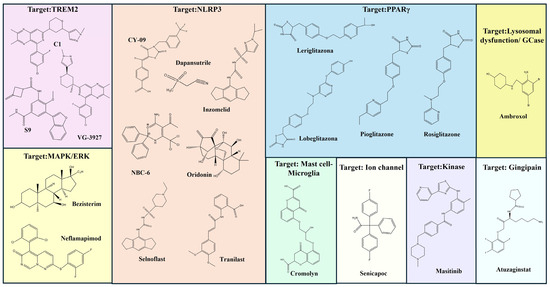
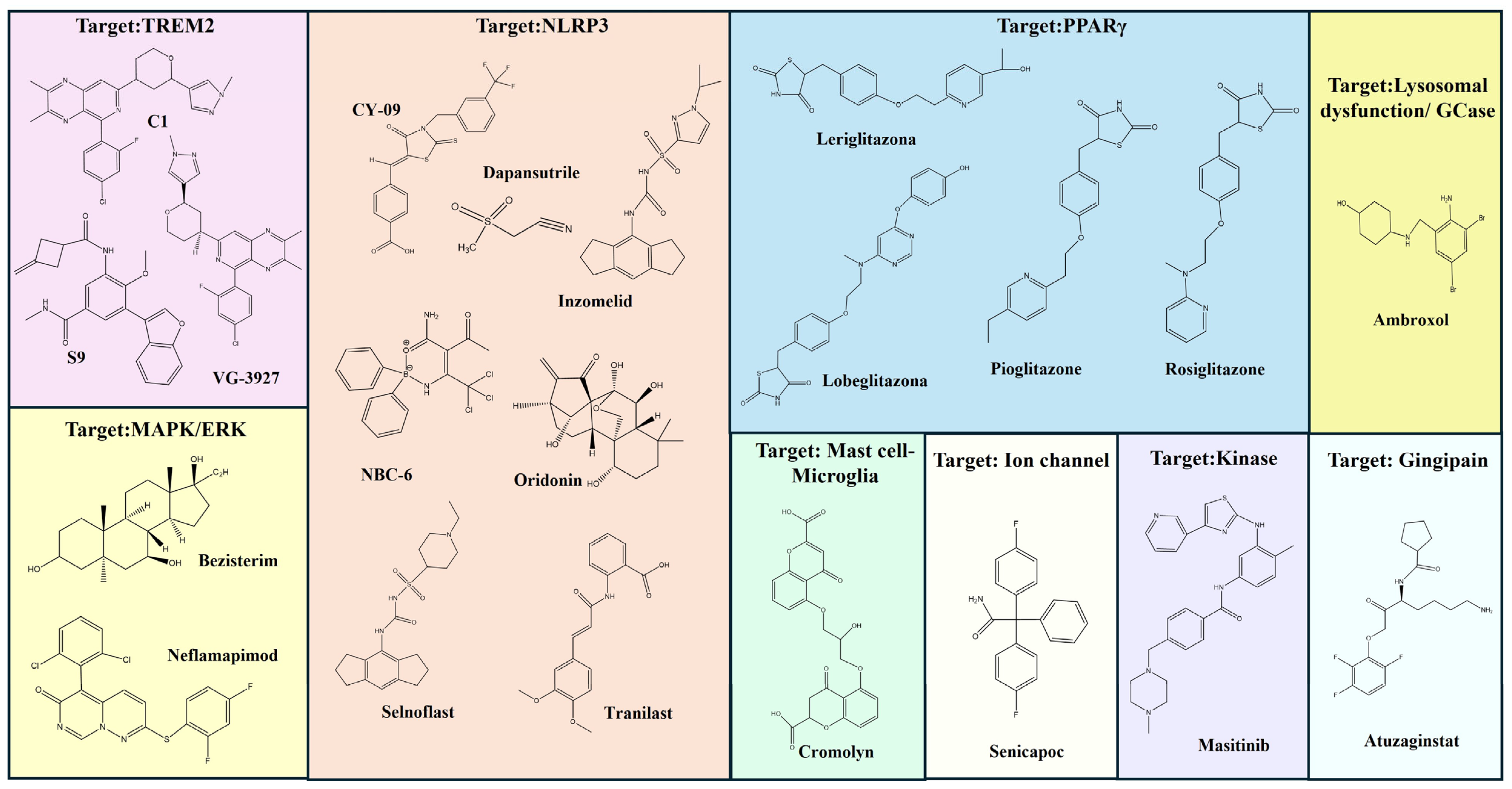
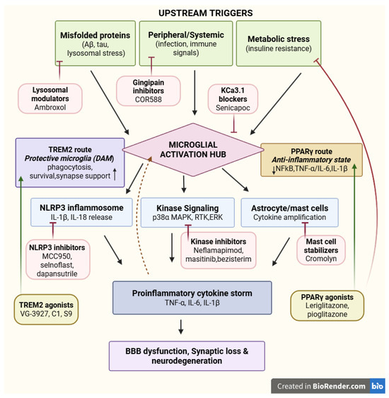
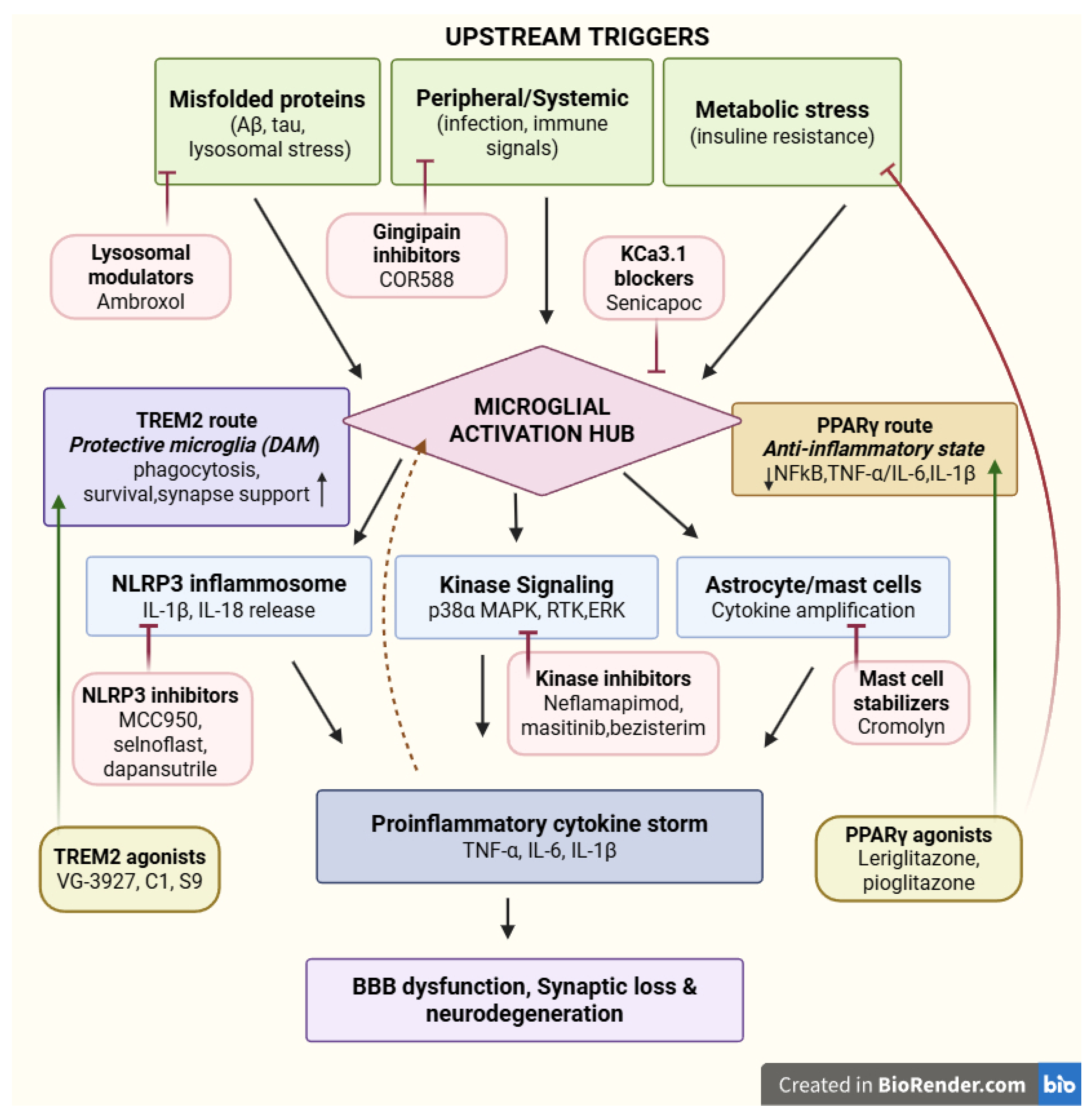

Disclaimer/Publisher’s Note: The statements, opinions and data contained in all publications are solely those of the individual author(s) and contributor(s) and not of MDPI and/or the editor(s). MDPI and/or the editor(s) disclaim responsibility for any injury to people or property resulting from any ideas, methods, instructions or products referred to in the content. |
© 2026 by the authors. Licensee MDPI, Basel, Switzerland. This article is an open access article distributed under the terms and conditions of the Creative Commons Attribution (CC BY) license.