


Abstract
Background/Objectives: Ferula varia Trautv. is a promising medicinal plant of the Apiaceae family and a potential source of biologically active secondary metabolites. The present study aimed to investigate the phytochemical composition and biological activities of F. varia root extract. Methods: The non-volatile compounds were characterized using high-performance liquid chromatography coupled with mass spectrometry (HPLC–QTOF–ESI-MS/MS) method. Furthermore, the total phenolic and flavonoid contents (TPC and TFC), antioxidant activity, and antimicrobial potential of the extract were evaluated using spectrophotometric and microdilution assays. Results: HPLC-ESI-QTOF-MS/MS analysis enabled the tentative identification of 25 metabolites in the F. varia root extract, predominantly sesquiterpene coumarins together with organic acids, phenolic acids, coumarins, sesquiterpene esters, and prenylated aromatic compounds. The extract contained 30.06 ± 3.00 mg GAE/gextr of total phenolics and 1.42 ± 0.04 mg RuE/gextr of total flavonoids. Antioxidant assays revealed Trolox equivalent antioxidant capacity (TEAC), total antioxidant capacity (TAC), ferric reducing antioxidant power (FRAP) values of 0.10 ± 0.00 mM, 110.31 ± 4.94 mg AsAE/gextr, and 21.95 ± 0.00 mg TE/gextr, respectively, while the 2,2-diphenyl-1-picrylhydrazyl (DPPH) assay showed weak radical scavenging activity (IC50 > 1 mg/mL). The extract also demonstrated stronger antimicrobial activity against yeasts than against Gram-positive and Gram-negative bacteria, with minimal inhibitory concentration values ranging from 2.5 to 5 mg/mL and 10–20 mg/mL, respectively. Conclusions: These findings indicate that F. varia roots are a promising source of bioactive compounds exhibiting antioxidant and antimicrobial potential.
1. Introduction
Kazakhstan possesses a rich vascular flora of approximately 7000 species. Among these, 278 species are officially designated as medicinal plants according to Order No. 77 of the Ministry of Ecology and Natural Resources (7 March 2023), which established the national list of medicinal plants [1]. Kazakhstan harbors a remarkably rich vascular flora, with at least 1406 medicinal plant species representing 612 genera and 134 families [2]. Owing to its strategic location at the biogeographic interface between Europe and Asia, the country forms an important part of the Central Asian biodiversity hotspot. Its floristic diversity is largely attributed to pronounced habitat heterogeneity and the convergence of contrasting climatic regions, ranging from the cold-temperate zones of Northern Eurasia to the warmer Irano-Turanian region in southern Kazakhstan. These unique ecological and climatic conditions have promoted the development of a highly diverse plant flora with considerable ecological, economic, and ethnomedicinal value. Consequently, Kazakhstan represents an important reservoir of medicinal plant resources and a valuable source of bioactive compounds for pharmacological and phytochemical research [3].
Comprising more than 180 species, the genus Ferula L. is a diverse member of the Apiaceae family, widely distributed throughout Central Asia, the Mediterranean region, and the Middle East [4]. In the flora of Kazakhstan, the genus Ferula is represented by 51 species, 15 of which are classified as endemic [5,6]. Species of this genus have long been used in traditional medicine owing to their diverse pharmacological properties, including antimicrobial, antioxidant, anti-inflammatory, antidiabetic, and anticancer activities [7,8,9]. These biological effects are mainly associated with the presence of biologically active secondary metabolites such as sesquiterpene coumarins, sulfur-containing compounds, phenolic acids, flavonoids, and essential oils [10]. Numerous representatives of the genus are considered valuable medicinal and aromatic plants and continue to attract increasing scientific interest as promising sources of natural bioactive compounds. Plants of this genus have significant economic value due to their richness in resinous constituents, essential oils, starch, and gums, together with their established uses in traditional medicine and as forage crops [11].
Among them, Ferula varia Trautv. is a relatively understudied species despite its occurrence in Central Asian flora and its potential ethnopharmacological importance. Earlier, it has been proposed to evaluate the quality of F. varia herb based on the content of the flavonoid cinaroside [12,13]. Previous phytochemical investigations of Ferula species have demonstrated that roots are particularly rich in characteristic terpenoid and phenolic constituents, many of which exhibit pronounced biological activities. Traditionally, the root extract of F. persica has been used as an antispasmodic, carminative, laxative, and expectorant, as well as for the treatment of diabetes and hypertension [14]. Sesquiterpene esters such as ferutinin, teferdin, and related compounds are regarded as chemotaxonomic markers of several Ferula species and are known for their antioxidant, antimicrobial, estrogenic, and cytotoxic properties [15].
Natural antioxidants derived from medicinal plants are currently receiving considerable attention because oxidative stress is closely associated with the development of numerous chronic disorders, including cardiovascular, neurodegenerative, inflammatory, and metabolic diseases [16,17,18]. Plant-derived antioxidants are therefore being actively investigated as safer alternatives to synthetic antioxidants used in the pharmaceutical, cosmetic, and food industries [19,20]. Similarly, the increasing emergence of antibiotic-resistant microorganisms has intensified the search for novel antimicrobial agents of natural origin [21]. Plant extracts and essential oils represent promising sources of such compounds due to their structural diversity and multitarget mechanisms of action.
Although several Ferula species have been extensively investigated, F. varia remains one of the least explored representatives of the genus, particularly with respect to the phytochemical composition of its underground parts. Previous studies have primarily focused on isolated biological effects or volatile constituents, whereas comprehensive investigations integrating high-resolution metabolite profiling with biological activity assessment are still lacking. Moreover, detailed characterization of the non-volatile metabolome of F. varia by UHPLC–QTOF–MS/MS has not been comprehensively reported, leaving significant gaps in the understanding of its chemical diversity and pharmacological potential. Addressing these gaps is essential for identifying bioactive metabolites that may serve as promising candidates for pharmaceutical, nutraceutical, and functional food applications.
The present study represents the first comprehensive HPLC–ESI–QTOF–MS/MS characterization of the secondary metabolites of F. varia roots. Several compounds tentatively identified in the extract, including olgoferin, karatavic acid, flavaprenin, ferugin, teferdin, ferutinin, ferusinkin A, badrakemone, karatavicin, and farnesiferone B, have previously been reported from other Ferula species [6,7] but are described here for the first time in F. varia. These findings substantially expand the known phytochemical profile of the species and further support the remarkable chemical diversity and chemotaxonomic significance of sesquiterpene coumarins within the genus Ferula. The study provides a scientific basis for the further investigation of F. varia roots as a promising source of natural compounds with antioxidant, antimicrobial, and other pharmacologically relevant activities.
2. Results
The results of the phytochemical characterization and biological evaluation of the F. varia root extract are presented in this section. The extract was characterized by determining its TFC, TPC and HPLC–ESI–QTOF–MS/MS profiling. Furthermore, its antioxidant capacity, α-amylase inhibitory activity, and antimicrobial activity against Gram-positive bacteria, Gram-negative bacteria, and pathogenic yeasts were evaluated using established in vitro assays.
2.1. Phytochemical Analysis of F. varia Root Extract
2.1.1. Quantification of TPC and TFC
The total phenolic and flavonoid contents of the F. varia root extract are presented in Table 1. The analysis revealed that the extract contained a relatively high concentration of phenolic compounds, with a TPC value of 30.06 ± 3.00 mg GAE/gextr. In contrast, the flavonoid fraction was considerably lower, with a TFC value of 1.42 ± 0.04 mg RuE/gextr.
Table 1.
Total phenolic and flavonoid contents of F. varia root extract.
2.1.2. HPLC-ESI-QTOF-MS/MS Characterization
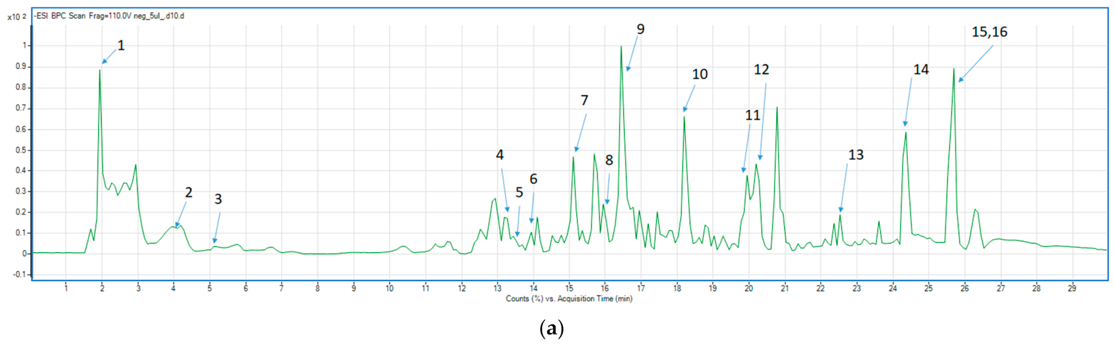
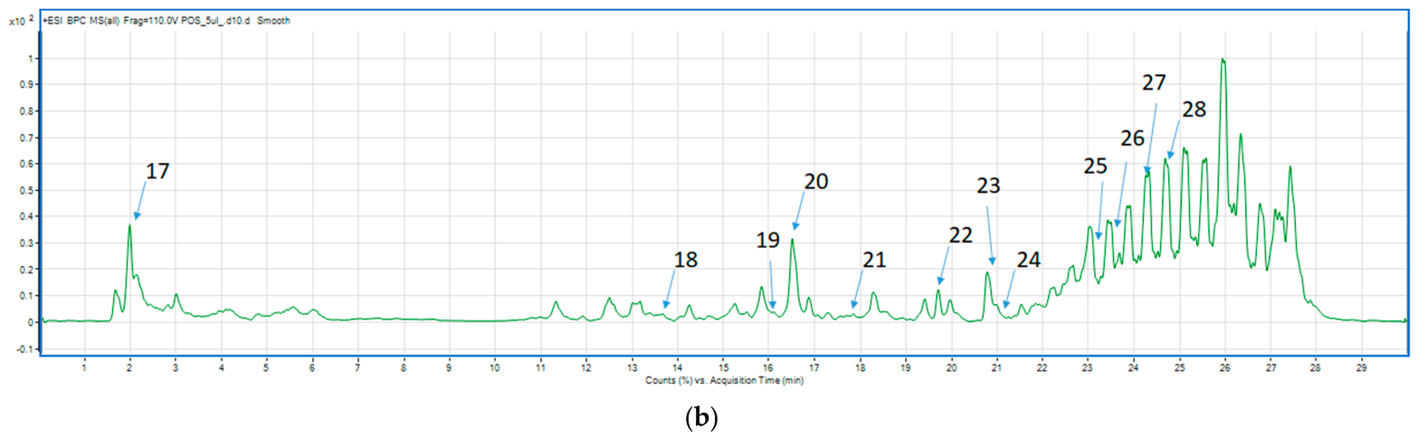
The phytochemical profile of the F. varia root extract was characterized by HPLC–ESI–QTOF–MS/MS analysis performed in both negative and positive ionization modes. A total of 25 metabolites were tentatively identified based on their retention times, accurate mass measurements, molecular formulae, mass fragmentation patterns, and comparison with previously reported literature data (Table 2). All compounds were assigned according to MSI identification level 2. A total of 15 metabolites were detected in the negative ionization mode and 12 metabolites in the positive ionization mode.
Table 2.
HPLC-ESI-QTOF-MS/MS analysis of the F. varia root extract in negative and positive ionization mode.
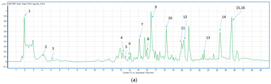
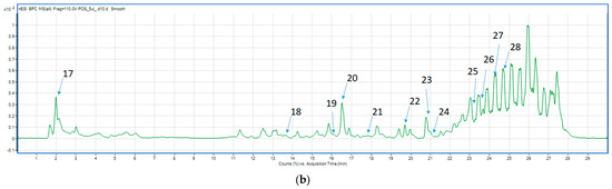
The annotated constituents included organic acids (citric and succinic acids), phenolic acids (ferulic, chlorogenic, and 3-methoxycinnamic acids), coumarins (bergapten, umbelliferone, 5,8-dihydroxypsoralen, and xanthotoxin), together with numerous sesquiterpene coumarins and related derivatives, including conferdione isomer, olgoferin, karatavic acid, conferone, ferugin, teferdin, ferutinin, ferusinkin A, badrakemone, galbanic acid, omega-acetoxyferprenin, karatavicin, and farnesiferone B. The chromatographic profile of the identified metabolites is presented in Figure 1, while their corresponding MS/MS spectra are provided in the Supplementary File (Figures S1–S27). Chlorogenic acid and umbelliferone were detected in both negative and positive ionization modes, providing complementary MS/MS fragmentation data for its tentative annotation.
Figure 1.
Base peak chromatogram (BPC) of the F. varia root extract (HPLC-ESI-QTOF-MS/MS): (a) negative ionization mode and (b) positive ionization mode.
Overall, the HPLC–ESI–QTOF–MS/MS analysis demonstrated that the F. varia root extract contains a chemically diverse mixture of organic acids, phenolic compounds, coumarins, and sesquiterpene esters, with the latter group representing characteristic constituents of the species.
2.2. Evaluation of Biological Activity of the F. varia Root Extract
2.2.1. Antioxidant Activity
The antioxidant potential of the F. varia root extract was evaluated using four complementary in vitro assays, namely TEAC, TAC by the phosphomolybdenum method, FRAP, and DPPH radical scavenging activity. The obtained results are summarized in Table 3.
Table 3.
Antioxidant activity of F. varia root extract (mean ± SD).
The extract exhibited measurable antioxidant activity in all tested systems. The TEAC assay demonstrated an antioxidant capacity of 0.10 ± 0.00 mM Trolox equivalents, indicating the ability of the extract constituents to neutralize ABTS•+ radicals. In the phosphomolybdenum assay, the extract showed a total antioxidant capacity of 110.31 ± 4.94 mg AsAE/gextr, reflecting a substantial overall reducing potential. The reducing activity of the extract was further confirmed by the FRAP assay, which yielded a value of 21.95 ± 0.00 mg TE/gextr. This result demonstrates the capability of the extract to reduce ferric (Fe3+) ions to their ferrous (Fe2+) form, suggesting the presence of electron-donating compounds. In contrast, the extract exhibited relatively weak free-radical scavenging activity in the DPPH assay, with an IC50 value exceeding 1.0 mg/mL. This finding indicates that relatively high concentrations of the extract were required to achieve 50% scavenging of DPPH radicals.
2.2.2. α-Amylase Inhibitory Activity
The α-Amylase inhibitory activity of the F. varia root extract is presented in Table 4. The extract exhibited an inhibitory potential of 6.06 ± 0.55 mg AcrE/gextr.
Table 4.
α-Amylase enzyme inhibitory potential of the extract (mean ± SD).
These results indicate the ability of the extract constituents to inhibit α-amylase activity and suggest the presence of compounds with enzyme inhibitory properties.
2.2.3. Antimicrobial and Antifungal Activity
The antimicrobial potential of the F. varia root extract was assessed against a range of Gram-positive and Gram-negative bacteria and pathogenic yeast strains based on minimum inhibitory concentration (MIC) and minimum bactericidal/fungicidal concentration (MBC/MFC) values. The results are presented in Table 5. Vancomycin, ciprofloxacin, and nystatin were used as the standard reference drugs (Table 6).
Table 5.
Antimicrobial activity of investigated F. varia extract (mg/mL).
Table 6.
Reference antibiotics activity against tested microbial strains (mg/L).
The extract exhibited antimicrobial activity against all tested microorganisms, although the degree of susceptibility varied among the strains. Among Gram-positive bacteria, the strongest inhibitory effect was observed against M. luteus, with MIC and MBC values of 5 and 10 mg/mL, respectively. Moderate activity was detected against B. cereus (MIC = 10 mg/mL), whereas S. aureus ATCC 25923, S. aureus ATCC BA1707, S. epidermidis, and E. faecalis demonstrated lower susceptibility, with MIC values of 20 mg/mL.
Among Gram-negative bacteria, MIC values ranged from 10 to 20 mg/mL. The highest sensitivity was observed for S. Typhimurium, E. coli, K. pneumoniae, and P. aeruginosa, each exhibiting MIC values of 10 mg/mL. P. mirabilis was comparatively less susceptible, showing both MIC and MBC values of 20 mg/mL. The strongest antimicrobial activity of the extract was observed against yeast strains. C. glabrata was the most sensitive microorganism tested, exhibiting MIC and MFC values of 2.5 and 5 mg/mL, respectively. In addition, C. albicans and C. parapsilosis showed pronounced susceptibility, with MIC and MFC values of 5 mg/mL.
For comparison, the antimicrobial activity of the F. varia root extract was evaluated alongside the corresponding reference antimicrobial agents. Vancomycin showed MIC values of 0.12–1.95 mg/L against the tested Gram-positive bacteria, while ciprofloxacin exhibited MIC values of 0.015–0.488 mg/L against Gram-negative bacteria. For the tested yeast strains, nystatin showed MIC values of 0.24–0.48 mg/L. The reference agents demonstrated substantially lower MIC values than the crude F. varia extract, confirming their higher antimicrobial potency under the experimental conditions.
Overall, the extract demonstrated stronger antifungal than antibacterial activity. The lowest MIC value recorded in the present study was observed against C. glabrata (2.5 mg/mL), indicating that this species was the most susceptible microorganism among all tested strains. In contrast, most bacterial strains required higher inhibitory concentrations (10–20 mg/mL), reflecting a comparatively lower sensitivity to the extract.
3. Discussion
The present study offers the first comprehensive characterization of the phytochemical profile and biological activities of F. varia root extract. Although several species of the genus Ferula have been extensively investigated and the aerial parts of F. varia have recently been characterized, detailed studies on the chemical composition and biological activities of the roots remain very limited. To the best of our knowledge, this study represents the first comprehensive HPLC–ESI–QTOF–MS/MS characterization of the secondary metabolite profile of F. varia roots. Previously, phytochemical investigations of F. varia roots have mainly focused on the isolation and structural elucidation of individual sesquiterpene lactones and their glycosides rather than comprehensive metabolite profiling and, more recently, to the characterization of phenolic constituents in the aerial parts [28]. Suzuki et al. [36] reported several new sesquiterpene lactones from the roots of F. varia, whereas Kurimoto et al. [37] subsequently described seven new sesquiterpene lactone glycosides isolated from the methanolic root extract. In contrast, the present study provides a substantially broader phytochemical overview by tentatively identifying 25 metabolites belonging to several structurally distinct classes, thereby considerably expanding the known chemical composition of F. varia roots.
Among the identified metabolites, sesquiterpene coumarins are regarded as characteristic chemotaxonomic markers of Ferula species and represent one of the largest groups of specialized metabolites within the genus [38,39]. Previous phytochemical investigations have shown that these compounds are widely distributed in the roots, rhizomes, and oleo-gum-resins of numerous Ferula species, including F. communis [40], F. assa-foetida [41,42], F. huber-morathii [43], F. sinkiangensis [44], F. latisecta [45], and F. flabelliloba [31]. Although sesquiterpene coumarins are well recognized as characteristic constituents of the genus, several compounds tentatively identified in the present study have not previously been reported from F. varia. Olgoferin and karatavic acid have previously been described in F. persica [10], conferone, conferdione derivatives, teferdin, ferutinin, and karatavicin were reported from F. assa-foetida [41,42], ferusinkin A was isolated from F. sinkiangensis [44], whereas farnesiferone B has mainly been reported from F. assa-foetida and several Iranian Ferula species. Their tentative identification in F. varia therefore considerably broadens the known distribution of these metabolites within the genus and highlights the rich chemodiversity of the species.
Their predominance in the present study therefore confirms the characteristic phytochemical pattern of underground organs of Ferula. The occurrence of several daucane-type sesquiterpene esters, including ferutinin and teferdin, is particularly noteworthy. These metabolites have repeatedly been reported as major constituents of Ferula roots and have attracted considerable attention because of their broad spectrum of biological activities, including antioxidant, antimicrobial, anti-inflammatory, estrogenic, and cytotoxic properties [46]. Ferutinin, in particular, is considered one of the characteristic constituents of several Ferula species and has been extensively investigated as a representative bioactive sesquiterpene ester [47].
Another group of detected compounds consisted of phenolic constituents, such as chlorogenic acid, ferulic acid, 3-methoxycinnamic acid, bergapten, umbelliferone, and xanthotoxin. Although these compounds were detected in lower diversity than sesquiterpene coumarins, they represent an important group of metabolites commonly reported in the Ferula genus [26]. Notably, chlorogenic acid, ferulic acid, and 3-methoxycinnamic acid have rarely been discussed as constituents of F. varia roots. Umbelliferone and furanocoumarins such as bergapten and xanthotoxin are also characteristic constituents of numerous Ferula species and have previously been described as taxonomically significant coumarin derivatives within the genus [39].
An interesting observation was the detection of chlorogenic acid in both negative and positive ionization modes. The negative ion mode produced the characteristic fragment ion at m/z 191, corresponding to quinic acid, together with the diagnostic fragment at m/z 161, whereas positive ionization generated the protonated molecular ion accompanied by the fragment at m/z 163. These fragmentation pathways are consistent with previously reported tandem mass spectrometric behavior of chlorogenic acid and are commonly used for its annotation in untargeted metabolomic studies.
The phytochemical profile obtained for the roots differs markedly from that previously reported for the aerial parts of F. varia, where phenolic constituents represented the dominant metabolite class [28]. In contrast, the present study demonstrated that the underground organs accumulate a considerably broader spectrum of specialized metabolites, with sesquiterpene coumarins representing the predominant constituents. The simultaneous detection of numerous sesquiterpene coumarins together with phenolic acids, simple coumarins, furanocoumarins, prenylated aromatic compounds, and sesquiterpene esters provides new evidence of the remarkable chemical diversity of F. varia roots. These findings substantially expand the phytochemical knowledge of the species and indicate that F. varia represents a richer source of bioactive metabolites than previously recognized.
In addition to the qualitative characterization of individual metabolites by HPLC–ESI–QTOF–MS/MS, the quantitative determination of TPC and TFC provides complementary information on the abundance of the major classes of antioxidant constituents. Compared with our previous study [28] on the aerial parts of F. varia, the root extract contained a higher level of total phenolics (30.06 ± 3.00 vs. 23.22 ± 1.02 mg GAE/gextr) but a lower flavonoid content (1.42 ± 0.04 vs. 2.83 ± 0.98 mg RUE/gextr), suggesting organ-specific accumulation of phenolic metabolites. TPC and TFC results are consistent with the HPLC–ESI–QTOF–MS/MS, which revealed the presence of. several phenolic acids, while flavonoids, if present, are likely to occur only in minor amounts. Nouioura et al. [48] showed that F. communis ethanol and methanol fruit extracts exhibited the highest TPC values of 62.20 ± 0.11 and 60.82 ± 0.32 mg GAE/g dry weight (DW), respectively, followed by the aqueous extract (44.04 ± 0.22 mg GAE/g DW). Lower TPC values were reported for acetone (19.76 ± 1.19 mg GAE/g DW), ethyl acetate (15.24 ± 0.60 mg GAE/g DW), chloroform (13.94 ± 0.92 mg GAE/g DW), and hexane (6.93 ± 0.43 mg GAE/g DW) extracts. In another study, Tunisian F. communis showed markedly higher TPC values in fruits (422 mg GAE/g DW), followed by flowers (207.21 mg GAE/g DW) and stems (129.86 mg GAE/g DW) [24]. A recent study on F. persica revealed the highest TPC was observed in the cyclopentyl methyl ether extract (78.3 mg GAE/g), followed by the ethyl acetate (76.3 mg GAE/g) and ethanol (74.5 mg GAE/g) extracts of the aerial parts, whereas root extracts contained 19.5–41.6 mg GAE/g [49].
According to the literature data, chlorogenic acid is one of the major hydroxycinnamic acid derivatives frequently associated with antioxidant activity [50], whereas ferulic acid contributes to the radical scavenging and reducing properties of plant extracts [51]. The observed differences in their content may be reflected in the antioxidant capacity determined by different in vitro assays. The antioxidant activity of F. varia root extract was evaluated using assays based on different reaction mechanisms. The extract exhibited moderate antioxidant activity. However, the response varied among the methods. The highest activity was observed in the phosphomolybdenum assay (110.31 mg AsAE/gextr), indicating a considerable total reducing capacity. The FRAP assay also demonstrated the ability of the extract to reduce Fe3+ ions (21.95 mg TE/gextr). In contrast, the DPPH assay showed weak radical scavenging activity (IC50 > 1.0 mg/mL). This difference suggests that the extract acts primarily through electron transfer rather than hydrogen atom transfer. Comparable findings have been documented in other Ferula species. Hydroethanolic extracts of F. asafoetida leaves exhibited stronger DPPH scavenging and ferric reducing activity than gum extracts owing to their higher phenolic and flavonoid contents [52]. The authors concluded that antioxidant capacity positively correlated with total phenolic concentration. In contrast, root extracts enriched in terpenoids and sesquiterpene coumarins often display only moderate DPPH scavenging despite measurable reducing capacity. Several reviews indicate that Ferula species are particularly rich in sesquiterpene coumarins, sulfur-containing metabolites, and terpenoids, compounds more frequently associated with antimicrobial and anti-inflammatory effects than with high radical-scavenging activity [8]. A recent study of F. communis reported substantially stronger DPPH activity for leaf extracts IC50 value of 0.263 ± 0.008 mg/mL, while methanolic fruit and root extracts exhibited IC50 values of 0.076 ± 0.039 mg/mL and 0.820 ± 0.031 mg/mL, respectively [53]. Likewise, antioxidant activity in F. sinkiangensis varied considerably among solvent fractions [54]. The ethyl acetate, n-butanol, and methanol fractions exhibited DPPH IC50 values of 34.1, 33.8, and 40.3 μg/mL, respectively. These values are nearly 25–30-fold lower than the activity observed for F. varia, indicating a much higher free-radical scavenging capacity. The authors attributed this effect to the enrichment of phenolic constituents in medium-polarity solvent fractions. The antioxidant activity of F. varia was lower than that reported for F. communis [48]. Methanolic and ethanolic extracts of F. communis exhibited DPPH IC50 values of 8.09 and 8.25 μg/mL, respectively, whereas acetone and aqueous extracts showed IC50 values of 34.23 and 37.28 μg/mL. In addition, the methanolic extract displayed the highest reducing power (EC50 = 17.63 μg) and total antioxidant capacity (72.15 ± 0.11 mg AAE/g). Overall, variations in antioxidant activity among Ferula species are largely determined by the extraction solvent, plant organ, and metabolite composition, particularly the abundance of phenolic compounds.
The F. varia root extract demonstrated measurable α-amylase inhibitory activity (6.06 ± 0.55 mg AcrE/gextr), indicating the presence of constituents capable of suppressing carbohydrate-hydrolyzing enzymes. Recent studies have demonstrated that sesquiterpene coumarins represent a promising class of natural α-amylase inhibitors [55]. Baccari et al. [56] reported that a series of semisynthetic arylidene derivatives obtained from the sesquiterpene coumarin coladonin, isolated from the roots of F. tunetana, exhibited potent α-amylase inhibitory activity, with IC50 values ranging from 7.24 to 28.98 μM. Ferulic acid identified in the F. varia root extract has previously been reported as an effective inhibitor of carbohydrate-hydrolyzing enzymes. Zheng et al. [57] demonstrated that ferulic acid inhibited α-amylase and α-glucosidase with IC50 values of 0.622 and 0.866 mg/mL, respectively.
The F. varia root extract exhibited antimicrobial activity against a range of Gram-positive and Gram-negative bacteria, as well as yeast strains. Notably, this study represents the first report on the antimicrobial activity of F. varia root extract, and the obtained findings were therefore compared with those reported for other Ferula species. The 80% methanolic root extract of F. communis exhibited greater activity against Gram-positive bacteria, particularly S. aureus (MIC = 16.67–18.66 mg/mL), whereas Gram-negative bacteria were less susceptible, with average MIC values exceeding 30 mg/mL [58]. Similar findings were reported for F. communis fruit extracts, where the ethanolic extract exhibited the strongest antibacterial activity, with MIC values of 0.312 mg/mL against E. coli and 0.625 mg/mL against B. subtilis and P. mirabilis, while the acetone extract showed MIC values of 2.5 mg/mL against all tested bacterial strains. The superior activity of the ethanolic extract was associated with its higher phenolic and flavonoid contents and greater antioxidant capacity [48]. Likewise, the chloroform extract of the aerial parts of F. caspica demonstrated the highest antibacterial activity against S. aureus (MIC = 32 μg/mL) and E. faecalis (MIC = 64 μg/mL), while E. coli and P. aeruginosa exhibited markedly lower susceptibility (MIC = 512 μg/mL). The same extract showed only moderate antifungal activity against Candida spp., with MIC values ranging from 128 to 256 μg/mL [59]. In contrast, the CO2 extract of F. asafoetida displayed potent activity against both Gram-positive and Gram-negative bacteria, with MIC values of 7.81 μg/mL for S. aureus and 15.63 μg/mL for E. coli, K. pneumoniae, and S. enterica, whereas C. albicans was considerably less susceptible (MIC = 62.5 μg/mL) [60].These findings suggest that the antimicrobial activity of Ferula species is strongly influenced by both the plant species and the extraction solvent, resulting in different susceptibility patterns among microbial groups. Unlike previously reported extracts, which generally showed greater efficacy against Gram-positive bacteria, the F. varia root extract demonstrated comparable or even higher activity against E. coli, S. Typhimurium, K. pneumoniae, and P. aeruginosa with MIC = 10 mg/mL, while the strongest antimicrobial effect was observed against C. glabrata with MIC = 2.5 mg/mL. For additional comparison, the antimicrobial activity of the F. varia root extract was evaluated alongside standard reference agents. Vancomycin showed MIC values of 0.12–1.95 mg/L against the tested Gram-positive bacteria, ciprofloxacin exhibited MIC values of 0.015–0.488 mg/L against Gram-negative bacteria, and nystatin showed MIC values of 0.24–0.48 mg/L against the tested yeast strains. As expected, the reference antimicrobial agents exhibited substantially greater potency than the crude F. varia extract. However, direct comparison of the numerical MIC values should be interpreted with caution because the reference drugs are purified antimicrobial compounds, whereas the plant extract represents a complex mixture of constituents and was tested at mg/mL concentrations. Nevertheless, the positive-control results confirm the expected susceptibility of the tested microorganisms and provide an appropriate reference framework for evaluating the antimicrobial activity of the F. varia root extract.
The observed antimicrobial activity may be related to the coumarins -rich composition of the extract, as well as phenolic acids. In the present extract, compounds such as ferutinin, umbelliferone, galbanic acid, ferulic and chlorogenic acids have been associated with antimicrobial activity. For example, ferutinin exhibited the strongest antibacterial activity against S. aureus, with MIC and MBC values of 25 μM [61]. Shahverdi et al. [62] reported that galbanic acid markedly enhanced the activity of β-lactam antibiotics against S. aureus, reducing the MICs of penicillin G and cephalexin by 64-fold and 128-fold, respectively. Borges et al. [63] reported that ferulic acid exhibited broad-spectrum antibacterial activity against E. coli, P. aeruginosa, S. aureus, and L. monocytogenes, with MIC values of 100 μg/mL for E. coli and P. aeruginosa and 1100–1250 μg/mL for Gram-positive bacteria, acting primarily through irreversible disruption of bacterial membrane integrity and increased membrane permeability. Previous studies have demonstrated that chlorogenic acid exhibits broad-spectrum antibacterial activity, with MIC values ranging from 20 to 80 μg/mL [64]. Umbelliferone was reported to exhibit potent antibacterial activity against B. cereus, B. subtilis, and K. pneumoniae, with MIC values of 0.31–0.62 mg/mL [61]. Additionally, umbelliferone showed antifungal activity against both susceptible and multidrug-resistant C. albicans strains, with MIC values of 125–175 μg/mL, highlighting its potential as a promising natural antifungal agent [65].
These findings indicate that the biological potential of F. varia roots is closely associated with their chemically diverse metabolite profile, particularly the presence of phenolic acids and characteristic sesquiterpene coumarins. Although the extract demonstrated promising antioxidant, enzyme inhibitory, antimicrobial, and antifungal properties in vitro. This study was limited to in vitro assays and tentative metabolite identification. Future investigations should focus on the isolation and quantitative determination of the major constituents, synergistic biological effects, elucidation of their mechanisms of action, and confirmation of efficacy and safety in appropriate in vivo models.
4. Materials and Methods
4.1. Plant Material
The wild-growing plant Ferula varia (Schrenk ex Fisch., C.A.Mey. & Avé-Lall.) Trautv., native to the flora of Kazakhstan, was collected during the flowering period in April 2025 as part of an expedition to the Northern Pre-Balkhash region of the Republic of Kazakhstan. The plant material was collected near the Bektatau Mountains, Aktogai District, Karaganda Region, at 47.74108° N, 74.29520° E. The underground parts of F. varia were taxonomically authenticated by botanist Dr. M. Yu. Ishmuratova, and a voucher specimen was deposited in the Herbarium of E.A. Buketov Karaganda University under accession number QAR04328.
4.2. Preparation of the Extract
Dried roots (19 g) were subjected to microwave-assisted extraction with 70% aqueous ethanol (w/w) at a plant material-to-solvent ratio of 1:10 (w/v) using an ETHOS X advanced microwave extraction system (Milestone, Sorisole, Italy). Extraction was performed at 800 W microwave power for 12 min [66]. The resulting extract was filtered, and the solvent was removed under reduced pressure using an IKA RV 8 rotary evaporator (IKA-Werke GmbH & Co. KG, Breisgau, Germany). The extraction yield was 20.9% (w/w).
4.3. Phytochemical Analysis
4.3.1. Determination of TPC
The TPC of the extracts was determined spectrophotometrically using the Folin–Ciocalteu reagent according to a previously described method [67]. Methanol was used to prepare the extract solutions and gallic acid stock solutions. For the assay, 20 µL of the sample solution, 1560 µL of ultrapure water, and 100 µL of Folin–Ciocalteu reagent were sequentially combined in a 96-deep-well plate using a 12-channel micropipette. Following incubation for 1–8 min, 300 µL of 20% sodium carbonate solution was added, and the mixture was thoroughly mixed. The reaction mixture was then incubated for 2 h at 25 °C in the dark. Subsequently, 300 µL of the reaction mixture was transferred to a 96-well microplate, and absorbance was measured at 760 nm. The TPC was calculated using a five-point calibration curve prepared with gallic acid at concentrations ranging from 0.01 to 1.0 mg/mL. All measurements were performed in triplicate. The gallic acid calibration curve was described by the equation y = 0.7897x + 0.0606, with an R2 value of 0.9915 (Figure S28). The results were expressed as the mean ± standard error and reported as milligrams of gallic acid equivalents per gram of extract (mg GAE/gextr).
4.3.2. Determination of TFC
The TFC was quantified using a spectrophotometric aluminum chloride (AlCl3) assay based on a previously published procedure [68]. Briefly, 80 µL of either the extract solution or rutin standard was mixed with 80 µL of AlCl3 reagent and 1840 µL of absolute ethanol in a 96-deep-well plate. For the blanks, AlCl3 was replaced with 10 µL of 99.8% acetic acid. The mixtures were allowed to react for 40 min in the dark, after which 300 µL of each reaction mixture was transferred to a 96-well microplate. Absorbance was subsequently recorded at 415 nm using a microplate reader. Quantification was performed against a seven-point rutin calibration curve covering 0.01–1.0 mg/mL, described by the equation y = 1.6177x + 0.0479 (R2 = 0.9987; Figure S29). The results were calculated as the mean ± standard error and expressed as milligrams of rutin equivalents per gram of extract (mg RuE/gextr).
4.3.3. HPLC–ESI–QTOF–MS/MS Analysis
Phytochemical profiling of the extract was conducted using an Agilent 1200 HPLC system coupled with an Agilent 6210 Quadrupole Time-of-Flight (QTOF) mass spectrometer (Agilent Technologies, Santa Clara, CA, USA). The analysis was performed in both negative (ESI−) and positive (ESI+) electrospray ionization mode with a Dual AJS ESI ion source (Agilent Technologies, Santa Clara, CA, USA). Chromatographic separation was achieved using a binary mobile-phase system comprising solvent A (water containing 0.1% formic acid) and solvent B (acetonitrile containing 0.1% formic acid). The gradient program was set as follows: 0–2 min, 99% A/1% B; 2–10 min, a linear change to 80% A/20% B; 10–15 min, 60% A/40% B; 15–17 min, 5% A/95% B; 17–22 min, 5% A/95% B; and 22.1–25 min, restoration of the initial composition (99% A/1% B). The mobile-phase flow rate was 0.2 mL/min, with a total chromatographic run time of 30 min. UV detection was performed at 290 nm. A 5 μL sample volume was injected using the standard injection mode. Mass spectrometric data were acquired in Auto MS/MS mode over an m/z range of 100–1400 for MS scans and 60–1400 for MS/MS scans, with an acquisition rate of 1 spectrum/s for both modes. The ion source was operated under the following conditions: capillary voltage, 3000 V; nozzle voltage, 1000 V; fragmentor voltage, 110 V; skimmer voltage, 65 V; and octopole RF peak voltage, 750 V. Nitrogen was employed as both the drying and sheath gas at a flow rate of 12 L/min. The drying and sheath gas temperatures were maintained at 250 °C and 300 °C, respectively, while the nebulizer pressure was set to 35 psig. Data acquisition and processing were performed using MassHunter software, version 10.0. Compound identification was based on a combination of UV spectral characteristics, retention times, and interpretation of the observed fragmentation patterns. The obtained data were compared with reference information from established mass spectral databases, including MassBank [69] and PubChem [70], as well as relevant scientific literature. The detected compounds were subsequently annotated in accordance with the identification confidence levels recommended by the Metabolomics Standards Initiative (MSI) [71].
4.4. Assessment of Biological Activity
4.4.1. Trolox Equivalet Antioxidant Capacity Assay (TEAC)
The ABTS•+ radical cation-scavenging activity of the samples was determined according to the method of Re et al. [72], with minor modifications. To generate the ABTS•+ radical cation, 7 mM ABTS and 2.5 mM K2S2O8 were dissolved in 10 mL of ultrapure water and incubated in the dark at room temperature for 16 h. Before analysis, the resulting ABTS•+ solution was diluted with absolute ethanol to obtain an absorbance of 0.7–0.8 at 734 nm. Sample solutions were prepared in methanol at a concentration of 1 mg/mL, while Trolox standard solutions were prepared at concentrations of 3.0, 2.0, 1.0, 0.5, 0.25, and 0.125 mM. For the assay, 10 µL of each sample solution was mixed with 990 µL of the ABTS•+ working solution. For the control, 10 µL of methanol was added instead of the sample or Trolox solution. After incubation for 30 min, the decrease in absorbance was measured at 734 nm and used to construct the Trolox calibration curve. The ABTS•+ scavenging activity was expressed as TEAC, mM and calculated using the linear regression equation obtained from the Trolox standards: y = 23.075x − 2.5945 (R2 = 0.9924; Figure S30).
4.4.2. Total Antioxidant Capacity (TAC)—Phosphomolybden Assay
The antioxidant capacity of the samples was assessed using the phosphomolybdenum assay following the procedure reported by Zengin et al. [73]. Sample solutions were prepared at a concentration of 1 mg/mL and mixed with freshly prepared phosphomolybdenum reagent containing 0.6 M sulfuric acid, 28 mM sodium phosphate, and 4 mM ammonium molybdate. The reaction mixtures were incubated at 95 °C for 90 min and subsequently allowed to cool to room temperature. Absorbance was then recorded at 695 nm. The TAC was determined using an ascorbic acid calibration curve (Figure S31) and expressed as milligrams of ascorbic acid equivalents per gram of extract (mg AsAE/gextr).
4.4.3. Ferric Reducing Antioxidant Power (FRAP)
Ferric ion-reducing capacity was determined using a modified FRAP assay based on the method reported by Wong et al. [74]. The required stock solutions consisted of 300 mM acetate buffer (pH 3.6), 10 mM 2,4,6-tris(2-pyridyl)-S-triazine (TPTZ) prepared in 40 mM HCl, and 20 mM FeCl3·6H2O. The FRAP working reagent was freshly prepared by combining the solutions in a ratio of 10:1:1 (acetate buffer:TPTZ:FeCl3). For the assay, 15 µL of each sample solution prepared in methanol containing 10% dimethyl sulfoxide (DMSO) was mixed with 285 µL of the freshly prepared FRAP reagent. The reaction mixtures were incubated at 37 °C for 30 min in the dark, after which the absorbance was measured at 593 nm. A Trolox calibration curve was used for quantification (Figure S32). The ferric ion-reducing antioxidant capacity was expressed as milligrams of Trolox equivalents per gram of dry extract (mg TE/gextr).
4.4.4. Free Radical Scavenging Activity (DPPH Assay)
The hydrogen-atom- and electron-donating capacities of the samples were assessed based on their ability to reduce the purple-colored DPPH radical. The DPPH radical-scavenging activity was determined according to the method of Brand-Williams et al. [75], with minor modifications. A fresh DPPH solution (0.08 mg/mL) was prepared daily in methanol and stored at 4 °C in the dark between measurements. Serial dilutions of the test samples (1.0–0.0078 mg/mL) and reference standards, including gallic acid (0.1–0.00078 mg/mL), ascorbic acid (0.50–0.0039 mg/mL), and Trolox (0.75–0.0058 mg/mL), were prepared in methanol containing 10% DMSO directly in 96-well microtiter plates. Methanol was used as the positive control in the final column of the plate. For the assay, 100 µL of each sample, essential oil, or standard solution was mixed with 100 µL of the DPPH solution using an Eppendorf Research® plus multichannel pipette (Eppendorf, Hamburg, Germany). The plates were incubated in the dark for 30 min, after which the decrease in absorbance was measured at 517 nm. Control wells contained 100 µL of methanol instead of the test solution and were mixed with 100 µL of DPPH solution. All measurements were performed in triplicate. The DPPH radical-scavenging activity was expressed as a percentage of inhibition, calculated using the following equation:
IC50 values were estimated from plots of DPPH radical-scavenging activity (%) as a function of the corresponding sample concentrations.
4.4.5. Antimicrobial Activity
The antimicrobial activity of the extract was assessed by the broth microdilution technique following the guidelines of the European Committee on Antimicrobial Susceptibility Testing (EUCAST) [76]. The antibacterial properties were evaluated against a panel of Gram-positive bacteria, including Staphylococcus aureus ATCC 25923, S. aureus ATCC BA1707, Staphylococcus epidermidis ATCC 12228, Micrococcus luteus ATCC 10240, Bacillus cereus ATCC 10876, and Enterococcus faecalis ATCC 29212, as well as Gram-negative bacteria, namely Salmonella Typhimurium ATCC 14028, Escherichia coli ATCC 25922, Proteus mirabilis ATCC 12453, Klebsiella pneumoniae ATCC 13883, and Pseudomonas aeruginosa ATCC 9027. Mueller–Hinton (MH) broth was used as the culture medium for bacterial strains. Antifungal activity was determined against Candida glabrata ATCC 90030, Candida albicans ATCC 102231, and Candida parapsilosis ATCC 22019 using MH broth supplemented with 2% glucose. Prior to the assay, the extract was dissolved in DMSO, diluted with the corresponding culture medium, and subjected to serial two-fold dilutions. The resulting dilutions were dispensed into sterile 96-well microtiter plates (Nunc, Roskilde, Denmark) to obtain final extract concentrations ranging from 20 to 0.156 mg/mL. Fresh microbial cultures were suspended in sterile 0.85% NaCl and standardized to a turbidity equivalent to 0.5 McFarland. The standardized inocula were subsequently added to the wells to achieve final concentrations of 5 × 10⁵ CFU/mL for bacteria and 5 × 10⁴ CFU/mL for yeasts. The plates were incubated at 35 °C for 18–20 h. The MIC was established as the lowest extract concentration that prevented visible microbial growth. Growth inhibition was assessed visually and confirmed spectrophotometrically by measuring absorbance at 600 nm using a BioTek ELx800 microplate reader (BioTek Instruments, Inc., Winooski, VT, USA). To determine the MBC/MFC, 5 μL aliquots from each well were transferred onto extract-free MH agar plates and incubated at 35 °C for 24 h. The lowest concentration resulting in complete inhibition of microbial growth was recorded as the MBC or MFC, respectively. All experiments were conducted in triplicate, and the highest value obtained was reported. The assay included appropriate controls: DMSO at a final concentration of 1% (v/v), an inoculum control without extract, and an extract control without microbial inoculum.
4.4.6. -Amylase Enzyme Inhibition Assay
The inhibitory effect of the samples on α-amylase activity was evaluated using the Caraway–Somogyi iodine/potassium iodide (I2/KI) colorimetric method [77,78], with minor modifications. A 0.05% substrate solution was prepared by dissolving 10 mg of soluble potato starch in 20 mL of ultrapure water, followed by boiling for 10 min and cooling to room temperature prior to use. Acarbose (0.01–0.1 mg/mL prepared in buffer) was used as the reference inhibitor. For the assay, 20 mM sodium phosphate buffer (pH 6.9) was dispensed into 96-well flat-bottom microplates using a multichannel automatic pipette (Eppendorf, Germany). Subsequently, 25 μL of the sample solution and 50 μL of α-amylase solution (0.8 U/mL in buffer) were added, and the reaction mixtures were incubated at 37 °C for 10 min. Following pre-incubation, 50 μL of the starch substrate solution was added, and the mixtures were further incubated at 37 °C for 10 min. The enzymatic reaction was terminated by adding 25 μL of 1 M HCl, followed by the addition of 100 μL of the I2/KI reagent. Sample blanks were prepared using all assay components except the enzyme, which was replaced with 50 μL of buffer. Control wells contained all reaction components with 25 μL of the solvent used to prepare the samples instead of the sample solution and represented 100% enzyme activity. Absorbance was measured at 630 nm for both samples and blanks. The percentage inhibition of α-amylase activity (Inh%) was calculated using the following equation:
4.5. Statistical Analysis
Statistical analyses were conducted using SigmaPlot software (version 12.0). Data obtained from the inhibition assays are presented as the mean ± standard error of the mean (SEM). The IC50 values for the DPPH radical-scavenging assay were estimated by nonlinear regression analysis.
5. Conclusions
The present study provides the first comprehensive phytochemical characterization and biological evaluation of F. varia root extract. HPLC–ESI–QTOF–MS/MS analysis revealed a chemically diverse metabolite profile dominated by sesquiterpene coumarins, together with phenolic acids, coumarins, organic acids, sesquiterpene esters, and prenylated aromatic compounds.
The biological evaluation demonstrated that the extract possesses moderate antioxidant activity, measurable α-amylase inhibitory potential, and broad-spectrum antimicrobial activity against Gram-positive and Gram-negative bacteria as well as pathogenic yeasts, with the highest susceptibility observed for C. glabrata. The observed biological effects are likely associated with the combined action of the identified phenolic acids and characteristic sesquiterpene coumarins, which have previously been reported to exhibit antioxidant, enzyme inhibitory, antimicrobial, and antifungal properties.
The present study substantially expands the phytochemical knowledge of F. varia roots. Several metabolites previously reported from other Ferula species were tentatively identified here for the first time in F. varia roots, demonstrating that the underground organs of this species represent a considerably richer source of specialized metabolites than previously recognized.
Overall, the results indicate that F. varia roots represent a promising source of biologically active secondary metabolites and support their potential as a valuable raw material for the development of phytopharmaceuticals and other plant-derived bioactive products.
Supplementary Materials
The following supporting information can be downloaded at: https://www.mdpi.com/article/10.3390/molecules31172990/s1, Figure S1: MS/MS fragmentation spectrum of bergapten in negative mode; Figure S2: MS/MS fragmentation spectrum of citric acid in negative mode; Figure S3: MS/MS fragmentation spectrum of succinic acid in negative mode; Figure S4: MS/MS fragmentation spectrum of ferulic acid in negative mode; Figure S5: MS/MS fragmentation spectrum of conferdione isomer in negative mode; Figure S6: MS/MS fragmentation spectrum of olgoferin in negative mode; Figure S7: MS/MS fragmentation spectrum of karatavic acid in negative mode, Figure S8: MS/MS fragmentation spectrum of chlorogenic acid in negative mode; Figure S9: MS/MS fragmentation spectrum of flavaprenin in negative mode; Figure S10: MS/MS fragmentation spectrum of umbelliferone in negative mode; Figure S11: MS/MS fragmentation spectrum of 3-methoxycinnamic acid in negative mode; Figure S12: MS/MS fragmentation spectrum of conferone in negative mode; Figure S13: MS/MS fragmentation spectrum of ferugin in negative mode; Figure S14: MS/MS fragmentation spectrum of teferdin in negative mode; Figure S15: MS/MS fragmentation spectrum of ferutinin in negative mode; Figure S16: MS/MS fragmentation spectrum of 5,8-dihydroxypsoralen in positive mode; Figure S17: MS/MS fragmentation spectrum of xanthotoxin in positive mode; Figure S18: MS/MS fragmentation spectrum of myristicin in positive mode; Figure S19: MS/MS fragmentation spectrum of chlorogenic acid in positive mode; Figure S20: MS/MS fragmentation spectrum of ferusinkin A in positive mode; Figure S21: MS/MS fragmentation spectrum of umbelliferone in positive mode; Figure S22: MS/MS fragmentation spectrum of badrakemone in positive mode; Figure S23: MS/MS fragmentation spectrum of galbanic acid in positive mode; Figure S24: MS/MS fragmentation spectrum of omega-acetoxyferprenin in positive mode; Figure S25: MS/MS fragmentation spectrum of karatavicin in positive mode; Figure S26: MS/MS fragmentation spectrum of farnesiferone B in positive mode; Figure S27: MS/MS fragmentation spectrum of badrakemin in positive mode; Figure S28: Gallic acid calibration curve; Figure S29: Rutin calibration curve; Figure S30: Calibration curve for Trolox; Figure S31: Calibration curve obtained with ascorbic acid; Figure S32: Calibration curve for Trolox.
Author Contributions
Conceptualization, M.K.S., Y.K.L. and G.A.A.; methodology, Y.K.L., M.K.S., G.Ö., T.Ö. and W.K.-K.; formal analysis, Y.K.L., W.K.-K., G.Ö. and T.Ö.; investigation, M.K.S., Y.K.L., K.Z.B., A.S., G.K.K., M.Y.I., G.Ö., T.Ö., I.K.-G. and W.K.-K.; resources, G.A.A., M.Y.I., G.Ö., T.Ö. and W.K.-K.; data curation, M.K.S. and Y.K.L.; writing—original draft preparation, Y.K.L.; writing—review and editing, W.K.-K., I.K.-G., G.Ö., and T.Ö.; visualization, M.K.S.; supervision, G.A.A.; project administration, M.K.S. All authors have read and agreed to the published version of the manuscript.
Funding
This research was funded by the Ministry of Science and Higher Education of Republic of Kazakhstan, grant number AP26103521: “Kazakhstani Ferules: pharmacognostic and genetic studies for the creation of new herbal medicines”.
Institutional Review Board Statement
Not applicable.
Informed Consent Statement
Not applicable.
Data Availability Statement
The original contributions presented in this study are included in the article/Supplementary Materials. Further inquiries can be directed to the corresponding authors.
Conflicts of Interest
The authors declare no conflicts of interest.
Abbreviations
The following abbreviations are used in this manuscript:
| AcrE | Acarbose equivalent |
| AsA | Ascorbic acid equivalent |
| BPC | Base peak chromatogram |
| DBE | Number of double bonds |
| DMSO | Dimethyl sulfoxide |
| DPPH | 2,2-diphenyl-1-picrylhydrazyl |
| EUCAST | European Committee on Antimicrobial Susceptibility Testing |
| FRAP | Ferric reducing antioxidant power |
| GAE | Gallic acid equivalent |
| HPLC–QTOF–ESI-MS/MS | High-performance liquid chromatography coupled with mass spectrometry |
| MBC | Minimum bactericidal concentration |
| MFC | Minimum fungicidal concentration |
| MH | Mueller-Hinton |
| MIC | Minimum inhibitory concentration |
| MSI | Metabolomics Standards Initiative |
| RT | Retention time |
| RuE | Rutin equivalent |
| SEM | Standard error of the mean |
| TAC | Total antioxidant capacity |
| TE | Trolox equivalent |
| TEAC | Trolox equivalent antioxidant capacity |
| TFC | Total flavonoid content |
| TPC | Total phenolic content |
| TPTZ | 2,4,6-tris(2-pyridyl)-S-triazine |
References
- Ministry of Ecology and Natural Resources of the Republic of Kazakhstan. Order No. 77 of 7 March 2023 on the Approval of the List of Medicinal Plants. Available online: https://faolex.fao.org/docs/pdf/kaz221545.pdf (accessed on 24 July 2026).
- Grudzinskaya, L.M.; Gemejiyeva, N.G.; Nelina, N.V.; Karzhaubekova, G.G. Annotated List of Medicinal Plants in Kazakhstan: Reference Book; Institute of Botany and Phytointroduction, Ministry of Ecology of the Republic of Kazakhstan: Almaty, Kazakhstan, 2014; 228p. [Google Scholar]
- Gemejiyeva, N.G.; Grudzinskaya, L.M. Current State and Prospects for Studies on the Diversity of Medicinal Flora in Kazakhstan. In Vegetation of Central Asia and Environs; Egamberdieva, D., Öztürk, M., Eds.; Springer: Cham, Switzerland, 2018; pp. 239–262. [Google Scholar] [CrossRef] [Scilit]
- Plants of the World Online (POWO), Ferula L. Available online: https://powo.science.kew.org/taxon/30105171-2#publications (accessed on 24 July 2026).
- Aidarkhanova, G.S.; Kukhar, Y.V. Evaluation of Biological Activity of Ferula tatarica Fisch. ex Spreng. for Determination of Pharmaceutical Potential. J. Biol. Res. 2025, 1, 1–11. [Google Scholar] [CrossRef] [Scilit]
- Mukhtubayeva, S.; Ramazanov, A.; Ageev, D.; Takirova, M.; Myrzagaliyeva, A. Phytocenotic Characteristics and Resources of Ferula songarica in Central Kazakhstan (within the Ulytau and Karaganda Regions). Fundam. Exp. Biol. 2026, 31, 199–207. [Google Scholar] [CrossRef] [Scilit]
- Boghrati, Z.; Iranshahi, M. Ferula Species: A Rich Source of Antimicrobial Compounds. J. Herb. Med. 2019, 16, 100244. [Google Scholar] [CrossRef] [Scilit]
- Ghasemi, Z.; Rezaee, R.; Aslani, M.R.; Boskabady, M.H. Anti-Inflammatory, Anti-Oxidant, and Immunomodulatory Activities of the Genus Ferula and Their Constituents: A Review. Iran. J. Basic Med. Sci. 2021, 24, 1613–1623. [Google Scholar] [CrossRef] [Scilit] [PubMed]
- Bagheri, S.M.; Hakimizadeh, E.; Allahtavakoli, M. A Review on Antidiabetic Potential of Genus Ferula (Apiaceae). Curr. Tradit. Med. 2024, 10, e090323214509. [Google Scholar] [CrossRef] [Scilit]
- Razavi, S.M.; Janani, M. A New Ester Coumarin from Ferula persica Wild, Indigenous to Iran. Nat. Prod. Res. 2015, 29, 717–721. [Google Scholar] [CrossRef] [Scilit] [PubMed]
- Sarsenbayev, K.N. Medicinally Important Plants of Kazakhstan. In Vegetation of Central Asia and Environs; Egamberdieva, D., Öztürk, M., Eds.; Springer: Cham, Switzerland, 2018; pp. 263–289. [Google Scholar] [CrossRef] [Scilit]
- Kotenko, L.D.; Mamatkhanova, M.A.; Khalilov, R.M.; Mamatkhanov, A.U.; Sotimov, G.B. Standardization of Ferula varia Herb. Khim. Rastit. Syr. 2009, 4, 151–154. [Google Scholar]
- Mamatkhanova, M.A.; Khalilov, R.M.; Syrov, V.N.; Mamatkhanov, A.U.; Kotenko, L.D.; Satimov, G.B.; Madrakhimov, S.N. Technology for Cinaroside Production from the Aerial Part of Ferula varia and Evaluation of Its Hypoazotemic Activity. Pharm. Chem. J. 2009, 43, 160–162. [Google Scholar] [CrossRef] [Scilit]
- Mohammadhosseini, M.; Venditti, A.; Sarker, S.D.; Nahar, L.; Akbarzadeh, A. The Genus Ferula: Ethnobotany, Phytochemistry and Bioactivities—A Review. Ind. Crops Prod. 2019, 129, 350–394. [Google Scholar] [CrossRef] [Scilit]
- Miski, M. Biologically Active Sesquiterpenes from the Indigenous Ferula Species (Apiaceae) of Turkey. Planta Med. 2013, 79, PN32. [Google Scholar] [CrossRef] [Scilit]
- Liguori, I.; Russo, G.; Curcio, F.; Bulli, G.; Aran, L.; Della-Morte, D.; Gargiulo, G.; Testa, G.; Cacciatore, F.; Bonaduce, D.; et al. Oxidative Stress, Aging, and Diseases. Clin. Interv. Aging 2018, 13, 757–772. [Google Scholar] [CrossRef] [Scilit] [PubMed]
- Pham-Huy, L.A.; He, H.; Pham-Huy, C. Free Radicals, Antioxidants in Disease and Health. Int. J. Biomed. Sci. 2008, 4, 89–96. [Google Scholar] [PubMed]
- Sies, H. Oxidative Stress: Concept and Some Practical Aspects. Antioxidants 2020, 9, 852. [Google Scholar] [CrossRef] [Scilit] [PubMed]
- Sharifi-Rad, J.; Rodrigues, C.F.; Sharopov, F.; Docea, A.O.; Can Karaca, A.; Sharifi-Rad, M.; Kahveci Karıncaoglu, D.; Gülseren, G.; Şenol, E.; Demircan, E.; et al. Diet, Lifestyle and Cardiovascular Diseases: Linking Pathophysiology to Cardioprotective Effects of Natural Bioactive Compounds. Int. J. Environ. Res. Public Health 2020, 17, 2326. [Google Scholar] [CrossRef] [Scilit] [PubMed]
- Atanasov, A.G.; Zotchev, S.B.; Dirsch, V.M.; Supuran, C.T. International Natural Product Sciences Taskforce. Natural Products in Drug Discovery: Advances and Opportunities. Nat. Rev. Drug Discov. 2021, 20, 200–216. [Google Scholar] [CrossRef] [Scilit] [PubMed]
- Murray, C.J.L.; Ikuta, K.S.; Sharara, F.; Swetschinski, L.; Robles Aguilar, G.; Gray, A.; Han, C.; Bisignano, C.; Rao, P.; Wool, E.; et al. Global Burden of Bacterial Antimicrobial Resistance in 2019: A Systematic Analysis. Lancet 2022, 399, 629–655. [Google Scholar] [CrossRef] [Scilit] [PubMed]
- Zhuzzhan, K.E.; Kydyrbaeva, A.K.; Moldakaryzova, A.Z.; Kashaganova, Z.A.; Yerkekulova, K.K.; Tussupbekova, G.A. Phytochemical Studies of Ferula foetida Plant and Its Medical Significance. Vestn. Kazakh Natl. Med. Univ. 2022, 1, 565–570. [Google Scholar] [CrossRef] [Scilit]
- Naguib, Y.M.A. Pharmaceutically Active Composition Extracted from Ferula hermonis and Process of Its Extraction. U.S. Patent US6623768B1, 23 September 2003. Available online: https://patents.google.com/patent/US6623768B1/en (accessed on 24 August 2026).
- Rahali, F.Z.; Kefi, S.; Bettaieb Rebey, I.; Hamdaoui, G.; Tabart, J.; Kevers, C.; Franck, T.; Mouithys-Mickalad, A.; Sellami, I.H. Phytochemical Composition and Antioxidant Activities of Different Aerial Parts Extracts of Ferula communis L. Plant Biosyst. 2019, 153, 213–221. [Google Scholar] [CrossRef] [Scilit]
- Chen, Z.-Q.; Ma, S.-J.; Zhou, G.; Mohamma, A. Accumulation Profile of Umbelliferone and Ferulic Acid in Different Growth Years and Tissue Parts of Ferula sinkiangensis by HPLC–DAD Analysis. Sep. Sci. Plus 2025, 8, e70040. [Google Scholar] [CrossRef] [Scilit]
- Khosnutdinova, T.; Gemejiyeva, N.; Karzhaubekova, Z.; Sultanova, N. Coumarins of Genus Ferula L. (Apiaceae Lindl.). Eurasian Chem.-Technol. J. 2023, 25, 39–56. [Google Scholar] [CrossRef] [Scilit]
- Luca, S.V.; Gaweł-Bęben, K.; Strzępek-Gomółka, M.; Jumabayeva, A.; Sakipova, Z.; Xiao, J.; Marcourt, L.; Wolfender, J.-L.; Skalicka-Woźniak, K. Liquid–Liquid Chromatography Separation of Guaiane-Type Sesquiterpene Lactones from Ferula penninervis Regel & Schmalh. and Evaluation of Their In Vitro Cytotoxic and Melanin Inhibitory Potential. Int. J. Mol. Sci. 2021, 22, 10717. [Google Scholar] [CrossRef] [Scilit] [PubMed]
- Smagulov, M.K.; Levaya, Y.K.; Badekova, K.Z.; Ivasenko, S.A.; Atazhanova, G.A.; Gabe, V.; Ishmuratova, M.Y.; Kacergius, T. Phytochemical Profiling of Ferula varia Extract and Its Antibiofilm Activity Against Streptococcus mutans. Molecules 2025, 30, 4178. [Google Scholar] [CrossRef] [Scilit] [PubMed]
- PubChem. Ferugin (CID 184085). Available online: https://pubchem.ncbi.nlm.nih.gov/compound/Ferugin (accessed on 24 July 2026).
- Maiuolo, J.; Musolino, V.; Guarnieri, L.; Macrì, R.; Coppoletta, A.R.; Cardamone, A.; Serra, M.; Gliozzi, M.; Bava, I.; Lupia, C.; et al. Ferula communis L. (Apiaceae) Root Acetone–Water Extract: Phytochemical Analysis, Cytotoxicity and In Vitro Evaluation of Estrogenic Properties. Plants 2022, 11, 1905. [Google Scholar] [CrossRef] [Scilit] [PubMed]
- Iranshahi, M.; Kalategi, F.; Sahebkar, A.; Sardashti, A.; Schneider, B. New Sesquiterpene Coumarins from the Roots of Ferula flabelliloba. Pharm. Biol. 2010, 48, 217–220. [Google Scholar] [CrossRef] [Scilit] [PubMed]
- Zhang, M.M.; Kamoldinov, K.S.; Kodirova, D.R.; Zou, G.-A. Chemical Constituents from Ferula samarkandica. Chem. Nat. Compd. 2024, 60, 367–370. [Google Scholar] [CrossRef] [Scilit]
- Borisov, V.N.; Ban’kovskii, A.I.; Sheichenko, V.I.; Pimenov, M.G. Myristicin from Ferula equisetacea. Chem. Nat. Compd. 1973, 9, 266. [Google Scholar] [CrossRef] [Scilit]
- Kim, K.H.; Lee, H.J.; Jeong, S.J.; Lee, H.J.; Lee, E.O.; Kim, H.S.; Zhang, Y.; Ryu, S.Y.; Lee, M.H.; Lü, J.; et al. Galbanic Acid Isolated from Ferula assafoetida Exerts In Vivo Anti-Tumor Activity in Association with Anti-Angiogenesis and Anti-Proliferation. Pharm. Res. 2011, 28, 597–609. [Google Scholar] [CrossRef] [Scilit] [PubMed]
- Zdero, C.; Bohlmann, F.; Niemeyer, H.M. Diterpenes and 5-Methyl Coumarin Derivatives from Gypothamnium pinifolium and Plazia daphnoides. Phytochemistry 1988, 27, 2953–2959. [Google Scholar] [CrossRef] [Scilit]
- Suzuki, K.; Okasaka, M.; Kashiwada, Y.; Takaishi, Y.; Honda, G.; Ito, M.; Takeda, Y.; Kodzhimatov, O.K.; Ashurmetov, O.; Sekiya, M.; et al. Sesquiterpene Lactones from the Roots of Ferula varia and Their Cytotoxic Activity. J. Nat. Prod. 2007, 70, 1915–1918. [Google Scholar] [CrossRef] [Scilit] [PubMed]
- Kurimoto, S.; Suzuki, K.; Okasaka, M.; Kashiwada, Y.; Kodzhimatov, O.K.; Takaishi, Y. Sesquiterpene Lactone Glycosides from the Roots of Ferula varia. Chem. Pharm. Bull. 2012, 60, 913–919. [Google Scholar] [CrossRef] [Scilit] [PubMed][Green Version]
- Wang, J.; Zheng, Q.; Wang, H.; Shi, L.; Wang, G.; Zhao, Y.; Fan, C.; Si, J. Sesquiterpenes and Sesquiterpene Derivatives from Ferula: Their Chemical Structures, Biosynthetic Pathways, and Biological Properties. Antioxidants 2024, 13, 7. [Google Scholar] [CrossRef] [Scilit] [PubMed]
- Salehi, M.; Naghavi, M.R.; Bahmankar, M. A Review of Ferula Species: Biochemical Characteristics, Pharmaceutical and Industrial Applications, and Suggestions for Biotechnologists. Ind. Crops Prod. 2019, 139, 111511. [Google Scholar] [CrossRef] [Scilit]
- Yoshino, Y.; Imanishi, M.; Miyamoto, L.; Tsuji, D.; Akagi, R.; Tsuchiya, K.; Kashiwada, Y.; Tanaka, N. Dauferulins A–L, Daucane-Type Sesquiterpenes from the Roots of Ferula communis: Their Structures and Biological Activities. Fitoterapia 2024, 174, 105877. [Google Scholar] [CrossRef] [Scilit] [PubMed]
- Alamri, M.A.; Soliman, G.A.; Alamri, M.A.; Abdel-Rahman, R.F.; Alhalabi, A.W.; Almahmoud, R.S.; Alsagheir, I.M.; Alharbi, A.G.; Abdel-Kader, M.S. Decoding the Anti-Inflammatory Properties of Sesquiterpene Coumarins from the Active Fractions of Ferula assa-foetida through Integrated Experimental and In Silico Analyses. Sci. Rep. 2025, 15, 42145. [Google Scholar] [CrossRef] [Scilit] [PubMed]
- Abd El-Razek, M.H.; Ohta, S.; Ahmed, A.A.; Hirata, T. Sesquiterpene Coumarins from the Roots of Ferula assa-foetida. Phytochemistry 2001, 58, 1289–1295. [Google Scholar] [CrossRef] [Scilit] [PubMed]
- Eruçar, F.M.; Kuran, F.K.; Altıparmak Ülbegi, G.; Özbey, S.; Karavuş, Ş.N.; Arcan, G.G.; Yazıcı Tütüniş, S.; Tan, N.; Aksoy Sağırlı, P.; Miski, M. Sesquiterpene Coumarin Ethers with Selective Cytotoxic Activities from the Roots of Ferula huber-morathii Peşmen (Apiaceae) and Unequivocal Determination of the Absolute Stereochemistry of Samarcandin. Pharmaceuticals 2023, 16, 792. [Google Scholar] [CrossRef] [Scilit] [PubMed]
- Wang, J.L.; Sang, C.Y.; Wang, J.; Li, P.L.; Chai, T.; Naghavi, M.R.; Zhao, Y.M.; Yang, J.L. Sesquiterpene Coumarins from Ferula sinkiangensis and Their Anti-Pancreatic Cancer Effects. Phytochemistry 2023, 214, 113824. [Google Scholar] [CrossRef] [Scilit] [PubMed]
- Soltani, S.; Amin, G.-R.; Salehi-Sourmaghi, M.H.; Schneider, B.; Lorenz, S.; Iranshahi, M. Sulfur-Containing Compounds from the Roots of Ferula latisecta and Their Cytotoxic Activities. Fitoterapia 2018, 124, 108–112. [Google Scholar] [CrossRef] [Scilit] [PubMed]
- Nazari, Z.E.; Iranshahi, M. Biologically Active Sesquiterpene Coumarins from Ferula Species. Phytother. Res. 2011, 25, 315–323. [Google Scholar] [CrossRef] [Scilit] [PubMed]
- Macrì, R.; Musolino, V.; Gliozzi, M.; Carresi, C.; Maiuolo, J.; Nucera, S.; Scicchitano, M.; Bosco, F.; Scarano, F.; Ruga, S.; et al. Ferula L. Plant Extracts and Dose-Dependent Activity of Natural Sesquiterpene Ferutinin: From Antioxidant Potential to Cytotoxic Effects. Molecules 2020, 25, 5768. [Google Scholar] [CrossRef] [Scilit] [PubMed]
- Nouioura, G.; El Fadili, M.; El Barnossi, A.; Loukili, E.H.; Laaroussi, H.; Bouhrim, M.; Giesy, J.P.; Aboul-Soud, M.A.M.; Al-Sheikh, Y.A.; Lyoussi, B.; et al. Comprehensive Analysis of Different Solvent Extracts of Ferula communis L. Fruit Reveals Phenolic Compounds and Their Biological Properties via In Vitro and In Silico Assays. Sci. Rep. 2024, 14, 8325. [Google Scholar] [CrossRef] [Scilit] [PubMed]
- Mohammadnezhad, P.; Valdés, A.; Cifuentes, A. Optimization and Chemical Characterization of Extracts Obtained from Ferula persica var. latisecta Aerial Parts and Roots and Their Neuroprotective Evaluation. Nutrients 2024, 16, 4210. [Google Scholar] [CrossRef] [Scilit] [PubMed]
- Tošović, J.; Marković, S.; Dimitrić Marković, J.M.; Mojović, M.; Milenković, D. Antioxidative Mechanisms in Chlorogenic Acid. Food Chem. 2017, 237, 390–398. [Google Scholar] [CrossRef] [Scilit] [PubMed]
- Zheng, M.; Liu, Y.; Zhang, G.; Yang, Z.; Xu, W.; Chen, Q. The Antioxidant Properties, Metabolism, Application and Mechanism of Ferulic Acid in Medicine, Food, Cosmetics, Livestock and Poultry. Antioxidants 2024, 13, 853. [Google Scholar] [CrossRef] [Scilit] [PubMed]
- Niazmand, R.; Razavizadeh, B.M. Ferula asafoetida: Chemical Composition, Thermal Behavior, Antioxidant and Antimicrobial Activities of Leaf and Gum Hydroalcoholic Extracts. J. Food Sci. Technol. 2021, 58, 2148–2159. [Google Scholar] [CrossRef] [Scilit] [PubMed]
- Ed-Dahmani, I.; El Fadili, M.; Nouioura, G.; Kandsi, F.; Atki, Y.E.; Abuelizz, H.A.; Conte, R.; Lafdil, F.Z.; Taleb, A.; Abdellaoui, A.; et al. Ferula communis Leaf Extract: Antioxidant Capacity, UHPLC–MS/MS Analysis, and In Vivo and In Silico Toxicity Investigations. Front. Chem. 2025, 12, 1485463. [Google Scholar] [CrossRef] [Scilit] [PubMed]
- Zhang, H.; Lu, J.; Zhou, L.; Jiang, L.; Zhou, M. Antioxidant and Antitumor Effects of Ferula sinkiangensis K. M. Shen. Int. J. Clin. Exp. Med. 2015, 8, 20845–20852. [Google Scholar] [PubMed]
- Erfani, F.; Farhadi, F.; Iranshahi, M.; Boozari, M. Evaluation of the Anti-diabetic Activity of Purified Compounds of Ferula assafoetida by in vitro and in silico Methods. Nat. Prod. Commun. 2024, 19, 1–9. [Google Scholar] [CrossRef] [Scilit]
- Baccari, W.; Saidi, I.; Filali, I.; Znati, M.; Lazrag, H.; Tounsi, M.; Marchal, A.; Waffo-Teguo, P.; Ben Jannet, H. Semi-Synthesis, α-Amylase Inhibition, and Kinetic and Molecular Docking Studies of Arylidene-Based Sesquiterpene Coumarins Isolated from Ferula tunetana Pomel ex Batt. RSC Adv. 2024, 14, 4654–4665. [Google Scholar] [CrossRef] [Scilit] [PubMed]
- Zheng, Y.; Tian, J.; Yang, W.; Chen, S.; Liu, D.; Fang, H.; Zhang, H.; Ye, X. Inhibition Mechanism of Ferulic Acid against α-Amylase and α-Glucosidase. Food Chem. 2020, 317, 126346. [Google Scholar] [CrossRef] [Scilit] [PubMed]
- Yirdaw, B.; Kassa, T. Preliminary Phytochemical Screening and Antibacterial Effects of Root Bark of Ferula communis (Apiaceae). Vet. Med. Sci. 2023, 9, 1901–1907. [Google Scholar] [CrossRef] [Scilit] [PubMed]
- Kahraman, C.; Topcu, G.; Bedir, E.; Tatli, I.I.; Ekizoglu, M.; Akdemir, Z.S. Phytochemical Screening and Evaluation of the Antimicrobial and Antioxidant Activities of Ferula caspica M. Bieb. Extracts. Saudi Pharm. J. 2019, 27, 525–531. [Google Scholar] [CrossRef] [Scilit] [PubMed]
- Rakhymbayev, N.; Datkhayev, U.; Sagindykova, B.; Myrzakozha, D.; Zhakipbekov, K.; Iskakbayeva, Z. Component Composition and Antimicrobial Activity of Subcritical CO2 Extract of Ferula asafoetida L., Growing in the Territory of Kazakhstan. Sci. Rise Pharm. Sci. 2023, 2, 82–91. [Google Scholar] [CrossRef] [Scilit]
- Gilbert-Girard, S.; Reigada, I.; Savijoki, K.; Yli-Kauhaluoma, J.; Fallarero, A. Screening of Natural Compounds Identifies Ferutinin as an Antibacterial and Anti-Biofilm Compound. Biofouling 2021, 37, 791–807. [Google Scholar] [CrossRef] [Scilit] [PubMed]
- Shahverdi, A.R.; Fakhimi, A.; Zarrini, G.; Dehghan, G.; Iranshahi, M. Galbanic Acid from Ferula szowitsiana Enhanced the Antibacterial Activity of Penicillin G and Cephalexin against Staphylococcus aureus. Biol. Pharm. Bull. 2007, 30, 1805–1807. [Google Scholar] [CrossRef] [Scilit] [PubMed]
- Borges, A.; Ferreira, C.; Saavedra, M.J.; Simões, M. Antibacterial Activity and Mode of Action of Ferulic and Gallic Acids against Pathogenic Bacteria. Microb. Drug Resist. 2013, 19, 256–265. [Google Scholar] [CrossRef] [Scilit] [PubMed]
- Lou, Z.; Wang, H.; Zhu, S.; Ma, C.; Wang, Z. Antibacterial Activity and Mechanism of Action of Chlorogenic Acid. J. Food Sci. 2011, 76, M398–M403. [Google Scholar] [CrossRef] [Scilit] [PubMed]
- Kaviraj, M.; Jayappriyan, K.R. Evaluation of Umbelliferone’s Antifungal Efficacy against Multidrug-Resistant Candida albicans: A Combined In Vitro and In Silico Approach Investigating Mechanisms of Action and Therapeutic Potential. Microb. Pathog. 2026, 213, 108373. [Google Scholar] [CrossRef] [Scilit] [PubMed]
- Levaya, Y.K.; Badekova, K.Z.; Zholdasbayev, M.E.; Kurmantayeva, G.K.; Atazhanova, G.A.; Sadyrbekov, D.T.; Assanali, A. Efficient Microwave-Assisted Extraction of Polyphenol-Rich Extract from Salvia dumetorum Leaves. Compounds 2025, 5, 58. [Google Scholar] [CrossRef] [Scilit]
- Singleton, V.L.; Orthofer, R.; Lamuela-Raventós, R.M. Analysis of Total Phenols and Other Oxidation Substrates and Antioxidants by Means of Folin–Ciocalteu Reagent. Methods Enzymol. 1999, 299, 152–178. [Google Scholar] [CrossRef] [Scilit]
- Özek, G.; Ishmuratova, M.; Yur, S.; Göger, F.; Haliloglu, Y.; Başer, K.H.C.; Özek, T. Investigation of Galatella villosa and G. tatarica for Antioxidant, α-Amylase, Tyrosinase, Lipoxygenase and Xanthine Oxidase Inhibitory Activities. Nat. Prod. Commun. 2017, 12, 1934578X1701200827. [Google Scholar] [CrossRef] [Scilit]
- MassBank. Mass Spectral Database for the Identification of Small Chemical Molecules. Available online: https://massbank.eu/MassBank/ (accessed on 24 July 2026).
- PubChem. PubChem Database. Available online: https://pubchem.ncbi.nlm.nih.gov/ (accessed on 24 July 2026).
- Spicer, R.A.; Salek, R.; Steinbeck, C. A Decade after the Metabolomics Standards Initiative: It’s Time for a Revision. Sci. Data 2017, 4, 170138. [Google Scholar] [CrossRef] [Scilit] [PubMed]
- Re, R.; Pellegrini, N.; Proteggente, A.; Pannala, A.; Yang, M.; Rice-Evans, C. Antioxidant Activity Applying an Improved ABTS Radical Cation Decolorization Assay. Free Radic. Biol. Med. 1999, 26, 1231–1237. [Google Scholar] [CrossRef] [Scilit] [PubMed]
- Zengin, G.; Sarikurkcu, C.; Uyar, P.; Aktumsek, A.; Uysal, S.; Kocak, M.S.; Ceylan, R. Crepis foetida L. subsp. rhoeadifolia (Bieb.) Celak. as a Source of Multifunctional Agents: Cytotoxic and Phytochemical Evaluation. J. Funct. Foods 2015, 17, 698–708. [Google Scholar] [CrossRef] [Scilit]
- Wong, S.P.; Leong, L.P.; Koh, J.H.W. Antioxidant Activities of Aqueous Extracts of Selected Plants. Food Chem. 2006, 99, 775–783. [Google Scholar] [CrossRef] [Scilit]
- Brand-Williams, W.; Cuvelier, M.E.; Berset, C. Use of a Free Radical Method to Evaluate Antioxidant Activity. LWT—Food Sci. Technol. 1995, 28, 25–30. [Google Scholar] [CrossRef] [Scilit]
- Kahlmeter, G.; Brown, D.F.J.; Goldstein, F.W.; MacGowan, A.P.; Mouton, J.W.; Österlund, A.; Rodloff, A.C.; Steinbakk, M.; Soriano, F.; Stetsiuk, O.; et al. European Committee on Antimicrobial Susceptibility Testing (EUCAST) Technical Notes on Antimicrobial Susceptibility Testing. Clin. Microbiol. Infect. 2006, 12, 501–503. [Google Scholar] [CrossRef] [Scilit] [PubMed]
- Xiao, Z.; Storms, R.; Tsang, A. A Quantitative Starch–Iodine Method for Measuring α-Amylase and Glucoamylase Activities. Anal. Biochem. 2006, 351, 146–148. [Google Scholar] [CrossRef] [Scilit] [PubMed]
- Zengin, G.; Sarikurkcu, C.; Aktumsek, A.; Ceylan, R. Sideritis galatica Bornm.: A Source of Multifunctional Agents for the Management of Oxidative Damage, Alzheimer’s Disease and Diabetes Mellitus. J. Funct. Foods 2014, 11, 538–547. [Google Scholar] [CrossRef] [Scilit]

Disclaimer/Publisher’s Note: The statements, opinions and data contained in all publications are solely those of the individual author(s) and contributor(s) and not of MDPI and/or the editor(s). MDPI and/or the editor(s) disclaim responsibility for any injury to people or property resulting from any ideas, methods, instructions or products referred to in the content. |
© 2026 by the authors. Licensee MDPI, Basel, Switzerland. This article is an open access article distributed under the terms and conditions of the Creative Commons Attribution (CC BY) license.