


Abstract
Cyanobacteria are promising platforms for bioenergy, carbon sequestration, and bioproduct synthesis, but their photosynthetic efficiency is limited by suboptimal light utilization, electron transport constraints, and environmental stress. Titanium dioxide nanoparticles (n-TiO2) have emerged as powerful photocatalytic materials that can enhance light absorption, modulate electron transport, and influence the redox balance in biological systems. This review advances the concept of photocatalytic-biological coupling, in which n-TiO2 functions as artificial light amplifiers that augment cyanobacterial photosynthesis. Current evidence on the physicochemical properties of n-TiO2, their interactions with cyanobacterial cells, and their effects on photosystems, electron transport chains, and downstream metabolic processes is examined. Particular emphasis is placed on the integration of photophysical and biological mechanisms, including reactive oxygen species (ROS)-mediated signaling, proton motive force (PMF) enhancement, and adenosine triphosphate (ATP) synthesis. Emerging approaches, including nano–bio interface engineering, environmental biotechnology applications, and artificial intelligence-guided optimization, are highlighted. By bridging photophysics, cellular bioenergetics, and computational design within a unified mechanistic framework, this review establishes the scientific foundation needed to translate photocatalytic–biological coupling into scalable and biotechnologically deployable nano-enabled photosynthetic systems.
1. Introduction
The accelerating global transition toward carbon-neutral energy systems and sustainable bioeconomy models has generated an urgent need for biological platforms capable of converting solar energy into high-value chemical products with minimal environmental footprint. Responsible for a substantial fraction of global primary productivity and oxygen evolution, cyanobacteria drive ecosystem function across marine and freshwater environments while simultaneously fixing atmospheric carbon dioxide (CO2) through highly coordinated photosynthetic machinery [1]. Beyond their ecological roles, cyanobacteria have demonstrated substantial biotechnological versatility, converting sunlight and CO2 into commercially relevant products including biofuels, bioplastics, phycobiliproteins, and pharmaceutical compounds [2,3]. These microorganisms can theoretically achieve solar-to-biomass conversion efficiencies of approximately 8–10%, which are substantially higher than those of conventional terrestrial bioenergy crops [4,5]. The capacity of cyanobacteria to grow on non-arable land using non-potable water, coupled with their amenability to genetic modification, has further reinforced their candidacy as next-generation cell factories for negative-emission biotechnologies. Nevertheless, the practical deployment of cyanobacterial production systems continues to be constrained by a fundamental bottleneck: the photosynthetic efficiency achieved under applied conditions remains far below the theoretical maximum, limiting volumetric productivity and economic competitiveness relative to heterotrophic microbial platforms.
Cyanobacteria possess a highly organized and functional photosynthetic apparatus. However, this system is inherently vulnerable to several efficiency-limiting mechanisms that become increasingly significant under large-scale cultivation conditions. One major constraint is photoinhibition, arising from high-irradiance damage to the D1 reaction center protein of photosystem II (PSII), which activates non-photochemical quenching (NPQ) that diverts absorbed photon energy away from photochemistry as protective heat dissipation [6]. Additional limitations arise from spectral restrictions associated with phycobilisome antenna complexes composed of phycocyanin, allophycocyanin, and phycoerythrin, which absorb light only within specific wavelength ranges, leaving substantial portions of the solar spectrum underutilized [7]. Under blue-light conditions, these constraints become particularly significant because inefficient energy transfer to PSII disrupts photosystem balance and reduces oxygen evolution and carbon fixation efficiency [8]. At the bioenergetic level, saturation of the plastoquinone pool generates bottlenecks in the electron transport chain (ETC), reducing the proton motive force available to drive F0F1-ATP synthase and limiting the ATP and NADPH supply required for the Calvin–Benson cycle [6]. Environmental stressors including fluctuating temperature, salinity, and UV radiation further dysregulate cellular redox homeostasis, compromising photosynthetic membrane integrity and metabolic flux [9]. Collectively, these intrinsic and extrinsic constraints present a major engineering challenge and overcoming them is central to unlocking the full biotechnological potential of cyanobacterial production systems.
Recent advances in nanotechnology have introduced a new class of interventions with the capacity to directly address these photosynthetic limitations at the biophysical and biochemical levels. Among the diverse array of engineered nanomaterials, titanium dioxide nanoparticles (n-TiO2) have attracted considerable scientific attention owing to their strong photocatalytic activity, UV-responsive wide bandgap optical properties, remarkable chemical stability, and relatively favorable safety profile at moderate concentrations [10,11]. This profile distinguishes n-TiO2 from other widely investigated semiconductor photocatalysts considered for biological applications. Graphitic carbon nitride (g-C3N4) and cadmium sulfide (CdS) have comparatively narrower band gaps (~2.7 and ~2.4 eV, respectively, versus ~3.0–3.2 eV for n-TiO2), providing greater responsiveness to visible light [12,13]. However, these materials also present limitations relevant to their use in biological systems. g-C3N4 can exhibit rapid recombination of photogenerated electron–hole pairs and relatively poor charge transport, which can limit photocatalytic efficiency despite its favorable visible-light absorption [12,13]. In contrast, CdS is susceptible to photocorrosion under prolonged illumination, with the potential release of Cd2+ ions posing additional concerns for biological applications [13]. Although limited by its wider band gap and predominantly UV-driven photoactivity, n-TiO2 offers high chemical and photostability. These characteristics, together with its well-established photocatalytic properties, make n-TiO2 particularly suitable for controlled interactions with living photosynthetic systems. Thus, while g-C3N4 and CdS offer advantages in visible-light utilization, n-TiO2 provides a distinct combination of stability and established nano–bio compatibility that is relevant to cyanobacterial applications.
Conventionally deployed in environmental remediation, photovoltaics, and antimicrobial applications, n-TiO2 is now being actively repositioned as functional modulators of biological photosynthesis. Titanium-containing nanoparticles have demonstrated consistent capacity to enhance chlorophyll synthesis, increase photosynthetic gas exchange parameters, and promote biomass accumulation across diverse photosynthetic organisms including microalgae and crop plants, with effects that are concentration-dependent, crystal phase-specific, and governed by surface-to-volume interactions with cellular components [14]. In cyanobacterial systems specifically, n-TiO2 exposure has been linked to enhanced PSII activity, increased phycobiliprotein pigment accumulation, elevated intracellular ATP content, and modulation of ROS levels through mechanisms that remain incompletely characterized at the molecular level [15]. These observations establish a compelling but mechanistically underexplored foundation for employing n-TiO2 as an active participant in cyanobacterial photosynthetic enhancement rather than as passive additives.
Despite the growing body of literature reporting n-TiO2 effects on photosynthetic microorganisms, significant knowledge gaps that limit rational system design and translational application remain. Most existing studies have been performed under idealized monoculture conditions using a narrow range of nanoparticle concentrations, with limited mechanistic integration across the photophysical, bioenergetic, and molecular levels of biological response. The absence of a unifying conceptual framework has further constrained the interpretability and comparability of findings across research groups and organisms. This review addresses these gaps by introducing and elaborating the concept of photocatalytic–biological coupling. This mechanistic framework reconceptualizes n-TiO2 not as an external chemical additive but as a functional artificial light amplifier that integrates with cyanobacterial photosystems to enhance photon capture, facilitate electron transport, and augment ATP synthesis. We critically synthesize current evidence on n-TiO2 physicochemical properties, nano–bio interface dynamics, photosystem interactions, ROS-mediated signaling, and bioenergetic outcomes, while further examining emerging opportunities in AI-guided structural optimization and environmental biotechnology applications. By providing this integrated mechanistic account, this review establishes a scientific foundation for the rational development of nano-enabled cyanobacterial systems with enhanced photosynthetic efficiency, improved bioproduct yields, and greater ecological sustainability at industrially relevant scales.
2. Cyanobacterial Photosynthesis and Its Inherent Limitations
Cyanobacterial photosynthesis operates through a highly interconnected bioenergetic network in which photosynthetic and respiratory processes share components within the thylakoid membrane system, enabling rapid adaptation to dynamic environmental conditions. Unlike higher plants, cyanobacteria possess integrated electron transport pathways that provide substantial metabolic flexibility and support survival across fluctuating light and nutrient environments [9]. This organization facilitates efficient coordination between light harvesting, electron transport, and cellular energy generation while simultaneously linking photosynthetic performance to intracellular redox regulation. Such functional integration allows cyanobacteria to sustain growth under variable conditions but also introduces vulnerabilities that can limit overall photosynthetic efficiency [16].
Despite this highly coordinated organization, cyanobacterial photosynthesis operates well below its theoretical maximum efficiency owing to a combination of intrinsic biochemical constraints and environmental pressures. As introduced above, spectral restrictions of phycobilisomes and photoinhibition of PSII represent two foundational and interconnected limitations, with the former leaving substantial portions of the solar spectrum unexploited and the latter diverting excitation energy away from photochemistry through NPQ activation when irradiance exceeds electron transport capacity [9,10]. ROS generated under such conditions further suppress D1 protein synthesis, impair PSII phosphorylation, and compound the rate of net photoinactivation [17]. Importantly, the efficiency of excitation energy transfer within phycobilisomes is constrained by the sparse chromophore arrangement and the Förster-type resonance energy transfer mechanism, which becomes particularly inefficient under blue-light illumination where photosystem I and II excitation becomes imbalanced, reducing oxygen evolution and overall quantum yield [9]. To mitigate progressive photodamage, cells engage photoprotective energy dissipation pathways including state transitions and orange carotenoid protein-mediated quenching; however, these protective mechanisms necessarily reduce the fraction of absorbed photons that contribute to productive carbon fixation, establishing an inherent trade-off between cellular protection and photosynthetic productivity that remains one of the central challenges in optimizing cyanobacterial systems for biotechnological deployment [18].
These inefficiencies are further compounded at the level of electron transport, where overreduction of the shared plastoquinone pool creates kinetic bottlenecks that restrict electron flux, attenuate proton gradient formation across the thylakoid membrane, and constrain the ATP and NADPH synthesis required for carbon fixation and biosynthetic metabolism [19]. Unlike higher plants, cyanobacteria share their plastoquinone pool between photosynthetic and respiratory electron transport, meaning that respiratory activity can directly compete with photosynthetic electron flow under fluctuating light conditions, further limiting the efficiency of linear electron transport and ATP production [9]. Under conditions of environmental stress, cyanobacteria activate cyclic electron flow and alternative electron transport pathways to preserve photosystem integrity and re-establish redox balance, yet these protective responses divert energy resources away from biomass accumulation and productive metabolic output [6]. Salinity, temperature fluctuations, and ultraviolet radiation exacerbate these dynamics by simultaneously impairing PSII electron transfer, suppressing D1 protein turnover, elevating intracellular ROS, and disrupting thylakoid membrane integrity in ways that compound photoinhibitory damage [20,21,22]. Collectively, these converging constraints across light harvesting, electron transport, and energy transduction define precise and interconnected physiological targets for intervention. Through photocatalytic charge generation, n-TiO2 can extend light absorption beyond the narrow range of phycobilisomes, facilitate electron transfer kinetics at rate-limiting steps in the transport chain, and modulate intracellular redox balance by supporting dissipation of excess reductive pressure that would otherwise drive photoinhibition and oxidative damage [23], positioning photocatalytic–biological coupling as a multifunctional strategy (Figure 1) targeting the spectral, kinetic, and redox limitations that constrain cyanobacterial photosynthetic efficiency.
Figure 1.
Photocatalytic activation of n-TiO2 and its coupling to cyanobacterial photosynthesis. (A) UV excitation generates electron–hole pairs (e−/h+); excitation competes with recombination. (B) Surface reactions generate ROS: e− reduces O2 to superoxide and H2O2, while h+ oxidizes water to hydroxyl radical. (C) Charge carriers and ROS extend spectral light capture, supplement electron flux to the PQ pool/Cyt b6f, and drive redox buffering, which in turn moderates ROS output.
3. Physicochemical Properties of Titanium Dioxide Nanoparticles: Determinants of Photocatalytic Activity
The photocatalytic and biological activity of n-TiO2 is fundamentally governed by their intrinsic physicochemical properties, which determine the efficiency of charge carrier generation, the yield and speciation of reactive oxygen species, and the nature of interactions with photosynthetic machinery. These material properties are established at the point of synthesis but can evolve under experimental conditions, making their systematic characterization under biologically relevant conditions an essential prerequisite for producing reproducible and mechanistically interpretable findings across biological systems [24,25].
3.1. Crystal Phase Composition
Among the intrinsic physicochemical determinants of n-TiO2 photocatalytic performance, crystal phase composition most directly controls charge carrier recombination kinetics and ROS generation efficiency through its influence on electronic band structure. As illustrated in Figure 1, photocatalytic activity in n-TiO2 is initiated when UV irradiation promotes electrons (e−) from the valence band to the conduction band, generating electron–hole pairs (e−/h+) whose fate, whether productive surface redox chemistry or unproductive recombination, is critically determined by crystal phase composition [24,26]. Anatase consistently demonstrates superior photocatalytic activity under UV irradiation, attributable to its wider bandgap of approximately 3.2 eV compared to 3.0 eV for rutile, a conduction band position more negative than the O2/O2− redox potential that facilitates superoxide generation, and a substantially lower electron–hole recombination rate arising from its indirect bandgap nature and longer charge carrier lifetimes [24,27]. As shown in Figure 1, this slower recombination permits a greater fraction of photogenerated charge carriers to reach the nanoparticle surface and participate in productive redox reactions, directly translating into higher ROS yields and greater capacity for photocatalytic electron transfer to cyanobacterial photosynthetic components [26,28].
Rutile, while less photocatalytically active in isolation due to its direct bandgap and correspondingly faster recombination rate (Figure 1), exhibits stronger visible light absorption owing to its narrower bandgap of 3.0 eV, and within mixed-phase systems functions as a visible-light antenna that extends photoactivity beyond the UV range [29]. Mixed-phase materials, most prominently the widely studied P25 formulation (approximately 75% anatase, 25% rutile), exhibit photocatalytic activity substantially exceeding either pure phase alone; electron paramagnetic resonance spectroscopy has demonstrated that charges generated on rutile under visible light are stabilized through rapid electron transfer to lower-energy anatase lattice trapping sites, creating catalytic hot spots at phase boundaries that suppress recombination and prolong the lifetime of reactive surface species [29,30]. Brookite, the least studied polymorph, possesses shallow electron traps that extend hole lifetime relative to rutile, conferring intermediate photocatalytic activity; however, its charge carrier dynamics and specific role in cyanobacterial photosynthetic responses remain largely uncharacterized [31,32]. Crystal phase composition should therefore be confirmed by X-ray diffraction with Rietveld refinement to quantify phase ratios, and X-ray photoelectron spectroscopy to characterize surface oxidation states, as these data are essential for mechanistic attribution of observed biological effects to the specific charge separation and recombination dynamics depicted in Figure 1 [24].
3.2. Particle Size, Morphology, and Surface Area
Particle size and morphology are interconnected determinants of the surface-to-volume ratio, which governs the density of photocatalytically active surface sites and the probability of interaction with cyanobacterial cell surfaces and photosynthetic components. Smaller n-TiO2 particles present proportionally greater surface area per unit mass, increasing the density of surface hydroxyl groups available for ROS generation [24]. Morphological variation, encompassing spherical particles, nanorods, nanotubes, and nanosheets, introduces additional complexity by altering the distribution of exposed crystal facets, with anatase {001} facets demonstrating substantially higher photocatalytic reactivity than thermodynamically stable {101} facets due to differences in surface atomic geometry and hydroxylation density [33]. Surface area, quantified by N2 physisorption analysis, provides an integrated measure of the reactive interface available for photocatalysis and should be reported alongside particle size measurements to enable cross-study comparability [24]. Transmission electron microscopy and scanning electron microscopy provide direct visualization of primary particle size and morphology and should be reported alongside dynamic light scattering measurements of hydrodynamic diameter, as the discrepancy between these values directly quantifies the degree of aggregation occurring under experimental conditions, a critical parameter that substantially alters the effective biological dose delivered to cyanobacterial cells [25] (Table 1).
Table 1.
Physicochemical characterization of n-TiO2 in cyanobacterial photosynthesis research.
3.3. Bandgap Properties and Light Responsiveness
A fundamental but frequently underappreciated constraint on the biological efficacy of n-TiO2 in cyanobacterial systems is the spectral mismatch between the UV-confined absorption range of pristine anatase and the visible and red-enriched illumination conditions characteristic of photobioreactor cultivation, a consideration that critically affects the interpretation of experimental outcomes across studies [39,40]. Under UV-inclusive white fluorescent illumination, n-TiO2 photocatalysis proceeds efficiently, generating ROS that interact with cellular components; under red-filtered or narrow-band visible illumination, the same nanoparticles may function primarily through physical surface interactions rather than photocatalytic mechanisms, producing qualitatively distinct biological responses [40,41]. Surface modification strategies including nitrogen doping, carbon coating, and metal ion incorporation have been employed to extend the effective absorption range of n-TiO2 into the visible spectrum, with nitrogen-doped variants demonstrating photocatalytic activation under visible light below 500 nm [39,42]. Diffuse reflectance UV–visible spectroscopy with Tauc plot analysis enables accurate determination of the optical bandgap and identification of sub-bandgap absorption features introduced by doping or surface modification, providing data essential for predicting photocatalytic behavior under the specific illumination conditions of a given biological experiment [42] (Table 1).
Pure anatase n-TiO2 is intrinsically limited by its wide band gap (approximately 3.0 to 3.2 eV), which confines photoactivation to the UV fraction of sunlight and constrains its use in outdoor or field-deployed systems. Non-metal doping narrows this band gap and red-shifts absorption into the visible range. Nitrogen doping introduces N 2p states above the valence band [43], while carbon doping enhances charge separation through similar mid-gap states [37]. In cyanobacteria specifically, nitrogen-doped and carbon/nitrogen co-doped TiO2 formulations have shown markedly faster visible-light photocatalytic activity against the cyanotoxin microcystin-LR than undoped TiO2 [44]. At the physiological level, N-doped anatase produced a distinctly different response in Arthrospira platensis than pure anatase, eliciting an adaptive antioxidant polyphenol response rather than the 74.1% growth inhibition seen with undoped anatase [45], consistent with the concentration-dependent divergence already summarized in Table 2. This indicates that doping shifts not only the light regime of activation but also the threshold separating adaptive from inhibitory responses. Direct measurements of PSII efficiency, electron transport, and ATP synthase activity following exposure to doped n-TiO2 remain limited in cyanobacteria, despite evidence that pristine TiO2 can affect these photosynthetic and metabolic processes.
Table 2.
Concentration-dependent photosynthetic enhancement and inhibition thresholds of n-TiO2 across cyanobacteria and microalgae.
3.4. Surface Hydroxylation and ROS Generation Capacity
Surface hydroxyl groups (Ti-OH) serve as the primary photocatalytic sites where photogenerated holes and molecular oxygen undergo redox reactions to yield ROS, and their density directly governs both the magnitude and speciation of the photocatalytic oxidative output under a given illumination condition [28]. ROS regulation is an active area of photocatalysis research, with recent work demonstrating that structural engineering of alternative photocatalysts, such as carbon nitride, can selectively favor specific ROS species over others [51]. Single-atom catalyst systems have similarly shown that water availability at catalytic sites critically governs ROS generation pathways, underscoring principles that may extend to nanoparticle–biological interfaces generally [52]. The identity of ROS species generated, including hydroxyl radicals, superoxide, and hydrogen peroxide, reflects the interplay between surface hydroxylation density, crystal phase, light intensity, and the availability of electron acceptors in the surrounding medium, with each species conferring mechanistically distinct biological effects on cyanobacterial photosynthetic components [28,53]. Hydroxyl radicals, generated through direct hole transfer to surface-bound Ti-OH groups, are highly reactive and spatially confined oxidants that damage membrane lipids and PSII reaction center proteins within nanometer-scale proximity of the nanoparticle surface. Superoxide and hydrogen peroxide, produced via electron transfer to dissolved oxygen, are comparatively longer-lived and diffusible, enabling ROS-mediated signaling at distances beyond the immediate nano-bio contact zone and at sub-toxic concentrations relevant to oxidative eustress [28,54]. Species-resolved measurements directly confirm that crystal phase governs which ROS n-TiO2 predominantly generates and how far those species travel from the particle surface. Using terephthalic acid fluorescence probing with single-molecule detection, Kim et al. [55] showed that hydroxyl radicals formed on anatase desorb and diffuse freely into bulk solution, while hydroxyl radicals formed on rutile stay adsorbed at the surface and are largely unavailable to react with substrates in solution, a mobility difference proposed as the main reason anatase outperforms rutile in reactions involving dissolved substrate. Coumarin-based probes that distinguish surface-bound from bulk-diffused hydroxyl radicals provided further evidence for this difference and linked it to distinct surface reaction pathways: on anatase, trapped holes directly generate adsorbed hydroxyl radicals, whereas on rutile, hydroxyl radical formation proceeds through a surface Ti-peroxo intermediate. Consistent with these differences, H2O2 addition increases hydroxyl radical production on rutile but decreases it on anatase [28]. Hirakawa et al. [56] quantified this phase dependence across both major ROS using chemiluminescence and fluorescence probes, finding hydroxyl radical formation consistently higher on anatase while rutile more effectively stabilized superoxide, indicating that phase controls not just total ROS yield but the partitioning between hydroxyl radical and superoxide pathways. The ability of freely diffusing hydroxyl radicals and longer-lived superoxide and H2O2 species to reach cytoplasmic and membrane-distal targets, unlike surface-confined oxidants, highlights the importance of phase-dependent ROS speciation, rather than ROS yield alone, in interpreting biological outcomes in n-TiO2–cyanobacteria systems. Surface hydroxyl density, quantifiable by thermogravimetric analysis or diffuse reflectance infrared Fourier transform spectroscopy, is therefore a mechanistically critical parameter whose systematic characterization is essential for predicting the ROS speciation profile generated by a given n-TiO2 formulation and for distinguishing photocatalytic from physical interaction modes under the illumination conditions of a given biological experiment [24,28,57] (Table 1).
3.5. Systematic Characterization Framework
Reproducible and mechanistically interpretable findings across research groups require systematic and multiparametric characterization of n-TiO2 under biologically relevant conditions. Individual physicochemical parameters cannot be evaluated in isolation, as crystal phase, particle size, surface area, agglomeration state, bandgap, and surface hydroxylation collectively and interdependently govern both photocatalytic performance and biological interaction outcomes. Table 1 presents a recommended characterization workflow that maps each analytical technique to the specific physicochemical property it quantifies and its direct biological relevance in cyanobacterial photosynthesis research. Reporting this complete parameter set as a minimum standard would substantially improve cross-study comparability, enable mechanistic attribution of observed biological effects to specific nanoparticle properties, and support the rational design of n-TiO2 supplementation strategies for photosynthetic enhancement applications.
4. Mechanisms of Nano–Bio Interface Formation and Cellular Interactions Between n-TiO2 and Cyanobacteria
The biological identity of n-TiO2 is substantially transformed before cellular contact occurs. Upon introduction into culture media, nanoparticle surfaces rapidly adsorb proteins, polysaccharides, and lipids to form a structurally layered biomolecular corona comprising an inner hard corona of tightly and irreversibly bound molecules and an outer soft corona of loosely associated, rapidly exchanging species [58]. The ionic strength conditions characteristic of cyanobacterial mineral salt media further stabilizes hard corona formation by compressing the electrical double layer and promoting hydrophobic protein–surface contacts. This drives a transition from transient to persistent corona configurations that fundamentally redefine nanoparticle surface charge, aggregation kinetics, and cellular recognition relative to measurements conducted in simple aqueous suspension [35,59] (Figure 2).
Figure 2.
Mechanistic model of photocatalytic–biological coupling in cyanobacteria mediated by Titanium dioxide (TiO2) nanoparticles.
A critical but underappreciated dimension of the nano–bio interface in cyanobacterial systems is the interception of n-TiO2 by extracellular polymeric substances prior to cell wall contact. Cyanobacterial EPS are heterogeneous assemblages of proteins, polysaccharides, and humic compounds bearing amino, carboxyl, and phenolic functional groups that envelop nanoparticles to form eco-corona layers or crosslink them into bridged aggregates, profoundly altering colloidal stability, effective particle size, and ROS-generating surface chemistry [36,60]. Notably, the crystal phase and exposed facets of n-TiO2 govern the selectivity and extent of EPS adsorption. Consequently, the same nanoparticle formulation can produce divergent eco-corona compositions and aggregation states depending on strain-specific EPS secretion profiles, introducing biological variability that single-parameter physicochemical measurements cannot resolve [36,61].
Nanoparticles that successfully traverse the EPS matrix and corona layers engage the cyanobacterial cell wall and thylakoid membrane system, where the concentration regime becomes the decisive determinant of biological outcome. Direct ultrastructural evidence provides mechanistic insight into the penetration of n-TiO2 across the cyanobacterial cell envelope. In the cyanobacterium Anabaena variabilis, extracellular biomolecules, particularly extracellular DNA, mediated the disaggregation of n-TiO2 aggregates at the cell surface, increasing the number of individual particles small enough to cross the peptidoglycan layer and outer membrane [62]. The resulting increase in dispersed nanoparticles enhanced their ability to cross the peptidoglycan layer and outer membrane, facilitating cellular internalization. Internalization of n-TiO2 was associated with several ultrastructural alterations, including the formation of a membrane-associated mucilage layer, an increased number of cytoplasmic crystalline inclusions, expansion of the intra-thylakoid space, disruption of the plasma membrane, and elevated intracellular ROS levels. These findings indicate that particle size and aggregation state, not exposure concentration alone, determine whether n-TiO2 remains surface-adsorbed or becomes internalized, and that the thylakoid membrane system is a direct target of internalized particles rather than a downstream effect of surface oxidative stress. At low to moderate exposures, n-TiO2 association with thylakoid membranes modulates membrane fluidity and proton translocation efficiency in ways consistent with photosynthetic enhancement [63]. At excessive concentrations, physical intercalation into thylakoid membranes disrupts the lateral supramolecular organization of PSII and PSI complexes, impairing the spatially coordinated electron transport architecture on which efficient photosynthesis depends [63,64]. Strain-specific differences in EPS composition, cell wall architecture, and antioxidant capacity further modulate these outcomes at every interaction layer, producing divergent pigment fluorescence and ROS accumulation profiles across genetically distinct cyanobacterial backgrounds and underscoring the necessity of strain-resolved experimental designs in this field (Figure 2).
5. Photocatalytic–Biological Coupling Mechanism of n-TiO2 in Cyanobacteria
5.1. Enhancement of Light Capture
Among the most consequential interactions between n-TiO2 and cyanobacterial photosynthesis is the capacity of nanoparticles to augment photon availability at pigment complexes, directly addressing the spectral limitations inherent to phycobilisome-based light harvesting (Figure 3). Photoactivated n-TiO2 redistributes incident radiation spatially via light scattering and near-field optical enhancement, concentrating photons within the phycobilisome absorption range near the nanoparticle surface and thereby increasing local photon flux without spectral shift [65]. At optimized concentrations, this photon redistribution improves PSII excitation efficiency, increasing water oxidation rates and the release of electrons into the transport chain. In Fremyella diplosiphon, an optimal n-TiO2 concentration of 2 mg/L enhanced phycobiliprotein autofluorescence and PSII quantum efficiency [46], indicating that light harvesting and excitation energy transfer within the photosynthetic antenna may be important biological targets of photocatalytic–biological interactions in cyanobacterial systems. Evidence from plant chloroplast systems demonstrates that n-TiO2 exposure concurrently elevates RuBisCO and ATP synthase activity alongside increased NADPH production, indicating that light capture enhancement propagates through the full photosynthetic energy conversion cascade rather than being confined to the antenna level [66,67].
Figure 3.
Schematic representation of cyanobacterial photosynthetic electron transport and energy conversion.
5.2. Modulation of Electron Transport
Photocatalytically generated charge carriers on the surface of photoactivated n-TiO2 interact directly with components of the photosynthetic electron transport chain, providing supplementary electron flux that reduces recombination losses at rate-limiting steps. Critically, n-TiO2-derived electrons enter the transport chain at the level of the plastoquinone pool and cytochrome b6f complex, the major proton-pumping and electron-coupling hub whose turnover rate determines the efficiency of PMF generation across the thylakoid membrane [6,65] (Figure 3). In Synechocystis sp. PCC 6803, n-TiO2 treatment produces a substantial increase in photocurrent generation, directly demonstrating nanoparticle capacity to amplify photosystem-driven electron transport beyond levels achievable by photosynthesis alone [23]. The resulting strengthening of PMF drives F0F1-ATP synthase at higher rates, increasing ATP output and expanding the metabolic capacity available for carbon fixation and biosynthesis. Collectively, nanoparticle-mediated improvements in electron transport efficiency have been associated with photosynthetic performance gains approaching 30% under optimized conditions, establishing electron transport modulation as the primary bioenergetic mechanism through which photocatalytic–biological coupling translates into measurable productivity enhancement [46].
5.3. Photocatalytic Reactive Oxygen Species Generation: From Oxidative Stress to Adaptive Signaling
The dual role of n-TiO2-generated ROS as both productive signaling agents and cytotoxic oxidants is inseparable from the evolutionary context of ROS biology in oxygenic phototrophs. Cyanobacteria generate ROS endogenously during normal photosynthesis, particularly when electron transport outpaces CO2 fixation, producing superoxide at PSI that superoxide dismutase converts to H2O2 for subsequent catalase-mediated detoxification [53]. This pre-existing redox signaling infrastructure, encompassing superoxide dismutase, catalase, peroxiredoxins, glutathione, and carotenoids, confers an intrinsic capacity to perceive and respond adaptively to low-level exogenous ROS, including those generated by n-TiO2 photocatalysis, rather than treating them exclusively as damage signals [68,69]. Within this evolutionary framework, photoactivated n-TiO2 generates electron–hole pairs whose surface reactions with H2O and dissolved O2 produce hydroxyl radicals, superoxide, and H2O2 in proportions governed by crystal phase, surface area, and light intensity [28,70].
At sub-toxic concentrations, photocatalytically generated H2O2 is proposed to operate within the oxidative eustress regime, potentially inducing reversible post-translational modifications of redox-sensitive cysteine residues, including sulfenylation, disulfide bond formation, and S-glutathionylation, that may function as molecular switches activating protective gene expression programs, upregulating antioxidant defenses [53,71]. Direct evidence for these specific modifications in response to n-TiO2-generated ROS is currently limited in cyanobacteria; this mechanistic framework draws largely from redox signaling studies in plant and non-photosynthetic systems and has not yet been directly demonstrated in these microorganisms. Importantly, this photocatalytic ROS production is attributable to the nanoparticles themselves rather than to dissolved Ti ions, and its magnitude varies substantially with nanoparticle formulation and the ionic composition of the culture medium, a distinction that is critical for translating laboratory findings to environmentally and biotechnologically realistic deployment conditions [28,37]. As n-TiO2 concentration increases beyond the adaptive threshold of the antioxidant network, the response transitions from productive eustress to oxidative distress, manifested as lipid peroxidation quantified by malondialdehyde accumulation, protein carbonylation, nucleic acid damage, thylakoid membrane disruption, and progressive loss of cell viability [53,63]. Precisely defining this concentration-dependent biphasic transition for each cyanobacterial strain, culture condition, and nanoparticle formulation therefore represents a non-negotiable prerequisite for any biotechnological deployment of n-TiO2-augmented photosynthetic systems.
6. Effects of n-TiO2 on Cyanobacterial Photosystems and Electron Transport
Empirical evidence from diverse experimental systems demonstrates concentration-dependent effects of n-TiO2 on cyanobacterial photosystem function. At optimally low concentrations, n-TiO2 stabilizes the structural integrity of PSII reaction center proteins D1 and D2 by attenuating oxidative damage, thereby sustaining electron generation from water splitting [28,65]. Enhanced electron flow through the ETC reduces NPQ, ensuring that a greater proportion of absorbed photon energy is directed toward photochemistry rather than dissipated as heat [15].
Molecular-level evidence for n-TiO2–ETC interactions from transcriptomic studies indicates that low concentrations of n-TiO2 upregulate the expression of genes encoding photosystem subunits, including psaA, psaB (PSI), psbA (D1 protein of PSII), and atpB (ATP synthase β-subunit) in microalgal systems [49]. Conversely, exposure to environmentally relevant concentrations of n-TiO2 can suppress expression of these same genes, indicating that photosynthetic enhancement and inhibition occupy distinct concentration regimes [49] (Table 2). In the specific context of F. diplosiphon, strain B481-SD (overexpressing the sterol desaturase gene) exhibited enhanced PSII activity and pigment autofluorescence under n-TiO2 treatment, whereas the wild-type strain B481-WT showed differential sensitivity, underscoring the importance of genetic background in mediating nanoparticle responses [46] (Table 2). This strain-specific difference is consistent with the documented membrane-level effect of sterol desaturase overexpression: B481-SD maintains a higher baseline ratio of unsaturated to saturated fatty acids than B481-WT, and this elevated membrane unsaturation has been associated with enhanced tolerance to nanoparticle-induced and other oxidative stressors in this strain [9]. Greater membrane fluidity may buffer thylakoid membrane integrity against n-TiO2-induced oxidative stress, plausibly preserving PSII function and ATP synthase activity at concentrations that begin to compromise the less fluid membrane of the wild type. At excessive concentrations, n-TiO2 induces a dysregulation of photosystem function through overproduction of ROS, leading to PSII core protein damage, plastoquinone pool oxidation, and impaired proton gradient formation [15,48]. These concentration-dependent biphasic effects demand precise calibration of n-TiO2 dosage relative to cell density, light intensity, and species-specific sensitivity.
The apparent discrepancy between studies reporting enhancement and inhibition at similar n-TiO2 concentrations reflects differences in particle characteristics and species sensitivity rather than a true contradiction. F. diplosiphon B481-SD showed enhancement at 2.0 mg L−1 using pure anatase particles (~21 nm), whereas M. aeruginosa and Synechococcus cedrorum showed no enhancement and an IC25 of 2.7 mg L−1 using P25, a larger (81.5 nm) anatase-rutile mixed-phase particle [47,50] (Table 2). As discussed in Section 3, mixed-phase P25 generates a distinct ROS profile from pure anatase, and larger crystallite size reduces surface reactivity and cellular uptake efficiency. Species- and strain-specific differences in EPS production and cell wall structure further modulate nanoparticle–cell interactions, so a concentration that is sub-inhibitory for one organism can exceed the tolerance threshold of another. These findings underscore that n-TiO2 concentration alone is an insufficient predictor of biological outcome; crystal phase, particle size, and species identity must be considered jointly when comparing studies.
7. Bioenergetic Outcomes: ATP Synthesis and Metabolic Enhancement
The downstream consequence of enhanced electron transport and strengthened PMF is elevated F0F1-ATP synthase activity, which represents the critical conversion point at which photocatalytic–biological coupling translates into measurable metabolic gain. In cyanobacteria and chloroplasts, ATP synthase plays a pivotal role as a photosynthetic membrane complex responsible for producing ATP from ADP and inorganic phosphate utilizing a proton motive force gradient induced by photosynthesis, and the structural and functional similarities between cyanobacterial and chloroplast ATP synthase suggest that the cyanobacterial form is the evolutionary precursor to the chloroplast complex [72]. The F0 complex converts the potential energy of the thylakoid membrane into rotary force by binding and dissociating with protons, while the F1 complex utilizes this rotary force to catalyze ATP synthesis, serving as the site where potential energy is ultimately converted into biologically usable chemical energy [72,73]. Critically, ATP synthase can degrade ATP when the PMF across the thylakoid membrane falls below a certain threshold, meaning that stable and sufficient PMF is an absolute prerequisite for net ATP synthesis rather than hydrolysis [72,74]. The conditions promoted by low to moderate n-TiO2 concentrations, namely strengthened PMF and attenuated oxidative stress at photosynthetic membranes, directly satisfy this prerequisite and thereby sustain productive ATP synthase operation [74].
Fluctuating light and dark periods, common in outdoor cultivation and densely mixed photobioreactors, raise a distinct question from the steady-state case above, and one with direct bearing on bioprocess design: whether n-TiO2 exposure could inadvertently promote reverse, hydrolytic ATP synthase activity under these conditions. In cyanobacteria specifically, this risk is constrained by a lineage-specific safeguard: unlike the chloroplast enzyme, whose γ subunit carries a redox-active dithiol/disulfide switch that shuts down hydrolysis in the dark, the cyanobacterial γ subunit lacks this Cys-based redox regulation entirely, and ATP hydrolysis is instead suppressed by an ADP-dependent inhibitory mechanism together with the small membrane protein ATPθ, whose levels rise under dark or low-energy conditions specifically to arrest wasteful ATP hydrolysis [75]. N-TiO2 photocatalysis is itself light-driven; its PMF-enhancing effect should therefore switch off along with illumination rather than persist into dark periods, and the nanoparticles are not expected to actively drive reverse-mode hydrolysis once the ATPθ -mediated brake engages. The more plausible risk for bioprocess design instead sits at the transition between states: abrupt shifts between illuminated and shaded conditions, as occur through self-shading in dense outdoor cultures, diel cycling, or mixing-induced excursions in and out of the photic zone, could create a brief window before PMF collapses and ATPθ-mediated inhibition is fully established, and at higher, distress-regime n-TiO2 concentrations, membrane oxidative damage could in principle interfere with this regulatory system. Whether n-TiO2 exposure measurably widens this transition window under realistic fluctuating-light cultivation has not been directly tested, and we identify it here as an open question relevant to scale-up, where diurnal cycling and self-shading are unavoidable.
Increased ATP availability propagates through the cyanobacterial metabolic network by directly fueling carbon fixation through the Calvin–Benson cycle and supporting anabolic processes including pigment biosynthesis, protein synthesis, and lipid metabolism. The addition of low-concentration TiO2 nanoparticles effectively promotes cyanobacterial growth through increased chlorophyll levels, resulting in enhanced photosynthetic efficiency and facilitated accumulation of stored biomolecules [76]. In A. platensis, n-TiO2 treatment has been documented to improve total protein and pigment yields through effects linked mechanistically to PSII activity [15], while in F. diplosiphon, strain-specific increases in phycobiliprotein accumulation under optimized n-TiO2 exposure reflect the downstream biosynthetic consequences of enhanced photosynthetic energy supply [46]. Enhanced NADPH generation, as a product of amplified PSI activity, further broadens this metabolic benefit by supplying the reducing equivalents required for biosynthetic reductive pathways. The convergence of elevated ATP supply and increased NADPH availability therefore represents the metabolic translation of photocatalytic–biological coupling, in which upstream improvements in light capture and electron transport propagate through the bioenergetic network to yield measurable gains in biomass productivity, pigment accumulation, and bioproduct output.
8. Omics-Level Responses of Cyanobacteria to n-TiO2 Exposure
8.1. Transcriptomic Reprogramming
The ultrastructural consequences of n-TiO2 internalization documented in Section 4, including thylakoid disruption, membrane-associated mucilage formation, and elevated intracellular ROS, raise questions on the molecular programs driving these changes. Transcriptomic analyses have provided a genome-wide view of the gene expression changes induced by n-TiO2 exposure in photosynthetic microorganisms, revealing coordinated activation of stress-protective pathways alongside modulation of photosynthetic capacity. Transcriptomic evidence across multiple n-TiO2-exposed biological systems documents consistent upregulation of oxidative stress response networks, including antioxidant enzymes, DNA repair machinery, and heat shock proteins, as conserved transcriptional signatures of nanoparticle-induced ROS accumulation [77]. At the level of photosynthetic gene regulation, exposure of Chlorella vulgaris to environmentally relevant n-TiO2 concentrations induced divergent transcriptional responses, with significant repression of rbcL at 1.1 to 4.4 µg/L indicating early inhibition of carbon fixation, and upregulation of psaA and psaD at 8.8 to 17.6 µg/L suggesting compensatory reinforcement of photosystem I activity [49] (Table 3). These concentration-dependent, gene-specific responses demonstrate that molecular endpoints reveal sublethal effects not detectable through growth measurements alone [49], indicating the importance of multi-gene transcriptional profiling for characterizing nanoparticle-photosystem interactions (Table 3).
Table 3.
Photosynthesis-Related Gene Responses to Titanium dioxide nanoparticles in Cyanobacteria and Microalgae.
8.2. Proteomic Alterations
Proteomic studies of cyanobacteria exposed to abiotic stress and nanomaterials have demonstrated coordinated reprogramming of photosynthetic, chaperone and metabolic protein networks that complement and extend transcriptional responses. Using TiO2 enrichment combined with LC-MS/MS-based global phosphoproteomic analysis, Yang et al. identified 245 proteins harboring 410 phosphorylation sites across 280 phosphopeptides in Synechococcus sp. PCC 7002, with characterized phosphoproteins involved in two-component signal transduction pathways and photosynthetic reactions [78]. The study established that post-translational modification of photosynthetic signaling proteins represents a key dimension of the cyanobacterial stress response that cannot be captured by transcriptomics alone. A systematic survey of published proteomic studies confirmed that photosynthesis-related proteins, including those involved in PSII core function and phycobiliprotein assembly, are among the most consistently differentially abundant proteins across multiple cyanobacterial species subjected to diverse abiotic stressors [79].
Under n-TiO2 exposure, the protein quality control network is prominently activated. Studies using n-TiO2 in plant systems have reported proteomic changes associated with upregulation of stress-responsive proteins, including members of the chaperone network [80]. Chaperone systems such as GroEL/GroES and DnaK are well-established components of the universal stress proteome in cyanobacteria [79]. Their increased abundance under nanoparticle-induced proteotoxic stress reflects the cellular investment required to preserve photosynthetic complex integrity through protein refolding. Metabolic enzymes involved in carbon fixation and antioxidant metabolism also exhibit concentration-dependent abundance changes that parallel transcriptomic patterns, confirming that nanoparticle responses are coordinated across multiple regulatory levels.
8.3. Metabolomic Responses
Metabolomic profiling provides a direct functional readout of the metabolic consequences of n-TiO2 exposure by quantifying the net outcome of transcriptional, translational, and enzymatic changes on cellular metabolite pools. Untargeted metabolomic analysis of Chlamydomonas reinhardtii exposed to n-TiO2 has revealed that exposure reprograms lipid and amino acid metabolism, suppresses protein biosynthesis, and perturbs central pathways including the TCA cycle, photosynthesis, and carbohydrate metabolism, leading to disruptions in energy balance and metabolic homeostasis [81]. Consistent with the oxidative distress regime described in Section 5.3, this membrane damage is detectable metabolomically as elevated malondialdehyde accumulation and increased membrane permeability [15,48], providing the chemical footprint of a transition already established structurally (Section 4) and biochemically (Section 5.3). In A. platensis exposed to n-TiO2, toxicity was manifested through reduction in photosynthetic pigments and disappearance of phycobiliproteins, indicating that oxidative damage to photosynthetic components can precede and occur independently of bulk lipid peroxidation [15], and collectively these findings illustrate that n-TiO2-induced metabolomic transitions are concentration-dependent and disproportionately target the photosynthetic apparatus as a primary site of metabolic disruption.
8.4. Integrative Multi-Omics Approaches
The integration of transcriptomic, proteomic, and metabolomic data within systems biology frameworks offers the most comprehensive mechanistic account of cyanobacterial responses to n-TiO2 NPs and represents the frontier of understanding in this field. Multi-omics integration in cyanobacteria, encompassing transcriptomics, proteomics, metabolomics, and fluxomics, has established that effective deployment of these organisms as photosynthetic cell factories requires integrative metabolic modeling to identify rate-limiting steps and improve photosynthetic efficiency and bioproduct yields [43]. Applying these integrative frameworks to n-TiO2-exposed cyanobacterial systems holds promise for identifying master regulatory nodes whose modulation could amplify the beneficial photosynthetic enhancement effects of nanoparticles while suppressing toxicity pathways, and for building predictive metabolic models that guide rational optimization of n-TiO2 supplementation strategies in engineered bioproduction systems.
9. Artificial Intelligence-Guided Optimization of TiO2-Cyanobacterial Photosynthetic Systems
The integration of artificial intelligence into the optimization of n-TiO2-cyanobacterial photosynthetic systems represents a transformative methodological advancement that addresses the fundamental limitations of conventional iterative experimental screening. AI-driven computational frameworks, particularly protein structure prediction platforms such as AlphaFold2 and AlphaFold3, have achieved near-experimental accuracy in modeling three-dimensional protein conformations [82,83,84] (Table 4). These advances have enabled accurate structural prediction of key cyanobacterial photosynthetic targets, including ATP synthase subunits, PSII reaction center proteins such as the D1 subunit, and phycobiliprotein light-harvesting complexes. The AlphaFold Protein Structure Database, archiving predicted structures for over 214 million protein sequences, provides directly accessible structural inputs for modeling n-TiO2 interactions with cyanobacterial photosynthetic machinery [85]. Molecular docking platforms, including AutoDock Vina 1.2.0 and SwissDock 2024, extend these structural models into actionable screening workflows by estimating binding free energies and predicting ligand poses within ATP synthase and PSII core proteins, identifying molecular candidates capable of stabilizing photosynthetic complexes under n-TiO2 exposure or amplifying TiO2-mediated bioenergetic benefits [86,87] (Table 4).
At the systems level, machine learning models provide powerful predictive tools for resolving the concentration-dependent biological outcomes of n-TiO2 exposure in cyanobacterial systems. Quantitative structure–activity relationship models built on ensemble machine learning algorithms have been applied to predict n-TiO2 cytotoxicity outcomes, identifying particle size, surface area, zeta potential, and dosage as the dominant physicochemical determinants of biological response [88] (Table 4). Machine learning frameworks applied to nanomaterial-induced ROS dynamics have revealed the physicochemical parameters governing the critical transition between productive ROS-mediated adaptive signaling and oxidative cytotoxicity, and have demonstrated capacity to predict cytotoxic outcomes from nanoparticle descriptors prior to experimental validation, establishing a directly transferable framework for optimizing n-TiO2 exposure conditions in cyanobacterial photosynthetic systems [89]. AI-driven enzyme engineering platforms integrating protein structure prediction, generative sequence modeling, and molecular docking have demonstrated the capacity to accelerate rational mutagenesis, optimize catalytic function, and design novel biosynthetic enzymes with enhanced stability and activity, establishing a directly transferable framework for AI-guided enhancement of the cyanobacterial photosynthetic apparatus under n-TiO2 supplementation [90].
Complementing AI-driven approaches, statistical modeling frameworks provide a rigorous quantitative methodology for determining the optimal n-TiO2 concentrations that maximize cyanobacterial photosynthetic productivity. Multiple regression analysis coupled with design of experiments enables simultaneous quantification of the independent and interactive effects of n-TiO2 concentration, light intensity, and culture conditions on key photosynthetic output variables including biomass yield, chlorophyll content, and PSII quantum yield, providing a mathematically grounded basis for identifying dominant explanatory factors and characterizing their combined influence on photosynthetic performance [91]. Building on this foundation, nonlinear dose–response curve fitting applying four-parameter logistic and polynomial regression models is particularly well-suited to characterizing the biphasic concentration–response relationship consistently observed across cyanobacterial systems, enabling precise determination of EC50 thresholds that demarcate productive photosynthetic enhancement from oxidative toxicity [92,93]. Together, these statistical approaches translate empirical concentration–response data into quantitatively grounded supplementation thresholds that can be directly integrated into photobioreactor design protocols. The convergence of AI-guided structural biology, machine learning-based nanoparticle optimization, and statistical dose–response modeling therefore collectively establishes a rigorous, multiscale analytical framework that bridges molecular-level mechanistic understanding with scalable, reproducible, and biotechnologically deployable n-TiO2 supplementation strategies for enhanced cyanobacterial photosynthetic performance.
Table 4.
AI and Computational Tools for Optimizing Titanium dioxide nanoparticle (n-TiO2)–Cyanobacterial Photosynthetic Systems.
10. Challenges and Future Perspectives
Despite compelling laboratory evidence for n-TiO2-mediated enhancement of cyanobacterial photosynthesis, translating these findings into scalable biotechnological systems requires resolving several interconnected challenges spanning reproducibility, environmental risk, engineering feasibility, and techno-economic viability. Most existing studies have been conducted under idealized monoculture conditions with narrow concentration ranges and laboratory-scale culture volumes that bear limited resemblance to the optical, hydrodynamic, and chemical complexity of industrial photobioreactor environments. Addressing these challenges through coordinated experimental and computational approaches represents the critical next step toward realizing the full biotechnological potential of photocatalytic–biological coupling.
Among the challenges requiring immediate attention, the environmental fate and ecotoxicological consequences of n-TiO2 release into aquatic systems represent the most pressing concern for responsible deployment. Engineered n-TiO2 has been detected in municipal wastewater treatment plant effluents at concentrations ranging from less than 5 to 25 µg L−1, with influent levels reaching up to 1233 µg L−1 despite removal efficiencies exceeding 96% across multiple full-scale facilities [95,96]; sediment represents the dominant long-term environmental reservoir, with modeled freshwater concentrations reaching up to 25 mg kg−1, a compartmental partitioning that existing aquatic risk frameworks inadequately address [97,98]. Most ecotoxicological studies have focused on milligram-per-liter acute toxicity thresholds, failing to characterize sublethal effects on primary producer photosynthetic gene expression and microbial community composition that occur at environmentally relevant microgram-per-liter concentrations [49,98]. Any biotechnological deployment of n-TiO2-supplemented photobioreactor systems must therefore incorporate lifecycle analysis of nanoparticle fate after process termination, including effluent treatment protocols capable of quantitative nanoparticle removal before discharge into receiving water bodies.
Compounding the environmental challenge, the reproducibility of reported photosynthetic enhancement effects remains compromised by variability in nanoparticle physicochemical characterization, culture conditions, light regimes, and strain-specific responses. Studies employing identical nanoparticle concentrations but differing in crystal phase, particle size, illumination spectrum, or culture medium ionic composition produce qualitatively divergent outcomes that cannot be meaningfully compared or synthesized. The characterization framework presented in Table 1 represents a minimum reporting standard whose systematic adoption would substantially improve cross-study interpretability, and standardized experimental protocols modeled on OECD ecotoxicological testing guidelines should be developed and endorsed as prerequisites for publication in this field [15,49].
Beyond the laboratory, scaling n-TiO2 supplementation to industrially relevant photobioreactor systems introduces engineering and economic challenges that are entirely absent from the current literature. Self-shading in dense cyanobacterial cultures creates steep light gradients that drive photoinhibition at the illuminated surface while simultaneously imposing photo limitation in the culture interior, and the light-scattering properties of suspended n-TiO2 offer a potentially significant mitigation strategy by redistributing incident photons more uniformly throughout the culture volume [99]. However, the ionic composition of scaled growth media, which typically differs from laboratory BG-11 formulations in pH, salinity, and dissolved organic carbon content, will alter nanoparticle colloidal stability and effective photocatalytic surface area in ways that require systematic characterization before bench-scale optimized concentrations can be reliably translated [35]. The illumination spectrum of outdoor or greenhouse photobioreactors differs substantially from the UV-inclusive illumination of laboratory studies, meaning the photocatalytic contribution of unmodified n-TiO2 may be attenuated unless visible-light-active surface-modified formulations are specifically employed. Efficient recovery and reuse of n-TiO2 following cultivation is a further prerequisite for biotechnological viability, since particle loss both raises operating costs and increases the risk of environmental release. Membrane filtration, centrifugation, sedimentation, and magnetic functionalization of n-TiO2 have each been investigated as recovery strategies capable of preserving photocatalytic and biological activity across cultivation cycles [100], though their performance at production scale and their compatibility with continuous cyanobacterial cultivation remain to be established. Beyond these engineering considerations, nanoparticle material costs, feasibility of recovery and reuse across production cycles, risk of Ti contamination of harvested bioproducts, and requirements for effluent nanoparticle removal collectively demand rigorous techno-economic assessment and multidisciplinary collaboration between photobiologists, chemical engineers, and environmental scientists before deployment at scale can be responsibly pursued.
Future research should be directed toward resolving these challenges through integrated experimental and computational approaches. Multi-omics characterization of cyanobacterial responses to n-TiO2 across concentration gradients and illumination conditions is essential for identifying the master regulatory nodes governing the transition between productive photosynthetic enhancement and oxidative toxicity [83]. AI-guided identification of n-TiO2 surface modifications that maximize photosynthetic enhancement while minimizing cytotoxicity represents a particularly promising avenue, supported by the demonstrated capacity of machine learning frameworks to predict nanomaterial-induced ROS dynamics from physicochemical descriptors [88]. Molecular docking studies targeting ATP synthase subunits and PSII core proteins, enabled by AlphaFold-predicted structural models, should be prioritized to identify photosynthesis-stabilizing ligands amenable to nanoparticle surface conjugation [82,87]. Field-scale evaluation of cyanobacterial cultivation supplemented with optimized n-TiO2 concentrations, informed by nonlinear dose–response modeling and accompanied by rigorous environmental fate characterization, would provide the translational evidence base necessary to advance nano-enabled photosynthetic systems toward scalable and environmentally responsible biotechnological deployment [92].
11. Conclusions
Photocatalytic–biological coupling provides a mechanistically grounded framework for understanding how n-TiO2 enhances cyanobacterial photosynthesis at the interface of photophysics and cell biology. By functioning as artificial light amplifiers and ROS-calibrated redox modulators, n-TiO2 augment photon capture, accelerate electron transport, strengthen proton motive force, and elevate ATP synthase-driven energy output, collectively improving light utilization efficiency, pigment accumulation, and metabolic productivity across diverse cyanobacterial systems. These enhancements are concentration-dependent, strain-specific, and mediated by a dynamic nano-bio interface governed by nanoparticle physicochemical properties, cellular redox state, and the biochemical composition of the biological environment and reported enhancement or inhibition outcomes therefore cannot be extrapolated across studies without matching these parameters. Critically, the transition between productive photosynthetic enhancement and oxidative cytotoxicity is governed by a narrow and biologically defined ROS window whose precise boundaries vary across species, cultivation conditions, and nanoparticle formulations, underscoring the necessity of rigorous dose–response characterization as a prerequisite for any translational application. The integration of AI-guided structural biology, multi-omics systems analysis, and advanced nanomaterial engineering will be essential for resolving the mechanistic complexity of these interactions and for translating laboratory-scale findings into scalable, reproducible, and environmentally responsible biotechnological deployments in renewable energy production, carbon sequestration, and sustainable bioproduct synthesis.
Author Contributions
Conceptualization, S.M.M., M.S. and V.S.; Literature Search, M.S. and S.M.M.; original draft writing, review and editing, S.M.M. and M.S.; supervision and editing, V.S. All authors have read and agreed to the published version of the manuscript.
Funding
This research did not receive external funding.
Institutional Review Board Statement
Not applicable.
Data Availability Statement
All information has been included in the manuscript.
Acknowledgments
During the preparation of this manuscript, the authors used ChatGPT-5.0 to generate parts of Figure 2. The authors reviewed and edited the generated content and take full responsibility for the content of the figure and the manuscript.
Conflicts of Interest
The authors declare no conflicts of interest.
References
- Sánchez-Baracaldo, P.; Cardona, T. On the origin of oxygenic photosynthesis and Cyanobacteria. New Phytol. 2020, 225, 1440–1446. [Google Scholar] [CrossRef] [Scilit]
- Agarwal, P.; Soni, R.; Kaur, P.; Madan, A.; Mishra, R.; Pandey, J.; Singh, S.; Singh, G. Cyanobacteria as a promising alternative for sustainable environment: Synthesis of biofuel and biodegradable plastics. Front. Microbiol. 2022, 13, 939347. [Google Scholar] [CrossRef] [Scilit]
- Pagels, F.; Guedes, A.C.; Amaro, H.M.; Kijjoa, A.; Vasconcelos, V. Phycobiliproteins from cyanobacteria: Chemistry and biotechnological applications. Biotechnol. Adv. 2019, 37, 422–443. [Google Scholar] [CrossRef] [Scilit]
- Santos-Merino, M.; Torrado, A.; Davis, G.A.; Röttig, A.; Bibby, T.S.; Kramer, D.M.; Ducat, D.C. Improved photosynthetic capacity and photosystem I oxidation via heterologous metabolism engineering in cyanobacteria. Proc. Natl. Acad. Sci. USA 2021, 118, e2021523118. [Google Scholar] [CrossRef] [Scilit]
- Melis, A. Solar energy conversion efficiencies in photosynthesis: Minimizing the chlorophyll antennae to maximize efficiency. Plant Sci. 2009, 177, 272–280. [Google Scholar] [CrossRef] [Scilit]
- Nikkanen, L.; Solymosi, D.; Jokel, M.; Allahverdiyeva, Y. Regulatory electron transport pathways of photosynthesis in cyanobacteria and microalgae: Recent advances and biotechnological prospects. Physiol. Plant. 2021, 173, 514–525. [Google Scholar] [CrossRef] [Scilit]
- Stadnichuk, I.N.; Kusnetsov, V.V. Phycobilisomes and phycobiliproteins in the pigment apparatus of oxygenic photosynthetics: From cyanobacteria to tertiary endosymbiosis. Int. J. Mol. Sci. 2023, 24, 2290. [Google Scholar] [CrossRef] [Scilit]
- Luimstra, V.M.; Schuurmans, J.M.; Verschoor, A.M.; Hellingwerf, K.J.; Huisman, J.; Matthijs, H.C.P. Blue light reduces photosynthetic efficiency of cyanobacteria through an imbalance between photosystems I and II. Photosynth. Res. 2018, 138, 177–189. [Google Scholar] [CrossRef] [Scilit]
- Wyatt, L.; Gichuki, S.; Yalcin, Y.S.; Sitther, V. Impact of ascorbic acid on zero-valent iron nanoparticle and UV-B mediated stress in the cyanobacterium, Fremyella diplosiphon. Microorganisms 2023, 11, 1245. [Google Scholar] [CrossRef] [Scilit]
- Mullineaux, C.W. Co-existence of photosynthetic and respiratory activities in cyanobacterial thylakoid membranes. Biochim. Biophys. Acta (BBA)—Bioenerg. 2014, 1837, 503–511. [Google Scholar] [CrossRef] [Scilit]
- Rathore, C.; Yadav, V.K.; Gacem, A.; AbdelRahim, S.K.; Verma, R.K.; Chundawat, R.S.; Gnanamoorthy, G.; Yadav, K.K.; Choudhary, N.; Sahoo, D.K.; et al. Microbial synthesis of titanium dioxide nanoparticles and their importance in wastewater treatment and antimicrobial activities: A review. Front. Microbiol. 2023, 14, 1270245. [Google Scholar] [CrossRef] [Scilit]
- Wang, Q.; Li, Y.; Huang, F.; Song, S.; Ai, G.; Xin, X.; Zhao, B.; Zheng, Y.; Zhang, Z. Recent advances in g-C3N4-based materials and their application in energy and environmental sustainability. Molecules 2023, 28, 432. [Google Scholar] [CrossRef] [Scilit]
- Shivaji, K.; Sridharan, K.; Kirubakaran, D.D.; Velusamy, J.; Emadian, S.S.; Krishnamurthy, S.; Devadoss, A.; Nagarajan, S.; Das, S.; Pitchaimuthu, S. Biofunctionalized CdS quantum dots: A case study on nanomaterial toxicity in the photocatalytic wastewater treatment process. ACS Omega 2023, 8, 19413–19424. [Google Scholar] [CrossRef] [Scilit]
- Li, P.; Xia, Y.; Song, K.; Liu, D. The impact of nanomaterials on photosynthesis and antioxidant mechanisms in gramineae plants: Research progress and future prospects. Plants 2024, 13, 984. [Google Scholar] [CrossRef] [Scilit]
- Rudi, L.; Cepoi, L.; Chiriac, T.; Djur, S. Interactions between potentially toxic nanoparticles (Cu, CuO, ZnO, and TiO2) and the cyanobacterium Arthrospira platensis: Biological adaptations to xenobiotics. Nanomaterials 2025, 15, 46. [Google Scholar] [CrossRef] [Scilit]
- Grettenberger, C.L.; Abou-Shanab, R.; Hamilton, T.L. Limiting factors in the operation of photosystems I and II in cyanobacteria. Microb. Biotechnol. 2024, 17, e14519. [Google Scholar] [CrossRef] [Scilit]
- Nishiyama, Y.; Allakhverdiev, S.I.; Murata, N. Protein synthesis is the primary target of reactive oxygen species in the photoinhibition of photosystem II. Physiol. Plant. 2011, 142, 35–46. [Google Scholar] [CrossRef] [Scilit]
- Muzzopappa, F.; Kirilovsky, D. Changing color for photoprotection: The orange carotenoid protein. Trends Plant Sci. 2020, 25, 92–104. [Google Scholar] [CrossRef] [Scilit]
- Shimakawa, G. Electron transport in cyanobacterial thylakoid membranes: Are cyanobacteria simple models for photosynthetic organisms? J. Exp. Bot. 2023, 74, 3476–3487. [Google Scholar] [CrossRef] [Scilit]
- Murata, N.; Takahashi, S.; Nishiyama, Y.; Allakhverdiev, S.I. Photoinhibition of photosystem II under environmental stress. Biochim. Biophys. Acta (BBA)—Bioenerg. 2007, 1767, 414–421. [Google Scholar] [CrossRef] [Scilit]
- Singh, V.K.; Jha, S.; Rana, P.; Mishra, S.; Kumari, N.; Singh, S.C.; Anand, S.; Upadhye, V.; Sinha, R.P. Resilience and mitigation strategies of cyanobacteria under ultraviolet radiation stress. Int. J. Mol. Sci. 2023, 24, 12381. [Google Scholar] [CrossRef] [Scilit]
- Zhang, J.; Lee, K.P.; Liu, Y.; Kim, C. Temperature-driven changes in membrane fluidity differentially impact FILAMENTATION TEMPERATURE-SENSITIVE H2-mediated photosystem II repair. Plant Cell 2025, 37, koae323. [Google Scholar] [CrossRef] [Scilit]
- Li, Y.; Wang, H.; Tang, L.; Zhu, H. Titanium dioxide nanoparticles enhance photocurrent generation of cyanobacteria. Biochem. Biophys. Res. Commun. 2023, 672, 113–119. [Google Scholar] [CrossRef] [Scilit]
- Schneider, J.; Matsuoka, M.; Takeuchi, M.; Zhang, J.; Horiuchi, Y.; Anpo, M.; Bahnemann, D.W. Understanding TiO2 photocatalysis: Mechanisms and materials. Chem. Rev. 2014, 114, 9919–9986. [Google Scholar] [CrossRef] [Scilit]
- Lettieri, S.; Pavone, M.; Fioravanti, A.; Santamaria Amato, L.; Maddalena, P. Charge carrier processes and optical properties in TiO2 and TiO2-based heterojunction photocatalysts: A review. Materials 2021, 14, 1645. [Google Scholar] [CrossRef] [Scilit]
- Qian, R.; Zong, H.; Schneider, J.; Zhou, G.; Zhao, T.; Li, Y.; Yang, J.; Bahnemann, D.W.; Pan, J.H. Charge carrier trapping, recombination and transfer during TiO2 photocatalysis: An overview. Catal. Today 2019, 335, 78–90. [Google Scholar] [CrossRef] [Scilit]
- Luttrell, T.; Halpegamage, S.; Tao, J.; Kramer, A.; Sutter, E.; Batzill, M. Why is anatase a better photocatalyst than rutile? Model studies on epitaxial TiO2 films. Sci. Rep. 2014, 4, 4043. [Google Scholar] [CrossRef] [Scilit]
- Nosaka, Y.; Nosaka, A.Y. Generation and detection of reactive oxygen species in photocatalysis. Chem. Rev. 2017, 117, 11302–11336. [Google Scholar] [CrossRef] [Scilit]
- Hurum, D.C.; Agrios, A.G.; Gray, K.A.; Rajh, T.; Thurnauer, M.C. Explaining the enhanced photocatalytic activity of Degussa P25 mixed-phase TiO2 using EPR. J. Phys. Chem. B 2003, 107, 4545–4549. [Google Scholar] [CrossRef] [Scilit]
- Ohtani, B.; Prieto-Mahaney, O.O.; Li, D.; Abe, R. What is Degussa (Evonik) P25? Crystalline composition analysis, reconstruction from isolated pure particles and photocatalytic activity test. J. Photochem. Photobiol. A Chem. 2010, 216, 179–182. [Google Scholar] [CrossRef] [Scilit]
- Vequizo, J.J.M.; Matsunaga, H.; Ishiku, T.; Kamimura, S.; Ohno, T.; Yamakata, A. Trapping-induced enhancement of photocatalytic activity on brookite TiO2 powders: Comparison with anatase and rutile TiO2 powders. ACS Catal. 2017, 7, 2644–2651. [Google Scholar] [CrossRef] [Scilit]
- Yamakata, A.; Vequizo, J.J.M.; Matsunaga, H. Distinctive behavior of photogenerated electrons and holes in anatase and rutile TiO2 powders. J. Phys. Chem. C 2015, 119, 24538–24545. [Google Scholar] [CrossRef] [Scilit]
- Yang, H.G.; Sun, C.H.; Qiao, S.Z.; Zou, J.; Liu, G.; Smith, S.C.; Cheng, H.M.; Lu, G.Q. Anatase TiO2 single crystals with a large percentage of reactive facets. Nature 2008, 453, 638–641. [Google Scholar] [CrossRef] [Scilit]
- Liu, S.; Yu, J.; Jaroniec, M. Anatase TiO2 with dominant high-energy {001} facets: Synthesis, properties, and applications. Chem. Mater. 2011, 23, 4085–4093. [Google Scholar] [CrossRef] [Scilit]
- Gonzalo, S.; Llaneza, V.; Pulido-Reyes, G.; Fernández-Piñas, F.; Bonzongo, J.C.; Leganes, F.; Rosal, R.; García-Calvo, E.; Rodea-Palomares, I. A colloidal singularity reveals the crucial role of colloidal stability for nanomaterials in vitro toxicity testing: nZVI-microalgae colloidal system as a case study. PLoS ONE 2014, 9, e109645. [Google Scholar] [CrossRef] [Scilit]
- Du, T.; Meng, R.; Qian, L.; Wang, Z.; Li, T.; Wu, L. Formation of extracellular polymeric substances corona on TiO2 nanoparticles: Roles of crystalline phase and exposed facets. Water Res. 2024, 249, 120990. [Google Scholar] [CrossRef] [Scilit]
- Ghumro, S.S.; Lal, B.; Pirzada, T. Visible-light-driven carbon-doped TiO2-based nanocatalysts for enhanced activity toward microbes and removal of dye. ACS Omega 2022, 7, 4333–4341. [Google Scholar] [CrossRef] [Scilit]
- Fujishima, A.; Zhang, X.; Tryk, D.A. TiO2 photocatalysis and related surface phenomena. Surf. Sci. Rep. 2008, 63, 515–582. [Google Scholar] [CrossRef] [Scilit]
- Etacheri, V.; Di Valentin, C.; Schneider, J.; Bahnemann, D.; Pillai, S.C. Visible-light activation of TiO2 photocatalysts: Advances in theory and experiments. J. Photochem. Photobiol. C Photochem. Rev. 2015, 25, 1–29. [Google Scholar] [CrossRef] [Scilit]
- Glemser, M.; Heining, M.; Schmidt, J.; Becker, A.; Garbe, D.; Buchholz, R.; Brück, T. Application of light-emitting diodes (LEDs) in cultivation of phototrophic microalgae: Current state and perspectives. Appl. Microbiol. Biotechnol. 2016, 100, 1077–1088. [Google Scholar] [CrossRef] [Scilit] [PubMed]
- Miller, R.J.; Bennett, S.; Keller, A.A.; Pease, S.; Lenihan, H.S. TiO2 nanoparticles are phototoxic to marine phytoplankton. PLoS ONE 2012, 7, e30321. [Google Scholar] [CrossRef] [Scilit]
- Liu, Q.; Li, X.; Chen, Y.; Zhang, X.; Gao, B.; Ma, M.; Yang, H.; Yuan, S.; Zhang, Q. Strategies for Regulating Reactive Oxygen Species in Carbon Nitride-Based Photocatalysis. Molecules 2025, 30, 3586. [Google Scholar] [CrossRef] [Scilit]
- Asahi, R.; Morikawa, T.; Ohwaki, T.; Aoki, K.; Taga, Y. Visible-light photocatalysis in nitrogen-doped titanium oxides. Science 2001, 293, 269–271. [Google Scholar] [CrossRef] [Scilit]
- Choi, H.; Antoniou, M.G.; Pelaez, M.; de la Cruz, A.A.; Shoemaker, J.A.; Dionysiou, D.D. Mesoporous nitrogen-doped TiO2 for the photocatalytic destruction of the cyanobacterial toxin microcystin-LR under visible light irradiation. Environ. Sci. Technol. 2007, 41, 7530–7535. [Google Scholar] [CrossRef] [Scilit]
- Comotto, M.; Casazza, A.A.; Aliakbarian, B.; Caratto, V.; Ferretti, M.; Perego, P. Influence of TiO2 nanoparticles on growth and phenolic compounds production in photosynthetic microorganisms. Sci. World J. 2014, 2014, 961437. [Google Scholar] [CrossRef] [Scilit]
- Sayadujjhara, M.; Yalcin, Y.S.; Ghann, W.; Uddin, J.; Sitther, V. Strain-specific impact of titanium dioxide nanoparticles on Fremyella diplosiphon physiological and metabolic responses. Curr. Microbiol. 2025, 83, 53. [Google Scholar] [CrossRef] [Scilit]
- Zahra, Z.; Kim, S.Y.; Kim, H.Y.; Lee, H.; Lee, H.; Jeon, J.Y.; Kim, D.-M.; Kim, D.-M.; Hong, S.-J.; Cho, B.-K. Phycobiliproteins production enhancement and lipidomic alteration by titanium dioxide nanoparticles in Synechocystis sp. PCC 6803 culture. J. Agric. Food Chem. 2018, 66, 8522–8529. [Google Scholar] [CrossRef] [Scilit]
- Xu, K.; Li, Z.; Juneau, P.; Xiao, F.; Lian, Y.; Zhang, W.; Shu, L.; Jiang, H.; Zhang, K.; Wang, C. Toxic and protective mechanisms of cyanobacterium Synechocystis sp. in response to titanium dioxide nanoparticles. Environ. Pollut. 2021, 274, 116508. [Google Scholar] [CrossRef] [Scilit]
- Gutiérrez, G.G.; Rivas-Valdés, F.; Benavente, B.P.; Olivares, R.; Hepp, M.I.; Barra, R.O.; Urrutia, R. Toxicological responses of photosynthetic genes in Chlorella vulgaris exposed to environmentally relevant concentrations of TiO2 nanoparticles. Int. J. Mol. Sci. 2025, 26, 10271. [Google Scholar] [CrossRef] [Scilit]
- Binh, C.T.T.; Peterson, C.G.; Tong, T.; Gray, K.A.; Gaillard, J.F.; Kelly, J.J. Comparing acute effects of a nano-TiO2 pigment on cosmopolitan freshwater phototrophic microbes using high-throughput screening. PLoS ONE 2015, 10, e0125613. [Google Scholar] [CrossRef] [Scilit]
- Makuła, P.; Pacia, M.; Macyk, W. How to correctly determine the band gap energy of modified semiconductor photocatalysts based on UV–Vis spectra. J. Phys. Chem. Lett. 2018, 9, 6814–6817. [Google Scholar] [CrossRef] [Scilit]
- Latifi, A.; Ruiz, M.; Zhang, C.C. Oxidative stress in cyanobacteria. FEMS Microbiol. Rev. 2009, 33, 258–278. [Google Scholar] [CrossRef] [Scilit]
- Khedr, T.M.; El-Sheikh, S.M.; Ismail, A.A.; Kowalska, E.; Bahnemann, D.W. Photodegradation of Microcystin-LR Using Visible Light-Activated C/N-co-Modified Mesoporous TiO2 Photocatalyst. Materials 2019, 12, 1027. [Google Scholar] [CrossRef] [Scilit]
- Sies, H.; Jones, D.P. Reactive oxygen species (ROS) as pleiotropic physiological signalling agents. Nat. Rev. Mol. Cell Biol. 2020, 21, 363–383. [Google Scholar] [CrossRef] [Scilit] [PubMed]
- Kim, W.; Tachikawa, T.; Moon, G.-h.; Majima, T.; Choi, W. Molecular-level understanding of the photocatalytic activity difference between anatase and rutile nanoparticles. Angew. Chem. Int. Ed. 2014, 53, 14036–14041. [Google Scholar] [CrossRef] [Scilit]
- Hirakawa, T.; Yawata, K.; Nosaka, Y. Photocatalytic reactivity for O2·− and ·OH radical formation in anatase and rutile TiO2 suspension as the effect of H2O2 addition. Appl. Catal. A Gen. 2007, 325, 105–111. [Google Scholar] [CrossRef] [Scilit]
- Mueller, R.; Kammler, H.K.; Wegner, K.; Pratsinis, S.E. OH surface density of SiO2 and TiO2 by thermogravimetric analysis. Langmuir 2003, 19, 160–165. [Google Scholar] [CrossRef] [Scilit]
- Önal Acet, B.; Gül, D.; Stauber, R.H.; Odabaşı, M.; Acet, Ö. A review for uncovering the “protein–nanoparticle alliance”: Implications of the protein corona for biomedical applications. Nanomaterials 2024, 14, 823. [Google Scholar] [CrossRef] [Scilit]
- Bilardo, R.; Traldi, F.; Vdovchenko, A.; Resmini, M. Influence of surface chemistry and morphology of nanoparticles on protein corona formation. Wiley Interdiscip. Rev. Nanomed. Nanobiotechnol. 2022, 14, e1788. [Google Scholar] [CrossRef] [Scilit]
- Adeleye, A.S.; Keller, A.A. Interactions between algal extracellular polymeric substances and commercial TiO2 nanoparticles in aqueous media. Environ. Sci. Technol. 2016, 50, 12258–12265. [Google Scholar] [CrossRef] [Scilit]
- Pereira, S.; Zille, A.; Micheletti, E.; Moradas-Ferreira, P.; De Philippis, R.; Tamagnini, P. Complexity of cyanobacterial exopolysaccharides: Composition, structures, inducing factors and putative genes involved in their biosynthesis and assembly. FEMS Microbiol. Rev. 2009, 33, 917–941. [Google Scholar] [CrossRef] [Scilit]
- Cherchi, C.; Chernenko, T.; Diem, M.; Gu, A.Z. Impact of nano titanium dioxide exposure on cellular structure of Anabaena variabilis and evidence of internalization. Environ. Toxicol. Chem. 2011, 30, 861–869. [Google Scholar] [CrossRef] [Scilit]
- Middepogu, A.; Hou, J.; Gao, X.; Lin, D. Effect and mechanism of TiO2 nanoparticles on the photosynthesis of Chlorella pyrenoidosa. Ecotoxicol. Environ. Saf. 2018, 161, 497–506. [Google Scholar] [CrossRef] [Scilit]
- Liu, L.N. Distribution and dynamics of electron transport complexes in cyanobacterial thylakoid membranes. Biochim. Biophys. Acta (BBA)—Bioenerg. 2016, 1857, 256–265. [Google Scholar] [CrossRef] [Scilit]
- Vatankhah, A.; Aliniaeifard, S.; Moosavi-Nezhad, M.; Abdi, S.; Mokhtarpour, Z.; Reezi, S.; Tsaniklidis, G.; Fanourakis, D. Plants exposed to titanium dioxide nanoparticles acquired contrasting photosynthetic and morphological strategies depending on the growing light intensity: A case study in radish. Sci. Rep. 2023, 13, 5873. [Google Scholar] [CrossRef] [Scilit]
- Gao, F.; Hong, F.; Liu, C.; Zheng, L.; Su, M.; Wu, X.; Yang, F.; Wu, C.; Yang, P. Mechanism of nano-anatase TiO2 on promoting photosynthetic carbon reaction of spinach. Biol. Trace Elem. Res. 2006, 111, 239–253. [Google Scholar] [CrossRef] [Scilit] [PubMed]
- Lei, Z.; Mingyu, S.; Chao, L.; Liang, C.; Hao, H.; Xiao, W.; Xiaoqing, L.; Fan, Y.; Fengqing, G.; Fashui, H. Effects of nano-anatase TiO2 on photosynthesis of spinach chloroplasts under different light illumination. Biol. Trace Elem. Res. 2007, 119, 68–76. [Google Scholar] [CrossRef] [Scilit]
- Boden, J.S.; Konhauser, K.O.; Robbins, L.J.; Sánchez-Baracaldo, P. Timing the evolution of antioxidant enzymes in cyanobacteria. Nat. Commun. 2021, 12, 4742. [Google Scholar] [CrossRef] [Scilit]
- Qiao, Y.; Wang, W.; Lu, X. High light induced alka(e)ne biodegradation for lipid and redox homeostasis in cyanobacteria. Front. Microbiol. 2020, 11, 1659. [Google Scholar] [CrossRef] [Scilit]
- Zhang, C.; He, Q.; Zhan, S.; Zhou, F. Photocatalytic hydrogen peroxide production in natural seawater by AgQDs(0D)/Bi2O3(3D) hybrid structure for in situ bacterial inactivation. J. Photochem. Photobiol. A Chem. 2023, 435, 114293. [Google Scholar] [CrossRef] [Scilit]
- Sies, H.; Berndt, C.; Jones, D.P. Oxidative stress. Annu. Rev. Biochem. 2017, 86, 715–748. [Google Scholar] [CrossRef] [Scilit]
- Yi, S.; Guo, X.; Lou, W.; Mao, S.; Luan, G.; Lu, X. Structure, regulation, and significance of cyanobacterial and chloroplast adenosine triphosphate synthase in the adaptability of oxygenic photosynthetic organisms. Microorganisms 2024, 12, 940. [Google Scholar] [CrossRef] [Scilit]
- Junge, W.; Nelson, N. ATP synthase. Annu. Rev. Biochem. 2015, 84, 631–657. [Google Scholar] [CrossRef] [Scilit]
- Davis, G.A.; Kramer, D.M. Optimization of ATP synthase c-rings for oxygenic photosynthesis. Front. Plant Sci. 2020, 10, 1778. [Google Scholar] [CrossRef] [Scilit]
- Song, K.; Baumgartner, D.; Hagemann, M.; Muro-Pastor, A.M.; Maaß, S.; Becher, D.; Hess, W.R. AtpΘ is an inhibitor of F0F1 ATP synthase to arrest ATP hydrolysis during low-energy conditions in cyanobacteria. Curr. Biol. 2022, 32, 136–148.e5. [Google Scholar] [CrossRef] [Scilit]
- Shanmuganathan, R.; Le, Q.H.; Aloufi, A.S.; Gavurová, B.; Deepak, J.R.; Mosisa, E.; Praveenkumar, T.R. High efficiency lipid production, biochar yield and chlorophyll a content of Chlorella sp. microalgae exposed to seawater and TiO2 nanoparticles. Environ. Res. 2023, 232, 116263. [Google Scholar] [CrossRef] [Scilit]
- Wang, S.; Alenius, H.; El-Nezami, H.; Karisola, P. A new look at the effects of engineered ZnO and TiO2 nanoparticles: Evidence from transcriptomics studies. Nanomaterials 2022, 12, 1247. [Google Scholar] [CrossRef] [Scilit]
- Yang, M.K.; Qiao, Z.X.; Zhang, W.Y.; Xiong, Q.; Zhang, J.; Li, T.; Ge, F.; Zhao, J.-D. Global phosphoproteomic analysis reveals diverse functions of serine/threonine/tyrosine phosphorylation in the model cyanobacterium Synechococcus sp. strain PCC 7002. J. Proteome Res. 2013, 12, 1909–1923. [Google Scholar] [CrossRef] [Scilit]
- Babele, P.K.; Kumar, J.; Chaturvedi, V. Proteomic de-regulation in cyanobacteria in response to abiotic stresses. Front. Microbiol. 2019, 10, 1315. [Google Scholar] [CrossRef] [Scilit]
- Satti, S.H.; Raja, N.I.; Ikram, M.; Oraby, H.F.; Mashwani, Z.U.R.; Mohamed, A.H.; Singh, A.; Omar, A.A. Plant-based titanium dioxide nanoparticles trigger biochemical and proteome modifications in Triticum aestivum L. under biotic stress of Puccinia striiformis. Molecules 2022, 27, 4274. [Google Scholar] [CrossRef] [Scilit]
- Liu, W.; Li, M.; Li, W.; Keller, A.A.; Slaveykova, V.I. Metabolic alterations in alga Chlamydomonas reinhardtii exposed to nTiO2 materials. Environ. Sci. Nano 2022, 9, 2922–2938. [Google Scholar] [CrossRef] [Scilit]
- Abramson, J.; Adler, J.; Dunger, J.; Evans, R.; Green, T.; Pritzel, A.; Ronneberger, O.; Willmore, L.; Ballard, A.J.; Bambrick, J.; et al. Accurate structure prediction of biomolecular interactions with AlphaFold 3. Nature 2024, 630, 493–500. [Google Scholar] [CrossRef] [Scilit] [PubMed]
- Pathania, R.; Srivastava, A.; Srivastava, S.; Shukla, P. Metabolic systems biology and multi-omics of cyanobacteria: Perspectives and future directions. Bioresour. Technol. 2022, 343, 126007. [Google Scholar] [CrossRef] [Scilit]
- Jumper, J.; Evans, R.; Pritzel, A.; Green, T.; Figurnov, M.; Ronneberger, O.; Tunyasuvunakool, K.; Bates, R.; Žídek, A.; Potapenko, A. Highly accurate protein structure prediction with AlphaFold. Nature 2021, 596, 583–589. [Google Scholar] [CrossRef] [Scilit] [PubMed]
- Varadi, M.; Bertoni, D.; Magana, P.; Paramval, U.; Pidruchna, I.; Radhakrishnan, M.; Tsenkov, M.; Nair, S.; Mirdita, M.; Yeo, J. AlphaFold Protein Structure Database in 2024: Providing structure coverage for over 214 million protein sequences. Nucleic Acids Res. 2024, 52, D368–D375. [Google Scholar] [CrossRef] [Scilit]
- Bugnon, M.; Röhrig, U.F.; Goullieux, M.; Perez, M.A.S.; Daina, A.; Michielin, O.; Zoete, V. SwissDock 2024: Major enhancements for small-molecule docking with Attracting Cavities and AutoDock Vina. Nucleic Acids Res. 2024, 52, W324–W332. [Google Scholar] [CrossRef] [Scilit]
- Eberhardt, J.; Santos-Martins, D.; Tillack, A.F.; Forli, S. AutoDock Vina 1.2.0: New docking methods, expanded force field, and Python bindings. J. Chem. Inf. Model. 2021, 61, 3891–3898. [Google Scholar] [CrossRef] [Scilit] [PubMed]
- Roy, J.; Pore, S.; Roy, K. Prediction of cytotoxicity of heavy metals adsorbed on nano-TiO2 with periodic table descriptors using machine learning approaches. Beilstein J. Nanotechnol. 2023, 14, 939–950. [Google Scholar] [CrossRef] [Scilit]
- Ji, Z.; Yin, Z. Machine learning-driven prediction of reactive oxygen species dynamics for assessing nanomaterials’ cytotoxicity. Biomimetics 2025, 10, 718. [Google Scholar] [CrossRef] [Scilit]
- Khan, M.F.; Khan, M.T. AI-driven enzyme engineering: Emerging models and next-generation biotechnological applications. Molecules 2026, 31, 45. [Google Scholar] [CrossRef] [Scilit]
- Gérin, S.; Mathy, G.; Franck, F. Modeling the dependence of respiration and photosynthesis upon light, acetate, carbon dioxide, nitrate and ammonium in Chlamydomonas reinhardtii using design of experiments and multiple regression. BMC Syst. Biol. 2014, 8, 96. [Google Scholar] [CrossRef] [Scilit]
- Ritz, C.; Baty, F.; Streibig, J.C.; Gerhard, D. Dose-response analysis using R. PLoS ONE 2015, 10, e0146021. [Google Scholar] [CrossRef] [Scilit] [PubMed]
- Ritz, C.; Streibig, J.C. Bioassay analysis using R. J. Stat. Softw. 2005, 12, 1–22. [Google Scholar] [CrossRef] [Scilit]
- Vijayakumar, S.; Rahman, P.K.S.M.; Angione, C. A hybrid flux balance analysis and machine learning pipeline elucidates metabolic adaptation in cyanobacteria. iScience 2020, 23, 101818. [Google Scholar] [CrossRef] [Scilit]
- Kiser, M.A.; Westerhoff, P.; Benn, T.; Wang, Y.; Pérez-Rivera, J.; Hristovski, K. Titanium nanomaterial removal and release from wastewater treatment plants. Environ. Sci. Technol. 2009, 43, 6757–6763. [Google Scholar] [CrossRef] [Scilit]
- Westerhoff, P.; Song, G.; Hristovski, K.; Kiser, M.A. Occurrence and removal of titanium at full-scale wastewater treatment plants: Implications for TiO2 nanomaterials. J. Environ. Monit. 2011, 13, 1195–1203. [Google Scholar] [CrossRef] [Scilit]
- Praetorius, A.; Scheringer, M.; Hungerbühler, K. Development of environmental fate models for engineered nanoparticles—A case study of TiO2 nanoparticles in the Rhine River. Environ. Sci. Technol. 2012, 46, 6705–6713. [Google Scholar] [CrossRef] [Scilit]
- Farkas, J.; Peter, H.; Ciesielski, T.M.; Thomas, K.V.; Sommaruga, R.; Salvenmoser, W.; Weyhenmeyer, G.A.; Tranvik, L.J.; Jenssen, B.M. Impact of TiO2 nanoparticles on freshwater bacteria from three Swedish lakes. Sci. Total Environ. 2015, 535, 85–93. [Google Scholar] [CrossRef] [Scilit]
- Saccardo, A.; Bezzo, F.; Sforza, E. Microalgae growth in ultra-thin steady-state continuous photobioreactors: Assessing self-shading effects. Front. Bioeng. Biotechnol. 2022, 10, 977429. [Google Scholar] [CrossRef] [Scilit]
- Bockenstedt, J.; Vidwans, N.A.; Gentry, T.; Vaddiraju, S. Catalyst recovery, regeneration and reuse during large-scale disinfection of water using photocatalysis. Water 2021, 13, 2623. [Google Scholar] [CrossRef] [Scilit]
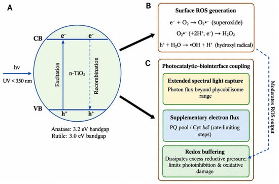
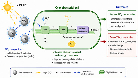
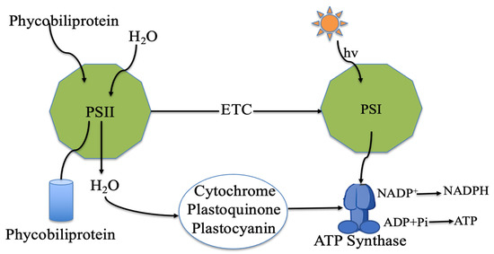
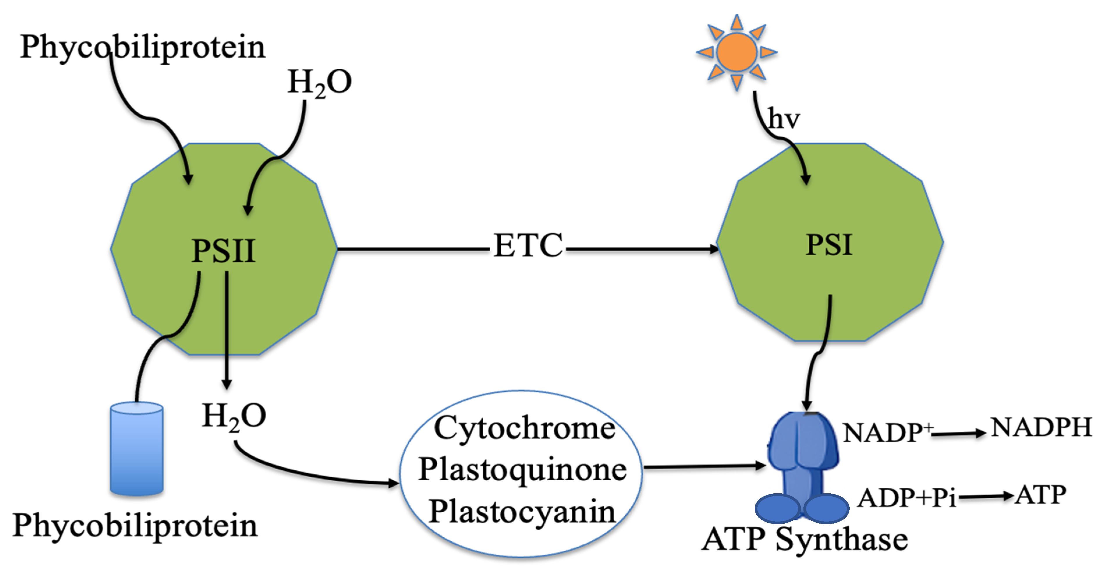

Disclaimer/Publisher’s Note: The statements, opinions and data contained in all publications are solely those of the individual author(s) and contributor(s) and not of MDPI and/or the editor(s). MDPI and/or the editor(s) disclaim responsibility for any injury to people or property resulting from any ideas, methods, instructions or products referred to in the content. |
© 2026 by the authors. Licensee MDPI, Basel, Switzerland. This article is an open access article distributed under the terms and conditions of the Creative Commons Attribution (CC BY) license.