


Abstract
Passiflora edulis seed oil (PESO) is a rich source of lipophilic bioactive compounds with potential to modulate oxidative stress and free glucose levels. This preclinical study aimed to evaluate the toxicological profile, antioxidant and glucose-modulating effects of PESO using hyperglycemic Danio rerio embryos (AB strain). PESO was chemically characterized by GC-FID/GC-MS after derivatization to fatty acid methyl esters. In animal studies, the median lethal concentration (LC50) of PESO at 96 h post-exposure (hpe) was determined using concentrations ranging from 12.5 to 400 μg/mL, and sublethal concentrations corresponding to 1/10 and 1/20 of the LC50 were selected for further investigations, including survival rate up to 144 hpe, continuous and pulsatile hyperglycemia assays (total free glucose was quantified in washed embryo or larval homogenates using an Accu-Chek Active® glucometer) (Roche Diabetes Care GmbH, Mannheim, Germany), total reactive oxygen species (ROS) production, and antioxidant gene expression at 96 hpe. Data were analyzed by nonlinear regression or analysis of variance followed by appropriate post hoc tests, with significance set at p ≤ 0.05. Chemical characterization identified linoleic acid (C18:2) as the predominant fatty acid. PESO exhibited an LC50 of 191.1 µg/mL. PESO at 9.5 and 19.1 µg/mL achieved embryonic survival above 85% and reduced total free glucose values in washed embryo or larval homogenates under pulsatile and continuous hyperglycemic conditions (p ≤ 0.05), although less effective than insulin (0.1 U/mL). PESO attenuated ROS production by 2% (w/v) glucose (p ≤ 0.05). PESO treatment was associated with distinct concentration-related expression profiles among the selected antioxidant-related genes: sod1 and cat expression increased at both concentrations, sod2, gpx1a, and gsr increased at 19.1 µg/mL, and nfe2l2a increased at 9.5 µg/mL (p ≤ 0.05). At the selected sublethal concentrations, PESO showed low embryotoxicity and was associated with reduced total free glucose values, partial attenuation of ROS accumulation, and changes in selected antioxidant-related transcripts in embryos exposed to hyperglycemic conditions.
1. Introduction
Traditional medicine highlights vegetable oils for their emollient properties and as vehicles for bioactive compounds [1,2]. Although historically associated with topical use, vegetable oils have attracted increasing scientific and commercial interest because fatty acids and minor constituents, including tocopherols, phytosterols, and polyphenols, have been reported in these lipid matrices and associated with antioxidant and metabolic properties [3]. These biological effects may be associated with the extraction methods used, as they play central roles in preserving chemical composition and ensuring physiological safety, with cold pressing being particularly effective at mitigating thermal degradation and solvent contamination [4].
Among vegetable oils, Passiflora edulis seed oil (PESO) stands out for its abundant seed availability, driven by its wide geographic distribution and near-continuous fruit production [5]. However, PESO remains largely unexplored, largely due to technological challenges related to its incorporation into aqueous systems and the scarcity of robust toxicological studies [4,6]. Toxicological assessment is particularly relevant because substances of natural origin are not inherently safe and may exhibit adverse effects, including carcinogenic, mutagenic, and teratogenic activities, as well as specific physiological effects, such as emmenagogue action, when not adequately investigated [7,8].
In this context, the zebrafish (Danio rerio) has been established as a highly efficient alternative model for toxicology, pharmacology, and metabolic studies. Notably, the zebrafish exhibits key features relevant to embryonic studies: the transparency of its embryos allows real-time observation of embryogenesis, and its genetic proximity to humans (with approximately 70% gene homology and orthologs for 84% of human disease-associated genes), combined with its easy handling, low maintenance cost, and high fecundity, make it an ideal model for experimental investigations of chemicals and pathophysiological processes [9,10].
Among the latter, hyperglycemia has been recognized as a key factor in systemic inflammation, increased ROS production, and structural damage to sensitive tissues [11]. In human pregnancy, maternal hyperglycemia can affect fetal growth, alter insulin secretion mechanisms, and increase the risk of chronic diseases later in life through fetal programming [11,12]. Thus, hyperglycemic embryonic models offer a unique opportunity to simultaneously evaluate the safety and bioactivity of natural compounds [13,14,15]. The present study aimed to establish the toxicological profile and evaluate the effects of PESO on total free glucose, ROS accumulation, selected antioxidant-related transcripts, and developmental safety in zebrafish embryos exposed to experimentally induced hyperglycemia.
2. Results
2.1. Fatty Acid Composition of PESO
The fatty acid composition of PESO was determined after derivatization to fatty acid methyl esters (FAMEs). The four principal compounds were assigned to their corresponding fatty acids: methyl palmitate, methyl linoleate, methyl cis-9-octadecenoate, and methyl octadecanoate (Table 1).
Table 1.
Fatty acid composition of Passiflora edulis seed oil determined as fatty acid methyl esters.
Linoleic acid (C18:2) was the predominant fatty acid, representing 66.93% of the total chromatographic peak area. Oleic acid (C18:1 cis-9) was the second most abundant fatty acid, accounting for 17.03%, followed by palmitic acid (C16:0; 11.24%) and stearic acid (C18:0; 4.80%). Overall, unsaturated fatty acids accounted for 83.96% of the detected FAMEs, whereas saturated fatty acids represented 16.04%. This profile confirms the predominance of unsaturated fatty acids in PESO, particularly linoleic acid.
2.2. Embryotoxicity, Median Lethal Concentration (LC50) Determination, and Survival Analysis
Physicochemical parameters (temperature and pH values) of the embryo medium were monitored throughout the 96 h post-exposure (hpe) period to ensure stable experimental conditions. The medium remained thermally stable, with a mean temperature of 26.1 ± 0.1 °C, and exhibited a nearly neutral pH (6.9 ± 0.1), with no significant fluctuations across PESO concentrations. No effects were observed in the embryos.
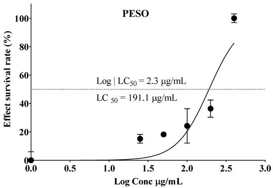
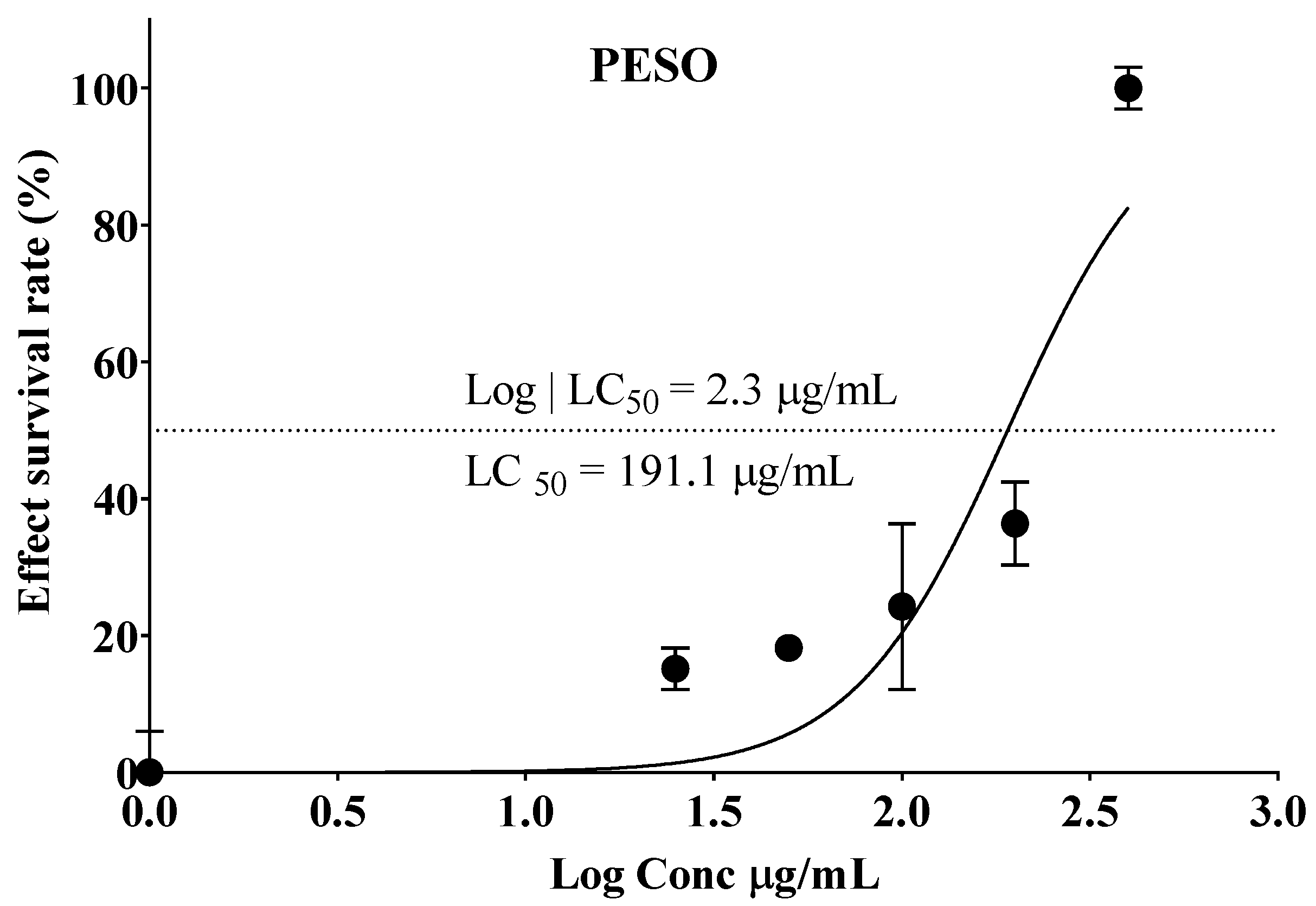
Embryo mortality increased in a concentration-dependent manner, particularly at 200 and 400 μg/mL, which also showed the highest incidence of developmental malformations. The concentration–response relationship was further characterized using nonlinear regression analysis (Figure 1), yielding an estimated median lethal concentration (LC50) of 191.1 µg/mL (95% CI: 123.5–269.0 µg/mL; logLC50 = 2.3).
Figure 1.
Concentration–response curves illustrating the effect of Passiflora edulis seed oil (PESO) on the survival of Danio rerio embryos after 96 h of exposure. Data are expressed as mean ± SEM from three independent biological experiments, each including 20 individually exposed embryos per PESO concentration, corresponding to a total of 60 embryos per concentration. LC50 values were calculated by nonlinear regression using the log(inhibitor) versus normalized response model with a variable slope in GraphPad Prism 9 (GraphPad Software, San Diego, CA, USA). The estimated LC50 for PESO was 191.1 µg/mL (95% CI: 123.5–269.0 µg/mL; logLC50 = 2.3).
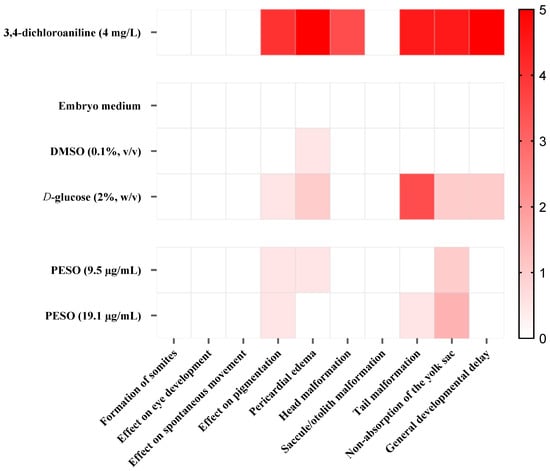
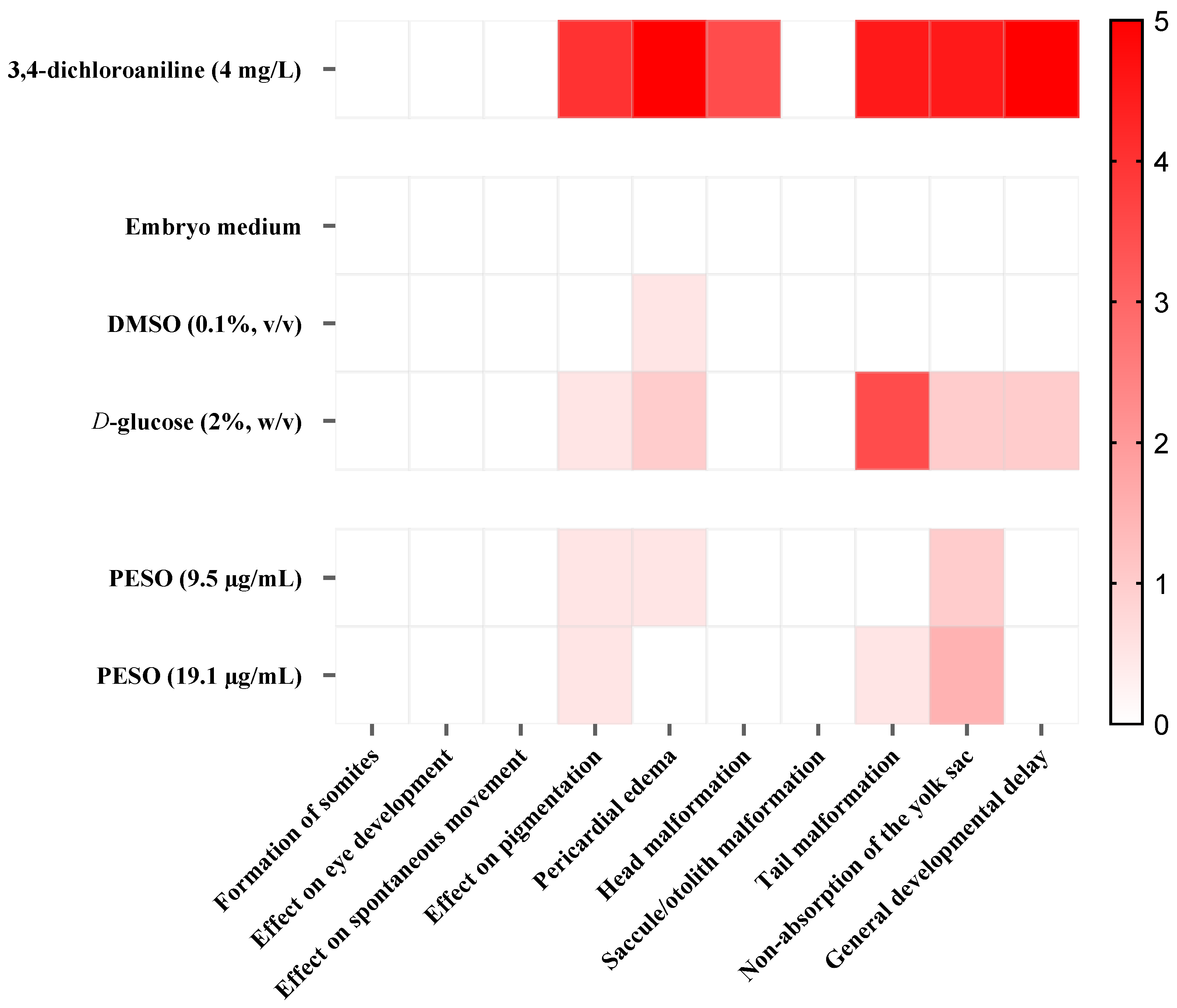
Based on the LC50 value determined for PESO, two sublethal concentrations (1/10 and 1/20 of the LC50; 19.1 and 9.5 μg/mL, respectively) were selected for subsequent embryotoxicity assessment. Developmental endpoints were systematically evaluated at 96 hpe to characterize potential morphological alterations associated with PESO exposure (Figure 2).
Figure 2.
Heatmap of developmental abnormalities in Danio rerio embryos exposed to Passiflora edulis seed oil (PESO). Evaluated endpoints included somite formation, eye development, spontaneous movement, pigmentation, pericardial edema, yolk sac malformation, tail malformation, non-absorption of the yolk sac, and general developmental delay. Positive control (3,4-dichloroaniline), negative control (embryo medium), solvent control (0.1% dimethylsulfoxide—DMSO), and D-glucose (2%, hyperglycemia control) groups are shown. Data are expressed as mean ± SEM (n = 3). Tested PESO concentrations were 19.1 µg/mL (1/10 LC50) and 9.5 µg/mL (1/20 LC50). Color intensity represents the descriptive severity score assigned to each developmental endpoint, ranging from 0 (no observable alteration; white) to 5 (highest observed severity; red). The heatmap is presented as a semiquantitative descriptive visualization.
The positive control (3,4-dichloroaniline) led to the highest frequency and diversity of abnormalities, including alterations in pigmentation, pronounced pericardial edema, tail and head malformations, impaired yolk sac absorption, and marked general developmental delay, confirming the assay’s sensitivity. As expected, embryos from the negative control and solvent control groups exhibited normal development, with only one isolated case of pericardial edema in the solvent control group (Figure 2).
Embryos exposed to 2% (w/v) D-glucose presented moderate developmental disturbances, particularly increased pigmentation defects and tail malformations. Notably, these alterations partially overlapped with those observed in the positive-control experiment, though they occurred at lower frequency and intensity.
The PESO exposure at concentrations of 9.5 µg/mL resulted in a low incidence of abnormalities, primarily mild pigmentation changes, discrete pericardial edema, and occasional yolk sac non-absorption. For PESO (at 19.1 µg/mL), the frequency of alterations remained low but slightly higher than shown above, with isolated occurrences of tail malformation, pigmentation defects, and mild yolk sac non-absorption. Overall, both PESO sublethal concentrations produced only subtle morphological effects, with no indication of severe structural abnormalities (Figure 2).
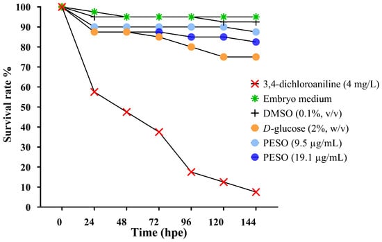
To further characterize potential sublethal effects of PESO, embryo survival was monitored for up to 144 hpe at the two selected concentrations (9.5 and 19.1 μg/mL) and compared with the positive control. As expected, the positive control (3,4-dichloroaniline) led to a marked and progressive decline in survival, dropping to less than 10% at 144 hpe, confirming assay responsiveness (Figure 3). Embryos from the negative and solvent controls maintained consistently high survival rates (>90%) throughout the experiment.
Figure 3.
Survival rates of Danio rerio embryos exposed to 1/10 and 1/20 of the LC50 concentrations of Passiflora edulis seed oil (PESO) over 144 h. Positive control (3,4-dichloroaniline), negative control (embryo medium), solvent control (0.1% DMSO), and D-glucose (2%, hyperglycemia control) groups are shown. Data are expressed as mean ± SEM (n = 3). Tested PESO concentrations were 19.1 µg/mL (1/10 LC50) and 9.5 µg/mL (1/20 LC50). Survival was monitored at 0, 24, 48, 72, 96, and 144 hpe.
Exposure to 2% (w/v) D-glucose resulted in a moderate reduction in survival after 96 hpe, decreasing to 75% at 144 hpe, which is consistent with the known impact of hyperglycemia on embryo viability.
At both PESO concentrations, embryo survival remained stable throughout the exposure period. Embryos treated with 9.5 μg/mL exhibited survival rates comparable to those of the control groups, with no significant decline over time. Similarly, embryos exposed to 19.1 μg/mL exhibited survival above 85% up to 144 hpe, indicating that these sublethal concentrations did not compromise viability. Overall, PESO at 1/10 and 1/20 of the LC50 did not induce time-dependent lethality, supporting the selection of these concentrations for subsequent functional assays (Figure 3).
2.3. Effects of PESO on Total Free Glucose in Washed Embryo and Larval Homogenates Under Hyperglycemic Conditions
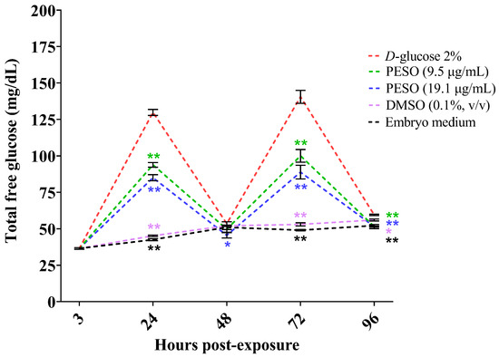
Under the pulsatile hyperglycemia protocol, zebrafish embryos were alternately exposed to glucose-containing medium (2% D-glucose) and embryo medium without glucose at defined developmental time points (3, 24, 48, 72, and 96 hpe) (Figure 4).
Figure 4.
Effect of Passiflora edulis seed oil (PESO) on total free glucose measured in washed embryo or larval homogenates under a pulsatile hyperglycemia protocol in Danio rerio. The pulsatile protocol was designed to evaluate embryo responses during alternating glucose-exposure and glucose-free recovery periods without pharmacological intervention; therefore, no insulin-treated comparison was included. Embryos were subjected to alternating periods of exposure to 2% D-glucose and glucose-free recovery intervals throughout development: 3–24 hpe (hyperglycemia), 24–48 hpe (normoglycemia), 48–72 hpe (hyperglycemia), and 72–96 hpe (normoglycemia). PESO (9.5 and 19.1 µg/mL) was administered during the respective exposure periods. Total free glucose was measured in washed embryo or larval homogenates at 3, 24, 48, 72, and 96 hpe using a portable glucometer. Statistical analysis was performed using two-way ANOVA followed by Dunnett’s post hoc test to compare each treatment group with the hyperglycemic control (2% D-glucose). Significance is indicated as * p < 0.05 and ** p < 0.001. Symbols were color-coded by treatment groups for clarity. Data are expressed as mean ± SEM (n = 3).
At 24, 72, and 96 hpe, total free glucose values measured in washed embryo or larval homogenates differed significantly in all PESO-treated groups compared with the hyperglycemic control exposed to 2% D-glucose, regardless of concentration (p < 0.05 or p < 0.001; Figure 4). Notably, at 48 hpe, corresponding to a glucose-free recovery phase in which all embryos were maintained in glucose-free embryo medium, only embryos previously treated with PESO at 19.1 µg/mL exhibited significantly lower total free glucose values than the 2% D-glucose control group (p < 0.05).
Moreover, at 96 hpe, embryos previously treated with PESO displayed lower total free glucose values in washed homogenates than those measured in both the 2% D-glucose group and the embryo-medium control. This response was more evident at the higher PESO concentration (19.1 µg/mL), suggesting a sustained reduction in the measured free glucose signal during the glucose-free recovery interval.
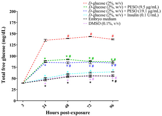
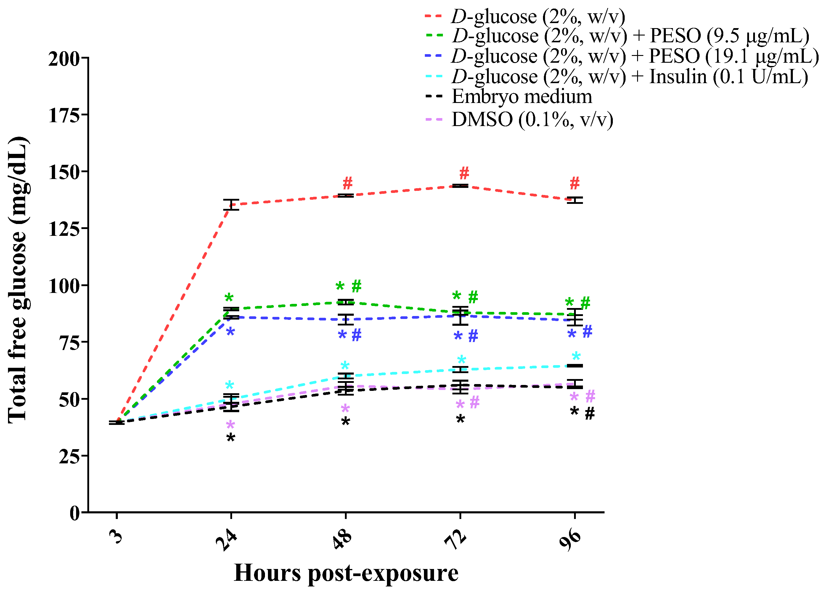
Continuous exposure of Danio rerio embryos to 2% D-glucose resulted in a significant increase in total free glucose values measured in washed embryo or larval homogenates from 24 hpe onward, with elevated values maintained through 96 hpe. Treatment with PESO at 9.5 and 19.1 µg/mL significantly reduced the measured total free glucose values compared with the 2% D-glucose group at all evaluated time points from 24 hpe (p ≤ 0.05). In both PESO-treated groups, the measured values remained below 90 mg/dL throughout the experimental period; however, these values represent glucometer readings obtained from homogenates and should not be interpreted as a systemic normoglycemic range (Figure 5). At 96 hpe, PESO reduced total free glucose values by approximately 36.5% and 38.5% at 9.5 and 19.1 µg/mL, respectively, relative to the D-glucose group, whereas insulin treatment (0.1 U/mL) produced a greater reduction of approximately 53.0%.
Figure 5.
Effect of Passiflora edulis seed oil (PESO) on total free glucose measured in washed embryo or larval homogenates under continuous hyperglycemic conditions in Danio rerio. Insulin (0.1 U/mL) was included as a pharmacological positive control in the continuous protocol to provide a reference under sustained hyperglycemic exposure. Glucose concentrations were measured at 3, 24, 48, 72, and 96 h post-exposure. PESO was tested at 9.5 and 19.1 µg/mL, and insulin (0.1 U/mL) was included as a positive control. Data were analyzed by two-way ANOVA, followed by Dunnett’s post hoc test vs. D-glucose 2% and vs. Insulin 0.1 U/mL group. Significance is indicated as * p ≤ 0.05 vs. D-glucose and # p ≤ 0.05 vs. insulin; symbols are color-coded by treatment group and positioned for clarity. Data are expressed as mean ± SEM (n = 3).
2.4. Effects of PESO on Oxidative Stress
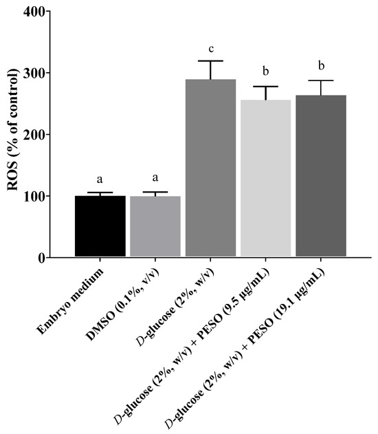
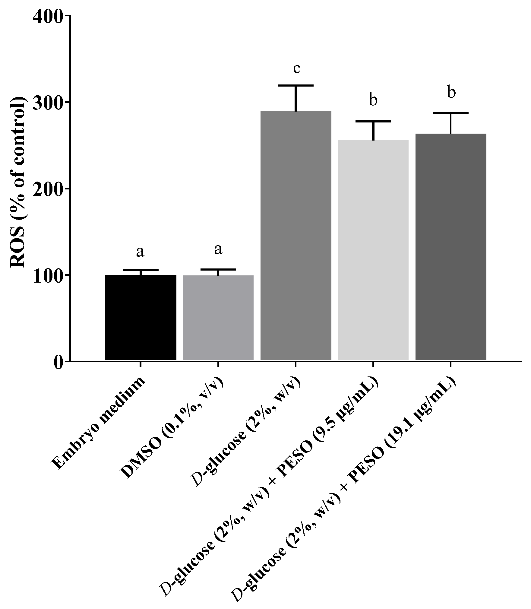
Continuous exposure of Danio rerio embryos to 2% (w/v) D-glucose for 96 h resulted in significantly higher levels of ROS compared with all other experimental groups, including the negative control (embryo medium), solvent control (0.1% dimethylsulfoxide—DMSO), and PESO-treated groups (9.5 and 19.1 µg/mL) (p ≤ 0.05). Treatment with PESO at both concentrations significantly attenuated ROS production relative to the hyperglycemic group (p ≤ 0.05). However, ROS levels in PESO-treated larvae remained significantly higher than those observed in the control groups (Figure 6).
Figure 6.
Effect of P. edulis seed oil (PESO) on reactive oxygen species (ROS) release in zebrafish larvae exposed to continuous hyperglycemia. ROS levels were quantified after 96 h of exposure to 2% D-glucose. Embryo medium was used as the control. Data is presented as mean ± SEM. Statistical analysis was performed using one-way ANOVA followed by Tukey’s post hoc test. Different letters denote statistically significant differences among groups (p ≤ 0.05).
2.5. Expression Profiles of Selected Antioxidant-Related
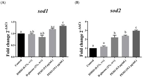
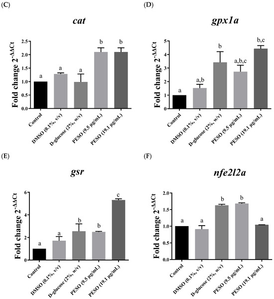
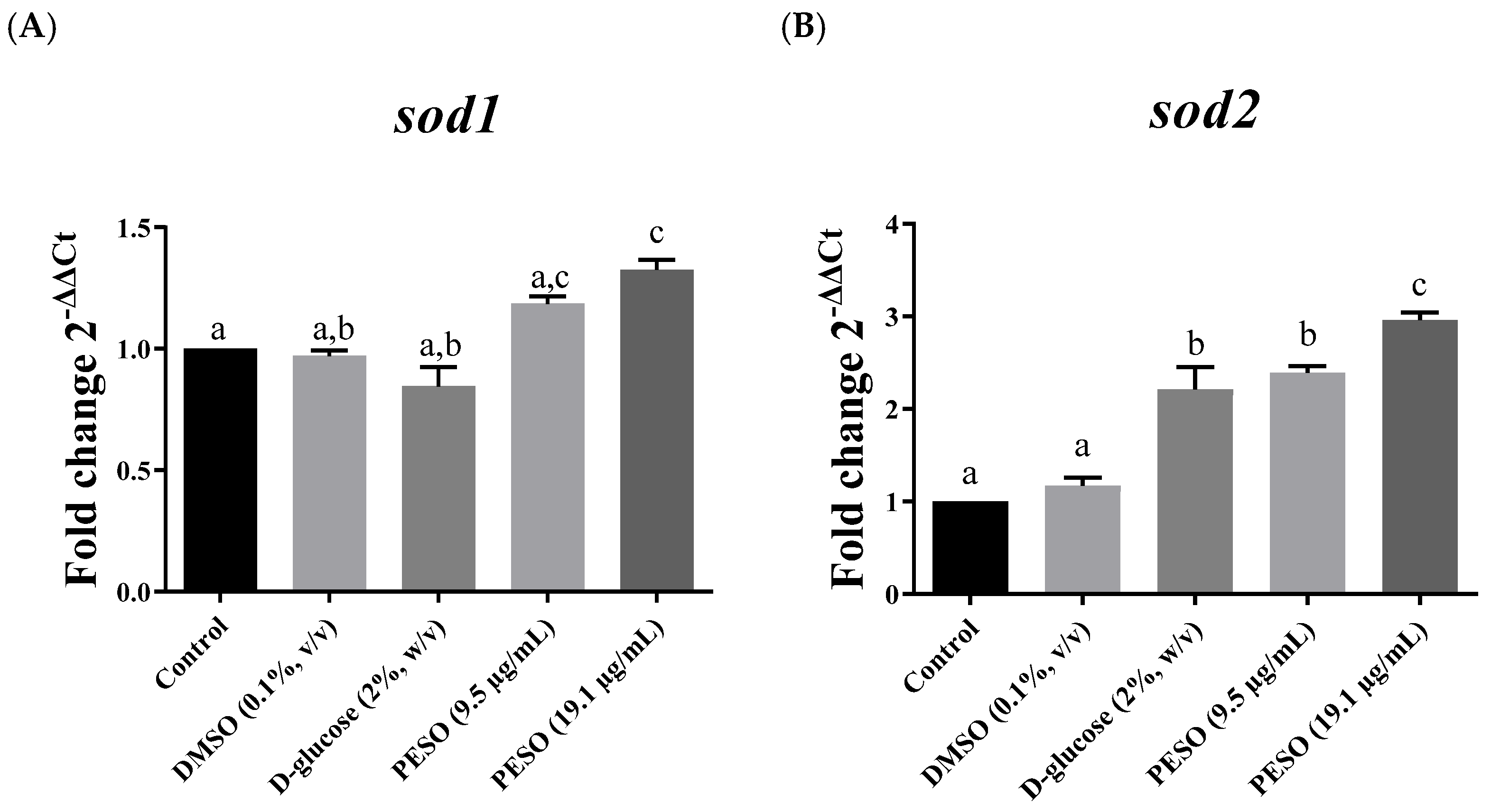
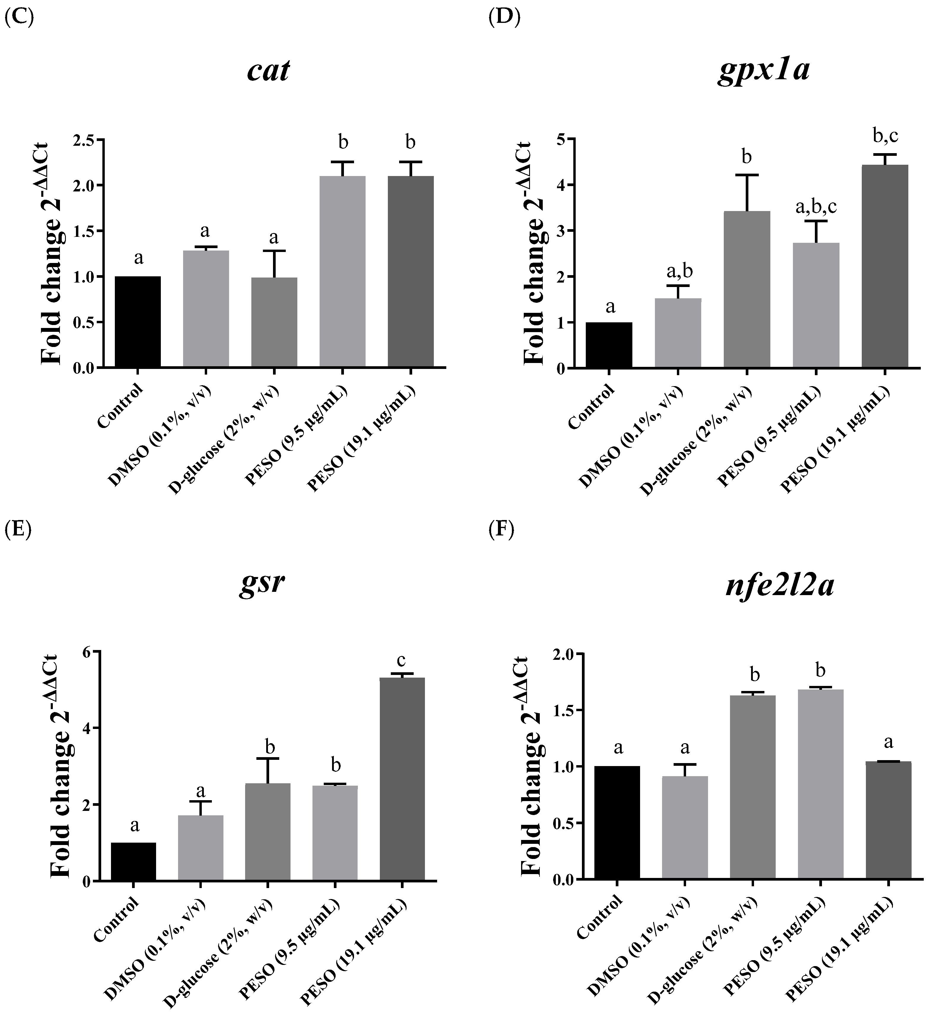
Among the analyzed antioxidant-related genes, distinct expression profiles were observed under hyperglycemic conditions depending on PESO concentration. The genes superoxide dismutase 2 (sod2), glutathione peroxidase 1a (gpx1a), and glutathione reductase (gsr) showed significantly higher expression in hyperglycemic embryos treated with PESO at 19.1 µg/mL (p ≤ 0.05). In contrast, catalase (cat) and superoxide dismutase 1 (sod1) were significantly upregulated in hyperglycemic embryos treated with PESO at both 9.5 and 19.1 µg/mL (p ≤ 0.05). Regarding the transcriptional regulator nuclear factor erythroid 2 related factor 2 (nfe2l2a), increased expression was observed in the hyperglycemic group (2% D-glucose) and in hyperglycemic embryos treated with PESO at 9.5 µg/mL (p ≤ 0.05) (Figure 7).
Figure 7.
Relative transcript abundance of selected antioxidant-related genes in zebrafish embryos exposed to DMSO (0.1%), 2% D-glucose, or PESO (9.5 and 19.1 µg/mL) for 96 hpe. Genes analyzed: (A) sod1 (superoxide dismutase 1), (B) sod2 (superoxide dismutase 2), (C) cat (catalase), (D) gpx1a (glutathione peroxidase 1a), (E) gsr (glutathione-disulfide reductase), and (F) nfe2l2a (nuclear factor erythroid 2-related factor 2). Data are presented as mean ± SEM. Statistical analysis was performed using one-way ANOVA followed by Tukey’s post hoc test. Different letters indicate statistically significant differences among groups (p ≤ 0.05).
3. Discussion
Embryogenesis constitutes a critical period of development during which exposure to adverse metabolic conditions, such as hyperglycemia, can compromise embryonic metabolism and contribute to oxidative stress, inflammation, and long-term developmental consequences [11,16,17]. In humans, hyperglycemia during this period, although managed with insulin therapy, limits the ability to effectively modulate associated inflammatory and redox processes [16,18]. This scenario highlights therapeutic gaps and reinforces the need for strategies that act in an integrated manner across these pathophysiological axes. In this context, the results of this study demonstrate that PESO, despite being a complex lipophilic matrix derived from an agro-industrial by-product, exhibits an adequate safety profile and exerts relevant modulatory effects on glycemia, oxidative stress, and the expression of antioxidant-related genes.
The chemical profile of PESO also supports its biological relevance. In the present study, linoleic acid (C18:2) was the predominant fatty acid, followed by palmitic and stearic acids. Similar findings have been reported for P. edulis seed oil, with linoleic acid consistently identified as the major fatty acid [4,19]. The predominance of linoleic acid is relevant, given that polyunsaturated fatty acids may influence membrane organization, redox homeostasis, and metabolic signaling pathways [20,21,22]. However, the chemical characterization performed in the present study was restricted to fatty acid methyl esters. Therefore, the presence and potential contribution of other minor constituents to the observed biological responses cannot be established from the present data.
In toxicological assessments, the determination of the LC50 is a fundamental parameter for contextualizing the biological safety of compounds in embryonic models. In this regard, the essential oil of Zingiber ottensii Valeton showed an LC50 of 1.003 µg/mL in zebrafish embryos [23], which was associated with pronounced developmental abnormalities, including morphological defects, reduced hatching rate, and decreased heart rate. Similarly, the toxicity of different essential oils has been evaluated, with reported LC50 values of 3.7 µg/mL for lemongrass, 14.4 µg/mL for thyme, and 5.3 µg/mL for oregano, along with embryonic developmental alterations such as reduced growth, spinal deformities, pericardial edema, decreased eye size, and failure of swim bladder inflation [24]. In contrast, Pogostemon cablin oil (patchouli oil) exhibited an LC50 of approximately 120 ppm (120 µg/mL) after 72 h of exposure, indicating an intermediate toxicity profile, with a progressive increase in mortality observed only at higher concentrations [25].
Since toxicological evidence involving fixed oils in zebrafish embryos remains limited, comparisons with essential oils were also considered to provide a broader perspective on the embryotoxic potential of plant-derived lipid compounds. Similarly, the LC50 reported for Pogostemon cablin oil (120 µg/mL at 72 h) was closer to that observed for PESO, which may be partially attributed to similarities in their lipid-rich composition [25]. Comparisons between PESO and essential oils should be interpreted cautiously. Fixed oils are predominantly composed of triglycerides and long-chain fatty acids, whereas essential oils consist mainly of volatile, low-molecular-weight secondary metabolites such as mono- and sesquiterpenes [26]. While both fixed and essential oils may exert antioxidant and metabolic effects, essential oils often exhibit greater biological activity, due to their lipophilicity, which facilitates membrane permeation and interaction with multiple molecular targets [27]. Consequently, LC50 values obtained for fixed and essential oils should not be analyzed as directly equivalent or used to establish the relative safety of these oil categories. Essential oil studies were included only to provide a broad toxicological context because embryotoxicity data for fixed plant oils remain limited. Moreover, the developing zebrafish possesses stage-dependent detoxification and antioxidant-defense capacities, which may influence susceptibility to different chemical matrices. Therefore, the LC50 value observed for PESO should be evaluated within the specific experimental conditions of the present study rather than as evidence that fixed oils are generally better tolerated than essential oils.
With respect to developmental outcome, several studies have demonstrated that exposure to fixed and/or essential oils during zebrafish embryogenesis is frequently associated with pronounced developmental abnormalities, including reduced hatching rates, decreased heart rate, impaired growth, spinal deformities, pericardial edema, reduced eye size, and failure of swim bladder inflation [23,24,25]. In the present study, embryos exposed to sublethal concentrations of PESO (1/10 and 1/20 of the LC50) exhibited only mild developmental alterations, mainly characterized by subtle pigmentation changes, discrete pericardial edema, and occasional incomplete yolk sac absorption. In contrast, embryos subjected to hyperglycemic conditions displayed markedly more severe developmental abnormalities, including pronounced pericardial edema, pigmentation alterations, malformations of the tail and head, impaired yolk sac absorption, and an overall delay in embryonic development.
The predominance of developmental abnormalities under hyperglycemic conditions is consistent with previous reports demonstrating that elevated glucose levels exert direct deleterious effects on zebrafish embryos. In particular, hyperglycemia has been associated with ocular defects, including alterations in retinal layer thickness, increased macrophage infiltration, and reductions in Müller glia and retinal ganglion cells, highlighting the susceptibility of neural and sensory tissues to glucose-induced metabolic stress during early development [28].
D-glucose has been widely employed as an exogenous agent to induce hyperglycemic conditions during zebrafish embryogenesis, at concentrations comparable to those reported in animal models and indicative of glycemic fluctuations observed under diabetic conditions [28,29]. The zebrafish model is particularly well-suited for investigating hyperglycemia-related developmental disturbances, as embryos exhibit fluctuating hyperglycemia within clinically relevant ranges and develop externally, allowing direct exposure to glucose [30,31]. This characteristic circumvents the placental barrier present in mammalian models, which can modulate glucose transfer and confound the direct assessment of embryonic effects. While mammalian systems remain essential for understanding complex maternal–fetal interactions, zebrafish embryos offer a unique and powerful platform for early mechanistic investigations of hyperglycemia-induced toxicity and developmental disruption [32,33].
Corroborating the findings discussed above, exposure to hyperglycemic conditions emerged as a major developmental stressor, inducing morphological abnormalities during early embryogenesis and being accompanied by higher total free glucose values in washed embryo or larval homogenates and increased ROS production. Sustained exposure to 2% D-glucose aggravated oxidative stress in zebrafish embryos, reinforcing the deleterious effects of the glucose-enriched microenvironment. Notably, co-exposure to PESO consistently attenuated these alterations under both pulsatile and continuous protocols, reducing the measured total free glucose values and limiting ROS accumulation compared with the hyperglycemic control. Under continuous exposure, the effect of PESO was less pronounced than that of insulin. Collectively, these findings support a modulatory effect of PESO on total free glucose measured in washed homogenates, together with attenuation of oxidative stress, rather than demonstrating a direct systemic hypoglycemic action [28,34,35].
From a mechanistic perspective, the concomitant reductions in total free glucose values and ROS accumulation raise the hypothesis that the lipid composition of PESO may contribute to the observed metabolic and redox-related responses. Linoleic and oleic acids, as well as minor constituents such as tocopherols and phytosterols, had been reported to influence energy- and redox-related processes in other experimental contexts [4]. However, the individual contribution of these constituents was not evaluated in the present study. Moreover, mitochondrial function, cellular energy metabolism, insulin signaling, glucose transport, and gluconeogenic pathways were not assessed. Therefore, the present findings do not demonstrate that PESO directly modulates these pathways or definitively improves the intracellular redox environment. Rather, they indicate an association between PESO exposure and lower total free glucose values, partial attenuation of ROS accumulation, and changes in selected antioxidant-related transcripts under glucose-induced metabolic stress. Further mechanistic studies are required to determine the cellular pathways underlying these responses [34,36].
Because zebrafish embryos were directly exposed to glucose and PESO in the absence of placental mediation, the observed responses occurred without a maternal or placental interface. Nevertheless, this experimental design does not establish a direct molecular protective mechanism of PESO. In addition, ROS levels were assessed only at 96 hpe; therefore, the present findings describe the oxidative status at the end of the continuous exposure period and do not establish the temporal onset or progression of ROS modulation. Time-resolved analyses, together with direct assessments of mitochondrial and metabolic function, will be required to characterize the mechanisms and dynamics of this response.
Within the zebrafish model, which enables direct embryonic exposure to glucose in the absence of placental modulation, PESO treatment was associated with lower total free glucose values and partial attenuation of ROS accumulation under glucose-induced metabolic stress. The concomitant occurrence of these responses suggests a possible relationship between free glucose reduction and redox modulation. However, because mitochondrial function, cellular energy metabolism, insulin signaling, glucose transport, and gluconeogenic pathways were not evaluated, the present findings do not establish a direct protective mechanism or an integrated mode of action. It is important to note that ROS levels were assessed only at 96 hpe; therefore, the present findings reflect the oxidative status at the end of the continuous exposure period and do not establish the temporal onset or progression of ROS modulation by PESO. Time-resolved analyses at earlier developmental stages will be required to characterize the dynamics of this response. These findings provide a basis for the subsequent discussion of the changes observed in selected antioxidant-related transcripts under glucose-induced metabolic stress [28].
Consistent with the previously observed attenuation of ROS levels, PESO treatment was associated with distinct concentration-related expression profiles among the selected antioxidant-related genes, suggesting the differential involvement of components of endogenous antioxidant defense. The upregulation of sod1 and cat (Figure 7) at both tested concentrations suggests the involvement of cytosolic antioxidant defenses across the evaluated PESO exposure levels. The corresponding enzymes constitute important components of the first line of defense against oxidative stress, with SOD1 catalyzing the dismutation of superoxide radicals and CAT preventing hydrogen peroxide accumulation following superoxide dismutation. Their responsiveness at the lower PESO concentration suggests that these antioxidant defenses may be recruited under glucose-induced oxidative stress even at the lower exposure level [37,38].
In contrast, the selective upregulation of sod2, gpx1a, and gsr at 19.1 µg/mL suggests the additional involvement of mitochondrial and glutathione-dependent antioxidant mechanisms at the higher PESO concentration. SOD2 operates within the mitochondrial compartment, detoxifying superoxide generated by the electron transport chain, whereas GPx1a and GSR are central components of the glutathione system, contributing to peroxide reduction and maintenance of intracellular redox homeostasis through glutathione recycling. The preferential upregulation of these genes at 19.1 µg/mL therefore indicates a concentration-associated expression profile involving mitochondrial and glutathione-dependent antioxidant defenses under glucose-induced oxidative stress. Notably, nfe2l2a was upregulated at 9.5 µg/mL, which is consistent with the role of Nrf2 as a redox-sensitive transcriptional regulator involved in antioxidant responses. Together, these findings indicate that, under glucose-induced metabolic stress, PESO treatment was associated with coordinated but distinct concentration-related expression profiles among the selected antioxidant-related genes. The lower concentration was associated primarily with nfe2l2a, sod1, and cat responses, whereas the higher concentration was additionally associated with sod2, gpx1a, and gsr expression [39,40,41].
Although PESO exhibited promising biological effects, certain methodological constraints must be considered. This investigation was conducted solely using a zebrafish embryonic model, which, despite being highly informative for early developmental, metabolic, and redox-related processes, does not encompass the full physiological complexity of mammalian systems. Notably, the absence of placental mediation in zebrafish embryos limits the direct translation of these findings to mammalian gestational contexts, in which the placenta plays a pivotal role in regulating glucose transport, redox homeostasis, and fetal metabolic programming. Accordingly, the translational implications of the protective responses observed under hyperglycemic conditions should be interpreted with appropriate caution. Furthermore, the experimental approach was restricted to exposure during early embryogenesis and did not address long-term, chronic, or reproductive outcomes.
The pulsatile and continuous protocols were designed to address distinct experimental questions. The pulsatile protocol evaluated embryonic responses during alternating periods of glucose exposure and glucose-free recovery without pharmacological intervention, whereas insulin was included in the continuous protocol as a pharmacological reference under sustained hyperglycemic exposure. Consequently, the absence of an insulin-treated group in the pulsatile protocol precludes the establishment of a common pharmacological benchmark across the two paradigms, and the magnitude of PESO-associated responses should not be directly compared between them.
Regarding the molecular analyses, although actb was selected after comparison with elfa and gapdh, normalization using multiple validated reference genes could provide additional robustness in future studies. PESO-only groups were included in the embryotoxicity and survival assessments but were not included in the ROS and gene expression experiments. Therefore, the present study cannot determine whether PESO alters basal redox status or antioxidant-related gene expression in embryos maintained without glucose exposure. The redox and transcriptional findings are thus restricted to the effects associated with PESO treatment in the presence of glucose-induced metabolic stress. In addition, the qPCR findings reflect relative transcript abundance and should not be interpreted as direct evidence of increased antioxidant enzyme function. Protein abundance and the enzymatic activities of SOD, CAT, GPx, and GSR were not evaluated, and the GSH/GSSG ratio was not measured. Consequently, the functional consequences of the observed transcriptional changes remain open. Protein abundance and the enzymatic activities of SOD, CAT, GPx, and GSR were not evaluated, and the GSH/GSSG ratio was not measured. Consequently, the functional consequences of the observed transcriptional changes remain to be established. Inflammation is retained only in the general background as a recognized consequence of hyperglycemia and is not attributed as an experimentally demonstrated effect of PESO.
Glucose was quantified as total free glucose in washed embryos or larval homogenates using a portable glucometer. The experimental design included an embryo-medium negative control, a 2% D-glucose hyperglycemic control, and an insulin-treated positive control, and embryos or larvae were rinsed three consecutive times before homogenization. Nonetheless, the efficacy of exogenous-glucose elimination was not autonomously substantiated by a washing-gradient experiment, and the glucometer readings were not corroborated by an independent enzyme-based quantification technique. Consequently, the findings are to be regarded as comparative assessments of total free glucose in washed homogenates instead of a direct or specific quantification of blood glucose or intracellular glucose. Future studies incorporating these methodological refinements, together with long-term evaluations in complementary experimental models, will be important for a more comprehensive assessment of both safety and efficacy, particularly when considering the potential application of bioactive compounds in gestational or pediatric contexts.
4. Materials and Methods
4.1. Chemical
All reagents were of analytical grade. DMSO, 3,4-dichloroaniline, D-glucose, phosphate-buffered saline (PBS), 2′,7′-dichlorodihydrofluorescein diacetate (H2DCFDA), and nuclease-free water were obtained from Sigma-Aldrich (St. Louis, MO, USA). RNAlater® Solution and PureLink™ RNA Mini Kit were purchased from Invitrogen/Thermo Fisher Scientific (Waltham, MA, USA). The High-Capacity cDNA Reverse Transcription Kit and SYBR® Green Master Mix were obtained from Applied Biosystems (Foster City, CA, USA). The QuantiNova® SYBR Green PCR Kit was acquired from Qiagen (Hilden, Germany).
Insulin (human recombinant) was obtained from Novo Nordisk (Bagsværd, Denmark). Accu-Chek Active® glucometer and test strips were supplied by Roche Diagnostics (Mannheim, Germany). Commercial zebrafish feed (Alcon Basic®, Camboriú, Brazil) and Artemia salina cysts (Artemia do RN®, Natal, Brazil) were also used. Gene-specific primers for sod1, sod2, cat, gpx1a, gsr, nfe2l2a, and actb were synthesized by Integrated DNA Technologies (IDT, Coralville, IA, USA).
4.2. Preparation of Passiflora edulis Seed Oil (PESO)
Seeds of Passiflora edulis were obtained from organically cultivated sour passion fruits supplied by Paraíso Agrícola Farm, a member of the local family farming cooperative in Matriz do Camaragibe, Alagoas, Brazil (9°09′24″ S, 35°31′45″ W). The botanical material was identified by researchers from the Herbarium MAC of the Instituto do Meio Ambiente do Estado de Alagoas, Brazil, and a voucher specimen was deposited under registration number MAC 78263, corresponding to Passiflora edulis Sims (Passifloraceae). The use of Brazilian genetic heritage was registered in SisGen under code A1CA908.
After harvest, the seeds were manually separated from the pulp, thoroughly washed under running water, and dried in a forced-air oven at 50 °C for 48 h. The dried seeds were then mechanically pressed using a 15-t hydraulic press (Marcon, MPH-15, Marília, Brazil) operated at ambient temperature (20–25 °C) without external heating or cooling. The resulting crude PESO was left undisturbed for ~48 h to allow sedimentation. The clarified upper fraction was carefully decanted, avoiding contact with the sediment layer, transferred to sterile amber containers, and stored at 4 °C until use.
4.2.1. Fatty Acid Transesterification
FAMEs were prepared according to the method described by Joseph and Ackman [42]. Briefly, PESO (25.0 ± 0.1 mg) was mixed with 1.5 mL of 0.5 mol/L NaOH in methanol and heated at 100 °C under stirring for 5 min. After cooling, 2.0 mL of 12% (v/v) BF3 in methanol was added, and the reaction mixture was maintained at 100 °C under stirring for 30 min. Subsequently, 1 mL of hexane was added, followed by vigorous mixing for 30 s and the addition of 5 mL of saturated NaCl solution. The mixture was refrigerated until complete phase separation. The upper organic phase was collected, combined with an additional 1 mL of hexane, concentrated, and stored at −18 °C under an N2 atmosphere until chromatographic analysis.
4.2.2. Gas Chromatography with Flame Ionization Detection (GC-FID)
FAMEs were analyzed using a GC-2010 Plus gas chromatograph equipped with a flame ionization detector (Shimadzu Corporation, Kyoto, Japan) and an NTS capillary column (30 m × 0.25 mm i.d. × 0.25 μm). Nitrogen was used as a carrier gas at a column flow rate of 0.71 mL/min. The oven temperature was initially set at 120 °C and increased to 300 °C at 6 °C/min. Analyses were performed in split mode by injecting 1 μL of the esterified oil solution prepared in HPLC-grade hexane.
4.2.3. Gas Chromatography–Mass Spectrometry (GC-MS)
GC-MS analysis was performed using a Shimadzu QP-2010 gas chromatograph coupled to a mass spectrometer and fitted with an SH-5MS capillary column (30 m × 0.25 mm i.d. × 0.25 μm; Shimadzu). Helium was used as the carrier gas at a flow rate of 0.71 mL/min. Mass spectra were acquired in electron ionization mode at 70 eV. The oven temperature was initially maintained at 125 °C for 5 min, increased to 300 °C at 6 °C/min, and held at this temperature for 10 min. A 1 μL aliquot of the esterified oil solution prepared in HPLC-grade hexane was injected in splitless mode.
4.2.4. Identification and Quantification of Fatty Acid Methyl Esters
FAMEs were identified by comparing their retention times with authentic fatty acid methyl ester standards and by analyzing their mass fragmentation patterns and retention indices, which were compared with spectral library data, including NIST and Wiley databases. Quantification was performed from GC-FID peak areas using methyl palmitate as an external reference standard. The calibration curve was constructed using methyl palmitate solutions ranging from 10 to 75 ppm, and FAME concentrations were expressed as µg/mL. Relative abundance was calculated as the percentage of the total chromatographic peak area.
4.3. Animal Model
All procedures involving adult zebrafish and their embryos were approved by the Animal Use Ethics Committee of the Federal University of Alagoas (UFAL; protocol no. 13/2022) and were conducted in accordance with applicable guidelines for the care and use of laboratory animals. Acute toxicity was assessed using the Fish Embryo Acute Toxicity (FET) test in accordance with OECD Guideline 236 [43]. The median lethal concentration (LC50) was determined and used to select sublethal concentrations for the subsequent embryotoxicity, metabolic, biochemical, and molecular assays [44].
Adult zebrafish (Danio rerio, AB strain) were maintained in a recirculating aquaculture system under controlled conditions (28 °C; pH 6.7–7.2; dissolved oxygen approximately 6 mg/L; conductivity 1600 µS/cm; total ammonia < 0.01 mg/L). Fish were kept at a density of 2 fish/L under a 14 h light/10 h dark photoperiod (lights on at 07:00; 190 lux) and were fed three times daily with Artemia salina nauplii and a commercial diet containing 60% protein and 15% lipids.
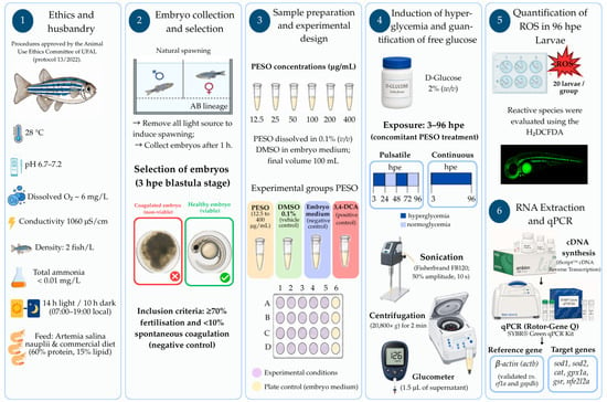
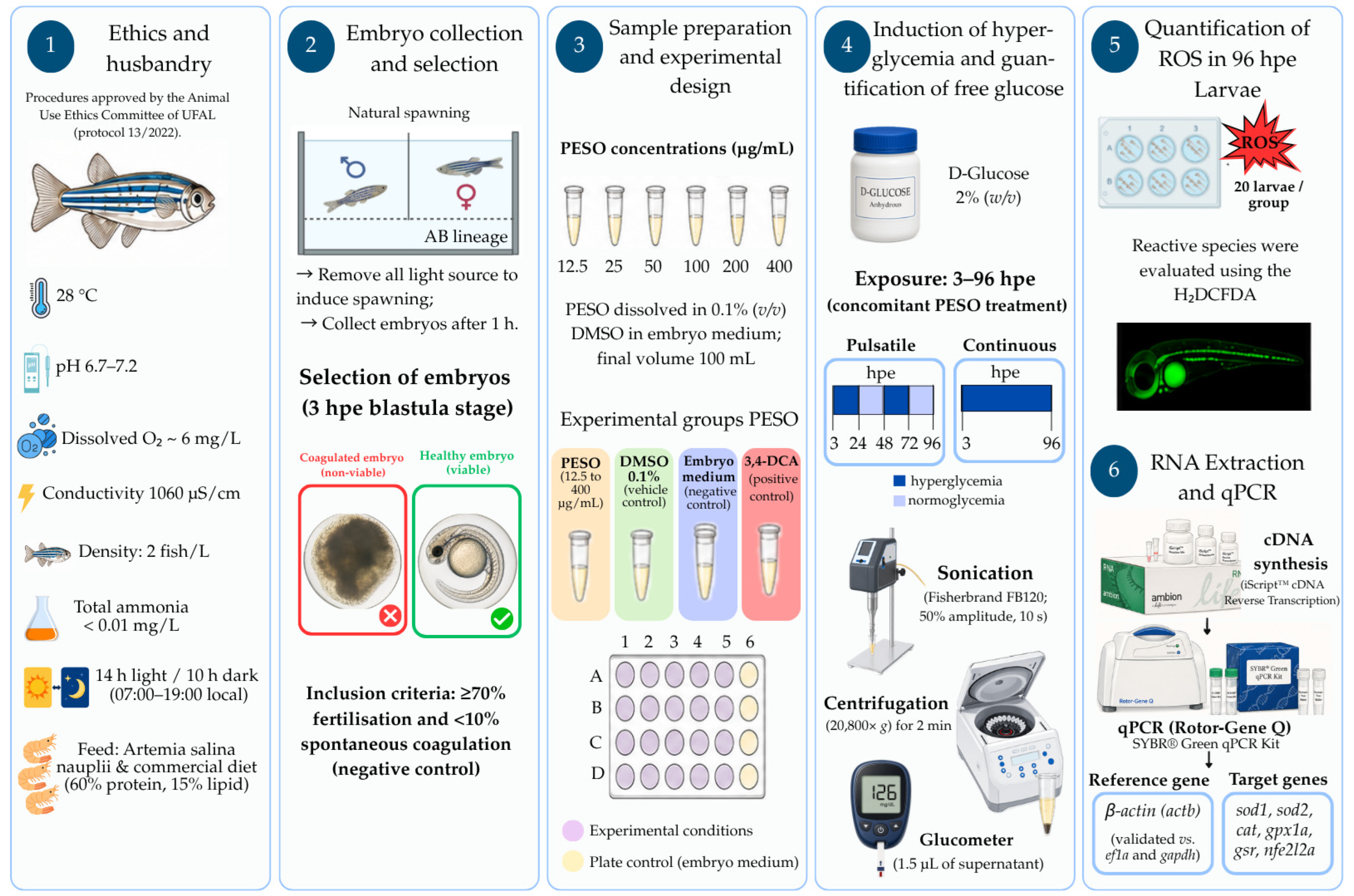
Embryos were obtained by natural spawning. Adult breeders were placed in mating tanks at a male-to-female ratio of 3:2 and separated overnight by a perforated translucent divider that permitted visual and chemical contact. At light onset, the divider was removed, and embryos were collected after 1 h. Fertilization and developmental stage were confirmed under a stereomicroscope (40×). Only normally developing embryos at the blastula stage (2–3 h post-exposure, hpe) from batches showing ≥70% viability and <10% spontaneous coagulation in the negative control were included. The experimental workflow is summarized in Figure 8.
Figure 8.
Schematic workflow of the experimental design used to evaluate the effects of PESO in Danio rerio.
The figure summarizes the main experimental steps, including embryo collection and selection, exposure to PESO and control groups, acute toxicity assessment, hyperglycemia induction, and subsequent biochemical and molecular analyses.
4.3.1. Sample Preparation, Experimental Design, and Sample-Size Determination
PESO working solutions were prepared in embryo medium using DMSO as the solvent, with a final DMSO concentration of 0.1% (v/v). All treatment and control solutions were freshly prepared. The controls were selected according to the objective of each assay: embryo medium served as the negative control, 0.1% DMSO as the solvent control, 2% (w/v) D-glucose as the hyperglycemic control, 3,4-dichloroaniline (4 mg/L) as the positive control for embryotoxicity, and insulin (0.1 U/mL) as the glycemic recovery control.
Three independent biological experiments were performed. In the acute toxicity and embryotoxicity assays, 20 embryos were allocated to each experimental group in each independent experiment, resulting in 60 embryos per group. Embryos were maintained individually in 24-well plates (one embryo per well), and four additional wells containing embryos in embryo medium were included as internal plate controls. For these assays, the individual embryo was considered the experimental unit. For glucose, ROS, and gene expression analyses, embryos or larvae were pooled, and each independently prepared pool was considered one experimental unit. Technical replicate readings were averaged and were not treated as independent biological observations.
Sample sizes were determined a priori in the experimental protocol approved by the institutional ethics committee. The number of embryos used in the toxicity and embryotoxicity assays was based on OECD Guideline 236 [40], whereas the pool sizes used for biochemical and molecular analyses were based on previously published zebrafish studies and on the minimum amount of biological material required for reliable glucose, ROS, and RNA measurements. The design also followed the reduction principle of the 3Rs. No post hoc adjustment of sample size was performed [43].
4.3.2. Acute Toxicity Assessment and Determination of LC50 in Danio rerio Embryos
Acute toxicity was evaluated in embryos exposed to PESO at 12.5, 25, 50, 100, 200, or 400 µg/mL for 96 h. At 2–3 hpe, viable embryos were randomly selected from the pooled spawn, rinsed, and transferred individually to 24-well plates containing 2 mL of the corresponding test solution per well. The assay included embryo medium as the negative control, 0.1% (v/v) DMSO as the solvent control, and 3,4-dichloroaniline (4 mg/L) as the positive control. Each group contained 20 embryos per independent experiment.
A semi-static exposure system was used, with complete renewal of all solutions every 24 h. Embryos were examined daily under a stereomicroscope throughout the 96-h exposure. Lethality was defined according to OECD Guideline 236 by the presence of at least one of the following endpoints: embryo coagulation, absence of somite formation, failure of tail detachment from the yolk sac, or absence of cardiac activity [43].
Hatching and non-lethal developmental alterations were also recorded but were not included in the LC50 calculation.
Cumulative mortality at 96 h was used to construct concentration–response curves. The LC50 was estimated by nonlinear regression, and concentrations corresponding to 1/10 and 1/20 of the LC50 were selected for the subsequent embryotoxicity and functional assays.
4.3.3. Embryotoxicity and Survival Assessment up to 144 Hpe
The developmental safety of the two selected sublethal PESO concentrations (9.5 and 19.1 µg/mL, corresponding to 1/20 and 1/10 of the LC50, respectively) was evaluated in an independent 144 hpe embryotoxicity assay. The experimental groups were embryo medium (negative control), 0.1% (v/v) DMSO (solvent control), 2% (w/v) D-glucose (hyperglycemic control), 3,4-dichloroaniline (4 mg/L; positive embryotoxicity control), PESO 9.5 µg/mL, and PESO 19.1 µg/mL. Each group comprised 20 embryos per independent experiment, and three independent biological experiments were conducted.
Exposure began at 2–3 hpe and was conducted under semi-static conditions, with complete renewal of the corresponding solutions every 24 h. Survival was recorded at the start of exposure and at 3, 24, 48, 72, 96, 120 and 144 hpe. The same OECD lethality criteria defined for the acute toxicity assay were applied, and non-viable embryos or larvae were removed when detected.
Developmental endpoints were assessed under a stereomicroscope and included somite formation, eye development, spontaneous movement, pigmentation, pericardial oedema, cranial and caudal malformations, yolk-sac malformation or incomplete absorption, and general developmental delay. The occurrence of each abnormality was summarized across the three independent experiments using the 0–5 ordinal scale employed for heatmap visualization, in which 0 indicated absence, and 5 represented the highest observed frequency.
4.3.4. Induction of Hyperglycemia and Quantification of Total Free Glucose in Washed Danio rerio Embryo and Larval Homogenates
Hyperglycemia was induced by exposing embryos to 2% (w/v) D-glucose from 3 to 96 hpe. The experimental groups included embryo medium, 0.1% (v/v) DMSO, 2% D-glucose, 2% D-glucose plus PESO at 9.5 or 19.1 µg/mL, and, in the continuous-exposure experiment, 2% D-glucose plus insulin (0.1 U/mL; 2 h exposure) as an insulin-treated positive control. Embryos were maintained in 6-well plates containing 10 mL of the corresponding solution per well [28]. For each treatment and sampling time, each biological replicate consisted of a pool of 60 embryos or larvae.
Two exposure paradigms, designed to address distinct experimental questions, were evaluated. The pulsatile protocol was conceived as an exploratory exposure–recovery paradigm to characterize the metabolic responses of zebrafish embryos during alternating periods of glucose exposure and recovery in glucose-free embryo medium, without pharmacological intervention. Embryos were exposed to glucose-containing solutions from 3 to 24 hpe and from 48 to 72 hpe and were maintained in glucose-free embryo medium during the recovery intervals from 24 to 48 hpe and from 72 to 96 hpe. PESO was administered during the corresponding glucose-exposure periods. Because this protocol was intended to evaluate embryo responses during glucose exposure and subsequent recovery without pharmacological interference, insulin was not included. In contrast, the continuous protocol was designed to evaluate PESO under sustained hyperglycemic conditions. Embryos remained exposed to 2% D-glucose, alone or in combination with PESO, from 3 to 96 hpe, with complete renewal of all solutions every 24 h. In this protocol, insulin was included as a pharmacological positive control to provide a reference response under continuous glucose exposure.
Independent pools were collected at 3, 24, 48, 72, and 96 hpe. At each sampling time, the exposure medium was completely removed, and embryos or larvae were rinsed three consecutive times with Milli-Q water. Only the washed biological material was transferred to sterile 2 mL tubes, and no exposure medium was included in the homogenates. Embryos or larvae were then euthanized by cooling and lysed by sonication in 200 μL PBS (Fisherbrand FB120; 50% amplitude, 10 s). Homogenates were centrifuged at 14,000 rpm (20,800× g) for 2 min, and the supernatants were retained for glucose quantification.
Total free glucose in the supernatants of washed embryo or larval homogenates was measured using an Accu-Chek Active® glucometer (Roche Diagnostics, Rotkreuz, Switzerland). For each biological sample, 1.5 μL of supernatant was applied to the test strip, and three consecutive technical readings were obtained. The mean of the three readings was used as the value for that biological replicate. Three independent biological replicates were analyzed for each treatment and sampling time. The analytical readout was interpreted as total free glucose in the washed homogenate and not as a direct measurement of blood glucose or selective intracellular glucose.
4.3.5. Quantification of ROS in Danio rerio Larvae
ROS production was quantified at 96 hpe as a terminal endpoint of the continuous hyperglycemia protocol. The groups comprised embryo medium, 0.1% DMSO, 2% D-glucose, and 2% D-glucose plus PESO at 9.5 or 19.1 µg/mL. For each condition, three independent biological replicates were analyzed, each consisting of a pool of 20 larvae. Larvae were euthanized by cooling, washed three times with ice-cold PBS (pH 7.4), lysed by sonication in 200 μL PBS (Fisherbrand FB120; 50% amplitude, 10 s), and centrifuged at 10,000× g for 10 min at 4 °C [45].
Reactive species were detected using 2′,7′-dichlorodihydrofluorescein diacetate (H2DCFDA; Thermo Fisher Scientific). For each reaction, 40 μL of supernatant was incubated with 40 μM H2DCFDA for 30 min at 37 °C in the dark. Fluorescence was measured using a GloMax® Multi Detection System (Promega Corporation, Madison, WI, USA) at 490 nm excitation and 530 nm emission.
Each biological sample was analyzed in technical quintuplicate, and the mean of the five readings was used as one biological value. ROS levels were expressed as a percentage of the negative control.
4.3.6. RNA Extraction and Quantitative Real-Time Polymerase Chain Reaction (qPCR)
Gene expression analyses were performed at 96 hpe using the same continuous-exposure groups evaluated in the ROS assay. Three independent biological replicates were analyzed per group, each consisting of a pool of 20 larvae. Larvae were euthanized by cooling and immediately preserved in RNAlater® (Thermo Fisher Scientific). Total RNA was extracted using the PureLink™ RNA Mini Kit (Life Technologies, Carlsbad, CA, USA) according to the manufacturer’s instructions, and RNA concentration and purity were assessed using a NanoDrop™ One spectrophotometer (Thermo Fisher Scientific, Waltham, MA, USA).
RNA samples were normalized to 100 ng/µL before cDNA synthesis using the High-Capacity cDNA Reverse Transcription Kit (Applied Biosystems). qPCR reactions were prepared in a final volume of 10 µL containing 10 ng cDNA, 5 µL SYBR® Green Master Mix, 1 µL forward primer, 1 µL reverse primer, 2 µL nuclease-free water, and 1 µL cDNA. Amplification was performed on a Rotor-Gene Q system (Qiagen, Hilden, Germany).
The relative transcript abundance of sod1, sod2, cat, gpx1a, gsr, and nfe2l2a was calculated using the 2−ΔΔCt method [46]. β-Actin (actb) [47] was used as the reference gene after empirical comparison with elongation factor 1-alpha (elfa) and glyceraldehyde-3-phosphate dehydrogenase (gapdh). Among the three candidate genes, actb showed the lowest intra-group and inter-group variability under glucose and PESO exposure and was therefore selected for normalization.
The transcriptional panel was selected a priori as a targeted exploratory set representing superoxide dismutation (sod1 and sod2), hydrogen peroxide removal (cat and gpx1a), glutathione recycling (gsr), and redox-responsive transcriptional regulation (nfe2l2a). The panel was not intended to provide a comprehensive characterization of the Nrf2–Keap1 regulatory pathway.
All qPCR reactions were performed in technical triplicate, and the mean cycle-threshold value was used for each biological replicate. Primer sequences, accession numbers, expected amplicon sizes, and melting temperatures are provided in Table 2. The reporting of the qPCR procedures followed MIQE recommendations [48,49].
Table 2.
Primer sequences for gene expression analysis and associated parameters.
4.4. Statistical Analysis
Statistical analyses were performed using GraphPad Prism 9.0 (GraphPad Software, San Diego, CA, USA). Data are presented as mean ± standard error of the mean (SEM), and p ≤ 0.05 was considered statistically significant. The value of n represents independent biological experiments or independently prepared biological pools. Technical replicate measurements were averaged before statistical analysis.
For the acute toxicity assay, the LC50 and its concentration–response curve were obtained by nonlinear regression using the log(inhibitor) versus normalized response model with a variable slope. Developmental abnormalities in the embryotoxicity assay were described in a heatmap using a semiquantitative ordinal scale ranging from 0 (no observable alteration) to 5 (highest observed severity). Because these scores represented descriptive summaries rather than embryo-level incidence data, no endpoint-specific inferential statistical analysis was performed.
Total free glucose values obtained from washed embryo or larval homogenates in the pulsatile and continuous hyperglycemia experiments were analyzed by two-way ANOVA to assess the effects of treatment, developmental time, and their interaction. Dunnett’s multiple-comparison test was used to compare treatment groups with hyperglycemic control. In the continuous model, a separate Dunnett comparison was also performed against the insulin-treated group.
ROS production and relative gene expression data were analyzed by one-way ANOVA followed by Tukey’s multiple-comparison test to identify differences among experimental groups.
4.5. Artificial Intelligence Statement
Generative artificial intelligence was used to assist in creating some graphical elements incorporated into the graphical abstract and the experimental workflow figure (Figure 8), based on detailed author-provided instructions. Figure 8 was subsequently redesigned, assembled, and reviewed by the authors to ensure scientific accuracy and consistency with the experimental design. No AI tools were used in the study design, data collection, analysis, interpretation, or generation of results. AI was also used for minor language refinement.
5. Conclusions
The zebrafish embryo model provided an efficient, low-cost, and ethically advantageous platform for the preliminary screening of developmental toxicity and glucose- and redox-related responses to plant-derived oils. Within this experimental framework, PESO displayed a favorable developmental safety profile and reduced total free glucose values in washed embryos or larval homogenates under experimentally induced hyperglycemic conditions during embryogenesis. Under glucose-induced metabolic stress, PESO also partially attenuated ROS accumulation and was associated with the modulation of selected antioxidant-related transcripts. However, because the embryos were directly exposed to glucose in the absence of a maternal organism, placental interface, and maternal metabolic regulation, this model does not reproduce mammalian gestational physiology or maternal–fetal glucose regulation. The findings should therefore be interpreted as preliminary evidence obtained specifically in an experimental model of hyperglycemia during embryogenesis.
Beyond the observed biological outcomes, the present findings also highlight the potential of agro-industrial residues as valuable sources of functional bioactive compounds, in line with sustainability, circular economy, and bioeconomy principles, with prospective benefits for food innovation and public health. To further substantiate these results, future investigations should include in vivo mammalian models to clarify the influence of placental regulation on the metabolic and redox actions of PESO and to assess long-term safety and developmental effects. Additionally, the development of formulation strategies to enhance oil solubility, bioavailability, and targeted delivery will be essential to support translational and nutraceutical applications.
From a nutritional and public health standpoint, the present findings extend beyond experimental observations of redox modulation. Diabetes mellitus remains a growing global concern, disproportionately affecting low- and middle-income settings, where access to comprehensive metabolic follow-up and adjunct therapeutic strategies may be limited. Although pharmacological management remains essential, dietary intervention represents the first-line approach and plays a central role in metabolic regulation. Thus, the identification of plant-derived oils associated with reduced total free glucose accumulation and oxidative imbalance under the tested experimental conditions, particularly those obtained from low-cost agro-industrial residues such as Passiflora edulis seeds, supports further investigation of these bioactive lipid matrices as potential ingredients for functional foods or nutritionally targeted formulations.
Author Contributions
Conceptualization, E.L.S.S.M., A.C.M.O., M.O.F.G. and J.A.X.; methodology, E.L.S.S.M., F.C.S., J.W.A.C.S., R.L.B.O., I.F.P.S., H.F.G., A.E.G.S. and J.I.S.; formal analysis, E.L.S.S.M., F.C.S., J.W.A.C.S., R.L.B.O. and I.F.P.S.; investigation, E.L.S.S.M., F.C.S., J.W.A.C.S., R.L.B.O. and I.F.P.S.; resources, H.F.G., A.E.G.S., J.I.S., A.C.M.O., M.O.F.G. and J.A.X.; data curation, E.L.S.S.M., F.C.S., J.W.A.C.S., R.L.B.O. and I.F.P.S.; validation, E.L.S.S.M., F.C.S., J.W.A.C.S., R.L.B.O. and I.F.P.S.; writing—original draft preparation, E.L.S.S.M.; writing—review and editing, E.L.S.S.M., F.C.S., J.W.A.C.S., R.L.B.O., I.F.P.S., H.F.G., A.E.G.S., J.I.S., A.C.M.O., M.O.F.G. and J.A.X.; visualization, E.L.S.S.M.; supervision, H.F.G., A.E.G.S., J.I.S., A.C.M.O., M.O.F.G. and J.A.X.; project administration, A.C.M.O., M.O.F.G. and J.A.X.; funding acquisition, A.C.M.O., M.O.F.G. and J.A.X. All authors have read and agreed to the published version of the manuscript.
Funding
The authors gratefully acknowledge the financial support of the Conselho Nacional de Desenvolvimento Científico e Tecnológico (CNPq), through the Universal Program (Call No. 18/2021, Grant No. 408964/2021-9—CNPq/MCTI/FNDCT); the Coordenação de Aperfeiçoamento de Pessoal de Nível Superior (CAPES)—RENORBIO/PROAP; and the Fundação de Amparo à Pesquisa do Estado de Alagoas (FAPEAL), under Grant No. E:60030.0000001755/2022; and the Instituto Nacional de Ciência e Tecnologia em Bioanalítica Lauro Kubota (INCTBio-LK; INCT_2024, Process No. 166463/2025-6), which supports the postdoctoral fellowship of Elaine L. S. S. Mendonça.
Institutional Review Board Statement
Institutional Review Board Statement: The animal study protocol was approved by the Ethics Committee on Animal Use (CEUA) of the Federal University of Alagoas (UFAL) (protocol code 13/2022) and conducted in accordance with relevant guidelines for the care and use of laboratory animals.
Data Availability Statement
All data supporting the findings of this study are included within the article.
Acknowledgments
The authors would like to thank the Laboratório de Sistema de Separação e Otimização de Processos (LASSOP/UFAL) for providing access to the 15 t Hydraulic Press (MARCON-MPH-15), used for oil extraction. During the preparation of this manuscript, the authors used Microsoft 365 Copilot in Microsoft Word, Version 2607 (Microsoft Corporation, Redmond, WA, USA), for language refinementThe authors have reviewed and edited the output and take full responsibility for the content of this publication.
Conflicts of Interest
The authors declare no conflicts of interest. The funders had no role in the design of the study; in the collection, analyses, or interpretation of data; in the writing of the manuscript; or in the decision to publish the results.
Abbreviations
The following abbreviations are used in this manuscript:
| PESO | Passiflora edulis seed oil |
| ROS | Reactive oxygen species |
| DMSO | Dimethyl sulfoxide |
| PBS | Phosphate-buffered saline |
| FET | Fish Embryo Acute Toxicity Test |
| NOAEL | No Observed Adverse Effect Level |
| LC50 | Median lethal concentration |
| LCmin | Minimum lethal concentration |
| rpm | Revolutions per minute |
| sod1 | Superoxide dismutase 1 |
| sod2 | Superoxide dismutase 2 |
| cat | Catalase |
| gpx1a | Glutathione peroxidase 1a |
| gsr | Glutathione reductase |
| nf2l2a | Nuclear factor erythroid 2–related factor 2 |
| elfa | Elongation factor 1-alpha |
| gapdh | Glyceraldehyde-3-phosphate dehydrogenase |
References
- Orchard, A.; Vuuren, S. Carrier oils in dermatology. Arch. Dermatol. Res. 2019, 311, 653–672. [Google Scholar] [CrossRef] [Scilit] [PubMed]
- Tian, M.; Bai, Y.; Tian, H.; Zhao, X. The Chemical Composition and Health-Promoting Benefits of Vegetable Oils-A Review. Molecules 2023, 28, 6393. [Google Scholar] [CrossRef] [Scilit] [PubMed]
- Vaughn, A.R.; Clark, A.K.; Sivamani, R.K.; Shi, V.Y. Natural Oils for Skin-Barrier Repair: Ancient Compounds Now Backed by Modern Science. Am. J. Clin. Dermatol. 2018, 19, 103–117. [Google Scholar] [CrossRef] [Scilit] [PubMed]
- Mendonça, E.L.S.S.; Rocha, T.S.; Gutierrez, M.A.M.; Botelho, R.M.; Borbely, A.U.; Goulart, H.F.; Santana, A.E.G.; Oliveira, A.C.M.; Xavier, J.A.; Goulart, M.O.F. Physicochemical, Antioxidant, and Antidiabetic Properties of Oil Extracted from Passiflora edulis Seeds Using Different Extraction Methods. ACS Food Sci. Technol. 2025, 5, 4279–4288. [Google Scholar] [CrossRef] [Scilit]
- Santana, F.C.d.; Shinagawa, F.B.; Araujo, E.d.S.; Costa, A.M.; Mancini-Filho, J. Chemical Composition and Antioxidant Capacity of Brazilian Passiflora Seed Oils. J. Food Sci. 2015, 143, 472–478. [Google Scholar] [CrossRef] [Scilit] [PubMed]
- Mostefa, N.; Djebli, N.; Khanh, P.N.; Ha, N.X.; Anh, H.T.N.; Ha, V.T.; Huong, T.T.; Anh, D.V.; Cuong, N.M. Anti-Alzheimer’s Activity of Polyphenolic Stilbene-Rich Acetone Fraction of the Oil-Removed Seeds of Passiflora edulis: In Vivo and in Silico Studies. Chem. Biodivers. 2023, 20, e202201051. [Google Scholar] [CrossRef] [Scilit] [PubMed]
- Ng’uni, T.; Klaasen, J.A.; Fielding, B.C. Acute toxicity studies of the South African medicinal plant Galenia africana. Toxicol. Rep. 2018, 5, 813–818. [Google Scholar] [CrossRef] [Scilit] [PubMed]
- Romagosa, C.M.R.; David, E.S.; Dulay, R.M.R. Embryo-toxic and teratogenic effects of Tinospora cordifolia leaves and bark extracts in zebrafish (Danio rerio) embryos. Asian J. Plant Sci. Res. 2016, 6, 37–41. [Google Scholar]
- Lantz-McPeak, S.; Guo, X.; Cuevas, E.; Dumas, M.; Newport, G.; Ali, S.; Paule, M.; Kanungo, J. Developmental toxicity assay using high content screening of zebrafish embryos. J. Appl. Toxicol. 2015, 35, 261–272. [Google Scholar] [CrossRef] [Scilit] [PubMed]
- Howe, K.; Clark, M.; Torroja, C.; Torrance, J.; Berthelot, C.; Muffato, M.; Collins, J.; Humphray, S.; McLaren, K.; Matthews, L.; et al. The zebrafish reference genome sequence and its relationship to the human genome. Nature 2013, 25, 498–503. [Google Scholar] [CrossRef] [Scilit] [PubMed]
- de Mendonça, E.; Fragoso, M.; de Oliveira, J.; Xavier, J.; Goulart, M.; de Oliveira, A. Gestational Diabetes Mellitus: The Crosslink among Inflammation, Nitroxidative Stress, Intestinal Microbiota and Alternative Therapies. Antioxidants 2022, 11, 129. [Google Scholar] [CrossRef] [Scilit] [PubMed]
- de Mendonça, E.; de Lima Macêna, M.; Bueno, N.B.; de Oliveira, A.C.M.; Mello, C.S. Premature birth, low birth weight, small for gestational age and chronic non-communicable diseases in adult life: A systematic review with meta-analysis. Early Hum. Dev. 2020, 149, 105154. [Google Scholar] [CrossRef] [Scilit] [PubMed]
- Dos Santos Melo, Y.L.; Luchiari, A.C.; Lopes, B.S.; Ferreira Rocha Silva, M.G.; Dos Santos Pais, T.; Procópio Gama Cortez, J.E.; da Silva Camillo, C.; Bezerra de Moura, S.A.; da Silva-Maia, J.K.; de Araújo Morais, A.H. Acute toxicity of trypsin inhibitor from tamarind seeds in embryo and adult zebrafish (Danio rerio). Toxicol. Rep. 2024, 13, 101766. [Google Scholar] [CrossRef] [Scilit] [PubMed]
- Khan, M.F.; Abutaha, N.; Nasr, F.A.; Alqahtani, A.S.; Noman, O.M.; Wadaan, M.A.M. Bitter gourd (Momordica charantia) possess developmental toxicity as revealed by screening the seeds and fruit extracts in zebrafish embryos. BMC Complement. Altern. Med. 2019, 19, 184. [Google Scholar] [CrossRef] [Scilit] [PubMed]
- de Santana Silva, L.L.; Alves, R.N.; de Paulo, D.V.; da Silva, J.D.F.; de Oliveira, A.P.S.; Coelho, L.; Navarro, D.; Napoleão, T.H.; do Amaral, I.P.G.; de Carvalho, P.S.M.; et al. Ecotoxicity of water-soluble lectin from Moringa oleifera seeds to zebrafish (Danio rerio) embryos and larvae. Chemosphere 2017, 185, 178–182. [Google Scholar] [CrossRef] [Scilit] [PubMed]
- Chiefari, E.; Arcidiacono, B.; Foti, D.; Brunetti, A. Gestational diabetes mellitus: An updated overview. J. Endocrinol. Investig. 2017, 40, 899–909. [Google Scholar] [CrossRef] [Scilit] [PubMed]
- Franzago, M.; Fraticelli, F.; Stuppia, L.; Vitacolonna, E. Nutrigenetics, epigenetics and gestational diabetes: Consequences in mother and child. Epigenetics 2019, 14, 215–235. [Google Scholar] [CrossRef] [Scilit] [PubMed]
- Guo, L.; Ma, J.; Tang, J.; Hu, D.; Zhang, W.; Zhao, X. Comparative Efficacy and Safety of Metformin, Glyburide, and Insulin in Treating Gestational Diabetes Mellitus: A Meta-Analysis. J. Diabetes Res. 2019, 2019, 9804708. [Google Scholar] [CrossRef] [Scilit] [PubMed]
- Liu, S.; Yang, F.; Li, J.; Zhang, C.; Ji, H.; Hong, P. Physical and chemical analysis of Passiflora seeds and seed oil from China. Int. J. Food Sci. Nutr. 2008, 59, 706–715. [Google Scholar] [CrossRef] [Scilit] [PubMed]
- Chañi-Paucar, L.O.; Santivañez, J.C.M.; Soto, J.P.; Portal-Cahuana, L.A.; Malaga, C.L.S.S.; Chagua-Rodríguez, P.; Flores, J.C.J.; Meireles, M.A.A. Supercritical Fluid Extraction of Amazonian Oils and Fats: Promising Species, Equipment, Yields, Composition, and Potential Uses. Processes 2025, 13, 948. [Google Scholar] [CrossRef] [Scilit]
- Takam, P.N.; Djikeng, F.T.; Kuate, D.; Kengne, A.P.N.; Tsafack, H.D.; Makamwé, I.; Oben, J.E. Passiflora edulis seed oil from west Cameroon: Chemical characterization and assessment of its hypolipidemic effect in high-fat diet–induced rats. Food Sci. Nutr. 2019, 7, 3751–3758. [Google Scholar] [CrossRef] [Scilit] [PubMed]
- Wu, J.H.Y.; Lemaitre, R.N.; King, I.B.; Song, X.; Psaty, B.M.; Siscovick, D.S.; Mozaffarian, D. Circulating Omega-6 Polyunsaturated Fatty Acids and Total and Cause-Specific Mortality: The Cardiovascular Health Study. Circulation 2014, 130, 1245–1253. [Google Scholar] [CrossRef] [Scilit] [PubMed]
- Thitinarongwate, W.; Mektrirat, R.; Nimlamool, W.; Khonsung, P.; Pikulkaew, S.; Okonogi, S.; Kunanusorn, P. Phytochemical and Safety Evaluations of Zingiber ottensii Valeton Essential Oil in Zebrafish Embryos and Rats. Toxics 2021, 9, 102. [Google Scholar] [CrossRef] [Scilit] [PubMed]
- da Silva, I.I.; da Silva, N.P.C.; Marrs, J.A.; Cadena, P.G. Essential Oils Produce Developmental Toxicity in Zebrafish Embryos and Cause Behavior Changes in Zebrafish Larvae. Biomedicines 2023, 11, 2821. [Google Scholar] [CrossRef] [Scilit] [PubMed]
- Wijaya, R.C. Lethal Concentration 50% of Patchouli Oil (Pogostemon cablin) Towards Zebrafish Embryo (Danio rerio). Herb-Med. J. 2020, 3, 1–6. [Google Scholar] [CrossRef] [Scilit]
- de Sousa, D.P.; Damasceno, R.O.S.; Amorati, R.; Elshabrawy, H.A.; de Castro, R.D.; Bezerra, D.P.; Nunes, V.R.V.; Gomes, R.C.; Lima, T.C. Essential Oils: Chemistry and Pharmacological Activities. Biomolecules 2023, 13, 1144. [Google Scholar] [CrossRef] [Scilit] [PubMed]
- Sultan, M.T.; Butt, M.S.; Karim, R.; Iqbal, S.Z.; Ahmad, S.; Zia-Ul-Haq, M.; Aliberti, L.; Ahmad, A.N.; De Feo, V. Effect of Nigella sativa fixed and essential oils on antioxidant status, hepatic enzymes, and immunity in streptozotocin induced diabetes mellitus. BMC Complement. Altern. Med. 2014, 14, 193. [Google Scholar] [CrossRef] [Scilit] [PubMed]
- Singh, A.; Castillo, H.; Brown, J.; Kaslin, J.; Dwyer, K.; Gibert, Y. High glucose levels affect retinal patterning during zebrafish embryogenesis. Sci. Rep. 2019, 11, 4121. [Google Scholar] [CrossRef] [Scilit] [PubMed]
- Rowe, C.J.; Delbridge-Perry, M.; Bonan, N.F.; Cohen, A.; Bentley, M.; DeCicco-Skinner, K.L.; Davidson, T.; Connaughton, V.P. Time dependent effects of prolonged hyperglycemia in zebrafish brain and retina. Front. Ophthalmol. 2022, 2, 947571. [Google Scholar] [CrossRef] [Scilit] [PubMed]
- Sargent, S.; Brennan, A.; Clark, J.K. Regenerative potential and limitations in a zebrafish model of hyperglycemia-induced nerve degeneration. Dev. Dyn. 2023, 252, 742–760. [Google Scholar] [CrossRef] [Scilit] [PubMed]
- Liang, H.; Zhang, R.; Zhou, L.; Wu, X.; Chen, J.; Li, X.; Chen, J.; Shan, L.; Wang, H. Corn stigma ameliorates hyperglycemia in zebrafish and GK rats of type 2 diabetes. J. Ethnopharmacol. 2024, 325, 117746. [Google Scholar] [CrossRef] [Scilit] [PubMed]
- Titialii-Torres, K.F.; Morris, A.C. Embryonic hyperglycemia perturbs the development of specific retinal cell types, including photoreceptors. J. Cell Sci. 2022, 135, jcs259187. [Google Scholar] [CrossRef] [Scilit] [PubMed]
- Konadu, B.; Cox, C.; Speed, J.; Gibert, Y. Zebrafish model of Gestational Diabetes. FASEB J. 2021, 35, e04152. [Google Scholar] [CrossRef] [Scilit]
- Thompson, E.; Hensley, J.; Taylor, R.S. Effect of High Glucose on Embryological Development of Zebrafish, Brachyodanio, Rerio through Wnt Pathway. Int. J. Mol. Sci. 2024, 25, 9443. [Google Scholar] [CrossRef] [Scilit] [PubMed]
- Jurczyk, A.; Roy, N.; Bajwa, R.; Gut, P.; Lipson, K.; Yang, C.; Covassin, L.; Racki, W.J.; Rossini, A.A.; Phillips, N.; et al. Dynamic glucoregulation and mammalian-like responses to metabolic and developmental disruption in zebrafish. Gen. Comp. Endocrinol. 2011, 170, 334–345. [Google Scholar] [CrossRef] [Scilit] [PubMed]
- Shrestha, A.P.; Saravanakumar, A.; Konadu, B.; Madireddy, S.; Gibert, Y.; Vaithianathan, T. Embryonic Hyperglycemia Delays the Development of Retinal Synapses in a Zebrafish Model. Int. J. Mol. Sci. 2022, 23, 9693. [Google Scholar] [CrossRef] [Scilit] [PubMed]
- Mills, M.G.; Gallagher, E.P. A targeted gene expression platform allows for rapid analysis of chemical-induced antioxidant mRNA expression in zebrafish larvae. PLoS ONE 2017, 12, e0171025. [Google Scholar] [CrossRef] [Scilit] [PubMed]
- Nguyen, T.; Thuy, T.T.L.; Ho, V.D.; Thai, P.; Ha, T.; Ty, P.; Duc, L.; Ton, T.H.D.; Cuong, L. Xanthine oxidase, α-glucosidase and α-amylase inhibitory activities of the essential oil from Piper lolot: In vitro and in silico studies. Heliyon 2023, 15, e19148. [Google Scholar] [CrossRef] [Scilit] [PubMed]
- Yamashita, A.; Deguchi, J.; Honda, Y.; Yamada, T.; Miyawaki, I.; Nishimura, Y.; Tanaka, T. Increased susceptibility to oxidative stress-induced toxicological evaluation by genetically modified nrf2a-deficient zebrafish. J. Pharmacol. Toxicol. Methods 2019, 96, 34–45. [Google Scholar] [CrossRef] [Scilit] [PubMed]
- Li, E.; Wang, Y.; Li, Q.; Li, L.; Wei, L. Protective Effects of Sal B on Oxidative Stress-Induced Aging by Regulating the Keap1/Nrf2 Signaling Pathway in Zebrafish. Molecules 2021, 26, 5239. [Google Scholar] [CrossRef] [Scilit] [PubMed]
- Uthaiah, C.A.; Devaru, N.C.; Shivakumar, N.H.; R, R.; Madhunapantula, S.V. Vitamin D Mitigates Hyperglycemia-Induced Cognition Decline in Danio rerio (Zebrafish) through the Activation of Antioxidant Mechanisms. Antioxidants 2022, 11, 2114. [Google Scholar] [CrossRef] [Scilit] [PubMed]
- Joseph, J.D.; Ackman, R.G. Capillary Column Gas Chromatographic Method for Analysis of Encapsulated Fish Oils and Fish Oil Ethyl Esters: Collaborative Study. J. AOAC Int. 1992, 75, 488–506. [Google Scholar] [CrossRef] [Scilit]
- Organisation for Economic Co-operation and Development (OECD). Test No. 236: Fish Embryo Acute Toxicity (FET). In OECD—Guidelines for the Testing of Chemicals, Section 2: Effects on Biotic Systems; OECD Publishing: Paris, France, 2013. [Google Scholar]
- Berkowitz, B. Avaliação Básica e Clínica de Novas Drogas. In Farmacologia Básica & Clínica, 9th ed.; Katzung, B.G., Ed.; Guanabara Koogan: Rio de Janeiro, Brazil, 2006; pp. 53–61. [Google Scholar]
- Marcon, M.; Mocelin, R.; Sachett, A.; Siebel, A.; Herrmann, A.; Piato, A. Enriched environment prevents oxidative stress in zebrafish submitted to unpredictable chronic stress. PeerJ 2018, 6, e5136. [Google Scholar] [CrossRef] [Scilit] [PubMed]
- Junior, F.C.d.S.; Agues-Barbosa, T.; Luchiari, A.C.; Medeiros, S.R.B.d. Genotoxicity and behavioral alterations induced by retene in adult zebrafish. J. Environ. Chem. Eng. 2021, 9, 106518. [Google Scholar] [CrossRef] [Scilit]
- Li, P.; Li, Z.; Zhong, L. Effects of low concentrations of triphenyltin on neurobehavior and the thyroid endocrine system in zebrafish. Ecotoxicol. Environ. Saf. 2019, 186, 109776. [Google Scholar] [CrossRef] [Scilit] [PubMed]
- Bustin, S.; Benes, V.; Garson, J.; Hellemans, J.; Huggett, J.; Kubista, M.; Mueller, R.; Nolan, T.; Pfaffl, M.; Shipley, G.; et al. The MIQE guidelines: Minimum information for publication of quantitative real-time PCR experiments. Clin. Chem. 2009, 55, 611–622. [Google Scholar] [CrossRef] [Scilit] [PubMed]
- Bustin, S.; Ruijter, J.; van den Hoff, M.; Kubista, M.; Pfaffl, M.; Shipley, G.; Tran, N.; Rödiger, S.; Untergasser, A.; Mueller, R.; et al. MIQE 2.0: Revision of the Minimum Information for Publication of Quantitative Real-Time PCR Experiments Guidelines. Clin. Chem. 2025, 71, 634–651. [Google Scholar] [CrossRef] [Scilit] [PubMed]

Disclaimer/Publisher’s Note: The statements, opinions and data contained in all publications are solely those of the individual author(s) and contributor(s) and not of MDPI and/or the editor(s). MDPI and/or the editor(s) disclaim responsibility for any injury to people or property resulting from any ideas, methods, instructions or products referred to in the content. |
© 2026 by the authors. Licensee MDPI, Basel, Switzerland. This article is an open access article distributed under the terms and conditions of the Creative Commons Attribution (CC BY) license.