


Abstract
A series of novel heterometallic rare-earth chloroterephthalate metal–organic frameworks with the general formula (TbxGd1−x)2(Cl-1,4-bdc)3·5H2O (x = 0–1) were synthesized via direct precipitation from aqueous solutions. The structural and photophysical properties of these compounds were studied in detail. All compounds exhibit bright luminescence upon UV excitation into the ligand absorption band due to an efficient antenna effect. The photoluminescence quantum yield shows a non-monotonic dependence on the concentration of the terbium ion with a maximum value of 71% achieved for the compound containing equal molar fractions of the lanthanide ions. The (Tb0.5Gd0.5)2(Cl-1,4-bdc)3·5H2O sample was evaluatedfor its utility in both qualitative and quantitative analysis of selected metal ions and in latent fingerprint development. It was shown to enable the detection of Cr(III), Fe(III), and Cu(II) ions through luminescence quenching, with the emission intensity being concentration-dependent. This behaviour highlights the compound’s potential as a basis for analytical protocols and materials aimed at the quantitative determination of these metal ions.
1. Introduction
Metal–organic frameworks (MOFs) have attracted considerable attention from the scientific community in recent years. The most vivid confirmation of this fact can be considered the Nobel Prize in Chemistry of 2025. Owing to their multifunctionality, MOFs find numerous applications ranging from chemistry and medicine to quantum technologies and forensic science. Among them, luminescent MOFs occupy a special place, in which rare-earth element (REE) ions serve as metal nodes. These compounds exhibit unique optical properties arising from f–f transitions of the REE ions, including narrow spectral lines, long excited state lifetimes (up to milliseconds), minimal sensitivity of the emission to the external environment (except for certain hypersensitive transitions), and high colour purity. These characteristics make REE-MOFs promising candidates for applications in organic light-emitting diodes [1,2], luminescent sensors [3,4], materials for bioimaging [5,6] and medicine [7,8], optical thermometers [9,10], photovoltaic systems [11,12], and other fields [13,14,15]. However, direct excitation of REE ions is inefficient due to the forbidden nature of f–f transitions, which results in low absorption coefficients and weak photoluminescence in inorganic REE compounds. Developing highly efficient materials requires overcoming this limitation. Additional factors that reduce emission intensity include concentration quenching (self-quenching) due to energy transfer between closely spaced active ions [16,17] and non-radiative relaxation through vibrations of O-H and C-H bonds of coordinated solvent molecules [18,19,20]. The latter issue can be addressed by replacing the solvent [21,22] and/or drying of compounds until complete removal of coordinated solvent [23]. The problem of self-quenching is often solved by introducing optically inactive REE ions (Gd3+, La3+, Y3+, Lu3+) into the crystalline matrix without disrupting the structure [24,25,26]. The forbidden nature of f–f transitions is often circumvented by the low local symmetry of REE ions in most of their compounds [15,25,27,28,29,30]. Compounds with high site symmetry generally exhibit lower quantum yields than their low-symmetry counterparts [31,32,33]. In this context, a widely adopted strategy involves the use of specially designed ligands that can efficiently absorb light and transfer the excitation energy to the metal ion. Following photon absorption, intersystem crossing populates the ligand triplet state (T1), which is then followed by energy transfer to an excited level of the REE ion. This mechanism is known as the antenna effect [34,35,36]. The efficiency of this process critically depends on the energy gap (ΔE) between the donor excited state (typically T1) of the ligand and the acceptor level of the REE ion. The optimal ΔE is generally considered to lie in the range of ΔE = 2500–3500 cm−1 [37]. An important means of tuning the T1 energy is chemical modification of the ligand. For example, introducing electron-withdrawing substituents such as halogens into the aromatic core of the ligand can lower the T1 energy, thereby adjusting it for optimal energy transfer to a specific REE ion [38,39]. Aromatic dicarboxylates, particularly terephthalates, rank among the most extensively studied antenna linkers for the development of luminescent REE-MOFs [30,40,41,42,43]. While unsubstituted REE terephthalates have been investigated in considerable detail, their halogenated derivatives have received substantially less attention. Most studies have focused on heavy halogens (iodine and bromine), owing to their pronounced influence on the supramolecular organization of the structures through halogen bonding [44,45] and the heavy-atom effect, which enhances intersystem crossing and the efficiency of REE ion luminescence [46,47,48]. Thus, the luminescence of lanthanide diiodo-, tetraiodo-, and tetrabromoterephthalates has been reported; however, a systematic analysis of the quantum yield dependence on the REE ion concentration has generally not been reported in these studiesin these studies [49,50].
Chloro-substituted terephthalates have also attracted attention, but investigation of them has followed a different direction. Several studies have addressed the structural chemistry of polychloroterephthalates of d-metals [51,52,53,54]; for REEs, such reports remain scarce [55]. Consequently, monochloroterephthalates as a class of ligands have received comparatively little attention. The available publications are mostly focused on their use in transition metal MOFs [56,57]. Comprehensive studies on the photophysical and structural properties of REE monochloroterephthalates are virtually absent from the literature.
In the present work, we studied in detail the structural and photophysical properties of a series of terbium–gadolinium monochloroterephthalate compounds over a wide range of luminescent ion concentrations, aiming to identify the composition with the highest quantum yield. We also demonstrate the potential of these materials for applications in analytical chemistry and fingerprinting.
2. Results and Discussion
2.1. Composition of Compounds
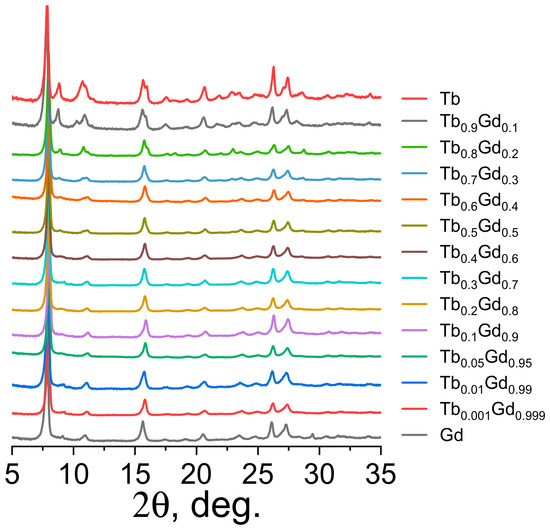
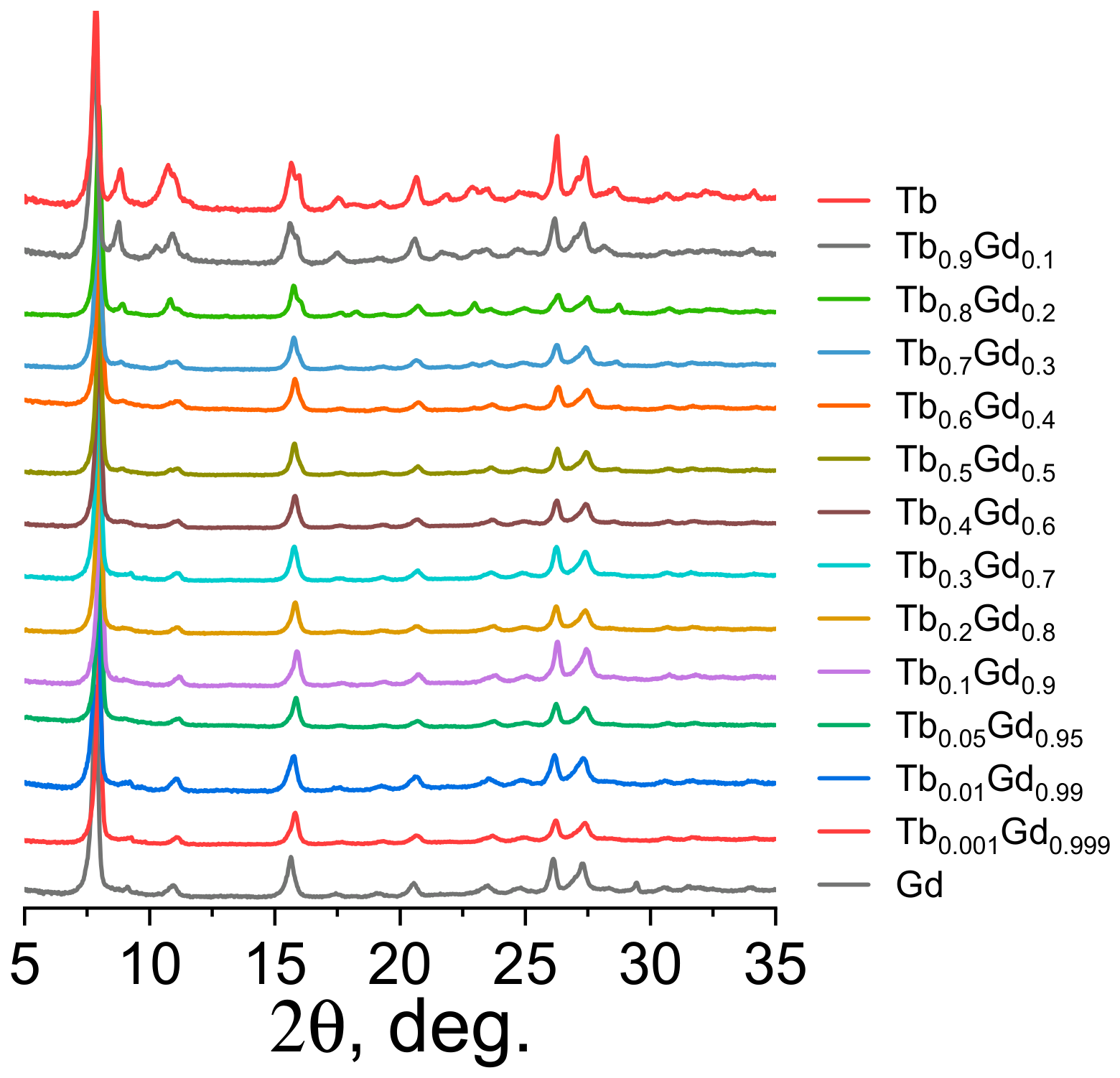
The powder X-ray diffraction (PXRD) patterns of the synthesized compounds are shown in Figure 1. The PXRD data indicate that the structure of the chloroterephthalate complexes is invariant with respect to the Tb/Gd ratio across the series, confirming the formation of solid solutions.
Figure 1.
Powder diffraction patterns of compounds of the series (TbxGd1−x)2(Cl-1,4-bdc)3∙5H2O (x = 0–1).
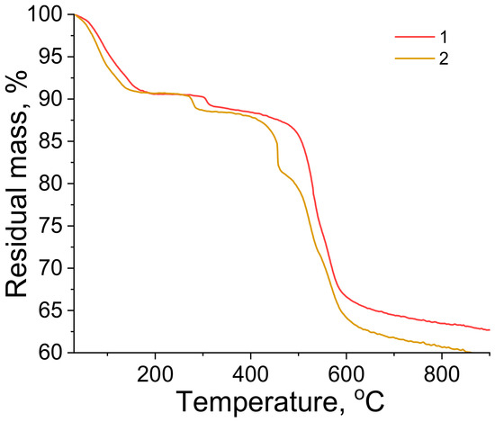
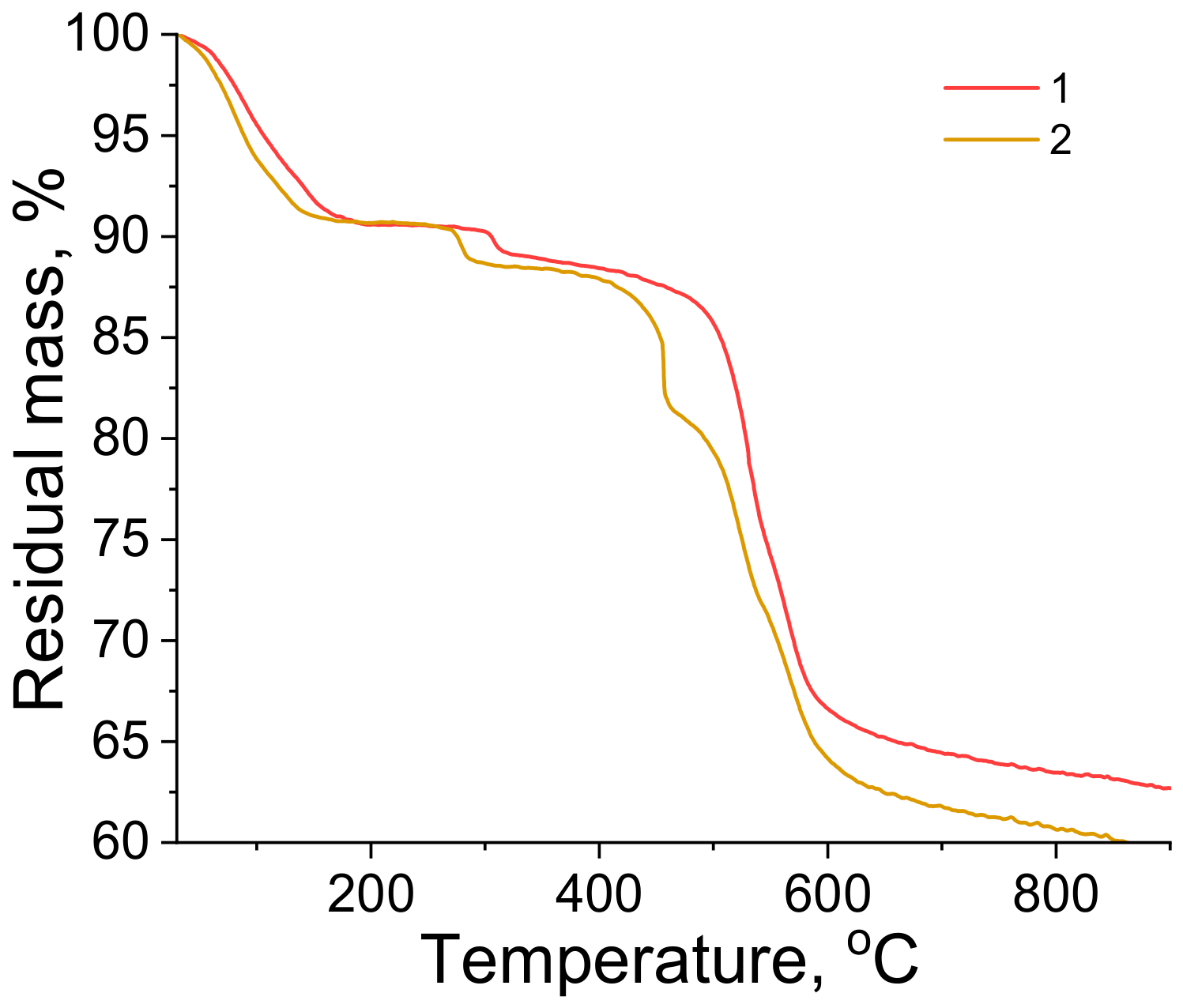
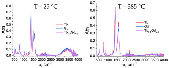
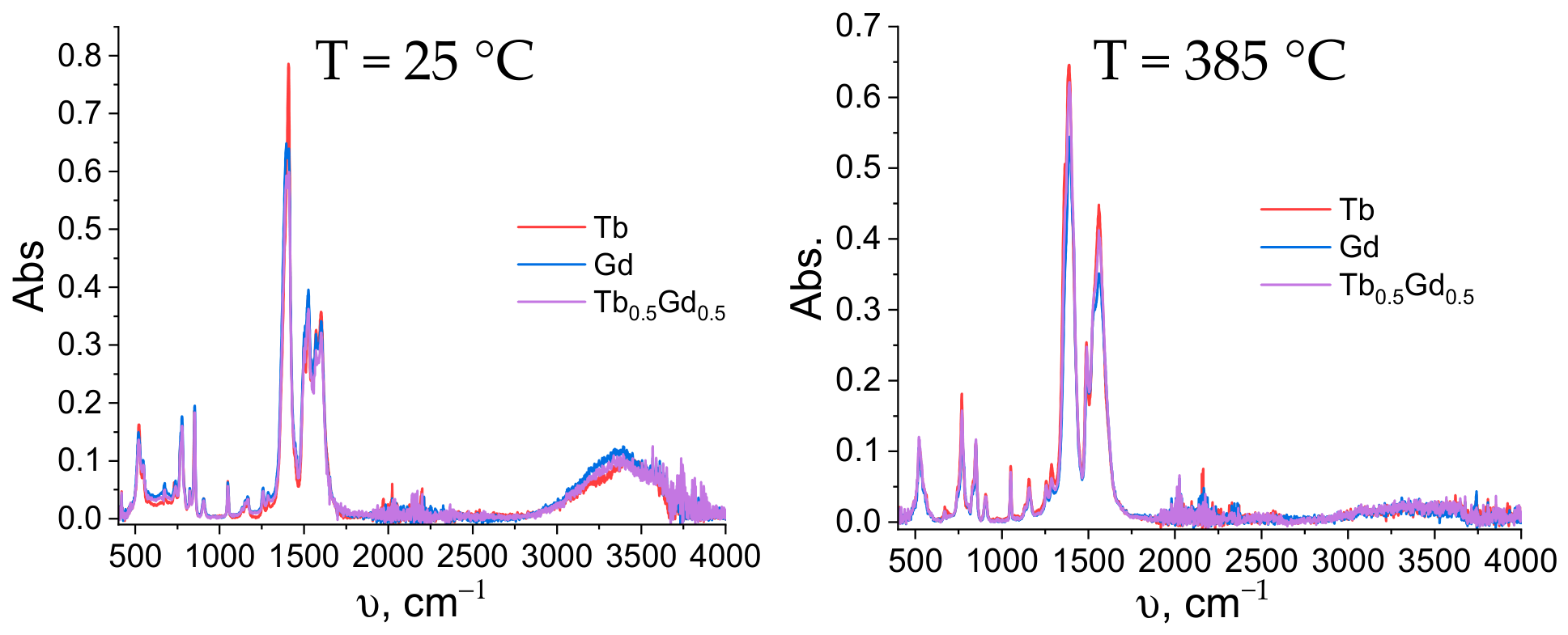
The composition of as-synthesized compounds was determined by thermogravimetric analysis (TGA), elemental analysis, energy dispersive X-ray spectroscopy (EDX), infrared (IR) spectroscopy, and variable-temperature powder X-ray diffraction (VT-PXRD). The EDX results (Table 1) showed that the metal ratios in the compounds correspond to the expected stoichiometric values. Thermogravimetric analysis (Figure 2) reveals several decomposition processes occurring at around 130–150 °C and 280–310 °C, which, most probably, correspond to the loss of water molecules. For the chloro-derivatives several decomposition processes occur at around 130–150 °C and 280-310 °C accompanied with mass loss 9.2–9.4 wt% that correspond to 5.2–5.3 water molecules per formula unit. This assumption was confirmed by the IR spectroscopy of the selected compounds, namely (TbxGd1−x)2(Cl-1,4-bdc)3∙5H2O (x = 0, 0.5, 1), before and after calcination at of 385 °C (Figure 3). The broad band centred around 3500 cm−1 is attributed to the O–H stretching vibrations of water molecules. The series of sharp bands observed in the 1270–1470 and 1470–1800 cm−1 ranges are assigned to the symmetric and asymmetric stretching modes, respectively, of the carboxylate (–COO−) groups in the chloroterephthalate ion [58,59]. One can observe that after calcination, the intensity of the 3500 cm−1 band significantly decreases and almost vanishes after 385 °C calcination, whereas the 1270–1470 and 1470–1800 cm−1 bands remain almost unchanged, which shows that the heating of these compounds up to 385 °C results in dehydration, whereas the organic ligand remains unchanged. In addition, the spectra exhibit bands in the range 600–800 cm−1, which are assigned to C–Cl stretching vibrations, and multiple peaks between 1400 and 1600 cm−1 corresponding to the aromatic ring skeletal vibrations. The C–H out-of-plane bending modes of the aromatic ring appear as weak bands near 750–850 cm−1.
Table 1.
Results of energy dispersive analysis (EDX).
Figure 2.
Thermogravimetric analysis curves for terbium (1) and gadolinium (2) compounds.
Figure 3.
FTIR spectra of neat Tb2(Cl-1,4-bdc)3∙5H2O, Gd2(Cl-1,4-bdc)3∙5H2O and of heterometallic (Tb0.5Gd0.5)2(Cl-1,4-bdc)3∙5H2O, measured after heating at temperatures of 25 and 385 °C.
The elemental analysis (Table 2) was additionally used to determine the number of crystallization water molecules in the halogenoterephthalates. The theoretical and experimental values given in Table 2 converge within the method’s error margin under the assumption that the chloro-derivatives contain 5 molecules of water of crystallization per formula unit. The IR spectra (Figure 3) of the chloroterephthalates, which are thermally stable up to higher temperatures, were recorded after heating at 385 °C. The disappearance of the band at 3300–3500 cm−1 indicates the removal of water and confirms that the observed mass loss originates from the release of water of crystallization and not from degradation of the organic framework.
Table 2.
Results of elemental analysis.
Thus, based on TGA, powder X-ray diffraction analysis, IR spectroscopy, and elemental analysis, the composition and the number of crystallization water molecules were determined for the obtained series of compounds: (TbxGd1−x)2(Cl-1,4-bdc)3·5H2O. The structures of the obtained compounds are similar to each other, a conclusion drawn from two cross-referenced factors: the matching powder X-ray diffraction patterns and the matching fine structure of the bands in the IR spectra of the compounds.
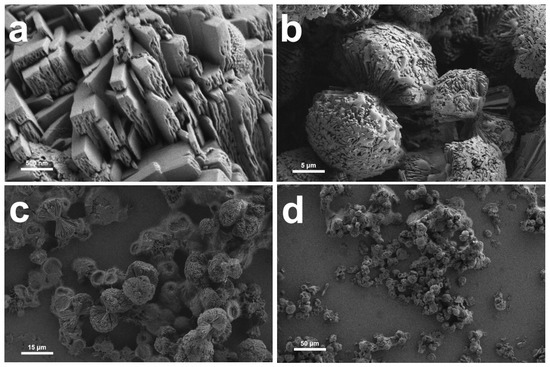
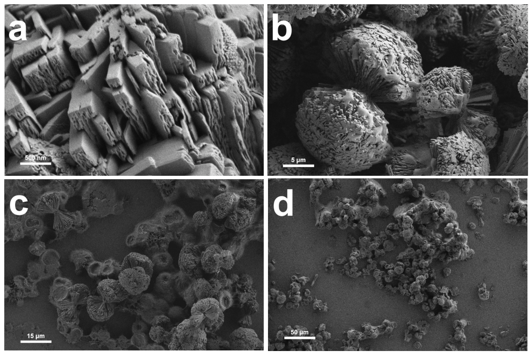
The morphology of the particles of the selected synthesized compounds was determined by scanning electron microscopy (SEM). In all samples, particles with an hourglass shape are formed (average diameter dav. = 15–30 μm). Hemispherical particles (dav. = 8–20 μm) begin to grow simultaneously from a single nucleation centre in opposite directions (Figure 4b–d). The growing halves of the particles consist of plate-like aggregates with varying thicknesses (30–400 nm). Analysis of the obtained images shows that the particle morphology does not depend on the composition of the compounds, which is consistent with the results of other analyses (XRD, TGA, IR) and confirms that the structure and phase composition are independent of the chemical composition.
Figure 4.
SEM images of Tb2(Cl-1,4-bdc)3∙5H2O samples: (a) magnification 30000:1 with the 500 nm scale bar (s.b.); (b) 3000:1 with the 5 μm s.b.; (c) 1000:1 with the 15 μm s.b.; (d) 300:1 with the 50 μm s.b.
2.2. Luminescence
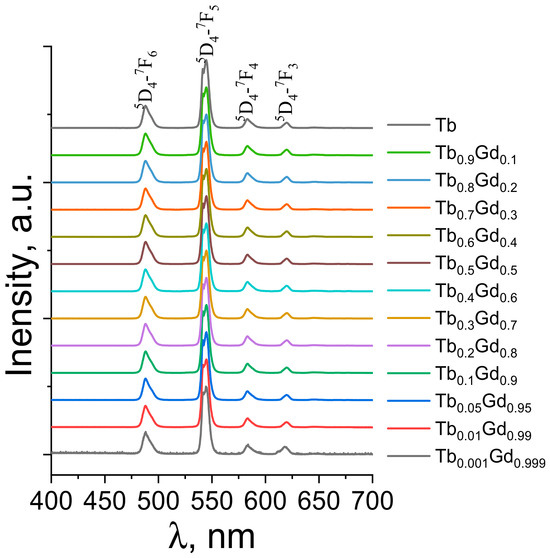
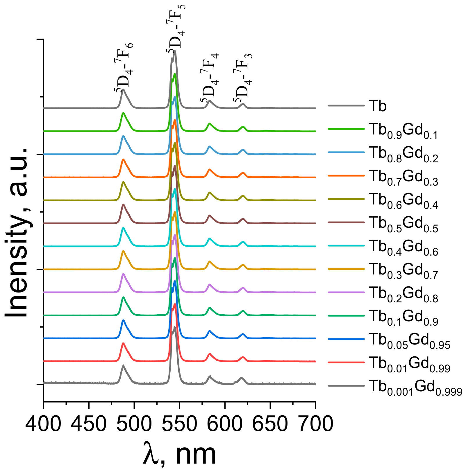
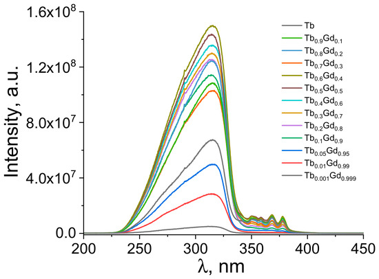
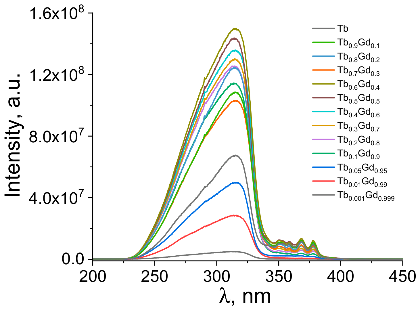
Figure 5 presents a comparison of the emission spectra for the investigated series of terbium-gadolinium chloroterephthalate MOFs, (TbxGd1−x)2(Cl-1,4-bdc)3∙5H2O (x = 0–1), upon 320 nm excitation into the absorption band of the terephthalate ion. The excitation wavelength was selected based on the luminescence excitation spectra (Figure 6) monitored at emission wavelengths of 544 nm (corresponding to their luminescence intensity maxima). The excitation spectra contain a broad band corresponding to the absorption of the terephthalate ion. In addition to the ligand-centred band, weak features are observed above 320 nm, which can be attributed to direct f–f transitions of Tb3+ ions (e.g., 7F6 → 5D4). These minor contributions do not affect the overall sensitization mechanism but are typical for concentrated terbium compounds. In the emission spectra, there are f-f transition bands of the lanthanide ion. This indicates the presence of an antenna effect (sensitized luminescence) in all the studied compounds. The emission bands in the spectra of the terbium compounds correspond to the following f-f transitions of the Tb3+ ion: 5D4 → 7F6 (488 nm), 5D4 → 7F5 (544 nm, highest intensity), 5D4 → 7F4 (583 nm), 5D4 → 7F3 (620 nm). This further confirms the X-ray phase analysis results regarding the uniform phase composition of the obtained compounds.
Figure 5.
Emission spectra of (TbxGd1−x)2(Cl-1,4-bdc)3∙5H2O (x = 0–1).
Figure 6.
Photoluminescence excitation spectra of (TbxGd1−x)2(Cl-1,4-bdc)3∙5H2O (x = 0–1).
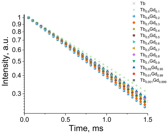
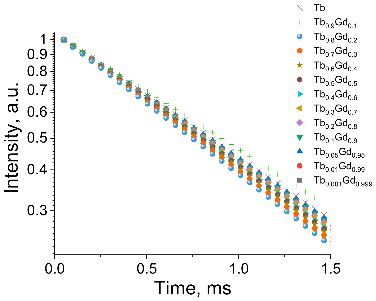
The photoluminescence decay kinetics curves for the synthesized compounds are presented in Figure 7. The curves were fitted by monoexponential decay function:
Figure 7.
Photoluminescence decay curves for the series (TbxGd1−x)2(Cl-1,4-bdc)3∙5H2O.
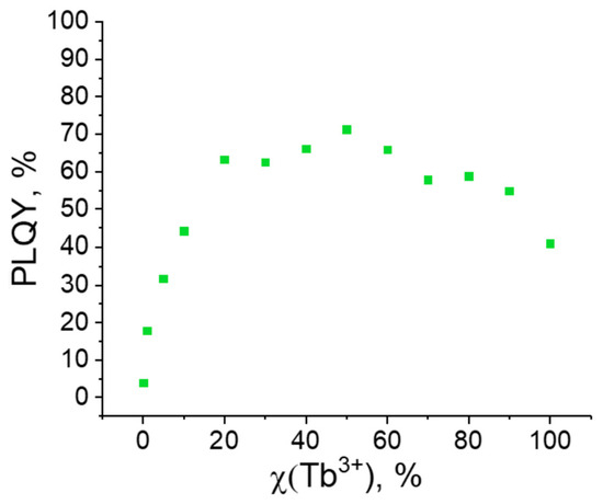
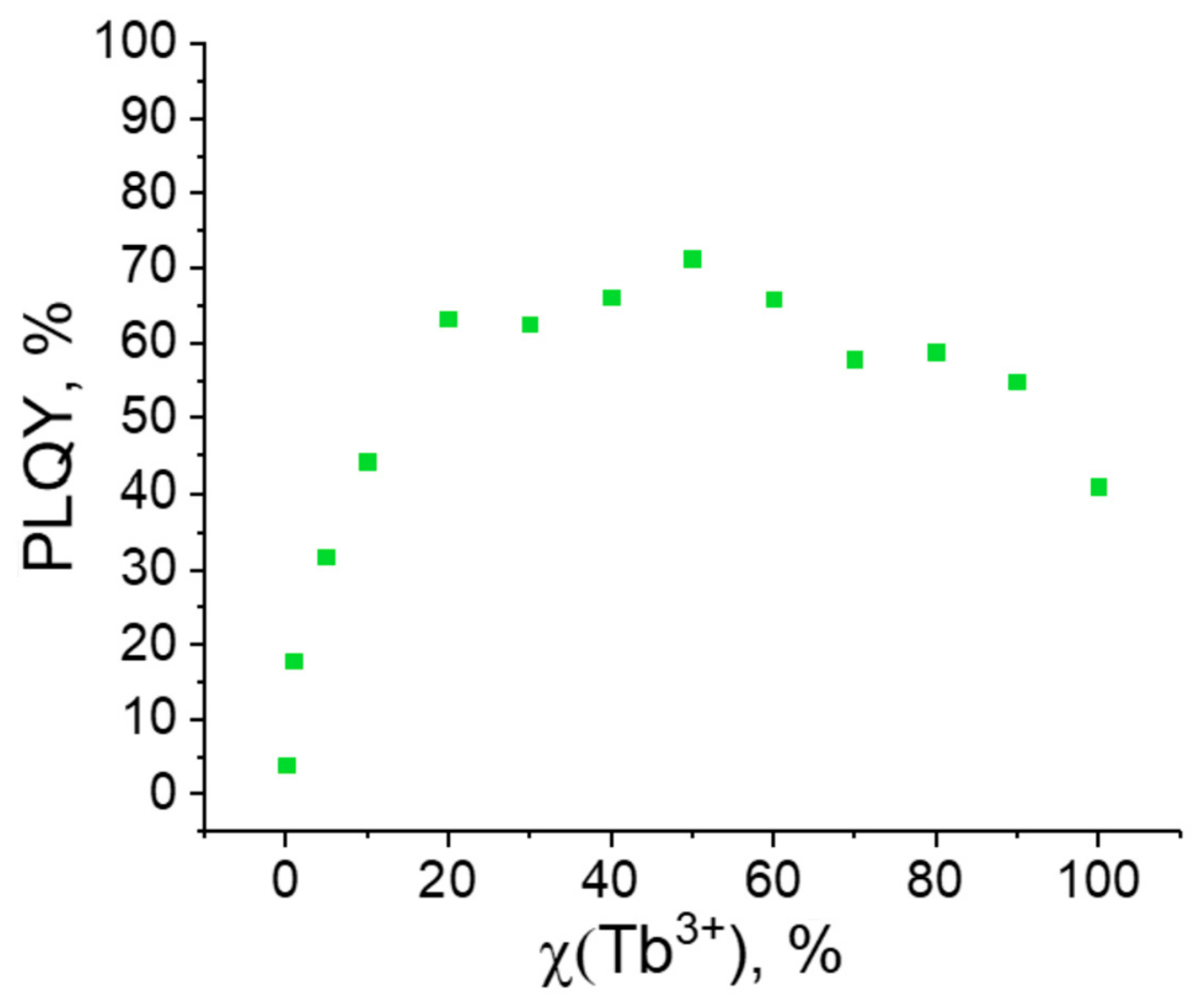
Based on the analysis of the luminescence decay curves, the lifetimes of the excited state (5D4) of the Tb3+ were determined (Figure 8; Table 3).
Figure 8.
Dependence of PLQYs on the composition of (TbxGd1−x)2(Cl-1,4-bdc)3∙5H2O samples (x = 0–1).
Table 3.
Lifetimes (τ) and photoluminescence quantum yields PLQY of (TbxGd1−x)2(Cl-1,4-bdc)3∙5H2O.
The lifetimes of the excited states of the luminescent ions gradually decrease with an increase in the concentration of these ions and a decrease in the concentration of Gd3+ ions. These results are consistent with our previous studies [24,25]; we attribute this phenomenon to a higher probability of energy transfer between closely spaced Tb3+ ions, as well as to quenching by impurities at higher concentrations of the luminescent ions. The following compound possesses the maximum value of quantum yield and luminescence intensity: (Tb0.5Gd0.5)2(Cl-1,4-bdc)3·5H2O (71 ± 1%). The plot of quantum yield dependence on the terbium mole fraction shows an initial gradual increase in intensity, associated with the increase in concentration of the luminescent ion (Tb3+), followed by a gradual decrease, which is attributed tothe dissipation of accumulated energy into thermal vibrations and self-quenching of the luminescent centres (concentration quenching).
Thus, the analysis of the photophysical properties of MOFs based on heterometallic terbium chloroterephthalates allowed us to identify the compositions exhibiting the most intense luminescence and highest quantum yields.
3. Application of (Tb0.5Gd0.5)2(Cl-1,4-bdc)3·5H2O
3.1. Heavy-Metal Detection
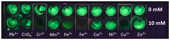
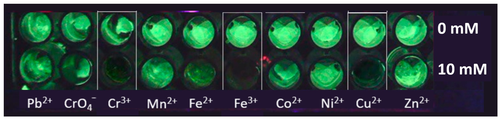
The effect of various ions in solution on the luminescent properties of (Tb0.5Gd0.5)2(Cl-1,4-bdc)3·5H2O, as the sample with the highest luminescence quantum yield, was studied. Prior to dosing, the MOF suspensions were subjected to ultrasonic treatment for 10 min in order to ensure complete deagglomeration and uniform dispersion. During the process of aliquoting, continuous agitation of the bulk suspension was maintained in order to ensure a uniform solid–liquid distribution and prevent any particle settling. First, screening was performed to identify ions that cause luminescence quenching. Aliquots of 20 µL of a suspension containing 4 × 10⁻⁶ mol of the luminescent compound were dispensed into the wells of a 96-well plate. To obtain the control samples (Figure 9, top row), 200 µL of distilled water was added to the suspension. For the test samples, 200 µL of a 10 mM solution of each salt (nitrates for metal cations, or the sodium salt for chromate) containing one of the following ions was added: Pb2+, CrO42−, Cr3+, Mn2+, Fe2+, Fe3+, Co2+, Ni2+, Cu2+, or Zn2+ (Figure 9, bottom row). The solutions in the wells were dried at 60 °C for 12 h. The plates were then placed in a UV chamber (254 nm), and photographs of the luminescent samples were taken under UV illumination (Figure 9). The (Tb0.5Gd0.5)2(Cl-1,4-bdc)3·5H2O samples emit green light, and addition of heavy metal ions decreses the intensity of emission (quenches the photoluminescence). The small red spots correspond to artefact reflection from 96-well plate and do not affect on the results.
Figure 9.
The influence of different ions on luminescence (Tb0.5Gd0.5)2(Cl-1,4-bdc)3·5H2O.
The screening results revealed that, among the ten tested species, only Cr3+, Fe3+, and Cu2+ cause significant luminescence quenching. To address potential concerns regarding salt hydrolysis and pH effects, a control experiment was conducted by holding the MOF in solutions at a pH of 1.5 and above for 12 h, followed by an ICP analysis for the content of lanthanide ions. It showed that when holding the solution at this pH, the concentration of lanthanide ions does not increase compared to the initial solution, which indicates the absence of destruction of these MOFs.
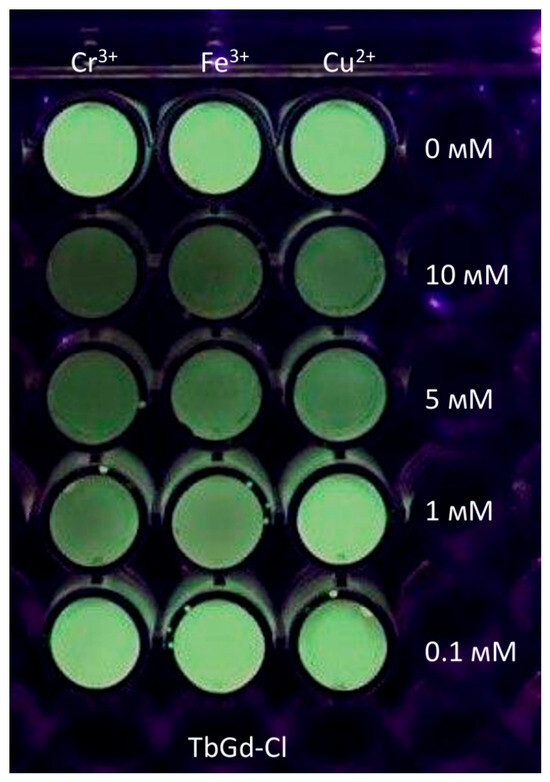
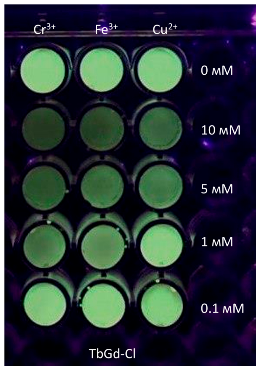
To evaluate the sensitivity of the synthesized compounds to these quenchers, the luminescence intensity was measured at various concentrations of the corresponding metal salts. For this purpose, different amounts of each salt were added to separate sets of solutions to obtain final concentrations in the range of 0.1 to 10 mM, along with a control sample containing only distilled water. The solutions in the wells were dried at 60 °C for 12 h. The plates were then placed in a UV chamber (254 nm), and photographs of the luminescent samples were taken under UV illumination. The results are presented in Figure 10.
Figure 10.
Screening results with different concentrations of metal ions.
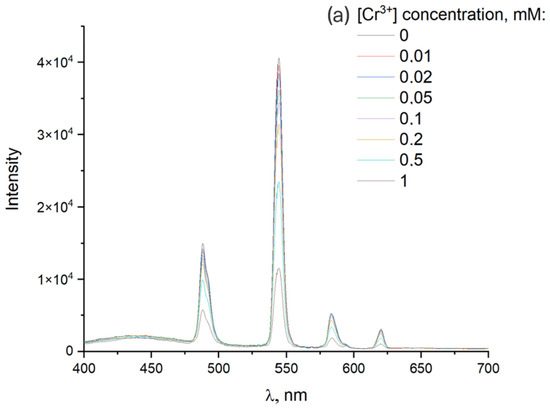
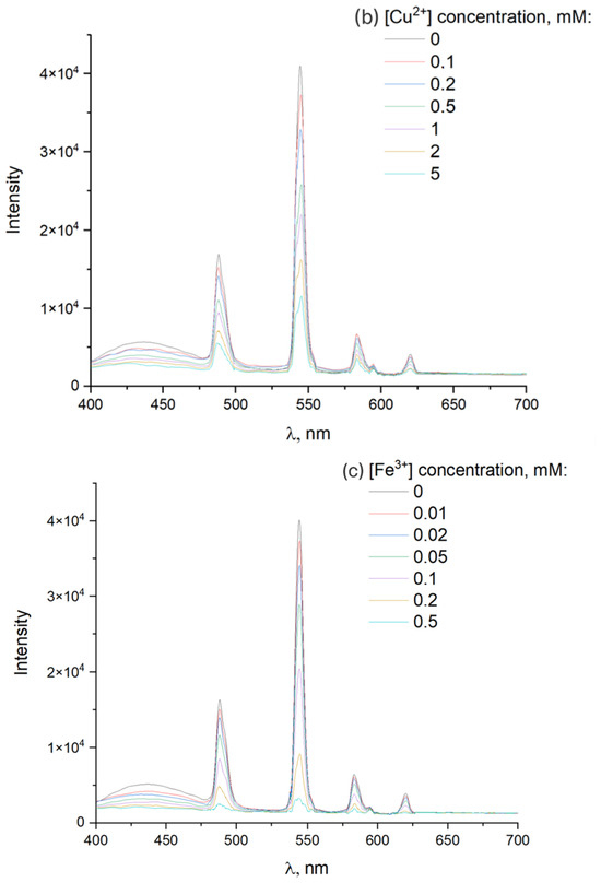
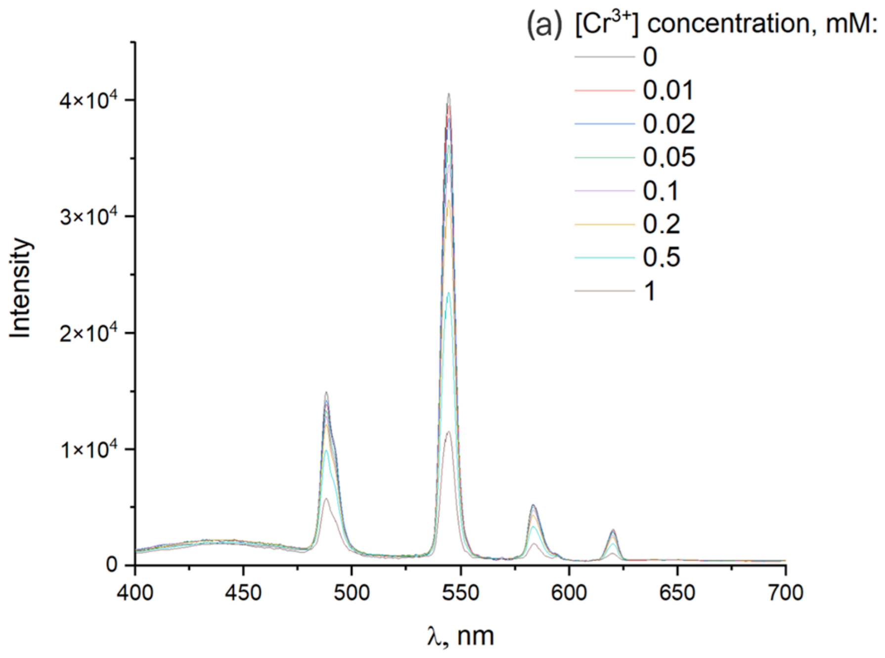
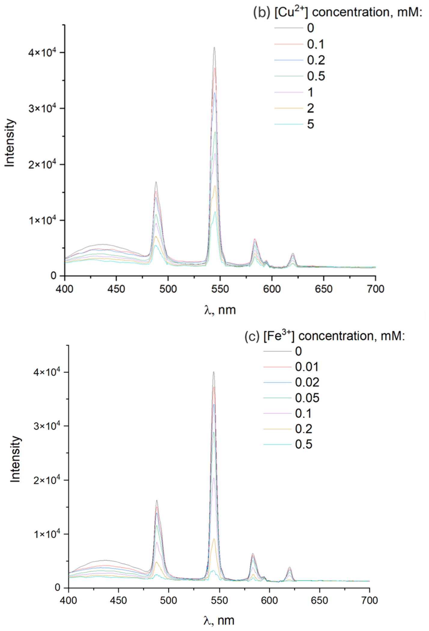
To study the sensing behaviour in more detail, luminescence spectra of terbium chloroterephthalate suspensions were recorded upon the addition of various concentrations of Cr3+, Fe3+, and Cu2+ ions. For this purpose, a suspension of (Tb0.5Gd0.5)2(Cl-1,4-bdc)3·5H2O with a concentration of 0.5 mg/mL was prepared. Microliter volumes of metal salt solutions with concentrations in the range of 0.0001–0.1 M were successively added to 3 mL of the suspension in a quartz cuvette, yielding final quencher ion concentrations of 0.001–5 mM. The luminescence spectra of the suspensions were recorded using an excitation wavelength of 320 nm (Figure 11).
Figure 11.
Emission spectra of (Tb0.5Gd0.5)2(Cl-1,4-bdc)3·5H2O with varying concentrations of added Cr3+ (a), Cu2+ (b), and Fe3+ (c) ions.
A noticeable decrease in luminescence intensity for Cr3+ and Fe3+ was observed at a concentration of 0.01 mM. Cu2+ ions exhibited the lowest quenching efficiency, with significant quenching observed only at 0.1 mM. Emission spectra of (Tb0.5Gd0.5)2(Cl-1,4-bdc)3·5H2O with varying concentrations of added Cr3+, Cu2+, and Fe3+ ions are shown below.
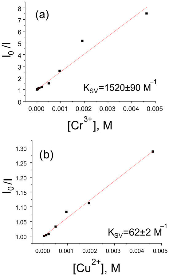
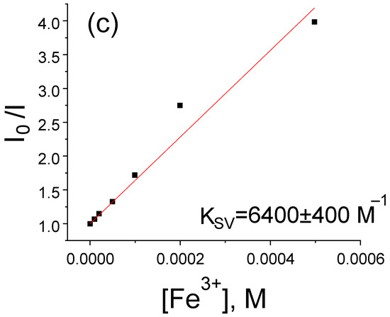
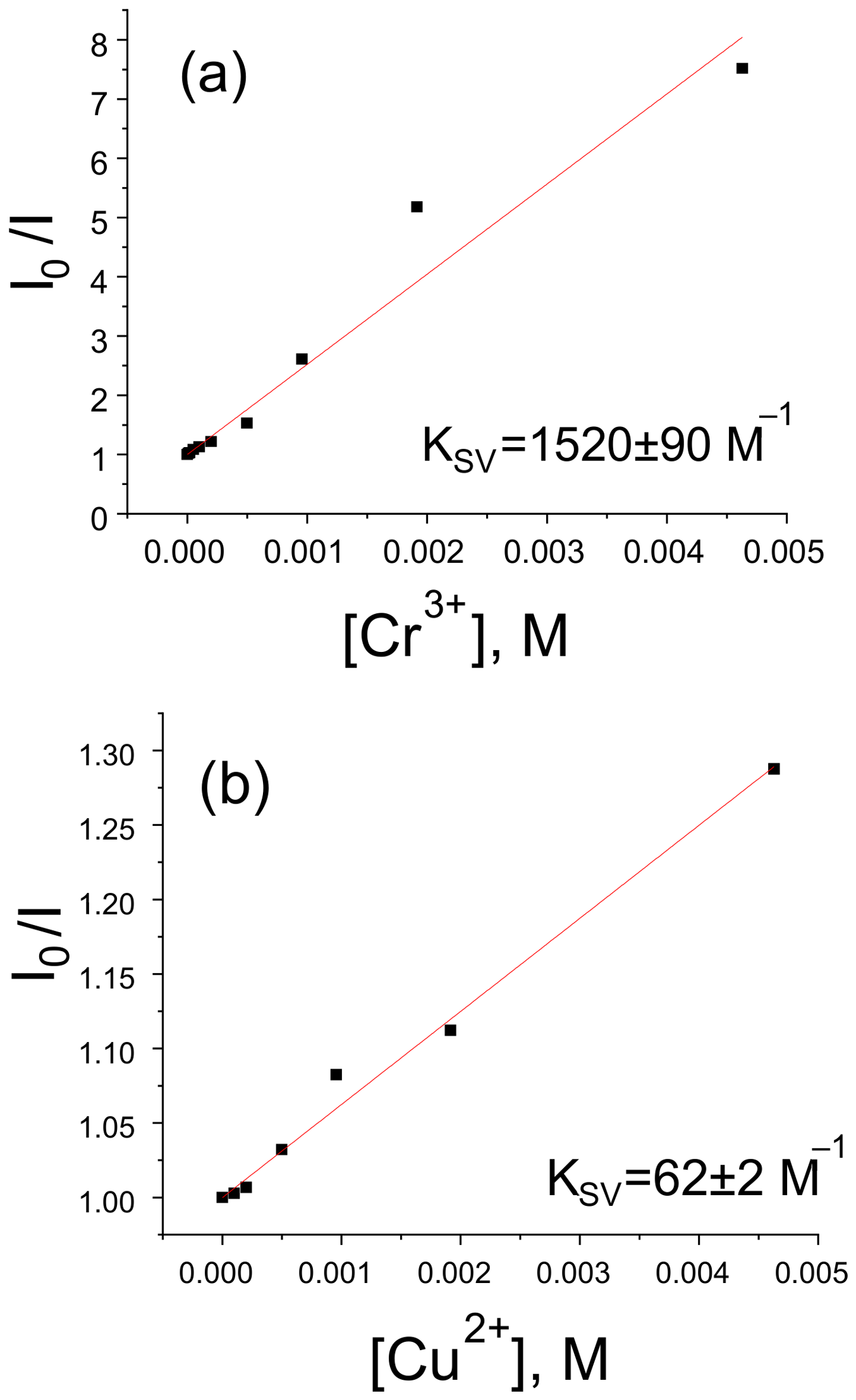
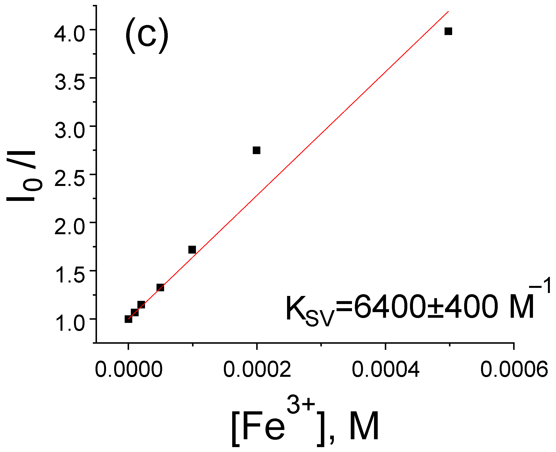
To quantitatively assess the quenching efficiency, plots of integrated luminescence versus quencher ion concentration were plotted in I0/I vs. [Mn+] coordinates, where I is the integrated luminescence intensity with the addition of a metal ion at [Mn+] concentration, and I0 is the integrated luminescence intensity in the absence of the additive. These dependencies (Figure 12) were approximated by linear functions of the form
where KSV is the quenching constant (Stern–Volmer constant).
Figure 12.
Dependence of I0/I (Tb0.5Gd0.5)2(Cl-1,4-bdc)3·5H2O on the concentration of Cr3+ (a), Cu2+ (b), and Fe3+ (c) ions for determining the Stern–Volmer constant.
A comparative analysis of the KSV values showed that Cr3+ and Fe3+ ions quench the luminescence of terbium chloroterephthalate more effectively. Although the detection limit (0.01 mM) is higher than that of some state-of-the-art luminescent sensors, the primary goal of this work was to demonstrate the feasibility of using these new MOFs as luminescent probes and to investigate the quenching mechanism. Further optimization of the material (e.g., by reducing particle size or surface functionalization) may improve the sensitivity.
3.2. Fingerprinting
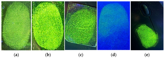
The capability of the synthesized compound for latent fingerprint development was tested on a range of surfaces. To prepare the latent fingerprints, the first author of this paper, Oleg Butorlin, as a volunteer, placed his right thumb onto the substrates without prior cleaning. Substrates included glass, aluminum foil, glossy cardboard, white paper, and the plastic body of a computer mouse. A fine layer of (Tb0.5Gd0.5)2(Cl-1,4-bdc)3·5H2O powder was then lightly distributed over each print with a feather brush; any surplus material was carefully swept away by gentle airflow from a blower. Imaging was performed under a 254 nm mercury vapour UV lamp, and a 500 nm long-pass filter was mounted in front of the camera to block the excitation light and enhance image contrast. Representative photographs are shown in Figure 13. Even on challenging backgrounds, with the exception of white paper, the method produced sharp, well-defined ridge patterns that could be discerned directly by eye, confirming the practical utility of these luminescent powders for fingerprint visualization.
Figure 13.
Luminescence images of latent fingerprints developed on transparent glass (a), aluminum foil (b), cardboard with a glossy surface (c), white paper (d), and computer mouse with a plastic case (e) with (Tb0.5Gd0.5)2(Cl-1,4-bdc)3·5H2O detected under the 254 nm illumination.
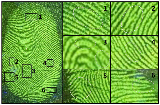
Close-up views (Figure 14) reveal all the characteristic ridge minutiae needed for reliable identification. The high-magnification luminescence images clearly display fine structural details: bifurcation (1), spur (2), sweat pores (3), island (4), closed delta (5), and loop (6). In contrast to conventional carbon-based powders—which often smudge fine features, stain the surrounding substrate, and become nearly invisible on dark surfaces—our formulation strongly highlights the fingerprint ridges without contaminating the background. The powder adheres selectively to the papillary residues, delivering excellent contrast, straightforward application, and faithful preservation of ridge detail, thereby substantially increasing the confidence and accuracy of forensic analyses.
Figure 14.
Luminescence images of latent fingerprints developed on transparent glass with (Tb0.5Gd0.5)2(Cl-1,4-bdc)3·5H2O detected under the 254 nm illumination in the dark field: the fine structure of latent fingerprint (in the dashed-line circle) bifurcation (1), spur (2), sweat pores (3) island (4), closed delta (5), loop (6).
4. Materials and Methods
Terbium (III) chloride hexahydrate and gadolinium (III) chloride hexahydrate were purchased from Chemcraft (Kaliningrad, Russia); 2-chlorobenzene-1,4-dicarboxylic (chloroterephtalic, H2(Cl-1,4-bdc)) acid (>98%), and sodium hydroxide (>99%) were purchased from Sigma-Aldrich Chemie GmbH (Taufkirchen, Germany) and used without additional purification. The heterometallic terephthalate compounds with a general formula of (TbxGd1−x)2(Cl-1,4-bdc)3·5H2O were prepared by mixing 0.2 M TbCl3 and 0.2 M GdCl3 with 2 mL of 0.3 M Na2(Cl-1,4-bdc) aqueous solution followed by ultrasonic treatment for 10 min. The Na2(Cl-1,4-bdc) aqueous solution was prepared by dissolving of the H2(Cl-1,4-bdc) in sodium hydroxide aqueous solution. The TbCl3 and GdCl3 solutions were mixed in stoichiometric amounts, and the total volume of the solutions was 1 mL The resulting white precipitates were separated from the mixture using centrifugation at 4000× g and washed three times with deionized water. The samples were then dried at 60 °C for 48 h. The Tb3+/Gd3+ ratios in the heterometallic terephthalates were confirmed using energy dispersive X-ray spectroscopy (EDX) (EDX spectrometer EDX-800P, Shimadzu, Kyoto, Japan). The Tb/Gd ratios obtained from EDX were consistent with the expected ratios of Tb3+/Gd3+ within 1 at.% accuracy. Powder X-ray diffraction (PXRD) measurements were performed with a D2 Phaser (Bruker, Billerica, MA, USA) X-ray diffractometer using Cu Kα radiation (λ = 1.54056 Å). SEM images were obtained using a scanning electron microscope Zeiss Merlin (Carl Zeiss AG, Jena, Germany). Thermogravimetry curves were obtained using a TG 209 F1 Libra thermo-microbalance (Netzsch, Hanau, Germany). Elemental (CHN) analysis was performed using the LECO TruSpec MICRO Elemental Analyzer (LECO Corporation, St. Joseph, MI, USA). The measurement of FTIR spectra were recorded using IRAffinity-1 spectrometer (Shimadzu, Kyoto, Japan). To carry out photoluminescence studies, the synthesized samples (20 mg) and potassium bromide (300 mg) were pressed into pellets (diameter 13 mm). The photoluminescence data were obtained with a Fluoromax-4 fluorescence spectrometer (Horiba Jobin Yvon, Kyoto, Japan) in perpendicular geometry. Lifetime measurements were performed with the same spectrometer using a pulsed Xe lamp (pulse duration: 3 μs) in a timescale of 50 μs–10 ms. The absolute values of the photoluminescence quantum yields were recorded using a Fluorolog-3 Quantaphi-2 (Horiba Scientific, Kyoto, Japan) device using an integration sphere. All measurements were performed at room temperature.
5. Conclusions
In this work, a series of heterometallic rare-earth chloroterephthalate metal–organic frameworks (MOFs) with the general formula (TbxGd1−x)2(Cl-1,4-bdc)3·5H2O was successfully synthesized and comprehensively characterized. The systematic investigation revealed several key findings. All obtained compounds form isostructural solid solutions across the entire composition range, as confirmed by XRD, TGA, IR spectroscopy, and EDX analysis. The synthesized MOFs exhibit bright photoluminescence under UV excitation due to an efficient antenna effect from the chlorosubstituted terephthalate ligand. The photoluminescence quantum yield shows a non-monotonic dependence on the terbium(III) concentration, reaching a maximum of 71 ± 1% for the composition (Tb0.5Gd0.5)2(Cl-1,4-bdc)3·5H2O. The luminescence enhancement in chloroterephthalates is explained by elimination of Tb3+ concentrational quenching by the partial substitution of luminescent Tb3+ ions with optically inactive Gd3+ ones without altering the crystalline structure. Screening against ten different ionic species showed that the luminescence of the MOF is significantly quenched by Cr3+, Fe3+, and Cu2+. The Stern–Volmer study confirmed that terbium chloroterephthalate is particularly sensitive to Cr3+ and Fe3+, with detection limits of 0.01 mM. These MOFs are therefore promising candidates for the selective luminescent detection of chromium and iron ions. The materials exhibit a distinctive particle morphology (hourglass-shaped aggregates of plate-like particles) and high thermal stability. The combination of high quantum yields, pure colour emission, and stability makes these heterometallic chloroterephthalate MOFs a promising new class of functional phosphors. The most immediate practical application lies in their use as highly effective luminescent fingerprint powders for forensic science, offering superior contrast, selectivity, and signal stability on various surfaces.
Author Contributions
Conceptualization, A.S.M. and N.A.B.; methodology, O.S.B. and A.S.P.; validation, O.S.B.; formal analysis, A.S.M., M.N.R. and O.S.B.; investigation, O.S.B., A.S.P. and A.E.M.; resources, A.S.M. and N.A.B.; data curation, M.N.R.; writing—original draft preparation, O.S.B., A.S.P. and M.Y.S.; writing—review and editing, A.E.M., N.A.B., A.S.M., M.N.R. and M.Y.S.; visualization, O.S.B., A.S.P. and A.E.M.; supervision, A.S.M., N.A.B. and M.Y.S.; project administration, A.S.M.; funding acquisition, A.S.M. All authors have read and agreed to the published version of the manuscript.
Funding
This work was supported by the Russian Science Foundation under grant no. 22-73-10040 (https://rscf.ru/en/project/22-73-10040/, accessed on 29 July 2026).
Institutional Review Board Statement
Not applicable.
Informed Consent Statement
Not applicable.
Data Availability Statement
The data presented in this study are available on request from the corresponding author.
Acknowledgments
The measurements were performed in the Research Park of Saint-Petersburg State University (Magnetic Resonance Research Centre, Chemical Analysis and Materials Research Centre, Cryogenic Department, Interdisciplinary Resource Centre for Nanotechnology, Centre for X-ray Diffraction Studies, Centre for Optical and Laser Materials Research, Thermogravimetric and Calorimetric Research Centre, and Centre for Innovative Technologies of Composite Nanomaterials).
Conflicts of Interest
The authors declare no conflicts of interest.
References
- Kaur, H.; Sundriyal, S.; Pachauri, V.; Ingebrandt, S.; Kim, K.-H.; Sharma, A.L.; Deep, A. Luminescent metal-organic frameworks and their composites: Potential future materials for organic light emitting displays. Coord. Chem. Rev. 2019, 401, 213077. [Google Scholar] [CrossRef] [Scilit]
- Utochnikova, V.V.; Latipov, E.V.; Dalinger, A.I.; Nelyubina, Y.V.; Vashchenko, A.A.; Hoffmann, M.; Kalyakina, A.S.; Vatsadze, S.Z.; Schepers, U.; Bräse, S.; et al. Lanthanide pyrazolecarboxylates for OLEDs and bioimaging. J. Lumin. 2018, 202, 38–46. [Google Scholar] [CrossRef] [Scilit]
- Yin, K.; Wu, S.; Zheng, H.; Gao, L.; Liu, J.; Yang, C.; Qi, L.-W.; Peng, J. Lanthanide Metal–Organic Framework-Based Fluorescent Sensor Arrays to Discriminate and Quantify Ingredients of Natural Medicine. Langmuir 2021, 37, 5321–5328. [Google Scholar] [CrossRef] [Scilit] [PubMed]
- Lustig, W.; Mukherjee, S.; Rudd, N.D.; Desai, A.V.; Li, J.; Ghosh, S.K. Metal–organic frameworks: Functional luminescent and photonic materials for sensing applications. Chem. Soc. Rev. 2017, 46, 3242–3285. [Google Scholar] [CrossRef] [Scilit] [PubMed]
- Liu, D.; Lu, K.; Poon, C.; Lin, W. Metal–Organic Frameworks as Sensory Materials and Imaging Agents. Inorg. Chem. 2014, 53, 1916–1924. [Google Scholar] [CrossRef] [Scilit] [PubMed]
- Amoroso, A.J.; Pope, S.J.A. Using lanthanide ions in molecular bioimaging. Chem. Soc. Rev. 2015, 44, 4723–4742. [Google Scholar] [CrossRef] [Scilit] [PubMed]
- Liu, Y.-Q.; Qin, L.-Y.; Li, H.-J.; Wang, Y.-X.; Zhang, R.; Shi, J.-M.; Wu, J.-H.; Dong, G.-X.; Zhou, P. Application of Lanthanide-Doped Upconversion Nanoparticles for Cancer Treatment: A Review. Nanomedicine 2021, 16, 2207–2242. [Google Scholar] [CrossRef] [Scilit] [PubMed]
- Zhao, L.; Zhang, W.; Wu, Q.; Fu, C.; Ren, X.; Lv, K.; Ma, T.; Chen, X.; Tan, L.; Meng, X. Lanthanide europium MOF nanocomposite as the theranostic nanoplatform for microwave thermo-chemotherapy and fluorescence imaging. J. Nanobiotechnol. 2022, 20, 133. [Google Scholar] [CrossRef] [Scilit] [PubMed]
- Zhao, D.; Yue, D.; Zhang, L.; Jiang, K.; Qian, G. Cryogenic Luminescent Tb/Eu-MOF Thermometer Based on a Fluorine-Modified Tetracarboxylate Ligand. Inorg. Chem. 2018, 57, 12596–12602. [Google Scholar] [CrossRef] [Scilit] [PubMed]
- Feng, T.; Ye, Y.; Liu, X.; Cui, H.; Li, Z.; Zhang, Y.; Liang, B.; Li, H.; Chen, B. A Robust Mixed-Lanthanide PolyMOF Membrane for Ratiometric Temperature Sensing. Angew. Chem. 2020, 132, 21936–21941. [Google Scholar] [CrossRef] [Scilit]
- Zhang, P.; Liang, L.; Liu, X. Lanthanide-doped nanoparticles in photovoltaics—More than just upconversion. J. Mater. Chem. C Mater. 2021, 9, 16110–16131. [Google Scholar] [CrossRef] [Scilit]
- Chen, D.; Wang, Y.; Hong, M. Lanthanide nanomaterials with photon management characteristics for photovoltaic application. Nano Energy 2012, 1, 73–90. [Google Scholar] [CrossRef] [Scilit]
- Zhang, Y.; Liu, S.; Zhao, Z.-S.; Wang, Z.; Zhang, R.; Liu, L.; Han, Z.-B. Recent progress in lanthanide metal–organic frameworks and their derivatives in catalytic applications. Inorg. Chem. Front. 2021, 8, 590–619. [Google Scholar] [CrossRef] [Scilit]
- Vahedigharehchopogh, N.; Kıbrıslı, O.; Erol, E.; Ersundu, M.Ç.; Ersundu, A.E. A straightforward approach for high-end anti-counterfeiting applications based on NIR laser-driven lanthanide doped luminescent glasses. J. Mater. Chem. C Mater. 2021, 9, 2037–2046. [Google Scholar] [CrossRef] [Scilit]
- Horrocks, W.D.; Sudnick, D.R. Handbook on the Physics and Chemistry of Rare Earths, 41st ed.; Elsevier: Amsterdam, The Netherlands, 1981; Volume 14. [Google Scholar]
- Lei, Y.; Zhou, S.; Ke, J.; Yu, L.; Wei, Y.; Liu, Y.; Hong, M. Breaking the concentration quenching limit of lanthanide emitter through multi-coupling of confined quasi-0D & 2D energy migration. Sci. China Chem. 2025, 68, 1372–1382. [Google Scholar] [CrossRef] [Scilit]
- Huang, D.; Li, F.; Ågren, H.; Chen, G. Inhibiting concentration quenching in Yb3+-Tm3+ upconversion nanoparticles by suppressing back energy transfer. Nat. Commun. 2025, 16, 4218. [Google Scholar] [CrossRef] [Scilit] [PubMed]
- Guseva, P.B.; Badikov, A.R.; Butorlin, O.S.; Toikka, Y.N.; Orlov, S.N.; Ryazantsev, M.N.; Bogachev, N.A.; Skripkin, M.Y.; Mereshchenko, A.S. Complexation of Lanthanides(III) Ions with Terephthalic Acid in Aqueous Solutions by Potentiometric Titration Combined with Photoluminescence Spectroscopy. Chemistry 2025, 7, 57. [Google Scholar] [CrossRef] [Scilit]
- Toikka, Y.N.; Badikov, A.R.; Bogachev, N.A.; Kolesnikov, I.E.; Skripkin, M.Y.; Orlov, S.N.; Mereshchenko, A.S. Luminescent properties and thermal stability of (Lu0.98Eu0.02)2bdc3·10H2O metal–organic frameworks. Mendeleev Commun. 2024, 34, 634–636. [Google Scholar] [CrossRef] [Scilit]
- She, J.-B.; Li, D.-D.; Gao, F.; Qiao, Z.-B.; Liu, H.-H.; Wei, W.; Peng, B. Judd-Ofelt analysis on trifluoroacetate europium complexes for liquid laser media. Chem. Res. Chin. Univ. 2013, 29, 166–170. [Google Scholar] [CrossRef] [Scilit]
- Miyata, K.; Nakanishi, T.; Fushimi, K.; Hasegawa, Y. Solvent-dependent luminescence of eight-coordinated Eu(III) complexes with bidentate phosphine oxide. J. Photochem. Photobiol. A Chem. 2012, 235, 35–39. [Google Scholar] [CrossRef] [Scilit]
- Fradgley, J.D.; Frawley, A.T.; Pal, R.; Parker, D. Striking solvent dependence of total emission and circularly polarised luminescence in coordinatively saturated chiral europium complexes: Solvation significantly perturbs the ligand field. Phys. Chem. Chem. Phys. 2021, 23, 11479–11487. [Google Scholar] [CrossRef] [Scilit] [PubMed]
- Yang, A.-H.; Zou, J.-Y.; Wang, W.-M.; Shi, X.-Y.; Gao, H.-L.; Cui, J.-Z.; Zhao, B. Two Three-Dimensional Lanthanide Frameworks Exhibiting Luminescence Increases upon Dehydration and Novel Water Layer Involving in Situ Decarboxylation. Inorg. Chem. 2014, 53, 7092–7100. [Google Scholar] [CrossRef] [Scilit] [PubMed]
- Butorlin, O.S.; Petrova, A.S.; Toikka, Y.N.; Kolesnikov, I.E.; Orlov, S.N.; Ryazantsev, M.N.; Bogachev, N.A.; Skripkin, M.Y.; Mereshchenko, A.S. The Structure and Optical Properties of Luminescent Europium Terephthalate Antenna Metal–Organic Frameworks Doped by Yttrium, Gadolinium, and Lanthanum Ions. Molecules 2024, 29, 3558. [Google Scholar] [CrossRef] [Scilit] [PubMed]
- Petrova, A.S.; Butorlin, O.S.; Toikka, Y.N.; Kolesnikov, I.E.; Orlov, S.N.; Ryazantsev, M.N.; Bogachev, N.A.; Skripkin, M.Y.; Mereshchenko, A.S. The Structure and Optical Properties of Luminescent Terbium Terephthalate Metal–Organic Frameworks Doped with Yttrium, Gadolinium, and Lanthanum Ions. Crystals 2024, 14, 966. [Google Scholar] [CrossRef] [Scilit]
- Maouche, R.; Belaid, S.; Benmerad, B.; Bouacida, S.; Freslon, S.; Daiguebonne, C.; Suffren, Y.; Calvez, G.; Bernot, K.; Roiland, C.; et al. Luminescence properties of lanthanide complexes-based molecular alloys. Inorganica Chim. Acta 2020, 501, 119309. [Google Scholar] [CrossRef] [Scilit]
- Binnemans, K. Interpretation of europium(III) spectra. Coord. Chem. Rev. 2015, 295, 1–45. [Google Scholar] [CrossRef] [Scilit]
- de Bettencourt-Dias, A. Introduction to Lanthanide Ion Luminescence. In Luminescence of Lanthanide Ions in Coordination Compounds and Nanomaterials; Wiley: Hoboken, NJ, USA, 2014; pp. 1–48. [Google Scholar] [CrossRef] [Scilit]
- Mironova, O.A.; Ryadun, A.A.; Sukhikh, T.S.; Konchenko, S.N.; Pushkarevsky, N.A. Synthesis and luminescence studies of lanthanide complexes (Gd, Tb, Dy) with phenyl- and 2-pyridylthiolates supported by a bulky β-diketiminate ligand. Impact of the ligand environment on terbium(III) emission. New J. Chem. 2020, 44, 19769–19779. [Google Scholar] [CrossRef] [Scilit]
- Nosov, V.G.; Toikka, Y.N.; Petrova, A.S.; Butorlin, O.S.; Kolesnikov, I.E.; Orlov, S.N.; Ryazantsev, M.N.; Kolesnik, S.S.; Bogachev, N.A.; Skripkin, M.Y.; et al. Brightly Luminescent (TbxLu1−x)2bdc3·nH2O MOFs: Effect of Synthesis Conditions on Structure and Luminescent Properties. Molecules 2023, 28, 2378. [Google Scholar] [CrossRef] [Scilit] [PubMed]
- Tu, D.; Liu, Y.; Zhu, H.; Li, R.; Liu, L.; Chen, X. Breakdown of Crystallographic Site Symmetry in Lanthanide-Doped NaYF 4 Crystals. Angew. Chem. Int. Ed. 2013, 52, 1128–1133. [Google Scholar] [CrossRef] [Scilit] [PubMed]
- You, W.; Tu, D.; Zheng, W.; Huang, P.; Chen, X. Lanthanide-doped disordered crystals: Site symmetry and optical properties. J. Lumin. 2018, 201, 255–264. [Google Scholar] [CrossRef] [Scilit]
- Shavaleev, N.M.; Eliseeva, S.V.; Scopelliti, R.; Bünzli, J.-C.G. Influence of Symmetry on the Luminescence and Radiative Lifetime of Nine-Coordinate Europium Complexes. Inorg. Chem. 2015, 54, 9166–9173. [Google Scholar] [CrossRef] [Scilit] [PubMed]
- Yin, H.-Q.; Wang, X.-Y.; Yin, X.-B. Rotation Restricted Emission and Antenna Effect in Single Metal–Organic Frameworks. J. Am. Chem. Soc. 2019, 141, 15166–15173. [Google Scholar] [CrossRef] [Scilit] [PubMed]
- Cao, W.; Tang, Y.; Cui, Y.; Qian, G. Energy Transfer in Metal–Organic Frameworks and Its Applications. Small Struct. 2020, 1, 2000019. [Google Scholar] [CrossRef] [Scilit]
- Alpha, B.; Ballardini, R.; Balzani, V.; Lehn, J.; Perathoner, S.; Sabbatini, N. Antenna effect in luminescent lanthanide cryptates: A photophysical study. Photochem. Photobiol. 1990, 52, 299–306. [Google Scholar] [CrossRef] [Scilit]
- Latva, M.; Takalo, H.; Mukkala, V.-M.; Matachescu, C.; Rodríguez-Ubis, J.C.; Kankare, J. Correlation between the lowest triplet state energy level of the ligand and lanthanide(III) luminescence quantum yield. J. Lumin. 1997, 75, 149–169. [Google Scholar] [CrossRef] [Scilit]
- Samuel, A.P.S.; Xu, J.; Raymond, K.N. Predicting Efficient Antenna Ligands for Tb(III) Emission. Inorg. Chem. 2009, 48, 687–698. [Google Scholar] [CrossRef] [Scilit] [PubMed]
- De Silva, C.R.; Li, J.; Zheng, Z.; Corrales, L.R. Correlation of Calculated Excited-state Energies and Experimental Quantum Yields of Luminescent Tb(III) β-diketonates. J. Phys. Chem. A 2008, 112, 4527–4530. [Google Scholar] [CrossRef] [Scilit] [PubMed]
- Toikka, Y.N.; Guseva, P.B.; Bogachev, N.A.; Kolesnik, S.S.; Glukhoedov, N.A.; Orlov, S.N.; Ryazantsev, M.N.; Skripkin, M.Y.; Mereshchenko, A.S. Ultrasound-Assisted Synthesis of Microcrystalline Lanthanide Terephthalates: Insights into Morphology and Structural Properties. Chemistry 2025, 7, 49. [Google Scholar] [CrossRef] [Scilit]
- Cui, Y.; Chen, B.; Qian, G. Lanthanide metal-organic frameworks for luminescent sensing and light-emitting applications. Coord. Chem. Rev. 2014, 273–274, 76–86. [Google Scholar] [CrossRef] [Scilit]
- Daiguebonne, C.; Kerbellec, N.; Guillou, O.; Bunzli, J.-C.; Gumy, F.; Catala, L.; Mallah, T.; Audebrand, N.; Gerault, Y.; Bernot, K.; et al. Structural and Luminescent Properties of Micro- and Nanosized Particles of Lanthanide Terephthalate Coordination Polymers. Inorg. Chem. 2008, 47, 3700–3708. [Google Scholar] [CrossRef] [Scilit] [PubMed]
- Reineke, T.M.; Eddaoudi, M.; Fehr, M.; Kelley, D.; Yaghi, O.M. From Condensed Lanthanide Coordination Solids to Microporous Frameworks Having Accessible Metal Sites. J. Am. Chem. Soc. 1999, 121, 1651–1657. [Google Scholar] [CrossRef] [Scilit]
- Smith, J.A.; Singh-Wilmot, M.A.; Carter, K.P.; Cahill, C.L.; Ridenour, J.A. Supramolecular assembly of lanthanide-2,3,5,6-tetrafluoroterephthalic acid coordination polymers via fluorine⋯fluorine interactions: A platform for luminescent detection of Fe 3+ and nitroaromatic compounds. New J. Chem. 2020, 44, 12317–12330. [Google Scholar] [CrossRef] [Scilit]
- Smith, J.A.; Singh-Wilmot, M.A.; Carter, K.P.; Cahill, C.L.; Ridenour, J.A. Lanthanide-2,3,5,6-Tetrabromoterephthalic Acid Metal–Organic Frameworks: Evolution of Halogen···Halogen Interactions across the Lanthanide Series and Their Potential as Selective Bifunctional Sensors for the Detection of Fe 3+, Cu 2+, and Nitroaromatics. Cryst. Growth Des. 2019, 19, 305–319. [Google Scholar] [CrossRef] [Scilit]
- Okuno, Y.; Cavagnero, S. Effect of heavy atoms on photochemically induced dynamic nuclear polarization in liquids. J. Magn. Reson. 2018, 286, 172–187. [Google Scholar] [CrossRef] [Scilit] [PubMed]
- Cui, G.; Fang, W. State-specific heavy-atom effect on intersystem crossing processes in 2-thiothymine: A potential photodynamic therapy photosensitizer. J. Chem. Phys. 2013, 138, 044315. [Google Scholar] [CrossRef] [Scilit] [PubMed]
- Utochnikova, V.V.; Grishko, A.Y.; Koshelev, D.S.; Averin, A.A.; Lepnev, L.S.; Kuzmina, N.P. Lanthanide heterometallic terephthalates: Concentration quenching and the principles of the ‘multiphotonic emission’. Opt. Mater. 2017, 74, 201–208. [Google Scholar] [CrossRef] [Scilit]
- Zaguzin, A.S.; Bondarenko, M.A.; Abramov, P.A.; Rakhmanova, M.I.; Sokolov, M.N.; Fedin, V.P.; Adonin, S.A. Two-Dimensional and Three-Dimensional Coordination Polymers Based on Ln(III) and 2,5-Diiodoterephthalates: Structures and Luminescent Behavior. Inorganics 2022, 10, 262. [Google Scholar] [CrossRef] [Scilit]
- Santos, J.C.C.; Pramudya, Y.; Krstić, M.; Chen, D.-H.; Neumeier, B.L.; Feldmann, C.; Wenzel, W.; Redel, E. Halogenated Terephthalic Acid ‘Antenna Effects’ in Lanthanide-SURMOF Thin Films. ACS Appl. Mater. Interfaces 2020, 12, 52166–52174. [Google Scholar] [CrossRef] [Scilit] [PubMed]
- Fang, Y.-Q.; Lu, M.; Lu, C.-X. Influence of solvent on the structures of two one-dimensional cobalt(II) coordination polymers with tetrachloroterephthalate. Acta Crystallogr. C 2009, 65, m86–m90. [Google Scholar] [CrossRef] [Scilit] [PubMed]
- Tu, Q.-L.; Wang, H.-D.; Chen, S.-C.; He, M.-Y.; Chen, Q. Resin-assisted solvothermal synthesis of a manganese(II) coordination polymer with tetrachloroterephthalate. Z. Naturforschung B 2015, 70, 705–709. [Google Scholar] [CrossRef] [Scilit]
- Zhang, Z.; Chen, S.; Xu, H.; He, M.; Chen, Q. Manipulation of Molecular and Supramolecular Structure in Cobalt(II) Complexes with Tetrachloroterephthalate through the Influence of Different Solvents. Z. Anorg. Allg. Chem. 2010, 636, 1629–1634. [Google Scholar] [CrossRef] [Scilit]
- Hua, J.-A.; Zhou, Y.-J.; Bian, Y.-J.; Tian, Y.; Zhao, Q.; Ma, X. Solvent-controlled assembly of two Zn(II) coordination polymers constructed from 1,3,5-tris(1-imidazolyl)benzene and 2,5-dichloroterephthalic acid with fluorescent recognition of carcinogenic dye in chloroform. J. Coord. Chem. 2020, 73, 282–296. [Google Scholar] [CrossRef] [Scilit]
- Ngom, F.; Blais, C.; Badiane, I.; Hénaff, C.; Camara, M.; Daiguebonne, C.; Guillou, O. Influence of halogens on the crystal packing of 3,4,5,6-tetra-chloro-phthalate-based lanthanide coordination compounds. CrystEngComm 2025, 27, 210–225. [Google Scholar] [CrossRef] [Scilit]
- Devic, T.; Horcajada, P.; Serre, C.; Salles, F.; Maurin, G.; Moulin, B.; Heurtaux, D.; Clet, G.; Vimont, A.; Grenèche, J.-M.; et al. Functionalization in Flexible Porous Solids: Effects on the Pore Opening and the Host−Guest Interactions. J. Am. Chem. Soc. 2010, 132, 1127–1136. [Google Scholar] [CrossRef] [Scilit] [PubMed]
- Horcajada, P.; Salles, F.; Wuttke, S.; Devic, T.; Heurtaux, D.; Maurin, G.; Vimont, A.; Daturi, M.; David, O.; Magnier, E.; et al. How Linker’s Modification Controls Swelling Properties of Highly Flexible Iron(III) Dicarboxylates MIL-88. J. Am. Chem. Soc. 2011, 133, 17839–17847. [Google Scholar] [CrossRef] [Scilit] [PubMed]
- Costa, B.A.; Nunes, W.D.G.; Bembo, L.H.; Siqueira, A.B.; Caires, F.; Leles, M.I.G.; Ionashiro, E.Y. Study of thermoanalytical behavior of heavier lanthanides terephthalates in air atmosphere. J. Therm. Anal. Calorim. 2018, 134, 1205–1210. [Google Scholar] [CrossRef] [Scilit]
- Nunes, W.D.G.; Teixeira, J.A.; Nascimento, A.L.C.S.D.; Caires, F.J.; Ionashiro, E.Y.; Ionashiro, M. A comparative study on thermal behavior of solid-state light trivalent lanthanide isonicotinates in dynamic dry air and nitrogen atmospheres. J. Therm. Anal. Calorim. 2016, 125, 397–405. [Google Scholar] [CrossRef] [Scilit]

Disclaimer/Publisher’s Note: The statements, opinions and data contained in all publications are solely those of the individual author(s) and contributor(s) and not of MDPI and/or the editor(s). MDPI and/or the editor(s) disclaim responsibility for any injury to people or property resulting from any ideas, methods, instructions or products referred to in the content. |
© 2026 by the authors. Licensee MDPI, Basel, Switzerland. This article is an open access article distributed under the terms and conditions of the Creative Commons Attribution (CC BY) license.