


Abstract
The performance of low-temperature detectors utilizing thermal effects is determined by their energy relaxation properties. Usually, heat transport experiments in mesoscopic structures are carried out in the steady state, where temperature gradients do not change in time. Here, we present an experimental study of dynamic thermal relaxation in a mesoscopic system—thin metallic film. We find that thermal relaxation of hot electrons in copper and silver films is characterized by several time constants, and that the annealing of the films changes them. In most cases, two time constants are observed, and we can model the system by introducing an additional thermal reservoir coupled to the film electrons. We determine the specific heat of this reservoir and its coupling to the electrons. We suspect that multiscale thermal relaxation arises from the complicated morphology of the films, in which the electron–phonon coupling strength in grains with different orientations varies.
1. Introduction
Investigation of thermal transport in mesoscopic devices is essential for the study of fundamental physics and for the development of low-temperature detectors [1]. Examples include transition edge sensors [2,3,4,5,6], hot-electron detectors [7,8,9,10,11,12] and other types of nano-calorimeters in the application of photon and particle detection [13,14,15,16], mass spectrometry [17,18] and in the field of quantum technologies [19,20,21,22]. In the steady state, thermal transport down to a single heat-conducting channel has been studied in recent decades [23,24,25,26,27]. In contrast, experiments carried out in the dynamic regime are rarely reported, mainly due to the lack of fast and sensitive thermometers to measure system temperature. Recently, several types of fast thermometers have been developed, which have made it possible to investigate the dynamic thermal relaxation in mesoscopic systems [28,29,30,31,32]. Such experiments are particularly important for low-temperature detectors utilizing thermal effects. Indeed, thermal relaxation determines the performance of the detector and its recovery time after a detection event. In the absence of detailed time-resolved measurements, the relaxation in the detector is usually assumed to be exponential, and its time constant is estimated from the parameters measured in the steady state [29,33]. Here we demonstrate the deficiency of this approach for a metallic film absorber, which may exhibit several relaxation times and may equilibrate very slowly.
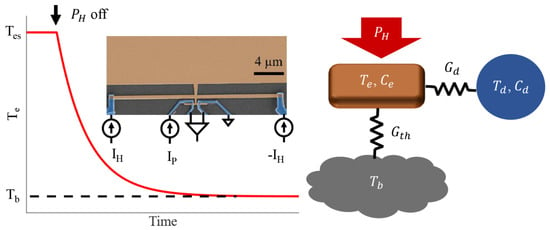
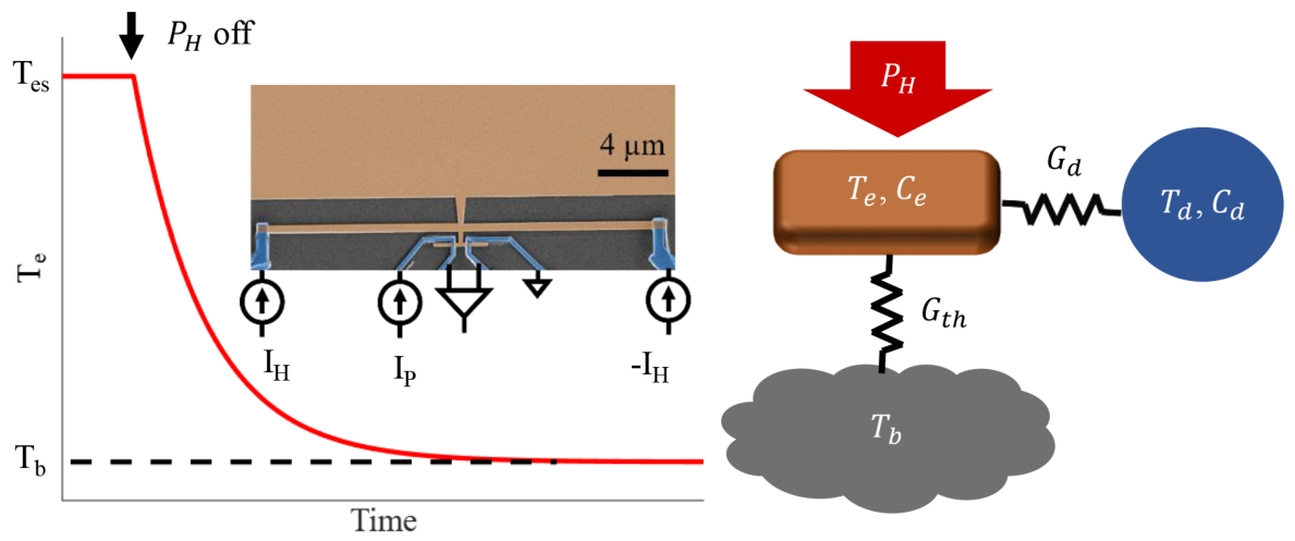
In this article, we mostly focus on the thermal relaxation process in a copper (Cu) film and study the time dependence of the electron temperature after the applied heating is turned off, as shown in Figure 1. Theory predicts that for a normal metal at low temperatures, thermal relaxation is governed by electron–phonon (e-ph) coupling [34], and then the relaxation time . Here is the Sommerfeld constant, which determines the heat capacity of electrons; , with V being the volume of the film; and is the characteristic electron–phonon coupling constant defining the heat flux between electron and phonon sub-systems with the temperatures and ; . For Cu, these two parameters take the values = 98 [35] and = 2 μ [34,36], which results in a relaxation time of μs at 100 mK. In our experiment, we show, contrary to theoretical prediction, that the relaxation of electron temperature in Cu films is characterized by several time constants. We have observed the same effects in silver (Ag) films (See Supplementary Material). Our results suggest that this phenomenon is caused by the combined effect of strong anisotropy of electron–phonon scattering rate in bulk Cu and Ag [37,38,39,40,41] and of the complex texture of thin films, which contain grains with different orientations relative to the substrate [42,43].
Figure 1.
(Left panel): Time dependence of the electron temperature in response to the external heating power . After is switched off, the electron temperature relaxes to the bath temperature . Inset: False-color SEM image of a typical sample with the measurement circuits (Cu is shown in brown, Al in blue). (Right panel): Thermal model of a heated Cu film on a dielectric substrate. Film electrons are thermally coupled to the bath () and to an additional thermal reservoir () by thermal conductances and .
2. Materials and Methods
One of our devices is shown as a false-color SEM image in the inset of Figure 1. Cu films of varying thicknesses are evaporated on substrate with the electron-beam evaporation technique. Superconducting aluminum (Al) is used for the galvanic connection to the Cu films. The large pad on top (partly shown) constitutes the main volume of the film to be studied. The long horizontal Cu wire in the middle is a heater; it is biased with currents of opposite polarities . The short wire at the bottom, which contacts two aluminum leads at both ends, behaves as a proximity JJ. It is used as a fast thermometer to monitor electron temperature in the Cu film. In the explored temperature range, its resolution was around 0.1 mK for a probing pulse of 2 μs duration (See Supplementary Material). The sample is cooled down with a home-made wet dilution refrigerator, and it is shielded against the environmental radiation with a sample holder shield at low temperature. Details of the measurement technique have been reported previously [30]. Thermal equilibration times between the heater, the thermometer, and the pad can be estimated as , where is the heat capacity of the corresponding film and is the heat conductance of the bridge connecting the two components. We find ns at 0.1 K for all measured devices, which is much shorter than thermal relaxation times (See Supplementary Material).
3. Results
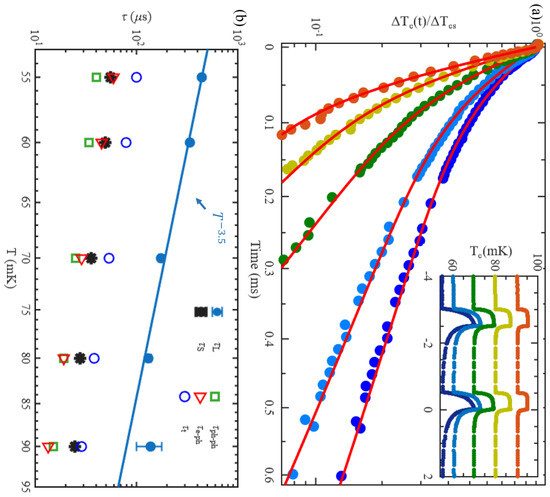
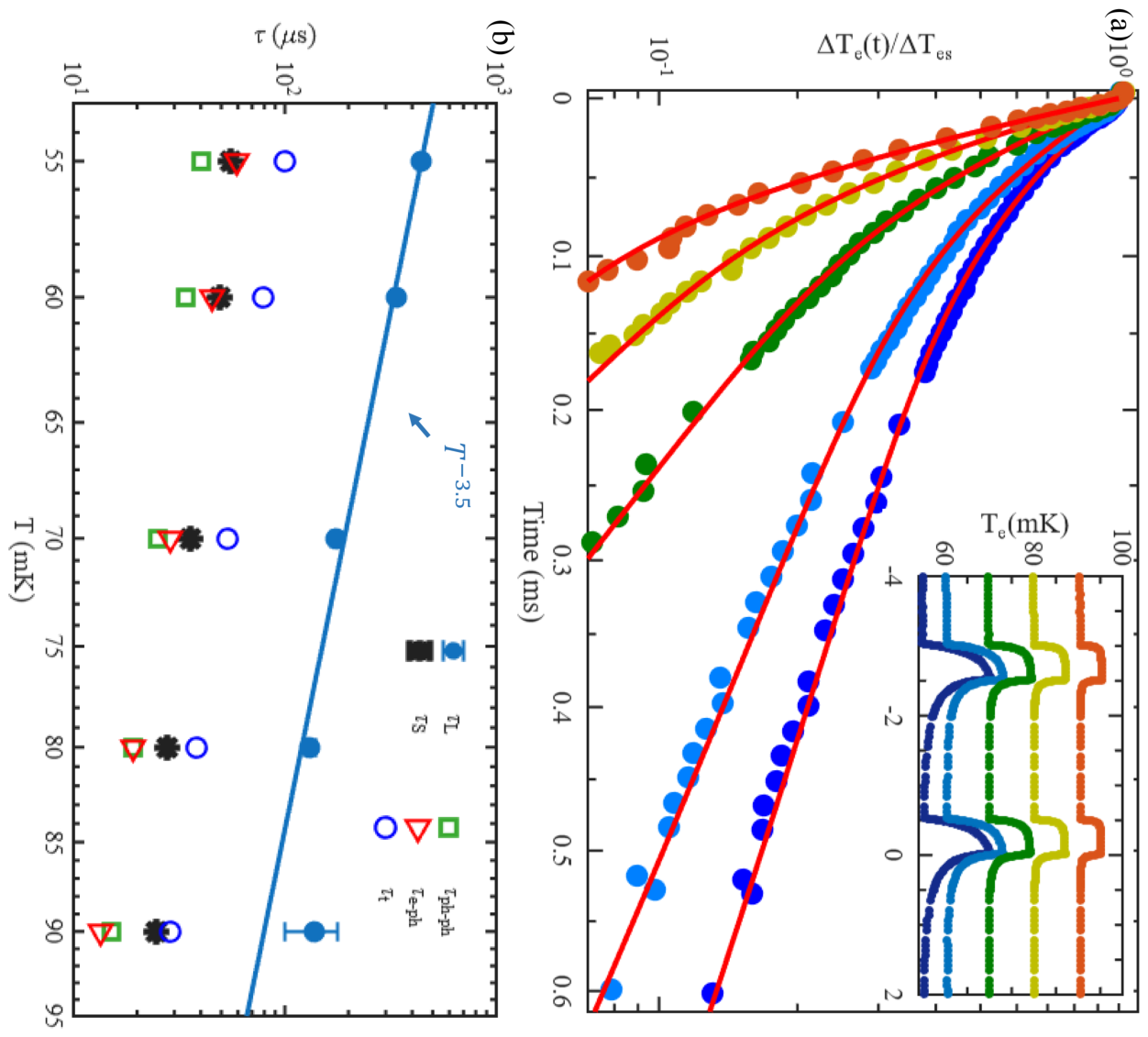
In the inset of Figure 2a, we show the time dependence of the electron temperature for the two heating current pulses with inverse polarities. Each current pulse has a width of 500 μs, and they were applied during the time intervals to ms and to 0 ms. The thickness of the Cu film was 300 nm. Before the heating pulse was applied, i.e., for ms, the electrons were in thermal equilibrium, with the bath temperature . After the heating was turned on, the electron temperature began to rise and finally reached the steady-state value . Identical responses to the two heating pulses confirm the accuracy of our measurement technique. The steady-state temperature rise is consistent with the previous DC measurements [44]. It decreases at high due to the increase in the thermal conductance between the electrons in the film and the environment.
Figure 2.
Thermal relaxation in a 300 nm thick Cu film. (a) Time dependence of the normalized electron temperature . Bath temperatures are = 55, 60, 70, 80, 90 mK from right to left. Red lines are fits with Equation (1). Inset: Electron temperature versus time. The values of are the same as in the main plot. (b) Temperature dependence of the two time constants and . Error bars are smaller than the marker size for most points. , and are the theory-predicted time constants given by Equation (4). The solid blue line shows a simple power-law fit of the dependence .
In order to investigate the dynamic thermal relaxation, we have recorded the time dependence of the electron temperature after the heating current is turned off. Figure 2a shows the normalized as a function of the time t. We have found, contrary to our expectation, that the dependence could not be fitted with an exponentially decaying function. However, we could very well fit the data with two exponents:
where a is a constant pre-factor and and are, respectively, the short and the long relaxation times. The nonlinear regression method has been used to fit the data with a, , and as free parameters. Ordinary Least Squares (OLS) regression of the fits varied between 99.97–99.99%. The red lines in Figure 2a show the corresponding fits. The times and for different temperatures are shown in Figure 2b. At low temperatures, the time constant is about one order of magnitude longer than , and at high temperatures, and differ even more. As a result, at high temperatures cannot be resolved, and the relaxation process takes a single exponential form with a time constant . This explains previous observations of very long relaxation times in Cu and AuPd films [9,28]. Due to the high sensitivity of our JJ thermometer at low temperatures, we can now clearly distinguish the two time constants. Performing simple power-law fits, we find that the relaxation times scale with temperature as and . We have also verified that the effect of the heating pulse amplitude on thermal relaxation is negligible for temperature increments within the range 6 mK 18 mK (See Supplementary Material).
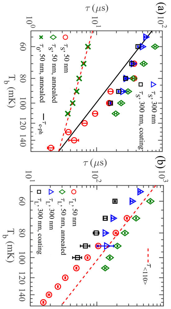
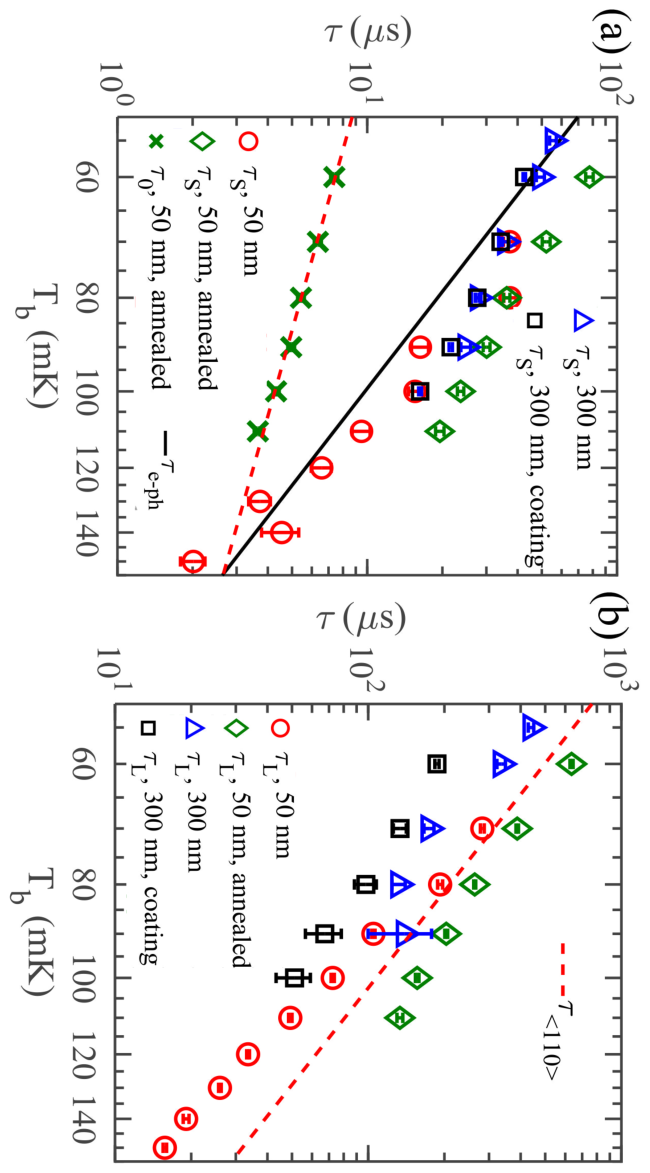
We have repeated the measurements with several films to test the dependence of the relaxation times on the film thickness, which has been observed previously [45]. Such dependence would suggest that multiple relaxation times may be caused by impurities or defects on the surface of the film. To verify this, we have fabricated a sample with a film thickness of 50 nm, in which the surface-to-volume ratio is increased by a factor of 6, and a sample with a 300 nm Cu film coated with a 5 nm thin layer of Al right after evaporation. The volumes of these two films were the same as the volume of the original 300 nm film, which was 120 μ. We have found that both films show two time constants, which are quite close to the ones of the 300 nm film without surface coating, as shown in Figure 3a. Finally, we have measured an annealed 50 nm thin Cu film in order to test the effect of the grain size and grain interface on thermal relaxation. SEM pictures of the sample show significant growth in the grain size after annealing (See Supplementary Material). Thermal relaxation of the annealed film changed significantly, and we needed three exponents with different relaxation times to fit the dependence with sufficient accuracy. These time constants are also shown in Figure 3. The longest and middle ones are of the same order of magnitude as the times and measured in other samples. The shortest time constant of the annealed film is about 6–10 times smaller than , and has a linear temperature dependence.
Figure 3.
Temperature dependence of thermal relaxation times for all samples. (a) Short time constant (for the annealed 50 nm film, we show both and the shortest relaxation time ). Black solid line shows the calculated electron–phonon time (4); red dashed line is the linear fit of of the annealed 50 nm film. (b) Longer relaxation time versus temperature. Red dashed line shows the electron–phonon relaxation time of electrons moving in <110> direction measured in bulk Cu, s [37].
4. Discussion
Summarizing these observations, we conclude that the appearance of an additional relaxation time cannot be explained by surface effects. One may alternatively relate it to magnetic impurities in the bulk of the films. However, a previous experiment [45] has ruled out such a possibility for Cu films. It was found in Ref. [45] that Cu films with a very low concentration of magnetic impurities still exhibited long relaxation time. In contrast, a less pure silver (Ag) film had only shown a short relaxation time , and its value was in good agreement with the predictions of free electron model. We show below that the origin of the multiscale thermal relaxation could be explained by the morphology of the films.
In order to analyze the two-scale relaxation of the electron temperature on a quantitative level, we propose a phenomenological model, in which the electrons in the Cu film are coupled to phonons and to an additional thermal reservoir, as shown in Figure 1. In this model, the time evolution of temperatures follows the equations
Here, is the heating power, A is the contact area between the film and the dielectric substrate, is the constant characterizing thermal boundary conductance between the phonons in the film and in the substrate, is the heat capacity of the phonons in the film, is the heat capacity of electrons. is the heat capacity of the thermal reservoir, is its temperature, is the constant characterizing the coupling between electrons and the reservoir, and is an unknown exponent, which we obtain from the fits. The value of this parameter may hint at the heat exchange mechanism between electrons and the additional thermal reservoir. For example, if the electronic system of the film is coupled to the additional thermal reservoir via electronic tunneling, then . If phonons mediate the heat exchange, one expects . In the latter case, the parameter depends on the acoustic properties of the interface between the copper film and the additional thermal reservoir, as well as on the contact area between them. The phonon heat capacity is usually very small, and one can put . Adopting this approximation and considering linearized versions of Equation (2), which are valid at sufficiently small , we obtain after an abrupt removal of the heating in the form of Equation (1) with the relaxation times and the pre-factor a having the form
The time , appearing above, characterizes the relaxation between electrons and the additional thermal reservoir, , and is the relaxation time of the electron temperature in the absence of the reservoir. It is given by the sum of two contributions:
In the above expressions, is the thermal conductance between electrons and the reservoir, is the thermal conductance between electrons and phonons, and is the thermal conductance between the phonons in the film and in the substrate. In Figure 2b we plot the times , and , given by Equation (4), with the constants = 2 μ and = 60 μ measured in an indepedent steady-state experiment [44]. We observe rather good agreement between the measured values of and the calculated times .
Next, we invert Equations (3) and (4) and express the heat capacity of the thermal reservoir, , and the thermal conductance, , in terms of measured parameters,
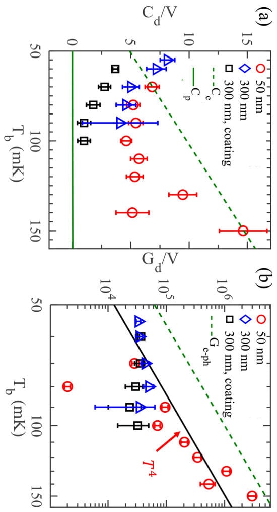
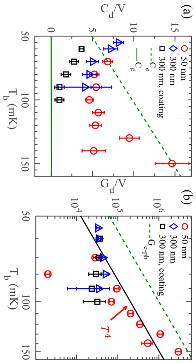
These values are plotted for three different films in Figure 4a,b. The heat capacity does not exhibit clear temperature dependence in the explored temperature range. Its value is rather high and comparable to the heat capacity of electrons . It is unlikely that low-concentration impurities would result in such a high value. One reason for that could be the existence of a significant number of grains in the film, which remain electrically decoupled from it. Such grains would form an additional thermal reservoir, which we have postulated in our model. In Figure 4b we show the temperature dependence of the thermal conductance . For the two 300 nm films is almost constant, while for the 50 nm film it roughly follows power-law dependence with the exponent close to 4, which corresponds to in Equation (2).
Figure 4.
(a) Temperature dependence of the specific heat of the additional thermal reservoir coupled to electrons (5), i.e., heat capacity normalized by the volume of the absorber, , for three different Cu films. Solid and dashed lines are the phonon and electron specific heats of the films, respectively. (b) Thermal conductance between the reservoir and the electrons (6) per unit volume, . The dashed line shows the electron–phonon thermal conductance ; the solid line is the fit of the data for the 50 nm thin film with dependence.
Our hypothesis about electrically decoupled grains in the film as a reason for the long thermal relaxation time is supported by other experiments. Previously, experiments [45] have demonstrated that thermal relaxation in a uniform silver film follows a single exponential decay with the time constant expected from the theory, while two exponents are needed for a silver film with different grain orientations (See Supplementary Material). Experiments with copper films have shown that grains with different crystal lattice orientations relative to the film surface, which can be expressed in terms of Miller indices , can simultaneously grow on substrate [42,43]. At the beginning of the growth process a film with <111> orientation is formed. Subsequently, grains with <110> orientation are nucleated at the boundaries of the <111> grains, which results in the growth of <110> texture. Afterwards, <111> grains can again nucleate at the boundaries between <110> grains and so on. In the end, one obtains a sandwiched structure of alternating textures, which might be poorly coupled electrically, especially in our films, which were in contact with atmosphere between fabrication and measurement. It is also well known that electron–phonon scattering rates in bulk Cu are strongly anisotropic [37]. Since in thin films phonon wave vectors are parallel to the surface and since the Fermi velocity of electrons is much higher than the speed of sound, phonons predominantly interact with electrons moving perpendicular to the film surface. Thus, in grains with a <110> orientation, for example, the scattering rate should be close to that of electrons moving in the <110> direction in the bulk material. Bulk relaxation time in the <110> direction is, indeed, significantly longer than that in the <111> direction, as magnetic resonance experiments have shown [37]. In Figure 3b, we plot measured electron–phonon relaxation time in the bulk Cu in the <110> direction, s [37,38,39], and find that its value is indeed close to the measured . Within this scenario, the high value of the pre-factor in front of the slowly decaying exponent observed for the 50 nm thin Cu film, , points to the dominant <110> texture. For the two 300 nm films, a drops from at low T to 0 at higher T (See Supplementary Material), which hints at <111> as preferred orientation.
Though the above arguments qualitatively explain our findings, further experiments and more detailed theoretical modeling are required in order to fully understand heat relaxation mechanisms in thin Cu films. For example, thermal relaxation in the annealed 50 nm film can only be fitted with three exponents and, therefore, cannot be described by the simple model (2). On the other hand, the pronounced effect of annealing on thermal relaxation points at the importance of the grain structure of the film. Yet another unclear issue is the nature of the thermal coupling between conducting and electrically decoupled grains, which is described by the thermal conductance . We expect such coupling to occur through tunnel barriers between the grains, which would result in temperature dependence, but the observed scaling differs from that. We have also considered an alternative thermal model, in which the additional reservoir couples to film phonons instead of electrons. However, this model results in much smaller values of the pre-factor a than the observed ones.
5. Conclusions
In summary, we have investigated dynamic thermal relaxation in Cu and Ag films at sub-kelvin temperatures. In contrast to uniform Ag thin films, which are well described by the free electron model [45,46], thermal relaxation in granular Cu and Ag films is complicated and characterized by two or more relaxation times. One possible explanation for this effect could be the existence of a thermal reservoir consisting of the grains weakly electrically coupled to the current-carrying electrons. Our experiment refines the understanding of non-equilibrium thermal transport in mesoscopic metallic structures and should help to further optimize the performance of the devices utilizing thermal effects and operating at low temperatures.
Supplementary Materials
The following supporting information can be downloaded at: https://www.mdpi.com/article/10.3390/e28080908/s1.
Author Contributions
Conceptualization, L.W. and J.P.P.; Methodology, L.W.; Validation, L.W.; Formal analysis, L.W., D.G. and Y.M.G.; Data curation, L.W.; Writing—original draft, L.W.; Writing—review & editing, L.W., D.G., Y.M.G. and J.P.P.; Supervision, J.P.P.; Project administration, J.P.P.; Funding acquisition, J.P.P. All authors have read and agreed to the published version of the manuscript.
Funding
This work was performed as part of the Academy of Finland Centre of Excellence program (Projects No. 312057).
Data Availability Statement
The original contributions presented in this study are included in the article/supplementary material. Further inquiries can be directed to the corresponding author.
Acknowledgments
We thank O.-P. Saira for technical help, as well as K. L. Viisanen and C. Enss for useful discussions. We acknowledge the provision of the fabrication facilities by Otaniemi Research Infrastructure for Micro and Nanotechnologies (OtaNano).
Conflicts of Interest
Author Dmitry Golubev was employed by HQS Quantum Simulations GmbH. The remaining authors declare that the research was conducted in the absence of any commercial or financial relationships that could be construed as a potential conflict of interest.
References
- Giazotto, F.; Heikkilä, T.T.; Luukanen, A.; Savin, A.M.; Pekola, J.P. Opportunities for mesoscopics in thermometry and refrigeration: Physics and applications. Rev. Mod. Phys. 2006, 78, 217–274. [Google Scholar] [CrossRef] [Scilit]
- Irwin, K.; Hilton, G. Transition-Edge Sensors. In Cryogenic Particle Detection; Enss, C., Ed.; Topics in Applied Physics; Springer: Berlin/Heidelberg, Germany, 2005; Volume 99, pp. 63–150. [Google Scholar]
- Karasik, B.S.; Sergeev, A.V.; Prober, D.E. Nanobolometers for THz Photon Detection. IEEE T. Thz. Sci. Techn. 2011, 1, 97–111. [Google Scholar] [CrossRef] [Scilit]
- Ullom, J.N.; Bennett, D.A. Review of superconducting transition-edge sensors for x-ray and gamma-ray spectroscopy. Supercond. Sci. Tech. 2015, 28, 084003. [Google Scholar] [CrossRef] [Scilit]
- Miaja-Avila, L.; O’Neil, G.C.; Joe, Y.I.; Alpert, B.K.; Damrauer, N.H.; Doriese, W.B.; Fatur, S.M.; Fowler, J.W.; Hilton, G.C.; Jimenez, R.; et al. Ultrafast Time-Resolved Hard X-Ray Emission Spectroscopy on a Tabletop. Phys. Rev. X 2016, 6, 031047. [Google Scholar] [CrossRef] [Scilit]
- Morgan, K.M. Hot science with cool sensors. Phys. Today 2018, 71, 28–34. [Google Scholar] [CrossRef] [Scilit]
- Nahum, M.; Martinis, J.M. Ultrasensitive-hot-electron microbolometer. Appl. Phys. Lett. 1993, 63, 3075–3077. [Google Scholar] [CrossRef] [Scilit]
- Kuzmin, L.; Chouvaev, D.; Tarasov, M.; Sundquist, P.; Willander, M.; Claeson, T. On the concept of a normal metal hot-electron microbolometer for space applications. IEEE Trans. Appl. Supercond. 1999, 9, 3186–3189. [Google Scholar] [CrossRef] [Scilit]
- Govenius, J.; Lake, R.E.; Tan, K.Y.; Möttönen, M. Detection of Zeptojoule Microwave Pulses Using Electrothermal Feedback in Proximity-Induced Josephson Junctions. Phys. Rev. Lett. 2016, 117, 030802. [Google Scholar] [CrossRef] [Scilit] [PubMed]
- Efetov, D.K.; Shiue, R.J.; Gao, Y.; Skinner, B.; Walsh, E.D.; Choi, H.; Zheng, J.; Tan, C.; Grosso, G.; Peng, C.; et al. Fast thermal relaxation in cavity-coupled graphene bolometers with a Johnson noise read-out. Nat. Nanotechnol. 2018, 13, 797–801. [Google Scholar] [CrossRef] [Scilit] [PubMed]
- Kokkoniemi, R.; Govenius, J.; Vesterinen, V.; Lake, R.E.; Gunyhó, A.M.; Tan, K.Y.; Simbierowicz, S.; Grönberg, L.; Lehtinen, J.; Prunnila, M.; et al. Nanobolometer with ultralow noise equivalent power. Commun. Phys. 2019, 2, 124. [Google Scholar] [CrossRef] [Scilit] [PubMed]
- Khurgin, J.; Bykov, A.Y.; Zayats, A.V. Hot-electron dynamics in plasmonic nanostructures: Fundamentals, applications and overlooked aspects. eLight 2024, 4, 15. [Google Scholar] [CrossRef] [Scilit]
- Wang, H.; Wang, F.; Xia, H.; Wang, P.; Li, T.X.; Li, J.Z.; Wang, Z.; Sun, J.M.; Wu, P.S.; Ye, J.F.; et al. Direct observation and manipulation of hot electrons at room temperature. Natl. Sci. Rev. 2021, 8, nwaa295. [Google Scholar] [CrossRef] [Scilit] [PubMed]
- Karasik, B.S.; Pereverzev, S.V.; Soibel, A.; Santavicca, D.F.; Prober, D.E.; Olaya, D.; Gershenson, M.E. Energy-resolved detection of single infrared photons with λ = 8 μm using a superconducting microbolometer. Appl. Phys. Lett. 2012, 101, 052601. [Google Scholar] [CrossRef] [Scilit]
- Doriese, W.B.; Abbamonte, P.; Alpert, B.K.; Bennett, D.A.; Denison, E.V.; Fang, Y.; Fischer, D.A.; Fitzgerald, C.P.; Fowler, J.W.; Gard, J.D.; et al. A practical superconducting-microcalorimeter X-ray spectrometer for beamline and laboratory science. Rev. Sci. Instrum. 2017, 88, 053108. [Google Scholar] [CrossRef] [Scilit] [PubMed]
- Kuzmin, L.S.; Pankratov, A.L.; Gordeeva, A.V.; Zbrozhek, V.O.; Shamporov, V.A.; Revin, L.S.; Blagodatkin, A.V.; Masi, S.; de Bernardis, P. Photon-noise-limited cold-electron bolometer based on strong electron self-cooling for high-performance cosmology missions. Commun. Phys. 2019, 2, 104. [Google Scholar] [CrossRef] [Scilit]
- Hilton, G.C.; Martinis, J.M.; Wollman, D.A.; Irwin, K.D.; Dulcie, L.L.; Gerber, D.; Gillevet, P.M.; Twerenbold, D. Impact energy measurement in time-of-flight mass spectrometry with cryogenic microcalorimeters. Nature 1998, 391, 672–675. [Google Scholar] [CrossRef] [Scilit] [PubMed]
- Novotný, O.; Allgeier, S.; Enss, C.; Fleischmann, A.; Gamer, L.; Hengstler, D.; Kempf, S.; Krantz, C.; Pabinger, A.; Pies, C.; et al. Cryogenic micro-calorimeters for mass spectrometric identification of neutral molecules and molecular fragments. J. Appl. Phys. 2015, 118, 104503. [Google Scholar] [CrossRef] [Scilit]
- Narla, A.; Shankar, S.; Hatridge, M.; Leghtas, Z.; Sliwa, K.M.; Zalys-Geller, E.; Mundhada, S.O.; Pfaff, W.; Frunzio, L.; Schoelkopf, R.J.; et al. Robust Concurrent Remote Entanglement Between Two Superconducting Qubits. Phys. Rev. X 2016, 6, 031036. [Google Scholar] [CrossRef] [Scilit]
- Romero, G.; García-Ripoll, J.J.; Solano, E. Microwave Photon Detector in Circuit QED. Phys. Rev. B 2009, 102, 173602. [Google Scholar] [CrossRef] [Scilit] [PubMed]
- Barzanjeh, S.; Guha, S.; Weedbrook, C.; Vitali, D.; Shapiro, J.H.; Pirandola, S. Microwave Quantum Illumination. Phys. Rev. Lett. 2015, 114, 080503. [Google Scholar] [CrossRef] [Scilit] [PubMed]
- Opremcak, A.; Pechenezhskiy, I.V.; Howington, C.; Christensen, B.G.; Beck, M.A.; Leonard, E.; Suttle, J.; Wilen, C.; Nesterov, K.N.; Ribeill, G.J.; et al. Measurement of a superconducting qubit with a microwave photon counter. Science 2018, 361, 1239–1242. [Google Scholar] [CrossRef] [Scilit] [PubMed]
- Schwab, K.; Henriksen, E.A.; Worlock, J.M.; Roukes, M.L. Measurement of the quantum of thermal conductance. Nature 2000, 404, 974–977. [Google Scholar] [CrossRef] [Scilit] [PubMed]
- Chiatti, O.; Nicholls, J.T.; Proskuryakov, Y.Y.; Lumpkin, N.; Farrer, I.; Ritchie, D.A. Quantum Thermal Conductance of Electrons in a One-Dimensional Wire. Phys. Rev. B 2006, 97, 056601. [Google Scholar] [CrossRef] [Scilit] [PubMed]
- Jezouin, S.; Parmentier, F.D.; Anthore, A.; Gennser, U.; Cavanna, A.; Jin, Y.; Pierre, F. Quantum limit of heat flow across a single electronic channel. Science 2013, 342, 601–604. [Google Scholar] [CrossRef] [Scilit] [PubMed]
- Mosso, N.; Drechsler, U.; Menges, F.; Nirmalraj, P.; Karg, S.; Riel, H.; Gotsmann, B. Heat transport through atomic contacts. Nat. Nanotechnol. 2017, 12, 430–433. [Google Scholar] [CrossRef] [Scilit] [PubMed]
- Banerjee, M.; Heiblum, M.; Rosenblatt, A.; Oreg, Y.; Feldman, D.E.; Stern, A.; Umansky, V. Observed quantization of anyonic heat flow. Nature 2017, 545, 75–79. [Google Scholar] [CrossRef] [Scilit] [PubMed]
- Gasparinetti, S.; Viisanen, K.L.; Saira, O.P.; Faivre, T.; Arzeo, M.; Meschke, M.; Pekola, J.P. Fast Electron Thermometry for Ultrasensitive Calorimetric Detection. Phys. Rev. Appl. 2015, 3, 014007. [Google Scholar] [CrossRef] [Scilit]
- Schmidt, D.R.; Yung, C.S.; Cleland, A.N. Nanoscale radio-frequency thermometry. Appl. Phys. Lett. 2003, 83, 1002–1004. [Google Scholar] [CrossRef] [Scilit]
- Wang, L.B.; Saira, O.P.; Pekola, J.P. Fast thermometry with a proximity Josephson junction. Appl. Phys. Lett. 2018, 112, 013105. [Google Scholar] [CrossRef] [Scilit]
- Zgirski, M.; Foltyn, M.; Savin, A.; Norowski, K.; Meschke, M.; Pekola, J. Nanosecond Thermometry with Josephson Junctions. Phys. Rev. Appl. 2018, 10, 044068. [Google Scholar] [CrossRef] [Scilit]
- Karimi, B.; Pekola, J.P. Noninvasive Thermometer Based on the Zero-Bias Anomaly of a Superconducting Junction for Ultrasensitive Calorimetry. Phys. Rev. Appl. 2018, 10, 054048. [Google Scholar] [CrossRef] [Scilit]
- Wei, J.; Olaya, D.; Karasik, B.S.; Pereverzev, S.V.; Sergeev, A.V.; Gershenson, M.E. Ultrasensitive hot-electron nanobolometers for terahertz astrophysics. Nat. Nanotechnol. 2008, 3, 496–500. [Google Scholar] [CrossRef] [Scilit] [PubMed]
- Wellstood, F.C.; Urbina, C.; Clarke, J. Hot-electron effects in metals. Phys. Rev. B 1994, 49, 5942–5955. [Google Scholar] [CrossRef] [Scilit] [PubMed]
- Kittel, C. Introduction to Solid State Physics, 8th ed.; Table 2; Wiley: Hoboken, NJ, USA, 2005. [Google Scholar]
- Roukes, M.L.; Freeman, M.R.; Germain, R.S.; Richardson, R.C.; Ketchen, M.B. Hot electrons and energy transport in metals at millikelvin temperatures. Phys. Rev. Lett. 1985, 55, 422–425. [Google Scholar] [CrossRef] [Scilit] [PubMed]
- Doezema, R.E.; Koch, J.F. Magnetic Surface Levels in Cu-Determination of Electron-Phonon-Scattering Rates. Phys. Rev. B 1972, 6, 2071–2077. [Google Scholar] [CrossRef] [Scilit]
- Koch, J.F.; Doezema, R.E. Anisotropy of the Electron-Phonon Scattering Rate in Cu. Phys. Rev. Lett. 1970, 24, 507–510. [Google Scholar] [CrossRef] [Scilit]
- Gantmakher, V.F.; Gasparov, V.A. Anisotropy of the probability of electron scattering by phonons on the Fermi surface of copper. J. Exp. Theor. Phys. 1973, 64, 1712–1723. [Google Scholar]
- Gasparov, V.A. Anisotropy of the probability of electron-phonon scattering in silver. J. Exp. Theor. Phys. 1975, 68, 2259–2270. [Google Scholar]
- Johnson, P.B.; Goodrich, R.G. Anisotropy of the electron-phonon collision frequency on the Fermi surface of silver. Phys. Rev. B 1976, 14, 3286–3295. [Google Scholar] [CrossRef] [Scilit]
- Wei, H.L.; Huang, H.; Woo, C.H.; Zheng, R.K.; Wen, G.H.; Zhang, X.X. Development of ⟨110⟩ texture in copper thin films. Appl. Phys. Lett. 2002, 80, 2290–2292. [Google Scholar] [CrossRef] [Scilit]
- Huang, H.; Wei, H.L.; Woo, C.H.; Zhang, X.X. Copper thin film of alternating textures. Appl. Phys. Lett. 2003, 82, 4265–4267. [Google Scholar] [CrossRef] [Scilit]
- Wang, L.B.; Saira, O.P.; Golubev, D.S.; Pekola, J.P. Crossover between Electron-Phonon and Boundary-Resistance Limits to Thermal Relaxation in Copper Films. Phys. Rev. Appl. 2019, 12, 024051. [Google Scholar] [CrossRef] [Scilit]
- Viisanen, K.L.; Pekola, J.P. Anomalous electronic heat capacity of copper nanowires at sub-Kelvin temperatures. Phys. Rev. B 2018, 97, 115422. [Google Scholar] [CrossRef] [Scilit]
- Pinsolle, E.; Rousseau, A.; Lupien, C.; Reulet, B. Direct Measurement of the Electron Energy Relaxation Dynamics in Metallic Wires. Phys. Rev. Lett. 2016, 116, 236601. [Google Scholar] [CrossRef] [Scilit] [PubMed]
Disclaimer/Publisher’s Note: The statements, opinions and data contained in all publications are solely those of the individual author(s) and contributor(s) and not of MDPI and/or the editor(s). MDPI and/or the editor(s) disclaim responsibility for any injury to people or property resulting from any ideas, methods, instructions or products referred to in the content. |
© 2026 by the authors. Licensee MDPI, Basel, Switzerland. This article is an open access article distributed under the terms and conditions of the Creative Commons Attribution (CC BY) license.