


Highlights
What are the main findings?
- Cognitive aging in abstinent patients with AUD may be domain-specific.
- Perceptual reasoning and recovery may be affected by increasing age.
What are the implications of the main findings?
- The findings are not considered strong enough to evidence the vulnerability hypothesis and do not support accelerated aging at all.
- Replication of the findings is needed with larger sample size and the inclusion of older adults (>60 year).
Abstract
Background/Objectives: Chronic alcohol use accelerates biological and cognitive aging, yet it remains unclear how cognitive aging progresses during abstinence in alcohol use disorder (AUD). It is also unknown to what extent this follows models such as accelerated aging or the age-related decline as proposed by the vulnerability hypothesis. This study examined age-related changes and cognitive recovery during abstinence in patients with AUD. Methods: A total of 197 clinically admitted patients, referred for detoxification and extensive neuropsychological examination, were included. Neuropsychological testing was administered in the second and sixth week of admission using well-normed instruments. Using both multi-assessment and cross-sectional data, relationships between age and normed cognitive outcome scores were examined. Results: After six weeks of abstinence, age-related deviations were observed for perceptual reasoning (PRI), verbal comprehension (VCI), and short-term memory (SMI) but not for ten other cognitive indices. During admission, age significantly influenced the change in belonging to a specific recovery category. Each additional year of age reduced the odds of showing no cognitive impairment by 5% and reduced the odds of cognitive recovery by approximately 4%, compared to non-improvers. Conclusions: Age-related influences appear limited to specific cognitive functions and do not follow a uniform or easily interpretable pattern. Perceptual reasoning seems negatively affected after age 60 for participants with six weeks of abstinence. Older participants showed a reduced likelihood of cognitive recovery and a reduced likelihood of having no cognitive problems at all. The findings do not support accelerated aging and are still too weak to be considered evidence for the vulnerability hypothesis. Implications for future research are discussed.
1. Introduction
Normal, healthy aging is associated with neurobiological alterations, decline in various cognitive functions, and a susceptibility to develop neurodegenerative disorders such as Alzheimer’s disease [1,2,3,4]. However, there is considerable heterogeneity among older adults, and a substantial proportion maintains well-preserved cognitive functioning without evidence of impairment. Healthy lifestyle factors including, e.g., physical fitness, may contribute to retaining or even regaining cognitive abilities. Conversely, evidence suggests that chronic and excessive alcohol use can worsen cognitive processes, leading to accelerated biological aging [5,6,7] and a faster decline in cognitive functioning with age [3,8,9]. The relationship between aging and cognitive functioning in patients with alcohol use disorder (AUD) has been examined through the ‘premature aging’ hypothesis, which proposes that cognitive functioning of patients with AUD resembles that of older adults without AUD [10]. Findings from older studies investigating this hypothesis are mixed, ranging from full support [10], to partial [11], and no support [12,13].
The premature aging hypothesis has been refined into an accelerated aging hypothesis and a vulnerability hypothesis [14,15,16]. The accelerated aging hypothesis proposes that cognitive performance of the individual with AUD resembles that of an older normal person from the onset of chronic and excessive drinking, irrespective of a specific age. Three studies were found with evidence that may be considered to support accelerated aging in individuals with excessive alcohol use. A recent, longitudinal cohort study, measuring global intelligence during early adulthood and during late midlife, showed increased impairments of global cognitive functioning (0.12 point decline in IQ score) in men with heavy alcohol use (about 40 g per day) compared to men with lower weekly alcohol consumptions (1–14 units/week) [17]. In another 10-year longitudinal cohort study, participants with heavy alcohol use (≥36 g per day) showed, with increasing age, a faster decline in global cognitive functioning (2.4 extra years), short-term verbal memory (5.7 extra years) and executive functioning (1.5 extra years), compared to participants from less severe categories [18]. These findings illustrate a relation between the severity of alcohol use and cognitive performance, which translates into performance consistent with an older age. An older cross-sectional study reported that, regardless of their age, patients with AUD performed worse on a series of neuropsychological tasks than their age-matched controls, supporting accelerated aging [19]. The number of studies supporting accelerated aging is small, and they investigated only a narrow range of cognitive domains. For example, visuospatial functions and auditory or visual memory functions were either not examined or only minimally addressed, whereas these functions are considered characteristic cognitive domains impaired in patients with AUD [9].
The vulnerability hypothesis proposes that chronic and excessive drinking at an older age leads to substantially greater impairments compared to individuals at a younger age. Also here, only three studies have investigated the implications of this hypothesis, with mixed findings [19,20,21]. While results from the Noonberg et al. [19] study did not support the hypothesis, Kurihara et al. [20], using a cognitive screening instrument for patients with schizophrenia, found a tipping point in which participants of 53 years and older showed greater cognitive impairments (>2 SD) from age-related norms compared to younger participants (<2 SD). The contradictive findings may be partially explained by the limited range of cognitive domains assessed, the use of less suitable instruments to tap cognitive domains, or the absence of control groups. Another factor influencing the findings may be the period of abstinence, with considerable variation in abstinence duration [19] or the relatively short abstinence period of 4 weeks [20]. This may have influenced cognitive performance due to neurobiological disruptions occurring during detoxification [22]. In addition, cognitive impairments may (partly) resolve following prolonged abstinence, also influencing the relation between age and cognitive functioning [23]. From their longitudinal study examining abstinent older patients with AUD (mean age 71), Ross Cucurull et al. [21] tentatively suggested that recovery of cognitive functioning in older patients occurs more slowly, affecting the relation between age and cognitive performance. For this, however, they found no empirical support.
Despite long-standing interest in aging in patients with AUD, the studies discussed above do not provide information on two important points. Hallmark cognitive domains (e.g., memory, visuospatial functioning) were not or only marginally investigated, and duration of abstinence has not been sufficiently addressed. Cross-sectional studies employed short-term abstinence or considerable variation in abstinence duration, while the longitudinal studies did not investigate within-subject changes. While neurobiological studies support both accelerated aging and a vulnerability to brain aging with increasing age [5,24], it remains unclear how cognitive aging unfolds in abstinent patients with AUD, whether it follows an accelerated pattern or is characterized by significant deterioration at a particular age. The aim of this study is to examine age-related deviations during recovery of cognitive functions using a prospective cohort study in a defined period of abstinence. To this end, we investigate the relation between age and cognitive functioning ((working) memory, visuospatial functioning, speed, intelligence) after six weeks of abstinence and the relation between age and change in cognitive functioning during that same period. Identified relationships are controlled for factors known to be associated with both cognitive functioning and age [25,26].
2. Materials and Methods
2.1. Design and Participants
This prospective observational cohort study was conducted at specialized clinical addiction healthcare facilities in the Netherlands (Tactus Institute for Addiction Care). Data from routine care processes were collected between November 2017 and August 2024. Patients with AUD admitted for clinical detoxification and neuropsychological examination were included. Patients were excluded in case of pregnancy, severe somatic or neurological disorders (e.g., lupus, congenital epilepsy, acquired brain injury), acute psychiatric disorders (psychosis, suicidal ideation), or insufficient Dutch language skills. The research was performed in accordance with the Declaration of Helsinki and approved by Tactus Institutional Review Board, executed following institutional guidelines. All participants gave written informed consent.
2.2. Procedure
Patients were referred to the clinical facilities for inpatient alcohol detoxification and extensive neuropsychological assessment. At admission, demographic information was collected, as well as information on alcohol, polysubstance use (cocaine, cannabis, MDMA, amphetamines), and tobacco use. As part of routine care, patients completed a cognitive screening using the Montreal Cognitive Assessment (MoCA) twice [27]: in the second and sixth week of admission. At one point in time, during the sixth week, patients underwent extensive neuropsychological assessment, including the WAIS-IV and WMS-IV. The timing of these assessments aligns with prior research and established recommendations on abstinence duration to minimize detoxification related cognitive disturbances [22,28]. By means of twice-weekly urinalysis, abstinence from alcohol and other substances during admission was verified. All neuropsychological tests were performed by a trained psychologist according to appropriate user guidelines [29,30].
2.3. Measurements
Information on age, education level and gender were registered as well as quantities of previous alcohol use (e.g., ‘the number of years of daily alcohol use’, ‘the age of onset of daily alcohol use’), all via a clinical interview based on the Measurements in the Addictions for Triage and Evaluation [31]. The education level was classified according to the seven categories model of Verhage [32] and subsequently merged into three categories, ‘Low’ (levels 1 and 2), ‘Average’ (levels 3, 4 and 5) and ‘High’ (levels 6 and 7). Substance use disorders were classified following DSM-5 criteria (American Psychiatric Association, 2013). All diagnoses were established by a certified clinical neuropsychologist.
2.3.1. Montreal Cognitive Assessment (MoCA)
The MoCA is a concise screening tool for measuring cognitive functioning using various domains of neuropsychological functioning (visuospatial skills, attention, language, memory, abstraction and orientation) [27]. The uncorrected total score (0–30) was used as an outcome measure for the purpose of this research. A score < 26 indicates a possible cognitive impairment. The instrument is available in parallel versions (7.1, 7.2 and 7.3, 8.1 and 8.2). The 7.1, or more recently the 8.1 version, was used during the first assessment, and the 7.2 or 8.2 version during the second assessment. The instrument is translated into Dutch [33], is well normed [34], is validated and shows good test–retest reliability [35].
2.3.2. Wechsler Adult Intelligence Score Fourth Edition (WAIS-IV)
The WAIS-IV is an individually administered test used to assess the cognitive and intellectual abilities of a person aged 16 to 85 years [36]. The WAIS-IV is widely used in neuropsychological assessments [37] and has acknowledged value when assessing neuropsychological dysfunction [38] and the relationship between aging and cognition [39].
For the purpose of this research we used the original four-factor model (Verbal Comprehension Index (VCI), Perceptual Reasoning Index (PRI), Auditive Working Memory Index (AWMI), Processing Speed Index (PSI)) and three additional indexes from the alternative five-factor model (Fluid Intelligence (FI), Short-term Memory (SMI), Visual Information Processing (VIP)) based on the Cattell–Horn–Carroll theory of cognitive abilities [40]. The other two indexes (Crystallized Intelligence (CI), Processing Speed (PSI)) were not used since these are identical to the VCI and PSI from the original model. Both models are considered complementary for interpreting WAIS-IV results [30,41,42]. A total of 10 core subtests (Similarities [SI], Vocabulary [VC], Information [IN], Block Design [BD], Matrix Reasoning [MR], Visual Puzzles [VP], Digit Span [DS], Arithmetic [AR], Coding [CD], Symbol Search [SS]), and three supplemental subtests (Figure Weights [FW], Picture Completion [PC], Letter-Number Sequencing [LN]), were administered. With these subtests, all indexes of both the four- and five-factor model could be calculated. Raw scores are translated to age-related normed scores (M = 100, SD = 15) using the WAIS-IV manual. Scores ≤ 76 are considered deviating from the norm (≤1.5 SD).
Reliability scores for the Dutch WAIS-IV version are considered good (varying around or above 0.90, and above 0.80 for split-half reliability), as are its criterium and construct validity properties [29].
2.3.3. Wechsler Memory Scale Fourth Edition (WMS-IV)
The WMS-IV is an individually administered test used to assess auditive and visual memory (immediate retrieval (I), delayed recall (II) and recognition) and visual working memory functions. It has versions for adults (16–69 years) and older adults (65–90 years). For the purpose of this research, the latter version was only administered when the participant was 70 years or older. The adult version consists of six subtests, covering Auditive Memory (AMI) (Logical Memory I/II, Verbal Paired Associates I/II), Visual Memory (VMI) (Designs I/II, Visual Reproduction I/II) and Visual Working Memory (VWMI) (Serial Addition, Symbol Span). For each participant, a score was also calculated reflecting the capacity for immediate memory (Immediate Memory Index, IMI) and delayed memory (Delayed Memory Index, DMI). All raw scores were translated to age-related scores (percentiles) using Q-interactive software (https://qiactive.com/choose-share/login/auth (accessed on 1 March 2026)). Scores ≤ 5th percentile are considered deviated from the norm (≤1.5 SD). The version for older adults does not contain the Designs task and the tasks underlying the WMI.
The WMS is well normed, and the reliability of the index scores is considered good (internal consistency between 0.87 and 0.97, interrater reliability between 0.77 and 1.00) [43]. Together with the WAIS-IV, the WMS-IV provides a comprehensive evaluation of one’s neurocognitive status [38].
2.4. Sample Size
We determined our sample size using G*power (version 3.1.9.6) [44] and calculated an approximation of the sample size for an F test (multiple linear regression). Based on previous studies [17,18], we expected a small to medium effect size (f2 = 0.10). Alpha was set at 0.05, and we accepted a power of 0.80. We estimated the maximum number of predictors to be used between one and four, depending on the specific model. For the analysis of both our cross-sectional and longitudinal data, a sample size between 85 and 125 participants was calculated in order to detect an effect using multiple linear regression.
2.5. Outcome Measures
Cognitive functioning as the dependent variable is reflected by the normed scores on the WAIS-IV (Verbal Comprehension, Perceptual Reasoning, Working Memory, Speed, Full Scale Intelligence Quotient, Fluent Intelligence, Short Term Memory, Visual Performance) and WMS (Auditive Memory, Visual Memory, Short Term Memory, Immediate Memory, Extended Memory).
One variable was created reflecting cognitive recovery, consisting of four categories: improvement (group A), no improvement (group B), deterioration (group C) or no cognitive impairments (D). Group assignment was based on a combination of the two MoCA scores (week 2 and week 6 of clinical admission). Participants were assigned to group A when they scored <26 at the first assessment and ≥26 in the second, to group B when the first and second score were <26, to group C when the first score was ≥26 and the second score was <26, and to group D when both scores were ≥26.
Variables that are both associated with cognitive functioning and age were considered confounders. Demographic (gender, age, education level) and drinking-related (severity of alcohol use disorder, use of other non-prescribed substances, polysubstance use (yes/no), tobacco use disorder (yes/no), years of daily alcohol use, age of onset of daily alcohol use, and neurological injury (yes/no)) variables were considered in this regard. For all confounder analyses we used Chi-square tests (Somers’ d for ordinal X binary analyses, Fisher’s Exact Test for binary X binary analyses), parametric or non-parametric tests if applicable. To detect candidate confounders, we used a more tolerant p-value (p < 0.10) to minimize type II error.
2.6. Statistical Analysis
Baseline characteristics were described using the appropriate measures for descriptive statistics. The percentage of participants scoring ≥ 1.5 SD below normal indicated possible cognitive impairment, in line with other studies in the field [35,45]. Normality was judged by visual inspection of the data. In case of doubt, skewness and kurtosis were calculated to Z-scores [46]. A score between −2 and +2 was accepted for normality [47]. In case of missing data, variables were listwise removed from the analysis. When participants completed the older version of the WMS-IV, the Visual Memory Index score was not available for analysis. Given the exploratory character of our research, and to find new possible leads that could improve our understanding of premature aging, we did not correct for multiple testing [48].
We took the following steps to examine age-related deviations in relation to cognitive functioning after six weeks of abstinence. First, variables that could serve as candidate confounders in the relation between age and cognitive functioning were identified. Stratification of data was considered when a confounder substantially influenced the cognitive outcome measures, and the relation between the confounder and age, or between the confounder and the outcome measure, could not be interpreted simply as (non-)linear. Next, we analyzed whether age predicted cognitive functioning by means of a linear relationship, using linear regression analyses. Since the scores on cognitive functioning are already corrected for age, a significant model presents a deviation from the age norm. For every significant regression model found (p < 0.05), we examined the effect of candidate confounders on this relationship by adding one candidate confounder to the model at a time. During this step, a model and the contribution of age, or the confounder, are considered significant when p < 0.05. Finally, non-linear associations were examined using spline regression. Three excess-age predictors (excess40, excess50, excess60) were created, reflecting the numbers of years a participant’s age exceeded 40, 50 or 60 years, respectively, and coded as zero when age was equal to or below the reference value. For each cognitive outcome, a model including all three predictors was estimated. Significant models (p < 0.05) were interpreted as evidence for non-linear age-related effects. Given the possible high correlation among predictors, individual coefficients were not interpreted independently; instead, the pattern of coefficients was examined to characterize changes in the trajectory of cognitive functioning across age range (e.g., increases, plateaus, or declines after ages 40, 50 or 60). The R2-values were not interpreted due to possible overfitting.
To analyze the relation between age and neurocognitive recovery, we used a multinomial logistic regression analysis. We examined whether age predicted assignment to either one of the four recovery categories, using group B as the reference category (p < 0.05). Nagelkerke R2 was used to determine the goodness of fit, using the following qualification criteria: poor (0–0.1), modest (0.1–0.3), moderate (0.3–0.5) and strong (>0.5) [49]. Statistical analysis was carried out using the Statistical Package for Social Sciences software, SPSS, IBM (version 29).
3. Results
3.1. Descriptives of Participants
A total of 197 patients with (mostly severe) AUD were included in this study (see Table 1). Participants were on average 43.9 years old, with the majority under 60 years old (88%). Substantial amounts of participants (16.9–37.1%) were impaired (≤1.5 SD) on tasks; see Appendix A. Participants with a high education level did so on three out of thirteen tasks (10.0–13.3%). Not all outcome measures were available for all included participants for two reasons. First, intelligence testing, particularly extensive intelligence testing using all WAIS-IV subtests, was not conducted if participants had completed a comparable assessment within two years prior to admission. Second, fewer data were available for the WMS because this instrument was gradually introduced across clinics during the eight-year data collection period. Third, a total of 110 participants completed the MoCA at both assessment points, with 154 participants at the first time point and 112 participants at the second. Data were incomplete for several reasons, for example, due to illness, premature discharge, or the MoCA was not completed in full. The missing data showed a random pattern. Analysis comparing participants that completed both MoCA assessments with participants that completed only one, or none of the assessments, yielded no differences with regard to demographic variables (see Table 1 for demographic factors).
Table 1.
Demographic and drinking-related variables (n = 197).
The MoCA scores showed no cognitive impairments for 47 participants compared to 34 participants with cognitive impairments on both time measurements; an improvement was seen for 19 participants, while 10 participants showed deterioration of cognitive function (see Table 2).
Table 2.
Overview mean age for four recovery categories (n = 110).
3.2. Confounders Influencing the Relationship Between Age and Cognitive Functioning
Confounder analyses revealed that education level, age of onset of daily alcohol use, and polysubstance use may influence the relationship between age and cognitive functioning (see Appendix A).
3.2.1. Education Level
The effect of age on education level was significant (F (2, 192) = 3.812, p = 0.024). Bonferroni-adjusted post hoc comparisons showed that the high education level group was about 7.8 years older than the low education level group (MD = 7.8, 95% CI [0.94, 14.63], p = 0.02), whereas no significant difference was found between these groups with the average education level group (p = 0.46, p = 0.17). Education level affected 13 out of 15 (90%) of the outcome measures (p < 0.05), except VWMI. Since the relation between education level and age is not linear, or non-linear, data was stratified by education level, yielding different mean scores per outcome. Substantial amounts of participants with a low (39.4–71.4%) and average education level (14.3–34.9%) were impaired (≤1.5 SD) on all tasks (see Table 3). The age distribution across education levels was largely comparable, with a slightly higher proportion of older participants in the high-education level (see Appendix A).
3.2.2. Age of Onset of Daily Alcohol Use
Age of onset correlated with both age (r (171) = 0.534, p < 0.001) and 12 out of 15 outcome measures (p < 0.05), except VPI, PSI and VWMI. When age of onset was added as a confounder to the models that showed significant associations with age, it did not contribute significantly to any of the outcomes.
3.2.3. Polysubstance Use
Participants with polysubstance use were significantly younger (M = 38.0, SD = 11.3) than participants with AUD only ((M = 48.3, SD = 12.1), t (181) = 5.837, p < 0.001). Polysubstance use correlated with 5 out of 15 outcome measures (p < 0.05). When polysubstance was added as a confounder to the models that showed significant associations with age, it did not contribute significantly to any of the outcomes.
3.2.4. Linear Effect of Age on Cognitive Functioning After Six Weeks Abstinence
After stratification for education level, regression analyses using age as the predictor for cognitive functioning indicated two indices (VCI, SMI) that were significantly predicted by age, both in participants with an average education level and those with a high education level (see Table 4).
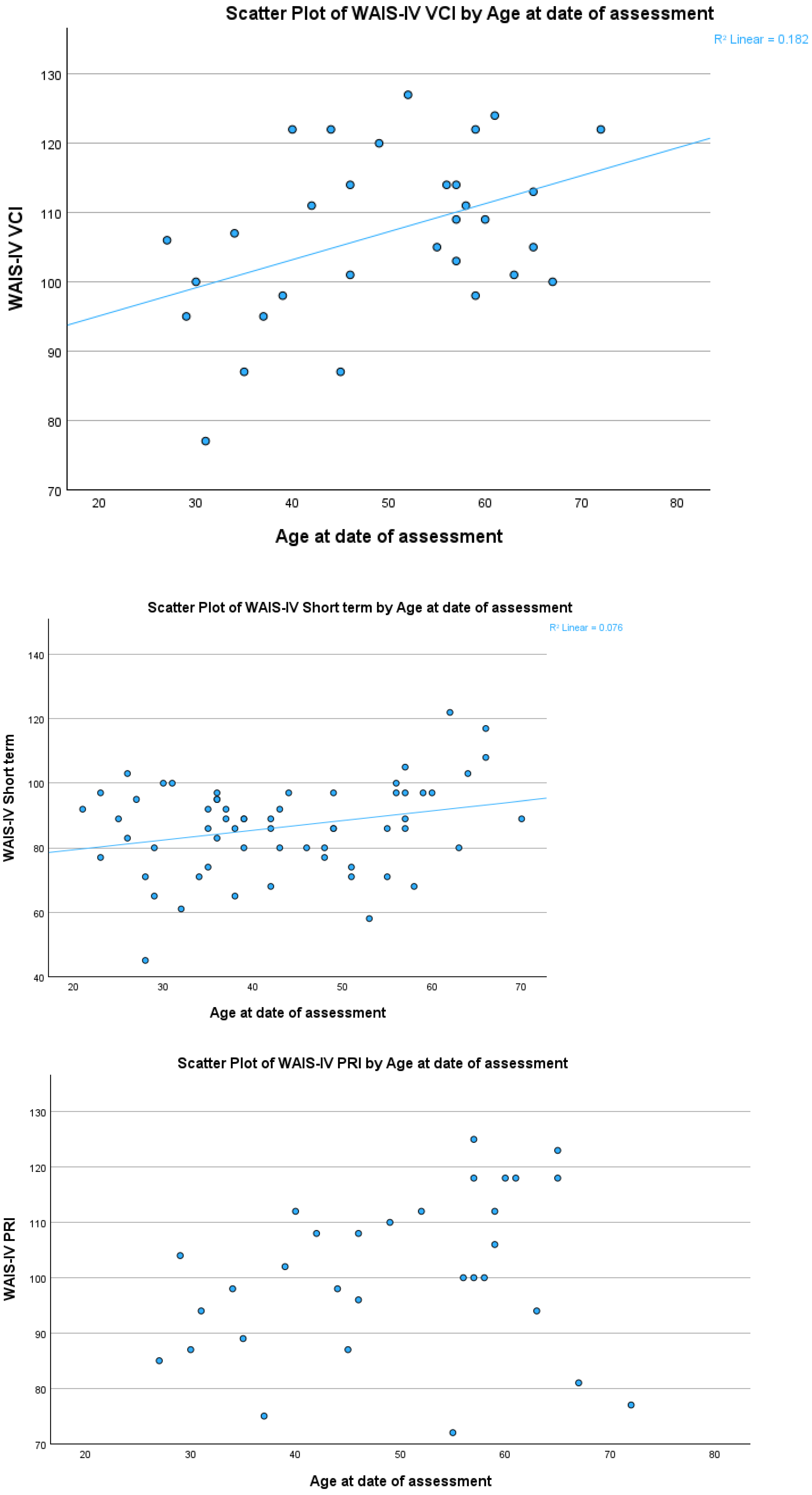
In participants with an average education level, the SMI score increased by 0.303 points per year of age (F (1, 59) = 4.883, p = 0.031, 95% CI [0.03–0.58], R2 = 0.076). In participants with a high education level, age predicted the VCI score with an increase of 0.405 points per year of age (F (1, 29) = 6.450, p = 0.017, 95% CI [0.08–0.73], R2 = 0.182).
Table 3.
Overview of outcome measures stratified per education level.
Table 4.
Regression analysis cognitive functioning stratified by education level.
3.2.5. Non-Linear Effect of Age on Cognitive Functioning After Six Weeks Abstinence
When examining the potential non-linear relation between age and cognitive functioning across education levels, no significant models were identified for participants with a low education level.
Among participants with an average education level, the spline model indicated that age significantly predicted SMI (F(3, 57) = 4.08, p = 0.011, R2 = 0.18), pointing toward a non-linear pattern. Although none of the individual spline segments reached significance (excess40: B = −0.65, p = 0.267; excess50: B = 2.53, p = 0.061; excess60: B = −1.23, p = 0.504), the segment after age 50 (95% CI [−0.12–5.18]) showed a near significant positive trend.
Among participants with a high education level, the linear spline model indicated that age significantly predicted PRI, F(3, 27) = 3.00, p = 0.048, R2 = 0.25. While no significant contributions were found after the age of 40 and 50 (excess 40: B = 0.55, p = 0.569; excess50: B = 0.66, p = 0.734), a near significant trend for a decrease in the outcome was found after the age of 60 (excess 60: B = −3.67, p = 0.056, 95% CI [−7.45–0.11]).
3.2.6. Effect of Age on Recovery of Cognitive Functioning
MoCA scores after two and six weeks of abstinence were available for 110 participants (see Table 2). When examining the potential relation between age and the degree of recovery, analysis yielded a statistically significant result (χ2(3) = 12.302, p = 0.006, Nagelkerke pseudo R2 = 0.115).
Compared to participants who showed no signs of improvement, each additional year of age was associated with the following:
- -
- A near significant decrease in the odds of belonging to the improved group (B = −0.41, SE = 0.24, Wald χ2(1) = 3.04, p = 0.081, OR = 0.96, 95% CI [0.92, 1.01]), corresponding to approximately a 4% decrease in odds per year.
- -
- A significant decrease in the odds of belonging to the group that deteriorated (B = −0.97, SE = 0.34, Wald χ2(1) = 8.21, p = 0.004, OR = 0.91, 95% CI [0.85, 0.97]), corresponding to approximately a 9% decrease in odds per year.
- -
- A significant decrease in the odds of belonging to the group without any cognitive impairments (B = −0.47, SE = 0.19, Wald χ2(1) = 6.05, p = 0.014, OR = 0.96, 95% CI [0.92, 0.99]), corresponding to approximately a 5% decrease in odds per year.
4. Discussion
The present study investigated age-related deviations in cognitive functioning and cognitive recovery across cognitive domains commonly affected in patients with AUD. Participants were six weeks abstinent. A total of 40% of participants showed no improvement or even a decline in cognitive functioning, and substantial proportions with low (up to 71.4%) or average education (up to 34.9%) continued to perform below normative levels. Age-related deviations were identified for perceptual reasoning (PRI), verbal comprehension (VCI), and short-term memory (SMI). Specifically, a non-linear association between age and PRI was found in highly educated participants, while age-related improvements in VCI and SMI were observed in participants with high and average education levels, respectively. For SMI, non-linear effects indicated a specific change after age 50.
We investigated cognitive domains often affected in patients with AUD (working memory, auditive and visual memory, and visuospatial functioning). Only visuospatial functioning (reflected by PRI) and working memory (reflected by SMI) could be partly explained by age-related deviations, but not in domains reflecting auditive and visual memory. These null effects may partly reflect recovery associated with abstinence, as certain cognitive functions, such as processing speed, have been shown to recover within weeks of cessation [23]. In contrast, recovery of memory-related functions may require longer periods of abstinence [23,50]. Consistent with this, a substantial proportion of participants in the current sample continued to perform below normative levels on memory and other cognitive domains. However, as this is not explained by age-related deviation, other factors contribute to the observed impairments. Chronic and excessive alcohol use may accumulate several risk factors, such as (repeatedly experiencing) nutritional deficiencies, seizures, and neurotoxic effects [51], as well as increased risk for cerebrovascular accidents [52], all of which may negatively affect cognitive function. Alternatively, pre-existing cognitive and neurobiological characteristics, such as smaller gray and white matter volume, may also contribute to the lower cognitive functions observed [53,54,55].
The data in our study suggest that age-related influences may be limited to specific functions and do not follow a uniform or easily interpretable pattern. The deviation observed in PRI, for example, resembles patterns reported in healthy, non-AUD populations [56]. However, because these results are based on age-normed scores, they may reflect a negative deviation relative to age-adjusted expectations. One possible explanation is that increasing age increases sensitivity to the neurotoxic and cognitive effects of alcohol [57,58], potentially compromising the integrity of frontal and parietal brain regions that are critical for these functions [59,60]. Given the near-significant result and the low number of older participants (>60, N = 7), we must interpret this finding with restraint.
For VCI, the observed pattern aligns with findings in the general population, where VCI typically improves from age 20 to around age 60 [56]. In our sample, however, younger participants performed lower than older participants, possibly reflecting less accumulated crystallized verbal knowledge during chronic alcohol use. Alcohol-related cognitive impairment at a young age seems unlikely [61], as age of onset of chronic drinking did not affect the results in our study. It also cannot be excluded that performances within the normal range still resemble functioning below premorbid levels in patients with chronic and excessive alcohol use.
A similar, though more complex, pattern emerged for SMI. While a slight increase with age was observed, non-linear analyses also revealed better performance among participants aged 50 to 60. This contrasts with the age-related decline in SMI reported in healthy populations [62,63,64]. Given the limited variance explained by the model, this finding may represent a chance observation rather than a systematic pattern.
Analyses of cognitive recovery indicated that age influenced the likelihood of belonging to specific recovery categories. Compared to the group participants who did not improve, each additional year of age decreased the odds of having no cognitive impairments by 5% and was associated with a near-significant 4% decrease in the odds of showing recovery. These results may indicate increased vulnerability to the neurotoxic effects of alcohol and/or a slower recovery of cognitive functioning at older ages [21,57]. Finally, a 9% decrease in the odds of deterioration, compared to no improvement, was found. This finding is counterintuitive and might be affected by the small sample size of the older adults (>70 years) combined with relatively more highly educated individuals amongst these older patients. This finding needs to be replicated in a larger sample to be able to draw conclusions.
Overall, with respect to the overarching theme of premature aging, we found that older participants with predominantly severe AUD had a reduced chance of cognitive recovery and a reduced chance of having no cognitive problems at all and that perceptual reasoning after the age of 60 may be particularly susceptible to (prolonged) impairment during abstinence. Although it is plausible to expect that brain vulnerability—especially regarding alcohol use—increases with advancing age, this pattern is not as evident in our data. It should be taken into account that cognitive recovery was assessed using a screening instrument (the MoCA) and that our sample size was relatively small. Further research is therefore required to reach more definitive conclusions. Future studies might include a larger sample in which older adults with severe AUD are compared with education-matched younger adults with AUD, as well as with an older control group without AUD, across multiple cognitive domains and preferably assessed at two time points. No support was found for the accelerated aging hypothesis which suggests that age in patients with AUD is associated with systematically lower cognitive functioning compared to age-related peers.
This explorative study came with the following strengths. Firstly, we used data from a sample of typical treatment-seeking AUD patients admitted for clinical care. The characteristics (mean age of 43.9, 25% female, and 82% severe AUD) resemble those of other empirical studies in this field of research [65,66,67], increasing the external validity of our study. Secondly, we conducted the same standardized methodology for every patient, consisting of timely administration of assessments, a strict period of abstinence, verification of abstinence by means of urinalysis, and using well-established, widely used neuropsychological tests. The following limitations should be considered as they might have affected the validity of our findings. Firstly, we used age-normed scores to detect effects of age on cognitive functioning. This is a reasonable alternative, although a well-matched control group would have been preferable. While the instruments and the normed scores are well-accepted and widely used, participants with a higher level of education were more present when constructing the norm groups [68]. This may have lowered the sensitivity of the instruments to capture (subtle) differences in functioning among patients with lower education levels. In addition, the somewhat older participants in the high education group may also have affected the results, though the difference does not seem very big (about 8 years), and the distribution of age in the other education level groups is comparable. Given the need for stratification by education level in our study, future studies should aim to include controls matched for age, gender, and education level. Due to stratification, some of the analyses consisted of few observations, reducing power and limiting the detection of effects. The data on memory (WMS data) were especially limited, as was the size of the populations in the different recovery groups. The null results found may therefore be a result of low power (type II error) and should therefore be considered for replication in future research accounting for multiple testing to reduce type I error. Thirdly, our population consisted of only 2% of participants 70 years and older, while it is known that cognitive aging progresses after the age of 70 for several cognitive functions (reasoning, spatial visualization, memory, speed) [56,69]. Including older participants with AUD in empirical studies is therefore an important direction for future research, preferably using a multi-assessment design to capture recovery processes.
5. Conclusions
To conclude, our study predominantly found age-related effects on three cognitive indices (PRI, VCI and SMI) but not on ten other cognitive indices. Age-related effects in patients with predominantly severe AUD may be domain-specific, since only perceptual reasoning seems to be negatively affected after the age of 60 at six weeks of abstinence. Furthermore, we found that increasing age in this group of patients may reduce the odds of recovery and of not having any cognitive impairment at all. With regard to premature aging, our results do not support accelerated aging. Some of our findings may be consistent with the vulnerability hypothesis; however, due to study limitations we generally consider the results too uncertain to serve as evidence. Future studies should use control groups, include older patients with AUD, and account for age and education level to gain a more refined picture of cognitive aging during abstinence.
Author Contributions
Conceptualization, J.S., Y.C.M.R., H.A.D.H., J.I.M.E. and B.A.G.D.; methodology, J.S. and B.A.G.D.; formal analysis, J.S.; investigation, J.S.; data curation, J.S.; writing—review and editing, Y.C.M.R., H.A.D.H., J.I.M.E. and B.A.G.D.; supervision, B.A.G.D.; project administration, J.S. All authors have read and agreed to the published version of the manuscript.
Funding
This research received no external funding.
Institutional Review Board Statement
This research was performed in accordance with the Declaration of Helsinki and approved by Tactus Institutional Review Board (16-1-2023, IRB no.: 2025-21), executed following institutional guidelines. All participants gave written informed consent.
Informed Consent Statement
Informed consent was obtained from all subjects involved in the study.
Data Availability Statement
The data that support the findings of this study contain potentially identifying and/or sensitive patient information and hence are only available upon reasonable request to the scientific committee of Tactus Addiction Treatment (weco@tactus.nl). The provision of data will be considered by the Tactus Local Scientific Research Committee and the authors involved in this study. The sharing of patient data is subject to Dutch and European legal and ethical regulations.
Acknowledgments
We thank Femke Atsma, methodologist at the IQ Health of Radboudumc, for providing statistical advice.
Conflicts of Interest
The authors declare no conflicts of interest.
Appendix A
Table A1.
Results: Descriptives of participants.
Table A2.
Confounders influencing the relationship between age and cognitive functioning.
Figure A1.
Scatterplots of cognitive tasks vs. age at the date of assessment.
References
- Cordeiro, A.; Gomes, C.; Bicker, J.; Fortuna, A. Aging and cognitive resilience: Molecular mechanisms as new potential therapeutic targets. Drug Discov. Today 2024, 29, 104093. [Google Scholar] [CrossRef] [PubMed]
- Le Berre, A.-P.; Laniepce, A.; Segobin, S.; Pitel, A.-L.; Sullivan, E.V. Alcohol Use Disorder: Permanent and Transient Effects on the Brain and Neuropsychological Functions. In The Oxford Handbook of Adult Cognitive Disorders; Alosco, M.L., Stern, R.A., Eds.; Oxford University Press: Oxford, UK, 2019; pp. 306–337. [Google Scholar] [CrossRef]
- Zahr, N.M. Alcohol Use Disorder and Dementia: A Review. Alcohol Res. Curr. Rev. 2024, 44, 3. [Google Scholar] [CrossRef] [PubMed]
- Zanto, T.P.; Gazzaley, A. Aging of the frontal lobe. In Handbook of Clinical Neurology; Elsevier: Amsterdam, The Netherlands, 2019; Volume 163, pp. 369–389. [Google Scholar] [CrossRef]
- Guggenmos, M.; Schmack, K.; Sekutowicz, M.; Garbusow, M.; Sebold, M.; Sommer, C.; Smolka, M.N.; Wittchen, H.-U.; Zimmermann, U.S.; Heinz, A.; et al. Quantitative neurobiological evidence for accelerated brain aging in alcohol dependence. Transl. Psychiatry 2017, 7, 1279. [Google Scholar] [CrossRef] [PubMed]
- Wang, X.; Manza, P.; Li, X.; Ramos-Rolón, A.; Hager, N.; Wang, G.; Volkow, N.D.; Hu, Y.; Shi, Z.; Wiers, C.E. Reduced brain network segregation in alcohol use disorder: Associations with neurocognition. Addict. Biol. 2024, 29, e13446. [Google Scholar] [CrossRef]
- Jung, J.; McCartney, D.L.; Wagner, J.; Yoo, J.; Bell, A.S.; Mavromatis, L.A.; Rosoff, D.B.; Hodgkinson, C.A.; Sun, H.; Schwandt, M.; et al. Additive Effects of Stress and Alcohol Exposure on Accelerated Epigenetic Aging in Alcohol Use Disorder. Biol. Psychiatry 2023, 93, 331–341. [Google Scholar] [CrossRef]
- Bøstrand, S.M.K.; Vaher, K.; de Nooij, L.; Harris, M.A.; Cole, J.H.; Cox, S.R.; Marioni, R.E.; McCartney, D.L.; Walker, R.M.; McIntosh, A.M.; et al. Associations between alcohol use and accelerated biological ageing. Addict. Biol. 2022, 27, e13100. [Google Scholar] [CrossRef]
- Sullivan, E.V.; Pfefferbaum, A. Brain-behavior Relations and Effects of Aging and Common Comorbidities in Alcohol Use Disorder: A Review. Neuropsychology 2019, 33, 760. [Google Scholar] [CrossRef]
- Blusewicz, M.J.; Dustman, R.E.; Schenkenberg, T.; Beck, E.C. Neuropsychological correlates of chronic alcoholism and aging. J. Nerv. Ment. Dis. 1977, 165, 348–355. [Google Scholar] [CrossRef]
- Harker, J.O.; Riege, W.H. Memory impairments in alcoholism: Effects of age and education. J. Subst. Abus. 1989, 1, 417–430. [Google Scholar] [CrossRef]
- Grant, I.; Adams, K.M.; Reed, R. Aging, Abstinence, and Medical Risk Factors in the Prediction of Neuropsychologic Deficit Among Long-term Alcoholics. Arch. Gen. Psychiatry 1984, 41, 710–718. [Google Scholar] [CrossRef]
- Kramer, J.H.; Blusewicz, M.J.; Preston, K.A. The Premature Aging Hypothesis: Old Before Its Time? J. Consult. Clin. Psychol. 1989, 57, 257–262. [Google Scholar] [CrossRef] [PubMed]
- Evert, D.L.; Oscar-Berman, M. Alcohol-Related Cognitive Impairments: An Overview of How Alcoholism May Affect the Workings of the Brain. Alcohol Health Res. World 1995, 19, 89. [Google Scholar] [PubMed]
- Ellis, R.J.; Oscar-Berman, M. Alcoholism, Aging, and Functional Cerebral Asymmetries. Psychol. Bull. 1989, 106, 128–147. [Google Scholar] [CrossRef]
- Ryan, C.; Butters, N. Alcohol Consumption and Premature Aging. In Recent Developments in Alcoholism; Springer: Boston, MA, USA, 1984; Volume 2, pp. 223–250. [Google Scholar] [CrossRef]
- Grønkjaer, M.; Flensborg-Madsen, T.; Osler, M.; Sørensen, H.J.; Becker, U.; Mortensen, E.L. Adult-Life Alcohol Consumption and Age-Related Cognitive Decline from Early Adulthood to Late Midlife. Alcohol Alcohol. 2019, 54, 446–454. [Google Scholar] [CrossRef] [PubMed]
- Sabia, S.; Elbaz, A.; Britton, A.; Bell, S.; Dugravot, A.; Shipley, M.; Kivimaki, M.; Singh-Manoux, A. Alcohol consumption and cognitive decline in early old age. Neurology 2014, 82, 332–339. [Google Scholar] [CrossRef]
- Noonberg, A.; Goldstein, G.W.; Page, H.A. Premature Aging in Male Alcoholics: “Accelerated Aging” or “Increased Vulnerability”? Alcohol. Clin. Exp. Res. 1985, 9, 334–338. [Google Scholar] [CrossRef] [PubMed]
- Kurihara, K.; Shiroma, A.; Koda, M.; Shinzato, H.; Takaesu, Y.; Kondo, T. Age-related cognitive decline is accelerated in alcohol use disorder. Neuropsychopharmacol. Rep. 2023, 43, 587–595. [Google Scholar] [CrossRef]
- Ros-Cucurull, E.; Palma-Álvarez, R.F.; Cardona-Rubira, C.; García-Raboso, E.; Jacas, C.; Grau-López, L.; Abad, A.C.; Rodríguez-Cintas, L.; Ros-Montalbán, S.; Casas, M.; et al. Alcohol use disorder and cognitive impairment in old age patients: A 6 months follow-up study in an outpatient unit in Barcelona. Psychiatry Res. 2018, 261, 361–366. [Google Scholar] [CrossRef]
- Walvoort, S.J.W.; Wester, A.J.; Egger, J.I.M. Neuropsychologische diagnostiek en cognitieve functies bij alcoholabstinentie. Tijdschr. Psychiatr. 2013, 55, 101–111. [Google Scholar]
- Powell, A.; Sumnall, H.; Smith, J.; Kuiper, R.; Montgomery, C. Recovery of neuropsychological function following abstinence from alcohol in adults diagnosed with an alcohol use disorder: Systematic review of longitudinal studies. PLoS ONE 2024, 19, e0296043. [Google Scholar] [CrossRef]
- Tomasi, D.; Wiers, C.E.; Manza, P.; Shokri-Kojori, E.; Michele-Vera, Y.; Zhang, R.; Kroll, D.; Feldman, D.; McPherson, K.; Biesecker, C.; et al. Accelerated Aging of the Amygdala in Alcohol Use Disorders: Relevance to the Dark Side of Addiction. Cereb. Cortex 2021, 31, 3254–3265. [Google Scholar] [CrossRef]
- Bates, M.E.; Buckman, J.F.; Nguyen, T.T. A role for cognitive rehabilitation in increasing the effectiveness of treatment for alcohol use disorders. Neuropsychol. Rev. 2013, 23, 27–47. [Google Scholar] [CrossRef]
- Staudt, J.; Kok, T.; de Haan, H.A.; Walvoort, S.J.W.; Egger, J.I.M. Neurocognitive Recovery in Abstinent Patients with Alcohol Use Disorder: A Scoping Review for Associated Factors. Neuropsychiatr. Dis. Treat. 2023, 19, 2039–2054. [Google Scholar] [CrossRef] [PubMed]
- Nasreddine, Z.S.; Phillips, N.A.; Bédirian, V.; Charbonneau, S.; Whitehead, V.; Collin, I.; Cummings, J.L.; Chertkow, H. The Montreal Cognitive Assessment, MoCA: A brief screening tool for mild cognitive impairment. J. Am. Geriatr. Soc. 2005, 53, 695–699. [Google Scholar] [CrossRef] [PubMed]
- Bruijnen, C.J.W.H.; Walvoort, S.J.W.; Dijkstra, B.A.G.; De Jong, C.A.J.; Kessels, R.P.C. The course of cognitive performance during inpatient treatment in patients with alcohol use disorder with no, mild or major neurocognitive disorders. Alcohol Alcohol. 2021, 56, 89–100. [Google Scholar] [CrossRef]
- Wechsler, D. WAIS-IV-NL Nederlandstalige Bewerking, Technische Handleiding [The Dutch Language Version of the WAIS-IV: Technical Manual]; Pearson Assessment and Information B.V.: Amsterdam, The Netherlands, 2012. [Google Scholar]
- Lichtenberger, E.O.; Kaufman, A.S.; Kaufman, N.L. Essentials of WAIS-IV Assessment, 2nd ed.; John Wiley & Sons: Hoboken, NJ, USA, 2012; 512p. [Google Scholar]
- Schippers, G.M.; Broekman, T.G.; Buchholz, A.; Rutten, R. Introducing a new assessment instrument: The Measurements in the Addictions for Triage and Evalution (MATE). SUCHT 2009, 55, 209–218. [Google Scholar] [CrossRef]
- Verhage, F. Intelligentie en Leeftijd: Onderzoek bij Nederlanders van Twaalf tot Zevenenzeventig Jaar. [Intelligence and Age: Investigations on Dutch Persons from Twelve to Seventy-Seven Years]; Van Gorcum: Assen, The Netherlands, 1964. [Google Scholar]
- Thissen, A.J.A.M.; Van Bergen, F.; De Jonghe, J.F.M.; Kessels, R.P.C.; Dautzenberg, P.L.J. Applicability and validity of the Dutch version of the Montreal cognitive assessment (MoCA-D) in diagnosing MCI. Tijdschr. Gerontol. Geriatr. 2010, 41, 231–240. [Google Scholar] [CrossRef]
- Blanco-Campal, A.; Diaz-Orueta, U.; Navarro-Prados, A.B.; Burke, T.; Libon, D.J.; Lamar, M. Features and psychometric properties of the Montreal Cognitive Assessment: Review and proposal of a process-based approach version (MoCA-PA). Appl. Neuropsychol. Adult 2021, 28, 658–672. [Google Scholar] [CrossRef]
- Ridley, N.; Batchelor, J.; Draper, B.; Demirkol, A.; Lintzeris, N.; Withall, A. Cognitive screening in substance users: Diagnostic accuracies of the Mini-Mental State Examination, Addenbrooke’s Cognitive Examination–Revised, and Montreal Cognitive Assessment. J. Clin. Exp. Neuropsychol. 2018, 40, 107–122. [Google Scholar] [CrossRef]
- Wechsler, D. Wechsler Adult Intelligence Scale, 4th ed.; American Psychological Association (APA): Washington, DC, USA, 2008. [Google Scholar]
- Rabin, L.A.; Paolillo, E.; Barr, W.B. Stability in Test-Usage Practices of Clinical Neuropsychologists in the United States and Canada Over a 10-Year Period: A Follow-Up Survey of INS and NAN Members. Arch. Clin. Neuropsychol. 2016, 31, 206–230. [Google Scholar] [CrossRef]
- Cullum, C.M.; Larrabee, G.J. WAIS-IV Use in Neuropsychological Assessment. In WAIS-IV Clinical Use and Interpretation; Elsevier Inc.: Amsterdam, The Netherlands, 2010; pp. 167–187. [Google Scholar] [CrossRef]
- Salthouse, T.A.; Saklofske, D.H. Do the WAIS-IV Tests Measure the Same Aspects of Cognitive Functioning in Adults Under and Over 65? In WAIS-IV Clinical Use and Interpretation; Elsevier Inc.: Amsterdam, The Netherlands, 2010; pp. 217–235. [Google Scholar] [CrossRef]
- McGrew, K.S. CHC theory and the human cognitive abilities project: Standing on the shoulders of the giants of psychometric intelligence research. Intelligence 2009, 37, 1–10. [Google Scholar] [CrossRef]
- Ward, L.C.; Bergman, M.A.; Hebert, K.R. WAIS-IV subtest covariance structure: Conceptual and statistical considerations. Psychol. Assess. 2012, 24, 328–340. [Google Scholar] [CrossRef]
- Weiss, L.G.; Keith, T.Z.; Zhu, J.; Chen, H. WAIS-IV and Clinical Validation of the Four- and Five-Factor Interpretative Approaches. J. Psychoeduc. Assess. 2013, 31, 94–113. [Google Scholar] [CrossRef]
- Hendriks, M.P.H.; Bouman, Z.; Kessels, R.P.; Aldenkamp, A.P. Wechsler Memory Scale—Fourth Edition, Dutch Edition (WMS-IV-NL); Technical Manual; Pearson Assessment: Amsterdam, The Netherlands, 2014. [Google Scholar]
- Faul, F.; Erdfelder, E.; Lang, A.G.; Buchner, A. G*Power 3: A flexible statistical power analysis program for the social, behavioral, and biomedical sciences. Behav. Res. Methods 2007, 39, 175–191. [Google Scholar] [CrossRef]
- Bailey, K.; Bartholow, B.D.; Saults, J.S.; Lust, S.A. Give me just a little more time: Effects of alcohol on the failure and recovery of cognitive control. J. Abnorm. Psychol. 2014, 123, 152–167. [Google Scholar] [CrossRef] [PubMed]
- Ghasemi, A.; Zahediasl, S. Normality Tests for Statistical Analysis: A Guide for Non-Statisticians. Int. J. Endocrinol. Metab. 2012, 10, 486. [Google Scholar] [CrossRef]
- Hair, J.F.; Hult, G.T.; Ringle, C.; Sarstedt, M. A Primer on Partial Least Squares Structural Equation Modeling (PLS-SEM); SAGE Publications, Ltd.: Thousand Oaks, CA, USA, 2017; 374p. [Google Scholar]
- Tukey, J.W. We Need Both Exploratory and Confirmatory. Am. Stat. 1980, 34, 23–25. [Google Scholar] [CrossRef]
- Muijs, D. Doing Quantitative Research in Education with SPSS; SAGE Publications, Ltd.: Thousand Oaks, CA, USA, 2011. [Google Scholar] [CrossRef]
- Crowe, S.F.; Cammisuli, D.M.; Stranks, E.K. Widespread cognitive deficits in alcoholism persistent following prolonged abstinence: An updated meta-analysis of studies that used standardised neuropsychological assessment tools. Arch. Clin. Neuropsychol. 2019, 35, 31–45. [Google Scholar] [CrossRef]
- Clergue-Duval, V.; Coulbault, L.; Questel, F.; Cabé, N.; Laniepce, A.; Delage, C.; Boudehent, C.; Bloch, V.; Segobin, S.; Naassila, M.; et al. Alcohol Withdrawal Is an Oxidative Stress Challenge for the Brain: Does It Pave the Way toward Severe Alcohol-Related Cognitive Impairment? Antioxidants 2022, 11, 2078. [Google Scholar] [CrossRef] [PubMed]
- Nguyen Trung, M.L.; Ancion, A.; Tridetti, J.; Lancellotti, P. Cardiovascular effects of alcohol consumption. Rev. Med. Liege 2019, 74, 321–325. [Google Scholar]
- Squeglia, L.M.; Ball, T.M.; Jacobus, J.; Brumback, T.; McKenna, B.S.; Nguyen-Louie, T.T.; Sorg, S.F.; Paulus, M.P.; Tapert, S.F. Neural predictors of initiating alcohol use during adolescence. Am. J. Psychiatry 2017, 174, 172–185. [Google Scholar] [CrossRef]
- O’Halloran, L.; Nymberg, C.; Jollans, L.; Garavan, H.; Whelan, R. The potential of neuroimaging for identifying predictors of adolescent alcohol use initiation and misuse. Addiction 2017, 112, 719–726. [Google Scholar] [CrossRef]
- Melugin, P.R.; Nolan, S.O.; Siciliano, C.A. Bidirectional causality between addiction and cognitive deficits. In International Review of Neurobiology; Academic Press Inc.: Cambridge, MA, USA, 2021; Volume 157, pp. 371–407. [Google Scholar] [CrossRef]
- Salthouse, T. Selective review of cognitive aging. J. Int. Neuropsychol. Soc. 2010, 16, 754–760. [Google Scholar] [CrossRef]
- Deak, T.; Burzynski, H.E.; Nunes, P.T.; Day, S.M.; Savage, L.M. Adolescent Alcohol and the Spectrum of Cognitive Dysfunction in Aging. In Effects of Alcohol on the Brain Across the Lifespan; Advances in Experimental Medicine and Biology; Springer: Cham, Switzerland, 2025; Volume 1473, pp. 257–298. [Google Scholar] [CrossRef]
- Bates, M.E.; Bowden, S.C.; Barry, D. Neurocognitive impairment associated with alcohol use disorders: Implications for treatment. Exp. Clin. Psychopharmacol. 2002, 10, 193–212. [Google Scholar] [CrossRef] [PubMed]
- Daviet, R.; Aydogan, G.; Jagannathan, K.; Spilka, N.; Koellinger, P.D.; Kranzler, H.R.; Nave, G.; Wetherill, R.R. Associations between alcohol consumption and gray and white matter volumes in the UK Biobank. Nat. Commun. 2022, 13, 1175. [Google Scholar] [CrossRef] [PubMed]
- Sullivan, E.; Harris, R.A.; Pfefferbaum, A. Alcohol’s effects on brain and behavior. Alcohol Res. Health 2010, 33, 127–143. [Google Scholar] [PubMed] [PubMed Central]
- Kuhns, L.; Kroon, E.; Lesscher, H.; Mies, G.; Cousijn, J. Age-related differences in the effect of chronic alcohol on cognition and the brain: A systematic review. Transl. Psychiatry 2022, 12, 345. [Google Scholar] [CrossRef]
- Harada, C.N.; Natelson Love, M.C.; Triebel, K.L. Normal cognitive aging. Clin. Geriatr. Med. 2013, 29, 737–752. [Google Scholar] [CrossRef] [PubMed]
- Hester, R.L.; Kinsella, G.J.; Ong, B. Effect of age on forward and backward span tasks. J. Int. Neuropsychol. Soc. 2004, 10, 475–481. [Google Scholar] [CrossRef]
- Murman, D.L. The Impact of Age on Cognition. Semin. Hear. 2015, 36, 111–121. [Google Scholar] [CrossRef]
- Bates, M.E.; Voelbel, G.T.; Buckman, J.F.; Labouvie, E.W.; Barry, D. Short-term neuropsychological recovery in clients with substance use disorders. Alcohol. Clin. Exp. Res. 2005, 29, 367–377. [Google Scholar] [CrossRef] [PubMed]
- Ioime, L.; Guglielmo, R.; Affini, G.F.; Quatrale, M.; Martinotti, G.; Callea, A.; Savi, E.; Janiri, L. Neuropsychological performance in alcohol dependent patients: A one-year longitudinal study. Psychiatry Investig. 2018, 15, 505–513. [Google Scholar] [CrossRef]
- Maillard, A.; Poussier, H.; Boudehent, C.; Lannuzel, C.; Vicente, A.; Vabret, F.; Cabe, N.; Pitel, A.-L. Short-term neuropsychological recovery in alcohol use disorder: A retrospective clinical study. Addict. Behav. 2020, 105, 106350. [Google Scholar] [CrossRef] [PubMed]
- Pearson. WAIS-IV-NL Psychometrische Eigenschappen Deel 2 van 3; Pearson Assessment and Information B.V.: Amsterdam, The Netherlands, 2012. [Google Scholar]
- Salthouse, T. Consequences of age-related cognitive declines. Annu. Rev. Psychol. 2012, 63, 201–226. [Google Scholar] [CrossRef] [PubMed]

Disclaimer/Publisher’s Note: The statements, opinions and data contained in all publications are solely those of the individual author(s) and contributor(s) and not of MDPI and/or the editor(s). MDPI and/or the editor(s) disclaim responsibility for any injury to people or property resulting from any ideas, methods, instructions or products referred to in the content. |
© 2026 by the authors. Licensee MDPI, Basel, Switzerland. This article is an open access article distributed under the terms and conditions of the Creative Commons Attribution (CC BY) license.