


Toxins 2020, 12(4), 273; https://doi.org/10.3390/toxins12040273 - 23 Apr 2020
Cited by 20 | Viewed by 4171
Abstract
►
Show Figures
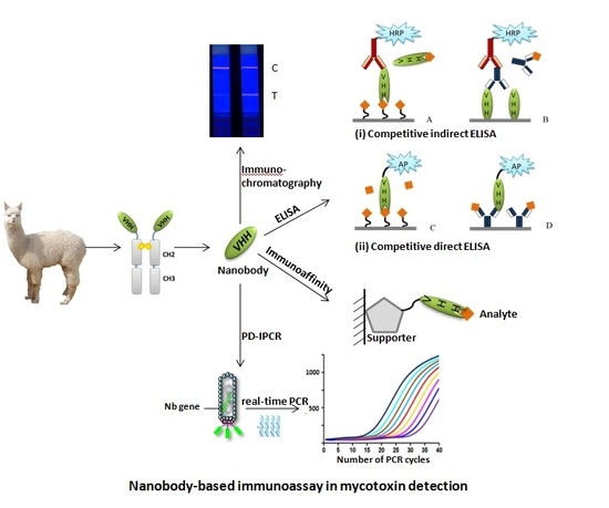
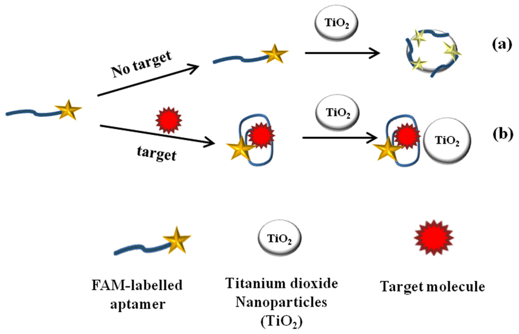
Anti-idiotypic nanobodies, usually expressed by gene engineering protocol, has been shown as a nontoxic coating antigen for toxic compound immunoassays. We here focused on how to increase immunoassay sensitivity by changing the nanobody’s primary sequence. In the experiments, two anti-idiotype nanobodies against monoclonal
[...] Read more.
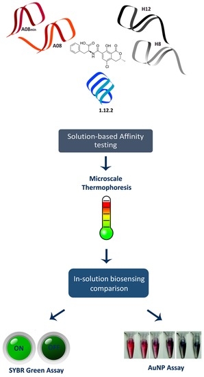
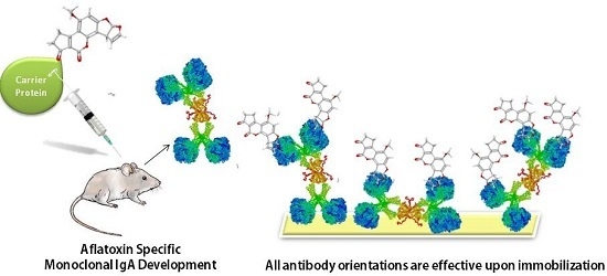
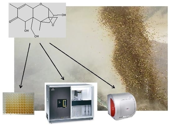
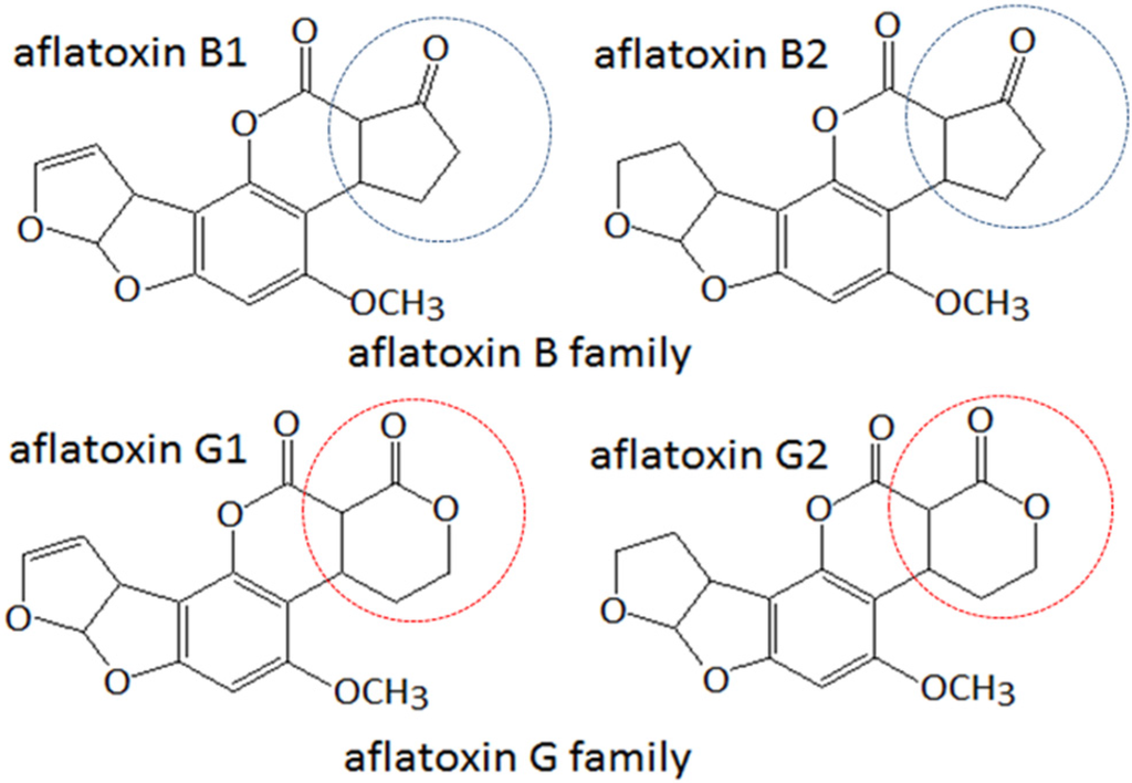
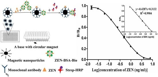
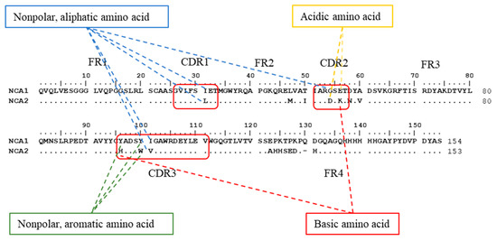
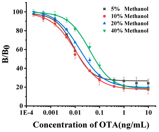
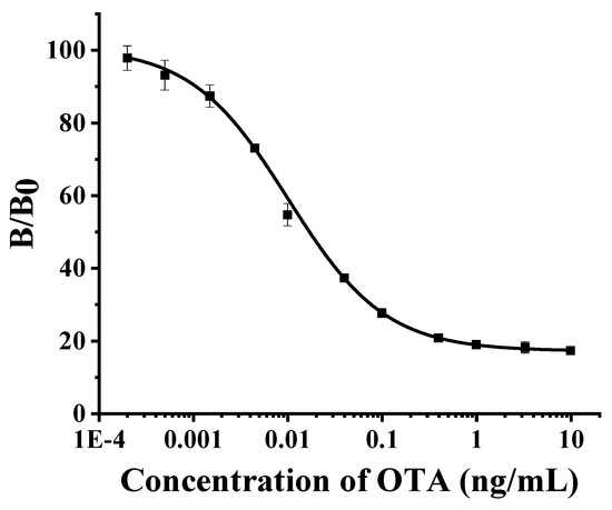
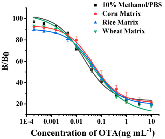
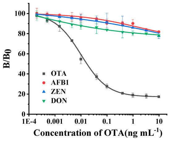
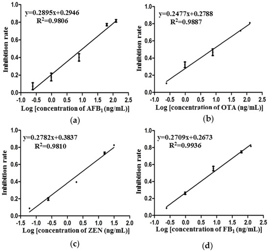
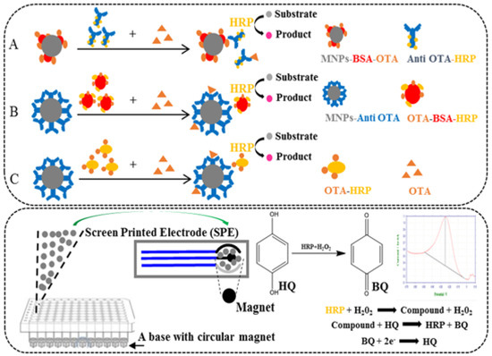
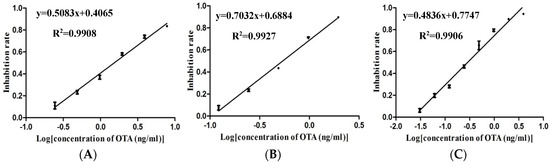
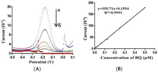
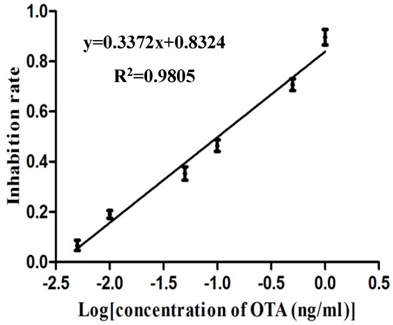
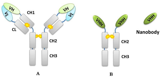
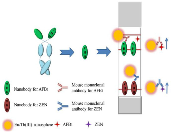
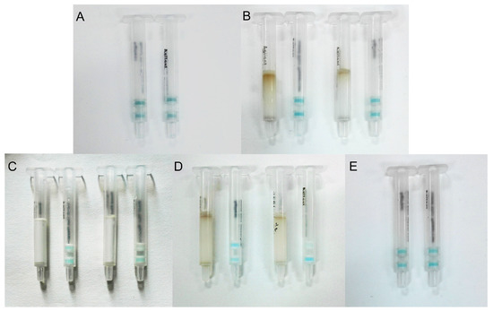
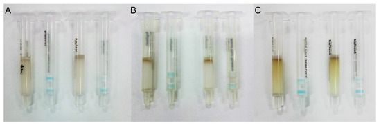
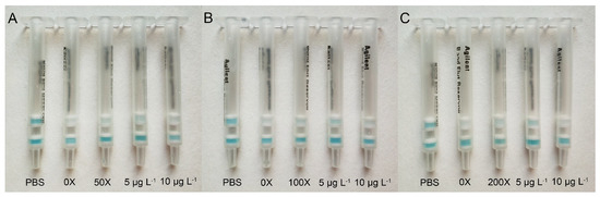
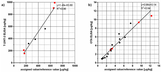
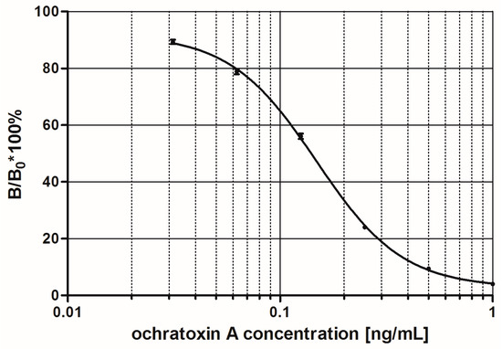
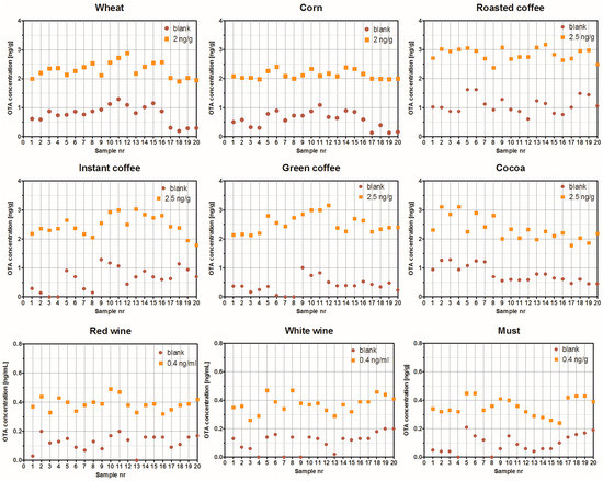
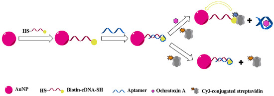
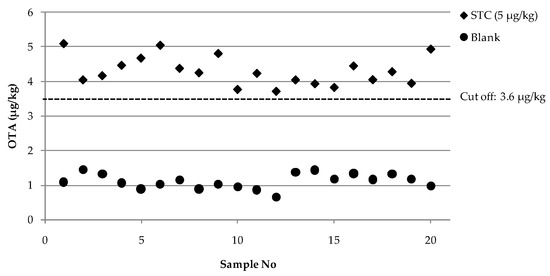
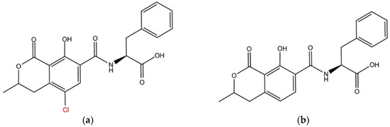
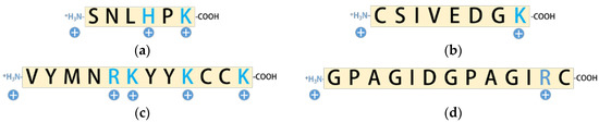
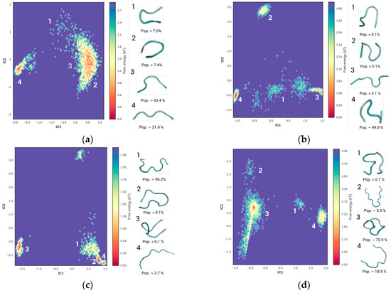
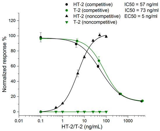
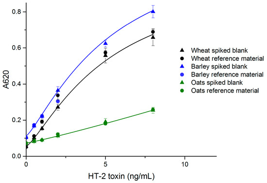
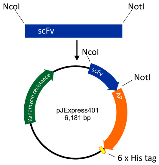
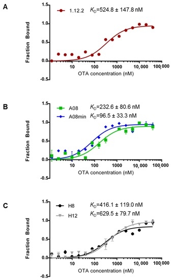
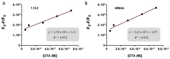
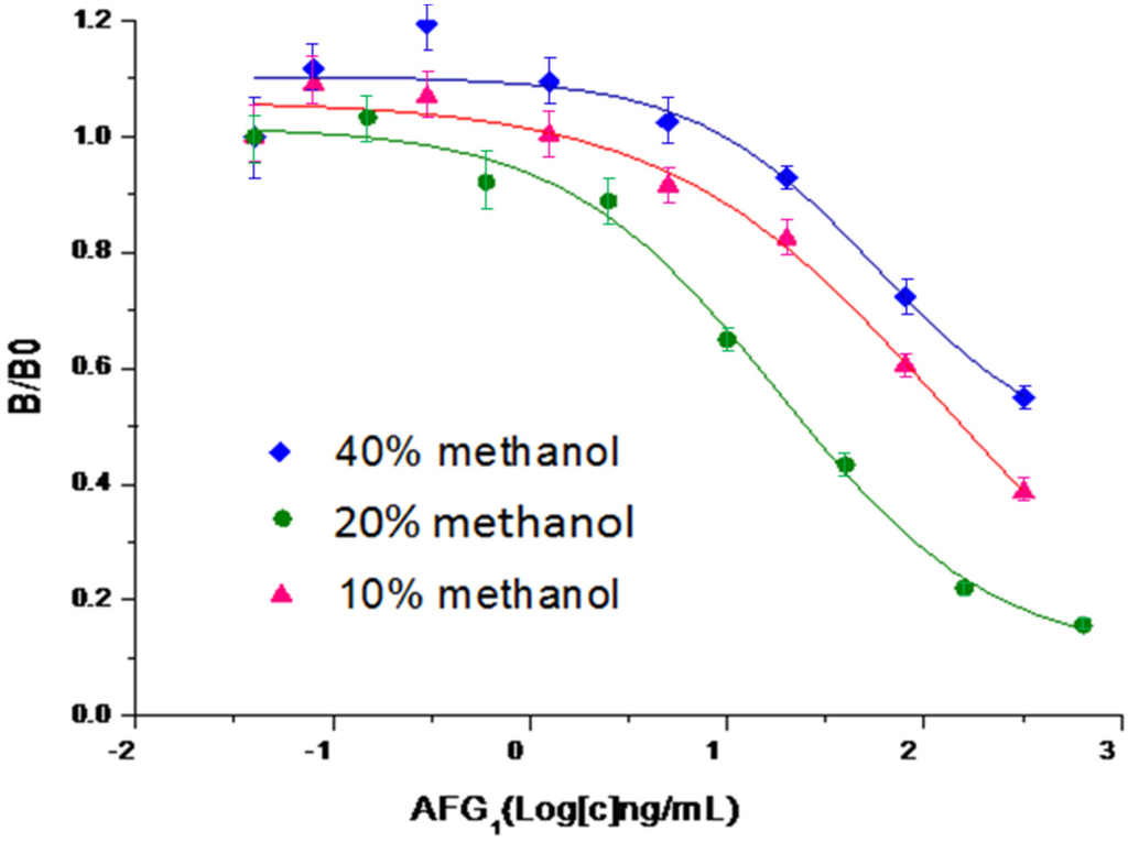
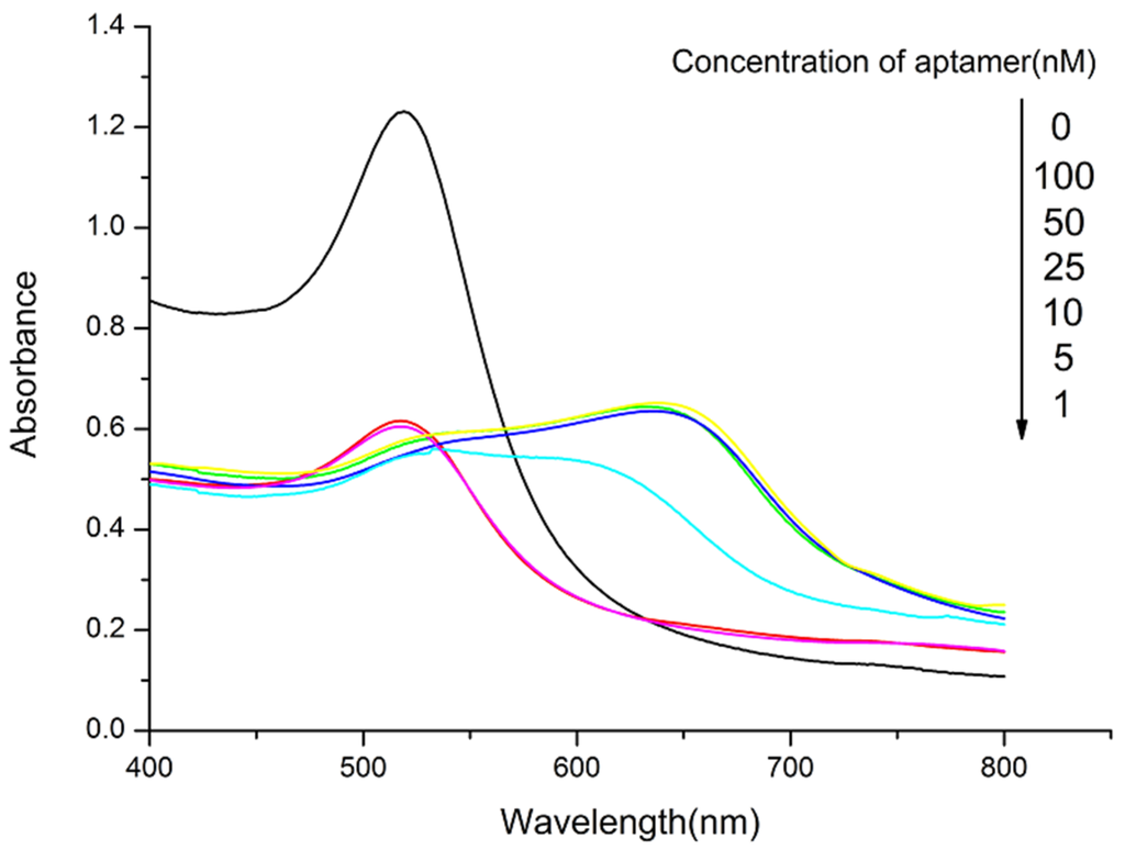
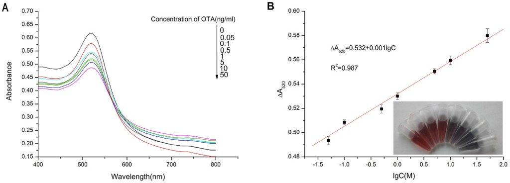
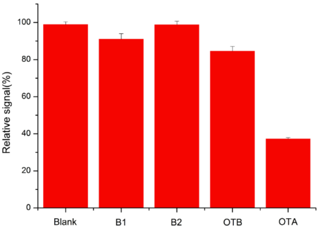
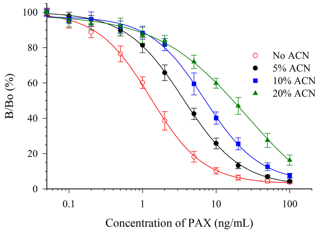
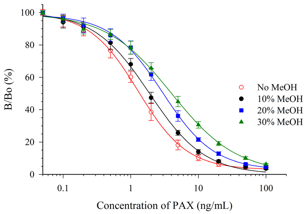
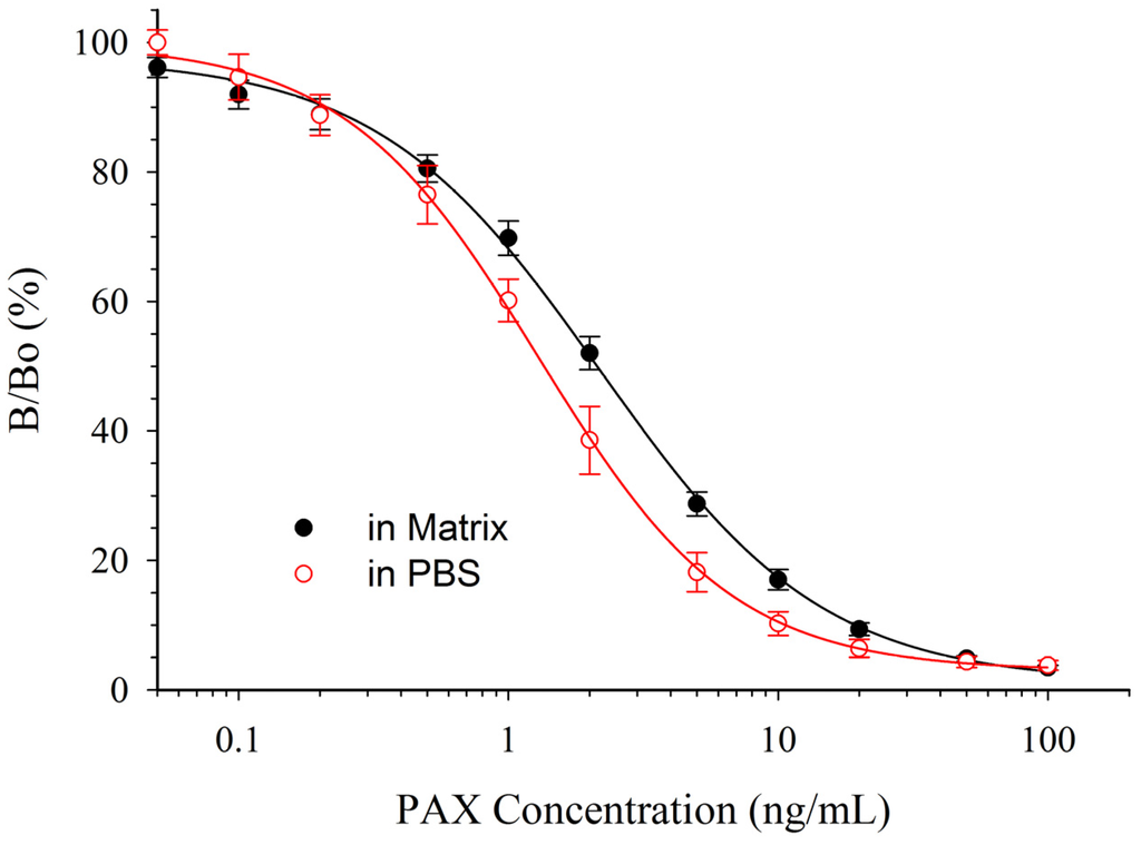
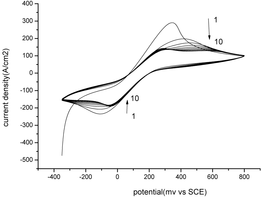
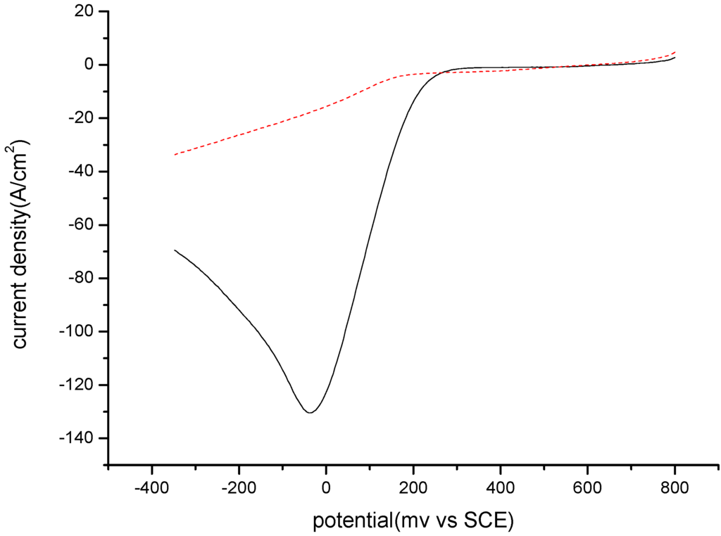
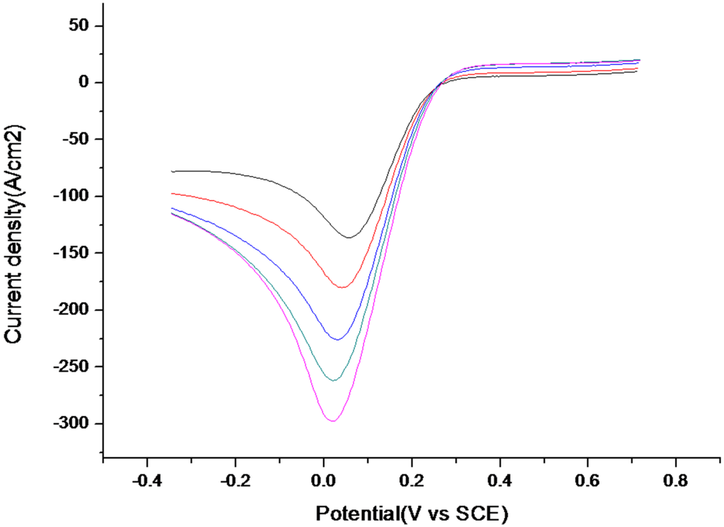
Anti-idiotypic nanobodies, usually expressed by gene engineering protocol, has been shown as a nontoxic coating antigen for toxic compound immunoassays. We here focused on how to increase immunoassay sensitivity by changing the nanobody’s primary sequence. In the experiments, two anti-idiotype nanobodies against monoclonal antibody 1H2, which is specific to ochratoxin A, were obtained and named as nontoxic coating antigen 1 (NCA1) and nontoxic coating antigen 2 (NCA2). Three differences between the nanobodies were discovered. First, there are six amino acid residues (AAR) of changes in the complementarity determining region (CDR), which compose the antigen-binding site. One of them locates in CDR1 (I–L), two of them in CDR2 (G–D, E–K), and three of them in CDR3 (Y–H, Y–W). Second, the affinity constant of NCA1 was tested as 1.20 × 10⁸ L mol−1, which is about 4 times lower than that of NCA2 (5.36 × 10⁸ L mol−1). Third, the sensitivity (50% inhibition concentration) of NCA1 for OTA was shown as 0.052 ng mL−1, which was 3.5 times lower than that of nontoxic coating antigen 2 (0.015 ng mL−1). The results indicate that the AAR changes in CDR of the anti-idiotypic nanobodies, from nonpolar to polar, increasing the affinity constant may enhance the immunoassay sensitivity. In addition, by using the nontoxic coating antigen 2 to substitute the routine synthetic toxic antigen, we established an eco-friendly and green enzyme-linked immunosorbent assay (ELISA) method for rapid detection of ochratoxin A in cereals. The half-maximal inhibitory concentration (IC50) of optimized ELISA was 0.017 ng mL−1 with a limit of detection (LOD) of 0.003 ng mL−1. The optimized immunoassay showed that the average recoveries of spiked corn, rice, and wheat were between 80% and 114.8%, with the relative standard deviation (RSD) ranging from 3.1–12.3%. Therefore, we provided not only basic knowledge on how to improve the structure of anti-idiotype nanobody for increasing assay sensitivity, but also an available eco-friendly ELISA for ochratoxin A in cereals.
Full article
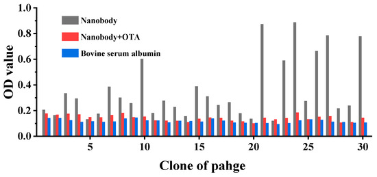
Figure 1


