Parasites Series: Parasitic Infection, Characterisation and Host Health, Parasitic Diversity and Host Range

A special issue of Pathogens (ISSN 2076-0817). This special issue belongs to the section "Parasitic Pathogens".

Deadline for manuscript submissions: closed (31 March 2025) | Viewed by 13122

Special Issue Editors


E-Mail Website
Guest Editor
Vector and Water-Borne Pathogens Research Group, School of Veterinary & Life Sciences, Murdoch University, South Street, Murdoch, Perth, Australia
Interests: haemoparasites; Trypanosoma; haemoproteidae; wildlife; protozoa; nematoda

E-Mail Website
Guest Editor
Vector and Waterborne Pathogen Research Group, College of Science, Health, Engineering and Education, Murdoch University, Perth, WA, Australia
Interests: molecular epidemiology; public health; protozoa; water and food-borne parasites

E-Mail Website
Guest Editor
Department of Medical Laboratory Sciences, Faculty of Applied Health Sciences, The Hashemite University, Zarqa, Jordan
Interests: protozoa; Cryptosporidium; in vitro culture; genotyping and subtyping; diagnosis

Special Issue Information

Dear Colleagues,

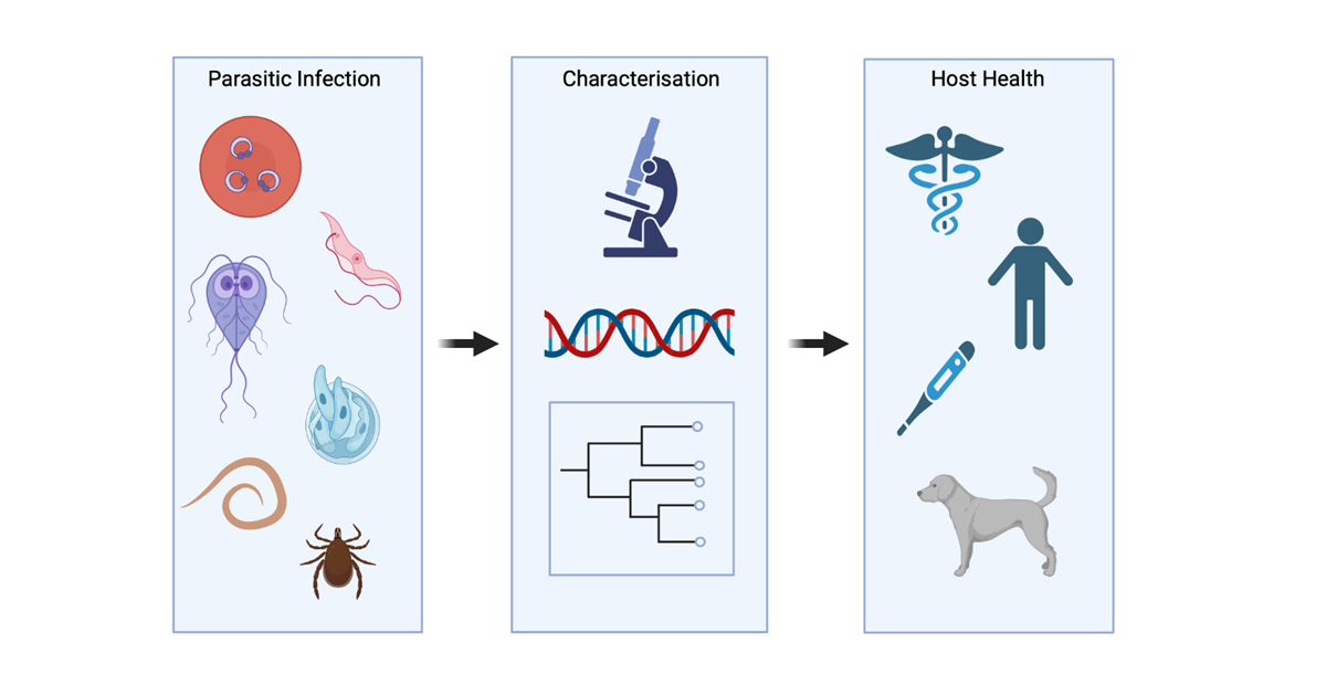
Parasitism is classically defined as a relationship between two living organisms in which one benefits at the expense of the other. Helminths, ectoparasites and protozoa are the three main types of organisms that cause such infections, and vary in size, complexity, and medical and/or veterinary concern. Their life cycles can range from intracellular parasitism to exoparasitism, with completion requiring either a single host species or multiple successive hosts. Parasite transmission roots vary highly, ranging from environmental contamination (contaminated food, water, and fomites) to being reliant on vectors for their continued development, reproduction, and host infection. As natural stressors, parasites often have detrimental effects on their hosts, with clinical severity ranging from asymptomatic to symptomatic, with the outcome of such infections often dependent on the immune status of their host. Acute-to-chronic infections can develop with symptoms associated to helminths and gastrointestinal protozoans (e.g., Cryptosporidium spp. and Giardia) generally leading to malabsorption, vomiting, diarrhoea, and weight loss, while infections with bloodborne parasites (e.g., Plasmodium spp. and Trypanosoma spp.) tend to be associated with haemolytic anaemia, fatigue, joint and muscle pain, headache, flu-like symptoms, encephalitis and behavioural changes. However, interchanges of symptoms can occur, adversely affecting the health and productivity of animal and human populations worldwide. The need to characterise and ascertain all aspects of parasitic infections through biological, chemical, molecular, and epidemiological studies is imperative to address knowledge gaps in the field, which will ultimately facilitate management strategies and treatment options to maintain stable and healthy populations on both a domestic and global scale.

Dr. Jill M. Austen
Dr. Alireza Zahedi
Prof. Dr. Nawal S. H. Hijjawi
Guest Editors

Manuscript Submission Information

Manuscripts should be submitted online at www.mdpi.com by registering and logging in to this website. Once you are registered, click here to go to the submission form. Manuscripts can be submitted until the deadline. All submissions that pass pre-check are peer-reviewed. Accepted papers will be published continuously in the journal (as soon as accepted) and will be listed together on the special issue website. Research articles, review articles as well as short communications are invited. For planned papers, a title and short abstract (about 100 words) can be sent to the Editorial Office for announcement on this website.

Submitted manuscripts should not have been published previously, nor be under consideration for publication elsewhere (except conference proceedings papers). All manuscripts are thoroughly refereed through a single-blind peer-review process. A guide for authors and other relevant information for submission of manuscripts is available on the Instructions for Authors page. Pathogens is an international peer-reviewed open access monthly journal published by MDPI.

Please visit the Instructions for Authors page before submitting a manuscript. The Article Processing Charge (APC) for publication in this open access journal is 2200 CHF (Swiss Francs). Submitted papers should be well formatted and use good English. Authors may use MDPI's English editing service prior to publication or during author revisions.

Keywords

  • parasites
  • host health
  • protozoa
  • helminths
  • ectoparasites
  • diagnosis
  • health impacts

Benefits of Publishing in a Special Issue

  • Ease of navigation: Grouping papers by topic helps scholars navigate broad scope journals more efficiently.
  • Greater discoverability: Special Issues support the reach and impact of scientific research. Articles in Special Issues are more discoverable and cited more frequently.
  • Expansion of research network: Special Issues facilitate connections among authors, fostering scientific collaborations.
  • External promotion: Articles in Special Issues are often promoted through the journal's social media, increasing their visibility.
  • e-Book format: Special Issues with more than 10 articles can be published as dedicated e-books, ensuring wide and rapid dissemination.

Further information on MDPI's Special Issue policies can be found here.

Published Papers (6 papers)

Order results
Result details
Select all
Export citation of selected articles as:

Research

Jump to: Review

17 pages, 4883 KiB  
Article
Prevalence of Blastocystis spp. and Other Gastrointestinal Pathogens Among Patients Admitted to Research Hospitals in Campania Region, Italy
by Marianna Ascierto, Annalisa Chianese, Francesco Foglia, Emiliana Finamore, Luciana Petrullo, Carla Zannella, Anna De Filippis, Maria Grazia Coppola and Massimiliano Galdiero
Pathogens 2025, 14(5), 425; https://doi.org/10.3390/pathogens14050425 - 27 Apr 2025
Viewed by 99
Abstract
Background. Blastocystis spp. is a common protozoan found in the gastrointestinal tract, typically existing as a non-pathogenic organism in humans and other animals. However, it can become pathogenic when the immune system is compromised due to bacterial, viral, fungal, or other parasitic infections, [...] Read more.
Background. Blastocystis spp. is a common protozoan found in the gastrointestinal tract, typically existing as a non-pathogenic organism in humans and other animals. However, it can become pathogenic when the immune system is compromised due to bacterial, viral, fungal, or other parasitic infections, as well as systemic conditions, leading to symptomatic blastocystosis. Methods. Fecal samples were collected from patients at the University Hospital of Campania “Luigi Vanvitelli” and Cotugno Hospital in Naples. Among these samples, those that tested positive for Blastocystis spp. and were associated with other microbial infections were further analyzed. Bacterial co-infections were identified using immunochromatographic tests (ICTs) and matrix-assisted laser desorption/ionization time-of-flight mass spectrometry (MALDI-TOF MS). Viral infections were detected using chemiluminescent immunoassay (CLIA), while fungal infections were diagnosed through microscopic examination and molecular biology techniques. Additionally, co-infections with other parasites were identified through microscopic analysis after Ridley’s concentration and Giemsa staining (O&P). Results. Out of the 2050 stool samples collected, 121 were positive for Blastocystis spp., of which 75 were associated with other infections. We identified the vacuolar form in patients co-infected with bacteria (n = 22), viruses (n = 30), fungi (n = 3), and other parasites (n = 20). Conclusions. Our findings indicated a higher incidence of the vacuolar form of Blastocystis spp. in symptomatic and immunocompromised patients, suggesting that a weakened immune system may increase the risk of contracting Blastocystis and other microbial infections. Full article
Show Figures

Figure 1

11 pages, 14331 KiB  
Article
Fatal Toxoplasmosis in Red Kangaroos (Macropus rufus) in East China
by Haiyan Gong, Quan Wang, Yinghong Jin, Suoping Qiu, Zhaoguo Chen, Xiangan Han, Zongyan Chen and Wei Jiang
Pathogens 2025, 14(2), 202; https://doi.org/10.3390/pathogens14020202 - 19 Feb 2025
Viewed by 750
Abstract
Background: As a ubiquitous apicomplexan parasite, Toxoplasma gondii causes huge economic losses and poses a great threat to the health of animals, including humans, worldwide. In some kangaroo species, T. gondii can be fatal. To date, little information is available on T. gondii [...] Read more.
Background: As a ubiquitous apicomplexan parasite, Toxoplasma gondii causes huge economic losses and poses a great threat to the health of animals, including humans, worldwide. In some kangaroo species, T. gondii can be fatal. To date, little information is available on T. gondii infection in the red kangaroos in east China. At a zoo in east China, thirteen red kangaroos consecutively developed clinical signs from July to November 2016, resulting in the deaths of seven, three of which were analyzed in this study. Methods: In the present study, ascitic fluid, blood and samples from the brain, heart, liver, spleen, lung, kidney, and mesenteric lymph nodes of three dead red kangaroos were collected. The pathogen was explored through microscopic observation, nested PCR, immunofluorescence antibody test (IFAT), hematoxylin–eosin (HE) staining, and immunohistochemistry (IHC) staining, respectively. Meanwhile, the potential source of the infection was also investigated by testing the blood of stray cats in the zoo for T. gondii using nested PCR. Results: Three dead red kangaroos were subjected to a necropsy, and organisms resembling T. gondii were detected in their ascitic fluids under microscope. This infection was further confirmed by a nested PCR assay, which resulted in a successful amplification and sequencing of the 433 bp fragment of the T. gondii 5.8S rRNA gene in all the dissected tissues, including heart, liver, spleen, lung, kidney, lymph nodes, cecum, and brain, as well as in body fluids (blood and ascitic fluid). Furthermore, the tachyzoites were observed in the heart, liver, spleen, lymph nodes, cecum, and brain through IFAT and HE staining. Administration of classic drugs (sulfadiazine and pyrimethamine) against T. gondii significantly alleviated the clinical signs of the sick kangaroos. The possible source of this infection was traced to a native stray cat, as T. gondii DNA was detected in its blood. Conclusions: In the present study, lethal T. gondii infection in red kangaroos has been described for the first time in east China, highlighting the necessity and urgency for close and long-term surveillance of this parasite infection in captive animals. The same strain of T. gondii detected in kangaroos as that found in stray cats wandering in the same area emphasizes the importance of controlling stray cat populations to mitigate the risk of Toxoplasma transmission to other animals. Full article
Show Figures

Figure 1

13 pages, 614 KiB  
Article
A Comparative Study of Genetic Diversity and Multiplicity of Infection in Uncomplicated Plasmodium falciparum Infections in Selected Regions of Pre-Elimination and High Transmission Settings Using MSP1 and MSP2 Genes
by Olusegun Philip Akoniyon, Moses Akiibinu, Matthew A. Adeleke, Rajendra Maharaj and Moses Okpeku
Pathogens 2024, 13(2), 172; https://doi.org/10.3390/pathogens13020172 - 13 Feb 2024
Cited by 1 | Viewed by 2691
Abstract
Background: Understanding the genetic structure of P. falciparum population in different regions is pivotal to malaria elimination. Genetic diversity and the multiplicity of infection are indicators used for measuring malaria endemicity across different transmission settings. Therefore, this study characterized P. falciparum infections [...] Read more.
Background: Understanding the genetic structure of P. falciparum population in different regions is pivotal to malaria elimination. Genetic diversity and the multiplicity of infection are indicators used for measuring malaria endemicity across different transmission settings. Therefore, this study characterized P. falciparum infections from selected areas constituting pre-elimination and high transmission settings in South Africa and Nigeria, respectively. Methods: Parasite genomic DNA was extracted from 129 participants with uncomplicated P. falciparum infections. Isolates were collected from 78 participants in South Africa (southern Africa) and 51 in Nigeria (western Africa). Allelic typing of the msp1 and msp2 genes was carried out using nested PCR. Results: In msp1, the K1 allele (39.7%) was the most common allele among the South African isolates, while the RO33 allele (90.2%) was the most common allele among the Nigerian isolates. In the msp2 gene, FC27 and IC3D7 showed almost the same percentage distribution (44.9% and 43.6%) in the South African isolates, whereas FC27 had the highest percentage distribution (60.8%) in the Nigerian isolates. The msp2 gene showed highly distinctive genotypes, indicating high genetic diversity in the South African isolates, whereas msp1 showed high genetic diversity in the Nigerian isolates. The RO33 allelic family displayed an inverse relationship with participants’ age in the Nigerian isolates. The overall multiplicity of infection (MOI) was significantly higher in Nigeria (2.87) than in South Africa (2.44) (p < 0.000 *). In addition, heterozygosity was moderately higher in South Africa (1.46) than in Nigeria (1.13). Conclusions: The high genetic diversity and MOI in P. falciparum that were observed in this study could provide surveillance data, on the basis of which appropriate control strategies should be adopted. Full article
Show Figures

Figure 1

19 pages, 1871 KiB  
Article
Insights into the Genetic Diversity of Leishmania (Viannia) panamensis in Panama, Inferred via Multilocus Sequence Typing (MLST)
by Daniel Mendieta, Vanessa Vásquez, Luis Jaén, Vanessa Pineda, Azael Saldaña, José Eduardo Calzada and Franklyn Samudio
Pathogens 2023, 12(5), 747; https://doi.org/10.3390/pathogens12050747 - 22 May 2023
Viewed by 2713
Abstract
Leishmaniasis is a disease caused by parasites of the genus Leishmania and transmitted by sand fly vectors. Tegumentary leishmaniasis is the most prevalent clinical outcome in Latin America, afflicting people from 18 countries. In Panama, the annual incidence rate of leishmaniasis is as [...] Read more.
Leishmaniasis is a disease caused by parasites of the genus Leishmania and transmitted by sand fly vectors. Tegumentary leishmaniasis is the most prevalent clinical outcome in Latin America, afflicting people from 18 countries. In Panama, the annual incidence rate of leishmaniasis is as high as 3000 cases, representing a major public health problem. In endemic regions, L. panamensis is responsible for almost eighty percent of human cases that present different clinical outcomes. These differences in disease outcomes could be the result of the local interplay between L. panamensis variants and human hosts with different genetic backgrounds. The genetic diversity of L. panamensis in Panama has only been partially explored, and the variability reported for this species is based on few studies restricted to small populations and/or with poor resolutive markers at low taxonomic levels. Accordingly, in this study, we explored the genetic diversity of sixty-nine L. panamensis isolates from different endemic regions of Panama, using an MLST approach based on four housekeeping genes (Aconitase, ALAT, GPI and HSP70). Two to seven haplotypes per locus were identified, and regional differences in the genetic diversity of L. panamensis were observed. A genotype analysis evidenced the circulation of thirteen L. panamensis genotypes, a fact that might have important implications for the local control of the disease. Full article
Show Figures

Figure 1

13 pages, 932 KiB  
Article
In Vitro Susceptibility of Cryptosporidium parvum to Plant Antiparasitic Compounds
by Sandamalie Ranasinghe, Alireza Zahedi, Anthony Armson, Alan J. Lymbery and Amanda Ash
Pathogens 2023, 12(1), 61; https://doi.org/10.3390/pathogens12010061 - 30 Dec 2022
Cited by 4 | Viewed by 2842
Abstract
Cryptosporidium parvum is a significant cause of watery diarrhoea in humans and other animals worldwide. Although hundreds of novel drugs have been evaluated, no effective specific chemotherapeutic intervention for C. parvum has been reported. There has been much recent interest in evaluating plant-derived products [...] Read more.
Cryptosporidium parvum is a significant cause of watery diarrhoea in humans and other animals worldwide. Although hundreds of novel drugs have been evaluated, no effective specific chemotherapeutic intervention for C. parvum has been reported. There has been much recent interest in evaluating plant-derived products in the fight against gastrointestinal parasites, including C. parvum. This study aimed to identify extracts from 13 different plant species that provide evidence for inhibiting the growth of C. parvum in vitro. Efficacy against C. parvum was detected and quantified using quantitative PCR and immunofluorescence assays. All plant extracts tested against C. parvum showed varying inhibition activities in vitro, and none of them produced a cytotoxic effect on HCT-8 cells at concentrations up to 500 µg/mL. Four plant species with the strongest evidence of activity against C. parvum were Curcuma longa, Piper nigrum, Embelia ribes, and Nigella sativa, all with dose-dependent efficacy. To the authors' knowledge, this is the first time that these plant extracts have proven to be experimentally efficacious against C. parvum. These results support further exploration of these plants and their compounds as possible treatments for Cryptosporidium infections. Full article
Show Figures

Graphical abstract

Review

Jump to: Research

11 pages, 860 KiB  
Review
Lungworms of Non-Ruminant Terrestrial Mammals and Humans in Iran
by Sina Mohtasebi, Alireza Sazmand, Salman Zafari, Guilherme G. Verocai and Domenico Otranto
Pathogens 2023, 12(6), 759; https://doi.org/10.3390/pathogens12060759 - 25 May 2023
Cited by 2 | Viewed by 2767
Abstract
With over 300 terrestrial and aquatic mammalian species, Iran is considered a country with an ample mastofauna. Although many studies have assessed the distribution of gastrointestinal helminth parasites in animals and humans in Iran, lungworms have not received adequate attention. Following a previous [...] Read more.
With over 300 terrestrial and aquatic mammalian species, Iran is considered a country with an ample mastofauna. Although many studies have assessed the distribution of gastrointestinal helminth parasites in animals and humans in Iran, lungworms have not received adequate attention. Following a previous article in which we reviewed the diversity and prevalence of lungworm infections in pastoral and wild ruminants of Iran, this report compiles the available scientific information about the occurrence of lungworms in non-ruminant mammals and humans from 1980 to 2022 to provide insights into the epidemiology of these infections. International and national scientific databases were searched, and twenty-six articles in peer-reviewed journals, one conference paper, and one D.V.M. thesis were included in the study. In total, 10 species belonging to seven genera, including Dictyocaulus, Deraiophoronema, Protostrongylus, Crenosoma, Eucoleus, Aelurostrongylus, and Metastrongylus, were reported in the respiratory tract or feces of humans, domestic animals (i.e., camels, equids, dogs, and cats), and wildlife species (i.e., hedgehogs, wild boars, and hares). Most of the studies (22/28) were performed using post-mortem examinations. The overall prevalence of respiratory nematode infection varied according to animal species in camels (14.83%), equids (13.31%), dogs (5%), wild boars (45.66%), hedgehogs (42.57%), and hares (1.6%). In addition, pulmonary capillariasis caused by Eucoleus aerophilus was reported in a 9 year old child. The prevalence of lungworm species in domestic camels, equids, and dogs, combined with a lack of labeled anthelmintic products, supports the need to improve our understanding of these important nematode parasites and inform the development of sustainable control strategies. From a zoo and wildlife medicine point of view, there is a shortage of information about the presence and prevalence of lungworm infections in the majority of mammalian species, pending epidemiological studies that integrate classical parasitology and molecular methods. Full article
Show Figures

Figure 1

Back to TopTop