



Mitochondrial and Endoplasmic Reticulum Dynamics in Cellular Stress Responses

A special issue of Life (ISSN 2075-1729). This special issue belongs to the section "Physiology and Pathology".

Deadline for manuscript submissions: 25 September 2026 | Viewed by 2393
Editors


E-Mail Website
Guest Editor
Jiangsu Key Laboratory of Sericultural Biology and Biotechnology, School of Biotechnology, Jiangsu University of Science and Technology, Zhenjiang 212100, China
Interests: mitochondrial function; endoplasmic reticulum homeostasis; cell stress; gene function; physiopathology

E-Mail Website
Guest Editor
Key Laboratory of Environmental Medicine Engineering, Ministry of Education, School of Public Health, Southeast University, Nanjing 210009, China
Interests: mitochondrial dynamics; endoplasmic reticulum stress; oogenesis; aging; cell imaging; reproductive health

E-Mail Website
Guest Editor
Institutes of Agricultural Science and Technology Development, College of Veterinary Medicine, Yangzhou University, Yangzhou 225009, China
Interests: influenza; PEDV
Special Issues, Collections and Topics in MDPI journals

Special Issue Information

Dear Colleagues,

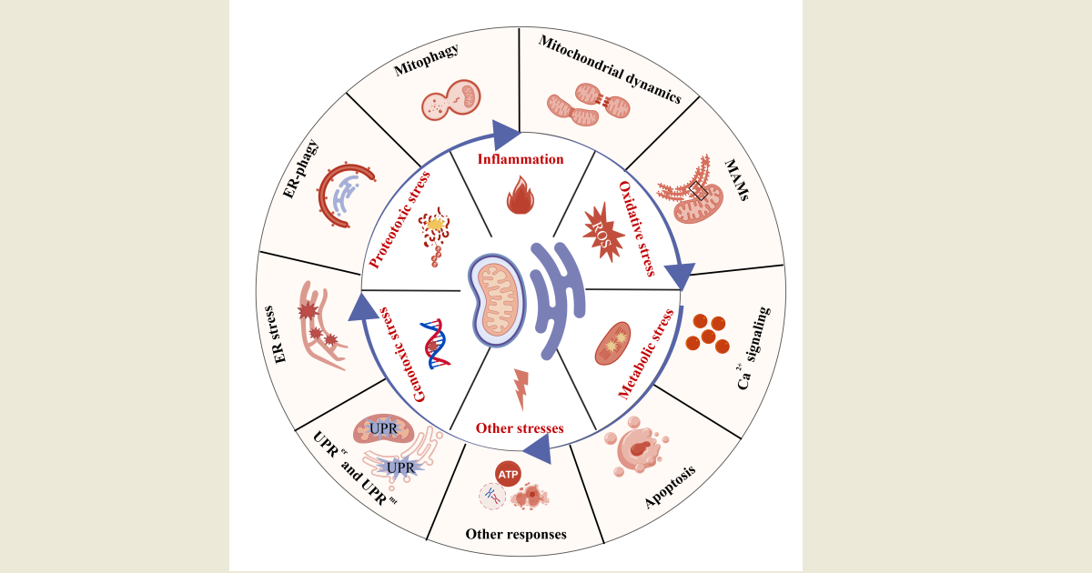
Cells are continuously exposed to various stressors that trigger their protective mechanisms to enhance resilience. The capacity of stress responses to adapt and survive by restoring homeostasis is an integral component of cellular physiology. Among the key organelles involved in these responses, the mitochondria and endoplasmic reticulum play pivotal roles in maintaining cellular homeostasis. The dynamic interplay between these organelles is crucial for mediating cellular adaptation and fate decisions during stress.

This Special Issue aims to provide a comprehensive overview of the latest advancements in understanding the dynamics of mitochondria and endoplasmic reticulum in cellular stress responses. We particularly welcome studies addressing mitochondria-endoplasmic reticulum contact sites, Ca2+ signaling, mitochondrial dynamics (fission, fusion, and motility) during stress, endoplasmic reticulum stress, UPRer and UPRmt, ER-phagy and mitophagy. Contributions on redox/ROS signaling in apoptosis versus survival decisions, as well as organelle quality control, biogenesis, and metabolic rewiring in response to stress, are also encouraged.

Dr. Chengmin Li
Dr. Xing Duan
Dr. Changchao Huan
Guest Editors

Manuscript Submission Information

Manuscripts should be submitted online at www.mdpi.com by registering and logging in to this website. Once you are registered, click here to go to the submission form. Manuscripts can be submitted until the deadline. All submissions that pass pre-check are peer-reviewed. Accepted papers will be published continuously in the journal (as soon as accepted) and will be listed together on the special issue website. Research articles, review articles as well as short communications are invited. For planned papers, a title and short abstract (about 250 words) can be sent to the Editorial Office for assessment.

Submitted manuscripts should not have been published previously, nor be under consideration for publication elsewhere (except conference proceedings papers). All manuscripts are thoroughly refereed through a single-anonymized peer-review process. A guide for authors and other relevant information for submission of manuscripts is available on the Instructions for Authors page. Life is an international peer-reviewed open access monthly journal published by MDPI.

Please visit the Instructions for Authors page before submitting a manuscript. The Article Processing Charge (APC) for publication in this open access journal is 2600 CHF (Swiss Francs). Submitted papers should be well formatted and use good English. Authors may use MDPI's English editing service prior to publication or during author revisions.


Keywords

  • mitochondria
  • endoplasmic reticulum
  • MAMs
  • cellular responses
  • physiopathology
  • homeostasis
  • cross-talk
  • stress
  • signaling
  • regulation

Benefits of Publishing in a Special Issue

  • Ease of navigation: Grouping papers by topic helps scholars navigate broad scope journals more efficiently.
  • Greater discoverability: Special Issues support the reach and impact of scientific research. Articles in Special Issues are more discoverable and cited more frequently.
  • Expansion of research network: Special Issues facilitate connections among authors, fostering scientific collaborations.
  • External promotion: Articles in Special Issues are often promoted through the journal's social media, increasing their visibility.
  • Reprint: MDPI Books provides the opportunity to republish successful Special Issues in book format, both online and in print.

Further information on MDPI's Special Issue policies can be found here.

Published Papers (2 papers)

Order results
Result details
Select all
Export citation of selected articles as:

Research

Jump to: Review

19 pages, 4917 KB  
Article
Spermine Ameliorates DSS-Induced Ulcerative Colitis in Mice by Improving Mitophagy and Intestinal Microbiota
by Ran Yu, Yamei Liu, Yating Zheng, Saisai Chen, Ling Tong, Jichun Wang, Chengmin Li and Chuanjian Zhang
Life 2026, 16(3), 417; https://doi.org/10.3390/life16030417 - 4 Mar 2026
Viewed by 968
Abstract
Spermine (Spe) plays a critical role in maintaining the integrity of the intestinal barrier and promoting intestinal development. However, the therapeutic role of Spe on ulcerative colitis (UC) remains unclear. This study aims to research the impact and mechanism of Spe on dextran [...] Read more.
Spermine (Spe) plays a critical role in maintaining the integrity of the intestinal barrier and promoting intestinal development. However, the therapeutic role of Spe on ulcerative colitis (UC) remains unclear. This study aims to research the impact and mechanism of Spe on dextran sulfate sodium (DSS)-induced colitis in mice. Twenty-eight C57BL/6 mice were orally administered Spe before and during DSS treatment to evaluate its protective effects. Lipopolysaccharides (LPSs) were used to construct an in vitro UC model in IEC-6 cells. The study indicates that Spe treatment upregulated the expression of tight junction protein occludin and inhibited NLRP3 mediated inflammatory response by downregulating the levels of NLRP3, Caspase-1, IL-1β, IL-18 and TNF-α in the colon of DSS-treated mice. In addition, Spe enhanced mitophagy in colitis mice by increasing expressions of mitophagy factors (PINK1, Parkin, LC3-II) in DSS-treated mice. PINK1-mediated mitophagy helps alleviate LPS-induced mitochondrial damage in IEC-6 cells. Furthermore, Spe regulates the gut microbiota composition in mice with colitis by increasing the abundance of unclassified Muribaculaceae, reducing the levels of Firmicutes and Blautia, and lowering the Firmicutes/Bacteroidetes ratio. In conclusion, spermine exhibited treatment efficacy on DSS-induced colitis by inhibiting NLRP3-mediated inflammatory response, promoting mitophagy and improving intestinal microbial dysbiosis. Full article
Show Figures

Figure 1

Review

Jump to: Research

20 pages, 1690 KB  
Review
Mitochondrial Adaptations to Exercise Training in Equine Skeletal Muscle: A Narrative Review
by Vlad Cocioba, Paula Nistor, Daniel George Bratu, Șerban Blaga, Bianca Cornelia Zanfira, Călin Mircu and Ioan Huțu
Life 2026, 16(6), 1008; https://doi.org/10.3390/life16061008 - 16 Jun 2026
Viewed by 583
Abstract
The horse represents one of the most physiologically specialized athletic mammals, capable of sustaining both high-intensity and prolonged exercise. Central to this remarkable performance capacity is the metabolic adaptability of skeletal muscle and its mitochondrial network. This narrative review synthesizes current evidence from [...] Read more.
The horse represents one of the most physiologically specialized athletic mammals, capable of sustaining both high-intensity and prolonged exercise. Central to this remarkable performance capacity is the metabolic adaptability of skeletal muscle and its mitochondrial network. This narrative review synthesizes current evidence from equine, human, and rodent studies on exercise-induced mitochondrial remodeling in equine skeletal muscle. A comprehensive literature search was conducted across PubMed, Web of Science, and Scopus using terms related to equine exercise physiology, mitochondrial biology, and skeletal muscle metabolism. Preference was given to peer-reviewed original research and review articles. Mitochondria regulate oxidative phosphorylation, substrate oxidation, redox signaling, and cellular responses to metabolic stress induced by exercise. Training induces extensive mitochondrial adaptations, including mitochondrial biogenesis, remodeling of the respiratory chain, enhanced oxidative phosphorylation efficiency, and increased metabolic flexibility. These adaptations are believed to contribute to improvements in aerobic capacity, delayed fatigue onset, and enhanced recovery following exercise, although direct mechanistic evidence in horses remains limited. In equine skeletal muscle, mitochondrial plasticity is closely linked to muscle fiber composition and the distribution of oxidative and glycolytic fibers. Exercise-induced signaling pathways involving AMP-activated protein kinase (AMPK), Ca2+-dependent kinases, and the transcriptional coactivator PGC-1α regulate mitochondrial biogenesis and metabolic remodeling. In addition, mitochondrial dynamics, including fusion, fission, and mitophagy, maintain mitochondrial quality and functional efficiency during repeated training stimuli. Experimental studies in Thoroughbred and Standardbred horses demonstrate that training has been associated with increases in mitochondrial density and respiratory capacity in equine skeletal muscle, contributing directly to improved aerobic performance and metabolic efficiency. However, mitochondrial adaptations must be interpreted within the broader context of musculoskeletal adaptation, as metabolic improvements may occur faster than structural adaptation of tendons and ligaments. This review synthesizes current knowledge on exercise-induced mitochondrial remodeling in equine skeletal muscle, while highlighting the limited mechanistic evidence available in horses and the need for more standardized longitudinal studies. Full article
Show Figures

Figure 1

Back to TopTop