


Cells 2025, 14(23), 1902; https://doi.org/10.3390/cells14231902 - 1 Dec 2025
Cited by 1 | Viewed by 1581
Abstract
►
Show Figures
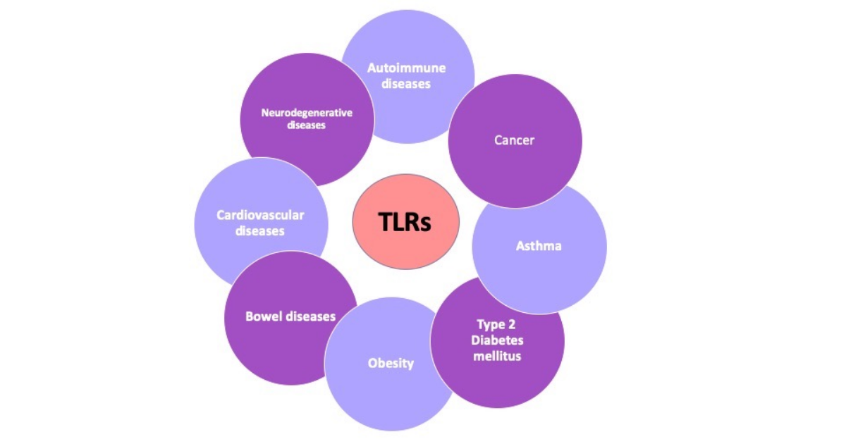
Inborn errors of immunity (IEIs), formerly referred to as primary immunodeficiencies (PID), represent a heterogeneous group of hereditary disorders that significantly increase patients’ susceptibility to severe and recurrent infections. Toll-like receptors (TLRs) play a pivotal role in host defense as fundamental components of
[...] Read more.
Inborn errors of immunity (IEIs), formerly referred to as primary immunodeficiencies (PID), represent a heterogeneous group of hereditary disorders that significantly increase patients’ susceptibility to severe and recurrent infections. Toll-like receptors (TLRs) play a pivotal role in host defense as fundamental components of innate immunity, while also linking it to adaptive immune responses. This review summarizes advances in understanding the involvement of TLRs in the pathogenesis of IEIs in children. It highlights genetic defects such as deficiencies in MyD88, IRAK-4, NEMO, and TLR3, which lead to distinct clinical phenotypes, for example, increased susceptibility to bacterial infections or herpes simplex virus type-1 (HSV-1) encephalitis. The review also examines more complex disorders, including chronic granulomatous disease (CGD), common variable immunodeficiency (CVID), and X-linked agammaglobulinemia (XLA), in which TLR signaling may be either impaired or dysregulated. This analysis demonstrates the growing importance of functional assays evaluating TLR activity as a diagnostic tool complementary to genetic testing, as well as their potential to precisely characterize immunological phenotypes. Furthermore, current therapeutic perspectives are discussed, including the use of TLR agonists, which have shown promising results in oncology, the role of gene therapy as a causal treatment option, and a proposed diagnostic algorithm incorporating TLR-based evaluation. Despite significant progress, substantial knowledge gaps remain, particularly regarding the full spectrum of TLR signaling abnormalities across IEI subtypes. The conclusions emphasize the need for large-scale, international studies to achieve a comprehensive understanding of pathogenic mechanisms and to develop more targeted and effective therapeutic interventions for children affected by these rare disorders.
Full article

Figure 1


