New Advances and Perspectives in Nanotechnology for the Treatment of Cancer Cells

A special issue of Cells (ISSN 2073-4409). This special issue belongs to the section "Cell Methods".

Deadline for manuscript submissions: 20 March 2026 | Viewed by 15746

Special Issue Editor


E-Mail Website
Guest Editor
School of Biosystems and Food Engineering, University College Dublin, Belfield, D04 V1W8 Dublin, Ireland
Interests: nanotechnology; immunotherapeutics; cancer biotherapeutics; proliferation; regulating factors; cell culture; biomedical application; nutraceuticals; cancer treatment

Special Issue Information

Dear Colleagues,

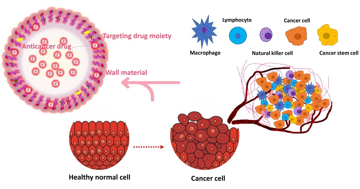
In recent decades, nanotechnology has already had a great impact on cancer detection and therapy. The application of nanotechnology to the healthcare aspects of biomedicine is rapidly progressing in advanced treatment strategies, particularly in the detection and monitoring of cancer progression and treatment in the field of cancer biotherapeutics. This type of cutting-edge technology in the current pharmaceutical industry is manifested by a recently developed nanomedicine approach to the treatment various types of cancer, advances that have unlocked new abilities in developed datasets to provide biological insight into cancer.
In the Special Issue of Cells, entitled “New Advance and perspectives in Nanotechnology for the Treatment of Cancer Cells”, we would like to showcase current advances in the field of nanotechnology and related approaches to the study of cancer detection and treatment. This applies is related to advances in underlying technologies, as well as those relating to key biological and therapeutic findings. Potential topics include, but are not limited to, nanotechnology research with a cancer focus involving the development of nanotechnology methods, nanotechnology-based biological and clinical findings, small-molecule and immunotherapeutic target discovery, therapeutic mechanisms of action, cancer immunology, advancements in sample preparation techniques, single-cell analysis, comprehensive metadata analysis, AI/statistics technology, etc. The perspectives for new advances/developments in cancer biotherapeutics directing to the clinical approaches of therapeutic drug delivery by nanotechnology and based approaches will be addressed. Interdisciplinary research is highly valued and encouraged. We welcome the submission of both original research articles and reviews.

Dr. Karuppusamy Shanmugapriya
Guest Editor

Manuscript Submission Information

Manuscripts should be submitted online at www.mdpi.com by registering and logging in to this website. Once you are registered, click here to go to the submission form. Manuscripts can be submitted until the deadline. All submissions that pass pre-check are peer-reviewed. Accepted papers will be published continuously in the journal (as soon as accepted) and will be listed together on the special issue website. Research articles, review articles as well as short communications are invited. For planned papers, a title and short abstract (about 100 words) can be sent to the Editorial Office for announcement on this website.

Submitted manuscripts should not have been published previously, nor be under consideration for publication elsewhere (except conference proceedings papers). All manuscripts are thoroughly refereed through a single-blind peer-review process. A guide for authors and other relevant information for submission of manuscripts is available on the Instructions for Authors page. Cells is an international peer-reviewed open access semimonthly journal published by MDPI.

Please visit the Instructions for Authors page before submitting a manuscript. The Article Processing Charge (APC) for publication in this open access journal is 2700 CHF (Swiss Francs). Submitted papers should be well formatted and use good English. Authors may use MDPI's English editing service prior to publication or during author revisions.

Keywords

  • nanotechnology
  • immunotherapeutics
  • cancer biotherapeutics
  • proliferation
  • regulating factors
  • cell culture
  • biomedical application
  • nutraceuticals
  • cancer treatment

Benefits of Publishing in a Special Issue

  • Ease of navigation: Grouping papers by topic helps scholars navigate broad scope journals more efficiently.
  • Greater discoverability: Special Issues support the reach and impact of scientific research. Articles in Special Issues are more discoverable and cited more frequently.
  • Expansion of research network: Special Issues facilitate connections among authors, fostering scientific collaborations.
  • External promotion: Articles in Special Issues are often promoted through the journal's social media, increasing their visibility.
  • Reprint: MDPI Books provides the opportunity to republish successful Special Issues in book format, both online and in print.

Further information on MDPI's Special Issue policies can be found here.

Published Papers (7 papers)

Order results
Result details
Select all
Export citation of selected articles as:

Research

Jump to: Review

19 pages, 10760 KiB  
Article
Celastrol-Loaded Hyaluronic Acid/Cancer Cell Membrane Lipid Nanoparticles for Targeted Hepatocellular Carcinoma Prevention
by Peng He, Manshu Zou, Chanjuan Zhang, Yaning Shi and Li Qin
Cells 2024, 13(21), 1819; https://doi.org/10.3390/cells13211819 - 4 Nov 2024
Cited by 3 | Viewed by 1483
Abstract
Hepatocellular carcinoma (HCC) is the third leading cause of cancer-related deaths worldwide, and its prevention and treatment face severe challenges. It is crucial to improve the targeting of drugs on tumor cells and tissues. Celastrol (CeT), as an active ingredient of traditional Chinese [...] Read more.
Hepatocellular carcinoma (HCC) is the third leading cause of cancer-related deaths worldwide, and its prevention and treatment face severe challenges. It is crucial to improve the targeting of drugs on tumor cells and tissues. Celastrol (CeT), as an active ingredient of traditional Chinese medicine, possesses strong antitumor effects, especially in triggering apoptosis of HCC. However, due to its toxicity and lack of targeting, its application is greatly limited. HMCLPs, a nano-biomimetic platform carrying CeT with controllable drug release, enhanced targeting, and immunocompatibility, were developed for the first time, which can be used for the treatment of HCC. By utilizing homologous cell membranes and hyaluronic acid (HA), HMCLPs can precisely target tumor regions and release CeT in a controlled manner. Both in vitro and in vivo studies have demonstrated that HMCLPs loaded with CeT significantly increased the accumulation of reactive oxygen species (ROS), induced mitochondrial damage, and triggered apoptosis of HCC cells, resulting in effective treatment with minimal adverse reaction. The development of HMCLPs as a nanocarrier system for CeT delivery offers a promising therapeutic strategy for HCC. This innovative approach improves the targeted delivery and bioavailability of CeT, dramatically induces apoptosis in HCC cells, and exerts its powerful antitumor effects while minimizing systemic toxicity. The present study highlights the potential of combining innovative nanocarriers with powerful natural compounds such as CeT to enhance efficacy and reduce toxicity. Full article
Show Figures

Figure 1

14 pages, 1684 KiB  
Article
Evaluation of Vincamine Loaded with Silver Nanoparticles as a New Potential Therapeutic Agent Against Ehrlich’s Solid Carcinoma in Mice
by Naief Dahran, Mohamed S. Othman, Mohamed E. Ghoniem, Mai A. Samak, Mohamed T. Elabbasy, Sofian T. Obeidat, Ghada M. Aleid, Shimaa Abo Elnaga, Azza M. Khaled, Aya A. Altaleb and Ahmed E. Abdel Moneim
Cells 2024, 13(21), 1762; https://doi.org/10.3390/cells13211762 - 24 Oct 2024
Cited by 1 | Viewed by 1327
Abstract
Vincamine, a monoterpenoid indole alkaloid with vasodilatory properties, is extracted from the leaves of Vinca minor. The present study aimed to determine the potential anticancer effects of vincamine loaded in silver nanoparticles (VCN-AgNPs) in mice with Ehrlich solid carcinoma (ESC). After tumor [...] Read more.
Vincamine, a monoterpenoid indole alkaloid with vasodilatory properties, is extracted from the leaves of Vinca minor. The present study aimed to determine the potential anticancer effects of vincamine loaded in silver nanoparticles (VCN-AgNPs) in mice with Ehrlich solid carcinoma (ESC). After tumor transplantation, the mice were divided into five groups: ESC, ESC+Cisplatin (CPN; 5 mg/kg), ESC+VCN (40 mg/kg), ESC+AgNPs (6 mg/kg), and ESC+VCN-AgNPs (20 mg/kg). The administration of VCN-AgNPs to ESC-bearing mice improved their survival rate and reduced their body weight, tumor size, and tumor weight compared to the ESC group. Furthermore, VCN-AgNPs intensified oxidative stress in tumor tissues, as evidenced by elevated levels of lipid peroxidation (LPO) and nitric oxide (NO), along with a reduction in the levels of the antioxidants investigated (GSH, GPx, GR, SOD, CAT, and TAC). Furthermore, VCN-AgNPs increased the apoptotic proteins Bax and caspase-3, decreased the anti-apoptotic protein (Bcl-2), increased the inflammatory markers TNF-α and IL-1β, and inhibited angiogenesis by lowering VEGF levels in tumor tissues, all of which led to apoptosis. Furthermore, histopathological studies showed that VCN-AgNPs suppressed the progression of Ehrlich carcinoma and induced the formation of clusters of necrotic and fragmented tumor cells. VCN-AgNPs possess cytotoxic and genotoxic effects against ESC because of their pro-oxidant, pro-apoptotic, pro-inflammatory, and antiangiogenic effects. Additionally, the combination of VCN-AgNPs was more effective and safer than chemically synthesized AgNPs, as indicated by an increase in the lifespan of animals and the total tumor inhibition index. Full article
Show Figures

Graphical abstract

15 pages, 3889 KiB  
Article
Understanding Macrophage Interaction with Antimony-Doped Tin Oxide Plasmonic Nanoparticles
by Olexiy Balitskii, Viktoriya Ivasiv, Maria Porteiro-Figueiras, Phattadon Yajan, Mira Witzig, Aura Maria Moreno-Echeverri, José Muñetón Díaz, Barbara Rothen-Rutishauser, Alke Petri-Fink and Sandeep Keshavan
Cells 2024, 13(17), 1468; https://doi.org/10.3390/cells13171468 - 1 Sep 2024
Viewed by 1601
Abstract
Antimony-doped tin oxide nanoparticles (ATO NPs) have emerged as a promising tool in biomedical applications, namely robust photothermal effects upon near-infrared (NIR) light exposure, enabling controlled thermal dynamics to induce spatial cell death. This study investigated the interplay between ATO NPs and macrophages, [...] Read more.
Antimony-doped tin oxide nanoparticles (ATO NPs) have emerged as a promising tool in biomedical applications, namely robust photothermal effects upon near-infrared (NIR) light exposure, enabling controlled thermal dynamics to induce spatial cell death. This study investigated the interplay between ATO NPs and macrophages, understanding cellular uptake and cytokine release. ATO NPs demonstrated biocompatibility with no impact on macrophage viability and cytokine secretion. These findings highlight the potential of ATO NPs for inducing targeted cell death in cancer treatments, leveraging their feasibility, unique NIR properties, and safe interactions with immune cells. ATO NPs offer a transformative platform with significant potential for future biomedical applications by combining photothermal capabilities and biocompatibility. Full article
Show Figures

Figure 1

15 pages, 3737 KiB  
Article
Green-Synthesized Silver and Selenium Nanoparticles Using Berberine: A Comparative Assessment of In Vitro Anticancer Potential on Human Hepatocellular Carcinoma Cell Line (HepG2)
by Azza M. Khaled, Mohamed S. Othman, Sofian T. Obeidat, Ghada M. Aleid, Shimaa M. Aboelnaga, Alaa Fehaid, Heba M. R. Hathout, Ashraf A. Bakkar, Ahmed E. Abdel Moneim, Islam M. El-Garawani and Dalia S. Morsi
Cells 2024, 13(3), 287; https://doi.org/10.3390/cells13030287 - 5 Feb 2024
Cited by 24 | Viewed by 3368
Abstract
A well-known natural ingredient found in several medicinal plants, berberine (Ber), has been shown to have anticancer properties against a range of malignancies. The limited solubility and bioavailability of berberine can be addressed using Ber-loaded nanoparticles. In this study, we compared the in [...] Read more.
A well-known natural ingredient found in several medicinal plants, berberine (Ber), has been shown to have anticancer properties against a range of malignancies. The limited solubility and bioavailability of berberine can be addressed using Ber-loaded nanoparticles. In this study, we compared the in vitro cytotoxic effects of both Ber-loaded silver nanoparticles (Ber-AgNPs) and Ber-loaded selenium nanoparticles (Ber-SeNPs) in the human liver cancer cell line (HepG2) and mouse normal liver cells (BNL). The IC50 values in HepG2 for berberine, Ber-AgNPs, Ber-SeNPs, and cisplatin were 26.69, 1.16, 0.04, and 0.33 µg/mL, respectively. Our results show that Ber and its Ag and Se nanoparticles exerted a good antitumor effect against HepG2 cells by inducing apoptosis via upregulating p53, Bax, cytosolic cytochrome C levels, and caspase-3 activity, and the down-regulation of Bcl-2 levels. Similarly, incubation with Ber and both Ber-NPs (Ag and Se) led to a significant dose-dependent elevation in inflammatory markers’ (TNF-α, NF-κB, and COX-2) levels compared to the control group. In addition, it led to the arrest of the G1 cell cycle by depleting the expression of cyclin D1 and CDK-2 mRNA. Furthermore, Ber and both Ber-NPs (Ag and Se) caused a significant dose-dependent increase in LDH activity in HepG2 cells. Furthermore, our findings offer evidence that Ber and its nanoparticles intensified oxidative stress in HepG2 cells. Furthermore, the migration rate of cells subjected to berberine and its nanoforms was notably decreased compared to that of control cells. It can be inferred that Ber nanoparticles exhibited superior anticancer efficacy against HepG2 compared to unprocessed Ber, perhaps due to their improved solubility and bioavailability. Furthermore, Ber-SeNPs exhibited greater efficacy than Ber-AgNPs, possibly as a result of the inherent anticancer characteristics of selenium. Full article
Show Figures

Figure 1

12 pages, 1133 KiB  
Article
Exploring the Potential of Montmorillonite as an Antiproliferative Nanoagent against MDA-MB-231 and MCF-7 Human Breast Cancer Cells
by Alireza Ghannad Sabzevari, Hossein Sabahi, Mohsen Nikbakht, Mehdi Azizi, Hassan Dianat-Moghadam and Zohreh Amoozgar
Cells 2024, 13(2), 200; https://doi.org/10.3390/cells13020200 - 22 Jan 2024
Cited by 2 | Viewed by 2526
Abstract
Unlike MCF-7 cells, MDA-MB-231 cells are unresponsive to hormone therapy and often show resistance to chemotherapy and radiotherapy. Here, the antiproliferative effect of biocompatible montmorillonite (Mt) nanosheets on MDA-MB-231 and MCF-7 human breast cancer cells was evaluated by MTT assay, flow cytometry, and [...] Read more.
Unlike MCF-7 cells, MDA-MB-231 cells are unresponsive to hormone therapy and often show resistance to chemotherapy and radiotherapy. Here, the antiproliferative effect of biocompatible montmorillonite (Mt) nanosheets on MDA-MB-231 and MCF-7 human breast cancer cells was evaluated by MTT assay, flow cytometry, and qRT-PCR. The results showed that the Mt IC50 for MDA-MB-231 and MCF-7 cells in a fetal bovine serum (FBS)-free medium was ~50 and ~200 µg/mL, and in 10% FBS medium ~400 and ~2000 µg/mL, respectively. Mt caused apoptosis in both cells by regulating related genes including Cas-3, P53, and P62 in MDA-MB-231 cells and Bcl-2, Cas-8, Cas-9, P53, and P62 in MCF-7 cells. Also, Mt arrested MCF-7 cells in the G0/G1 phase by altering Cyclin-D1 and P21 expression, and caused sub-G1 arrest and necrosis in both cells, possibly through damaging the mitochondria. However, fewer gene expression changes and more sub-G1 arrest and necrosis were observed in MDA-MB-231 cells, confirming the higher vulnerability of MDA-MB-231 cells to Mt. Furthermore, MDA-MB-231 cells appeared to be much more vulnerable to Mt compared to other cell types, including normal lung fibroblast (MRC-5), colon cancer (HT-29), and liver cancer (HepG2) cells. The higher vulnerability of MDA-MB-231 cells to Mt was inferred to be due to their higher proliferation rate. Notably, Mt cytotoxicity was highly dependent on both the Mt concentration and serum level, which favors Mt for the local treatment of MDA-MB-231 cells. Based on these results, Mt can be considered as an antiproliferative nanoagent against MDA-MB-231 cells and may be useful in the development of local nanoparticle-based therapies. Full article
Show Figures

Figure 1

Review

Jump to: Research

21 pages, 2459 KiB  
Review
Enhancing Proton Therapy Efficacy Through Nanoparticle-Mediated Radiosensitization
by Jie Ma, Hao Shen and Zhaohong Mi
Cells 2024, 13(22), 1841; https://doi.org/10.3390/cells13221841 - 7 Nov 2024
Cited by 4 | Viewed by 1580
Abstract
Proton therapy, characterized by its unique Bragg peak, offers the potential to optimize the destruction of cancer cells while sparing healthy tissues, positioning it as one of the most advanced cancer treatment modalities currently available. However, in comparison to heavy ions, protons exhibit [...] Read more.
Proton therapy, characterized by its unique Bragg peak, offers the potential to optimize the destruction of cancer cells while sparing healthy tissues, positioning it as one of the most advanced cancer treatment modalities currently available. However, in comparison to heavy ions, protons exhibit a relatively lower relative biological effectiveness (RBE), which limits the efficacy of proton therapy. The incorporation of nanoparticles for radiosensitization presents a novel approach to enhance the RBE of protons. This review provides a comprehensive discussion of the recent advancements in augmenting the biological effects of proton therapy through the use of nanoparticles. It examines the various types of nanoparticles that have been the focus of extensive research, elucidates their mechanisms of radiation sensitization, and evaluates the factors influencing the efficiency of this sensitization process. Furthermore, this review discusses the latest synergistic therapeutic strategies that integrate nanoparticle-mediated radiosensitization and outlines prospective directions for the future application of nanoparticles in conjunction with proton therapy. Full article
Show Figures

Figure 1

15 pages, 1366 KiB  
Review
Molecular Mechanisms of the Therapeutic Effect of Selenium Nanoparticles in Hepatocellular Carcinoma
by Elena G. Varlamova
Cells 2024, 13(13), 1102; https://doi.org/10.3390/cells13131102 - 26 Jun 2024
Cited by 11 | Viewed by 2461
Abstract
This review describes and summarizes, for the first time, the molecular mechanisms of the cytotoxic effect of selenium nanoparticles of various origins on hepatocellular carcinoma cells. The text provides information from recent years indicating the regulation of various signaling pathways and endoplasmic reticulum [...] Read more.
This review describes and summarizes, for the first time, the molecular mechanisms of the cytotoxic effect of selenium nanoparticles of various origins on hepatocellular carcinoma cells. The text provides information from recent years indicating the regulation of various signaling pathways and endoplasmic reticulum stress by selenium nanoparticles; the pathways of cell death of liver cancer cells as a result of exposure to selenium nanoparticles are considered. Particular attention is paid to the participation of selenoproteins and selenium-containing thioredoxin reductases and glutathione peroxidases in these processes. Previously, there were no reviews that fully reflected the cytotoxic effects of selenium nanoparticles specifically in hepatocellular carcinoma, despite the fact that many reviews and experimental articles have been devoted to the causes of this disease and the molecular mechanisms of regulation of cytotoxic effects by other agents. The relevance of this review is primarily explained by the fact that despite the development of various drugs and approaches for the treatment and prevention of hepatocellular carcinoma, this disease is still the fourth leading cause of death in the world. For this reason, a complete understanding of the latest trends in the treatment of oncology of various etiologies, especially hepatocellular carcinoma, is extremely important. Full article
Show Figures

Graphical abstract

Back to TopTop