


cells-logo

Journal Browser

► Journal Browser

Membrane Damage and Repair in Organelles

A Topical Collection in Cells (ISSN 2073-4409) belonging to the section "Intracellular and Plasma Membranes".

Viewed by 5407

Editor


E-Mail Website
Collection Editor
Faculty of Science and Engineering, Iwate University, Morioka 020-8551, Japan
Interests: membrane traffic; ArfGAP; exosomes

Topical Collection Information

Dear Colleagues,

Organelle membranes play critical roles to compartmentalize organelle contents from cytosol and to maintain organelle functions in cells. Organelle membranes can be damaged by bacterial escape during infection, inorganic materials internalized from outside, and environmental change (such as osmolarity change). When organelle membranes are damaged, many molecular events are expected to occur. For example, organelle membrane tension is decreased, ions (such as Ca2+) leak into cytosol, proteins (such as Galectins) enter lumen from cytosol, sphingomyelin (in the luminal side of the membrane) is exposed to cytosol, the ESCRT complex is recruited to repair damaged membranes, and autophagy occurs to degrade damaged organelles. The process of membrane damage and repair has initially been studied in the plasma membrane; however, recent studies have started to reveal the molecular events for organelles.

This Topical Collection focuses on membrane damage and repair in organelles and provides an overview of membrane damage caused by pathogen escape, inorganic materials, other methods used to damage organelles, the physical properties of organelle membranes, lipid characteristics, signaling after membrane damage, membrane repair, and the degradation of damaged organelles. This Topical Collection highlights the recent advances and future research directions that are necessary to understand the membrane damage and repair in organelles.

Dr. Yoko Shiba
Collection Editor

Manuscript Submission Information

Manuscripts should be submitted online at www.mdpi.com by registering and logging in to this website. Once you are registered, click here to go to the submission form. All submissions that pass pre-check are peer-reviewed. Accepted papers will be published continuously in the journal (as soon as accepted) and will be listed together on the collection website. Research articles, review articles as well as communications are invited. For planned papers, a title and short abstract (about 250 words) can be sent to the Editorial Office for assessment.

Submitted manuscripts should not have been published previously, nor be under consideration for publication elsewhere (except conference proceedings papers). All manuscripts are thoroughly refereed through a single-anonymized peer-review process. A guide for authors and other relevant information for submission of manuscripts is available on the Instructions for Authors page. Cells is an international peer-reviewed open access semimonthly journal published by MDPI.

Please visit the Instructions for Authors page before submitting a manuscript. The Article Processing Charge (APC) for publication in this open access journal is 2700 CHF (Swiss Francs). Submitted papers should be well formatted and use good English. Authors may use MDPI's English editing service prior to publication or during author revisions.

Keywords

  • membrane damage
  • organelle
  • pathogen
  • inorganic material
  • galectin

Published Papers (2 papers)

2026

Jump to: 2025

32 pages, 1536 KB  
Review
Initial Molecular Detection of Membrane Damage in Post-Golgi Compartments
by Yoko Shiba and Nana Saito
Cells 2026, 15(15), 1375; https://doi.org/10.3390/cells15151375 - 30 Jul 2026
Viewed by 628
Abstract
Mammalian cells contain numerous membrane-bound organelles, of which endosomes serve as the initial destination for endocytosed molecules. Therapeutic agents are also internalized by cells and transported to endosomes or phagosomes and subsequently delivered to lysosomes for degradation. Therefore, these agents require drug delivery [...] Read more.
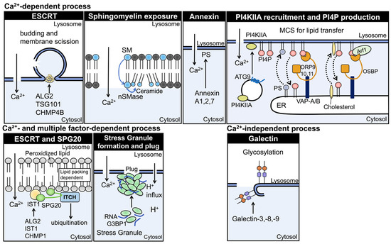
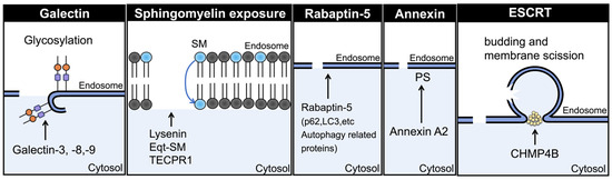
Mammalian cells contain numerous membrane-bound organelles, of which endosomes serve as the initial destination for endocytosed molecules. Therapeutic agents are also internalized by cells and transported to endosomes or phagosomes and subsequently delivered to lysosomes for degradation. Therefore, these agents require drug delivery systems (DDSs) that enable their escape from endosomes into the cytosol before lysosomal degradation; however, endosomal escape is a major limitation of current DDSs. Studies of bacterial phagosomal escape have revealed mechanisms by which host cells detect damage to organelle membranes. These membrane damage-sensing molecules also recognize membrane damage caused by artificial DDSs or physical energy-based insults. In this review, we summarize the molecular mechanisms underlying the early stages of membrane damage in the plasma membrane, lysosomes and bacteria-containing vacuoles (BCVs) to better understand the early stages of endosomal membrane damage in the absence of pathogens. We summarize recent advances in galectins, endosomal sorting complexes required for transport (ESCRT) complexes, sphingomyelin, stress granules, and phosphatidylinositol 4-phosphate (PI4P) at membrane contact sites, as well as annexins. We also discuss the recruitment kinetics of these molecules to damaged membranes. Although the recruitment kinetics vary depending on cell type and experimental conditions, this information provides a timeframe for the events following membrane damage, including damage sensing, membrane repair, and degradation of damaged organelles. We also discuss a potential fourth event, fusion between the plasma membrane and endosomes or lysosomes for membrane repair in the annexin section. Finally, we summarize approaches for inducing “sterile” endosomal membrane damage. Future development of these approaches may facilitate the design of novel DDSs and physical energy-based strategies for manipulating specific organelles. Full article
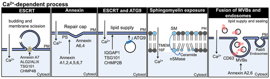
►▼ Show Figures

Figure 1

2025

Jump to: 2026

15 pages, 4731 KB  
Article
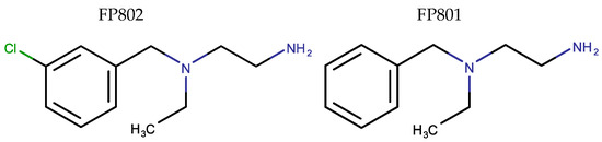
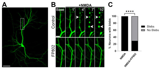
Pharmacological Targeting of the NMDAR/TRPM4 Death Signaling Complex with a TwinF Interface Inhibitor Prevents Excitotoxicity-Associated Dendritic Blebbing and Organelle Damage
by Omar A. Ramírez, Andrea Hellwig, Zihong Zhang and Hilmar Bading
Cells 2025, 14(3), 195; https://doi.org/10.3390/cells14030195 - 28 Jan 2025
Cited by 5 | Viewed by 3533
Abstract
Focal swellings of dendrites (“dendritic blebbing”) together with structural damage of mitochondria and the endoplasmic reticulum (ER) are morphological hallmarks of glutamate neurotoxicity, also known as excitotoxicity. These pathological alterations are generally thought to be caused by the so-called “overactivation” of N-methyl-D-aspartate receptors [...] Read more.
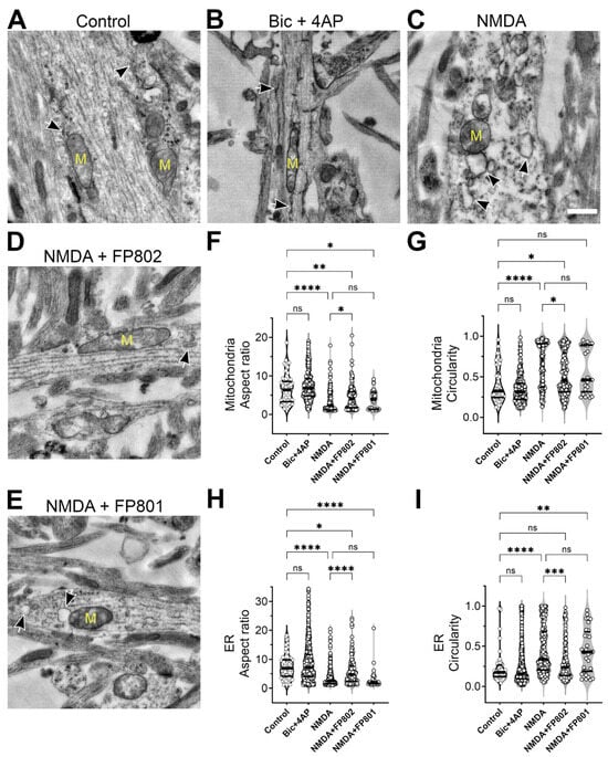
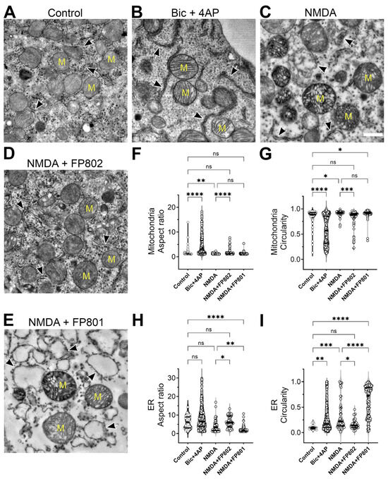
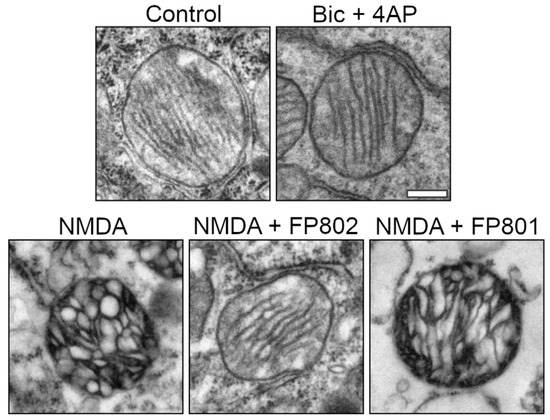
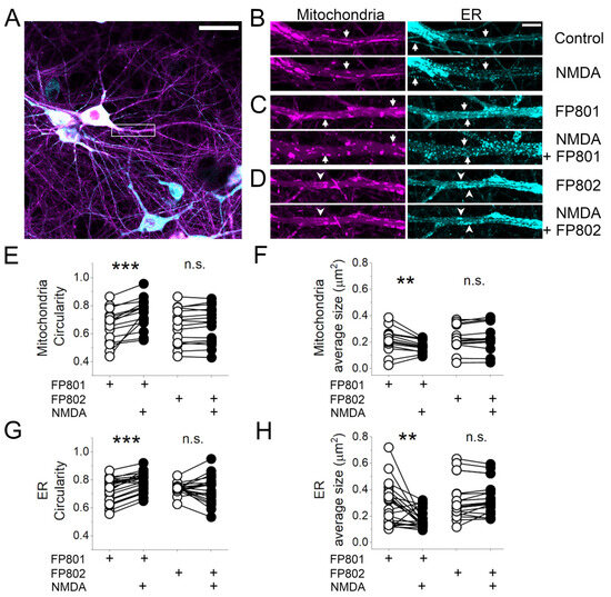
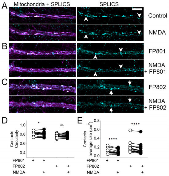
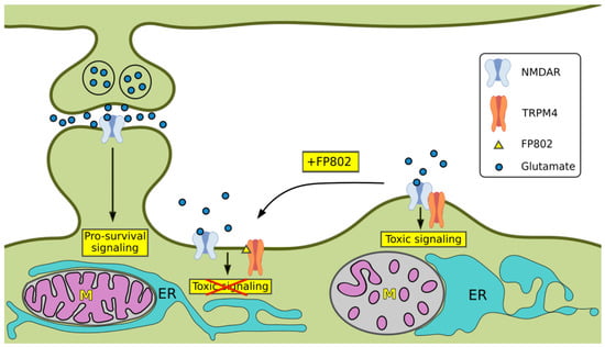
Focal swellings of dendrites (“dendritic blebbing”) together with structural damage of mitochondria and the endoplasmic reticulum (ER) are morphological hallmarks of glutamate neurotoxicity, also known as excitotoxicity. These pathological alterations are generally thought to be caused by the so-called “overactivation” of N-methyl-D-aspartate receptors (NMDARs). Here, we demonstrate that the activation of extrasynaptic NMDARs, specifically when forming a protein–protein complex with TRPM4, drives these pathological traits. In contrast, strong activation of synaptic NMDARs fails to induce cell damage despite evoking plateau-type calcium signals that are comparable to those generated by activation of the NMDAR/TRPM4 complex, indicating that high intracellular calcium levels per se are not toxic to neurons. Using confocal laser scanning microscopy and transmission electron microscopy, we show that disrupting the NMDAR/TRPM4 complex using the recently discovered small-molecule TwinF interface inhibitor FP802 inhibits the NMDA-induced neurotoxicity-associated dendritic blebbing and structural damage to mitochondria and the ER. It also prevents, at least in part, the disruption of ER–mitochondria contact sites. These findings establish the NMDAR/TRPM4 complex as the trigger for the structural damage of dendrites and intracellular organelles associated with excitotoxicity. They also suggest that activation of the NMDAR/TRPM4 complex, in addition to inducing high-amplitude, plateau-type calcium signals, generates a second signal required for glutamate neurotoxicity (“two-hit hypothesis”). As structural damage to organelles, particularly mitochondria, is a common feature of many human neurodegenerative diseases, including Alzheimer’s disease and amyotrophic lateral sclerosis (ALS), TwinF interface inhibitors have the potential to provide neuroprotection across a broad spectrum of these diseases. Full article
►▼ Show Figures

Figure 1

Back to TopTop