


Highlights
What are the main findings?
- •
- Advanced heart failure, LVAD support, and heart transplantation can be conceptualized as a continuous immunological trajectory rather than isolated clinical stages; we introduce the concept of the “recipient immune biography” to describe how cumulative inflammatory, infectious, device-related, and sensitizing exposures shape the immune state encountered by the donor graft.
- •
- Post-transplant alloimmunity is determined not only by donor-antigen recognition but also by immune-cell fate, clonality, memory, and tissue localization, integrating effector, regulatory, exhaustion-associated, Tfh–B-cell–humoral, and tissue-resident memory programs.
What are the implications of the main findings?
- •
- LVAD support may be viewed as an immunological interphase in which heart-failure-associated immune abnormalities may resolve, persist, or be remodeled, potentially modifying the immune substrate subsequently exposed to donor alloantigens.
- •
- Future immune surveillance should move beyond isolated circulating biomarkers toward longitudinal, multimodal phenotyping that integrates blood biomarkers, high-dimensional cellular profiling, single-cell transcriptomics, TCR/BCR clonotyping, graft molecular analysis, and spatial immunology.
Abstract
Advanced heart failure (HF) left ventricular assist device (LVAD) support and heart transplantation (HTx) are commonly considered distinct clinical stages, although each occurs within an immune system shaped by preceding inflammatory and antigenic exposures. This review introduces the concept of the recipient immune biography, describing the cumulative immunological history that accompanies patients from advanced HF through mechanical circulatory support to transplantation. We examine how HF-associated innate and adaptive immune activation may be modified by mechanical unloading while simultaneously being reshaped by blood–device interactions, hemolysis, complement activation, infection, transfusion, and sensitization during LVAD support. Following transplantation, this preconditioned immune repertoire encounters persistent donor alloantigen through direct, indirect, and semidirect allorecognition. We integrate T-cell effector, regulatory, exhaustion-associated, and memory states with T follicular helper–B-cell interactions, plasma-cell persistence, donor-specific antibodies, and tissue-resident immunity. Emphasis is placed on the limitations of peripheral biomarkers for representing graft-localized immune processes. We propose a Longitudinal Immune Trajectory integrating serial peripheral biomarkers with cellular, clonal, molecular, and spatial profiling to reconstruct patient-specific immune remodeling across the HF–LVAD–HTx continuum.
1. Advanced Heart Failure as a State of Immune Priming
Advanced HF is accompanied by sustained innate and adaptive immune activation driven by recurrent myocardial injury, hypoperfusion, congestion, and metabolic stress [1,2]. DAMP-mediated activation of TLR and NLRP3 pathways promotes inflammatory cytokine release and monocyte–macrophage activation, while persistent inflammation may alter effector, regulatory, and antigen-experienced lymphocyte populations [3,4,5]. Thus, patients reaching LVAD implantation or HTx carry a preconditioned immune state shaped by HF, infections, transfusions, interventions, and other sensitizing exposures [6,7]. This pre-transplant state should not, however, be interpreted as necessarily donor-specific or directly alloreactive.
We use the term recipient immune biography as an integrative framework describing the cumulative immunological consequences of myocardial injury, systemic inflammation, infection, transfusion, device exposure, sensitizing events, and therapeutic interventions preceding transplantation. It is not synonymous with adaptive immune memory, trained immunity, immune imprinting, or HLA sensitization, but encompasses their potential contributions to the immune state encountered at advanced surgical therapy. Importantly, we do not propose this as a novel immunological mechanism or a prospectively validated clinical construct; rather, it provides a conceptual framework for integrating established biological processes across the HF–LVAD–HTx continuum.
This framework is biologically relevant because antigen-experienced lymphocytes differ from naïve cells in activation thresholds, costimulatory dependence, trafficking, and effector capacity [8,9]. LVAD support further reshapes this pre-existing immune state through the opposing effects of hemodynamic recovery and new device-related stimuli, including biomaterial exposure, altered shear, hemolysis, complement activation, and infection. HTx subsequently introduces persistent donor alloantigen into this already conditioned immune environment [6,10]. Whether components of the preceding phenotype persist sufficiently to influence subsequent alloimmunity remains unresolved (Figure 1).
Figure 1.
The Recipient Immune Biography: Immune Remodeling Across Advanced Heart Failure, LVAD Support, and Heart Transplantation. The schematic depicts the progressive remodeling of recipient immunity across advanced HF, durable LVAD support, and HTx. In advanced HF, recurrent myocardial injury, metabolic and mitochondrial stress, tissue hypoperfusion, and congestion promote DAMP release and innate immune activation. LVAD support simultaneously removes selected HF-associated inflammatory stimuli and introduces blood–device interactions, altered shear stress, hemolysis, complement activation, infection, and sensitizing exposures. Following HTx, the resulting recipient immune repertoire encounters persistent donor alloantigen through direct, indirect, and semidirect allorecognition, generating competing effector, regulatory, memory, and tissue-resident programs. Symbols indicating cellular activation, dysfunction/apoptosis, and mitochondrial stress are defined directly within the figure. Abbreviations: APC, antigen-presenting cell; DAMP, damage-associated molecular pattern; HF, heart failure; HTx, heart transplantation; LVAD, left ventricular assist device; T cell, T lymphocyte. Created by T Urbanowicz on 25 September 2026 using www.figurelabs.ai (certificate ID: FL-PUB-20260924-6TIQ4K).
This narrative review was informed by a structured search of PubMed/MEDLINE, Scopus, and Web of Science for English-language publications through September 2026 that address immune remodeling in advanced HF, durable LVAD support, and HTx. Cardiac-specific human evidence was prioritized and supplemented by mechanistically informative non-cardiac transplantation and experimental studies when corresponding cardiac data were limited. Reference lists of relevant primary studies and reviews were additionally screened. Study selection was narrative and emphasized biological relevance, clinical applicability, and mechanistic contribution; no formal meta-analysis, risk-of-bias assessment, or quantitative evidence grading was performed.
2. LVAD as a Driver of Immune Remodeling
Left ventricular assist device (LVAD) implantation represents a major transition in the immune trajectory of advanced heart failure (HF) [11]. Mechanical unloading restores cardiac output, improves end-organ perfusion, reduces venous congestion, and partially removes several stimuli responsible for chronic immune activation in the failing circulation [12]. At the same time, however, durable support introduces a permanent blood-contacting artificial surface, non-physiological shear forces, hemolysis, altered hemostatic signaling, complement activation, and a persistent risk of device-related infection [13,14,15]. The resulting phenotype is likely to reflect the balance between these opposing forces and may evolve substantially with duration of support.
Mechanical unloading attenuates upstream inflammatory drivers, including hypoxia, congestion, neurohormonal activation, and recurrent myocardial stress, and longitudinal HM3 studies have documented reductions in CRP and selected inflammatory cytokines [12]. Conversely, persistent blood–device contact activates platelet, leukocyte, coagulation, and complement pathways, while non-physiological shear promotes cellular injury [13,16].
Protein adsorption, complement-derived C3a/C5a, and DAMP release—including ATP, mitochondrial DNA, and HMGB1—may engage TLR–MYD88–NF-κB and NLRP3 signaling, linking hemocompatibility-related injury to innate inflammation. LVAD support should therefore be viewed neither as uniformly pro- nor anti-inflammatory, but as a dynamic balance between resolution of HF-associated inflammatory signaling and device-conditioned immune stimulation.
Historical LVAD studies have described monocyte and T-cell activation, increased CD95/Fas expression, activation-induced CD4+ T-cell death, impaired proliferative responses, and susceptibility to opportunistic infection [17,18]. Subsequent continuous-flow studies demonstrated broader changes across innate and adaptive compartments, although contemporary observations indicate substantial interindividual and temporal heterogeneity [19,20]. Importantly, contemporary observations suggest that these immune phenotypes are heterogeneous and may evolve with duration of support, while the pre-implant immune state itself appears to influence subsequent susceptibility to infection [21].
Contemporary durable support is dominated by the fully magnetically levitated HeartMate 3 (HM3), whose flow characteristics and reduced hemolytic burden differ substantially from earlier pulsatile and axial-flow systems [22,23]. Consequently, the marked CD95-mediated lymphocyte apoptosis and immune dysfunction reported in historical cohorts cannot automatically be extrapolated to contemporary support. Interpretation across device generations is further complicated by differences in support duration and cumulative exposure to infection, bleeding, transfusion, and other sensitizing events.
The principal immune transitions across advanced HF, historical and contemporary LVAD support, and subsequent heart transplantation are summarized in Table 1.
Table 1.
Immune Remodeling Across Advanced Heart Failure, LVAD Support, and Heart Transplantation: From Recipient Immune Biography to Donor-Specific Alloimmunity.
Device-related infection adds sustained microbial stimulation to the LVAD immune environment and may reinforce innate activation while altering adaptive differentiation and memory [36,37].
More broadly, LVAD-associated immune remodeling appears heterogeneous: inflammatory activation may coexist with regulatory adaptation, impaired cellular responsiveness, or partial recovery as HF-related inflammatory stimuli recede [19,20]. It remains unknown whether these changes persist at the transcriptional, epigenetic, or clonal level after prolonged support. This distinction is important: transient adaptation to LVAD support should not be equated with immune reprogramming.
Resolving this question will require longitudinal phenotyping from pre-implant HF through stable LVAD support and, where applicable, transplantation [38,39] (Figure 2).
Figure 2.
LVAD Support as an Immunological Interphase: Resolution, Persistence, and Remodeling of the Pre-Existing Immune State. The schematic conceptualizes LVAD support as a dynamic immunological interphase rather than a uniformly pro- or anti-inflammatory state. Mechanical unloading and restoration of systemic perfusion may attenuate selected HF-associated inflammatory drivers, whereas persistent blood–device contact, altered shear forces, hemolysis, complement and platelet activation, infection, transfusion, and sensitizing events introduce new immune stimuli. Their interaction may result in partial resolution of HF-associated inflammation, persistence of pre-existing abnormalities, or emergence of device-conditioned immune states. These trajectories are not mutually exclusive and may evolve with duration of support and intervening clinical events. Abbreviations: C3, complement component 3; C9, complement component 9; HF, heart failure; LVAD, left ventricular assist device; O2, oxygen; T cell, T lymphocyte. Created by T Urbanowicz on 24 September 2026 using www.figurelabs.ai (certificate ID: FL-PUB-20260924-6TIQ4K).
3. The Cellular Transition from Immune Priming to Alloreactivity
Heart transplantation introduces a qualitatively different immunological challenge into an immune system already conditioned by advanced heart failure and, in many recipients, by prolonged mechanical circulatory support [40]. Unlike the sterile inflammatory signals that dominate during progressive myocardial injury, transplantation provides a persistent source of highly immunogenic non-self HLA molecules, together with tissue injury resulting from donor management, organ preservation, ischemia–reperfusion, and surgical implantation [41,42]. Transplantation marks the point at which this preceding immune history is confronted with donor specificity [43].
At the cellular level, productive T-cell activation requires the integration of three interconnected signals. Recognition of peptide–HLA complexes by the T-cell receptor (TCR) provides antigen specificity, but TCR engagement alone is insufficient to determine the magnitude or functional character of the ensuing response. A second, costimulatory signal is classically provided through CD28 engagement by CD80/CD86 expressed on antigen-presenting cells (APCs), allowing antigen recognition to progress toward effective activation rather than anergy or incomplete responsiveness. A third layer is provided by the surrounding cytokine milieu, which shapes subsequent differentiation, expansion, and functional polarization of the responding T-cell population [44]. TCR engagement, costimulation, and cytokine-dependent signaling converge on intracellular programs governing clonal expansion, metabolic adaptation, survival, and differentiation. The integrated strength, duration, and context of these signals influence whether donor-reactive clones preferentially acquire effector, regulatory, memory, or dysfunction/exhaustion-associated states [45,46]. Allorecognition determines how donor antigen is encountered, whereas intracellular signal integration helps determine the subsequent functional fate of the responding clone.
Donor alloantigens are encountered through direct, indirect, and semidirect allorecognition. Direct recognition of intact donor HLA–peptide complexes contributes prominently to early alloimmunity, whereas indirect presentation of processed donor antigens by recipient APCs may sustain CD4+ T-cell help, humoral immunity, and chronic graft injury. Semidirect presentation, in which recipient APCs acquire intact donor HLA, provides an additional bridge between these pathways [41,42,47,48]. Classical sensitizing events—including transfusion, pregnancy, prior transplantation, and mechanical circulatory support—add further heterogeneity [49]. Thus, donor alloantigen encounters a recipient repertoire shaped by prior cellular memory, clonality, and humoral sensitization rather than an immunologically neutral host.
4. Cell Fate Matters: Effector Function, Regulation, Exhaustion, and Memory
Recognition of donor antigen initiates alloimmunity but does not, by itself, determine its biological consequence. Following activation, alloreactive T cells enter differentiation programs shaped by the strength and persistence of TCR signaling, costimulation, cytokine exposure, metabolic conditions, and tissue-derived signals [50,51].
These influences determine whether responding cells predominantly acquire effector, regulatory, dysfunction/exhaustion-associated, or memory properties. This distinction is particularly relevant after transplantation because, unlike a self-limited infection, the principal source of alloantigen persists for the lifetime of a functioning graft. Immune control must therefore be maintained in the continued presence of the donor organ [52,53].
4.1. Effector T-Cell Programs
Productive alloimmune activation generates complementary CD4+ and CD8+ effector programs. Th1- and Th17-associated responses amplify graft inflammation through IFN-γ- dependent macrophage activation, enhanced antigen presentation, chemokine production, and leukocyte recruitment, whereas activated CD8+ T cells exert direct cytotoxicity via perforin–granzyme and death receptor pathways. Single-cell studies of cardiac allograft rejection confirm the expansion of proliferative and cytotoxic CD8+ populations, accompanied by IFN-γ-, TNF-, granzyme-, and chemokine-associated programs [54]. Within the graft, these responses converge on shared cellular targets and interact closely with macrophages, endothelial cells, fibroblasts, and other tissue-resident populations [55,56].
4.2. Regulatory T Cells and Counter-Regulatory Control
Counter-regulatory mechanisms operate simultaneously. Regulatory T cells (Tregs), particularly CD4+CD25^high^FOXP3+ populations, restrain conventional T-cell responses through complementary mechanisms involving CTLA-4-dependent modulation of costimulation, IL-10 and TGF-β signaling, and CD39–CD73-mediated adenosine generation [57,58]. Peripheral Treg abundance alone does not establish suppressive competence, stability, tissue localization, or the functional balance between regulatory and effector populations [59,60].
4.3. Inhibitory Signaling and Exhaustion-Associated T-Cell States
Persistent TCR engagement may progressively induce inhibitory receptors, transcriptional and metabolic remodeling, and reduced proliferative or cytokine-producing capacity [61,62]. PD-1, TIM-3, LAG-3, and TIGIT are associated with such states, but none independently establishes exhaustion; PD-1, in particular, may reflect recent activation rather than irreversible dysfunction [63,64]. In the setting of persistent alloantigen, inhibitory signaling may restrain effector activity and contribute to the equilibrium required for long-term graft survival, as indirectly illustrated by the rejection risk associated with immune checkpoint inhibition in solid-organ recipients [65,66,67].
The term “exhaustion” should therefore be used cautiously after transplantation. Reduced function or inhibitory receptor expression may reflect progressive dysfunction, adaptive restraint, pharmacological immunosuppression, or overlapping states [65,68,69]. Defining transplantation-associated exhaustion consequently requires functional and molecular evidence rather than inference from surface phenotype alone [61].
The principal T-cell states and their interpretative limitations are summarized in Table 2. These states should be regarded as dynamic and potentially overlapping rather than fixed identities: persistent alloantigen may drive individual clones through effector, regulatory, inhibitory, and memory-associated programs over time.
Table 2.
Cellular States of T-Cell Alloimmunity After Heart Transplantation: Effector Function, Regulation, Exhaustion, and Memory.
4.4. Circulating and Tissue-Resident Memory T Cells
Central-memory T cells retain substantial proliferative reserve, whereas effector-memory populations support rapid peripheral responses [53,86]. Tissue-resident memory T cells (T_RM), by contrast, persist within non-lymphoid tissues and can respond locally to antigen without repeated recruitment from blood [52,53,87]. Peripheral phenotyping may therefore underestimate the organization and persistence of donor-reactive memory within the graft [62,88].
Post-transplant memory develops on a pre-existing antigen-experienced repertoire shaped by previous inflammatory and sensitizing exposures. Heterologous immunity may permit pre-existing clones to cross-react with allogeneic HLA, while subsequent donor-antigen exposure can select and expand donor-reactive clonotypes [12,89,90]. Clonality and functional state should be considered complementary rather than interchangeable dimensions of alloimmune memory.
Critically, clonality and functional state are not interchangeable. Related alloreactive cells may occupy different transcriptional, metabolic, or functional states according to antigen exposure, regulatory pressure, and tissue localization. Effector, regulatory, dysfunction/exhaustion-associated, and memory programs may therefore coexist within the same recipient—and potentially within the same graft—as evolving cellular trajectories rather than mutually exclusive terminal phenotypes (Figure 3).
Figure 3.
Cell Fate After Alloantigen Recognition: From Effector Activation to Regulation, Exhaustion, and Memory. The schematic illustrates how alloantigen recognition translates into divergent yet potentially overlapping T-cell fates. Integration of TCR signaling, costimulation, cytokine exposure, metabolic conditions, immunosuppressive therapy, and graft-derived cues may promote effector, regulatory, inhibitory/exhaustion-associated, or memory programs. These states should not be interpreted as fixed or mutually exclusive phenotypes; persistent donor-antigen exposure may permit dynamic transitions among them over time. Tissue localization further modifies their biological consequences, linking cellular state to endothelial and parenchymal injury, immune regulation, or durable immune surveillance within the graft. Abbreviations: APC, antigen-presenting cell; CD, cluster of differentiation; CTLA-4, cytotoxic T-lymphocyte-associated protein 4; IFN-γ, interferon gamma; IL, interleukin; LAG-3, lymphocyte-activation gene 3; MHC, major histocompatibility complex; PD-1, programmed cell death protein 1; T_CM, central memory T cell; TCR, T-cell receptor; T_EM, effector memory T cell; TGF-β, transforming growth factor beta; Th1, T helper 1 cell; Th17, T helper 17 cell; TIGIT, T-cell immunoreceptor with immunoglobulin and ITIM domains; TIM-3, T-cell immunoglobulin and mucin-domain containing-3; Treg, regulatory T cell; T_RM, tissue-resident memory T cell. Created by T Urbanowicz on 24 September 2026 using www.figurelabs.ai (certificate ID: FL-PUB-20260924-18CC9Q).
5. Adaptive Immune Memory Beyond T Cells: Tfh–B-Cell–Plasma-Cell–DSA Axis
5.1. Tfh–B-Cell Cooperation and Germinal-Center Responses
Adaptive immune memory after HTx extends beyond antigen-experienced T cells to the Tfh–B-cell axis. Tfh-derived CD40L and cytokine signals, particularly IL-21, support donor-reactive B-cell activation, germinal-center selection, affinity maturation, class switching, and differentiation into memory B cells and antibody-secreting populations [91,92,93,94]. Because donor alloantigen persists, these processes may remain active despite immunosuppression, with the balance between persistence and quiescence shaped by antigen exposure and regulatory mechanisms.
Within germinal centers, somatic hypermutation, affinity-dependent selection, and class-switch recombination progressively reshape the responding repertoire [72,95]. Because the allograft provides a persistent alloantigen, this process occurs in an environment of continuing antigenic exposure in which T-cell help, immunosuppression, and regulatory mechanisms jointly determine whether B-cell clones persist, mature, become quiescent, or undergo further differentiation [96].
5.2. Memory B Cells and Long-Lived Plasma Cells
Memory B cells and long-lived plasma cells represent biologically distinct forms of humoral persistence. Memory B cells preserve donor-reactive recall capacity and can rapidly generate antibody-secreting progeny after renewed stimulation, whereas long-lived plasma cells may maintain antibody production with limited dependence on renewed B-cell activation [97,98]. Consequently, serum antibody measurements cannot reconstruct the size, clonal diversity, or recall potential of the underlying memory B-cell compartment [99,100]. Conversely, depletion of circulating B cells may leave established long-lived plasma-cell populations relatively unaffected [101].
5.3. DSA and Antibody-Mediated Effector Mechanisms
Preformed DSA reflects sensitization preceding transplantation, whereas de novo DSA (dnDSA) indicates the emergence or renewed detectability of a donor-directed humoral response after transplantation [102,103]. Persistent class II DSA, particularly HLA-DQ antibodies, has been associated with adverse long-term cardiac allograft outcomes [104]. Yet DSA detection alone incompletely defines pathogenic potential, which is influenced by specificity, persistence, subclass, avidity, complement-binding capacity, and antigen density and accessibility within the graft [105]. Mean fluorescence intensity should likewise be interpreted with caution, as it is a semiquantitative assay signal rather than a direct measure of antibody concentration or pathogenicity [106,107].
DSA connects adaptive humoral memory to tissue-level effector mechanisms through the graft endothelium. Binding to donor HLA can activate complement and promote endothelial activation, altered vascular permeability, leukocyte recruitment, and microvascular inflammation [108,109].
Antibody-coated vascular targets may simultaneously engage FcγRIIIa/CD16a on NK cells, thereby coupling donor specificity to antibody-dependent cellular cytotoxicity and inflammatory signaling [110,111]. Complement activation, NK-cell responses, endothelial signaling, and myeloid recruitment may therefore operate concurrently rather than as isolated mechanisms [112]. Consistent with this integrated model, molecular studies of antibody-mediated rejection identify prominent NK-cell- and IFN-γ-associated signatures, including in some cases without detectable circulating DSA [113].
The cellular architecture linking donor-antigen recognition to humoral memory and graft injury is summarized in Table 3. The central implication is that circulating DSA represents a downstream product of a broader cellular network rather than a complete representation of the donor-specific humoral state.
Table 3.
The Tfh–B-Cell–Plasma-Cell–DSA Axis: Cellular Architecture and Effector Mechanisms of Humoral Alloimmune Memory.
Repeated endothelial activation provides a plausible continuum between donor-specific humoral memory, microvascular inflammation, antibody-mediated rejection, and chronic vascular remodeling [126,127,128]. These processes may remain biologically active despite preserved graft function, emphasizing that clinical stability does not necessarily imply immunological quiescence.
Accordingly, integrating antibody phenotypes with BCR/TCR clonality and tissue-resolved cellular profiling may better reconstruct the cellular origins and persistence of humoral alloimmunity than DSA measurement alone [129,130].
Throughout the following sections, evidence derived directly from human heart transplantation is distinguished from mechanistic observations obtained in experimental models or other solid-organ transplants; the latter are used to define biologically plausible mechanisms rather than to imply cardiac-specific validation.
6. The Transplanted Heart as a Tissue Immune Ecosystem
Following transplantation, the heart becomes a dynamic immune ecosystem in which donor-derived cells interact with infiltrating recipient leukocytes, endothelium, fibroblasts, and other stromal populations [131,132]. These interactions shape antigen recognition, immune regulation, cytotoxicity, antibody-mediated injury, repair, and vascular remodeling [53,133]. The graft should therefore be viewed not merely as a target of systemic alloimmunity, but as a spatially organized immune–vascular–stromal compartment whose biology depends on cellular identity, origin, state, and localization [134].
In contrast to circulating memory populations, tissue-resident memory (T-RM) cells exemplify this principle by remaining positioned within the graft. T-RM may persist within the graft and provide a locally positioned memory compartment capable of rapid antigen re-encounter [135]. Their contribution to human cardiac allograft biology remains incompletely defined.
6.1. Macrophages and the Myeloid Compartment
The graft macrophage compartment evolves dynamically from donor-resident populations toward increasing recipient monocyte-derived chimerism. Experimental cardiac transplantation suggests functional divergence between donor CCR2− and CCR2+ macrophages, with the latter favoring inflammatory recruitment and the former potentially supporting graft homeostasis [136]. Human single-nucleus studies similarly indicate progressive replacement of donor-resident macrophages by recipient-derived populations capable of inflammatory, reparative, or profibrotic states [137]. This temporal and functional heterogeneity renders simple M1/M2 classification inadequate [138]. Macrophages integrate injury sensing, antigen presentation, interferon and complement signaling, phagocytosis, inflammation, and repair [139]. Their biological significance depends less on abundance than on origin, transcriptional state, localization, and participation in multicellular graft networks [140,141].
6.2. NK Cells and Antibody-Dependent Effector Responses
Natural killer (NK) cells occupy a strategically important interface between humoral recognition and cellular tissue injury. Engagement of FcγRIIIa (CD16a) by IgG bound to donor vascular targets can induce NK-cell degranulation, cytokine release, and antibody-dependent cellular cytotoxicity, thereby providing a cellular effector pathway downstream of donor-specific antibodies [142]. This mechanism is particularly relevant within the microvasculature, where NK-cell activation is closely associated with antibody-mediated rejection and microvascular inflammation [124,143]. NK-derived IFN-γ may further amplify endothelial and myeloid activation, extending the consequences of antibody recognition beyond the initial Fc receptor interaction [144]. Notably, molecular studies identify NK-cell-associated and IFN-γ-associated programs in antibody-mediated rejection even when circulating DSA are not detected [145]. NK cells should be viewed within a broader vascular immune circuit that integrates antibody binding, endothelial activation, Fc receptor signaling, and local myeloid responses; their tissue activation and localization may be more informative than their peripheral blood abundance.
6.3. Fibroblasts and Vascular Smooth Muscle Cells
The stromal and vascular compartments are equally active participants in this ecosystem. Fibroblasts respond to inflammatory and tissue-injury signals by remodeling extracellular matrix and producing cytokines, chemokines, and growth factors that influence immune-cell recruitment and persistence [53]. Signals derived from T cells and macrophages promote fibroblast activation, creating reciprocal circuits in which inflammation and repair become progressively intertwined [146]. Within the vascular wall, inflammatory signaling can shift VSMCs from a contractile phenotype toward proliferative, migratory, and matrix-remodeling states that directly contribute to neointimal expansion in Cardiac allograft vasculopathy (CAV) [147].
6.4. Immune–Stromal Cellular Neighborhoods and CAV
Single-cell and spatial studies increasingly resolve these processes as multicellular tissue neighborhoods rather than independent cellular compartments. Macrophages, activated fibroblasts, endothelial cells, pericytes, lymphocytes, and phenotypically altered vascular cells may occupy spatially organized niches in which inflammatory and remodeling programs coexist, particularly around the graft microvasculature [116,120]. Chronic graft injury is therefore unlikely to follow a simple linear sequence in which inflammation ends before fibrosis begins. Instead, persistent immune activation, endothelial injury, stromal activation, and vascular remodeling may evolve in parallel and reinforce one another through reciprocal cell–cell signaling [148]. Such networks provide a plausible biological mechanism by which repeated or incompletely resolved alloimmune injury is translated into progressive fibrosis and CAV [118].
The principal immune, vascular, and stromal populations that constitute the transplanted-heart ecosystem are summarized in Table 4. Their biological relevance resides not simply in cellular identity or abundance, but in the convergence of origin, functional state, spatial localization, and cellular interaction within myocardial and vascular niches.
Table 4.
Cellular Architecture of the Transplanted Heart: Immune, Vascular, and Stromal Components of the Graft Ecosystem.
The characteristic concentric intimal expansion of donor coronary arteries can be viewed as the anatomical consequence of this prolonged multicellular dialogue. Adaptive and innate immune responses activate the endothelium and promote leukocyte recruitment; T cells, NK cells, and macrophages provide cytotoxic and cytokine signals; fibroblasts and vascular cells alter their phenotype, proliferative behavior, and extracellular matrix production; and the remodeled tissue environment subsequently modifies immune cell recruitment and function. As illustrated in Figure 4, alloimmune activation, endothelial injury, innate effector responses, and stromal remodeling are therefore better understood as an interconnected network that progressively reorganizes the graft microenvironment rather than as sequential, independent events.
Figure 4.
Transplanted Heart as an Immune–Vascular–Stromal Ecosystem. The schematic depicts the cardiac allograft as an integrated immune–vascular–stromal ecosystem in which alloimmune activation, endothelial injury, complement activity, leukocyte recruitment, and stromal remodeling are interconnected rather than sequential processes. T cells, antibody-mediated mechanisms, and innate immune populations interact with endothelial cells, fibroblasts, and VSMCs to generate multicellular tissue niches that sustain inflammation, extracellular matrix remodeling, intimal expansion, and fibrosis. Such local programs may persist despite apparent peripheral or histological quiescence and provide a biological framework linking recurrent immune injury to progressive CAV and chronic graft remodeling. Abbreviations: CAV, cardiac allograft vasculopathy; VSMCs, vascular smooth muscle cells. Created by T Urbanowicz on 24 September 2026 using www.figurelabs.ai (certificate ID: FL-PUB-20260924-BI9CYI).
Peripheral profiling nevertheless captures only part of this biology. Single-cell RNA sequencing of peripheral blood from 40 heart-transplant recipients identified 745 cell-specific differentially expressed genes in high-grade CAV, along with modest expansion of CD4+ central memory T cells and CD14+ and CD16+ monocytes [154]. These observations demonstrate a systemic immune signature of CAV but cannot reconstruct the coronary lesion itself: circulating cells provide a correlate of tissue disease rather than its spatial architecture. Spatial transcriptomic studies of human cardiac biopsies further reveal molecular heterogeneity within conventional rejection categories involving T cells, endothelial cells, fibroblasts, and myeloid populations [39]. Histologically similar biopsies may therefore represent biologically distinct molecular states and potentially different trajectories.
This distinction shifts the relevant unit of biological interpretation from the individual cell toward the multicellular neighborhood. A macrophage-derived chemokine acquires greater mechanistic meaning when its receptor-bearing lymphocyte occupies the same vascular niche; analogous relationships apply to growth-factor signaling between immune cells, fibroblasts, and VSMCs [155]. Immune populations shape endothelial and stromal behavior, while resident graft cells, in turn, regulate leukocyte recruitment, retention, and differentiation [156]. Once established, such networks may sustain fibrosis and vascular remodeling even after overt rejection has subsided [123], potentially explaining why established structural programs cannot necessarily be reversed by intensifying conventional lymphocyte-directed immunosuppression.
This tissue-centered perspective also changes the interpretation of apparent clinical stability. A biopsy without active cellular rejection may still contain endothelial, macrophage, fibroblast, and VSMC programs established by previous injury. Histology captures the dominant pattern at a particular moment; late graft disease reflects the accumulated consequences of immune injury, adaptation, and incomplete repair. Accordingly, preserved graft function and absence of overt rejection should not be equated with tissue-level immunological quiescence.
7. From Peripheral Biomarkers to Spatial Immunology: Toward Precision Immune Phenotyping
Contemporary immune profiling can interrogate circulating biomarkers, cellular phenotypes, alloantibodies, clonal repertoires, transcriptional states, and spatially resolved graft populations at unprecedented resolution [62]. The principal challenge is therefore no longer the generation of additional biomarkers, but their biological integration across compartments and over time to reconstruct the evolving recipient–graft immune state [157].
7.1. Conventional and Emerging Circulating Biomarkers
Endomyocardial biopsy, DSA, and dd-cfDNA interrogate related, but distinct dimensions of graft biology: histological injury, donor-directed humoral recognition, and graft-cell injury, respectively. None alone establishes the complete immune state. Precision phenotyping should therefore determine which biological process an abnormality represents, where it occurs, and how it evolves relative to the recipient’s preceding immune state.
Beyond DSA and dd-cfDNA, several complementary biomarker classes may provide information on biological processes that are incompletely captured by conventional rejection surveillance. Non-HLA antibodies and autoantibodies directed against vascular, endothelial, or intracellular antigens have been associated with graft injury in selected transplant populations and may identify humoral activity not explained by conventional anti-HLA DSA. Their interpretation remains challenging, however, because assay standardization, pathogenic thresholds, antigen specificity, and incremental prognostic value remain heterogeneous. Accordingly, these antibodies should currently be regarded as complementary biological signals rather than interchangeable surrogates for established DSA testing.
Soluble inflammatory mediators provide a different layer of information. Cytokines and chemokines reflecting IFN-γ-, TNF-, IL-6-, IL-1-, and related inflammatory programs may indicate ongoing immune activation but generally lack sufficient graft specificity when interpreted in isolation, particularly in recipients with concurrent infection, systemic inflammation, or tissue injury. Soluble suppression of tumorigenicity 2 (sST2), reflecting myocardial stress and inflammatory–fibrotic signaling, similarly offers potentially useful prognostic information but cannot by itself distinguish alloimmune injury from broader cardiovascular stress. Their principal value may therefore lie in multimarker trajectories rather than isolated thresholds.
Extracellular vesicles (EVs), including exosomes, represent a potentially richer source of graft- and immune-derived molecular information because they transport proteins, lipids, messenger RNA, microRNA, and other nucleic-acid cargo between cells. Donor- or graft-associated EV signatures could theoretically provide information regarding endothelial activation, immune-cell communication, and tissue injury before these processes become apparent through conventional functional measures. Translation remains limited by pre-analytical variability, isolation methodology, vesicle heterogeneity, normalization, and the absence of broadly validated clinical thresholds.
A conceptually distinct biomarker is Torque Teno Virus (TTV) load. Rather than directly measuring graft injury or donor-specific immunity, TTV replication has been investigated as an indirect indicator of the net state of immunosuppression. Higher viral loads may reflect greater pharmacological immune suppression, whereas lower levels may identify relatively preserved immune competence. This distinction is clinically important because immune surveillance after HTx involves two complementary questions: whether graft-directed immune injury is occurring and whether the recipient is globally over- or under-immunosuppressed. Combining injury-associated biomarkers such as dd-cfDNA and DSA with markers reflecting systemic immune competence may therefore provide a more biologically informative framework for individualized immunosuppression than escalation or reduction based on any single assay. Prospective validation remains necessary before such multimarker strategies can be used as stand-alone treatment algorithms.
7.2. Peripheral Cellular Phenotyping: Opportunities and Biological Limitations
Peripheral blood provides the practical backbone for such longitudinal assessment because it permits repeated sampling without graft injury [124]. Flow cytometry can resolve activated, regulatory, effector, memory, B-cell, NK-cell, and myeloid populations; soluble mediators reflect systemic inflammatory activity; DSA introduces donor specificity; and gene-expression profiling and dd-cfDNA provide complementary information on immune activation and graft injury. Importantly, these signals are not interchangeable. Clinical studies combining peripheral gene-expression profiling with dd-cfDNA already support the principle that multimodal assessment can distinguish biological states that remain unresolved when either measurement is interpreted alone [158].
The principal limitation of peripheral biomarkers is that they compress complex tissue biology into a circulating signal. Increased dd-cfDNA indicates graft-cell injury without identifying its cellular mechanism [125,145]. DSA establishes donor-directed antibody recognition but cannot determine whether antibody production reflects an active germinal-center response, reactivated memory B cells, or persistent long-lived plasma cells [159]. Likewise, PD-1 expression may accompany activation, adaptive restraint, or progressive dysfunction, while enumeration of regulatory or memory populations provides little information regarding clonality, transcriptional state, or tissue localization. Increasing the number of circulating biomarkers may therefore improve detection without proportionately improving biological interpretation [160].
7.3. Single-Cell and Clonal Resolution of the Immune Response
Single-cell RNA sequencing resolves transcriptionally distinct states across lymphoid and myeloid compartments, identifying activation, regulatory, cytotoxic, metabolic, interferon-responsive, and exhaustion-associated programs. Its principal value lies in assigning biological signals to specific cell populations and functional states.
Integration with TCR and BCR sequencing extends this resolution across time by tracing clonal persistence and evolution. T-cell clonotypes may be followed across transplantation-related transitions and into circulating or tissue-resident memory compartments [88,161], while BCR sequencing can distinguish transient plasmablast responses from durable donor-reactive B-cell lineages [162]. These approaches shift immune profiling from static phenotype toward clonal history and cellular fate.
7.4. Spatial Immunology and Multilevel Precision Phenotyping
Spatial approaches add a dimension unavailable in blood biomarkers or dissociated single-cell analyses: tissue context. By localizing molecularly defined immune states relative to endothelium, donor antigen, fibroblasts, and vascular smooth muscle cells, they extend interpretation from biomarkers and individual cells to functionally organized multicellular niches.
These analytical levels are complementary rather than competing. As summarized in Table 5, their integration connects circulating signals with cellular phenotype, clonal history, graft-specific molecular programs, and spatially organized tissue interactions.
Table 5.
From Clinical Surveillance to Spatial Immunology: Translational Readiness and a Multilevel Framework for Precision Immune Phenotyping in Heart Transplantation.
7.5. The Longitudinal Immune Trajectory: Toward Patient-Specific Immune Reconstruction
The broader opportunity is to organize these measurements longitudinally rather than episodically. We propose a Longitudinal Immune Trajectory in which each immunological state is interpreted relative to those preceding it across advanced HF, LVAD support, transplantation, rejection, and CAV (Figure 5). Serial peripheral assessment would provide the longitudinal backbone, while selected high-information transitions could undergo deeper cellular, clonal, molecular, and spatial interrogation. This framework integrates immune-cell state, clonality, humoral responses, donor specificity, and tissue localization without requiring identical analytical depth at every clinical stage.
Figure 5.
The Longitudinal Immune Trajectory: From Peripheral Biomarkers to Spatial Precision Immunology. The schematic presents the proposed Longitudinal Immune Trajectory as a framework for reconstructing recipient immune remodeling across advanced HF, LVAD support, transplantation, stable post-transplant follow-up, rejection, and CAV. Serial peripheral measurements provide a longitudinal backbone, while selected clinical transitions may be interrogated at greater resolution using single-cell profiling, TCR/BCR clonotyping, graft molecular analysis, and spatial approaches. Integration across time, cellular state, clonality, donor specificity, and tissue localization is intended to distinguish transient immune activation from persistent or progressively remodeled states. The framework is conceptual and hypothesis-generating and should not be interpreted as a validated clinical surveillance algorithm. Abbreviations: BCR, B-cell receptor; CAV, cardiac allograft vasculopathy; dd-cfDNA, donor-derived cell-free DNA; DSA, donor-specific antibody; HF, heart failure; HTx, heart transplantation; LVAD, left ventricular assist device; NK, natural killer; PBMCs, peripheral blood mononuclear cells; RNA-seq, RNA sequencing; TCR, T-cell receptor; UMAP, Uniform Manifold Approximation and Projection. Created by T Urbanowicz on 24 September 2026 using www.figurelabs.ai (certificate ID: FL-PUB-20260924-MJLFPX).
The Longitudinal Immune Trajectory remains a conceptual rather than validated surveillance framework; optimal sampling intervals, assay combinations, thresholds, and predictive performance require prospective definition. If validated, however, this approach could shift surveillance from episodic detection toward patient-specific reconstruction of how immune states emerge, persist, and evolve across time and tissue.
8. Conclusions
Advanced HF, durable LVAD support, and HTx are best viewed as successive phases of a continuously remodeled immune trajectory rather than immunologically isolated states. At transplantation, donor alloantigen encounters a recipient immune repertoire shaped by preceding myocardial injury, inflammation, infection, transfusion, sensitization, therapeutic interventions, and, when applicable, prolonged blood–device interaction. Subsequent graft biology reflects dynamic interactions among effector, regulatory, memory, exhaustion-associated, humoral, innate, endothelial, and stromal programs.
Neither circulating biomarkers nor histological assessment alone can fully reconstruct this complexity. Longitudinal integration of graft-injury markers, alloantibodies, cellular phenotypes, clonality, molecular programs, and tissue-resolved information may ultimately provide a more biologically coherent representation of recipient–graft immunity. The recipient immune biography and Longitudinal Immune Trajectory should therefore be regarded as conceptual frameworks whose clinical value requires prospective validation.
Author Contributions
Conceptualization, Z.K., T.U., M.K., G.M.R., B.K., C.P., P.M., M.R., P.S., K.G. and E.G.; investigation, Z.K., T.U., M.K., G.M.R., B.K., C.P., P.M., M.R., P.S., K.G. and E.G.; writing—original draft preparation, Z.K., T.U., M.K., G.M.R., B.K., C.P., P.M., M.R., P.S., K.G. and E.G.; writing—review and editing, Z.K., T.U., M.K., G.M.R., B.K., C.P., P.M., M.R., P.S., K.G. and E.G.; supervision, Z.K. and T.U.; project administration, Z.K. and T.U. All authors have read and agreed to the published version of the manuscript.
Funding
This research received no external funding.
Institutional Review Board Statement
Not applicable.
Informed Consent Statement
Not applicable.
Data Availability Statement
No new data were created or analyzed in this study. Data sharing is not applicable to this article.
Acknowledgments
The Figures, including graphical abstract (ID: FL-PUB-20260924-XXUHRN) were prepared by the authors using FigureLabs.ai as an illustration tool. All scientific content, labels, relationships, and interpretations were defined and verified by the authors. The text was corrected using Grammarly version 1.195.0.0. The authors have reviewed and edited the output and take full responsibility for the content of this publication.
Conflicts of Interest
The authors declare no conflicts of interest.
Abbreviations
The following abbreviations are used in this manuscript:
| ADCC | antibody-dependent cellular cytotoxicity |
| APC | antigen-presenting cell |
| ATP | adenosine triphosphate |
| BCR | B-cell receptor |
| C3 | complement component 3 |
| C9 | complement component 9 |
| CAV | cardiac allograft vasculopathy |
| CCR2 | C-C chemokine receptor type 2 |
| CD | cluster of differentiation |
| CD40L | CD40 ligand |
| CRP | C-reactive protein |
| CTLA-4 | cytotoxic T-lymphocyte-associated protein 4 |
| DAMP | damage-associated molecular pattern |
| dd-cfDNA | donor-derived cell-free DNA |
| DSA | donor-specific antibody |
| FcγRIIIa | Fc gamma receptor IIIa |
| FOXP3 | forkhead box P3 |
| HF | heart failure |
| HM3 | HeartMate 3 |
| HMGB1 | high-mobility group box 1 |
| HLA | human leukocyte antigen |
| HTx | heart transplantation |
| IFN-γ | interferon gamma |
| IgG | immunoglobulin G |
| IL | interleukin |
| LAG-3 | lymphocyte-activation gene 3 |
| LVAD | left ventricular assist device |
| MFI | mean fluorescence intensity |
| MHC | major histocompatibility complex |
| mtDNA | mitochondrial DNA |
| mTOR | mechanistic target of rapamycin |
| MYD88 | myeloid differentiation primary response 88 |
| NFAT | nuclear factor of activated T cells |
| NK | natural killer |
| NLRP3 | NLR family pyrin domain containing 3 |
| PBMC | peripheral blood mononuclear cell |
| PD-1 | programmed cell death protein 1 |
| PD-L1 | programmed death-ligand 1 |
| RNA-seq | RNA sequencing |
| scRNA-seq | single-cell RNA sequencing |
| TCR | T-cell receptor |
| T_CM | central memory T cell |
| T_EM | effector memory T cell |
| Tfh | T follicular helper cell |
| TGF-β | transforming growth factor beta |
| Th1 | T helper 1 cell |
| Th17 | T helper 17 cell |
| TIGIT | T-cell immunoreceptor with immunoglobulin and ITIM domains |
| TIM-3 | T-cell immunoglobulin and mucin-domain containing-3 |
| TLR | Toll-like receptor |
| TNF | tumor necrosis factor |
| Treg | regulatory T cell |
| T_RM | tissue-resident memory T cell |
| UMAP | Uniform Manifold Approximation and Projection |
| VSMC | vascular smooth muscle cell |
References
- Boulet, J.; Sridhar, V.S.; Bouabdallaoui, N.; Tardif, J.C.; White, M. Inflammation in heart failure: Pathophysiology and therapeutic strategies. Inflamm. Res. 2024, 73, 709–723. [Google Scholar] [CrossRef] [Scilit] [PubMed]
- Hlavacek, D.; Cinkajzlova, A.; Kollar, M.; Kasperova, B.J.; Melenovsky, V.; Mraz, M.; Haluzik, M.; Ivak, P.; Stemberkova Hubackova, S. Impact of a durable left ventricular assist device on inflammation and cardiac remodelling in subjects with advanced heart failure. Open Heart 2026, 13, e004189. [Google Scholar] [CrossRef] [Scilit] [PubMed]
- Zuo, B.; Fan, X.; Xu, D.; Zhao, L.; Zhang, B.; Li, X. Deciphering the mitochondria-inflammation axis: Insights and therapeutic strategies for heart failure. Int. Immunopharmacol. 2024, 139, 112697–112708. [Google Scholar] [CrossRef] [Scilit] [PubMed]
- Han, H.; Xu, J.; Zhou, Z.; Zhong, C.; Li, H.; Liu, A.; Cui, J. Inflammatory mechanisms and targeted drugs in heart failure. Clin. Exp. Hypertens. 2026, 48, 2608225–2608240. [Google Scholar] [CrossRef] [Scilit] [PubMed]
- Martini, E.; Cremonesi, M.; Felicetta, A.; Serio, S.; Puccio, S.; Pelamatti, E.; van Beek, J.J.P.; Papadopoulou, V.; Catalano, C.; Fanuele, F.; et al. Autoimmune-Like Mechanism in Heart Failure Enables Preventive Vaccine Therapy. Circ. Res. 2025, 136, 4–25. [Google Scholar] [CrossRef] [Scilit] [PubMed]
- Gerogianni, A.; Sørensen, G.; Hoel, T.N.; McAdam, K.E.; Schjalm, C.; Gude, E.; Sjöström, D.J.; Henriksson, C.; Mohlin, C.; Barratt-Due, A.; et al. Implantation of a Continuous-Flow Left Ventricular Assist Device During Cardiopulmonary Bypass Is Associated with a Significant and Transient Acute Thromboinflammatory Response. Int. J. Mol. Sci. 2026, 27, 4594. [Google Scholar] [CrossRef] [Scilit] [PubMed]
- McComish, J.S.; Brewster, J.; Carreiro, E.; Chow, K.; Hogan, C.; Kanellis, J.; Lee, D.; Paul, S.; Whitlam, J.; Zantomio, D.; et al. Human leukocyte antigen sensitization from red cell transfusions: Rate and risk factors in renal and nonrenal patients requiring transfusion. Am. J. Transplant. 2026, 26, 291–303. [Google Scholar] [CrossRef] [Scilit] [PubMed]
- Alexander, K.L.; Ford, M.L. The Entangled World of Memory T Cells and Implications in Transplantation. Transplantation 2024, 108, 137–147. [Google Scholar] [CrossRef] [Scilit] [PubMed]
- Rose, J.R.; Akdogan-Ozdilek, B.; Rahmberg, A.R.; Powell, M.D.; Hicks, S.L.; Scharer, C.D.; Boss, J.M. Distinct transcriptomic and epigenomic modalities underpin human memory T cell subsets and their activation potential. Commun. Biol. 2023, 6, 363–382. [Google Scholar] [CrossRef] [Scilit] [PubMed]
- Tarafder, A.; Han, D.; Griffith, B.P.; Wu, Z.J. Computational and Experimental Assessment of Shear-Induced Blood Trauma by HeartMate II, HeartMate 3, and BrioVAD. ASAIO J. 2026, 72, 390–397. [Google Scholar] [CrossRef] [Scilit] [PubMed]
- Moeller, C.M.; Valledor, A.F.; Oren, D.; Rubinstein, G.; Sayer, G.T.; Uriel, N. Evolution of Mechanical Circulatory Support for advanced heart failure. Prog. Cardiovasc. Dis. 2024, 82, 135–146. [Google Scholar] [CrossRef] [Scilit] [PubMed]
- Yin, M.Y.; Maneta, E.; Kyriakopoulos, C.P.; Michaels, A.T.; Genovese, L.D.; Indaram, M.B.; Wever-Pinzon, O.; Singh, R.; Tseliou, E.; Taleb, I.; et al. Cardiac Reverse Remodeling Mediated by HeartMate 3 Left Ventricular Assist Device: Comparison to Older Generation Devices. ASAIO J. 2024, 70, 1060–1066. [Google Scholar] [CrossRef] [Scilit] [PubMed]
- Nascimbene, A.; Bark, D.; Smadja, D.M. Hemocompatibility and biophysical interface of left ventricular assist devices and total artificial hearts. Blood 2024, 143, 661–672. [Google Scholar] [CrossRef] [Scilit] [PubMed]
- Hollis, I.B.; Jennings, D.L.; Krim, S.; Ton, V.K.; Ducharme, A.; Cowger, J.; Looby, M.; Eulert-Green, J.J.; Bansal, N.; Horn, E.; et al. An ISHLT consensus statement on strategies to prevent and manage hemocompatibility related adverse events in patients with a durable, continuous-flow ventricular assist device. J. Heart Lung Transplant. 2024, 43, 1199–1234. [Google Scholar] [CrossRef] [Scilit] [PubMed]
- Nguyen, A.; Shafii, A.; Loor, G.; Civitello, A.; Frazier, O.H.; Rosengart, T.; Liao, K. Driveline Infections Among Patients Supported with Left Ventricular Assist Devices: A Single Center Sixteen-Year Longitudinal Profile. Artif. Organs 2026, 50, 572–579. [Google Scholar] [CrossRef] [Scilit] [PubMed]
- Rauch, A.; Lebreton, G.; Moussa, M.D.; Vincentelli, A.; Flecher, E.; Bouchot, O.; Dambrin, C.; Barandon, L.; Rozec, B.; Jeanpierre, E.; et al. Prevention of hemorrhage after implantation of mechanical circulatory support with a purified von Willebrand factor concentrate: Results of the early terminated randomized controlled trial. J. Thromb. Haemost. 2026, 24, 408–417. [Google Scholar] [CrossRef] [Scilit] [PubMed]
- Itescu, S.; John, R. Interactions between the recipient immune system and the left ventricular assist device surface: Immunological and clinical implications. Ann. Thorac. Surg. 2003, 75, S58–S65. [Google Scholar] [CrossRef] [Scilit] [PubMed]
- Ankersmit, H.J.; Tugulea, S.; Spanier, T.; Weinberg, A.D.; Artrip, J.H.; Burke, E.M.; Flannery, M.; Mancini, D.; Rose, E.A.; Edwards, N.M.; et al. Activation-induced T-cell death and immune dysfunction after implantation of left-ventricular assist device. Lancet 1999, 354, 550–555. [Google Scholar] [CrossRef] [Scilit] [PubMed]
- Klaeske, K.; Messer, E.K.; Klein, S.; Sieg, F.; Eifert, S.; Haunschild, J.; Jawad, K.; Saeed, D.; Dashkevich, A.; Borger, M.A.; et al. Body mass index-dependent immunological profile changes after left ventricular assist device implantation. Front. Immunol. 2023, 14, 1256725–1256737. [Google Scholar] [CrossRef] [Scilit] [PubMed]
- Dieterlen, M.T.; Sauskat, M.; Klaeske, K.; Messer, E.K.; Jozwiak-Nozdrzykowska, J.; Dashkevich, A.; Borger, M.A.; Eifert, S.; Nozdrzykowski, M. Long-term left ventricular assist device support reinforces detrimental immunological effects. Front. Immunol. 2026, 17, 1705980. [Google Scholar] [CrossRef] [Scilit] [PubMed]
- Dieterlen, M.T.; Messer, E.K.; Klaeske, K.; Sieg, F.; Eifert, S.; Haunschild, J.; Jawad, K.; Saeed, D.; Dashkevich, A.; Borger, M.A. Pre-Implant Immune Status is Associated with Infection Risk After Left Ventricular Assist Device Implantation. J. Inflamm. Res. 2024, 17, 581–589. [Google Scholar] [CrossRef] [Scilit] [PubMed]
- Ripoll, J.G.; Orjuela, R.B.; Ortoleva, J.; Nabzdyk, C.S.; Dasani, S.; Bhowmik, S.; Balakrishna, A.; Hain, S.; Chang, M.G.; Bittner, E.A.; et al. HeartMate 3: Analysis of Outcomes and Future Directions. J. Cardiothorac. Vasc. Anesth. 2024, 38, 3224–3233. [Google Scholar] [CrossRef] [Scilit] [PubMed]
- He, X.; Narayanaswamy, K.; Fischer, M.B.; Schmitt, L.; Ruthmeier, T.; Messner, B.; Jakubek, S.; Zimpfer, D.; Granegger, M. In-Vitro Hemocompatibility Evaluation of the HeartMate 3 Under Realistic Operating Conditions. IEEE Trans. BioMed. Eng. 2025, 72, 3000–3006. [Google Scholar] [CrossRef] [Scilit] [PubMed]
- Alcaide, P.; Kallikourdis, M.; Emig, R.; Prabhu, S.D. Myocardial Inflammation in Heart Failure with Reduced and Preserved Ejection Fraction. Circ. Res. 2024, 134, 1752–1766. [Google Scholar] [CrossRef] [Scilit] [PubMed]
- Lu, Y.; Xia, N.; Cheng, X. Regulatory T Cells in Chronic Heart Failure. Front. Immunol. 2021, 12, 732794. [Google Scholar] [CrossRef] [Scilit] [PubMed]
- Rosenbaum, A.N.; Antaki, J.F.; Behfar, A.; Villavicencio, M.A.; Stulak, J.; Kushwaha, S.S. Physiology of Continuous-Flow Left Ventricular Assist Device Therapy. Compr. Physiol. 2021, 12, 2731–2767. [Google Scholar] [CrossRef] [Scilit]
- Karnatak, R.; Sandkovsky, U. Risk of Infections with Long-Term Left Ventricular Assist Device Support. Cureus 2023, 15, e41412. [Google Scholar] [CrossRef] [Scilit] [PubMed]
- Jain, R.; Habal, M.V.; Clerkin, K.J.; Latif, F.; Restaino, S.W.; Zorn, E.; Takeda, K.; Naka, Y.; Yuzefpolskaya, M.; Farr, M.A.; et al. De Novo Human Leukocyte Antigen Allosensitization in Heartmate 3 Versus Heartmate II Left Ventricular Assist Device Recipients. ASAIO J. 2022, 68, 226–232. [Google Scholar] [CrossRef] [Scilit] [PubMed]
- Benichou, G.; Lancia, H.H. Intercellular transfer of MHC molecules in T cell alloimmunity and allotransplantation. BioMed. J. 2024, 47, 100749–100758. [Google Scholar] [CrossRef] [Scilit] [PubMed]
- Chau, V.Q.; Feinman, J.; Fullin, K.; Mahmood, K.; Oliveros, E.; Mitter, S.S.; Pinney, S.P.; Mancini, D.M.; Lala, A.; Moss, N. De novo human leukocyte antigen allosensitization patterns in patients bridged to heart transplantation using left ventricular assist devices. Transpl. Immunol. 2022, 72, 101567. [Google Scholar] [CrossRef] [Scilit] [PubMed]
- Colvin, M.M.; Cook, J.L.; Chang, P.P.; Hsu, D.T.; Kiernan, M.S.; Kobashigawa, J.A.; Lindenfeld, J.; Masri, S.C.; Miller, D.V.; Rodriguez, E.R.; et al. American Heart Association Heart Failure and Transplantation Committee of the Council on Clinical Cardiology; Council on Cardiovascular Disease in the Young; Council on Cardiovascular and Stroke Nursing; and Council on Cardiovascular Surgery and Anesthesia. Sensitization in Heart Transplantation: Emerging Knowledge: A Scientific Statement From the American Heart Association. Circulation 2019, 139, e553–e578. [Google Scholar] [PubMed]
- Uehara, M.; Solhjou, Z.; Banouni, N.; Kasinath, V.; Xiaqun, Y.; Dai, L.; Yilmam, O.; Yilmaz, M.; Ichimura, T.; Fiorina, P.; et al. Ischemia augments alloimmune injury through IL-6-driven CD4+alloreactivity. Sci. Rep. 2018, 8, 2461–2475. [Google Scholar] [CrossRef] [Scilit] [PubMed]
- Morelli, A.E.; Bracamonte-Baran, W.; Burlingham, W.J. Donor-derived exosomes: The trick behind the semidirect pathway of allorecognition. Curr. Opin. Organ Transplant. 2017, 22, 46–54. [Google Scholar] [PubMed]
- Basu, S.; Dudreuilh, C.; Shah, S.; Sanchez-Fueyo, A.; Lombardi, G.; Dorling, A. Activation and Regulation of Indirect Alloresponses in Transplanted Patients with Donor Specific Antibodies and Chronic Rejection. Transpl. Int. 2024, 37, 13196–131206. [Google Scholar] [CrossRef] [Scilit] [PubMed]
- Li, X.; Ding, R.; Cai, J. Tissue-resident memory T cells in organ transplantation: Implications for immune homeostasis and graft outcomes. Inflamm. Regen. 2026, 46, 24–38. [Google Scholar] [CrossRef] [Scilit] [PubMed]
- Inglis, S.S.; Suh, G.A.; Razonable, R.R.; Schettle, S.D.; Spencer, P.J.; Villavicencio, M.A.; Rosenbaum, A.N. Infections in Patients with Left Ventricular Assist Devices: Current State and Future Perspectives. ASAIO J. 2023, 69, 633–641. [Google Scholar] [CrossRef] [Scilit] [PubMed]
- Rushakoff, J.A.; Tam, P.C.K.; Kothadia, S.M.; Heldman, M.R.; Spahillari, A.; Trichon, B.; Keenan, J.E.; Schroder, J.N.; Milano, C.A.; Patel, C.B.; et al. Impact of HeartMate 3 left ventricular assist device infection on outcomes following heart transplant. J. Heart Lung Transplant. 2026, 45, 566–574. [Google Scholar] [CrossRef] [Scilit] [PubMed]
- Fernandez Valledor, A.; Moeller, C.M.; Rubinstein, G.; Oren, D.; Rahman, S.; Baranowska, J.; Lee, C.; Lorenzatti, D.; Righini, F.M.; Lotan, D.; et al. Durable left ventricular assist devices as a bridge to transplantation: What to expect along the way? Expert Rev. Med. Devices 2024, 21, 829–840. [Google Scholar] [CrossRef] [Scilit] [PubMed]
- Amancherla, K.; Taravella Oill, A.M.; Bledsoe, X.; Williams, A.L.; Chow, N.; Zhao, S.; Sheng, Q.; Bearl, D.W.; Perez, A.N.; Hoffman, R.D.; et al. Dynamic responses to rejection in the transplanted human heart revealed through spatial transcriptomics. bioRxiv 2025. [Google Scholar] [CrossRef] [Scilit] [PubMed]
- Nakayama, Y.; Fujiu, K.; Oshima, T.; Matsuda, J.; Sugita, J.; Matsubara, T.J.; Liu, Y.; Goto, K.; Kani, K.; Uchida, R.; et al. Heart failure promotes multimorbidity through innate immune memory. Sci. Immunol. 2024, 9, eade3814. [Google Scholar] [CrossRef] [Scilit] [PubMed]
- Praska, C.E.; Tamburrini, R.; Danobeitia, J.S. Innate immune modulation in transplantation: Mechanisms, challenges, and opportunities. Front. Transplant. 2023, 2, 1277669–1277680. [Google Scholar] [CrossRef] [Scilit] [PubMed]
- Assadiasl, S.; Safdel, S.; Gheitasi, M.; Nicknam, M.H. Heart Transplantation: Immunological Challenges Revisited. Curr. Cardiol. Rev. 2026, 22, e1573403X362826. [Google Scholar] [CrossRef] [Scilit] [PubMed]
- Kittleson, M.M. Management of the sensitized heart transplant candidate. Curr. Opin. Organ Transplant. 2023, 28, 362–369. [Google Scholar] [CrossRef] [Scilit] [PubMed]
- Yun, I.H.; Yang, J. Mechanisms of allorecognition and xenorecognition in transplantation. Clin. Transplant. Res. 2024, 38, 273–293. [Google Scholar] [CrossRef] [Scilit] [PubMed]
- Charmetant, X.; Pettigrew, G.J.; Thaunat, O. Allorecognition Unveiled: Integrating Recent Breakthroughs Into the Current Paradigm. Transpl. Int. 2024, 37, 13523–135534. [Google Scholar] [CrossRef] [Scilit] [PubMed]
- Ariail, E.; Garcia Espinoza, N.; Stephenson, A.C.; Spangler, J.B. Emerging approaches for T cell-stimulating platform development. Cell Syst. 2024, 15, 1198–1208. [Google Scholar] [CrossRef] [Scilit] [PubMed]
- DeFilippis, E.M.; Kransdorf, E.P.; Jaiswal, A.; Zhang, X.; Patel, J.; Kobashigawa, J.A.; Baran, D.A.; Kittleson, M.M. Detection and management of HLA sensitization in candidates for adult heart transplantation. J. Heart Lung Transplant. 2023, 42, 409–422. [Google Scholar] [CrossRef] [Scilit] [PubMed]
- Fu, J.; Wang, Z.; Martinez, M.; Obradovic, A.; Jiao, W. Plasticity of intragraft alloreactive T cell clones in human gut correlates with transplant outcomes. J. Exp. Med. 2024, 221, e20230930. [Google Scholar] [CrossRef] [Scilit] [PubMed]
- Abou-Daya, K.I.; Tieu, R.; Zhao, D.; Rammal, R.; Sacirbegovic, F.; Williams, A.L.; Shlomchik, W.D.; Oberbarnscheidt, M.H.; Lakkis, F.G. Resident memory T cells form during persistent antigen exposure leading to allograft rejection. Sci. Immunol. 2021, 6, eabc8122. [Google Scholar] [CrossRef] [Scilit] [PubMed]
- Sarraj, B.; Ye, J.; Akl, A.I.; Chen, G.; Wang, J.J.; Zhang, Z.; Abadja, F.; Abecassis, M.; Miller, S.D.; Kansas, G.S.; et al. Impaired selectin-dependent leukocyte recruitment induces T-cell exhaustion and prevents chronic allograft vasculopathy and rejection. Proc. Natl. Acad. Sci. USA 2014, 111, 12145–12150. [Google Scholar] [CrossRef] [Scilit] [PubMed]
- Tonnerre, P.; Wolski, D.; Subudhi, S.; Aljabban, J.; Hoogeveen, R.C.; Damasio, M.; Drescher, H.K.; Bartsch, L.M.; Tully, D.C.; Sen, D.R.; et al. Differentiation of exhausted CD8+ T cells after termination of chronic antigen stimulation stops short of achieving functional T cell memory. Nat. Immunol. 2021, 22, 1030–1041. [Google Scholar] [CrossRef] [Scilit] [PubMed]
- Tieu, R.; Zeng, Q.; Zhao, D.; Zhang, G.; Feizi, N.; Manandhar, P.; Williams, A.L.; Popp, B.; Wood-Trageser, M.A.; Demetris, A.J.; et al. Tissue-resident memory T cell maintenance during antigen persistence requires both cognate antigen and interleukin-15. Sci. Immunol. 2023, 8, eadd8454. [Google Scholar] [CrossRef] [Scilit] [PubMed]
- Nicosia, M.; Fairchild, R.L.; Valujskikh, A. Memory T Cells in Transplantation: Old Challenges Define New Directions. Transplantation 2020, 104, 2024–2034. [Google Scholar] [CrossRef] [Scilit] [PubMed]
- Kong, D.; Huang, S.; Miao, X.; Li, J.; Wu, Z.; Shi, Y.; Liu, H.; Jiang, Y.; Yu, X.; Xie, M.; et al. The dynamic cellular landscape of grafts with acute rejection after heart transplantation. J. Heart Lung Transplant. 2023, 42, 160–172. [Google Scholar] [CrossRef] [Scilit] [PubMed]
- Tang, Y.; Wang, J.; Zhang, Y.; Li, J.; Chen, M.; Gao, Y.; Dai, M.; Lin, S.; He, X.; Wu, C.; et al. Single-Cell RNA Sequencing Identifies Intra-Graft Population Heterogeneity in Acute Heart Allograft Rejection in Mouse. Front. Immunol. 2022, 13, 832573. [Google Scholar] [CrossRef] [Scilit] [PubMed]
- Chen, Z.; Xu, H.; Li, Y.; Zhang, X.; Cui, J.; Zou, Y.; Yu, J.; Wu, J.; Xia, J. Single-Cell RNA sequencing reveals immune cell dynamics and local intercellular communication in acute murine cardiac allograft rejection. Theranostics 2022, 12, 6242–6257. [Google Scholar] [CrossRef] [Scilit] [PubMed]
- Nam, M. Regulatory T cell therapy in solid organ transplantation: Clinical applications and laboratory monitoring strategies. Clin. Transplant. Res. 2025, 39, 230–242. [Google Scholar] [CrossRef] [Scilit] [PubMed]
- Tripathi, S.; Martin-Moreno, P.L.; Kavalam, G.; Schreiber, B.L.; Waaga-Gasser, A.M.; Chandraker, A. Adenosinergic Pathway and Linked Suppression: Two Critical Suppressive Mechanisms of Human Donor Antigen Specific Regulatory T Cell Lines Expanded Post Transplant. Front. Immunol. 2022, 13, 849939–849950. [Google Scholar] [CrossRef] [Scilit] [PubMed]
- Rahimifard, K.; Shahbazi, M.; Oliaei, F.; Akbari, R.; Tarighi, M.; Mohammadnia-Afrouzi, M. Increased frequency of CD39+CD73+ regulatory T cells and Deltex-1 gene expression level in kidney transplant recipients with excellent long-term graft function. Transpl. Immunol. 2023, 78, 101823–101829. [Google Scholar] [CrossRef] [Scilit] [PubMed]
- Tran, L.M.; Thomson, A.W. Detection and Monitoring of Regulatory Immune Cells Following Their Adoptive Transfer in Organ Transplantation. Front. Immunol. 2020, 11, 614578–614587. [Google Scholar] [CrossRef] [Scilit] [PubMed]
- Angeletti, A.; Cantarelli, C.; Riella, L.V.; Fribourg, M.; Cravedi, P. T-cell Exhaustion in Organ Transplantation. Transplantation 2022, 106, 489–499. [Google Scholar] [CrossRef] [Scilit] [PubMed]
- Franco, F.; Jaccard, A.; Romero, P.; Yu, Y.R.; Ho, P.C. Metabolic and epigenetic regulation of T-cell exhaustion. Nat. Metab. 2020, 2, 1001–1012. [Google Scholar] [CrossRef] [Scilit] [PubMed]
- Zheng, K.; Zheng, X.; Yang, W. The Role of Metabolic Dysfunction in T-Cell Exhaustion During Chronic Viral Infection. Front. Immunol. 2022, 13, 843242–843250. [Google Scholar] [CrossRef] [Scilit] [PubMed]
- Chen, J.; Chen, Z.; Xu, L.; Jia, J.; Rui, K.; Tian, J. PD-1+ CD8+ T cells: Roles of PD-1 beyond an exhaustion marker. Semin. Immunopathol. 2026, 48, 11–23. [Google Scholar] [CrossRef] [Scilit] [PubMed]
- Wang, Y.; Wu, Y.; Shen, Y.; Chen, Y.; Zhao, Y.; Zeng, X.; He, K. The multifaceted landscape of T cell exhaustion in organ transplantation: From molecular mechanisms (epigenetics, transcription, metabolism) to induction strategies. Genes Dis. 2025, 13, 101965. [Google Scholar] [CrossRef] [Scilit] [PubMed]
- Zou, D.; Zhang, X.; Chen, W. The Functional Adaptability of Hyporesponsive T Cells and its Impact on Transplant Outcomes. Curr. Transpl. Rep. 2023, 10, 147–152. [Google Scholar] [CrossRef] [Scilit] [PubMed]
- Remon, J.; Auclin, E.; Zubiri, L.; Schneider, S.; Rodriguez-Abreu, D.; Minatta, N.; Gautschi, O.; Aboubakar, F.; Muñoz-Couselo, E.; Pierret, T.; et al. Immune checkpoint blockers in solid organ transplant recipients and cancer: The INNOVATED cohort. ESMO Open 2024, 9, 103004. [Google Scholar] [CrossRef] [Scilit] [PubMed]
- Ando, S.; Perkins, C.M.; Sajiki, Y.; Chastain, C.; Valanparambil, R.M.; Wieland, A.; Hudson, W.H.; Hashimoto, M.; Ramalingam, S.S.; Freeman, G.J.; et al. mTOR regulates T cell exhaustion and PD-1-targeted immunotherapy response during chronic viral infection. J. Clin. Investig. 2023, 133, e160025. [Google Scholar] [CrossRef] [Scilit] [PubMed]
- Wei, X.; Weber, S.; Yin, D.; Allabauer, I.; Jobst-Schwan, T.; Wiesener, M.; Schiffer, M.; Dudziak, D.; Lehmann, C.H.K.; Woelfle, J.; et al. Pharmacodynamic Effect of mTOR Inhibition-based Immunosuppressive Therapy on T- and B-cell Subsets After Renal Transplantation. Transplant. Direct 2024, 10, e1666. [Google Scholar] [CrossRef] [Scilit] [PubMed]
- Zhang, Z.; Zhao, N.; Miao, Y.; Sun, Y.; Liu, C.; Cao, N.; Liu, C.; Ding, Y.; Li, J.; Li, N. The dual role of T cells in solid organ transplant rejection and immune tolerance. Front. Immunol. 2026, 17, 1787579–1787593. [Google Scholar] [CrossRef] [Scilit] [PubMed]
- Lee, Y.J.; Cho, M.L. Targeting T helper 17 cells: Emerging strategies for overcoming transplant rejection. Clin. Transplant. Res. 2024, 38, 309–325. [Google Scholar] [CrossRef] [Scilit] [PubMed]
- Liu, X.; Liu, B.; Qi, H. Germinal center reaction and output: Recent advances. Curr. Opin. Immunol. 2023, 82, 102308–102316. [Google Scholar] [CrossRef] [Scilit] [PubMed]
- Zou, D.; Yi, S.G.; Dai, Y.; Truong, L.; Gaber, L.W.; Knight, R.J.; Xiao, X.; Ghobrial, R.M.; Li, X.C.; Zhao, Z.; et al. CD8+ T cell differentiation into NK-like effector cells drives transplant rejection. Mol. Ther. 2026, 34, 2709–2724. [Google Scholar] [CrossRef] [Scilit] [PubMed]
- Lee, J.; Kim, D.; Min, B. Tissue Resident Foxp3+Regulatory T Cells: Sentinels and Saboteurs in Health and Disease. Front. Immunol. 2022, 13, 865593–865609. [Google Scholar] [CrossRef] [Scilit] [PubMed]
- Rha, M.S.; Jeong, H.W.; Ko, J.H.; Choi, S.J.; Seo, I.H.; Lee, J.S.; Sa, M.; Kim, A.R.; Joo, E.-J.; Ahn, J.Y.; et al. PD-1-Expressing SARS-CoV-2-Specific CD8+ T Cells Are Not Exhausted, but Functional in Patients with COVID-19. Immunity 2021, 54, 44–52.e3. [Google Scholar] [CrossRef] [Scilit] [PubMed]
- Yokose, T.; Szuter, E.S.; Rosales, I.; Guinn, M.T.; Liss, A.S.; Baba, T.; Ruddy, D.A.; Piquet, M.; Azzi, J.; Cosimi, A.B.; et al. Dysfunction of infiltrating cytotoxic CD8+ T cells within the graft promotes murine kidney allotransplant tolerance. J. Clin. Investig. 2024, 134, e179709. [Google Scholar] [CrossRef] [Scilit] [PubMed]
- Hong, J.J.; Amancha, P.K.; Rogers, K.; Ansari, A.A.; Villinger, F. Re-evaluation of PD-1 expression by T cells as a marker for immune exhaustion during SIV infection. PLoS ONE 2013, 8, e60186. [Google Scholar] [CrossRef] [Scilit] [PubMed]
- Su, C.A.; Fairchild, R.L. Memory T Cells in Transplantation. Curr. Transplant. Rep. 2014, 1, 137–146. [Google Scholar] [CrossRef] [Scilit] [PubMed]
- Koritzinsky, E.H.; Tsuda, H.; Fairchild, R.L. Endogenous memory T cells with donor-reactivity: Early post-transplant mediators of acute graft injury in unsensitized recipients. Transpl. Int. 2021, 34, 1360–1373. [Google Scholar] [CrossRef] [Scilit] [PubMed]
- Morris, A.B.; Pinelli, D.F.; Liu, D.; Wagener, M.; Ford, M.L. Memory T cell-mediated rejection is mitigated by FcγRIIB expression on CD8+ T cells. Am. J. Transplant. 2020, 20, 2206–2215. [Google Scholar] [CrossRef] [Scilit] [PubMed]
- Li, J.; Yang, B.; Pan, T.; Zhou, X.; Ma, Z.; Tang, D.; Xie, B.; Liu, J.; Chen, Z.; Lan, P. BHLHE40 Orchestrates Effector Tissue-Resident Memory CD8+ T Cells and Limits Long-Term Survival of Kidney Graft. Adv. Sci. 2026, 13, e20518. [Google Scholar] [CrossRef] [Scilit] [PubMed]
- Khorki, M.E.; Shi, T.; Cianciolo, E.E.; Burg, A.R.; Chukwuma, P.C.; Picarsic, J.L.; Morrice, M.K.; Woodle, E.S.; Maltzman, J.S.; Ferguson, A.; et al. Prior viral infection primes cross-reactive CD8+ T cells that respond to mouse heart allografts. Front. Immunol. 2023, 14, 1287546. [Google Scholar] [CrossRef] [Scilit] [PubMed]
- Amir, A.L.; D’Orsogna, L.J.; Roelen, D.L.; van Loenen, M.M.; Hagedoorn, R.S.; de Boer, R.; van der Hoorn, M.A.W.G.; Kester, M.G.D.; Doxiadis, I.I.N.; Falkenburg, J.H.F.; et al. Allo-HLA reactivity of virus-specific memory T cells is common. Blood 2010, 115, 3146–3157, Erratum in Blood 2010, 116, 5079. [Google Scholar] [CrossRef] [Scilit] [PubMed]
- Ningoo, M.; Cruz-Encarnación, P.; Khilnani, C.; Heeger, P.S.; Fribourg, M. T-cell receptor sequencing reveals selected donor-reactive CD8+ T cell clones resist antithymocyte globulin depletion after kidney transplantation. Am. J. Transplant. 2024, 24, 755–764. [Google Scholar] [CrossRef] [Scilit] [PubMed]
- Dunlap, G.S.; DiToro, D.; Henderson, J.; Shah, S.I.; Manos, M.; Severgnini, M.; Weins, A.; Guleria, I.; Ott, P.A.; Murakami, N.; et al. Clonal dynamics of alloreactive T cells in kidney allograft rejection after anti-PD-1 therapy. Nat. Commun. 2023, 14, 1549–1557. [Google Scholar] [CrossRef] [Scilit] [PubMed]
- Asada, N.; Ginsberg, P.; Gagliani, N.; Mittrücker, H.W.; Panzer, U. Tissue-resident memory T cells in the kidney. Semin. Immunopathol. 2022, 44, 801–811. [Google Scholar] [CrossRef] [Scilit] [PubMed]
- Fu, J.; Sykes, M. Emerging Concepts of Tissue-resident Memory T Cells in Transplantation. Transplantation 2022, 106, 1132–1142. [Google Scholar] [CrossRef] [Scilit] [PubMed]
- Jiao, W.; Martinez, M.; Muntnich, C.B.; Zuber, J.; Parks, C.; Obradovic, A.; Tian, G.; Wang, Z.; Long, K.D.; Waffarn, E.; et al. Dynamic establishment of recipient resident memory T cell repertoire after human intestinal transplantation. eBioMedicine 2024, 101, 105028. [Google Scholar] [CrossRef] [Scilit] [PubMed]
- Karahan, G.E.; Claas, F.H.J.; Heidt, S. Heterologous Immunity of Virus-Specific T Cells Leading to Alloreactivity: Possible Implications for Solid Organ Transplantation. Viruses 2021, 13, 2359. [Google Scholar] [CrossRef] [Scilit] [PubMed]
- Jiao, W.; Shen, Y.; Long, K.D.; Young, T.; Bay Muntnich, C.; Prada Rey, A.; Khwajazadah, M.; Wang, J.H.; Mohan, V.; Rogers, K.; et al. Immunogenomic landscape of the T cell repertoire after human lung transplantation and its clinical relevance. Am. J. Transplant. 2026; Epub ahead of print. [CrossRef] [Scilit]
- La Muraglia, G.M., 2nd; Wagener, M.E.; Ford, M.L.; Badell, I.R. Circulating T follicular helper cells are a biomarker of humoral alloreactivity and predict donor-specific antibody formation after transplantation. Am. J. Transplant. 2020, 20, 75–87. [Google Scholar] [CrossRef] [Scilit] [PubMed]
- Dvorscek, A.R.; McKenzie, C.I.; Robinson, M.J.; Ding, Z.; Pitt, C.; O’DOnnell, K.; Zotos, D.; Brink, R.; Tarlinton, D.M.; Quast, I. IL-21 has a critical role in establishing germinal centers by amplifying early B cell proliferation. EMBO Rep. 2022, 23, e54677. [Google Scholar] [CrossRef] [Scilit] [PubMed]
- Luo, W.; Conter, L.; Elsner, R.A.; Smita, S.; Weisel, F.; Callahan, D.; Wu, S.; Chikina, M.; Shlomchik, M. IL-21R signal reprogramming cooperates with CD40 and BCR signals to select and differentiate germinal center B cells. Sci. Immunol. 2023, 8, eadd1823. [Google Scholar] [CrossRef] [Scilit] [PubMed]
- Linterman, M.A.; Yu, D.; Webb, L.M.C.; Vinuesa, C.G. Targeting T follicular helper cells for lifelong health. Nat. Rev. Drug Discov. 2026; Epub ahead of print. [CrossRef] [Scilit]
- Victora, G.D.; Nussenzweig, M.C. Germinal Centers. Annu. Rev. Immunol. 2022, 40, 413–442. [Google Scholar] [CrossRef] [Scilit] [PubMed]
- Khiew, S.H.; Jain, D.; Chen, J.; Yang, J.; Yin, D.; Young, J.S.; Dent, A.; Sciammas, R.; Alegre, M.L.; Chong, A.S. Transplantation tolerance modifies donor-specific B cell fate to suppress de novo alloreactive B cells. J. Clin. Investig. 2020, 130, 3453–3466. [Google Scholar] [CrossRef] [Scilit] [PubMed]
- Yue, W.; Liu, J.; Li, X.; Wang, L.; Li, J. Memory B cells and long-lived plasma cells in AMR. Ren. Fail. 2022, 44, 1604–1614. [Google Scholar] [CrossRef] [Scilit] [PubMed]
- Reinke, J.G.; Schorr, C.; Lee, K.P. Why are long-lived plasma cells long-lived? Front. Immunol. 2026, 17, 1785563–1785578. [Google Scholar] [CrossRef] [Scilit] [PubMed]
- Wehmeier, C.; Karahan, G.E.; Krop, J.; de Vaal, Y.; Langerak-Langerak, J.; Binet, I.; Schaub, S.; Roelen, D.L.; Claas, F.H.J.; Heidt, S. Swiss Transplant Cohort Study. Donor-specific B Cell Memory in Alloimmunized Kidney Transplant Recipients: First Clinical Application of a Novel Method. Transplantation 2020, 104, 1026–1032. [Google Scholar] [CrossRef] [Scilit] [PubMed]
- Altulea, D.; van den Born, J.C.; Diepstra, A.; Bungener, L.; Terpstra, D.; Hepkema, B.G.; Lammerts, R.; Heeringa, P.; Heidt, S.; Otten, H.; et al. Memory B-cell derived donor-specific antibodies do not predict outcome in sensitized kidney transplant recipients: A retrospective single-center study. Front. Immunol. 2024, 15, 1360627–1360638. [Google Scholar] [CrossRef] [Scilit] [PubMed]
- Zhang, Z.; Markmann, C.; Yu, M.; Agarwal, D.; Rostami, S.; Wang, W.; Liu, C.; Zhao, H.; Ochoa, T.; Parvathaneni, K.; et al. Immunotherapy targeting B cells and long-lived plasma cells effectively eliminates pre-existing donor-specific allo-antibodies. Cell Rep. Med. 2023, 4, 101336. [Google Scholar] [CrossRef] [Scilit] [PubMed]
- Zhang, X.; Kransdorf, E.; Levine, R.; Patel, J.K.; Kobashigawa, J.A. HLA-DQ mismatches stimulate de novo donor specific antibodies in heart transplant recipients. Hum. Immunol. 2020, 81, 330–336. [Google Scholar] [CrossRef] [Scilit] [PubMed]
- Marco, I.; López-Azor García, J.C.; González Martín, J.; Severo Sánchez, A.; García-Cosío Carmena, M.D.; Sierra, E.M.; Bagudá, J.d.J.; Calvo, J.C.; Pérez, F.J.H.; Delgado, J.F. De Novo Donor-Specific Antibodies after Heart Transplantation: A Comprehensive Guide for Clinicians. J. Clin. Med. 2023, 12, 7474. [Google Scholar] [CrossRef] [Scilit] [PubMed]
- Bedford, A.; Jervis, S.; Worthington, J.; Lowe, M.; Poulton, K. Human leukocyte antigen epitope mismatch loads and the development of de novo donor-specific antibodies in cardiothoracic organ transplantation. Int. J. Immunogenet. 2022, 49, 30–38. [Google Scholar] [CrossRef] [Scilit] [PubMed]
- Al-Awadhi, S.; Raynaud, M.; Louis, K.; Bouquegneau, A.; Taupin, J.L.; Aubert, O.; Loupy, A.; Lefaucheur, C. Complement-activating donor-specific anti-HLA antibodies in solid organ transplantation: Systematic review, meta-analysis, and critical appraisal. Front. Immunol. 2023, 14, 1265796–1265814. [Google Scholar] [CrossRef] [Scilit] [PubMed]
- Maguire, C.H.; Schinstock, C.A.; Tambur, A.R. Measuring human leukocyte antigen alloantibodies: Beyond a binary decision. Curr. Opin. Organ Transplant. 2020, 25, 529–535. [Google Scholar] [CrossRef] [Scilit] [PubMed]
- Piazza, A.; Rombolà, G.; Maggiore, U.; Ciappi, D.; Cozzi, E.; de Stefano, M.C.; Crocchiolo, R.; Ricci, A.; Cardillo, M.; Miotti, V.; et al. AIBT Working Group. A comprehensive comparative assessment of mean fluorescence intensity of luminex single antigen bead tests between laboratories and commercial platforms: A report from the Italian Histocompatibility Network. Hum. Immunol. 2025, 86, 111611–111623. [Google Scholar] [CrossRef] [Scilit] [PubMed]
- Franco-Acevedo, A.; Comes, J.; Mack, J.J.; Valenzuela, N.M. New insights into maladaptive vascular responses to donor specific HLA antibodies in organ transplantation. Front. Transplant. 2023, 2, 1146040–1146060. [Google Scholar] [CrossRef] [Scilit] [PubMed]
- Cumpelik, A.; Heeger, P.S. Effects of the complement system on antibody formation and function: Implications for transplantation. Curr. Opin. Organ Transplant. 2022, 27, 399–404. [Google Scholar] [CrossRef] [Scilit] [PubMed]
- Charreau, B. Cellular and Molecular Crosstalk of Graft Endothelial Cells During AMR: Effector Functions and Mechanisms. Transplantation 2021, 105, e156–e167. [Google Scholar] [CrossRef] [Scilit] [PubMed]
- Bailly, E.; Macedo, C.; Ossart, J.; Louis, K.; Gu, X.; Ramaswami, B.; Bentlejewski, C.; Zeevi, A.; Randhawa, P.; Lefaucheur, C.; et al. Interleukin-21 promotes Type-1 activation and cytotoxicity of CD56dimCD16brightnatural killer cells during kidney allograft antibody-mediated rejection showing a new link between adaptive and innate humoral allo-immunity. Kidney Int. 2023, 104, 707–723. [Google Scholar] [CrossRef] [Scilit] [PubMed]
- Franco-Acevedo, A.; Pathoulas, C.L.; Murphy, P.A.; Valenzuela, N.M. The Transplant Bellwether: Endothelial Cells in Antibody-Mediated Rejection. J. Immunol. 2023, 211, 1276–1285. [Google Scholar] [CrossRef] [Scilit] [PubMed]
- Madill-Thomsen, K.S.; Hidalgo, L.G.; Mackova, M.; Demko, Z.; Prewett, A.; Campbell, P.; Felius, J.; Gong, T.; Hall, S.; Kim, D.H.; et al. Trifecta-Heart Study Group. Comparing DSA-negative and DSA-positive antibody-mediated rejection in heart transplants: Results from the Trifecta-Heart study. J. Heart Lung Transplant. 2026, 45, 1046–1057. [Google Scholar] [CrossRef] [Scilit] [PubMed]
- Baan, C.C.; de Graav, G.N.; Boer, K. T Follicular Helper Cells in Transplantation: The Target to Attenuate Antibody-Mediated Allogeneic Responses? Curr. Transplant. Rep. 2014, 1, 166–172. [Google Scholar] [CrossRef] [Scilit] [PubMed]
- Zeng, S.; Crichton, E.S.; Ford, M.L.; Badell, I.R. Memory T follicular helper cells drive donor-specific antibodies independent of memory B cells and primary germinal center and alloantibody formation. Am. J. Transplant. 2023, 23, 1511–1525. [Google Scholar] [CrossRef] [Scilit] [PubMed]
- Deng, G.; Zhang, Y.; Li, L.; He, J.; Jia, C.; Di, J.; Sun, Q. Macrophage-associated TLR9 signaling contributes to chronic cardiac allograft fibrosis via macrophage-to-myofibroblast transition and paracrine fibroblast activation. Int. Immunopharmacol. 2026, 187, 117194–117207. [Google Scholar] [CrossRef] [Scilit] [PubMed]
- Young, C.; Brink, R. The unique biology of germinal center B cells. Immunity 2021, 54, 1652–1664. [Google Scholar] [CrossRef] [Scilit] [PubMed]
- David, P.; Roquero, P.; Coutance, G. New Insights Into Pathogenesis, Diagnosis, and Management of Cardiac Allograft Vasculopathy. Can. J. Cardiol. 2026, 42, 335–345. [Google Scholar] [CrossRef] [Scilit] [PubMed]
- Louis, K.; Macedo, C.; Bailly, E.; Lau, L.; Ramaswami, B.; Marrari, M.; Landsittel, D.; Chang, A.; Chandran, U.; Fadakar, P.; et al. Coordinated Circulating T Follicular Helper and Activated B Cell Responses Underlie the Onset of Antibody-Mediated Rejection in Kidney Transplantation. J. Am. Soc. Nephrol. 2020, 31, 2457–2474. [Google Scholar] [CrossRef] [Scilit] [PubMed]
- Thorp, E.B.; Ananthakrishnan, A.; Lantz, C.W. Decoding immune cell interactions during cardiac allograft vasculopathy: Insights derived from bioinformatic strategies. Front. Cardiovasc. Med. 2025, 12, 1568528. [Google Scholar] [CrossRef] [Scilit] [PubMed]
- Aloisio, A.; Baldovini, C.; Thaunat, O.; Potena, L. Managing antibody-mediated rejection in heart transplant. Expert Opin. Biol. Ther. 2026, 26, 125–131. [Google Scholar] [CrossRef] [Scilit] [PubMed]
- Tseliou, E.; Drakos, S.G.; Thorp, E.B. Macrophages orchestrate the landscape of advanced cardiac allograft vasculopathy. Nat. Cardiovasc. Res. 2026, 5, 389–391. [Google Scholar] [CrossRef] [Scilit] [PubMed]
- Pighi, M.; Gratta, A.; Marin, F.; Bellamoli, M.; Lunardi, M.; Fezzi, S.; Zivelonghi, C.; Pesarini, G.; Tomai, F.; Ribichini, F. Cardiac allograft vasculopathy: Pathogenesis, diagnosis and therapy. Transplant. Rev. 2020, 34, 100569–100579. [Google Scholar] [CrossRef] [Scilit] [PubMed]
- Rodgers, N.; Gerding, B.; Cusi, V.; Vaida, F.; Tada, Y.; Morris, G.P.; Adler, E.D.; Stehlik, J.; Kim, P.J. Comparison of two donor-derived cell-free DNA tests and a blood gene-expression profile test in heart transplantation. Clin. Transplant. 2023, 37, e14984. [Google Scholar] [CrossRef] [Scilit] [PubMed]
- Khush, K.K.; Patel, J.; Pinney, S.; Kao, A.; Alharethi, R.; DePasquale, E.; Ewald, G.; Berman, P.; Kanwar, M.; Hiller, D.; et al. Noninvasive detection of graft injury after heart transplant using donor-derived cell-free DNA: A prospective multicenter study. Am. J. Transplant. 2019, 19, 2889–2899. [Google Scholar] [CrossRef] [Scilit] [PubMed]
- Abrahimi, P.; Liu, R.; Pober, J.S. Blood Vessels in Allotransplantation. Am. J. Transplant. 2015, 15, 1748–1754. [Google Scholar] [CrossRef] [Scilit] [PubMed]
- Lion, J.; Maitre, M.L.; de Truchis, C.; Taupin, J.L.; Poussin, K.; Haziot, A.; Chong, E.; Glotz, D.; Mooney, N. Restriction of interleukin-6 alters endothelial cell immunogenicity in an allogenic environment. Clin. Transplant. 2022, 36, e14815. [Google Scholar] [CrossRef] [Scilit] [PubMed]
- Boulet, J.; Kelleher, J.; Wanderley, M.R.B., Jr.; Nohria, A.; Andersson, C.; Kim, M.; Mehra, M.R. Outcomes of untreated subclinical antibody-mediated rejection after heart transplantation. Prog. Cardiovasc. Dis. 2023, 81, 48–53. [Google Scholar] [CrossRef] [Scilit] [PubMed]
- Owen, M.C.; Li, D.Y.; Shin, H.; Gu, W.; Parvathaneni, A.; Kadyrov, F.F.; Wang, X.; Sticco-Ivins, M.; Bonnici, G.; Nelson, S.L.; et al. Single-cell and spatial transcriptomics identify immune-stromal interactions in cardiac allograft vasculopathy. Nat. Cardiovasc. Res. 2026, 5, 439–455. [Google Scholar] [CrossRef] [Scilit] [PubMed]
- Ohm, B.; Jungraithmayr, W. B Cell Immunity in Lung Transplant Rejection—Effector Mechanisms and Therapeutic Implications. Front. Immunol. 2022, 13, 845867–845880. [Google Scholar] [CrossRef] [Scilit] [PubMed]
- Kopecky, B.J.; Dun, H.; Amrute, J.M.; Lin, C.Y.; Bredemeyer, A.L.; Terada, Y.; Bayguinov, P.O.; Koenig, A.L.; Frye, C.C.; Fitzpatrick, J.A.J.; et al. Donor Macrophages Modulate Rejection After Heart Transplantation. Circulation 2022, 146, 623–638. [Google Scholar] [CrossRef] [Scilit] [PubMed]
- Chang, Y.; Li, X.; Cheng, Q.; Hu, Y.; Chen, X.; Hua, X.; Fan, X.; Tao, M.; Song, J.; Hu, S. Single-cell transcriptomic identified HIF1A as a target for attenuating acute rejection after heart transplantation. Basic Res. Cardiol. 2021, 116, 64–73. [Google Scholar] [CrossRef] [Scilit] [PubMed]
- Chen, T.; Sun, X.; Gong, H.; Chen, M.; Li, Y.; Zhang, Y.; Wang, T.; Huang, X.; Wen, Z.; Xue, J.; et al. Host CD34+ cells are replacing donor endothelium of transplanted heart. J. Heart Lung Transplant. 2023, 42, 1651–1665. [Google Scholar] [CrossRef] [Scilit] [PubMed]
- Ma, Z.; Zhou, X.; Jia, W.; Tan, X.; Huang, X.; Wang, J.; Sun, L.; Li, Q.; Zhao, X.; Yuan, N.; et al. Setdb1 ablation in macrophages attenuates fibrosis in heart allografts. Proc. Natl. Acad. Sci. USA 2025, 122, e2424534122, Erratum in Proc. Natl. Acad. Sci. USA 2025, 122, e2520875122. [Google Scholar] [CrossRef] [Scilit] [PubMed]
- Szabo, P.A. Axes of heterogeneity in human tissue-resident memory T cells. Immunol. Rev. 2023, 316, 23–37. [Google Scholar] [CrossRef] [Scilit] [PubMed]
- Kopecky, B.J.; Frye, C.; Terada, Y.; Balsara, K.R.; Kreisel, D.; Lavine, K.J. Role of donor macrophages after heart and lung transplantation. Am. J. Transplant. 2020, 20, 1225–1235. [Google Scholar] [CrossRef] [Scilit] [PubMed]
- Li, X.; Turaga, D.; Li, R.G.; Tsai, C.R.; Quinn, J.N.; Zhao, Y.; Wilson, R.; Carlson, K.; Wang, J.; Spinner, J.A.; et al. The Macrophage Landscape Across the Lifespan of a Human Cardiac Allograft. Circulation 2024, 149, 1650–1666. [Google Scholar] [CrossRef] [Scilit] [PubMed]
- Terada, Y.; Li, W.; Amrute, J.M.; Bery, A.I.; Liu, C.R.; Nunna, V.; Frye, C.C.; Dun, H.; Koenig, A.L.; Luehmann, H.P.; et al. Tissue-resident CCR2+macrophage TREM-1/3 signaling is necessary for monocyte and neutrophil recruitment to injured hearts. Cell Rep. 2025, 44, 115380–115407. [Google Scholar] [CrossRef] [Scilit] [PubMed]
- Pham, M.X.; Teuteberg, J.J.; Kfoury, A.G.; Starling, R.C.; Deng, M.C.; Cappola, T.P.; Kao, A.; Anderson, A.S.; Cotts, W.G.; Ewald, G.A.; et al. Gene-expression profiling for rejection surveillance after cardiac transplantation. N. Engl. J. Med. 2010, 362, 1890–1900. [Google Scholar] [CrossRef] [Scilit] [PubMed]
- Wu, Z.; Liang, J.; Zhu, S.; Liu, N.; Zhao, M.; Xiao, F.; Li, G.; Yu, C.; Jin, C.; Ma, J.; et al. Single-cell analysis of graft-infiltrating host cells identifies caspase-1 as a potential therapeutic target for heart transplant rejection. Front. Immunol. 2023, 14, 1251028–1251044. [Google Scholar] [CrossRef] [Scilit] [PubMed]
- Duan, W.; Li, Y.; Xie, Q.; Zheng, Z.; Yang, J.; Yu, J.; Jian, Q. Single-cell profiling identifies an AIM2-dependent macrophage program amplifying T cell-mediated acute cardiac allograft rejection. Int. Immunopharmacol. 2026, 184, 116951–116966. [Google Scholar] [CrossRef] [Scilit] [PubMed]
- Parkes, M.D.; Halloran, P.F.; Hidalgo, L.G. Evidence for CD16a-Mediated NK Cell Stimulation in Antibody-Mediated Kidney Transplant Rejection. Transplantation 2017, 101, e102–e111. [Google Scholar] [CrossRef] [Scilit] [PubMed]
- Paul, P.; Picard, C.; Sampol, E.; Lyonnet, L.; Di Cristofaro, J.; Paul-Delvaux, L.; Lano, G.; Nicolino-Brunet, C.; Ravis, E.; Collart, F.; et al. Genetic and Functional Profiling of CD16-Dependent Natural Killer Activation Identifies Patients at Higher Risk of Cardiac Allograft Vasculopathy. Circulation 2018, 137, 1049–1059. [Google Scholar] [CrossRef] [Scilit] [PubMed]
- Diebold, M.; Allmer, D.M.; Vietzen, H.; Böhmig, G.A. The Role of Natural Killer Cells in Kidney Transplantation. Transplantation 2026, 110, e1779–e1788. [Google Scholar] [CrossRef] [Scilit] [PubMed]
- Lee, D.H.; Usmani, A.; Wu, R.; Wicks, T.; Noh, C.Y.; Burke, R.; Ravichandran, V.; Wolf-Doty, T.; Dumitru, I.; Oliveira, G.H.; et al. Relationship between donor-derived cell-free DNA and tissue-based rejection-related transcripts in heart transplantation. J. Heart Lung Transplant. 2024, 43, 1118–1125. [Google Scholar] [CrossRef] [Scilit] [PubMed]
- Xing, K.; Chang, Y.; An, Y.; Chen, X.; Zhu, X.; Huang, J.; Li, P.; Xu, M.; Sheng, Y.; Du, X.; et al. Blocking AREG-EGFR signaling attenuates pan-arterial fibrosis in chronic cardiac allograft rejection. Theranostics 2026, 16, 4603–4618. [Google Scholar] [CrossRef] [Scilit] [PubMed]
- Nevarez-Mejia, J.; Pickering, H.; Sosa, R.A.; Valenzuela, N.M.; Fishbein, G.A.; Baldwin, W.M., 3rd; Fairchild, R.L.; Reed, E.F. Spatial multiomics of arterial regions from cardiac allograft vasculopathy rejected grafts reveal novel insights into the pathogenesis of chronic antibody-mediated rejection. Am. J. Transplant. 2024, 24, 1146–1160. [Google Scholar] [CrossRef] [Scilit] [PubMed]
- Wagner, M.J.; Khoury, M.; Conway, J.; Freed, D.H. Cardiac allograft vasculopathy and the endothelial glycocalyx: A missing link? Transplant. Rev. 2025, 39, 100936. [Google Scholar] [CrossRef] [Scilit] [PubMed]
- Habal, M.V.; Miller, A.M.I.; Rao, S.; Lin, S.; Obradovic, A.; Khosravi-Maharlooei, M.; See, S.B.; Roy, P.; Shihab, R.; Ho, S.H.; et al. T cell repertoire analysis suggests a prominent bystander response in human cardiac allograft vasculopathy. Am. J. Transplant. 2021, 21, 1465–1476. [Google Scholar] [CrossRef] [Scilit] [PubMed]
- Moore, C.; Gao, B.; Roskin, K.M.; Vasilescu, E.M.; Addonizio, L.; Givertz, M.M.; Madsen, J.C.; Zorn, E. B cell clonal expansion within immune infiltrates in human cardiac allograft vasculopathy. Am. J. Transplant. 2020, 20, 1431–1438. [Google Scholar] [CrossRef] [Scilit] [PubMed]
- Louis, K.; Tabib, T.; Macedo, C.; Wang, J.; Cantalupo, P.; Chandran, U.; Gu, X.; Lucas, M.; Randhawa, P.; Abundis, M.; et al. High-dimensional profiling of immune responses to kidney transplant reveals heterogeneous T helper 1 and B cell effectors associated with rejection. Am. J. Transplant. 2025, 25, 706–719. [Google Scholar] [CrossRef] [Scilit] [PubMed]
- Kummer, L.; Zaradzki, M.; Vijayan, V.; Arif, R.; Weigand, M.A.; Immenschuh, S.; Wagner, A.H.; Larmann, J. Vascular Signaling in Allogenic Solid Organ Transplantation—The Role of Endothelial Cells. Front. Physiol. 2020, 11, 443–462. [Google Scholar] [CrossRef] [Scilit] [PubMed]
- Warunek, J.J.; Fan, L.; Zhang, X.; Wang, S.; Sanders, S.M.; Li, T.; Mathews, L.R.; Dwyer, G.K.; Wood-Trageser, M.A.; Traczek, S.; et al. Dysregulated Treg repair responses lead to chronic rejection after heart transplantation. J. Clin. Investig. 2024, 134, e173593. [Google Scholar] [CrossRef] [Scilit] [PubMed]
- Amancherla, K.; Schlendorf, K.H.; Chow, N.; Sheng, Q.; Freedman, J.E.; Rathmell, J.C. Single-cell RNA-sequencing identifies unique cell-specific gene expression profiles in high-grade cardiac allograft vasculopathy. J. Heart Lung Transplant. 2025, 44, 1321–1330. [Google Scholar] [CrossRef] [Scilit] [PubMed]
- Glinton, K.; DeBerge, M.; Yeap, X.Y.; Zhang, J.; Forbess, J.; Luo, X.; Thorp, E.B. Acute and chronic phagocyte determinants of cardiac allograft vasculopathy. Semin. Immunopathol. 2018, 40, 593–603. [Google Scholar] [CrossRef] [Scilit] [PubMed]
- Balam, S.; Buchtler, S.; Winter, F.; Schmidbauer, K.; Neumayer, S.; Talke, Y.; Renner, K.; Geissler, E.K.; Mack, M. Donor-But Not Recipient-Derived Cells Produce Collagen-1 in Chronically Rejected Cardiac Allografts. Front. Immunol. 2022, 12, 816509. [Google Scholar] [CrossRef] [Scilit] [PubMed]
- Teuteberg, J.; Patel, S.; Baran, D.A.; Shah, P.; Sayer, G.; Nguyen, A.B.; Ravichandran, A.; Hall, S.; Pinney, S.; DePasquale, E.; et al. Multimodal molecular testing provides prognostic value for heart transplant recipients. J. Heart Lung Transplant. 2026, 45, 388–400. [Google Scholar] [CrossRef] [Scilit] [PubMed]
- Moeller, C.M.; Valledor, A.F.; Oren, D.; Rahman, S.; Rahman, A.; Regan, M.; Lee, C.; Hertz, A.; Rubinstein, G.; Baranowska, J.; et al. Clinical Utility of Combined Donor-Derived Cell-Free DNA and Peripheral Gene-Expression-Profiling in Heart Transplant Recipients. Clin. Transplant. 2025, 39, e70340. [Google Scholar] [CrossRef] [Scilit] [PubMed]
- Louis, K.; Macedo, C.; Metes, D. Targeting T Follicular Helper Cells to Control Humoral Allogeneic Immunity. Transplantation 2021, 105, e168–e180. [Google Scholar] [CrossRef] [Scilit] [PubMed]
- Blank, C.U.; Haining, W.N.; Held, W.; Hogan, P.G.; Kallies, A.; Lugli, E.; Lynn, R.C.; Philip, M.; Rao, A.; Restifo, N.P.; et al. Defining ’T cell exhaustion. Nat. Rev. Immunol. 2019, 19, 665–674. [Google Scholar] [CrossRef] [Scilit] [PubMed]
- Hullegie-Peelen, D.M.; Tejeda-Mora, H.; Dieterich, M.; Heidt, S.; Bindels, E.M.J.; Hoogduijn, M.J.; Hesselink, D.A.; Baan, C.C. Tissue-resident memory T cells in human kidney transplants have alloreactive potential. Am. J. Transplant. 2024, 24, 1406–1413. [Google Scholar] [CrossRef] [Scilit] [PubMed]
- Russum, S.; Sayin, I.; Shwetar, J.; Baughan, E.; Jeong, J.C.; Kim, A.; Reyentovich, A.; Moazami, N.; Zeevi, A.; Chong, A.S.; et al. Donor HLA-DQ reactive B cells clonally expand under chronic immunosuppression and include atypical CD21lowCD27− B cells with high-avidity germline B-cell receptors. bioRxiv 2024. [Google Scholar] [CrossRef] [Scilit] [PubMed]
- Panicker, A.J.; Prokop, L.J.; Hacke, K.; Jaramillo, A.; Griffiths, L.G. Outcome-based Risk Assessment of Non-HLA Antibodies in Heart Transplantation: A Systematic Review. J. Heart Lung Transplant. 2024, 43, 1450–1467. [Google Scholar] [CrossRef] [Scilit] [PubMed]
- Radomsky, L.M.; Kühne, J.F.; Beushausen, K.; Keil, J.; Knigina, L.; Scheibner, Y.; Görler, A.; Ruhparwar, A.; Ius, F.; Bara, C.L.; et al. Core signature of rejection-associated cytokines and chemokines in endomyocardial biopsies after heart transplantation. Front. Cardiovasc. Med. 2025, 12, 1612258–1612271. [Google Scholar] [CrossRef] [Scilit] [PubMed]
- Lee, G.Y.; Choi, J.O.; Ju, E.S.; Lee, Y.J.; Jeon, E.S. Role of Soluble ST2 as a Marker for Rejection after Heart Transplant. Korean Circ. J. 2016, 46, 811–820. [Google Scholar] [CrossRef] [Scilit] [PubMed][Green Version]
- Aquino, A.; Korneva, L.; Osipova, M.; Bortsova, M.; Simonenko, M.; Sambur, D.; Golovkin, I.; Kudryavtsev, I.; Zaikova, E.; Kostareva, A.; et al. Plasma Extracellular Vesicle Profiles After Heart Transplantation are Associated with the Type of Allograft Rejection. Front. Biosci. (Landmark Ed) 2026, 31, 50478–50492. [Google Scholar] [CrossRef] [Scilit] [PubMed]
- Madill-Thomsen, K.S.; Hidalgo, L.G.; Mackova, M.; Campbell, P.; Demko, Z.; Felius, J.; Gong, T.; Hall, S.; Kim, D.H.; Kuczaj, A.; et al. Defining the relationships among four tests for assessing antibody-mediated rejection in heart transplants in a prospective, observational study. J. Heart Lung Transplant. 2026, 45, 659–670. [Google Scholar] [CrossRef] [Scilit] [PubMed]
- Iske, J.; Wiegmann, B.; Ius, F.; Chichelnitskiy, E.; Ludwig, K.; Kühne, J.F.; Hitz, A.M.; Beushausen, K.; Keil, J.; Iordanidis, S.; et al. Immediate major dynamic changes in the T- and NK-cell subset composition after cardiac transplantation. Eur. J. Immunol. 2023, 53, e2250097. [Google Scholar] [CrossRef] [Scilit] [PubMed]
- Huang, A.; Liu, K.; Yin, Z.; Liu, J.; Wei, H.; Xing, S.; Qu, Y.; Huang, L.; Li, L.; Li, C.; et al. IL-35 Stabilizes Treg Phenotype to Protect Cardiac Allografts in Mice. Transplantation 2024, 108, 161–174. [Google Scholar] [CrossRef] [Scilit] [PubMed]
- Aschauer, C.; Jelencsics, K.; Hu, K.; Heinzel, A.; Gregorich, M.G.; Vetter, J.; Schaller, S.; Winkler, S.M.; Weinberger, J.; Pimenov, L.; et al. Prospective Tracking of Donor-Reactive T-Cell Clones in the Circulation and Rejecting Human Kidney Allografts. Front. Immunol. 2021, 12, 750005–750017. [Google Scholar] [CrossRef] [Scilit] [PubMed]
- Zhang, H.; Zhang, D.; Hu, Y.; Zhang, H.; Zhang, Z.; Hu, X. Multi-Omic Profiling of T Cell-Mediated Rejection After Kidney Transplantation Reveals B Cell Receptor Repertoire Expansion and Its Prognostic Relevance. FASEB J. 2026, 40, e71407. [Google Scholar] [CrossRef] [Scilit] [PubMed]
- Halloran, P.F.; Madill-Thomsen, K.S. The Molecular Microscope Diagnostic System: Assessment of Rejection and Injury in Heart Transplant Biopsies. Transplantation 2023, 107, 27–44. [Google Scholar] [CrossRef] [Scilit] [PubMed]
- Amancherla, K.; Oill, A.M.T.; Bledsoe, X.; Williams, A.L.; Chow, N.; Smith, A.J.; Farrow, M.A.; Zhao, S.; Sheng, Q.; Bearl, D.W.; et al. Dynamic cellular programs of human cardiac allograft rejection revealed by spatial transcriptomics. Nat. Cardiovasc. Res. 2026, 5, 819–834. [Google Scholar] [CrossRef] [Scilit] [PubMed]
- Zou, Z.; Han, J.; Zhu, Z.; Zheng, S.; Xu, X.; Liu, S. Transforming heart transplantation care with multi-omics insights. J. Transl. Med. 2025, 23, 710–741. [Google Scholar] [CrossRef] [Scilit] [PubMed]
- Bektur, S.; Cadaret, L. The Association Between Gene Expression Profiling Scores and Malignancy in Adult Heart Transplant Recipients. Clin. Transplant. 2026, 40, e70593. [Google Scholar] [CrossRef] [Scilit] [PubMed]
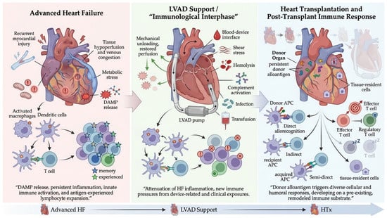
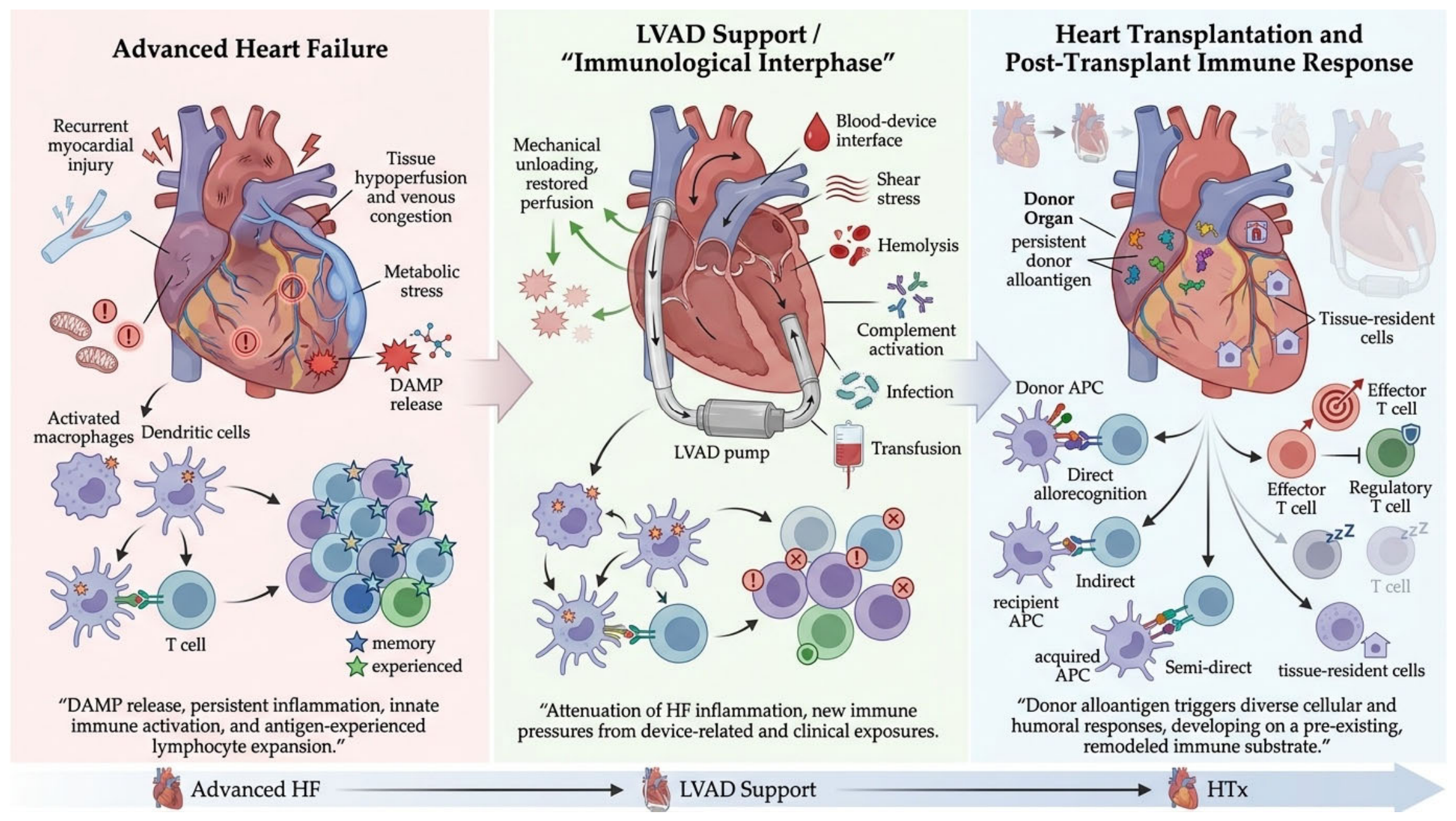
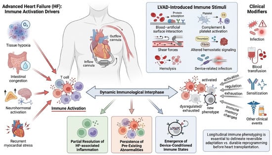
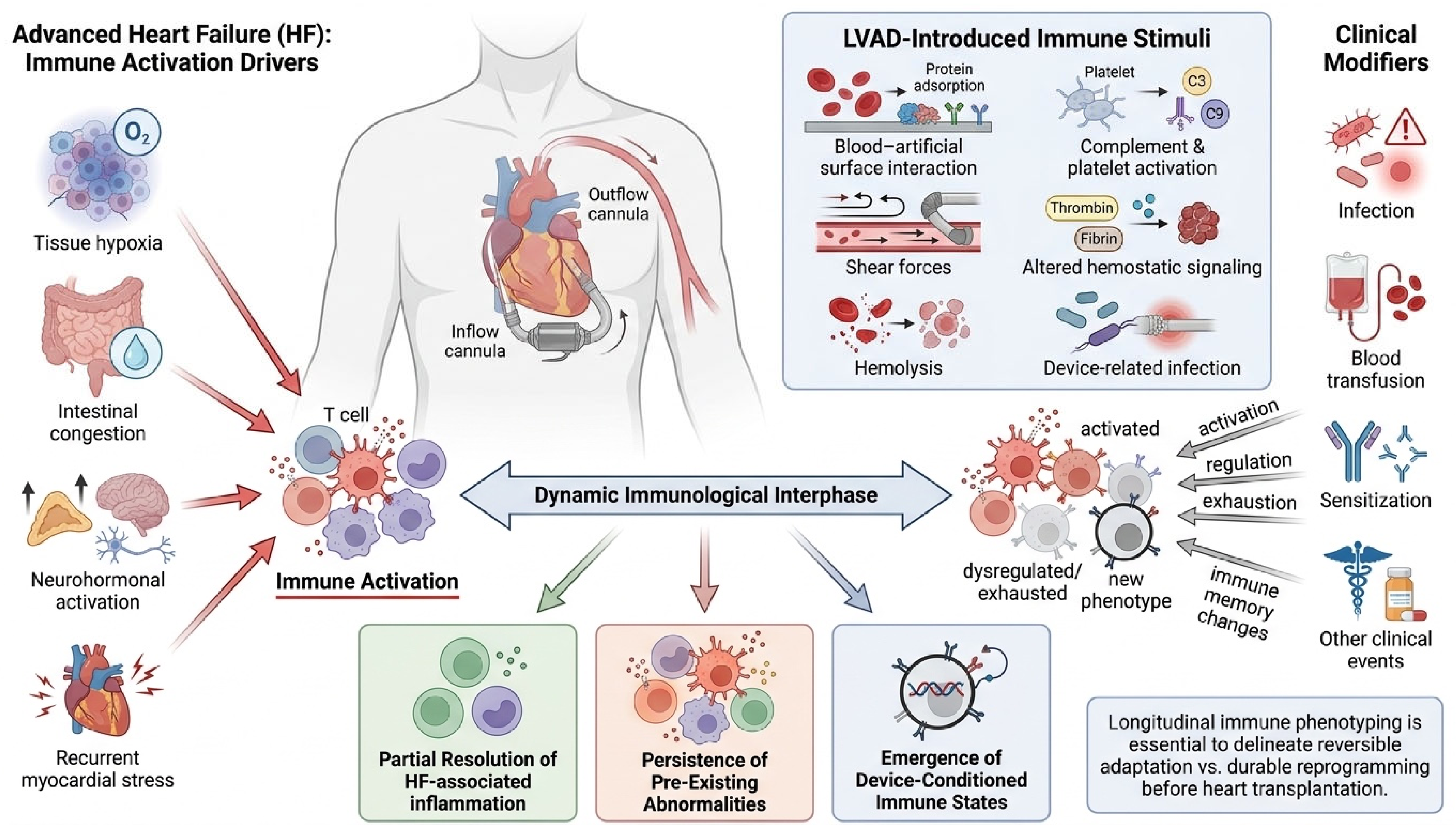
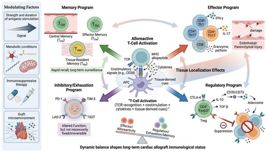
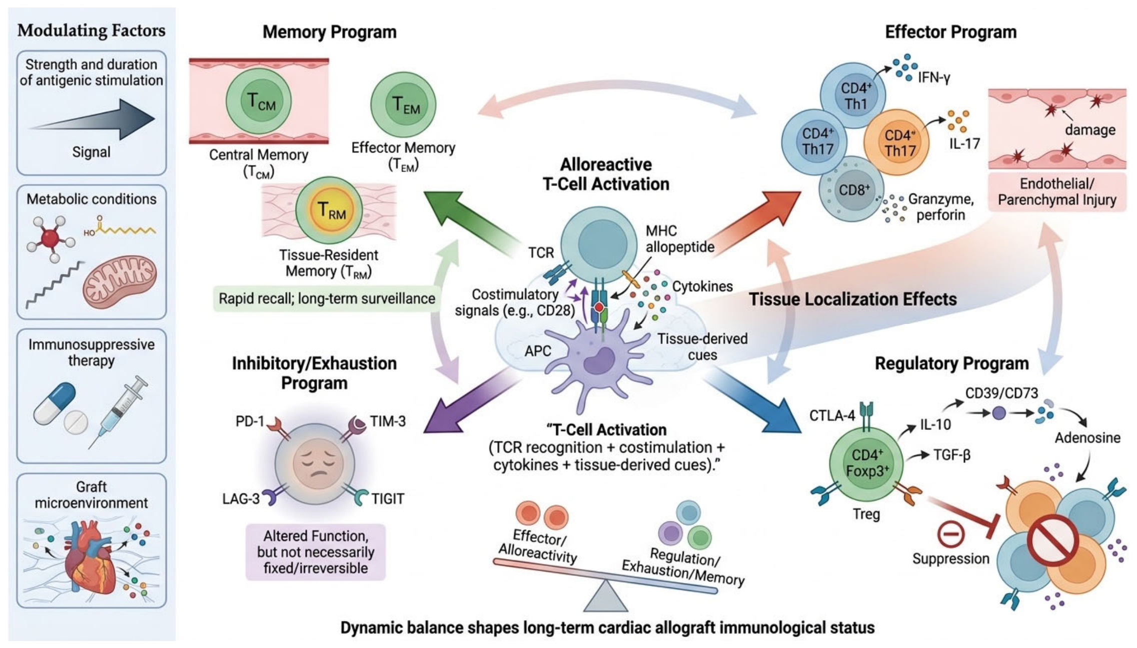
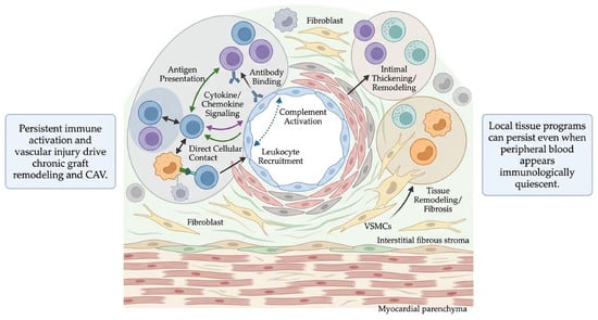
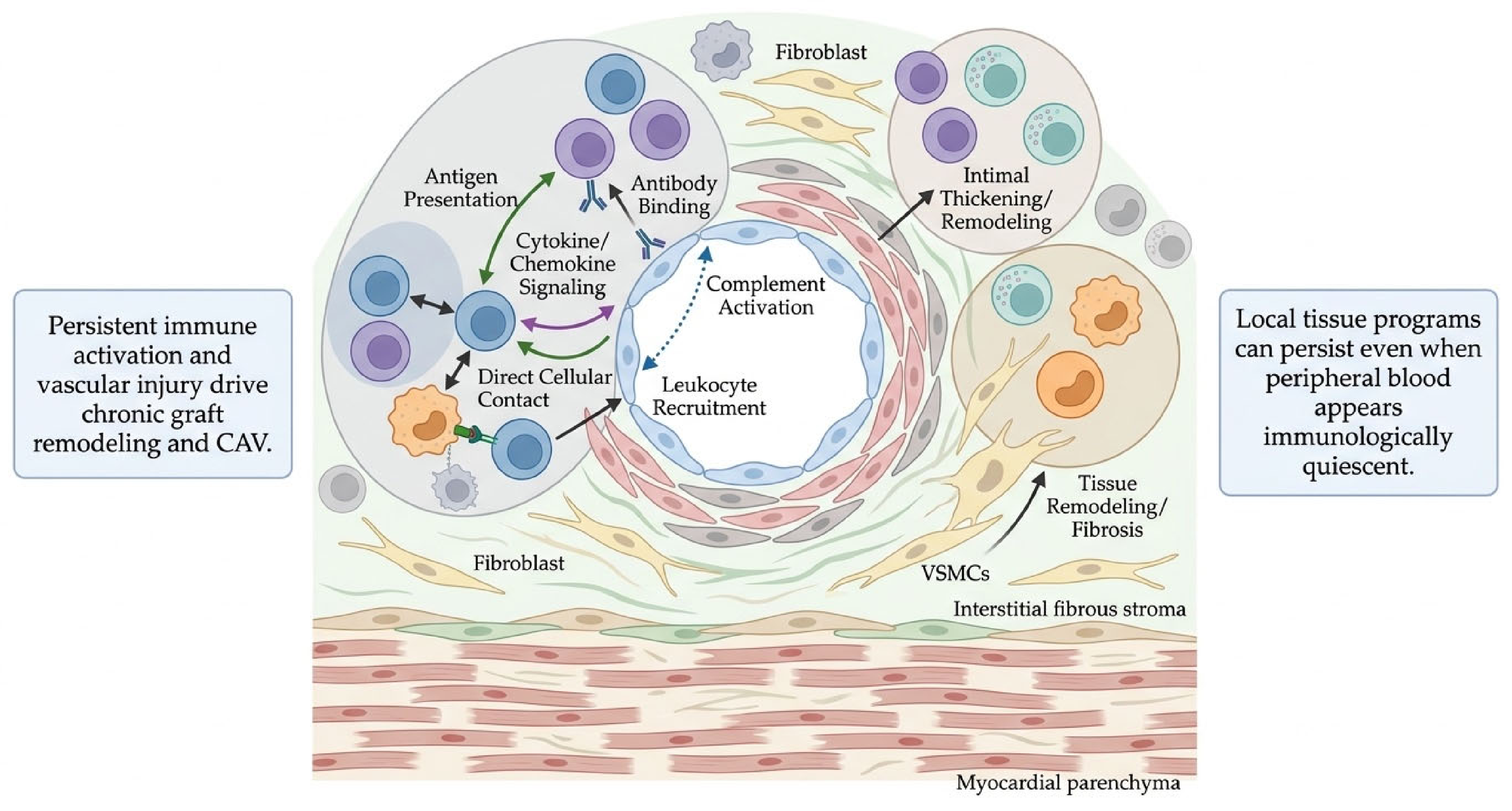
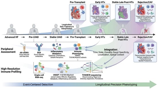
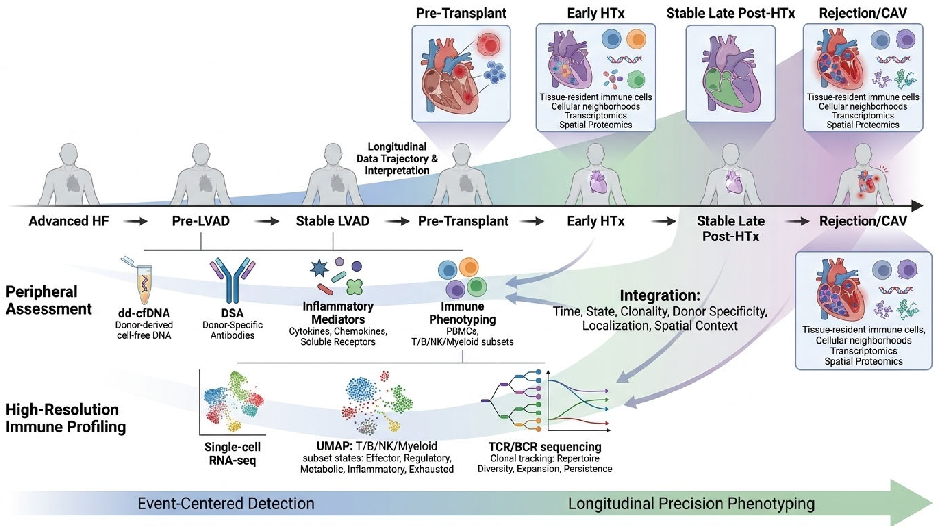

Disclaimer/Publisher’s Note: The statements, opinions and data contained in all publications are solely those of the individual author(s) and contributor(s) and not of MDPI and/or the editor(s). MDPI and/or the editor(s) disclaim responsibility for any injury to people or property resulting from any ideas, methods, instructions or products referred to in the content. |
© 2026 by the authors. Licensee MDPI, Basel, Switzerland. This article is an open access article distributed under the terms and conditions of the Creative Commons Attribution (CC BY) license.