


Abstract
Acidity, hypoxia, redox imbalance, and altered enzyme activity in the tumor microenvironment (TME) can be used to regulate nanocarrier structure and drug release. However, these conditions vary among tumors and across regions within individual tumors and overlap with certain normal physiological and inflammatory processes. This review examines TME-responsive nanomedicines for chemotherapy, phototherapy, immunomodulation, and theranostics in terms of their triggering conditions, response mechanisms, and carrier materials. Particular attention is given to matching activation thresholds to biological compartments, the relationship between delivery and release, and the biological interpretation of imaging signals. By comparing representative original studies, we examine the benefits of multiresponsive designs, potential off-target activation, and manufacturing complexity, while distinguishing the clinical use of conventional nanoformulations from clinical validation of TME-responsive mechanisms. Current studies demonstrate the feasibility of conditional drug release and local microenvironment modulation, but their clinical value remains to be established through consistent product quality, evidence of in vivo activation, and comparative efficacy studies.
1. Introduction
The tumor microenvironment (TME) comprises malignant cells and the surrounding stromal, immune, vascular, and extracellular components. Interactions among these components influence metabolism, invasion, and treatment responses [1,2,3,4]. Acidity, hypoxia, redox imbalance, and the activity of matrix-remodeling enzymes offer opportunities for responsive delivery, but these features are neither stable nor uniformly tumor-specific. Their intensity and spatial distribution vary with cancer type, anatomical site, perfusion, and prior treatment. Neural signaling provides an additional layer of regulation in some tumors [2,5]; it should not yet be regarded as a broadly established trigger for nanocarrier activation.
Differences between the TME and normal tissues provide a rationale for responsive delivery, but carrier selectivity also depends on biodistribution and activation conditions [6]. Ionizable groups or stimulus-sensitive linkages can be incorporated into polymeric, lipid, and inorganic materials to induce structural transitions, changes in surface properties, and drug release [7,8]. Existing nanoformulations have improved the formulation properties and pharmacokinetics of hydrophobic drugs, and some products reduce specific toxicities; these benefits depend on the drug, formulation, and indication [9,10,11]. In comparison, systems combining TME-triggered release with combination therapy and imaging remain predominantly at the preclinical and early clinical stages [12,13,14].
Previous reviews have addressed pH-responsive delivery [8], TME-targeted therapy [15], stimuli-responsive imaging [16], and transformable and biomimetic nanocarriers [14]. Recent studies have further combined delivery with microenvironment monitoring and modulation [12]. Building on this work, we compare activation ranges, tissue transport limitations, and imaging measures under different stimulus conditions and assess the suitability of individual designs in relation to manufacturing requirements. Related approaches, including receptor-targeted polysaccharide delivery [17], are also discussed, but receptor binding and stimulus-triggered release are assessed separately to distinguish their mechanisms of action.
2. Tumor Microenvironment Characteristics and the Molecular Basis of Responsive Nanocarrier Design
2.1. Key Physicochemical, Metabolic, and Neural Features of the Tumor Microenvironment
2.1.1. Extracellular Acidification in Tumors
Extracellular acidification in tumors is associated with metabolic reprogramming, acid–base transport, and local perfusion. Many tumor cells maintain high glycolytic flux even when oxygen is available, a phenomenon known as the Warburg effect [15,18,19]. Although glycolysis produces less ATP per glucose molecule than oxidative phosphorylation, increased flux and associated branch pathways can support energy production and biosynthesis [20]. Lactate accumulation and acidification often occur together, and their effects extend beyond local pH. Using tracer studies, Faubert et al. demonstrated that lactate can enter the tricarboxylic acid cycle in human lung tumors [21]. Other studies have linked tumor-derived lactate to changes in macrophage phenotype and suppression of T-cell and natural killer (NK)-cell function [22,23].
Phosphoinositide 3-kinase (PI3K)/protein kinase B (Akt)/mechanistic target of rapamycin (mTOR) signaling, hypoxia-inducible factor (HIF) regulation, and alterations in certain tumor suppressor genes can modify glucose uptake and metabolic enzyme expression, depending on the tumor’s genetic background and oxygen supply [3,24,25,26]. Under hypoxia or impaired von Hippel–Lindau (VHL) protein function, reduced HIF-α degradation can promote the expression of glycolysis-related genes. Tumor cells also maintain intracellular pH through several acid–base regulatory proteins: monocarboxylate transporters (MCTs) mediate lactate/H+ cotransport, the Na+/H+ exchanger 1 (NHE1) and vacuolar-type ATPase (V-ATPase) mediate proton transport, and carbonic anhydrase IX (CAIX) catalyzes CO2 hydration and contributes to intracellular and extracellular acid–base regulation [8,27,28]. Abnormal perfusion can limit the clearance of acidic metabolites. Normal extracellular fluid generally has a pH close to 7.4, whereas extracellular pH values of approximately 6.3–6.8 have been measured in some tumors; these values depend on tumor location, measurement method, and sampling time [8,27].
Acid-labile linkages and ionizable groups can alter carrier structure or drug release within specific pH ranges. The intended activation site must be defined: mild extracellular acidity in tumors differs from endosomal and lysosomal acidity, and normal cells also contain acidic organelles. Thus, whether a formulation that releases drug only at pH 5 is effective in tumor interstitium near pH 6.8 depends on its full pH–time response profile [8,27,29].
2.1.2. Regional Hypoxia
Rapid tumor growth can outpace oxygen supply, while disorganized vasculature causes uneven perfusion and fluctuations in oxygen availability. Regions with an oxygen partial pressure (pO2) below 10 mmHg are commonly classified as hypoxic for operational purposes, although measured values depend on tumor type, location, sampling method, and time [30,31]. Hypoxia may be chronic or intermittent; neither a fixed diffusion distance nor a single oxygen threshold describes all tumors. In particular, the most oxygen-deprived regions may also be the least accessible to circulating nanocarriers.
Insufficient oxygen reduces oxygen-dependent prolyl hydroxylation of HIF-α and its subsequent VHL-mediated recognition and degradation, thereby increasing hypoxia-related transcription. In a separate regulatory pathway, regulated in development and DNA damage responses 1 (REDD1) inhibits mTOR signaling through the complex formed by tuberous sclerosis proteins 1 and 2 (TSC1/TSC2) [32]. REDD1 can also regulate HIF-1α stability by affecting mitochondrial reactive oxygen species (ROS), an effect that can occur independently of mTOR complex 1 (mTORC1) in the models studied [33].
Hypoxia and associated metabolic changes can affect tumor immunity. Accumulation of metabolites such as lactate and adenosine, together with transforming growth factor beta (TGF-β)- and vascular endothelial growth factor (VEGF)-related signaling, can suppress effector-cell function and alter the states of dendritic cells, macrophages, regulatory T cells, and myeloid-derived suppressor cells [15,19,34]. These changes contribute to an immunosuppressive environment. However, the degree of hypoxia alone neither defines an immunologically cold tumor nor directly predicts responses to different treatments; immune-cell infiltration, antigen presentation, and prior treatment are also relevant.
Hypoxia-related signaling can contribute to invasion and treatment resistance, making improved oxygenation a target of some combination therapies. Preclinical studies, including work on the BMDI platform described in Section 4.5.2, have reported greater tumor inhibition when local oxygen generation is combined with chemotherapy and photothermal therapy [35]; these findings must be interpreted in relation to the contributions of the individual treatment components.
Hypoxia-responsive chemistry and oxygen-generating carriers address different objectives: the former uses low oxygen availability to promote activation, whereas the latter attempts to alleviate oxygen deficiency. Neither mechanism alone demonstrates tumor accumulation. Candidate systems should be evaluated at controlled oxygen levels and in poorly perfused tumor regions, with delivery, activation, and treatment response measured explicitly [30,31,36].
2.1.3. Oxidative Stress Associated with Elevated ROS and High Intracellular Glutathione Levels
ROS participate in cell signaling but can also damage cells when their accumulation exceeds antioxidant buffering capacity. Horak et al. found that REDD1 loss increased mitochondrial ROS and stabilized HIF-1α, whereas antioxidant treatment reduced HIF-1α levels in that model [33]. Gao et al. also observed HIF-dependent effects of antioxidants in specific tumor models [37]. These findings indicate reciprocal regulation between ROS and hypoxia signaling but do not support ROS reduction as a universal strategy for inhibiting metastasis. For ROS-responsive carriers, the local oxidant species and their balance with antioxidant systems are more informative than a single measure of total ROS.
Glutathione (GSH) is synthesized from glutamate, cysteine, and glycine through the actions of glutamate–cysteine ligase and glutathione synthetase. Glutathione reductase converts oxidized glutathione to GSH using NADPH as the source of reducing equivalents [38]. Intracellular GSH is generally present at millimolar concentrations, whereas extracellular concentrations are much lower; the frequently cited 100–1000-fold difference primarily describes the gradient between intracellular and extracellular compartments. Normal cells also contain high intracellular GSH levels. Cancer-associated nuclear factor erythroid 2-related factor 2 (NRF2) signaling, cystine uptake, and cysteine availability can enhance antioxidant capacity, with effects on treatment resistance that vary among tumors [38,39]. This distribution makes GSH more suitable as a post-internalization release signal, while tumor selectivity also depends on the preceding delivery process.
2.1.4. Tumor-Associated Matrix-Remodeling Enzymes
Tumor cells, fibroblasts, and immune cells can produce matrix metalloproteinases (MMPs), hyaluronidases, and cathepsins that contribute to extracellular matrix turnover and invasion [39,40,41]. Enzyme expression and catalytic activity are influenced by localization, endogenous inhibitors, and the local milieu; some of these enzymes also participate in normal lysosomal function and tissue repair. For delivery systems, the actual rate of substrate cleavage reflects activation more directly than total protein abundance. Cleavage-induced changes in particle size or surface properties, together with matrix density and interstitial pressure, determine tissue penetration.
Local proteolysis can alter carrier transport through tissue by removing a shell, exposing ligands, or releasing smaller particles [41,42]. Relevant designs and original studies are discussed in Section 3.1.5.
2.1.5. Abnormal Tumor Vasculature
Tumor angiogenesis is regulated by signals including VEGF and angiopoietin-2 (Ang2), but the structure and function of newly formed vessels are often heterogeneous. Abnormal endothelial junctions, basement membranes, and pericyte coverage, together with vascular compression, can cause inadequate perfusion, leakage, and fluctuations in oxygen supply [19,39]. These changes interact with oxygen consumption by tumor cells to shape the distribution of hypoxia. Insufficient functional lymphatic drainage within tumors can further impair tissue fluid clearance; vascular abnormalities and lymphatic dysfunction should be understood as related but distinct processes.
Vascular leakage, inadequate lymphatic drainage, and matrix mechanics jointly influence tumor interstitial fluid pressure. Elevated interstitial pressure can reduce transvascular convective transport, while dense matrix and solid stress further restrict particle diffusion and vascular perfusion [39,40,41,43]. Lysyl oxidase (LOX), MMPs, and other factors participate in matrix remodeling, but their effects on drug distribution vary with matrix composition and the mode of remodeling. Accordingly, the effects of local oxygen generation or stimulus-triggered release remain contingent on carriers reaching the relevant tumor regions [34,43].
2.1.6. Emerging Regulatory Features of the Neural Microenvironment
Interactions between tumors and the nervous system have been investigated in pancreatic cancer, glioma, breast cancer, and other settings, although the neural structures and signaling mechanisms differ among tumors. Neurotransmitters can affect tumor or stromal cells through adrenergic, cholinergic, and other receptors, and some models link neural activity to invasion, immune regulation, and treatment responses [2,5]. Perineural invasion, paracrine signaling, and direct synapse-like connections have distinct biological implications and should not be combined into a single universal mechanism. Existing evidence suggests potential delivery targets, but the suitability of neural signals as nanocarrier triggers requires separate validation.
2.2. Interrelationships Among TME Features and Implications for Nanodrug Delivery System Design
2.2.1. Limitations of Passive and Active Targeting
The enhanced permeability and retention (EPR) effect describes preferential macromolecular accumulation and retention associated with tumor vascular and drainage characteristics; it does not establish that drugs have reached malignant cells. Whole-tumor measurements may include intravascular material, extracellular particles, and particles taken up by macrophages. Sindhwani et al. demonstrated an important contribution of active transendothelial transport in the models studied [44], challenging explanations based exclusively on endothelial gaps. Nevertheless, the relative contributions of paracellular and transcellular transport should be evaluated for each carrier and tumor context rather than assumed to be universal.
Ligand-mediated targeting can enhance receptor-dependent uptake after carriers reach accessible cells, but it does not necessarily increase total tumor deposition or overcome poor perfusion. High-affinity binding near blood vessels may also limit deeper distribution. Endothelial transcytosis, reversible ligand exposure, size-transformable carriers, and specific matrix or vascular interventions offer complementary approaches, each requiring appropriate transport and safety studies [9,11,44]. TME responsiveness acts downstream of, or together with, these delivery processes and cannot replace them. Imaging evidence of heterogeneous nanoparticle deposition in humans and its translational implications are discussed in Section 5.2.2.
2.2.2. Advantages of TME-Responsive Nanodelivery Systems
Metabolic and structural changes within the TME can influence one another. Poor perfusion limits oxygen supply and drug entry, lactate accumulation and acidification can alter immune-cell function, and antioxidant systems such as GSH can reduce the effects of treatments that rely on oxidative damage [19,38,45]. Matrix remodeling also changes tissue permeability and conditions for cell migration [41]. These processes do not always change in the same direction; carrier design therefore requires identification of the principal delivery or treatment barriers in a given lesion.
Responsive carriers can regulate distinct delivery steps separately. For example, extracellular removal of an acid-sensitive shell can promote uptake, followed by intracellular cleavage of a reduction-sensitive linkage to release the drug. Coordination between these steps depends on carrier distribution, triggering conditions, and reaction time; insufficient activation at the first step can limit subsequent release [12,13,14]. Overall treatment effects should be compared with those of single-responsive and nonresponsive carriers at comparable doses and dosing schedules, with drug exposure measured concurrently. Pharmacodynamic effects can then be compared at similar exposure levels to distinguish increased exposure from altered cellular sensitivity.
3. Design Strategies for TME-Responsive Nanodrug Delivery Systems
Acidity, redox conditions, hypoxia, and enzyme activity can trigger changes in carrier structure, drug release, or biological activity (Figure 1). The responsive groups, relevant compartments, and carrier materials associated with these stimuli are discussed below; the relationship between imaging signals and treatment processes is addressed separately in Section 3.3.
Figure 1.
Design strategies for tumor microenvironment-responsive nanomedicine.
Responsiveness becomes relevant only after a carrier reaches the corresponding compartment. Tissue accumulation and local activation therefore require separate measurements; their spatial and temporal overlap determines effective drug exposure.
The figure summarizes illustrative conditions and response mechanisms for pH-, ROS-, hypoxia-, GSH-, and enzyme-responsive systems. Extracellular pH values of approximately 6.3–6.8 have been reported in some tumors; intracellular GSH concentrations of 2–10 mM also occur in normal cells; H2O2 concentrations of 50–100 μM represent experimental conditions rather than a universal concentration in human tumors; and pO2 < 10 mmHg is an operational criterion for hypoxia. Activation depends on the biological compartment, local stimulus availability, substrate accessibility, and reaction kinetics. Oxidation-triggered carrier changes are distinguished from ROS-generating treatment modules. Bioreductive activation encompasses hydrophilicity changes, linkage cleavage, and prodrug activation, depending on the chemical structure and reductive pathway. Multiresponsive designs may enhance release, but AND/OR logic and therapeutic synergy require separate experimental validation. The median tumor accumulation of approximately 0.7% of the injected dose was derived from a preclinical literature dataset [46] and does not represent a universal human delivery efficiency or intracellular drug release. Abbreviations: PBAE, poly(β-amino ester); PDMAEMA, poly(2-(dimethylamino)ethyl methacrylate); CDT, chemodynamic therapy; HAPs, hypoxia-activated prodrugs; TPZ, tirapazamine; HA, hyaluronic acid; MMPs, matrix metalloproteinases; ROS, reactive oxygen species; GSH, glutathione.
3.1. Selection of Response Types and Their Molecular Mechanisms
This section discusses the chemistry of pH-, ROS-, hypoxia-, GSH-, and enzyme-responsive systems, followed by a comparison of how stimulus combinations operate. Table 1 summarizes reference conditions, responsive groups, and representative systems for each stimulus.
Table 1.
Comparison of major endogenous TME stimuli and corresponding responsive design strategies.
To facilitate comparison, we assess “smart” responsiveness in four respects: stimulus selectivity relative to relevant physiological or inflammatory conditions; response profiles, ranges, and times under defined conditions; leakage without triggering and active-drug release after triggering; and functional gains over nonresponsive carriers and free drugs. Tumor-selective activation additionally requires localization evidence at the tissue or cellular level; increased whole-tumor fluorescence reflects only part of this process. These criteria guide the comparative analysis in this review, but no uniform numerical thresholds have been established.
3.1.1. pH Responsiveness
The mildly acidic extracellular space of some tumors and the more acidic endosomal and lysosomal compartments constitute distinct design targets. Endosomal and lysosomal acidification also occurs in normal cells. pH-responsive formulations should therefore be characterized across the intended extracellular and intracellular pH ranges, with exposure time, buffer capacity, protein content, and dilution conditions specified, rather than assessed only by comparing pH 7.4 with pH 5 [8,71,72]. The main strategies involve protonation-driven transitions and cleavage of acid-labile linkages.
The responsiveness of ionizable polymers depends on their composition and assembly. Shenoy et al. used solvent displacement to prepare poly(ethylene oxide) (PEO)-modified poly(β-amino ester) (PBAE) particles approximately 150–200 nm in diameter. The polymer dissolved rapidly below pH 6.5, and cellular paclitaxel delivery was evaluated against non-pH-responsive poly(ε-caprolactone) (PCL) particles [47]. Loh et al. constructed triblock micelles from poly(2-(dimethylamino)ethyl methacrylate) (PDMAEMA) and poly[(R)-3-hydroxybutyrate] and observed pH- and temperature-dependent doxorubicin release [48]. Metal–phenolic networks employ a different mechanism: Ejima et al. prepared films and capsules through pH-dependent coordination between Fe(III) and polyphenols and observed corresponding disassembly [49]. These systems involve polymer dissolution, changes in micellar properties, and dissociation of coordination structures, respectively; their response ranges should be compared at the formulation level.
Acid-labile linkages can respond to local pH changes and trigger drug release. Bae et al. linked drugs to polymers through hydrazone bonds to construct micelles responsive to intracellular acidic conditions [50]; Gillies and Fréchet investigated acid-sensitive micelles containing acetal structures [51]; and Shen and Ryser used a cis-aconityl spacer in daunorubicin–macromolecule conjugates [52]. Substituents, linkage accessibility, and the surrounding medium all influence hydrolysis kinetics, requiring comparison of acid-sensitive systems under comparable conditions. The epirubicin micelle NC-6300 has undergone a phase Ib study in humans; its dosing and safety findings are discussed in Section 5.1 [53].
3.1.2. ROS Responsiveness
The reactivity, lifetime, and subcellular distribution of ROS vary among species. H2O2 concentrations of 50–100 μM, commonly used in the literature, provide a range of conditions for release experiments, but their relevance to a given tumor depends on the measurement method and compartment [55,56]. Inflammation and activated phagocytes also generate oxidants. ROS responsiveness therefore primarily describes chemical changes in a carrier under defined oxidative conditions; tissue selectivity additionally depends on distribution and reaction time.
Oxidation-sensitive structures include thioethers, thioketals, and arylboronic esters. Napoli et al. constructed vesicles from a triblock copolymer containing poly(ethylene glycol) (PEG) and poly(propylene sulfide); oxidation of thioethers increased segment hydrophilicity and destabilized the vesicles [59]. Thioketal cleavage and boronic ester oxidation promote release through changes in linkage structure, while selenium-containing linkages may respond to both oxidative and reductive conditions [57,58,62,63]. These reactions differ in species selectivity and rate. Comparisons of release profiles should therefore account for oxidant composition, concentration, and exposure time.
Stimulus-responsive release can also be combined with ROS generation, but oxidative cleavage, oxygen generation, and Fenton-like reactions should be considered separately. In the MnO2-coated mesoporous silica system developed by Lin et al., GSH-mediated reduction released Mn2+ and consumed GSH; in an HCO3−-containing medium, Mn2+ promoted the formation of ·OH from H2O2, enhancing chemodynamic therapy (CDT) [54]. This mechanism differs from systems in which MnO2 participates in oxygen generation from H2O2 [35]. Organic–inorganic hybrid carriers can provide both loading space and reactive sites, but functional compatibility depends on reaction rates, substrate availability, and the stability of each component [73,74].
3.1.3. Hypoxia Responsiveness
Hypoxic regions can support bioreductive activation, but oxygen partial pressure is not the sole determinant of reaction rate. Reductase expression, electron-donor availability, carrier accessibility, and fluctuations in oxygen levels also contribute [36,60].
Nitroimidazoles and azo linkages are common hypoxia-associated responsive structures. Thambi et al. conjugated a 2-nitroimidazole derivative to carboxymethyl dextran to prepare polymeric nanoparticles whose properties changed under hypoxic conditions [61]. Reductive cleavage of azo bonds and bioreduction of certain quinone prodrugs can also be used in delivery designs [60,75]. These reactions depend on enzyme activity, electron donors, and oxygen levels. For example, two-electron reduction mediated by NAD(P)H quinone oxidoreductase 1 (NQO1) does not itself constitute an oxygen-specific switch. Experiments must therefore distinguish the effects of low oxygen from those of altered reductase expression.
Hypoxia-activated prodrugs (HAPs), including tirapazamine (TPZ), AQ4N, and PR-104, undergo bioreduction to generate more cytotoxic active species. Their selectivity depends on reductive pathways, oxygen levels, and tissue transport, and their parent compounds should not be regarded as uniformly nontoxic [60]. Preclinical studies have used nanocarriers to modify HAP exposure and release in an attempt to improve efficacy; clinical benefit requires further evaluation [36,60]. Another strategy supplies oxygen to tumors or generates it in situ, for example, through MnO2- or CaO2-based systems, to alleviate oxygen limitation and support oxygen-dependent treatments [36,76]. These approaches modify the microenvironment and differ mechanistically from carrier cleavage directly triggered by hypoxia.
3.1.4. GSH Responsiveness
GSH can modify carrier linkages through thiol–disulfide exchange. Cleavage of disulfide bonds located at crosslinking sites, between blocks, or within hydrophobic side chains can cause de-crosslinking, shell shedding, or changes in hydrophilicity and hydrophobicity, respectively [62,63]. Ryu et al. incorporated hydrophilic triethylene glycol units and hydrophobic alkyl side chains containing disulfides into amphiphilic copolymers. GSH induced disassembly and cargo release, and doxorubicin (DOX)-loaded micelles were also evaluated for cytotoxicity in MCF-7 cells [66]. This example links changes in bond structure to micelle destabilization, with evidence derived primarily from chemical release studies and cell experiments.
Reduction reactions involving metal oxides provide another design route. For example, the reaction of MnO2 with GSH can consume reducing equivalents while altering carrier structure; in related chemodynamic systems, GSH depletion may also weaken cellular antioxidant defenses [54]. This mechanism differs from ROS-mediated cleavage of organic linkages. In GSH-dependent release systems, the rate of material-induced GSH consumption, intracellular replenishment, and the amount required for reaction jointly influence activation.
3.1.5. Enzyme Responsiveness
Enzyme-responsive carriers use substrate cleavage to change their structure or expose functional components. MMPs acting on the extracellular matrix and proteases within endosomes or lysosomes occupy different compartments and require different peptide sequences and activation steps [42,67,68]. Hyaluronic acid can participate in receptor binding and serve as a substrate for enzymatic degradation; increased uptake and hyaluronidase-induced structural changes are distinct processes [69]. Carrier accessibility, substrate specificity, and local enzyme activity therefore jointly determine where activation occurs.
Wong et al. developed a multistage size-changing system in which approximately 100 nm gelatin particles carried approximately 10 nm quantum dots; MMP-2-mediated gelatin degradation released the smaller components [70]. Transport studies in collagen gels and tumor models showed broader diffusion after release of the smaller particles. The quantum dots served primarily as tracers, and the experiments focused on how enzyme-induced size changes affected tissue penetration. Unlike enzyme-sensitive linkages that directly release drugs, these designs first alter the transport unit; subsequent drug action depends on the form of the payload.
3.1.6. Multiresponsive Designs and Their Comparison
Single-responsive systems can control a specific step with fewer components, as illustrated by acid-induced dissolution of PBAE and reductive disassembly of disulfide-containing carriers [47,66]. Combined designs aim to address different limitations in distribution, internalization, and release. The mixed polyprodrug micelles MPPMs-1 developed by Luo et al. provide a comparison of input conditions within the same formulation: at 12 h, cumulative DOX release exceeded 50% at pH 6.5 without a reducing agent, was approximately 40% at pH 7.4 with 10 mM dithiothreitol (DTT), and exceeded 85% when both inputs were combined; release at pH 7.4 without DTT remained below 15% at 48 h [77]. Because either input alone produced substantial release, this system demonstrates dual-stimulus-enhanced release rather than a strict AND gate. DTT is an experimental reducing agent, and these results cannot be directly converted into release rates under physiological GSH conditions.
Such comparisons establish the effects of responsive chemistry, whereas comparisons of efficacy with single-responsive carriers address a separate question involving drug loading, circulation, cellular uptake, and active-drug exposure. Multiple modules may improve successive delivery steps, but spatial separation of activating compartments or mismatched reaction times may also limit release [14,62,78]. The advantages of multiresponsive systems should therefore be judged from results obtained with matched carriers. An AND gate requires all necessary inputs, whereas an OR gate permits either input to produce an output; the logic of a sequential cascade depends on whether the preceding step supplies a necessary condition for the next [14,62,68]. For pH/GSH combinations, the spatial distinction between acidic organelles and the relatively GSH-rich cytosol must also be considered.
3.2. Carrier Materials
Carrier material selection directly determines the drug-loading capacity, stability, biodistribution, and responsive behavior of a nanodelivery system. Based on chemical composition and structural features, current TME-responsive nanocarriers can be grouped into four major classes (Figure 2): liposomes and lipid nanoparticles, polymeric nanoparticles, inorganic nanoparticles, and biomimetic membrane nanocarriers [12,67].
Figure 2.
Representative nanocarrier classes and stimuli-responsive design strategies.
The columns summarize lipid, polymeric, inorganic/hybrid, and biomimetic carriers. Colors identify endogenous conditions: acidic pH (orange), intracellular GSH (purple), ROS (red), hypoxia (blue), and enzyme activity (green); gray denotes external stimuli. Illustrations are schematic; the inorganic example is an MSN. Table 2 summarizes formulation- and indication-specific clinical evidence, which does not validate all TME-responsive variants. Abbreviations: LNPs, lipid nanoparticles; GSH, glutathione; ROS, reactive oxygen species; PAMAM, poly(amidoamine); MSNs, mesoporous silica nanoparticles; AuNPs, gold nanoparticles; IONPs, iron oxide nanoparticles; MOFs, metal–organic frameworks; PEG, poly(ethylene glycol); 2D, two-dimensional. (Created with Figdraw).
Table 2.
Comparison of major nanocarrier classes for TME-responsive drug delivery.
3.2.1. Liposomes and Lipid Nanoparticles
Liposomes consist of lipid bilayers enclosing an aqueous lumen, allowing hydrophilic and hydrophobic drugs to be loaded into the lumen and membrane, respectively. Liposomal drugs such as Doxil and Onivyde are in clinical use [79,80]. pH-responsive designs can employ membranes containing dioleoylphosphatidylethanolamine (DOPE), which promote nonbilayer phase formation and membrane destabilization under appropriate compositional and acidic conditions. Acid-labile PEG–lipid linkages can alter uptake or membrane interactions through deshielding [69,71]. The activation range is determined by the phase behavior and release kinetics of the complete formulation.
Lipid nanoparticles (LNPs) used for nucleic acid delivery generally contain ionizable lipids, helper lipids, cholesterol, and PEG–lipids; their internal structure need not resemble that of classical liposomes. Ionizable lipids such as DLin-MC3-DMA, SM-102, and ALC-0315 become protonated at low pH, facilitating nucleic acid encapsulation and interactions with endosomal membranes. Surface charge near physiological pH depends on composition and apparent pKa [9,92]. The use of LNPs for mRNA delivery demonstrates the feasibility of manufacturing and delivery for this formulation class, but whether the extracellular tumor environment effectively triggers their function requires formulation-specific evaluation.
3.2.2. Polymeric Nanoparticles
The structure, degradation, and release behavior of polymeric nanoparticles can be tuned. Poly(lactic-co-glycolic acid) (PLGA), PCL, and poly(amino acids) are widely studied scaffold materials; drug loading and encapsulation efficiency vary with the drug, polymer, and preparation method [93,94]. Incorporating ionizable blocks or acid-, reduction-, or oxidation-sensitive linkages can confer corresponding structural changes [47,50,59]. Drug content has different implications for physical encapsulation and prodrug conjugation: the former is primarily influenced by partitioning and diffusion, whereas the latter also depends on linkage cleavage and regeneration of the active drug. Formulation comparisons should therefore consider both excipient burden and the releasable drug fraction.
Dendrimers use branched structures and surface groups to regulate drug loading and cellular interactions. The generation, terminal modifications, and medium conditions of poly(amidoamine) (PAMAM) dendrimers alter their protonation, conformation, and release behavior [68,71]. Polymeric micelles, in contrast, assemble from amphiphilic polymers, with hydrophobic regions encapsulating or conjugating drugs and hydrophilic segments providing colloidal stability [93,94]. Surface functionalization of dendrimers and self-assembly of micelles offer distinct design possibilities and lead to different formulation-dependent patterns of toxicity, dilution stability, and active-drug release.
3.2.3. Inorganic Nanoparticles
Inorganic nanoparticles can combine drug delivery with imaging or catalytic functions. Gold, iron oxide, MnO2, and mesoporous silica differ substantially in their properties; their applications and safety should therefore be discussed in relation to their specific composition, morphology, and surface modifications [13,74,83,84,85,95].
Mesoporous silica has tunable pores and surfaces that support drug loading and the attachment of responsive pore caps [83,84]. Pore architecture and gating determine loading and diffusion, while serum proteins, dilution, and enzymes can affect cap stability. Its in vivo fate includes dissolution of the silica framework, retention of intact particles, and clearance of dissolved species. Elemental analysis and particle localization provide different information; combining them helps distinguish degradation from excretion.
The optical properties of gold nanostructures depend on their size and morphology and can support photothermal conversion or surface-enhanced Raman scattering (SERS). In some systems containing Au–S linkages, thiol exchange can alter surface ligands or release attached components. This is a ligand-exchange process and should not be broadly described as GSH-mediated reductive bond cleavage [85,95]. Iron oxide can provide magnetic resonance imaging (MRI) contrast, while its catalytic activity depends on iron oxidation state, coordination, and reaction conditions. Feraheme (ferumoxytol) is approved for iron replacement in specific adult populations with iron deficiency anemia; its safety data cannot directly substitute for evaluation of a new cancer formulation [86]. MnO2 systems can participate in oxygen generation or undergo GSH-mediated reduction that consumes GSH and releases Mn2+. The contribution of each reaction to imaging and treatment depends on the conditions [35,54].
Metal–organic frameworks (MOFs) are formed by coordination between metal nodes and organic ligands; their pore structures and coordination stability can be used to regulate loading and release. Some iron-containing MOFs exhibit catalytic activity, but acid-sensitive dissociation and ROS generation remain dependent on metal coordination and medium composition [74]. Two-dimensional materials, including black phosphorus, MXenes, and MoS2, have also been investigated for phototherapy [13]. Because these materials differ markedly in degradation, ion release, and organ retention, their optical performance should be evaluated together with acceptable doses and long-term in vivo fate.
3.2.4. Biomimetic Membrane Nanocarriers
Hu et al. coated polymeric cores with erythrocyte membranes to produce nanoparticles that retained some membrane proteins and exhibited prolonged circulation, with their behavior verified in mice [87]. Subsequent studies have used tumor-cell membranes, macrophage membranes, and hybrid membranes to modulate cellular interactions [88,89]. These functions depend on membrane source, protein orientation, and coating integrity. Control of cell source, membrane-to-core ratio, residual cytoplasmic components, and pathogens during preparation affects batch consistency. Membrane protein identification and receptor-binding or cellular potency assays therefore serve different quality assessment purposes.
Extracellular vesicle composition and yield vary with the producer cells and the culture, isolation, and storage conditions. The term exosome refers to a subclass with an established endosomal origin; otherwise, extracellular vesicle is the more accurate term [90,91]. For therapeutic vesicles, particle concentration and size characterize the formulation’s particle population, free cargo and co-isolated contaminants affect purity, and cellular or molecular potency assays assess function. Natural vesicle composition may also change in response to local enzymes or other environmental conditions. For engineered formulations, responses arising from sensitive linkages or functional components should be validated separately from membrane-related effects on distribution and uptake. Table 2 compares the major carrier classes, representative size data, and the scope and limitations of their clinical evidence.
3.3. Theranostic Design
Theranostic design requires clear identification of the process represented by an imaging signal. Carrier deposition, active-drug release, and tissue response to treatment follow different time courses and cannot be inferred interchangeably from the same signal intensity. Existing systems can be classified by the purpose of imaging: distribution tracking, intervention guidance, or response monitoring. Validation requirements depend on the intended use [16,96].
3.3.1. Types of Imaging Probes
Near-infrared-I (NIR-I) fluorescence can be used to examine superficial lesions and those accessible by endoscopy or during surgery, offering rapid acquisition and real-time guidance [16,97]. Near-infrared-II (NIR-II) imaging can reduce scattering effects when suitable wavelengths and detection conditions are used. Hong et al. performed in vivo imaging in small animals using PEG-modified Ag2S quantum dots emitting at approximately 1200 nm, providing original research evidence for this probe class [98]. The effective depth of both windows depends on tissue optical properties, probe brightness, and instrumentation. When activatable fluorescence is used to monitor drug release, background activation and calibration of the signal against drug release must also be considered.
Photoacoustic imaging detects acoustic signals generated by optical absorption to provide information on local vasculature or oxygenation [99]. Wavelength, optical fluence, transducer frequency, and tissue geometry jointly determine resolution and depth. Probe concentration and optical fluence within tissue also affect the signal; interpretation of apparent oxygenation changes therefore depends on spectral unmixing and system calibration. Manufacturing and clearance data for nanoparticulate contrast agents further determine the conditions for their use.
Responsive MRI contrast depends jointly on probe concentration, relaxivity, water exchange, and the imaging sequence. Released components, such as manganese species, may also have a different distribution from the original carrier [35,54,74]. Positron emission tomography (PET) uses radiolabels to quantify whole-body distribution, and 64Cu-MM-302 has been used to measure deposition in human lesions [96]; implementation requires label stability, dosimetry, and a radiopharmaceutical supply. Raman/SERS provides vibrational spectroscopic information, and gold particles carrying reporter molecules can be used at superficial or endoscopically accessible sites [95]. These modalities measure different entities at different scales; the value of combining them depends on whether they provide complementary information about specific aspects of distribution or response. Table 3 summarizes the imaging modalities, their practical limitations, and representative probes discussed in this section.
Table 3.
Comparison of imaging modalities for theranostic nanoplatforms.
3.3.2. Strategies for Integrating Imaging and Therapeutic Modules
Combinations of imaging and therapeutic modules can serve different purposes, discussed below in terms of distribution tracking, intervention guidance, and response monitoring [12,100].
Distribution tracking usually involves co-delivery of imaging and therapeutic components within the same formulation. Retention of the label in the carrier and concordant pharmacokinetics are prerequisites for inferring drug distribution from imaging. If the carrier, label, and payload separate in vivo, the signal may diverge from drug exposure. Distribution imaging should therefore be combined with tissue drug measurements and, where necessary, cellular localization analyses [96].
Intervention guidance uses imaging to select a treatment site or time, for example, by applying light after sufficient lesion uptake. Peak carrier signal may not coincide with the optimal treatment time because release, tissue oxygenation, and clearance have different kinetics. The value of imaging guidance can be assessed against a prespecified fixed schedule or conventional guidance at the same drug and light doses [16].
Response monitoring uses release-activated probes, enzyme-sensitive reporters, or degradation-associated MRI signals to track local activation and tissue responses [16]. Release signals must be quantitatively related to the amount of active drug actually released, while cell-death-associated signals must correspond to independent biological endpoints. Changes in pH, perfusion, and clearance can all affect the readout. If the signal is intended to guide treatment adjustments, the effects of those adjustments on efficacy or toxicity also require validation.
64Cu-MM-302 is a radiolabeled, human epidermal growth factor receptor 2 (HER2)-targeted liposomal doxorubicin formulation. A PET/CT study in 19 patients with HER2-positive metastatic breast cancer demonstrated differences in particle deposition among lesions [96]. This study supports distribution imaging in humans; local release and the effects of treatment adjustments based on imaging require additional drug measurements and intervention studies.
4. Representative Applications of TME-Responsive Smart Theranostic Nanosystems
4.1. TME-Responsive Chemotherapeutic Nanocarriers for Reversing Multidrug Resistance
4.1.1. Inherent Limitations of Free Chemotherapeutic Drugs
Exposure of normal tissues to free chemotherapeutic drugs can limit the administered dose. Some tumors also develop cross-resistance to drugs with different structures or mechanisms of action, termed multidrug resistance (MDR) [101]. In addition to intracellular drug efflux and damage repair, perfusion, hypoxia, and matrix barriers can affect drug access and activity [102]. Designing carriers to address these distinct processes helps separate the contributions of improved delivery from those of reversal of cellular resistance.
4.1.2. pH-Sensitive Drug Delivery Systems for Mitigating MDR
MDR involves mechanisms including drug efflux, regulation of cell death, and damage repair. Among ATP-binding cassette (ABC) transporters, P-glycoprotein (P-gp), breast cancer resistance protein (BCRP), and multidrug resistance-associated protein 1 (MRP1) can reduce intracellular drug exposure; changes in apoptotic pathways and detoxification enzymes can also alter drug sensitivity [101,102]. P-gp uses ATP to drive transport, and some carrier studies therefore combine regulation of drug release with modulation of energy metabolism [103].
Xu et al. used a pentaerythritol scaffold and orthoester linkages to attach D-α-tocopheryl polyethylene glycol 1000 succinate (TPGS), producing star-shaped amphiphilic copolymer micelles capable of loading DOX. The drug-loaded micelles had a hydrodynamic diameter of approximately 101.3 nm and a drug-loading content of 10.8% [103]. Acidic conditions promoted linkage hydrolysis and micelle disassembly. In MCF-7/ADR cells, blank micelles reduced ATP levels, while drug-loaded micelles improved DOX uptake and cytotoxicity. Three-dimensional spheroid and tumor-bearing mouse experiments further demonstrated inhibitory effects in resistant models. Changes in release and TPGS-associated biological effects jointly contributed to treatment, addressing delivery and cellular resistance, respectively.
4.2. TME Stimulus-Responsive Combination Therapy Involving Photodynamic or Photothermal Treatment
4.2.1. TME-Related Limitations of Conventional Photodynamic and Photothermal Therapy
Photothermal therapy (PTT) depends on local energy deposition and heat dissipation, whereas photodynamic therapy (PDT) requires photochemical reactions within a treatment volume accessible to light. Light attenuation, uneven formulation distribution, and injury to irradiated normal tissues can limit both treatments. Hypoxia particularly restricts oxygen-dependent PDT, while antioxidant defenses and stress responses may also reduce efficacy [104,105]. Superficial mouse tumors provide relatively accessible irradiation conditions; for deep human lesions, endoscopic or interstitial light delivery further influences the treatable volume.
4.2.2. TME-Responsive Nanoparticle-Mediated Photodynamic Therapy
Wang et al. integrated an iridium photosensitizer with a copper-containing MOF to produce Ir1@FA@MOFs. Copper coordination and GSH-mediated reduction altered electron transfer, shifting the photodynamic process from type II toward a more hypoxia-tolerant type I pathway. Antitumor and immune-related effects were observed in 4T1 tumor-bearing mice [106]. This design adapts to hypoxia by altering the photosensitization pathway; its residual oxygen requirement depends on the electron-transfer processes and subsequent radical reactions involved.
4.2.3. TME-Responsive Nanoparticle-Mediated Photothermal Therapy
BMDI combines IR780-mediated photothermal activity with a MnO2/bovine serum albumin (BSA) drug-loading structure to support DOX treatment in preclinical models [35]; its composition and findings are detailed in Section 4.5.2. Compared with the photosensitization-pathway modification discussed above, irradiation in this system primarily provides a thermal effect. Drug exposure and tissue temperature jointly influence combination-treatment outcomes, and even at the same incident power, the thermal dose varies with absorption and heat dissipation.
4.3. TME-Responsive Nanosystems for Remodeling the Immunosuppressive Microenvironment and Enhancing Cancer Immunotherapy
T-cell function is jointly regulated by antigen recognition, costimulation, cytokines, and metabolic state. Immune checkpoint blockade, chimeric antigen receptor T (CAR-T) cells, and oncolytic viruses act through different mechanisms, but all may be affected by local nutrient competition, hypoxia, and suppressive metabolites [19,107,108,109]. These factors can alter effector-cell proliferation, mitochondrial function, and sustained responses. Responsive delivery has therefore been used to deliver regulatory components to specific immune cells or modify local metabolic conditions; its effects must be evaluated in relation to the target cells and immune context.
The PPD/MPC adjuvant micelles developed by Meng et al. underwent size reduction and charge reversal under mildly acidic conditions, exposing a mannose-containing core and promoting uptake by tumor-associated macrophages, dendritic cells, and myeloid-derived suppressor cells. The study linked changes in immune-cell function to signaling through Toll-like receptors 4 and 9 (TLR4 and TLR9) and observed antitumor effects in cellular and animal models. The regulation of multiple cell populations arose from a single pH-responsive delivery process; carrier activation should therefore be distinguished from the subsequent immune responses. A correction to some images and quantitative analyses has been published, with the authors stating that the main conclusions were unchanged [110].
4.4. TME-Responsive Multimodal Combination Therapy
Zhang et al. assembled copper-doped carbon dots, MnO2 nanosheets, and DOX into the MDCC platform [111]. The study used 808 nm irradiation to generate photothermal effects and 660 nm irradiation to produce singlet oxygen, together with Fenton-like ·OH generation and DOX activity. In the models studied, the authors also observed improved oxygenation, reduced HIF-1α and programmed death-ligand 1 (PD-L1) expression, immunogenic cell death (ICD), and changes in immune-cell function. Compared with Ir1@FA@MOFs, which emphasizes regulation of the photosensitization pathway, and BMDI, which combines chemotherapy and photothermal activity, MDCC integrates more treatment components and activates optical processes at different wavelengths. These comparisons primarily identify differences in the processes targeted. Because formulations, models, and irradiation protocols differ, the available results are insufficient to rank the efficacy of the three systems.
4.5. TME-Responsive Theranostic Nanoplatforms
4.5.1. Clinical Value of Theranostics
The clinical use of theranostic platforms depends on whether imaging supplies information missing from conventional monitoring. For example, imaging of particle deposition may identify poorly supplied lesions, while release-related signals may indicate whether a drug has been activated locally. These applications require different signal-calibration methods. If imaging is used to select irradiation timing or adjust doses, comparative studies must evaluate how these decisions affect efficacy and safety [16,96].
4.5.2. BMDI: A BSA-Stabilized, MnO2-Based Multifunctional Theranostic Platform
Yuan et al. used a one-pot method to integrate MnO2, DOX, and IR780 into BSA-stabilized nanostructures, producing BMDI particles approximately 30 nm in size by transmission electron microscopy. DOX and IR780 loading contents were 2.12 ± 0.11% and 1.04 ± 0.07%, respectively [35]. The system exhibited H2O2-associated oxygen generation, GSH-responsive release, and manganese-related MRI contrast and was used for combined chemotherapy and photothermal therapy in cells and tumor-bearing mice. For this formulation, the relatively low drug-loading fraction means that a therapeutic drug dose is accompanied by a corresponding carrier burden. BSA source, the distribution and clearance of released manganese, and the photothermal dose received by the lesion are composition-related issues for subsequent evaluation. Tumor inhibition and histological observations in the original study support short-term preclinical assessment; an MRI-guided dosing-adjustment protocol has not been established.
5. Status and Barriers of Clinical Translation
5.1. Clinical Translation of TME-Responsive Smart Nanodelivery Systems
Conventional nanoformulations are already used clinically. Doxil and Onivyde are liposomal anticancer drugs, and Abraxane is albumin-bound paclitaxel [79,80,81]; Feraheme is used for iron replacement in iron deficiency anemia [86]. These products provide experience in the manufacturing, administration, and safety assessment of specific carriers, but their indications and mechanisms should be discussed separately from those of engineered TME-responsive formulations. Table 2 accordingly presents clinical evidence for specific products.
The pH-sensitive epirubicin micelle NC-6300 has undergone dose-finding studies in humans. In a phase Ib study enrolling 29 patients, the recommended phase II dose was 150 mg/m2; hematological toxicities and preliminary antitumor activity were observed [53]. The study provided dosing, pharmacokinetic, and tolerability data that informed dosing in a subsequent angiosarcoma expansion cohort.
The expansion cohort, published in 2022, enrolled 10 patients with angiosarcoma who received 150 mg/m2 intravenously every 3 weeks. Investigator-assessed median progression-free survival was 5.4 months, and the objective response rate was 30% (3/10). Grade 3–4 neutropenia and thrombocytopenia occurred in 80% and 40% of patients, respectively [82]. This small, open-label, nonrandomized study added activity and safety data for this indication. Neither early trial established superiority over conventional treatment through a randomized comparison or independently quantified the contribution of pH-triggered release to clinical outcomes.
Clinical use of a carrier formulation, human testing of a responsive formulation, and clinical benefit attributable to a response mechanism address different questions. Early clinical studies of NC-6300 and deposition imaging with 64Cu-MM-302 provide evidence for some of these aspects [53,82,96]. For TME-responsive theranostic platforms, local activation still needs to be linked to effective drug exposure and treatment outcomes.
5.2. Major Barriers to Translation of TME-Responsive Smart Nanodelivery Systems
5.2.1. Substantial Heterogeneity of the Tumor Microenvironment
Heterogeneity affects the proportion of a tumor that meets a carrier’s activation requirements. Less acidic lesions may fail to activate a pH switch with a narrow threshold range; intermittent hypoxia may shorten the period available for bioreduction; high GSH levels may accelerate reductive release while scavenging oxidants required for treatment; and enzyme abundance may not predict substrate cleavage when inhibitors are present [8,36,38,42]. Different stimuli may also be spatially separated, making the probability of simultaneously satisfying multiple inputs lower than tumor-homogenate measurements might suggest.
Selection of activation thresholds should account for both the tumor regions in which activation is possible and exposure in normal and inflamed tissues. This match may change when treatment alters perfusion or metabolism [8,36,38,42].
5.2.2. Limited In Vivo Delivery Efficiency and Uncertainty of the EPR Effect
For intravenously administered formulations intended to release drugs after internalization, nanoparticles generally undergo blood circulation, vascular transport, interstitial migration, and cellular uptake. Systems intended for extracellular release do not require intact carriers to enter cancer cells. Particle size, protein adsorption, and phagocytic clearance can affect several steps simultaneously [9,11]. Evaluation of improved targeting should identify the principal limiting step and use corresponding measures of distribution or drug exposure.
The median tumor delivery of approximately 0.7% of the injected dose reported by Wilhelm et al. was derived from a heterogeneous preclinical literature dataset [46]. It is neither a universally applicable human EPR value nor the fraction of drug reaching the cancer-cell cytosol. Human evidence also does not support a simple assertion that EPR is absent: 64Cu-MM-302 PET/CT revealed marked differences in deposition among patient lesions [96]. These findings support measuring delivery in the intended patient population. Ligand-mediated uptake, endothelial transport, size changes, or local administration may help, provided they improve the rate-limiting step without causing unacceptable exposure of normal tissues.
5.2.3. Balancing Responsiveness and In Vivo Stability
Circulatory stability and local release place different demands on structural stability. Overly stable linkages may limit release after tumor arrival, whereas excessive sensitivity can increase leakage in the circulation or normal tissues. Triggered release rates should therefore be evaluated together with stability under nontriggering conditions [12,14].
Acidic vesicles in normal cells, oxidants produced during inflammation, and proteases involved in tissue repair can all activate nominally TME-responsive carriers outside tumors. Uptake by hepatic or splenic phagocytes may expose carriers to precisely these conditions. Increasing sensitivity therefore requires balancing tumor coverage against premature activation. For designs requiring two inputs, selectivity depends on the probability that the stimulus combination occurs in tumor and nontumor compartments. If either input can trigger release, adding response mechanisms may broaden off-target activation. Plasma incubation, models of inflamed tissue, and matched nonresponsive carrier controls can be selected according to the expected exposure compartments.
5.2.4. Structural Complexity, Scale-Up, and Quality Control
To enable targeting, responsive release, imaging, microenvironment modulation, and combination treatment, many smart nanosystems contain multiple components, including inorganic materials, lipid or polymer scaffolds, targeting ligands, responsive linkages, chemotherapeutic agents, nucleic acid drugs, photosensitizers, and imaging probes [12,14,58,89]. Although structural complexity can confer multiple functions, it also complicates preparation, characterization, manufacturing scale-up, and quality control.
Scale-up can alter nucleation and self-assembly. Changes in mixing time, flow-rate ratio, concentration, and addition sequence can affect particle size, drug loading, and activation range. The reproducibility advantages of microfluidic or impinging-jet mixing depend on maintaining suitable mixing conditions at larger scales. Purification must control free drug and residual reagents while confirming that operations such as tangential-flow filtration do not alter the carrier. For the final formulation, sterilization or aseptic processing, container adsorption, reconstitution after lyophilization, and stability after dilution are all relevant to administration. Comparisons among laboratory, pilot, and clinical batches should include release and potency assays [112,113].
5.2.5. Uncertainty Regarding Biosafety and Long-Term In Vivo Fate
The in vivo fate of inorganic materials requires composition-specific investigation. Persistent gold structures may be retained in phagocyte-rich organs; silica dissolution depends on structure and the biological environment; iron oxide degradation does not eliminate dose- or coating-related risks; and MnO2 degradation releases manganese species whose distribution and clearance must be measured. MOFs can also generate metal ions and degradation products of organic ligands. Loss of particle morphology or imaging signal does not demonstrate elimination from the body. Repeated-dose studies should combine tissue elemental analysis at successive time points, differentiation between particles and ions where feasible, excreta recovery, and histopathological examination over an appropriate recovery period [83,84,85,95,113].
Unintended activation of TME-responsive systems outside tumors may also cause aberrant drug release in the liver, spleen, kidneys, or inflamed tissues. Preclinical evaluation must therefore extend beyond short-term antitumor efficacy to examine tissue distribution, metabolism and excretion, immunogenicity, hemocompatibility, long-term toxicity, and repeated-dose safety systematically [9,10].
5.2.6. Limited Predictive Value of Preclinical Models
Many TME-responsive smart nanomedicines show favorable efficacy in two-dimensional cell cultures and subcutaneous tumor models, but these models do not fully reproduce human tumor vasculature, the immune microenvironment, matrix barriers, metastatic processes, or patient heterogeneity [9,10]. Strategies that depend on hypoxia, dense extracellular matrix, tumor-associated macrophages, or an immunosuppressive microenvironment are particularly susceptible to oversimplification in conventional models.
Patient-derived tumor organoids, orthotopic models, patient-derived xenografts, and humanized immune models can provide additional information on cellular heterogeneity, local anatomy, or aspects of the immune context, respectively, but each has limitations. Model selection should match the question being tested, such as tissue penetration, activation in hypoxic regions, or immune-cell regulation [9,10,69].
5.3. Quality Assessment and Regulatory Challenges for TME-Responsive Nanomedicines
TME-responsive formulations commonly contain a drug, a carrier, and a responsive structure, sometimes supplemented with targeting or imaging components. Changes in composition can simultaneously affect distribution, release, and safety. Early pharmaceutical development should therefore identify the quality risks associated with these features [113,114].
The quality target product profile should connect the intended route of administration, dose, indication, and activation compartment to measurable product attributes. In addition to identity, purity, sterility, and endotoxin levels, relevant attributes include particle-size distribution and aggregation, surface chemistry, component stoichiometry, drug loading, free and bound drug, residual solvents or metals, degradation products, and storage stability. Analytical methods based on complementary principles are needed when a single method cannot distinguish intact carriers, aggregates, and free components. The FDA guidance on drug products containing nanomaterials, issued in 2022, supports product-specific evaluation rather than an assumption that all nanoformulations present the same risks [113].
Responsive-release assays should detect performance differences caused by changes in linkage density, component ratios, or processing. Relevant measures include baseline leakage, activation range, response time, and active-drug recovery. Designs combining stimuli also require testing of each input separately. Protein binding and analytical recovery in biological media affect interpretation. Batch acceptance limits should be established using exposure, potency, and safety data for the formulation, and the methods should have adequate precision and stability-indicating capability [112,113].
Carriers and their degradation products may alter organ exposure, immune responses, and repeated-dose tolerability. Safety studies should account for pharmacokinetics and tissue clearance and cover the expected number of doses and an appropriate recovery period [9,11,12,16]. For multicomponent platforms, existing safety data for individual components are insufficient to describe the assembled formulation; component interactions should also be evaluated.
Quality by design (QbD) can structure these development activities. Under ICH Q8(R2), the quality target product profile (QTPP) provides the starting point for identifying critical material attributes, critical quality attributes, and critical process parameters and establishing their relationships with formulation performance [112]. For responsive formulations, these relationships encompass not only size and stability but also release and potency under stimulus conditions. Their continued validity should be tested after scale-up or process changes.
For a given product, material characterization, release assays, and in vivo studies should yield mutually interpretable results. For example, release differences caused by altered linkage density must be linked to drug exposure or potency before they can support batch control or process changes. Such product-specific evaluation helps establish the clinical relevance of quality attributes [112,113].
6. Future Perspectives
6.1. The Role of Artificial Intelligence Prediction Models in Nanocarrier Design
Machine learning methods within artificial intelligence (AI) have been used to screen ionizable lipids. Xu et al. developed the AGILE platform by combining molecular pretraining, a lipid library, and cellular transfection experiments. The platform identified H9 from approximately 12,000 virtual structures and subsequently identified R6 for macrophage delivery [115]. The study included cellular transfection and some validation by intramuscular administration, showing that the model could narrow the candidate pool while revealing cell-specific preferences for lipid structures. Its prediction endpoint was mRNA delivery; it did not address clinical prediction of drug release triggered by local tumor stimuli.
Wang et al. developed separate models for LNP apparent pKa and mRNA delivery and synthesized candidate lipids for mouse studies after two screening rounds [116]. LQ089, identified in the second round, produced a luciferase-expression area under the curve that did not differ significantly from that of the SM-102 control. The pKa model performed better within the 6–7 range, where training samples were concentrated, with larger deviations outside that range; predictions for some uncommon structures were also limited. These findings indicate that model value can be evaluated through the experimental performance of new formulations, while applicability remains constrained by the training data and prediction endpoints.
These studies offer methodological guidance for modeling TME-responsive formulations. For such carriers, an initial target could be the fraction released under specified stimulus, medium, and time conditions, followed by assessment of whether model-based screening improves the experimental hit rate. Inputs should be restricted to material composition, structure, and experimental conditions available at prediction time, with measured release fractions or response curves as targets; low-activity and failed results should also be included in the dataset. Samples along a release curve from the same formulation should remain in the same data partition to prevent information from that formulation being shared between training and test sets. Independent validation across structural families, batches, or laboratories, followed by prospective measurements of new materials, constitutes the validation approach proposed here. Prediction of human efficacy would additionally require tissue distribution and clinical data.
6.2. Personalized Nanomedicine
Patient stratification can begin by matching standardized formulations to suitable populations. Beyond diagnostic categories, candidate measures should indicate whether carriers can reach a lesion and whether local conditions support activation. The following workflow is proposed for development; its clinical predictive value requires prospective validation.
The target compartment and required duration of action should first be defined for the indication, followed by selection of feasible assays. Functional imaging can provide information on delivery or hypoxia, and biopsies can be used to measure enzyme activity or related molecular markers. Routine liquid biopsy cannot yet directly replace measurements of local pH, oxygen partial pressure, or intracellular GSH. Assay results should be related to formulation activation in ex vivo samples or models to prespecify sampling times and stratification cutoffs, with reproducibility and interlesion differences evaluated.
Early clinical studies can combine imaging, tissue drug measurements, and pharmacokinetics to assess whether distribution and activation coincide. The 64Cu-MM-302 study provides an example of deposition imaging [96], whereas release assessment requires corresponding drug measurements. Subsequent comparative studies should examine the relationship between stratification biomarkers and treatment benefit, distinguishing predictive effects from general prognostic associations. If treatment changes perfusion or metabolism, the stability of the stratification measures should be reassessed.
A limited set of reproducibly manufactured formulations is well suited to stratification studies. Replacing a ligand, linkage, or drug within a platform may alter pharmacokinetics and activation behavior; modular designs therefore still require comparability studies and, where necessary, additional clinical evaluation. Patient selection and formulation development should proceed together.
7. Conclusions
TME-responsive nanocarriers have achieved several forms of conditional release and microenvironment modulation in preclinical studies. Their efficacy depends on the combined effects of carrier distribution, local stimuli, and reaction kinetics. Existing evidence supports further investigation, but the extent of activation in humans and its contribution to treatment outcomes remain major uncertainties.
Further development should select response mechanisms for defined indications and delivery barriers and compare them with simpler formulations under comparable dosing regimens, while assessing differences in exposure and pharmacodynamic effects.
Manufacturing scale-up and clinical dose selection require reproducible formulations, suitable release and potency assays, and data on carrier and degradation-product clearance. Imaging-guided treatment should be compared with conventional monitoring to establish its effects on treatment decisions and outcomes.
Clinical evaluation can test the relationships among delivery, activation, and treatment benefit in predefined patient populations while also assessing toxicity and quality of life. AI-assisted screening and modular design can support this process, but their contributions must be evaluated through actual formulations and study results.
Funding
This work was supported by the National Natural Science Foundation of China (No. 82304389) and the Scientific Research Program of the Beijing Municipal Education Commission (No. KM202310025023).
Institutional Review Board Statement
Not applicable.
Informed Consent Statement
Not applicable.
Data Availability Statement
No new data were created or analyzed in this study. Data sharing is not applicable to this article.
Conflicts of Interest
The authors declare no conflicts of interest.
References
- Brunner, J.S.; Finley, L.W.S. Metabolic Determinants of Tumour Initiation. Nat. Rev. Endocrinol. 2023, 19, 134–150. [Google Scholar] [CrossRef] [Scilit]
- Zheng, Y.; Li, L.; Shen, Z.; Wang, L.; Niu, X.; Wei, Y.; Sun, S.; Zhao, J. Mechanisms of Neural Infiltration-Mediated Tumor Metabolic Reprogramming Impacting Immunotherapy Efficacy in Non-Small Cell Lung Cancer. J. Exp. Clin. Cancer Res. 2024, 43, 284. [Google Scholar] [CrossRef] [Scilit]
- Khalaf, K.; Hana, D.; Chou, J.T.-T.; Singh, C.; Mackiewicz, A.; Kaczmarek, M. Aspects of the Tumor Microenvironment Involved in Immune Resistance and Drug Resistance. Front. Immunol. 2021, 12, 656364. [Google Scholar] [CrossRef] [Scilit]
- de Visser, K.E.; Joyce, J.A. The Evolving Tumor Microenvironment: From Cancer Initiation to Metastatic Outgrowth. Cancer Cell 2023, 41, 374–403. [Google Scholar] [CrossRef] [Scilit]
- Hanahan, D.; Monje, M. Cancer Hallmarks Intersect with Neuroscience in the Tumor Microenvironment. Cancer Cell 2023, 41, 573–580. [Google Scholar] [CrossRef] [Scilit]
- Peng, S.; Xiao, F.; Chen, M.; Gao, H. Tumor-Microenvironment-Responsive Nanomedicine for Enhanced Cancer Immunotherapy. Adv. Sci. 2022, 9, e2103836. [Google Scholar] [CrossRef] [Scilit]
- Deng, X.; Liu, X.; Zhao, L. Multifunctional Nanoplatforms for Tumor Microenvironment Remodeling: Toward Precision and Intelligent Cancer Therapy. Mater. Today Bio 2025, 35, 102385. [Google Scholar] [CrossRef] [Scilit]
- Ding, H.; Tan, P.; Fu, S.; Tian, X.; Zhang, H.; Ma, X.; Gu, Z.; Luo, K. Preparation and Application of pH-Responsive Drug Delivery Systems. J. Control. Release 2022, 348, 206–238. [Google Scholar] [CrossRef] [Scilit]
- Mitchell, M.J.; Billingsley, M.M.; Haley, R.M.; Wechsler, M.E.; Peppas, N.A.; Langer, R. Engineering Precision Nanoparticles for Drug Delivery. Nat. Rev. Drug Discov. 2021, 20, 101–124. [Google Scholar] [CrossRef] [Scilit]
- Shi, J.; Kantoff, P.W.; Wooster, R.; Farokhzad, O.C. Cancer Nanomedicine: Progress, Challenges and Opportunities. Nat. Rev. Cancer 2017, 17, 20–37. [Google Scholar] [CrossRef] [Scilit]
- Blanco, E.; Shen, H.; Ferrari, M. Principles of Nanoparticle Design for Overcoming Biological Barriers to Drug Delivery. Nat. Biotechnol. 2015, 33, 941–951. [Google Scholar] [CrossRef] [Scilit]
- He, J.; Xue, W.; Li, Y. Tumor Microenvironment-Responsive Nanomedicine: Monitoring and Modulating the Tumor Microenvironment for Precision Cancer Therapy. Int. J. Nanomed. 2026, 21, 560983. [Google Scholar] [CrossRef] [Scilit]
- Pallares, R.M.; Consolino, L.; Wang, A.; De Lorenzi, F.; Kiessling, F.; Schroeder, A.; Lammers, T. Targeting Metastasis with Nanomedicine. Nat. Rev. Bioeng. 2026, 4, 47–66. [Google Scholar] [CrossRef] [Scilit]
- Sheng, J.; Yuan, W.; Zhang, M.; Chen, Z.; Qi, Y.; Zhao, Y.; Zhang, S. Overcoming Tumor Microenvironment Barriers: Transformable and Bioinspired Nanomedicine Strategies for Deep Tumor Penetration. J. Nanobiotechnol. 2026, 24, 189. [Google Scholar] [CrossRef] [Scilit]
- Thakkar, S.; Sharma, D.; Kalia, K.; Tekade, R.K. Tumor Microenvironment Targeted Nanotherapeutics for Cancer Therapy and Diagnosis: A Review. Acta Biomater. 2020, 101, 43–68. [Google Scholar] [CrossRef] [Scilit]
- Mi, P. Stimuli-Responsive Nanocarriers for Drug Delivery, Tumor Imaging, Therapy and Theranostics. Theranostics 2020, 10, 4557–4588. [Google Scholar] [CrossRef] [Scilit]
- Raza, M.A.; Khatoon, N.; Parveen, R.; Nirisha, V.; Thakur, Y.; Thakur, Y.; Lata, K.; Shifana, A.S.; Disouza, J.; Saoji, S.D.; et al. Natural Macromolecules Polysaccharide-Based Drug Delivery Systems Targeting Tumor Necrosis Factor Alpha Receptor for the Treatment of Cancer: A Review. Int. J. Biol. Macromol. 2025, 318, 145145. [Google Scholar] [CrossRef] [Scilit]
- Li, S.; Gong, J.; Kang, B.; Wang, Z.; Ma, Y.; Xia, X.; Yan, H. Targeting Glycolytic Metabolism in Cancer Therapy: Current Approaches and Future Perspectives. Cells 2026, 15, 362. [Google Scholar] [CrossRef] [Scilit]
- Arner, E.N.; Rathmell, J.C. Metabolic Programming and Immune Suppression in the Tumor Microenvironment. Cancer Cell 2023, 41, 421–433. [Google Scholar] [CrossRef] [Scilit]
- Keibler, M.A.; Wasylenko, T.M.; Kelleher, J.K.; Iliopoulos, O.; Vander Heiden, M.G.; Stephanopoulos, G. Metabolic Requirements for Cancer Cell Proliferation. Cancer Metab. 2016, 4, 16. [Google Scholar] [CrossRef] [Scilit]
- Faubert, B.; Li, K.Y.; Cai, L.; Hensley, C.T.; Kim, J.; Zacharias, L.G.; Yang, C.; Do, Q.N.; Doucette, S.; Burguete, D.; et al. Lactate Metabolism in Human Lung Tumors. Cell 2017, 171, 358–371.e9. [Google Scholar] [CrossRef] [Scilit]
- Colegio, O.R.; Chu, N.-Q.; Szabo, A.L.; Chu, T.; Rhebergen, A.M.; Jairam, V.; Cyrus, N.; Brokowski, C.E.; Eisenbarth, S.C.; Phillips, G.M.; et al. Functional Polarization of Tumour-Associated Macrophages by Tumour-Derived Lactic Acid. Nature 2014, 513, 559–563. [Google Scholar] [CrossRef] [Scilit]
- Brand, A.; Singer, K.; Koehl, G.E.; Kolitzus, M.; Schoenhammer, G.; Thiel, A.; Matos, C.; Bruss, C.; Klobuch, S.; Peter, K.; et al. LDHA-Associated Lactic Acid Production Blunts Tumor Immunosurveillance by T and NK Cells. Cell Metab. 2016, 24, 657–671. [Google Scholar] [CrossRef] [Scilit]
- Gupta, S.; Roy, A.; Dwarakanath, B.S. Metabolic Cooperation and Competition in the Tumor Microenvironment: Implications for Therapy. Front. Oncol. 2017, 7, 68. [Google Scholar] [CrossRef] [Scilit]
- Lyu, S.; Gildor, N.; Zhang, Q.; Liao, C. Rewiring Cancer Metabolism: Oncogenic Signaling Pathways and Targeted Therapeutics. Sci. China Life Sci. 2025, 68, 3281–3302. [Google Scholar] [CrossRef] [Scilit]
- Kato, Y.; Maeda, T.; Suzuki, A.; Baba, Y. Cancer Metabolism: New Insights into Classic Characteristics. Jpn. Dent. Sci. Rev. 2018, 54, 8–21. [Google Scholar] [CrossRef] [Scilit]
- Blaszczak, W.; Swietach, P. What Do Cellular Responses to Acidity Tell Us About Cancer? Cancer Metastasis Rev. 2021, 40, 1159–1176. [Google Scholar] [CrossRef] [Scilit]
- Lee, S.-H.; McIntyre, D.; Honess, D.; Hulikova, A.; Pacheco-Torres, J.; Cerdán, S.; Swietach, P.; Harris, A.L.; Griffiths, J.R. Carbonic Anhydrase IX Is a pH-Stat That Sets an Acidic Tumour Extracellular pH In Vivo. Br. J. Cancer 2018, 119, 622–630. [Google Scholar] [CrossRef] [Scilit]
- Zhang, H.; Li, L.; Li, W.; Yin, H.; Wang, H.; Ke, X. Endosomal pH, Redox Dual-Sensitive Prodrug Micelles Based on Hyaluronic Acid for Intracellular Camptothecin Delivery and Active Tumor Targeting in Cancer Therapy. Pharmaceutics 2024, 16, 1327. [Google Scholar] [CrossRef] [Scilit]
- Brahimi-Horn, M.C.; Chiche, J.; Pouysségur, J. Hypoxia and Cancer. J. Mol. Med. 2007, 85, 1301–1307. [Google Scholar] [CrossRef] [Scilit]
- Bennewith, K.L.; Dedhar, S. Targeting Hypoxic Tumour Cells to Overcome Metastasis. BMC Cancer 2011, 11, 504. [Google Scholar] [CrossRef] [Scilit]
- Brugarolas, J.; Lei, K.; Hurley, R.L.; Manning, B.D.; Reiling, J.H.; Hafen, E.; Witters, L.A.; Ellisen, L.W.; Kaelin, W.G. Regulation of mTOR Function in Response to Hypoxia by REDD1 and the TSC1/TSC2 Tumor Suppressor Complex. Genes Dev. 2004, 18, 2893–2904. [Google Scholar] [CrossRef] [Scilit]
- Horak, P.; Crawford, A.R.; Vadysirisack, D.D.; Nash, Z.M.; DeYoung, M.P.; Sgroi, D.; Ellisen, L.W. Negative Feedback Control of HIF-1 Through REDD1-Regulated ROS Suppresses Tumorigenesis. Proc. Natl. Acad. Sci. USA 2010, 107, 4675–4680. [Google Scholar] [CrossRef] [Scilit]
- Yang, M.; Li, J.; Gu, P.; Fan, X. The Application of Nanoparticles in Cancer Immunotherapy: Targeting Tumor Microenvironment. Bioact. Mater. 2021, 6, 1973–1987. [Google Scholar] [CrossRef] [Scilit]
- Yuan, X.; Yin, Y.; Zan, W.; Sun, X.; Yang, Q. Hybrid Manganese Dioxide-Bovine Serum Albumin Nanostructure Incorporated with Doxorubicin and IR780 for Enhanced Breast Cancer Chemo-Photothermal Therapy. Drug Deliv. 2019, 26, 1254–1264. [Google Scholar] [CrossRef] [Scilit]
- Pan, Y.; Liu, L.; Mou, X.; Cai, Y. Nanomedicine Strategies in Conquering and Utilizing the Cancer Hypoxia Environment. ACS Nano 2023, 17, 20875–20924. [Google Scholar] [CrossRef] [Scilit]
- Gao, P.; Zhang, H.; Dinavahi, R.; Li, F.; Xiang, Y.; Raman, V.; Bhujwalla, Z.M.; Felsher, D.W.; Cheng, L.; Pevsner, J.; et al. HIF-Dependent Antitumorigenic Effect of Antioxidants In Vivo. Cancer Cell 2007, 12, 230–238. [Google Scholar] [CrossRef] [Scilit]
- Bansal, A.; Simon, M.C. Glutathione Metabolism in Cancer Progression and Treatment Resistance. J. Cell Biol. 2018, 217, 2291–2298. [Google Scholar] [CrossRef] [Scilit]
- Catalano, V.; Turdo, A.; Di Franco, S.; Dieli, F.; Todaro, M.; Stassi, G. Tumor and Its Microenvironment: A Synergistic Interplay. Semin. Cancer Biol. 2013, 23, 522–532. [Google Scholar] [CrossRef] [Scilit]
- Siegler, E.L.; Kim, Y.J.; Wang, P. Nanomedicine Targeting the Tumor Microenvironment: Therapeutic Strategies to Inhibit Angiogenesis, Remodel Matrix, and Modulate Immune Responses. J. Cell. Immunother. 2016, 2, 69–78. [Google Scholar] [CrossRef] [Scilit]
- Cassani, M.; Fernandes, S.; Pagliari, S.; Cavalieri, F.; Caruso, F.; Forte, G. Unraveling the Role of the Tumor Extracellular Matrix to Inform Nanoparticle Design for Nanomedicine. Adv. Sci. 2025, 12, e2409898. [Google Scholar] [CrossRef] [Scilit]
- Li, M.; Zhao, G.; Su, W.-K.; Shuai, Q. Enzyme-Responsive Nanoparticles for Anti-Tumor Drug Delivery. Front. Chem. 2020, 8, 647. [Google Scholar] [CrossRef] [Scilit]
- Puiggalí, J. From Nanoparticle Design to Clinical Translation in Cancer Therapy. Int. J. Mol. Sci. 2026, 27, 2253. [Google Scholar] [CrossRef] [Scilit]
- Sindhwani, S.; Syed, A.M.; Ngai, J.; Kingston, B.R.; Maiorino, L.; Rothschild, J.; MacMillan, P.; Zhang, Y.; Rajesh, N.U.; Hoang, T.; et al. The Entry of Nanoparticles into Solid Tumours. Nat. Mater. 2020, 19, 566–575. [Google Scholar] [CrossRef] [Scilit]
- Roy, N.; Paira, P. Glutathione Depletion and Stalwart Anticancer Activity of Metallotherapeutics Inducing Programmed Cell Death: Opening a New Window for Cancer Therapy. ACS Omega 2024, 9, 20670–20701. [Google Scholar] [CrossRef] [Scilit]
- Wilhelm, S.; Tavares, A.J.; Dai, Q.; Ohta, S.; Audet, J.; Dvorak, H.F.; Chan, W.C.W. Analysis of Nanoparticle Delivery to Tumours. Nat. Rev. Mater. 2016, 1, 16014. [Google Scholar] [CrossRef] [Scilit]
- Shenoy, D.; Little, S.; Langer, R.; Amiji, M. Poly(Ethylene Oxide)-Modified Poly(β-Amino Ester) Nanoparticles as a pH-Sensitive System for Tumor-Targeted Delivery of Hydrophobic Drugs. 1. In Vitro Evaluations. Mol. Pharm. 2005, 2, 357–366. [Google Scholar] [CrossRef] [Scilit]
- Loh, X.J.; Ong, S.J.; Tung, Y.T.; Choo, H.T. Dual Responsive Micelles Based on Poly[(R)-3-Hydroxybutyrate] and Poly(2-(Di-Methylamino)Ethyl Methacrylate) for Effective Doxorubicin Delivery. Polym. Chem. 2013, 4, 2564–2574. [Google Scholar] [CrossRef] [Scilit]
- Ejima, H.; Richardson, J.J.; Liang, K.; Best, J.P.; van Koeverden, M.P.; Such, G.K.; Cui, J.; Caruso, F. One-Step Assembly of Coordination Complexes for Versatile Film and Particle Engineering. Science 2013, 341, 154–157. [Google Scholar] [CrossRef] [Scilit]
- Bae, Y.; Fukushima, S.; Harada, A.; Kataoka, K. Design of Environment-Sensitive Supramolecular Assemblies for Intracellular Drug Delivery: Polymeric Micelles That Are Responsive to Intracellular pH Change. Angew. Chem. Int. Ed. 2003, 42, 4640–4643. [Google Scholar] [CrossRef] [Scilit]
- Gillies, E.R.; Fréchet, J.M.J. A New Approach Towards Acid Sensitive Copolymer Micelles for Drug Delivery. Chem. Commun. 2003, 1640–1641. [Google Scholar] [CrossRef] [Scilit]
- Shen, W.C.; Ryser, H.J. Cis-Aconityl Spacer Between Daunomycin and Macromolecular Carriers: A Model of pH-Sensitive Linkage Releasing Drug from a Lysosomotropic Conjugate. Biochem. Biophys. Res. Commun. 1981, 102, 1048–1054. [Google Scholar] [CrossRef] [Scilit]
- Chawla, S.P.; Goel, S.; Chow, W.; Braiteh, F.; Singh, A.S.; Grilley Olson, J.E.; Osada, A.; Bobe, I.; Riedel, R.F. A Phase 1b Dose Escalation Trial of NC-6300 (Nanoparticle Epirubicin) in Patients with Advanced Solid Tumors or Advanced, Metastatic, or Unresectable Soft-Tissue Sarcoma. Clin. Cancer Res. 2020, 26, 4225–4232. [Google Scholar] [CrossRef] [Scilit]
- Lin, L.-S.; Song, J.; Song, L.; Ke, K.; Liu, Y.; Zhou, Z.; Shen, Z.; Li, J.; Yang, Z.; Tang, W.; et al. Simultaneous Fenton-Like Ion Delivery and Glutathione Depletion by MnO2-Based Nanoagent to Enhance Chemodynamic Therapy. Angew. Chem. Int. Ed. 2018, 57, 4902–4906. [Google Scholar] [CrossRef] [Scilit]
- Huang, W.; Zhang, P.; Zhao, E.; Xiong, J.; Wang, S.; Lv, Z. Potential Application and Prospects of ROS-Sensitive Biomaterials in Cancer Therapy: A Immune Microenvironment Triggered Nanomaterial. Discov. Oncol. 2025, 16, 185. [Google Scholar] [CrossRef] [Scilit]
- Wang, X.; Xiong, X. Mitochondrial Reactive Oxygen Species (mROS) Generation and Cancer: Emerging Nanoparticle Therapeutic Approaches. Int. J. Nanomed. 2025, 20, 6085–6119. [Google Scholar] [CrossRef] [Scilit]
- Yang, Y.; Sun, W. Recent Advances in Redox-Responsive Nanoparticles for Combined Cancer Therapy. Nanoscale Adv. 2022, 4, 3504–3516. [Google Scholar] [CrossRef] [Scilit]
- Lin, J.; Li, D.; Li, C.; Zhuang, Z.; Chu, C.; Ostrikov, K.K.; Thompson, E.W.; Liu, G.; Wang, P. A Review on Reactive Oxygen Species (ROS)-Inducing Nanoparticles Activated by Uni- or Multi-Modal Dynamic Treatment for Oncotherapy. Nanoscale 2023, 15, 11813–11833. [Google Scholar] [CrossRef] [Scilit]
- Napoli, A.; Valentini, M.; Tirelli, N.; Müller, M.; Hubbell, J.A. Oxidation-Responsive Polymeric Vesicles. Nat. Mater. 2004, 3, 183–189. [Google Scholar] [CrossRef] [Scilit]
- Xu, X.-X.; Chen, S.-Y.; Yi, N.-B.; Li, X.; Chen, S.-L.; Lei, Z.; Cheng, D.-B.; Sun, T. Research Progress on Tumor Hypoxia-Associative Nanomedicine. J. Control. Release 2022, 350, 829–840. [Google Scholar] [CrossRef] [Scilit]
- Thambi, T.; Deepagan, V.G.; Yoon, H.Y.; Han, H.S.; Kim, S.-H.; Son, S.; Jo, D.-G.; Ahn, C.-H.; Suh, Y.D.; Kim, K.; et al. Hypoxia-Responsive Polymeric Nanoparticles for Tumor-Targeted Drug Delivery. Biomaterials 2014, 35, 1735–1743. [Google Scholar] [CrossRef] [Scilit]
- Andrade-Gagnon, B.; Oh, J.K. Recent Advances in the Synthesis and Shell-Sheddable Disassembly of Acid/Glutathione-Degradable Block Copolymer Nanoassemblies for Drug Delivery. Polym. Chem. 2024, 15, 3709–3735. [Google Scholar] [CrossRef] [Scilit]
- Ding, H.; Zhou, C.; Li, T. Nanomedicines with Versatile GSH-Responsive Linkers for Cancer Theranostics. ACS Biomater. Sci. Eng. 2024, 10, 5977–5994. [Google Scholar] [CrossRef] [Scilit]
- Meng, X.; Shen, Y.; Zhao, H.; Lu, X.; Wang, Z.; Zhao, Y. Redox-Manipulating Nanocarriers for Anticancer Drug Delivery: A Systematic Review. J. Nanobiotechnol. 2024, 22, 587. [Google Scholar] [CrossRef] [Scilit]
- Liu, P.; Hao, L.; Liu, M.; Hu, S. Glutathione-Responsive and -Exhausting Metal Nanomedicines for Robust Synergistic Cancer Therapy. Front. Bioeng. Biotechnol. 2023, 11, 1161472. [Google Scholar] [CrossRef] [Scilit]
- Ryu, J.-H.; Roy, R.; Ventura, J.; Thayumanavan, S. Redox-Sensitive Disassembly of Amphiphilic Copolymer Based Micelles. Langmuir 2010, 26, 7086–7092. [Google Scholar] [CrossRef] [Scilit]
- Tong, T.; Yan, Y.; Shuai, Q. Review of Stimuli-Responsive Polymeric Prodrug Nanomaterials for Cancer Therapy. ACS Appl. Nano Mater. 2026, 9, 7956–7969. [Google Scholar] [CrossRef] [Scilit]
- Bhattacharya, S.; Prajapati, B.G.; Singh, S. A Critical Review on the Dissemination of pH and Stimuli-Responsive Polymeric Nanoparticular Systems to Improve Drug Delivery in Cancer Therapy. Crit. Rev. Oncol. Hematol. 2023, 185, 103961. [Google Scholar] [CrossRef] [Scilit]
- Meng, W.; Huang, L.; Guo, J.; Xin, Q.; Liu, J.; Hu, Y. Innovative Nanomedicine Delivery: Targeting Tumor Microenvironment to Defeat Drug Resistance. Pharmaceutics 2024, 16, 1549. [Google Scholar] [CrossRef] [Scilit]
- Wong, C.; Stylianopoulos, T.; Cui, J.; Martin, J.; Chauhan, V.P.; Jiang, W.; Popovic, Z.; Jain, R.K.; Bawendi, M.G.; Fukumura, D. Multistage Nanoparticle Delivery System for Deep Penetration into Tumor Tissue. Proc. Natl. Acad. Sci. USA 2011, 108, 2426–2431. [Google Scholar] [CrossRef] [Scilit]
- Chen, Z.; Wang, X.; Zhao, N.; Chen, H.; Guo, G. Advancements in pH-Responsive Nanocarriers: Enhancing Drug Delivery for Tumor Therapy. Expert Opin. Drug Deliv. 2023, 20, 1623–1642. [Google Scholar] [CrossRef] [Scilit]
- Liu, Y.; Si, L.; Jiang, Y.; Jiang, S.; Zhang, X.; Li, S.; Chen, J.; Hu, J. Design of pH-Responsive Nanomaterials Based on the Tumor Microenvironment. Int. J. Nanomed. 2025, 20, 705–721. [Google Scholar] [CrossRef] [Scilit]
- Wei, D.; Sun, Y.; Zhu, H.; Fu, Q. Stimuli-Responsive Polymer-Based Nanosystems for Cancer Theranostics. ACS Nano 2023, 17, 23223–23261. [Google Scholar] [CrossRef] [Scilit]
- Pantwalawalkar, J.; Mhettar, P.; Nangare, S.; Mali, R.; Ghule, A.; Patil, P.; Mohite, S.; More, H.; Jadhav, N. Stimuli-Responsive Design of Metal–Organic Frameworks for Cancer Theranostics: Current Challenges and Future Perspective. ACS Biomater. Sci. Eng. 2023, 9, 4497–4526. [Google Scholar] [CrossRef] [Scilit]
- George Joy, J.; Sharma, G.; Kim, J.-C. Tailoring Polymeric Nanocarriers for Hypoxia-Specific Drug Release: Insights into Design and Applications in Clinics. Chem. Eng. J. 2024, 496, 153978. [Google Scholar] [CrossRef] [Scilit]
- Liu, Z.; Liu, X.; Zhang, W.; Gao, R.; Wei, H.; Yu, C.-Y. Current Advances in Modulating Tumor Hypoxia for Enhanced Therapeutic Efficacy. Acta Biomater. 2024, 176, 1–27. [Google Scholar] [CrossRef] [Scilit]
- Luo, J.; Zhang, S.; Zhu, P.; Liu, W.; Du, J. Fabrication of pH/Redox Dual-Responsive Mixed Polyprodrug Micelles for Improving Cancer Chemotherapy. Front. Pharmacol. 2022, 12, 802785. [Google Scholar] [CrossRef] [Scilit]
- Shinde, S.; Shah, S.; Famta, P.; Wagh, S.; Pandey, G.; Sharma, A.; Vambhurkar, G.; Jain, A.; Srivastava, S. Next-Generation Transformable Nanomedicines: Revolutionizing Cancer Drug Delivery and Theranostics. Mol. Pharm. 2025, 22, 2783–2806. [Google Scholar] [CrossRef] [Scilit]
- U.S. National Library of Medicine. DailyMed: DOXIL (Doxorubicin Hydrochloride Liposome Injection), Prescribing Information. Available online: https://dailymed.nlm.nih.gov/dailymed/fda/fdaDrugXsl.cfm?setid=917eddf9-1d3f-4abe-8c81-d33b066e5bae&type=display (accessed on 23 September 2026).
- U.S. Food and Drug Administration. ONIVYDE (Irinotecan Liposome Injection), Prescribing Information. December 2024. Available online: https://www.accessdata.fda.gov/drugsatfda_docs/label/2024/207793s018lbl.pdf (accessed on 23 September 2026).
- U.S. Food and Drug Administration. ABRAXANE (Paclitaxel Protein-Bound Particles for Injectable Suspension), Prescribing Information. 2020. Available online: https://www.accessdata.fda.gov/drugsatfda_docs/label/2020/021660s047lbl.pdf (accessed on 23 September 2026).
- Riedel, R.F.; Chua, V.; Moradkhani, A.; Krkyan, N.; Ahari, A.; Osada, A.; Chawla, S.P. Results of NC-6300 (Nanoparticle Epirubicin) in an Expansion Cohort of Patients with Angiosarcoma. Oncologist 2022, 27, 809–e765. [Google Scholar] [CrossRef] [Scilit]
- Dutta Gupta, Y.; Mackeyev, Y.; Krishnan, S.; Bhandary, S. Mesoporous Silica Nanotechnology: Promising Advances in Augmenting Cancer Theranostics. Cancer Nanotechnol. 2024, 15, 9. [Google Scholar] [CrossRef] [Scilit]
- Zhu, Y.; Bai, Y.; He, J.; Qiu, X. Advances in the Stimuli-Responsive Mesoporous Silica Nanoparticles as Drug Delivery System Nanotechnology for Controlled Release and Cancer Therapy. 3 Biotech 2023, 13, 274. [Google Scholar] [CrossRef] [Scilit]
- Fernandes, D.A. Multifunctional Gold Nanoparticles for Cancer Theranostics. 3 Biotech 2024, 14, 267. [Google Scholar] [CrossRef] [Scilit]
- U.S. National Library of Medicine. DailyMed: FERAHEME (Ferumoxytol Injection), Prescribing Information. Available online: https://www.dailymed.nlm.nih.gov/dailymed/fda/fdaDrugXsl.cfm?setid=32b0e320-a739-11dc-a704-0002a5d5c51b&type=display (accessed on 23 September 2026).
- Hu, C.M.J.; Zhang, L.; Aryal, S.; Cheung, C.; Fang, R.H.; Zhang, L. Erythrocyte Membrane-Camouflaged Polymeric Nanoparticles as a Biomimetic Delivery Platform. Proc. Natl. Acad. Sci. USA 2011, 108, 10980–10985. [Google Scholar] [CrossRef] [Scilit]
- Guo, H.; Guo, M.; Xia, Z.; Shao, Z. Membrane-Coated Nanoparticles as a Biomimetic Targeted Delivery System for Tumour Therapy. Biomater. Transl. 2024, 5, 33–45. [Google Scholar] [CrossRef] [Scilit]
- Dhas, N.; García, M.C.; Kudarha, R.; Pandey, A.; Nikam, A.N.; Gopalan, D.; Fernandes, G.; Soman, S.; Kulkarni, S.; Seetharam, R.N.; et al. Advancements in Cell Membrane Camouflaged Nanoparticles: A Bioinspired Platform for Cancer Therapy. J. Control. Release 2022, 346, 71–97. [Google Scholar] [CrossRef] [Scilit]
- Chen, Z.; Xiong, M.; Tian, J.; Song, D.; Duan, S.; Zhang, L. Encapsulation and Assessment of Therapeutic Cargo in Engineered Exosomes: A Systematic Review. J. Nanobiotechnol. 2024, 22, 18. [Google Scholar] [CrossRef] [Scilit]
- Welsh, J.A.; Goberdhan, D.C.I.; O’Driscoll, L.; Buzas, E.I.; Blenkiron, C.; Bussolati, B.; Cai, H.; Di Vizio, D.; Driedonks, T.A.P.; Erdbrügger, U.; et al. Minimal Information for Studies of Extracellular Vesicles (MISEV2023): From Basic to Advanced Approaches. J. Extracell. Vesicles 2024, 13, e12404. [Google Scholar] [CrossRef] [Scilit]
- Hou, X.; Zaks, T.; Langer, R.; Dong, Y. Lipid Nanoparticles for mRNA Delivery. Nat. Rev. Mater. 2021, 6, 1078–1094. [Google Scholar] [CrossRef] [Scilit]
- Jin, Z.; Al Amili, M.; Guo, S. Tumor Microenvironment-Responsive Drug Delivery Based on Polymeric Micelles for Precision Cancer Therapy: Strategies and Prospects. Biomedicines 2024, 12, 417. [Google Scholar] [CrossRef] [Scilit]
- Wang, Q.; Atluri, K.; Tiwari, A.K.; Babu, R.J. Exploring the Application of Micellar Drug Delivery Systems in Cancer Nanomedicine. Pharmaceuticals 2023, 16, 433. [Google Scholar] [CrossRef] [Scilit]
- Kesharwani, P.; Ma, R.; Sang, L.; Fatima, M.; Sheikh, A.; Abourehab, M.A.S.; Gupta, N.; Chen, Z.-S.; Zhou, Y. Gold Nanoparticles and Gold Nanorods in the Landscape of Cancer Therapy. Mol. Cancer 2023, 22, 98. [Google Scholar] [CrossRef] [Scilit]
- Lee, H.; Shields, A.F.; Siegel, B.A.; Miller, K.D.; Krop, I.; Ma, C.X.; LoRusso, P.M.; Munster, P.N.; Campbell, K.; Gaddy, D.F.; et al. 64Cu-MM-302 Positron Emission Tomography Quantifies Variability of Enhanced Permeability and Retention of Nanoparticles in Relation to Treatment Response in Patients with Metastatic Breast Cancer. Clin. Cancer Res. 2017, 23, 4190–4202. [Google Scholar] [CrossRef] [Scilit]
- Zhang, Y.; Xing, J.; Jiang, J.; Liao, M.; Pan, G.; Wang, Y. Hypoxia-Responsive Nanoparticles for Fluorescence Diagnosis and Therapy of Cancer. Theranostics 2025, 15, 1353–1375. [Google Scholar] [CrossRef] [Scilit]
- Hong, G.; Robinson, J.T.; Zhang, Y.; Diao, S.; Antaris, A.L.; Wang, Q.; Dai, H. In Vivo Fluorescence Imaging with Ag2S Quantum Dots in the Second Near-Infrared Region. Angew. Chem. Int. Ed. 2012, 51, 9818–9821. [Google Scholar] [CrossRef] [Scilit]
- Sridharan, B.; Lim, H.G. Advances in Photoacoustic Imaging Aided by Nano Contrast Agents: Special Focus on Role of Lymphatic System Imaging for Cancer Theranostics. J. Nanobiotechnol. 2023, 21, 437. [Google Scholar] [CrossRef] [Scilit]
- Li, F.; Tang, Z.; Zheng, Y.; Jiang, T.; Deng, L.; Dai, W.; Zhao, Y.; Zheng, N.; Liu, S.; Fan, Y.; et al. Responsive Nanomedicine Strategies Achieve Pancreatic Cancer Precise Theranostics. Bioact. Mater. 2026, 55, 334–375. [Google Scholar] [CrossRef] [Scilit]
- Zhou, L.; Wang, H.; Li, Y. Stimuli-Responsive Nanomedicines for Overcoming Cancer Multidrug Resistance. Theranostics 2018, 8, 1059–1074. [Google Scholar] [CrossRef] [Scilit]
- Chen, S.; Yang, K.; Tuguntaev, R.G.; Mozhi, A.; Zhang, J.; Wang, P.C.; Liang, X.-J. Targeting Tumor Microenvironment with PEG-Based Amphiphilic Nanoparticles to Overcome Chemoresistance. Nanomed. Nanotechnol. Biol. Med. 2016, 12, 269–286. [Google Scholar] [CrossRef] [Scilit]
- Xu, Y.; Wang, S.; Yang, L.; Dong, Y.; Zhang, Y.; Yan, G.; Tang, R. pH-Sensitive Micelles Self-Assembled from Star-Shaped TPGS Copolymers with Ortho Ester Linkages for Enhanced MDR Reversal and Chemotherapy. Asian J. Pharm. Sci. 2021, 16, 363–373. [Google Scholar] [CrossRef] [Scilit]
- Xue, Q.; Zeng, S.; Ren, Y.; Pan, Y.; Chen, J.; Chen, N.; Wong, K.K.Y.; Song, L.; Fang, C.; Guo, J.; et al. Relief of Tumor Hypoxia Using a Nanoenzyme Amplifies NIR-II Photoacoustic-Guided Photothermal Therapy. Biomed. Opt. Express 2024, 15, 59–76. [Google Scholar] [CrossRef] [Scilit]
- Jiang, Z.; Li, T.; Cheng, H.; Zhang, F.; Yang, X.; Wang, S.; Zhou, J.; Ding, Y. Nanomedicine Potentiates Mild Photothermal Therapy for Tumor Ablation. Asian J. Pharm. Sci. 2021, 16, 738–761. [Google Scholar] [CrossRef] [Scilit]
- Wang, P.; Yu, L.-B.; Shen, Q.-H.; Dao, J.; Di, Z.-Y.; Li, Z.-Y.; Zhang, X.-Y.; Hu, Q.-Y.; Tan, C.-P. Synergistic Copper-Coordination/Glutathione Reduction Drives an In Situ Type II-to-Type I Photodynamic Switch in Iridium-Based Photosensitizer Nanocomposites for Potentiated Cancer Immunotherapy. Adv. Mater. 2025, 37, e06349. [Google Scholar] [CrossRef] [Scilit]
- Morad, G.; Helmink, B.A.; Sharma, P.; Wargo, J.A. Hallmarks of Response, Resistance, and Toxicity to Immune Checkpoint Blockade. Cell 2021, 184, 5309–5337. [Google Scholar] [CrossRef] [Scilit]
- Liu, G.; Rui, W.; Zhao, X.; Lin, X. Enhancing CAR-T Cell Efficacy in Solid Tumors by Targeting the Tumor Microenvironment. Cell. Mol. Immunol. 2021, 18, 1085–1095. [Google Scholar] [CrossRef] [Scilit]
- DePeaux, K.; Delgoffe, G.M. Metabolic Barriers to Cancer Immunotherapy. Nat. Rev. Immunol. 2021, 21, 785–797. [Google Scholar] [CrossRef] [Scilit]
- Meng, S.; Du, H.; Li, X.; Zheng, X.; Zhao, P.; Yuan, Z.; Huang, S.; Zhao, Y.; Dai, L. Correction to “An Adjuvant Micelle-Based Multifunctional Nanosystem for Tumor Immunotherapy by Remodeling Three Types of Immunosuppressive Cells”. ACS Nano 2025, 19, 42716–42717. [Google Scholar] [CrossRef] [Scilit]
- Zhang, H.; Sun, M.; Zhu, X.; Lu, Y.; Lan, Z.; Yu, B.; Wu, X.; Jin, Y. Smart MnO2 Nanosheet–Copper Carbon Dot Nanoplatform Enabling Multimodal Therapy to Reverse Hypoxia and Reprogram the Tumor Immune Microenvironment. ACS Appl. Mater. Interfaces 2026, 18, 23491–23516. [Google Scholar] [CrossRef] [Scilit]
- International Council for Harmonisation (ICH). ICH Harmonised Tripartite Guideline: Pharmaceutical Development Q8(R2). 2009. Available online: https://database.ich.org/sites/default/files/Q8_R2_Guideline.pdf (accessed on 23 September 2026).
- U.S. Food and Drug Administration. Drug Products, Including Biological Products, that Contain Nanomaterials: Guidance for Industry. April 2022. Available online: https://www.fda.gov/regulatory-information/search-fda-guidance-documents/drug-products-including-biological-products-contain-nanomaterials-guidance-industry (accessed on 23 September 2026).
- U.S. Food and Drug Administration. Considering Whether an FDA-Regulated Product Involves the Application of Nanotechnology: Guidance for Industry. 2014. Available online: https://www.fda.gov/regulatory-information/search-fda-guidance-documents/considering-whether-fda-regulated-product-involves-application-nanotechnology (accessed on 23 September 2026).
- Xu, Y.; Ma, S.; Cui, H.; Chen, J.; Xu, S.; Gong, F.; Golubovic, A.; Zhou, M.; Wang, K.C.; Varley, A.; et al. AGILE Platform: A Deep Learning Powered Approach to Accelerate LNP Development for mRNA Delivery. Nat. Commun. 2024, 15, 6305. [Google Scholar] [CrossRef] [Scilit]
- Wang, W.; Chen, K.; Jiang, T.; Wu, Y.; Wu, Z.; Ying, H.; Yu, H.; Lu, J.; Lin, J.; Ouyang, D. Artificial Intelligence-Driven Rational Design of Ionizable Lipids for mRNA Delivery. Nat. Commun. 2024, 15, 10804. [Google Scholar] [CrossRef] [Scilit]
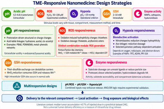
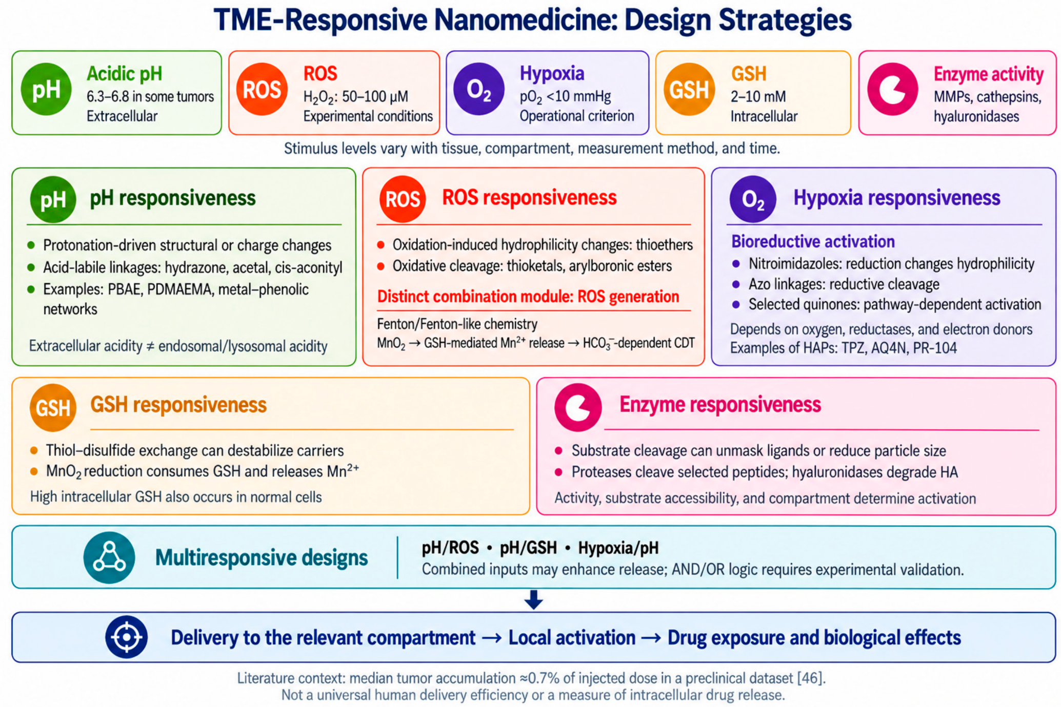
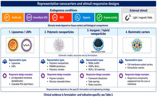
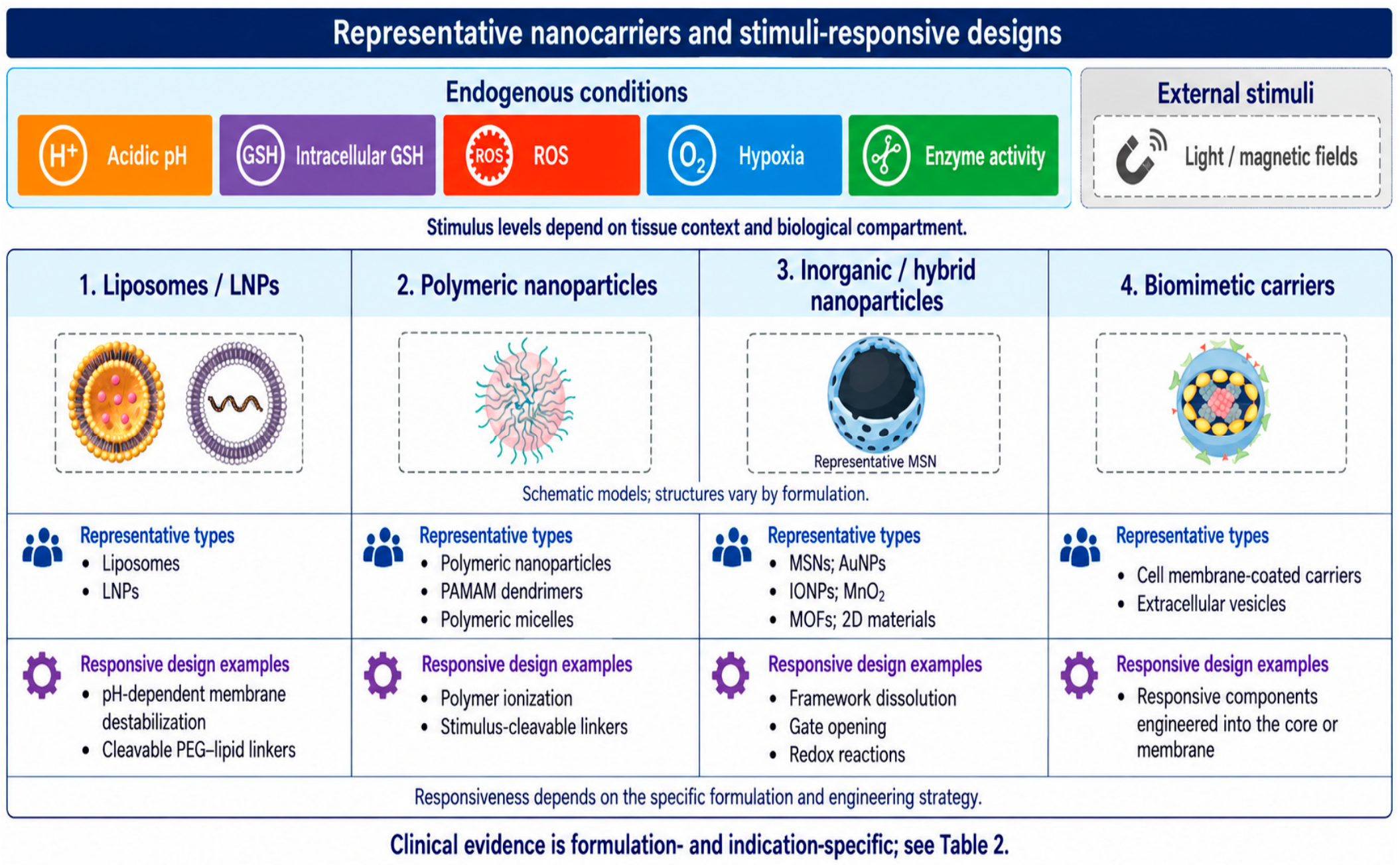

Disclaimer/Publisher’s Note: The statements, opinions and data contained in all publications are solely those of the individual author(s) and contributor(s) and not of MDPI and/or the editor(s). MDPI and/or the editor(s) disclaim responsibility for any injury to people or property resulting from any ideas, methods, instructions or products referred to in the content. |
© 2026 by the authors. Licensee MDPI, Basel, Switzerland. This article is an open access article distributed under the terms and conditions of the Creative Commons Attribution (CC BY) license.