


Abstract
Background: Giant cell arteritis (GCA) is a vasculitis mostly affecting older people. Primary membranous nephropathy (MN) is an immune-mediated glomerulopathy recently shown to be caused by autoantibodies. We report a case of both illnesses in an older gentleman. Case Presentation: A 63-year-old man developed systemic inflammatory signs and symptoms and near simultaneous nephrotic-range proteinuria. Investigations demonstrated both giant cell arteritis (GCA) and membranous nephropathy, with the presence of the M-type phospholipase A2 receptor (PLA2R) within subepithelial immune deposits. Renal function remained normal throughout the course. The patient achieved complete clinical and biochemical remission of both illnesses coincident with corticosteroid therapy. Conclusions: Although rare cases of concurrent GCA and MN have been reported, GCA occurring with PLA2R-positive MN has not been documented. This case raises the possibility of overlapping immunological pathways and may offer clues to the pathogenesis of GCA.
1. Introduction
Giant cell arteritis (GCA) is a granulomatous large-vessel vasculitis that primarily affects older adults and commonly presents with headache, constitutional symptoms, and elevated inflammatory markers [1]. The blood vessel inflammation is associated with activated macrophages, dendritic cells and T cells, and a prominent role for IL-6 has been established [2]. The deposition of immunoglobulin or complement in blood vessels is not considered a predominant mechanism in classical GCA.
Membranous nephropathy (MN), in contrast, is an autoimmune glomerular disease mediated by autoantibodies targeting podocyte antigens, most often the M-type phospholipase A2 receptor (PLA2R) [3]. Improvements in immunological diagnostic testing in MN and other conditions have revealed hitherto unknown autoantibodies that might be important in immune-mediated illnesses where culprit autoantigens have remained elusive.
Renal manifestations in GCA are unusual and typically ischemic due to large-vessel involvement [4]. Yet glomerulonephritis associated with GCA does occur, possibly more commonly than generally thought, as routine GCA laboratory studies might overlook podocyte injury. This is the first case documenting concomitant biopsy-proven GCA together with PLA2R-positive MN. The simultaneous onset of both illnesses, together with apparent steroid responsiveness in this case, generates hypotheses regarding shared immunologic processes involving GCA and MN.
2. Materials and Methods
Routine laboratory studies were performed by Labcorp, Inc., Indianapolis, IN, USA. Testing for antibodies to PLA2R by ELISA was performed at Mayo Clinic Laboratories, Rochester, MN, USA, as was immunofluorescence staining of the renal biopsy for PLA2R. Routine histology of paraffin-embedded specimens used standard protocols at Temple University Hospital and Stanford University Hospital and staining performed using hematoxylin and eosin, periodic acid–Schiff (PAS) reagents, and the Jones protocol. Immunofluorescence staining was performed at Temple on snap-frozen samples using commercially obtained fluorescein-conjugated antibodies to IgG, kappa and lambda immunoglobulin light chains, and C3. One block was multiple toluidine blue-stained, and one-micron-thick sections were prepared for electron microscopy.
3. Results
3.1. Case Presentation Time Line
3.1.1. Onset of Symptoms and First Corticosteroid Exposure
A 63-year-old man, in good health except for well-controlled hypertension, presented in April 2024 with six weeks of intermittent fever, fatigue, weight loss, and malaise several weeks after recovering from an apparent viral upper-respiratory illness. He reported occipital headaches, worse when recumbent, but denied jaw claudication or visual changes. There was no family history of autoimmune or of renal disease. Initial treatment with short courses of azithromycin and doxycycline provided no relief. A week’s trial of prednisolone (Medrol dose-pack) was followed by symptom resolution, but these recurred shortly after discontinuation.
A physical examination was notable for a blood pressure of 151/78 but was otherwise normal. Laboratory studies included normocytic anemia (hemoglobin 11.2 g/dL, a year previously 14–15 g/dL), thrombocytosis (platelets 457 × 109/L), an elevated erythrocyte sedimentation rate (ESR) of 45 mm/hour and C-reactive protein (73 mg/L). The white blood cell count was 10.2 k/mm3 with neutrophils 74.8% and lymphocytes 11.2%. Antinuclear antibodies were not detected by immunofluorescence (negative ANA), and anti-neutrophil cytoplasmic antigen autoantibodies (ANCAs), anti-myeloperoxidase, anti-PR3, and rheumatoid factor were also absent. Creatine kinase, C3, C4 and thyroid-stimulating hormone were normal. Blood cultures, a hepatitis B/C serologic panel, human immunodeficiency virus testing, the antistreptolysin-O titer, and a QuantiFERON–TB Gold assay yielded negative results. Urinalysis was not initially performed. Computed tomography of the chest (CT) demonstrated mild cardiomegaly, and small pericardial effusion CT examination of the abdomen and pelvis revealed mild narrowing of the celiac artery, deemed clinically insignificant. A transthoracic echocardiogram showed a normal ejection fraction (60–65%) and a small pericardial effusion without tamponade physiology. The prostate specific antigen was 3.7 ng/mL, and a colonoscopy performed in April 2024 was normal.
3.1.2. Diagnosis of GCA
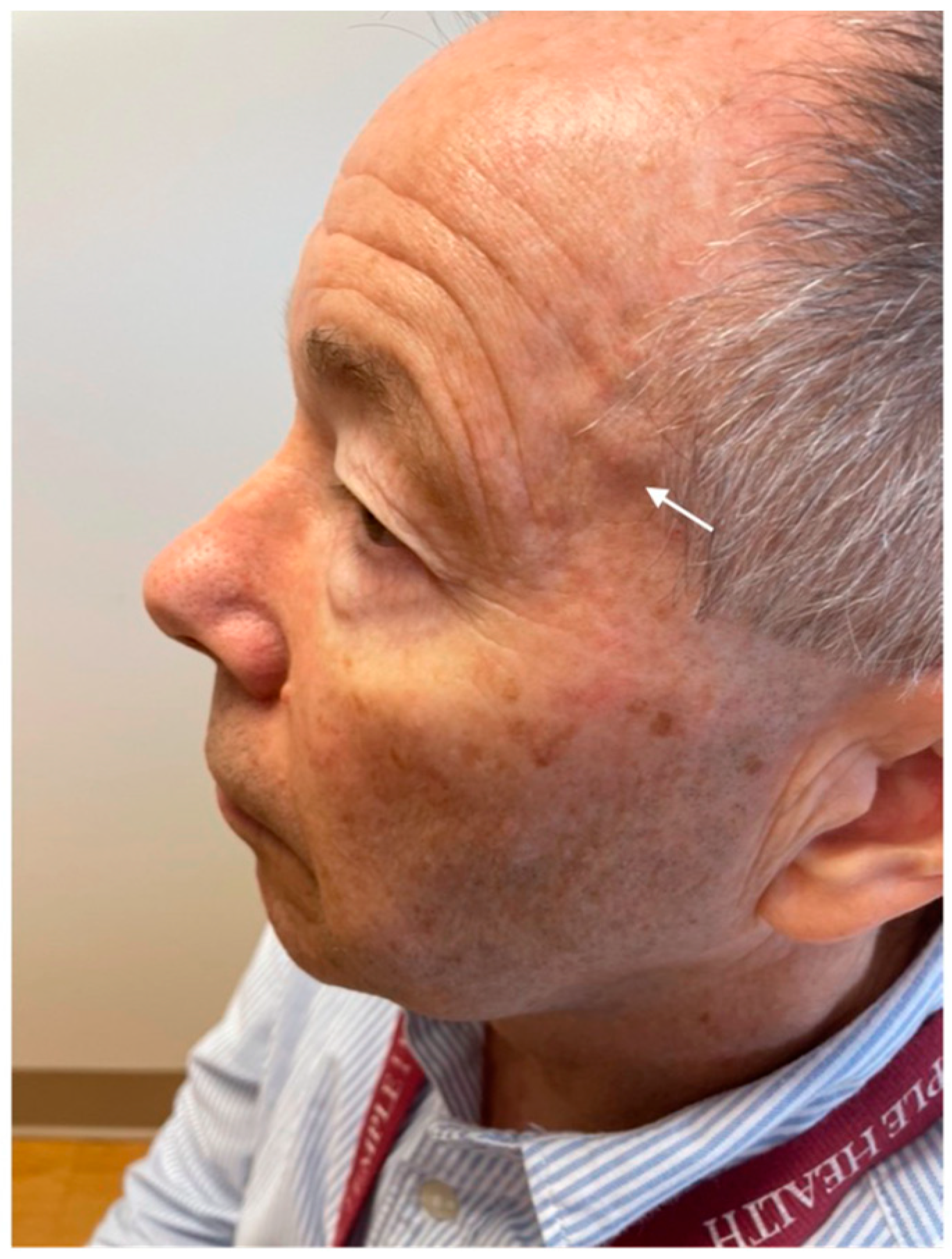
Because of persistent systemic inflammation, he was referred to Rheumatology one month later, in May 2024. A physical exam was notable for nontender swelling of the left temporal artery (Figure 1), periorbital edema, and 1+ pedal edema. The ESR was 108 mm/hr. A working diagnosis of GCA was made, and prednisone therapy was initiated at 40 mg po daily.
Figure 1.
Clinical image demonstrating swelling of the left temporal artery (arrow) and periorbital edema.
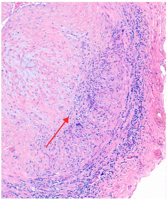
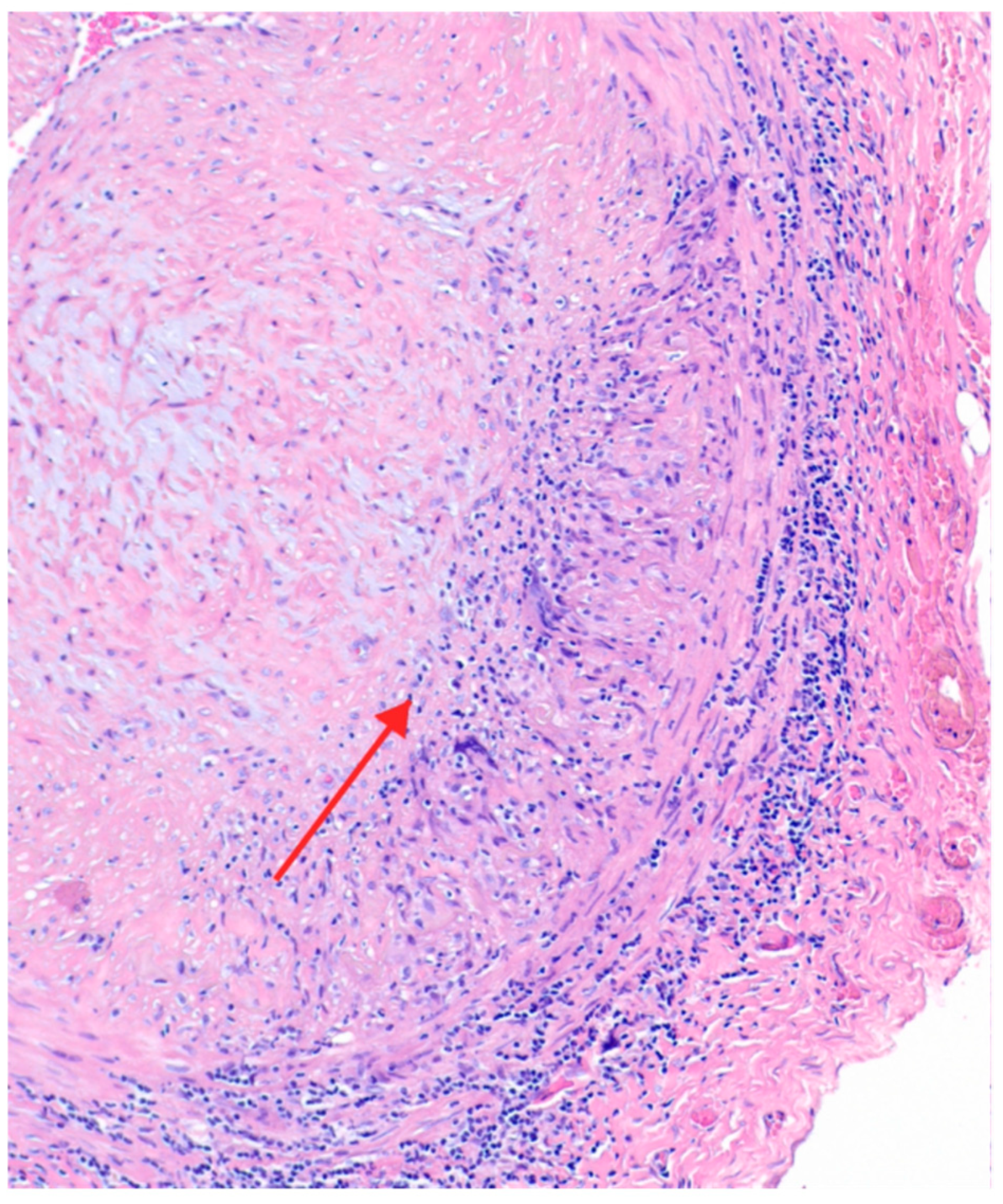
A temporal artery biopsy was performed in May 2024, approximately six weeks after initial exposure to prednisone. Medial granulomatous inflammation with giant cells and adventitial mononuclear cell infiltrates (Figure 2) was consistent with GCA. Investigations to search for PLA2R in the temporal artery biopsy were not performed, nor was staining for immunoglobulin and complement.
Figure 2.
Temporal artery biopsy showing the characteristic panarteritis of giant cell arteritis. There is severe luminal narrowing from intimal hyperplasia, medial granulomatous inflammation with giant cells (arrow) and prominent adventitial mononuclear cell infiltrates. (H&EX100).
Celiac artery narrowing was noted, but in the absence of signs or symptoms of mesenteric vascular insufficiency, this finding was not further investigated. Similarly, in the absence of apparent clinical significance, the small pericardial effusion received no further workup.
3.1.3. Discovery of Proteinuria
A urinalysis during the May 2024 Rheumatology workup showed a urine protein/creatinine ratio (UPCR) of 9869 mg/gm, Microscopic examination of the urine showed 5–10 isomorphic RBCs, rare WBCs and tubular cell clumps, and rare cellular casts. Serum albumin was 3.3 g/dL and serum creatinine was 0.81 mg/dL.
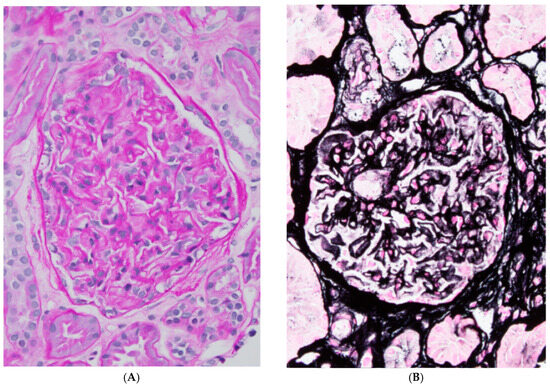
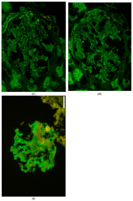
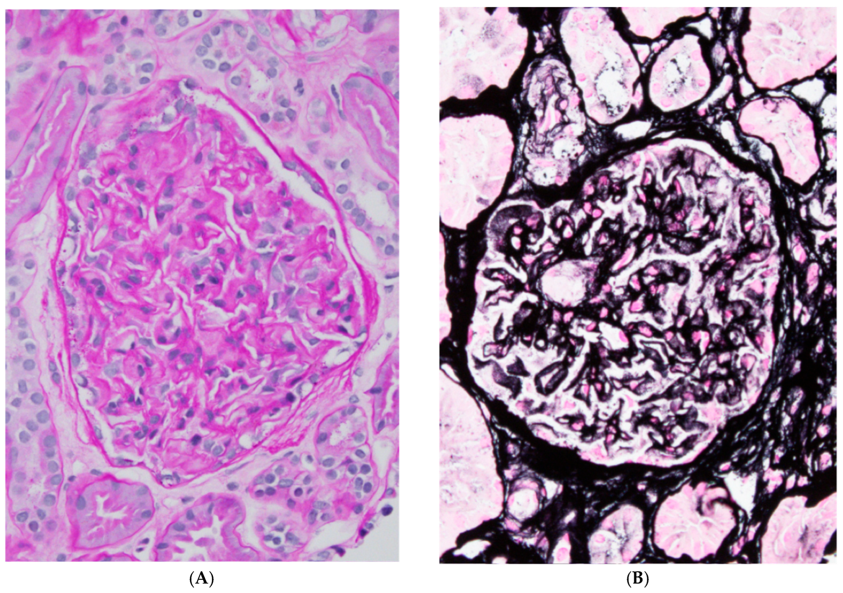
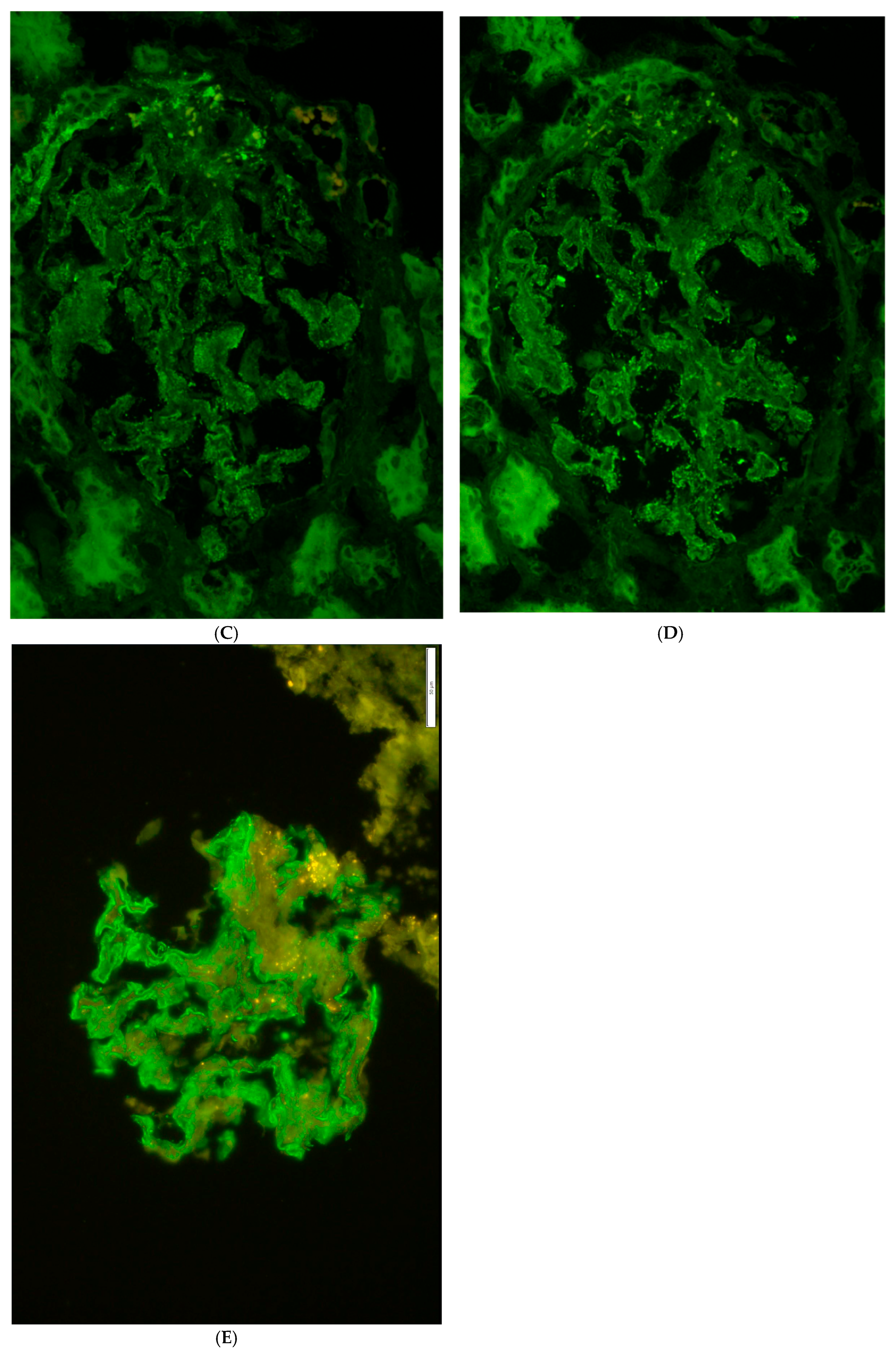
A renal biopsy was consistent with membranous nephropathy (Figure 3A–E). Twenty-eight glomeruli were examined. The glomeruli had diffusely thickened basement membranes with numerous “spikes and holes” on a Jones stain. No endocapillary hypercellularity, fibrinoid necrosis, segmental sclerosis, or crescent formation was seen. Mild acute tubular injury with open lumina and epithelial cell flattening was present. There was mild (20%) interstitial fibrosis and tubular atrophy.
Figure 3.
(A) PAS stain of the glomeruli at 400× magnification showing thickened glomerular basement membranes. (B) Jones stain at 400× magnification showing the numerous “spikes and holes” within the glomerular basement membranes. (C) C3 immunofluorescence stain showing bright, diffuse, global, granular, glomerular capillary wall staining (400×). (D) IgG immunofluorescent stain showing bright, diffuse, global, granular, glomerular, and capillary wall staining (400×). (E) PLA2R immunofluorescence showing positive staining.
There was no significant interstitial inflammation. The arteries and arterioles were unremarkable without inflammation, necrosis, or thrombosis. Twenty-four glomeruli were present for immunofluorescence evaluation. There was diffuse, global, granular, glomerular capillary wall positive staining for IgG (4+ on a 0–4, scale), C3 (4+), immunoglobulin kappa light chains (4+), and immunoglobulin lambda light chains (4+). Specific staining for IgG4 was not done.
Electron microscopy (EM). Two glomeruli were present for evaluation, neither globally sclerotic. Ultrastructural examination of one glomerulus revealed extensive (>80%) podocyte foot process effacement with microvillous transformation. The glomerular basement membranes were diffusely thickened. There was a normal amount of mesangial cellularity and matrix material. There were numerous subepithelial, intramembranous, and rare transmembranous immune complex-mediated electron-dense deposits, and no subendothelial or mesangial electron-dense deposits. There were no tubuloreticular inclusions. There were no deposits within the tubular basement membranes. No EM images are available at this time.
Demonstration of PLA2R in renal biopsy. Immunofluorescence staining at Mayo Clinic Laboratories documented PLA2R in the glomerulus (Figure 3E). Testing for serum PLA2R antibodies by ELISA at Mayo Clinic Laboratories revealed a level < 14 RU (relative units) per mL, with values of >20 RU/mL considered positive. A second serum sample sent in June 2024 was also negative for anti-PLA2R antibodies.
3.1.4. Additional Testing
A bone marrow biopsy was normal, and testing (Labcorp, Raritan, NJ, Inc.) for mutations associated with VEXAS syndrome [5] was negative. Serum protein electrophoresis was normal except for the slightly low albumin of 3.1 g/dL, and immunofixation was normal. Testing for anti-glomerular basement membrane antibody was not done.
3.1.5. Treatment and Clinical Course
The patient was given oral prednisone at 40 mg daily, with a gradual taper after four weeks. Telmisartan and hydrochlorothiazide were added for hypertension and antiproteinuric benefit, along with calcium and vitamin D supplementation. His headaches promptly disappeared, he reported feeling much better, and ESR and CRP declined to normal levels after one month of 40 mg daily doses of prednisone. UPCR declined promptly (see Table 1). BP stabilized in the 130/80 range after telmisartan was started, and albumin rose to 3.7 gm/dL when measured on 8 January 2025.
Table 1.
Clinical summary.
Prednisone was tapered as shown and stopped in October 2025. He has remained asymptomatic, and a complete blood count and comprehensive metabolic panel were normal as of April 2026. No further proteinuria has been detected. No adverse events of treatment were noted.
4. Discussion
This is a case of the concurrent recognition of GCA and PLA2R-positive membranous nephropathy in a single patient. Both conditions appeared to resolve rapidly with corticosteroid therapy. Although the concurrence might be entirely coincidental, we wondered whether this case could lend clues to the pathogenesis of GCA.
Could there be a link between PLA2R membranous nephropathy and GCA? These two rheumatic diseases are thought to have distinct underlying mechanisms. Primary PLA2R-positive MN is mediated by autoantibodies directed against the M-type phospholipase A2 receptor expressed on the surface membrane of podocytes [6] with rare extra-renal manifestations reported [7]. In sharp contrast to primary PLA2R-positive MN, a persuasive body of evidence implicates T-cell-mediated inflammation as the primary mechanism of GCA [8]. Autoantibodies are not believed to play a role, although there are reports of autoantibodies to phospholipids [9], endothelial cells [10], and intermediate filaments [11]. It is possible that some cases of apparent GCA may actually represent ANCA-associated vasculitis [12].
Rarity of renal involvement in GCA. Classic GCA seldom causes primary glomerular disease; most reported renal manifestations are ischemic or related to other overlapping vasculitides. Historical case reports and reviews emphasize that glomerulonephritis in GCA is uncommon but has been described [13].
Prior case reports of GCA with membranous nephropathy. Earlier literature contains scattered case reports describing GCA coexisting with membranous nephropathy, but these predate PLA2R testing and therefore could not distinguish primary (PLA2R-positive) from secondary MN [14].
Pathogenetic hypotheses. The PLA2R is expressed in podocytes, yet mRNA encoding this molecule has been detected in other tissues [15]. Autoantibodies might bind to multiple tissues, or cross-reactive antibodies might result from epitope spreading triggered by systemic inflammation, or unrecognized shared antigen expression. Our findings encourage the search for evidence of PLA2R-containing immune complexes in GCA tissues.
We would like to venture a second hypothesis to explain how antigens such as PLA2R might induce autoimmune disease. T-cell autoreactivity to the autoantigen (PLA2R) could lead to inflammation and direct damage to vessel walls via activated macrophages, causing vasculitis. PLA2R might at the same time induce circulating autoantibodies, with the potential to deposit in the glomerulus in the form of immune complexes. In unusual patients such as reported here, both processes might occur simultaneously. This hypothesis, like the one in the preceding paragraph, is highly speculative. We are also aware of the pitfalls stemming from drawing conclusions from a single case, but we emphasize that the case leads to a testable hypothesis—namely the testing of GCA patients for evidence of PLA2R autoimmunity.
Treatment and outcomes. The management of PLA2R-positive MN commonly involves immunosuppressive strategies tailored to patient risk; corticosteroids alone can produce remission in some cases, though modern guidelines often favor targeted B-cell depletion (e.g., rituximab) for high-risk disease [16]. Effective immunosuppression for systemic vasculitis (GCA) may also suppress the B-cell response driving PLA2R antibody production.
Limitations of this Report. This is a report of a single case. The concurrence of the two diagnoses (both more common in older individuals) might be coincidental. The two immunological processes might not necessarily be causally related. The diagnosis rests on the demonstration of the tissue deposition of autoantigen, but antibody specific for PLA2R was not detectable in the serum. Finally, the apparent response to corticosteroid therapy might instead reflect spontaneous resolution, which is known for both illnesses.
Clinical implications. When systemic inflammatory symptoms and renal abnormalities present together, clinicians should consider dual pathology and pursue targeted diagnostic testing (e.g., PLA2R serology/biopsy staining, timely temporal artery biopsy with expert pathology review). It is notable that serum antibodies to PLA2R were undetectable, despite deposition of the antigen in glomeruli. This is consistent with reported experience and emphasizes the need for searching for the PLA2R antigen in renal biopsy tissue, despite a negative test for anti-PLA2R in serum.
5. Conclusions
This case represents a rare coexistence of concurrently recognized GCA and PLA2R-positive MN, with an apparent favorable response to corticosteroid therapy. The recognition of a possible mechanistic connection between these two diseases could broaden our understanding of GCA.
Author Contributions
Conceptualization and methodology, K.A. and P.L.C.; writing—original draft preparation, K.A.; review and editing, K.A. and P.L.C.; assembling, organizing and interpreting data, and critically evaluating drafts and revisions of the manuscript, J.M.; performing pathology studies and interpretation, G.J.B. and J.H. All authors have read and agreed to the published version of the manuscript.
Funding
This research received no external funding.
Institutional Review Board Statement
Temple University IRB review and approval are not applicable because this research does not meet the federal regulatory definition of human subject research.
Informed Consent Statement
The patient has provided written informed consent for publishing this paper, and has given permission for his photograph to be used.
Data Availability Statement
The original contributions presented in this study are included in the article. Further inquiries can be directed to the corresponding author.
Acknowledgments
We are grateful to Sanjeev Sethi (Mayo Clinic) for an expert review of the renal pathology and assistance with the interpretation of PLA2R immunostaining.
Conflicts of Interest
The authors declare no conflicts of interest.
Abbreviations
The following abbreviations were used in the manuscript:
| GCA | giant cell arteritis |
| MN | membranous nephropathy |
| PLA2R | M-type phospholipase 2 receptor |
| CT | computed tomography |
| UPCR | urinary protein to creatinine ratio |
| CRP | C-reactive protein |
References
- Kermani, T.A. Giant Cell Arteritis. Ann. Intern Med. 2026, 179, ITC65–ITC80, Erratum in Ann. Intern. Med. 2026. https://doi.org/10.7326/ANNALS-26-02836. [Google Scholar] [CrossRef] [Scilit] [PubMed]
- Cayrou, C.; Gallet, F.; Ly, K.; Fauchais, A.L.; Bouanat, S.; Parreau, S. Unraveling giant cell arteritis: From immunopathology to emerging targeted therapies (2026 update). Autoimmun. Rev. 2026, 25, 104122. [Google Scholar] [CrossRef] [Scilit] [PubMed]
- Filippone, E.J.; Farber, J.L. Contemporary review of primary membranous nephropathy. Front. Immunol. 2026, 17, 1836347. [Google Scholar] [CrossRef] [Scilit] [PubMed] [PubMed Central]
- Maritati, F.; Iannuzzella, F.; Pavia, M.P.; Pasquali, S.; Vaglio, A. Kidney involvement in medium- and large-vessel vasculitis. J. Nephrol. 2016, 29, 495–505. [Google Scholar] [CrossRef] [Scilit] [PubMed]
- Assavarittirong, C.; Grant, C.; Nayak, S.S.; Nguyen, A.L. VEXAS Syndrome: Clinical Features, Hematologic Involvement, and Clinical Outcomes of Current and Emerging Therapies. Hematol. Rep. 2026, 18, 30. [Google Scholar] [CrossRef] [Scilit] [PubMed] [PubMed Central]
- Zhang, C.; Xu, Z.; Feng, Y.; Kong, J.; Wang, Y.; Xu, F.; Yang, M. Immune-mediated membranous nephropathy: Innovations in pathogenetic modeling and mechanistic insights. Int. Rev. Immunol. 2025, 44, 443–453. [Google Scholar] [CrossRef] [Scilit] [PubMed]
- Romagnani, P.; Tang, S.C.W.; Weins, A.; Huber, T.B.; Osafo, C.; Anders, H.J. Podocytopathies. Nat. Rev. Dis. Primers 2025, 11, 87. [Google Scholar] [CrossRef] [Scilit] [PubMed]
- Ibrahim, H.E.; De Bari, C. Giant cell arteritis: Update on pathogenesis and clinical implications. Curr. Opin. Rheumatol. 2025, 37, 72–79. [Google Scholar] [CrossRef] [Scilit] [PubMed]
- Kuret, T.; Lakota, K.; Hočevar, A.; Burja, B.; Čučnik, S.; Sodin-Semrl, S. Evaluating the utility of autoantibodies for disease activity and relapse in giant cell arteritis. J. Biol. Regul. Homeost. Agents 2018, 32, 313–319. [Google Scholar] [PubMed]
- Régent, A.; Dib, H.; Ly, K.H.; Agard, C.; Tamby, M.C.; Tamas, N.; Weksler, B.; Federici, C.; Broussard, C.; Guillevin, L.; et al. Identification of target antigens of anti-endothelial cell and anti-vascular smooth muscle cell antibodies in patients with giant cell arteritis: A proteomic approach. Arthritis Res. Ther. 2011, 13, R107. [Google Scholar] [CrossRef] [Scilit] [PubMed] [PubMed Central]
- Dasgupta, B.; Duke, O.; Kyle, V.; Macfarlane, D.G.; Hazleman, B.L.; Panayi, G.S. Antibodies to intermediate filaments in polymyalgia rheumatica and giant cell arteritis: A sequential study. Ann. Rheum. Dis. 1987, 46, 746–749. [Google Scholar] [CrossRef] [Scilit] [PubMed] [PubMed Central]
- Delaval, L.; Samson, M.; Schein, F.; Agard, C.; Tréfond, L.; Deroux, A.; Dupuy, H.; Garrouste, C.; Godmer, P.; Landron, C.; et al. French Vasculitis Study Group and French Study Group for Giant Cell Arteritis. Temporal Arteritis Revealing Antineutrophil Cytoplasmic Antibody-Associated Vasculitides: A Case-Control Study. Arthritis Rheumatol. 2021, 73, 286–294. [Google Scholar] [CrossRef] [Scilit] [PubMed]
- Crowder, S.; Pant, P.; Madruga, M.; Carlan, S. An Unusual Case of Renal-Limited Giant Cell Arteritis. Cureus 2026, 18, e112089. [Google Scholar] [CrossRef] [Scilit] [PubMed] [PubMed Central]
- El-Sayed, Z.A.; El-Awady, H.M.; Hassan, Z.E.; Adham, T.M.; Mostafa, H.M.; Elhefnawy, N.G. Giant cell arteritis in a 12-year-old girl presenting with nephrotic syndrome. Case Rep. Rheumatol. 2014, 2014, 491937. [Google Scholar] [CrossRef] [Scilit] [PubMed] [PubMed Central]
- Granata, F.; Petraroli, A.; Boilard, E.; Bezzine, S.; Bollinger, J.; Del Vecchio, L.; Gelb, M.H.; Lambeau, G.; Marone, G.; Triggiani, M. Activation of cytokine production by secreted phospholipase A2 in human lung macrophages expressing the M-type receptor. J. Immunol. 2005, 174, 464–474. [Google Scholar] [CrossRef] [Scilit] [PubMed]
- Floege, J.; Ayoub, I.; Brix, S.R.; Campbell, K.N.; Furie, R.; Nachman, P.H.; Tang, S.C.W.; Tomas, N.M.; Vivarelli, M.; Cheung, M.; et al. Targeting B cells in immune-mediated kidney diseases: Conclusions from a Kidney Disease: Improving Global Outcomes (KDIGO) Controversies Conference. Kidney Int. 2026, 110, 548–565. [Google Scholar] [CrossRef] [Scilit] [PubMed]


Disclaimer/Publisher’s Note: The statements, opinions and data contained in all publications are solely those of the individual author(s) and contributor(s) and not of MDPI and/or the editor(s). MDPI and/or the editor(s) disclaim responsibility for any injury to people or property resulting from any ideas, methods, instructions or products referred to in the content. |
© 2026 by the authors. Licensee MDPI, Basel, Switzerland. This article is an open access article distributed under the terms and conditions of the Creative Commons Attribution (CC BY) license.