


Abstract
Ultrasound is widely used in pediatric imaging because it is safe, portable, real-time, and free of ionizing radiation, but interpretation remains qualitative and operator-dependent. Ultrasound radiomics can extract quantitative features from standard grayscale images, providing potential biomarkers of tissue patterns not readily apparent on visual assessment. This structured narrative review summarizes pediatric ultrasound radiomics applications across organ systems and key barriers to clinical translation. Current evidence suggests promise in neonatal brain injury, neurodevelopmental assessment, liver disease staging, renal characterization, oncologic risk stratification, and emerging lung and muscle applications. However, most studies remain limited by retrospective single-center designs, small cohorts, acquisition variability, segmentation inconsistency, and limited external validation. Future progress will require standardized workflows, prospective multicenter validation, and clinically interpretable integration into decision-support systems.
1. Introduction
Over recent years, ultrasound has become a primary imaging modality in pediatrics because of its safety, accessibility, portability, low cost, real-time capability, and lack of ionizing radiation [1,2,3,4]. These advantages are particularly important in children, who may require repeated imaging and who face unique challenges including heightened radiation sensitivity, the potential need for sedation during longer examinations such as magnetic resonance imaging, small body size, and age-dependent anatomy [2]. These factors have reinforced ultrasound’s central role in pediatric diagnostic workflows, particularly for bedside imaging, longitudinal assessment, and evaluation of medically vulnerable patients [5,6].
Despite this versatility, conventional ultrasound interpretation remains largely qualitative and operator-dependent, with image acquisition and reporting influenced by sonographer technique, scanner settings, acoustic windows, and radiologist experience [7,8]. This subjectivity limits reproducibility across institutions and over time, particularly when subtle parenchymal or treatment-related tissue changes fall below the threshold of visual detectability on routine grayscale imaging [9,10]. These limitations have driven growing interest in radiomics—the extraction of quantitative imaging features from medical images to investigate associations with clinical, biological, or outcome-based information [11,12,13,14,15,16,17,18]. Applied to ultrasound, radiomics converts grayscale pixel data into numerical descriptors of signal intensity, spatial texture, morphology, and heterogeneity, capturing tissue patterns not readily appreciable by visual assessment alone and potentially supporting disease characterization, risk stratification, treatment monitoring, and outcome prediction [19,20,21].
The application of radiomics to pediatric ultrasound is a natural extension of this framework, given that ultrasound already underpins bedside evaluation, longitudinal follow-up, and disease surveillance in children. Current studies suggest potential applications in neonatal brain imaging, liver disease, renal characterization, oncologic risk stratification, lung disease, and muscle assessment. However, pediatric ultrasound radiomics remains an emerging research area rather than a routine clinical tool. Evidence is limited by small cohort sizes, single-center study designs, heterogeneous acquisition protocols, variable segmentation methods, limited reproducibility assessment, and insufficient external validation. Ultrasound radiomics also require particular methodological caution because image appearance is highly sensitive to acquisition parameters, scanner vendor, operator technique, speckle, artifacts, patient motion, and segmentation variability [22,23,24,25,26,27,28,29].
Unlike prior reviews that have largely addressed radiomics broadly or adult-dominant ultrasound applications, this structured narrative review aims to evaluate the current applications of radiomics in pediatric ultrasound imaging across organ systems and disease categories; identify methodological limitations and translational challenges that constrain clinical adoption; and outline a practical path toward reproducible, validated, and clinically meaningful ultrasound radiomic biomarkers in pediatric radiology [22,30].
2. Overview of Ultrasound Radiomics
This section provides a brief overview of the ultrasound radiomics workflow and major feature categories needed to interpret the current literature.
2.1. Radiomics Workflow in Ultrasound
The ultrasound radiomics workflow generally includes image acquisition, region-of-interest segmentation, preprocessing, feature extraction, feature selection, model development, and validation [Figure 1]. Each step can influence the final radiomic output and should be carefully considered in pediatric ultrasound studies.
Figure 1.
Created in BioRender. Schoeman, S. (2026) https://BioRender.com/suzbpzt, accessed on 24 May 2026. Schematic showing the steps of pediatric ultrasound radiomics workflow. Standardized US images are first acquired using consistent imaging parameters (1), followed by manual or semi-automated segmentation of the region of interest (ROI) corresponding to the target lesion or tissue (2). Preprocessing techniques including intensity normalization and denoising are then applied to enhance feature reproducibility prior to analysis (3). Radiomic features are then extracted from the ROI (4), these include higher-order texture features derived from matrices such as the gray-level co-occurrence matrix (GLCM) and gray-level run-length matrix (GLRM). In panel 4, the grayscale squares represent discretized pixel intensity values within the ROI, while the colored squares illustrate quantified spatial relationships between neighboring gray levels. These values correspond to frequency or probability of pixel intensity occurrences and are subsequently used to calculate texture metrics. Following extraction, redundant or unstable features are removed through quality control processes (5). Finally, after modeling is applied to associate features to relevant outcomes (6), final model performance is evaluated using metrics such as true or false positive rate and receiver operating characteristic curve (AUC) (7).
The first step is image acquisition. Image quality and feature values may be affected by scanner type, transducer frequency, gain, depth, focal zone placement, dynamic range, reconstruction parameters, and operator technique [22]. For ultrasound radiomics, standardization of acquisition protocols is particularly important because radiomic features are highly sensitive to technical variability.
The second step is region-of-interest segmentation, in which the target tissue or lesion is delineated manually, semiautomatically, or automatically [23]. Accurate segmentation is critical because extracted radiomic features are directly dependent on the selected region of interest. Inconsistent boundary definition may substantially affect feature reproducibility and model performance, particularly in pediatric imaging, where small anatomy, motion, variable acoustic windows, and subtle tissue boundaries can make segmentation challenging.
Preprocessing is then performed to improve image consistency before feature extraction. Common preprocessing steps include resampling, intensity normalization, gray-level discretization, denoising, and image filtering [24]. These steps are intended to reduce nonbiological variation and improve feature robustness, although excessive preprocessing may alter biologically meaningful image information. Standardization efforts such as the Image Biomarker Standardization Initiative have emphasized the importance of harmonized preprocessing and feature definitions to improve reproducibility across studies and software platforms.
After preprocessing, feature extraction is performed using dedicated software platforms such as PyRadiomics, MaZda, or other validated feature-extraction tools [25,26,31]. These tools can compute a broad range of engineered features from the segmented region of interest, including first-order, texture, shape, and filtered features. Because radiomics datasets often contain a large number of potentially redundant variables, feature selection is usually required to reduce dimensionality and retain the most informative and stable features. Common approaches include statistical filtering, reproducibility testing, correlation analysis, and regularization methods such as least absolute shrinkage and selection operator [16,27,28,29]. Selected features may then be incorporated into statistical or machine learning models for classification, prediction, or outcome assessment. Proper model development requires internal validation and, ideally, external testing to assess generalizability.
2.2. Radiomic Feature Categories
Radiomic features are commonly grouped into first-order, texture, shape, and higher-order features according to the type of image information they describe [Figure 2] [32].
Figure 2.
Created in BioRender. Schoeman, S. (2026) https://BioRender.com/n2plbd6, accessed on 24 May 2026. Figure showing the categories of radiomics features studied in pediatric ultrasound imaging. First-order features quantify the distribution of pixel intensities within the region of interest (ROI) without considering spatial relationships. Shape and morphologic features describe ROI geometry and size characteristics. Texture features evaluate spatial relationships between neighboring pixel intensities using various matrices. The arrows indicate spatial relationships and directional comparisons used to construct texture matrices such as GLCM and GLRM. Higher-order features apply mathematical transformations: the gray scale box represents wavelet-decomposed image sub-bands capturing image information at different spatial frequencies and orientations (e.g., low-low [LL], low-high [LH] frequency components). The numerical grid illustrates transformed pixel pattern (such as LBP) encoding used to quantify fine-scale texture characteristics.
First-order features are statistical descriptors derived from the distribution of pixel intensities within the region of interest, without accounting for spatial relationships. Common examples include mean, variance, skewness, kurtosis, and entropy. These features characterize the overall grayscale intensity profile of the tissue and can reflect signal magnitude, dispersion, asymmetry, peakedness, and randomness.
Texture features quantify spatial variation and gray-level arrangement within the image and are often useful when tissue heterogeneity is of interest. One of the most widely used texture approaches is the gray-level co-occurrence matrix, which evaluates how often pairs of gray levels occur at a defined spatial relationship and captures local spatial organization [32,33]. Common gray-level co-occurrence matrix features include contrast, correlation, homogeneity, and entropy. Another important texture family is the gray-level run-length matrix, which captures the length of consecutive runs of pixels with the same gray level in a specified direction. Common gray-level run-length matrix features include short-run emphasis, long-run emphasis, and gray-level nonuniformity.
Shape features describe the geometry of a segmented lesion or structure, including size, contour, compactness, and irregularity. Although shape features are commonly used in CT and MRI radiomics, their use in ultrasound may be more limited because lesion and tissue boundaries are often affected by acoustic window, speckle, shadowing, and segmentation variability.
Higher-order features are generated after applying mathematical filters or transforms to the original image to emphasize patterns at different scales or frequencies [34]. Common examples include wavelet features, which decompose the image into multiscale frequency sub-bands, as well as fractal dimension and local binary pattern descriptors, which may capture structural complexity and local microtexture. These approaches may enhance sensitivity to fine-scale heterogeneity but can also be sensitive to preprocessing choices and image quality.
Figure 2 shows the major categories of radiomic features in pediatric ultrasound imaging. Radiomic features include first-order intensity features, texture features, shape features, and higher-order filtered features. These categories provide complementary information about tissue intensity, spatial organization, morphology, and multiscale heterogeneity.
2.3. Ultrasound-Specific Considerations
Although the general radiomics workflow is similar across imaging modalities, ultrasound radiomics requires special consideration. Unlike CT and MRI, ultrasound does not have a standardized grayscale intensity scale, and image appearance is strongly influenced by acquisition settings, scanner vendor, probe selection, transducer pressure, patient motion, speckle, attenuation, and artifacts. These factors may affect radiomic feature stability and reduce reproducibility across scanners, operators, institutions, and patient populations.
These challenges are especially important in pediatric imaging, where small anatomy, developmental variation, limited patient cooperation, and disease rarity may further complicate image acquisition and model validation. Therefore, pediatric ultrasound radiomics should be interpreted as an emerging quantitative research approach that requires careful attention to acquisition standardization, segmentation reliability, feature reproducibility, and external validation before routine clinical implementation.
3. Technical Challenges Unique to Ultrasound Radiomics
Despite its promise as a noninvasive biomarker platform, ultrasound radiomics remain particularly vulnerable to technical variability across the imaging and analytic pipeline. In contrast to CT and MRI, ultrasound is intrinsically operator-dependent, and image appearance is strongly influenced by acquisition settings and system-specific factors. As a result, the reproducibility of radiomic features can be more difficult to ensure, which remains a central barrier to robust model development and clinical translation [22,35,36,37].
3.1. Acquisition-Related Variability
A major limitation of ultrasound radiomics is the variability introduced during image acquisition. Probe frequency, focal depth, gain, dynamic range, transducer pressure, scanner manufacturer, and operator technique can all alter image appearance, even when imaging the same tissue target [35,37,38]. This dependence on acquisition conditions is especially problematic for radiomics because quantitative features may capture not only underlying tissue characteristics but also technical differences in image formation.
In a reproducibility study using phantom and clinical ultrasound data, only a minority of extracted ultrasound radiomic features remained stable across changes in machine, segmentation location, and feature-extraction platform, underscoring the sensitivity of many features to acquisition-related variation [37]. These findings highlight the need for standardized acquisition protocols and careful documentation of scanner settings in ultrasound radiomics studies.
3.2. Preprocessing and Normalization
Because ultrasound grayscale values are highly system-dependent, preprocessing and normalization are often required to improve comparability across images, scanners, and institutions [22,35,39]. However, unlike CT, where intensity values have a more standardized physical basis, ultrasound lacks a universal grayscale intensity scale. Therefore, preprocessing choices such as resampling, discretization, filtering, contrast enhancement, speckle reduction, and intensity normalization may substantially influence downstream radiomic features [22,39,40].
Accordingly, normalization should not be considered a simple technical adjustment. Instead, it represents a critical methodological step that must be explicitly defined, justified, and validated within each ultrasound radiomics workflow. Without transparent preprocessing strategies, radiomic models may inadvertently learn scanner- or protocol-specific differences rather than biologically meaningful imaging phenotypes [22,39,40].
3.3. Radiomic Feature Reproducibility and Robustness
Reproducibility is a central requirement for any radiomic biomarker intended for clinical translation. In ultrasound radiomics, feature reproducibility can be affected by multiple interacting factors, including scanner vendor, acquisition settings, operator technique, image quality, preprocessing strategy, segmentation approach, and feature-extraction software. In a dedicated ultrasound radiomics reproducibility study, only a minority of extracted features remained reproducible when ultrasound machine, segmentation location, or feature-extraction platform changed, underscoring the sensitivity of ultrasound-derived features to technical variation [37].
For this reason, reproducibility assessment should be incorporated early in ultrasound radiomics studies rather than treated as a secondary validation step. Features should be evaluated for stability across repeated acquisitions, different observers, alternative segmentations, preprocessing methods, and, when possible, different ultrasound platforms. Common approaches include intraclass correlation coefficients, test–retest analysis, interobserver and intraobserver agreement, phantom-based reproducibility testing, and image perturbation analysis [41,42,43]. These methods can help identify robust features that are less sensitive to technical variation and more likely to generalize across clinical settings. This is particularly important in pediatric ultrasound, where small anatomy, motion, variable acoustic windows, and age-related developmental changes may further affect image appearance. Prioritizing reproducible features may reduce overfitting, improve model generalizability, and increase the likelihood that ultrasound radiomics can evolve from exploratory research toward clinically reliable imaging biomarkers.
3.4. Speckle Noise and Image Artifacts
Speckle is an inherent property of ultrasound imaging that arises from interference among subresolution scatterers and substantially influences image texture [44,45]. Although speckle may contain biologically relevant information in some quantitative ultrasound applications, it can also reduce contrast resolution, obscure lesion boundaries, and introduce stochastic texture patterns that confound feature stability [44,45,46]. Additional artifacts, including acoustic shadowing, posterior enhancement, reverberation, attenuation-related distortion, and motion, may further introduce nonbiologic heterogeneity into the image [45,46] [Figure 3]. Because radiomics is fundamentally designed to quantify image heterogeneity, these artifact-related patterns can be inadvertently encoded as discriminatory features unless careful quality control and preprocessing are applied [35,44,46].
Figure 3.
Influence of image quality on radiomics analysis. Representative cranial ultrasound images are shown to illustrate the effect of image quality on radiomic assessment. The upper panel (a,b) includes good quality images with adequate visualization of cerebral parenchyma, enabling reliable segmentation and feature extraction. The lower panel (c,d) demonstrates images degraded by artifacts and noise, which obscure parenchymal texture and may reduce the robustness and reproducibility of radiomic measurements. This figure underscores the importance of rigorous image quality assurance in brain ultrasound radiomics workflows.
3.5. Segmentation Variability and Automated Segmentation
Segmentation represents another major source of error in ultrasound radiomics. Manual delineation is still widely used, particularly in pediatric and research settings, yet ultrasound lesion margins are often indistinct because of low contrast, speckle, and artifact contamination [35,41,47]. Consequently, interobserver and intraobserver variability in region-of-interest definition can have a substantial effect on extracted features and on subsequent model performance [41,47,48]. Prior radiomics work has shown that feature robustness may decline meaningfully when segmentations vary across readers, and ultrasound-specific studies similarly suggest that automated or semiautomated approaches may improve reproducibility compared with fully manual workflows [37,41,47,48]. Even so, segmentation automation in ultrasound remains an evolving area and still requires rigorous validation before widespread adoption [37,41,42,43,47,48].
Deep learning-based segmentation methods provide a promising strategy to address this bottleneck. Convolutional neural network architectures, particularly U-Net and its variants, have become widely used for biomedical image segmentation and are increasingly being developed for automated and semiautomated ultrasound segmentation [49,50]. These models can learn spatial and contextual features from annotated imaging datasets and may help improve boundary consistency, reduce operator-dependent variability, and standardize region-of-interest placement across readers and institutions. This is particularly relevant in pediatric ultrasound, where small anatomy, motion, variable acoustic windows, and heterogeneous image quality can further complicate manual segmentation.
Despite this promise, automated ultrasound segmentation remains an evolving area. Deep learning models are sensitive to training data quality, annotation standards, scanner type, acquisition parameters, and patient population. Therefore, rigorous internal and external validation, quality-control procedures, and human oversight remain necessary before large scale clinical incorporation.
3.6. Model Calibration and Clinical Utility
Most pediatric ultrasound radiomics studies report discrimination metrics such as AUC, sensitivity, specificity, and accuracy. While these measures indicate how well a model separates disease categories or severity groups, they do not determine whether predicted probabilities are clinically reliable. A model may achieve a high AUC yet still overestimate or underestimate absolute disease risk, limiting its usefulness for individualized decision-making and clinical implementation [51,52].
Calibration, which reflects the agreement between predicted probabilities and observed outcomes, is therefore an important but underreported component of model evaluation. It can be assessed using calibration plots, calibration intercept and slope, and the Brier score [53,54]. In pediatric ultrasound radiomics, this is particularly relevant because proposed applications often involve risk stratification, biopsy selection, follow-up planning, treatment guidance, or prognostic counseling. Poorly calibrated models may appear statistically accurate while providing misleading risk estimates that could affect patient management.
Clinical utility should also be evaluated before radiomics models are considered for practice. Approaches such as decision curve analysis and net benefit assessment can determine whether a model improves decision-making compared with existing clinical pathways, expert interpretation, or default strategies [53]. Given the small datasets, low disease prevalence in some applications, and potential consequences of unnecessary imaging, biopsy, or treatment escalation in children, future pediatric ultrasound radiomics studies should move beyond AUC alone and report discrimination, calibration, and clinical utility to support clinically interpretable and actionable prediction models.
3.7. Small Pediatric Cohorts and Limited Generalizability
These technical challenges are amplified in pediatric imaging, where ultrasound radiomics studies are frequently constrained by small cohorts, single-center design, and limited external validation. Small datasets reduce statistical power, increase the risk of overfitting, and are particularly problematic in radiomics because the number of extracted features can vastly exceed the number of available subjects [36,55,56]. More broadly, recent evaluations of the ultrasound radiomics literature have repeatedly identified high risk of bias related to small sample size, restricted case diversity, and lack of multicenter validation [55,56]. In pediatric applications, these issues are even more pronounced because of disease rarity, heterogeneous age- and development-related anatomy, and practical barriers to assembling large, harmonized datasets [22,35,36,39,57].
3.8. Need for End-to-End Standardization
Ultimately, the most important barrier to clinical translation is the lack of end-to-end standardization. Reliable ultrasound radiomics requires standardized acquisition protocols, transparent preprocessing workflows, reproducible feature definitions, robust segmentation methods, quality-control procedures, and external multicenter validation [23,35,36,39,49].
The Image Biomarker Standardization Initiative has substantially improved harmonization of radiomic feature definitions and benchmarking across software platforms, but standardization of the broader ultrasound workflow remains incomplete, particularly at the levels of acquisition, preprocessing, and image quality control [23,39]. Reporting frameworks such as CLAIM and CLEAR further emphasize that methodological transparency, reproducibility assessment, and validation are essential for trustworthy imaging AI and radiomics research [57,58]. Without these measures, the apparent diagnostic performance of ultrasound radiomics models may not translate into reliable clinical performance across scanners, operators, institutions, or pediatric patient populations.
4. Review Design
This review was conducted as a structured narrative review of the pediatric ultrasound (US) radiomics literature. Given the heterogeneity in the existing literature, regarding study design, organ/tissue systems, image acquisition technique, segmentation methodology, feature extraction pipelines, and outcome measures, a formal meta-analysis was not feasible. The review aimed to synthesize current applications of US radiomics within the sphere of pediatric imaging while critically evaluating each study’s methodologic quality, reproducibility, and translational readiness.
4.1. Literature Search Strategy
A literature search was performed using PubMed, Scopus, and Google Scholar databases for studies published through March 2026. Search terms included combinations of: “ultrasound radiomics”, “quantitative ultrasound”, “ultrasound texture analysis”, “pediatric”, “children”, and organ-specific/pathology terms (i.e., brain, renal, tumor, musculoskeletal).
4.2. Eligibility Criteria
- Studies were included if:
- The cohort included pediatric patients (<18 years);
- They applied grayscale US-based radiomic, texture, or quantitative feature extraction methodologies;
- They evaluated diagnostic, prognostic, predictive, or classification performance;
- They included original research data;
- They were published in English.
Studies were excluded that:
- Exclusively included adult (>18 years) subjects;
- Did not apply quantitative feature extraction based on grayscale US image information including other US modalities, e.g., Doppler parameters and advanced ultrasound techniques (e.g., elastography, CEUS) were not included or critically assessed.
- Were not clinical or translational research (review articles, editorials, case series or reports);
- Analyzed non-US imaging modalities (CT or MRI);
- Did not clearly define methodology employed for radiomic analysis (feature extraction or model development);
- Study Selection and Data Extraction
- Eligible studies were reviewed and categorized by primary clinical applications. This included:
- Brain imaging;
- Liver imaging;
- Renal Imaging;
- Oncologic Imaging;
- Emerging areas—including lung and musculoskeletal.
- The following variables were analyzed, when available, to enhance review uniformity:
- Study design, sample size, clinical application, and patient population;
- Ultrasound modality and acquisition parameters;
- Segmentation methodology and radiomic pipeline;
- Radiomic features extracted and models used;
- Study limitations including validation technique or image acquisition methods;
- Level of evidence (per Oxford Center for Evidence-Based Medicine guidelines) [59].
4.3. Evidence Synthesis
Given the substantial heterogeneity within the growing body of research on pediatric-specific US-radiomics application, findings were synthesized qualitatively, rather than quantitatively pooled. The literature was interpreted with primary emphasis on recurring methodological limitations and emerging/potential areas of promise relevant to the scope of pediatric imaging applications.
5. Current Clinical Applications of Ultrasound Radiomics in Pediatrics
Because detailed study-level methodology, including segmentation approach, feature classes, software, model type, validation strategy, performance metrics, limitations, and evidence level, are summarized in Table 1, this section focuses on the clinical task, principal findings, critical interpretation, and translational readiness of pediatric ultrasound radiomics applications.
The volume and maturity of available evidence differ substantially across organ systems; therefore, the depth of discussion varies accordingly. Neonatal brain imaging is discussed in greater detail because it currently represents the largest and most developed pediatric ultrasound radiomics literature, whereas liver, renal, oncologic, lung, and musculoskeletal applications remain more limited or emerging. To improve readability, common limitations, including small sample size, single-center design, heterogeneous acquisition, variable segmentation, and limited external validation, are considered collectively in this section and revisited in the future directions rather than repeated in detail for each application.
5.1. Brain Imaging
Neonatal brain imaging currently represents the most developed pediatric application of ultrasound radiomics, but the evidence remains stronger as proof-of-concept than as a clinically deployable tool. Across studies, radiomics has shown that visually subtle or diffuse white matter abnormalities can be quantified from routine cranial ultrasound images. This is clinically meaningful because conventional neurosonography is limited in detecting non-cystic or evolving injury patterns.
5.1.1. Preterm White Matter Injury and Early Detection
Narchi et al. evaluated 20 very-low-birth-weight neonates and showed that texture analysis of initial cranial ultrasound could distinguish transient periventricular echogenicity from echogenicity that later progressed to cystic periventricular leukomalacia (PVL), with AUC values of 0.98 and 0.92 in sagittal and coronal planes, respectively [60]. Although promising, this study is best interpreted as an early proof-of-concept because of its small retrospective cohort, limited follow-up, and lack of external validation.
You et al. evaluated 33 preterm neonates and linked increased textural heterogeneity with MRI-graded severe white matter injury (WMI), with entropy achieving an AUC of 0.865 [61]. This strengthened the biologic plausibility that radiomic features may reflect structural disorganization in injured white matter. However, the method did not reliably distinguish normal from mild WMI, which is a critical limitation because subtle non-cystic injury is often the most difficult and clinically relevant target.
Zhu et al. advanced the field by testing higher-dimensional and automated radiomics approaches for WMI detection in 158 preterm neonates, with reported AUC values of around 0.85 [62]. A later multiplanar study in 267 premature infants achieved a validation AUC of 0.91, suggesting that multi-plane radiomic assessment may better capture the spatial heterogeneity of WMI than single-plane analysis [62,63]. These studies are more methodologically mature than earlier texture-analysis work, but their clinical readiness remains limited by single-center development and incomplete external validation.
5.1.2. Longitudinal Monitoring and Neurodevelopmental Risk
Longitudinal monitoring may be one of the most clinically compelling directions for neonatal brain radiomics. Jung et al. showed that serial texture change helped distinguish evolving periventricular leukomalacia from resolving periventricular echogenicity, with the strongest longitudinal measure achieving an AUC of 0.82 [64]. Laccetta et al. also found that quantitative grayscale measures of periventricular white matter were associated with later neurodevelopmental outcomes [65]. These findings suggest that texture trajectories may provide more clinically meaningful information than single-time-point assessment, although longer-term outcome validation remains needed.
5.1.3. Expanding Applications Beyond Preterm Injury
The application of brain ultrasound radiomics to full-term exposure-related conditions is promising but remains exploratory. Zimina et al. studied 89 full-term newborns with diabetic fetopathy and reported that radiomic models could detect brain differences not apparent on conventional neurosonography, with the best regional model achieving an AUC of 0.85 [66]. Sultan et al. applied neonatal brain ultrasound radiomics to HIV-exposed uninfected and HIV-unexposed newborns, reporting AUC values of 0.72 in the basal ganglia and 0.76 in the periventricular white matter [67]. These studies broaden the potential role of ultrasound radiomics beyond preterm injury, particularly in settings where MRI may be unavailable.
Overall, neonatal brain ultrasound radiomics has progressed from early texture analysis to higher-dimensional modeling, multiplanar assessment, longitudinal monitoring, and automated segmentation. Its main promise lies in detecting subtle or diffuse tissue abnormalities that are visually inapparent or difficult to grade reproducibly. Future studies should determine whether radiomics improves risk stratification, monitoring, or decision-making beyond expert cranial ultrasound and whether radiomic markers correlate with longitudinal neurodevelopmental outcomes.
5.2. Liver Imaging
Compared with neonatal brain imaging, pediatric liver ultrasound radiomics is less developed but addresses an important clinical gap. Conventional ultrasound is widely used for screening and follow-up of diffuse liver disease, yet visual assessment of echogenicity and attenuation remains subjective and insensitive to early or mild disease [68,69,70]. Radiomics may improve objectivity, but current pediatric evidence is narrow and largely focused on steatosis.
5.2.1. Steatosis and Diffuse Liver Disease
The strongest pediatric evidence comes from Das et al., who evaluated 93 healthy controls and 39 children with NAFLD and showed that ultrasound texture analysis differentiated fatty liver disease from healthy liver tissue with an external validation AUC of 0.92 [71]. The model also outperformed simpler echogenicity-based measures such as the hepatorenal index, suggesting that radiomics may capture parenchymal fat-related texture changes beyond subjective grayscale interpretation.
However, the clinical role of this approach remains uncertain. Pediatric NAFLD/MASLD exists across a spectrum of fat burden, inflammation, fibrosis, and metabolic risk. Therefore, binary classification against healthy controls may not reflect the more difficult clinical tasks of grading disease severity, detecting early disease, or monitoring treatment response. Future studies should test whether radiomics adds value over MRI-PDFF, elastography, laboratory markers, and clinical risk scores.
5.2.2. Fibrosis and Pediatric-Specific Barriers
Pediatric fibrosis is an important potential application but remains under-validated. Adult studies suggest that ultrasound radiomics may capture fibrotic architectural distortion, but extrapolation to children is problematic [72,73]. Pediatric liver texture is influenced by growth, body habitus, inflammation, congestion, underlying diagnosis, and treatment status. Without age-specific reference ranges and disease-specific validation, radiomic features may reflect developmental or technical variation rather than fibrosis.
5.2.3. Critical Summary
The main promise of pediatric liver radiomics is improved objectivity in a modality already used widely for screening and follow-up. The next stage should focus on multicenter pediatric cohorts, standardized acquisition, age-adjusted normative data, and clinically relevant endpoints such as fibrosis stage, steatosis severity, progression, and treatment response.
5.3. Renal Imaging
Renal ultrasound radiomics is one of the more clinically plausible pediatric applications because ultrasound is already central to renal evaluation, and several renal diseases have functional, laboratory, or histologic reference standards. The key question is whether radiomics can improve upon subjective echogenicity, which is commonly used but poorly specific.
5.3.1. Parenchymal Disease Characterization
De Leon-Benedetti et al. evaluated 31 pediatric subjects with AKI, CKD, or healthy kidneys and reported that radiomics models achieved an accuracy of around 0.90 for group differentiation [Figure 4] [25]. The observed pattern of greater homogeneity in AKI and greater heterogeneity in CKD is biologically plausible and clinically relevant. This study supports the potential of radiomics for quantitative renal parenchymal characterization.
Figure 4.
Radiomics reveal quantitative renal texture differences beyond visual grayscale assessment. (a) Representative grayscale renal ultrasound images from CKD, AKI, and healthy control cases show broadly overlapping visual appearances, underscoring the difficulty of distinguishing these groups by routine inspection alone. In contrast, normalized radiomic feature profiling (b) demonstrates distinct texture signatures across disease states. This example illustrates the potential of ultrasound radiomics to uncover subtle parenchymal differences not readily visible on conventional imaging. This figure was adapted from De Leon-Benedetti et al. [26].
5.3.2. Glomerulonephropathy and Histologic Risk Stratification
Radiomics studies in biopsy-confirmed glomerulonephropathy are particularly important because pathology provides a strong reference standard. Kou et al. studied 313 biopsy-confirmed pediatric glomerulonephropathy cases and reported high validation AUCs for differentiating IgA nephropathy, minimal change disease, and Henoch–Schonlein purpura nephritis, ranging from 0.91 to 0.98 [74]. Chen et al. evaluated 440 biopsy-confirmed cases of Henoch–Schonlein purpura nephritis and reported a validation AUC of 0.87 for predicting histologic severity, particularly crescentic disease [75].
These results are promising, but they must be interpreted cautiously. High performance in single-center retrospective datasets may reflect disease selection, acquisition consistency, or overfitting rather than generalizable disease biology. The clinical value of renal radiomics will depend on whether it can predict histologic severity or disease progression in external cohorts and whether it changes decisions about biopsy, treatment intensity, or monitoring.
5.3.3. Hydronephrosis and Longitudinal Monitoring
Sloan et al. studied 90 pediatric patients with hydronephrosis and achieved an AUC of 0.86 for differentiating low- from high-grade disease [76]. This is a practical application because hydronephrosis grading is routine but can be subjective. However, the incremental value over expert interpretation and standardized grading systems still needs clarification.
Early post-angioplasty radiomics work by Schoeman et al. suggests that texture analysis may detect longitudinal renal parenchymal changes not evident visually [Figure 5] [77]. This direction is attractive because radiomics may be most useful as a monitoring biomarker. However, such changes must be correlated with perfusion, renal function, blood pressure response, and clinical outcomes before they can be interpreted as meaningful recovery or remodeling.
Figure 5.
Example of radiomics-derived detection of subtle interval renal change beyond visual grayscale assessment. Baseline pre-angioplasty (A) and 14-month follow-up (B) grayscale renal ultrasound images appear broadly similar on conventional visual inspection. In contrast, quantitative texture analysis (C), based on renal cortical radiomic features normalized to a splenic reference region, demonstrates measurable longitudinal shifts in multiple texture parameters. This representative case illustrates the potential of ultrasound radiomics to identify subtle parenchymal changes not readily visible on standard grayscale imaging.
5.3.4. Critical Summary
Renal ultrasound radiomics has strong translational rationale, but the field remains immature. The most important future step is to demonstrate incremental value beyond conventional ultrasound, laboratory tests, pathology, and clinical variables. Multicenter validation, patient-level splitting, standardized cortical and medullary ROI definitions, and outcome-based endpoints will be essential.
A review of the application of US radiomics in pediatric renal imaging has demonstrated promising results in the relatively small studies in which it has been explored. Future research is needed to validate these initial findings in order to transition this research from ‘pilot studies’ in limited samples to validated results with relevant clinical use cases.
5.4. Oncologic Imaging
Pediatric oncologic ultrasound radiomics is promising because many tumors have pathologic or molecular reference standards, but current evidence remains in its early stages. The potential value of radiomics is not in replacing biopsy or pathology, but in improving noninvasive risk stratification, preoperative planning, and lesion characterization.
5.4.1. Tumor Characterization and Prognostic Stratification
Zhu et al. evaluated 73 pediatric patients with peripheral neuroblastic tumors and reported strong differentiation of prognostic subsets, with a radiomics-only validation AUC of 0.918 [78]. This suggests that grayscale tumor texture may reflect biologically meaningful heterogeneity. These findings suggest that grayscale tumor texture may reflect biologically meaningful heterogeneity and could complement established clinical, histologic, and molecular risk factors.
5.4.2. Thyroid and Thoracic Applications
Li et al. evaluated 164 pediatric patients with papillary thyroid carcinoma and reported a validation AUC of 0.832 for predicting extrathyroidal extension, outperforming routine sonographic assessment [79]. This is clinically relevant because extrathyroidal extension affects operative planning. However, class imbalance, manual segmentation, and single-center validation limit immediate clinical use.
Wei et al. reported high performance for benign-malignant differentiation of subpleural pulmonary lesions in 609 patients, with validation AUC of 0.924 [80]. Although not exclusively pediatric, this study illustrates the potential of combining radiomics, deep learning features, and clinical variables. Its limitations also highlight key barriers for ultrasound radiomics: retrospective design, class imbalance, grayscale-only imaging, and use of non-DICOM images.
5.4.3. Critical Summary
Oncologic ultrasound radiomics should be viewed as an adjunctive risk-stratification tool rather than a stand-alone diagnostic method. Future studies must address small sample size, class imbalance, and external validation. The most valuable models will likely be multimodal, combining ultrasound radiomics with clinical features, pathology, molecular markers, and outcomes.
5.5. Emerging Applications
Lung and musculoskeletal applications illustrate the expanding scope of pediatric ultrasound radiomics, but these areas are heterogeneous and less mature. Many studies overlap with quantitative ultrasound rather than conventional radiomics, so interpretation requires caution.
5.5.1. Lung Ultrasound Radiomics
Lin et al. evaluated 150 neonatal lung disease cases and reported strong performance for neonatal respiratory distress syndrome classification, with a validation AUC of 0.951 [81]. This suggests that radiomics may reduce operator dependence in neonatal lung ultrasound.
Cai et al. evaluated 301 pediatric patients with acute dyspnea and reported an AUC of 0.976 for an integrated radiomic–clinical model for acute heart failure diagnosis [82]. Although promising, the high performance should be interpreted cautiously because of overfitting concerns and the need for comparison with expert clinical assessment.
Mohamed, Sultan, Venkatakrishna, and colleagues explored AI-assisted quantitative lung ultrasound analysis for pediatric pneumonia-related findings in a small pilot cohort [83]. This remains preliminary, but it highlights a clinically relevant direction: semi-automated quantification of consolidation, pleural abnormalities, and B-lines.
5.5.2. Musculoskeletal and Muscle Applications
Quantitative muscle ultrasound studies in Duchenne muscular dystrophy show that grayscale intensity and backscatter features can distinguish diseased from healthy muscle and correlate with functional status [84,85,86]. These findings support the broader value of ultrasound-derived quantitative biomarkers, although not all studies represent conventional radiomics.
Scatteromics and hip ultrasound radiomics further extend this concept. Advanced scatter-based analysis has shown potential for distinguishing ambulatory decline in Duchenne muscular dystrophy [87], while developmental dysplasia of the hip radiomics achieved validation AUC of 0.91 for detecting femoral head microstructural changes [88]. These findings are promising, but the clinical role of these methods remains to be defined.
5.5.3. Critical Summary
Emerging lung and musculoskeletal applications suggest that radiomics may expand beyond traditional organ-based imaging into bedside functional and structural monitoring. However, the evidence remains early, heterogeneous, and often single-center. Future studies should clarify whether these quantitative approaches provide incremental value beyond expert interpretation, clinical scores, and established quantitative ultrasound measures.
Table 1.
Summary of pediatric ultrasound radiomics studies across organ systems, including study population, clinical application, segmentation approach, radiomic feature-extraction methodology, model performance, major limitations, and level of evidence.
6. Future Directions Toward Clinical Translation and Real-World Implementation
Despite growing interest in ultrasound radiomics, several critical steps remain before these approaches can be translated into routine clinical care. Future progress will depend not only on technical refinement, but also on demonstrating that ultrasound radiomics can provide actionable information at the point of care. For clinical implementation, radiomic tools must be reliable across scanners and operators, interpretable to clinicians, integrated into existing ultrasound workflows, and supported by evidence that they improve diagnostic confidence, risk stratification, treatment monitoring, or patient outcomes. Therefore, the path forward is inherently multidisciplinary, requiring collaboration among imaging scientists, pediatric radiologists, sonographers, engineers, informatics teams, industry partners, and regulatory bodies.
6.1. Standardization as the Foundation for Clinical Use
A major priority for clinical translation is the standardization of ultrasound radiomics methodology. Unlike more standardized cross-sectional imaging modalities, ultrasound is highly operator-dependent and sensitive to acquisition parameters, including transducer selection, frequency, gain, depth, focal zone placement, transducer pressure, and vendor-specific image processing. These sources of variability can substantially influence radiomic feature stability and may limit reproducibility across institutions and platforms.
Accordingly, future efforts should focus on establishing clinically practical acquisition protocols that can be implemented by sonographers in routine practice. These protocols should define not only scanner settings and image planes, but also minimum image-quality requirements, acceptable artifact thresholds, and standardized documentation of acquisition parameters. In parallel, preprocessing and feature-extraction pipelines must be harmonized so that radiomic outputs are reproducible across scanners, vendors, operators, and clinical environments. Reproducibility studies will be essential to identify robust features that remain stable under real-world imaging conditions and can therefore serve as reliable ultrasound-derived imaging biomarkers.
6.2. Multicenter and Multi-Vendor Validation
Beyond technical standardization, multicenter and multi-vendor validation is essential to determine whether ultrasound radiomic signatures are generalizable. Much of the current literature remains based on relatively small, single-center cohorts, often with limited external validation. While these studies are valuable for proof of concept, they are insufficient to establish broadly applicable biomarkers.
Large, diverse datasets collected across institutions, patient populations, disease subtypes, age groups, and imaging platforms are needed to test the robustness and transportability of proposed models. This is particularly important in pediatric imaging, where disease rarity, developmental variation, and differences in scanning practice can limit generalizability. Multicenter validation should also evaluate whether radiomics provides incremental value beyond conventional ultrasound interpretation, Doppler assessment, contrast-enhanced ultrasound, laboratory markers, pathology, MRI, or clinical outcomes. Demonstrating this added value will be essential for clinical adoption.
6.3. Integration into the Ultrasound Workflow
For ultrasound radiomics to become clinically meaningful, it must be integrated into the workflow in a way that is efficient, interpretable, and minimally disruptive. Radiomics should not require complex offline analysis that delays reporting or depends on specialized research personnel. Instead, future systems should aim for automated or semiautomated workflows that include image-quality assessment, region-of-interest detection, segmentation, feature extraction, and quantitative output generation within or alongside existing ultrasound platforms and PACS/reporting environments.
Near real-time analysis may allow for radiomic outputs to be generated during image acquisition or interpretation. For example, radiomics could provide immediate feedback on whether image quality is sufficient, whether the region of interest has been appropriately captured, or whether quantitative texture features fall outside an expected range. Such integration would make radiomics more practical for clinical use and could reduce operator burden while improving consistency across examinations.
6.4. Clinically Actionable Decision Support
The greatest clinical value of ultrasound radiomics will likely come from decision-support tools rather than isolated feature values. Radiomic outputs should be translated into clinically understandable information, such as probability scores, risk categories, longitudinal change metrics, or alerts for abnormal tissue patterns. These outputs should be presented in a way that supports, rather than replaces, expert radiologist interpretation.
In practice, ultrasound radiomics may be most useful when combined with grayscale findings, Doppler features, contrast-enhanced ultrasound metrics, laboratory data, pathology results, and clinical context. Such integrated models could assist with lesion characterization, disease staging, treatment response assessment, risk stratification, and follow-up planning. Importantly, these tools should be designed around specific clinical questions, such as distinguishing acute from chronic kidney injury, identifying early brain maturation differences, monitoring liver fibrosis, assessing tumor risk, or quantifying muscle and lung disease progression.
6.5. Application-Specific Implementation Pathways
Future work in pediatric ultrasound radiomics should move beyond broad technical feasibility and focus on application-specific pathways to clinical relevance. In neonatal brain imaging, longitudinal and multiplanar radiomics approaches may be particularly valuable when linked to neurodevelopmental outcomes, as serial textural change may provide more clinically meaningful information than single-timepoint classification alone.
In liver imaging, future studies should prioritize pediatric-specific validation and evaluate how radiomics can complement elastography, Doppler, contrast-enhanced ultrasound, laboratory markers, and clinical staging. In renal applications, radiomics should be developed not only to differentiate disease categories but also to support treatment monitoring, prediction of functional decline, and correlation with pathology or clinical outcomes. In oncologic imaging, clinically useful models should focus on risk stratification, biopsy guidance, treatment response, and follow-up decisions. Emerging lung and muscle applications further suggest that ultrasound radiomics may support functional and interpretable biomarkers, including pleural-line analysis, disease monitoring, and AI-guided automated assessment.
Across these domains, successful translation will require models that answer focused clinical questions, produce interpretable outputs, and demonstrate measurable value beyond standard ultrasound interpretation.
6.6. AI-Enabled Ultrasound Radiomics Platforms
Future development of ultrasound radiomics may involve closer integration with artificial intelligence, including machine learning and deep learning methods. Rather than serving only as isolated quantitative descriptors, radiomic features could potentially be incorporated into broader multimodal models that combine handcrafted imaging features, learned image representations, and relevant clinical data.
In ultrasound, AI-based methods may eventually assist with selected components of the radiomics workflow, such as acquisition guidance, image-quality assessment, region-of-interest identification, segmentation, feature extraction, or predictive modeling. However, these applications remain largely investigational in pediatric ultrasound radiomics, and their clinical utility has not yet been established in routine practice. Therefore, AI-enabled radiomics platforms should be viewed as a potential future direction rather than a currently validated clinical solution.
Any future implementation will require transparent model development, rigorous validation, clear communication of uncertainty, and continued human oversight. For radiologists and clinicians to trust these tools, outputs must be interpretable, reproducible, and clinically relevant. Importantly, AI-based radiomics should support, rather than replace, expert ultrasound interpretation.
6.7. Regulatory Approval, Reporting, and Clinical Adoption
Successful translation will also depend on addressing regulatory and implementation challenges. Before clinical deployment, ultrasound radiomics tools must undergo rigorous technical and clinical validation to demonstrate reliability, reproducibility, safety, and clinical utility. Regulatory approval pathways will require evidence that these systems perform consistently across intended use settings, including different scanners, operators, institutions, and patient populations.
Equally important is physician acceptance. Even technically strong tools are unlikely to be adopted unless they are transparent, interpretable, easy to use, and clearly beneficial to patient care. Integration into radiology reporting systems will therefore be crucial. Rather than presenting large numbers of abstract features, future reports should provide concise, clinically meaningful summaries, such as risk estimates, quantitative change over time, or decision-support statements. Clinical adoption will ultimately depend not only on algorithmic performance, but also on workflow compatibility, clinician trust, reimbursement pathways, regulatory readiness, and evidence that these tools improve diagnostic confidence, efficiency, or patient outcomes.
Closing Perspective
Taken together, the future of ultrasound radiomics lies not simply in generating more features, but in building standardized, validated, and clinically actionable systems. Progress in standardization, external validation, workflow integration, AI-enabled automation, and regulatory readiness will determine whether ultrasound radiomics remains primarily a research tool or evolves into a practical component of precision imaging. The field is still in a developmental stage, but its trajectory suggests substantial potential for meaningful clinical impact.
7. Conclusions
Ultrasound radiomics is an emerging quantitative approach that may enhance pediatric ultrasound by providing objective biomarkers of tissue heterogeneity, disease classification, and longitudinal change beyond conventional visual assessment. Current evidence across brain, liver, renal, oncologic, lung, and musculoskeletal applications is encouraging, but remains limited by small retrospective single-center studies, heterogeneous acquisition protocols, variable segmentation methods, limited reproducibility testing, and insufficient external validation. Future progress will require standardized acquisition and analysis workflows, multicenter prospective validation, robust reproducibility assessment, and clinically interpretable tools that integrate smoothly into pediatric ultrasound practice. With these advances, ultrasound radiomics may evolve from an exploratory research method into a practical component of precision pediatric imaging.
Author Contributions
Conceptualization, M.M., S.S., H.J.O. and L.R.S. Literature search and screening, M.M., M.E., S.S. and M.A.-H. writing—original draft preparation, M.M., M.E., S.S., M.A.-H. and L.R.S.; writing—review and editing, H.J.O. and L.R.S.; supervision, H.J.O. and L.R.S. All authors have read and agreed to the published version of the manuscript.
Funding
This research received no external funding.
Institutional Review Board Statement
Not applicable.
Informed Consent Statement
Not applicable.
Data Availability Statement
No new data were created or analyzed in this study. Data sharing is not applicable to this article.
Conflicts of Interest
The authors declare no conflicts of interest.
References
- Hwang, M.; Piskunowicz, M.; Darge, K. Advanced ultrasound techniques for pediatric imaging. Pediatrics 2019, 143, e20182609. [Google Scholar] [CrossRef] [Scilit]
- Thukral, B.B. Problems and preferences in pediatric imaging. Indian. J. Radiol. Imaging 2015, 25, 359–364. [Google Scholar] [CrossRef] [Scilit] [PubMed]
- World Health Organization. Communicating Radiation Risks in Paediatric Imaging: Information to Support Health Care Discussions About Benefit and Risk; World Health Organization: Geneva, Switzerland, 2016. [Google Scholar]
- U.S. Food and Drug Administration. Ultrasound Imaging; U.S. Food and Drug Administration: Silver Spring, MD, USA, 2024.
- de Borja, C.; Watkins, R.; Woolridge, T. Common ultrasound applications for pediatric musculoskeletal conditions. Curr. Rev. Musculoskelet. Med. 2022, 15, 447–455. [Google Scholar] [CrossRef] [Scilit]
- Frush, D.P.; Callahan, M.J.; Coley, B.D.; Nadel, H.R.; Paul Guillerman, R. Comparison of the different imaging modalities used to image pediatric oncology patients: A COG diagnostic imaging committee/SPR oncology committee white paper. Pediatr. Blood Cancer 2023, 70, e30298. [Google Scholar] [CrossRef] [Scilit]
- Gamboa, H.E.; Molle-Rios, Z.; Anupindi, S.A. Underutilization of Bowel Ultrasound in North America in Children with Inflammatory Bowel Disease. Dig. Dis. 2020, 38, 390–397. [Google Scholar] [CrossRef] [Scilit] [PubMed]
- Kuban, K.; Adler, I.; Allred, E.N.; Batton, D.; Bezinque, S.; Betz, B.W.; Cavenagh, E.; Durfee, S.; Ecklund, K.; Feinstein, K.; et al. Observer variability assessing US scans of the preterm brain: The ELGAN study. Pediatr. Radiol. 2007, 37, 1201–1208. [Google Scholar] [CrossRef] [Scilit]
- Moran, C.M.; Thomson, A.J.W. Preclinical ultrasound imaging—A review of techniques and imaging applications. Front. Phys. 2020, 8, 124. [Google Scholar] [CrossRef] [Scilit]
- Rix, A.; Lederle, W.; Theek, B.; Lammers, T.; Moonen, C.; Schmitz, G.; Kiessling, F. Advanced ultrasound technologies for diagnosis and therapy. J. Nucl. Med. 2018, 59, 740–746. [Google Scholar] [CrossRef] [Scilit]
- Yu, L.; Che, M.; Wu, X.; Luo, H. Research on ultrasound-based radiomics: A bibliometric analysis. Quant. Imaging Med. Surg. 2024, 14, 4520–4539. [Google Scholar] [CrossRef] [Scilit] [PubMed]
- Cloutier, G.; Destrempes, F.; Yu, F.; Tang, A. Quantitative ultrasound imaging of soft biological tissues: A primer for radiologists and medical physicists. Insights Imaging 2021, 12, 127. [Google Scholar] [CrossRef] [Scilit]
- Sun, F.-Y.; Meng, D.-L.; Liu, L.; Cao, X.-Q.; Fu, L.; Meng, L.; Cui, X.-W.; Pan, X.-F. Ultrasound-based radiomics for the evaluation of breast cancer. Front. Oncol. 2025, 15, 1710405. [Google Scholar] [CrossRef] [Scilit] [PubMed]
- Raunig, D.L.; McShane, L.M.; Pennello, G.; Gatsonis, C.; Carson, P.L.; Voyvodic, J.T.; Wahl, R.L.; Kurland, B.F.; Schwarz, A.J.; Gönen, M.; et al. Quantitative imaging biomarkers: A review of statistical methods for technical performance assessment. Stat. Methods Med. Res. 2015, 24, 27–67. [Google Scholar] [CrossRef] [Scilit]
- Avanzo, M.; Wei, L.; Stancanello, J.; Vallières, M.; Rao, A.; Morin, O.; Mattonen, S.A.; El Naqa, I. Machine and deep learning methods for radiomics. Med. Phys. 2020, 47, e185–202. [Google Scholar] [CrossRef] [Scilit]
- Christensen, A.M.; Rosado-Mendez, I.M.; Hall, T.J. A Systematized Review of Quantitative Ultrasound Based on First-Order Speckle Statistics. IEEE Trans. Ultrason. Ferroelectr. Freq. Control 2024, 71, 872–886. [Google Scholar] [CrossRef] [Scilit]
- Oelze, M.L.; Mamou, J. Review of quantitative ultrasound: Envelope statistics and backscatter coefficient imaging and contributions to diagnostic ultrasound. IEEE Trans. Ultrason. Ferroelectr. Freq. Control 2016, 63, 336–351. [Google Scholar] [CrossRef] [Scilit]
- Theek, B.; Opacic, T.; Magnuska, Z.; Lammers, T.; Kiessling, F. Radiomic analysis of contrast-enhanced ultrasound data. Sci. Rep. 2018, 8, 11359. [Google Scholar] [CrossRef] [Scilit] [PubMed]
- van Timmeren, J.E.; Cester, D.; Tanadini-Lang, S.; Alkadhi, H.; Baessler, B. Radiomics in medical imaging-“how-to” guide and critical reflection. Insights Imaging 2020, 11, 91. [Google Scholar] [CrossRef] [Scilit] [PubMed]
- McCague, C.; Ramlee, S.; Reinius, M.; Selby, I.; Hulse, D.; Piyatissa, P.; Bura, V.; Crispin-Ortuzar, M.; Sala, E.; Woitek, R. Introduction to radiomics for a clinical audience. Clin. Radiol. 2023, 78, 83–98. [Google Scholar] [CrossRef] [Scilit]
- Varghese, B.A.; Cen, S.Y.; Hwang, D.H.; Duddalwar, V.A. Texture analysis of imaging: What radiologists need to know. AJR Am. J. Roentgenol. 2019, 212, 520–528. [Google Scholar] [CrossRef] [Scilit]
- Zwanenburg, A.; Vallières, M.; Abdalah, M.A.; Aerts, H.J.W.L.; Andrearczyk, V.; Apte, A.; Ashrafinia, S.; Bakas, S.; Beukinga, R.J.; Boellaard, R.; et al. The Image Biomarker Standardization Initiative: Standardized Quantitative Radiomics for High-Throughput Image-based Phenotyping. Radiology 2020, 295, 328–338. [Google Scholar] [CrossRef] [Scilit]
- van Griethuysen, J.J.M.; Fedorov, A.; Parmar, C.; Hosny, A.; Aucoin, N.; Narayan, V.; Beets-Tan, R.G.H.; Fillion-Robin, J.-C.; Pieper, S.; Aerts, H.J.W.L. Computational radiomics system to decode the radiographic phenotype. Cancer Res. 2017, 77, e104–e107. [Google Scholar] [CrossRef] [Scilit] [PubMed]
- Avery, E.; Sanelli, P.C.; Aboian, M.; Payabvash, S. Radiomics: A primer on processing workflow and analysis. Semin. Ultrasound CT MRI 2022, 43, 142–146. [Google Scholar] [CrossRef] [Scilit] [PubMed]
- De Leon-Benedetti, L.; Sultan, L.R.; Otero, H.J.; Morales-Tisnés, T.; Sims, J.; Fitzpatrick, K.; Fitzgerald, J.C.; Furth, S.; Laskin, B.L.; Viteri, B. Quantifying “medical renal disease”: A pediatric pilot study using ultrasound radiomics for differentiating acute kidney injury and chronic kidney disease. Diagnostics 2025, 15, 2112. [Google Scholar] [CrossRef] [Scilit]
- Al-Hasani, M.; Sultan, L.R.; Sagreiya, H.; Cary, T.W.; Karmacharya, M.B.; Sehgal, C.M. Ultrasound Radiomics for the Detection of Early-Stage Liver Fibrosis. Diagnostics 2022, 12, 2737. [Google Scholar] [CrossRef] [Scilit] [PubMed]
- Whybra, P.; Zwanenburg, A.; Andrearczyk, V.; Schaer, R.; Apte, A.P.; Ayotte, A.; Baheti, B.; Bakas, S.; Bettinelli, A.; Boellaard, R.; et al. The image biomarker standardization initiative: Standardized convolutional filters for reproducible radiomics and enhanced clinical insights. Radiology 2024, 310, e231319. [Google Scholar] [CrossRef] [Scilit]
- Zhang, W.; Guo, Y.; Jin, Q. Radiomics and its feature selection: A review. Symmetry 2023, 15, 1834. [Google Scholar] [CrossRef] [Scilit]
- Fournier, L.; Costaridou, L.; Bidaut, L.; Michoux, N.; Lecouvet, F.E.; de Geus-Oei, L.-F.; Boellaard, R.; Oprea-Lager, D.E.; Obuchowski, N.A.; Caroli, A.; et al. Incorporating radiomics into clinical trials: Expert consensus endorsed by the European Society of Radiology on considerations for data-driven compared to biologically driven quantitative biomarkers. Eur. Radiol. 2021, 31, 6001–6012. [Google Scholar] [CrossRef] [Scilit]
- Lambin, P.; Rios-Velazquez, E.; Leijenaar, R.; Carvalho, S.; van Stiphout, R.G.P.M.; Granton, P.; Zegers, C.M.L.; Gillies, R.; Boellard, R.; Dekker, A.; et al. Radiomics: Extracting more information from medical images using advanced feature analysis. Eur. J. Cancer 2012, 48, 441–446. [Google Scholar] [CrossRef] [Scilit]
- Szczypiński, P.M.; Strzelecki, M.; Materka, A.; Klepaczko, A. MaZda—A software package for image texture analysis. Comput. Methods Programs Biomed. 2009, 94, 66–76. [Google Scholar] [CrossRef] [Scilit] [PubMed]
- Haralick, R.M.; Shanmugam, K.; Dinstein, I. Textural Features for Image Classification. IEEE Trans. Syst. Man. Cybern. 1973, 3, 610–621. [Google Scholar] [CrossRef] [Scilit]
- Galloway, M.M. Texture analysis using gray level run lengths. Comput. Graph. Image Process. 1975, 4, 172–179. [Google Scholar] [CrossRef] [Scilit]
- Ojala, T.; Pietikainen, M.; Maenpaa, T. Multiresolution gray-scale and rotation invariant texture classification with local binary patterns. IEEE Trans. Pattern Anal. Mach. Intell. 2002, 24, 971–987. [Google Scholar] [CrossRef] [Scilit]
- Jia, Y.; Yang, J.; Zhu, Y.; Nie, F.; Wu, H.; Duan, Y.; Chen, K. Ultrasound-based radiomics: Current status, challenges and future opportunities. Med. Ultrason. 2022, 24, 451–460. [Google Scholar] [CrossRef] [Scilit]
- Traverso, A.; Wee, L.; Dekker, A.; Gillies, R. Repeatability and reproducibility of radiomic features: A systematic review. Int. J. Radiat. Oncol. Biol. Phys. 2018, 102, 1143–1158. [Google Scholar] [CrossRef] [Scilit] [PubMed]
- Cheng, M.-Q.; Chen, L.-D.; Hu, H.-T.; Zhang, J.-C.; Ruan, S.-M.; Huang, H.; Kuang, M.; Lu, M.-D.; Li, W.; Wang, W. Reproducibility of radiomics features from ultrasound images: Influence of image acquisition and processing. Eur. Radiol. 2022, 32, 5843–5851. [Google Scholar] [CrossRef] [Scilit]
- Kim, Y.H. Artificial intelligence in medical ultrasonography: Driving on an unpaved road. Ultrasonography 2021, 40, 313–317. [Google Scholar] [CrossRef] [Scilit]
- Bettinelli, A.; Marturano, F.; Avanzo, M.; Loi, E.; Menghi, E.; Mezzenga, E.; Pirrone, G.; Sarnelli, A.; Strigari, L.; Strolin, S.; et al. A Novel Benchmarking Approach to Assess the Agreement among Radiomic Tools. Radiology 2022, 303, 533–541. [Google Scholar] [CrossRef] [Scilit]
- Triantafyllou, M.; Vassalou, E.E.; Goulianou, A.M.; Tosounidis, T.H.; Marias, K.; Karantanas, A.H.; Klontzas, M.E. The Effect of Ultrasound Image Pre-Processing on Radiomics Feature Quality: A Study on Shoulder Ultrasound. J. Digit. Imaging Inform. Med. 2025, 38, 4184–4195. [Google Scholar] [CrossRef] [Scilit] [PubMed]
- Xue, C.; Yuan, J.; Lo, G.G.; Chang, A.T.Y.; Poon, D.M.C.; Wong, O.L.; Zhou, Y.; Chu, W.C.W. Radiomics feature reliability assessed by intraclass correlation coefficient: A systematic review. Quant. Imaging Med. Surg. 2021, 11, 4431–4460. [Google Scholar] [CrossRef] [Scilit]
- Zwanenburg, A.; Leger, S.; Agolli, L.; Pilz, K.; Troost, E.G.C.; Richter, C.; Löck, S. Assessing robustness of radiomic features by image perturbation. Sci. Rep. 2019, 9, 614. [Google Scholar] [CrossRef] [Scilit]
- Van Timmeren, J.E.; Leijenaar, R.T.H.; van Elmpt, W.; Wang, J.; Zhang, Z.; Dekker, A.; Lambin, P. Test-Retest Data for Radiomics Feature Stability Analysis: Generalizable or Study-Specific? Tomography 2016, 2, 361–365. [Google Scholar] [CrossRef] [Scilit] [PubMed]
- Michailovich, O.V.; Tannenbaum, A. Despeckling of medical ultrasound images. IEEE Trans. Ultrason. Ferroelectr. Freq. Control 2006, 53, 64–78. [Google Scholar] [CrossRef] [Scilit]
- Bottenus, N.; LeFevre, M.; Cleve, J.; Crowley, A.L.; Trahey, G. Resolution and speckle reduction in cardiac imaging. IEEE Trans. Ultrason. Ferroelectr. Freq. Control 2021, 68, 1131–1143. [Google Scholar] [CrossRef] [Scilit] [PubMed]
- Baad, M.; Lu, Z.F.; Reiser, I.; Paushter, D. Clinical significance of US artifacts. Radiographics 2017, 37, 1408–1423. [Google Scholar] [CrossRef] [Scilit]
- Jin, J.; Zhu, H.; Zhang, J.; Ai, Y.; Zhang, J.; Teng, Y.; Xie, C.; Jin, X. Multiple U-Net-Based Automatic Segmentations and Radiomics Feature Stability on Ultrasound Images for Patients with Ovarian Cancer. Front. Oncol. 2020, 10, 614201. [Google Scholar] [CrossRef] [Scilit]
- Yang, Y.; Chen, F.; Liang, H.; Bai, Y.; Wang, Z.; Zhao, L.; Ma, S.; Niu, Q.; Li, F.; Xie, T.; et al. CNN-based automatic segmentations and radiomics feature reliability on contrast-enhanced ultrasound images for renal tumors. Front. Oncol. 2023, 13, 1166988. [Google Scholar] [CrossRef] [Scilit] [PubMed]
- Rainio, O.; Roshan, E.; Hosseini, S.M.; Rehman, R.; Okenwa, J.; Klén, R. Deep learning for medical ultrasound image segmentation: A systematic review of the current research. J. Digit. Imaging Inform. Med. 2026; epub ahead of print. [CrossRef] [Scilit]
- Ronneberger, O.; Fischer, P.; Brox, T. U-Net: Convolutional networks for biomedical image segmentation. In Medical Image Computing and Computer-Assisted Intervention (MICCAI); Navab, N., Hornegger, J., Wells, W.M., Frangi, A.F., Eds.; Springer International Publishing: Cham, Switzerland, 2015; pp. 234–241. [Google Scholar]
- Steyerberg, E.W.; Vickers, A.J.; Cook, N.R.; Gerds, T.; Gonen, M.; Obuchowski, N.; Pencina, M.J.; Kattan, M.W. Assessing the performance of prediction models: A framework for traditional and novel measures. Epidemiology 2010, 21, 128–138. [Google Scholar] [CrossRef] [Scilit]
- Moons, K.G.M.; Altman, D.G.; Reitsma, J.B.; Ioannidis, J.P.A.; Macaskill, P.; Steyerberg, E.W.; Vickers, A.J.; Ransohoff, D.F.; Collins, G.S. Transparent Reporting of a multivariable prediction model for Individual Prognosis or Diagnosis (TRIPOD): Explanation and elaboration. Ann. Intern. Med. 2015, 162, W1–W73. [Google Scholar] [CrossRef] [Scilit]
- Vickers, A.J.; Elkin, E.B. Decision curve analysis: A novel method for evaluating prediction models. Med. Decis. Mak. 2006, 26, 565–574. [Google Scholar] [CrossRef] [Scilit] [PubMed]
- Assel, M.; Sjoberg, D.D.; Vickers, A.J. The Brier score does not evaluate the clinical utility of diagnostic tests or prediction models. Diagn. Progn. Res. 2017, 1, 19. [Google Scholar] [CrossRef] [Scilit]
- Moro, F.; Ciancia, M.; Sciuto, M.; Baldassari, G.; Tran, H.E.; Carcagnì, A.; Fagotti, A.; Testa, A.C. Performance of radiomics analysis in ultrasound imaging for differentiating benign from malignant adnexal masses: A systematic review and meta-analysis. Acta Obstet. Gynecol. Scand. 2025, 104, 1433–1442. [Google Scholar] [CrossRef] [Scilit]
- Demircioğlu, A. Reproducibility and interpretability in radiomics: A critical assessment. Diagn. Interv. Radiol. 2025, 31, 321–328. [Google Scholar] [CrossRef] [Scilit]
- Mongan, J.; Moy, L.; Kahn, C.E. Checklist for artificial intelligence in medical imaging (CLAIM): A guide for authors and reviewers. Radiol. Artif. Intell. 2020, 2, e200029. [Google Scholar] [CrossRef] [Scilit]
- Kocak, B.; Baessler, B.; Bakas, S.; Cuocolo, R.; Fedorov, A.; Maier-Hein, L.; Mercaldo, N.; Müller, H.; Orlhac, F.; dos Santos, D.P.; et al. CheckList for EvaluAtion of Radiomics research (CLEAR): A step-by-step reporting guideline for authors and reviewers endorsed by ESR and EuSoMII. Insights Imaging 2023, 14, 75. [Google Scholar] [CrossRef] [Scilit]
- OCEBM Levels of Evidence—Centre for Evidence-Based Medicine (CEBM), University of Oxford. Available online: https://www.cebm.ox.ac.uk/resources/levels-of-evidence/ocebm-levels-of-evidence (accessed on 16 May 2026).
- Narchi, H.; Mahmoud-Ghoneim, D.; Skinner, A.; Cogings, P. Texture analysis of periventricular echogenicity on neonatal cranial ultrasound predicts periventricular leukomalacia. J. Neonatal Perinat. Med. 2013, 6, 117–124. [Google Scholar] [CrossRef] [Scilit]
- You, S.K.; Choi, Y.H.; Park, S.J.; Cheon, J.-E.; Kim, I.-O.; Kim, W.-S.; Lee, M.S.; Cho, H.-H. Quantitative sonographic texture analysis in preterm neonates with white matter injury: Correlation of texture features with white matter injury severity. J. Ultrasound Med. 2015, 34, 1931–1940. [Google Scholar] [CrossRef] [Scilit]
- Zhu, J.; Yao, S.; Yao, Z.; Yu, J.; Qian, Z.; Chen, P. White matter injury detection based on preterm infant cranial ultrasound images. Front. Pediatr. 2023, 11, 1144952. [Google Scholar] [CrossRef] [Scilit]
- Zhu, T.; Zhang, S.; Jiang, W.; Chai, D.; Mao, J.; Wei, Y.; Xiong, J. A multiplanar radiomics model based on cranial ultrasound to predict the white matter injury in premature infants and an analysis of its correlation with neurodevelopment. J. Ultrasound Med. 2024, 43, 899–911. [Google Scholar] [CrossRef] [Scilit]
- Jung, H.N.; Suh, S.-I.; Park, A.; Kim, G.-H.; Ryoo, I. Early prediction of periventricular leukomalacia using quantitative texture analysis of serial cranial ultrasound scans in very preterm infants. Ultrasound Med. Biol. 2019, 45, 2658–2665. [Google Scholar] [CrossRef] [Scilit]
- Laccetta, G.; Di Chiara, M.; De Nardo, M.C.; Tagliabracci, M.; Travaglia, E.; De Santis, B.; Spiriti, C.; Dito, L.; Regoli, D.; Caravale, B.; et al. Quantitative ultrasonographic examination of cerebral white matter by pixel brightness intensity as marker of middle-term neurodevelopment: A prospective observational study. Sci. Rep. 2023, 13, 16816. [Google Scholar] [CrossRef] [Scilit]
- Zimina, N.D.; Pleshkov, M.O.; Voshchenko, A.O.; Fomina, S.V.; Samoilova, Y.G.; Kudlai, D.A.; Mitselya, E.V.; Tolmachev, I.V. Radiomics in assessing neurosonographic changes in newborns with diabetic fetopathy: Analysis of ultrasound images. Sib. J. Clin. Exp. Med. 2026, 40, 9. [Google Scholar] [CrossRef] [Scilit]
- Sultan, L.R.; Ramirez-Suarez, K.I.; Schenkel, S.R.; Schaeubinger, M.M.; Cerron-Vela, C.; Kgole, S.W.; Masasa, G.; Phale, B.; Makhema, J.; Mokane, T.; et al. Brain ultrasound radiomics identify textural differences in basal ganglia and white matter between full term newborns HIV-exposed uninfected and HIV-unexposed in Botswana. Early Hum. Dev. 2025, 210, 106368. [Google Scholar] [CrossRef] [Scilit]
- Lee, E.J.; Choi, M.; Ahn, S.B.; Yoo, J.-J.; Kang, S.H.; Cho, Y.; Song, D.S.; Koh, H.; Jun, D.W.; Lee, H.W. Prevalence of nonalcoholic fatty liver disease in pediatrics and adolescents: A systematic review and meta-analysis. World J. Pediatr. 2024, 20, 569–580. [Google Scholar] [CrossRef] [Scilit]
- Vos, M.B.; Abrams, S.H.; Barlow, S.E.; Caprio, S.; Daniels, S.R.; Kohli, R.; Mouzaki, M.; Sathya, P.; Schwimmer, J.B.; Sundaram, S.S.; et al. NASPGHAN Clinical Practice Guideline for the Diagnosis and Treatment of Nonalcoholic Fatty Liver Disease in Children: Recommendations from the Expert Committee on NAFLD (ECON) and the North American Society of Pediatric Gastroenterology, Hepatology and Nutrition (NASPGHAN). J. Pediatr. Gastroenterol. Nutr. 2017, 64, 319–334. [Google Scholar]
- Yoon, H.; Kim, J.; Lim, H.J.; Lee, M.-J. Quantitative liver imaging in children. Investig. Radiol. 2025, 60, 60–71. [Google Scholar] [CrossRef] [Scilit]
- Das, A.; Connell, M.; Khetarpal, S. Digital image analysis of ultrasound images using machine learning to diagnose pediatric nonalcoholic fatty liver disease. Clin. Imaging 2021, 77, 62–68. [Google Scholar] [CrossRef] [Scilit]
- Zhou, Z.; Gao, A.; Zhang, Q.; Wu, W.; Wu, S.; Tsui, P.-H. Ultrasound backscatter envelope statistics parametric imaging for liver fibrosis characterization: A review. Ultrason. Imaging 2020, 42, 92–109. [Google Scholar] [CrossRef] [Scilit]
- Wang, X.-M.; Zhang, X.-J. Role of radiomics in staging liver fibrosis: A meta-analysis. BMC Med. Imaging 2024, 24, 87. [Google Scholar] [CrossRef] [Scilit]
- Kou, J.; Li, Z.; You, Y.; Wang, R.; Chen, J.; Tang, Y. Automatic identification and classification of pediatric glomerulonephritis on ultrasound images based on deep learning and radiomics. J. Big Data 2024, 11, 165. [Google Scholar] [CrossRef] [Scilit]
- Chen, J.; Wen, Z.; Yang, X.; Jia, J.; Zhang, X.; Pian, L.; Zhao, P. Ultrasound-Based Radiomics for the Classification of Henoch-Schönlein Purpura Nephritis in Children. Ultrason. Imaging 2024, 46, 110–120. [Google Scholar] [CrossRef] [Scilit]
- Sloan, M.; Li, H.; Lescay, H.A.; Judge, C.; Lan, L.; Hajiyev, P.; Giger, M.L.; Gundeti, M.S. Pilot study of machine learning in the task of distinguishing high and low-grade pediatric hydronephrosis on ultrasound. Investig. Clin. Urol. 2023, 64, 588–596. [Google Scholar] [CrossRef] [Scilit]
- SPR 2024. Pediatr. Radiol. 2024, 54, 1–167. [CrossRef] [Scilit]
- Zhu, H.; Ye, J.; Luo, H.; Ni, S.; Pan, Y.; Zhao, Z.; Yang, Y. Ultrasonic-Based Radiomics Signature with Machine Learning for Differentiating Prognostic Subsets of Pediatric Peripheral Neuroblastic Tumors: A Retrospective Study. Ultrasound Med. Biol. 2025, 51, 852–859. [Google Scholar] [CrossRef] [Scilit]
- Li, J.; Xia, F.; Wang, X.; Jin, Y.; Yan, J.; Wei, X.; Zhao, Q. Multiclassifier radiomics analysis of ultrasound for prediction of extrathyroidal extension in papillary thyroid carcinoma in children. Int. J. Med. Sci. 2023, 20, 278–286. [Google Scholar] [CrossRef] [Scilit] [PubMed]
- Wei, L.; Zeng, J.; Feng, Y.; Liao, X.; Yang, H. Ultrasound-based deep learning radiomics for the differential diagnosis of benign and malignant subpleural pulmonary lesions. Front. Oncol. 2026, 16, 1786674. [Google Scholar] [CrossRef] [Scilit]
- Lin, W.; Ruan, J.; Liu, Z.; Liu, C.; Wang, J.; Chen, L.; Zhang, W.; Lyu, G. Exploring the diagnostic value of ultrasound radiomics for neonatal respiratory distress syndrome. BMC Pediatr. 2024, 24, 215. [Google Scholar] [CrossRef] [Scilit]
- Cai, J.; Tong, N.; Hang, C.; Qi, X.; Su, L.; Guo, S. A Lung Ultrasound Radiomics-Based Machine Learning Model for Diagnosing Acute Heart Failure in the Emergency Department. Diagnostics 2026, 16, 598. [Google Scholar] [CrossRef] [Scilit]
- Mohamed, M.K.; Sultan, L.R.; Venkatakkrishna, S.S.B.; Cary, T.W.; Workman, L.; Otero, H.; Zar, H.J.; Sehgal, C.M.; Andronikou, S. Automated detection of pediatric pneumonia via clinically driven AI analysis of lung ultrasound. Ultrasound Q. 2026, 42, e00740. [Google Scholar] [CrossRef] [Scilit] [PubMed]
- Shklyar, I.; Geisbush, T.R.; Mijialovic, A.S.; Pasternak, A.; Darras, B.T.; Wu, J.S.; Rutkove, S.B.; Zaidman, C.M. Quantitative muscle ultrasound in Duchenne muscular dystrophy: A comparison of techniques. Muscle Nerve 2015, 51, 207–213. [Google Scholar] [CrossRef] [Scilit]
- Pillen, S.; Arts, I.M.P.; Zwarts, M.J. Muscle ultrasound in neuromuscular disorders. Muscle Nerve 2008, 37, 679–693. [Google Scholar] [CrossRef] [Scilit] [PubMed]
- Wijntjes, J.; van der Hoeven, J.; Saris, C.G.J.; Doorduin, J.; van Alfen, N. Visual versus quantitative analysis of muscle ultrasound in neuromuscular disease. Muscle Nerve 2022, 66, 253–261. [Google Scholar] [CrossRef] [Scilit] [PubMed]
- Chuang, Y.-W.; Lin, C.-W.; Weng, W.-C.; Tsui, P.-H. Ultrasound scatteromics: A multimodal QUS-based solution for detecting ambulatory function deterioration in Duchenne muscular dystrophy. Ultrasonics 2025, 154, 107679. [Google Scholar] [CrossRef] [Scilit] [PubMed]
- Hao, J.; Wang, X.; Pan, Z.; Zuo, M.; Jia, Y.; Lv, Y.; Wang, Y.; Liu, X. Ultrasound radiomics for identifying microstructural changes in the femoral head with developmental dysplasia of the hip. Pediatr. Radiol. 2025, 55, 2745–2751. [Google Scholar] [CrossRef] [Scilit]
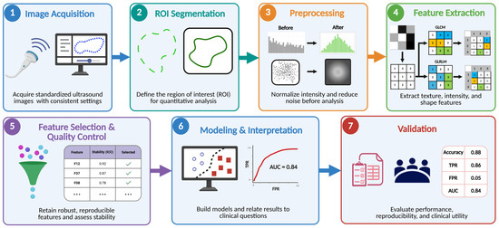
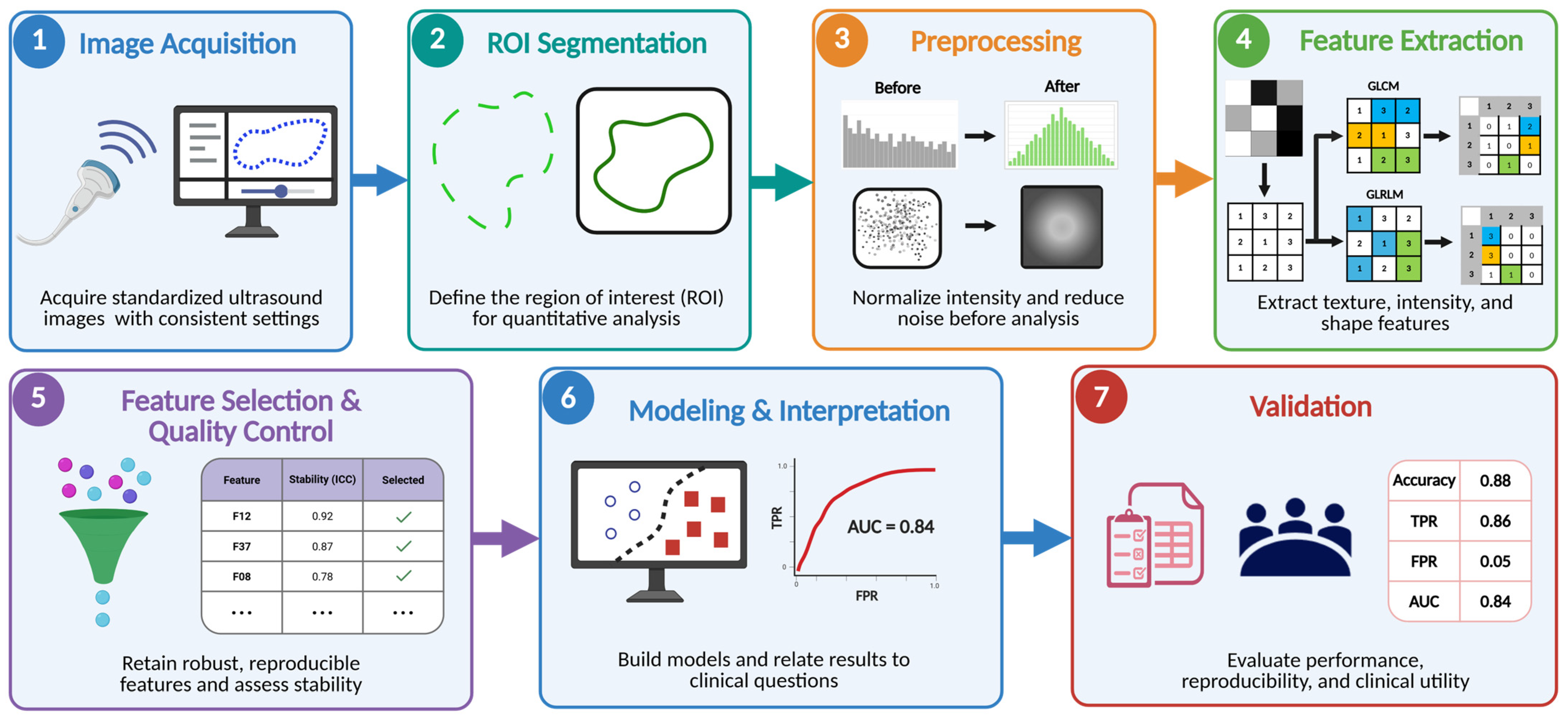
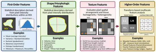
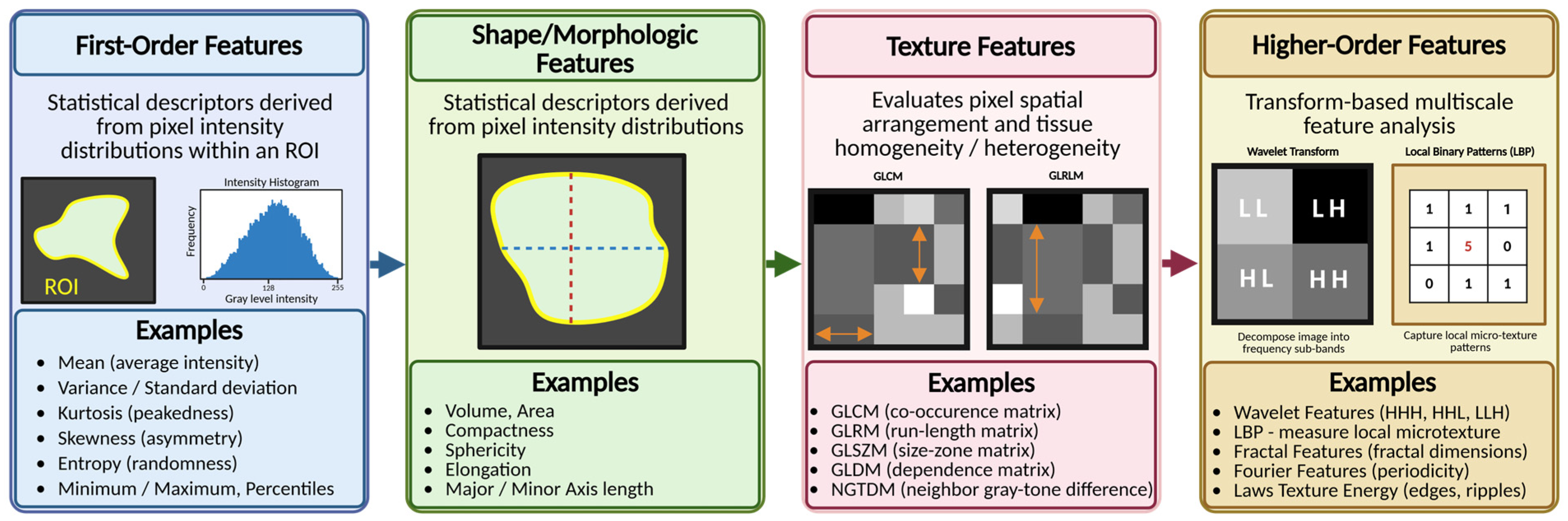
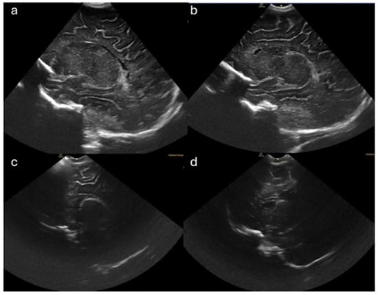
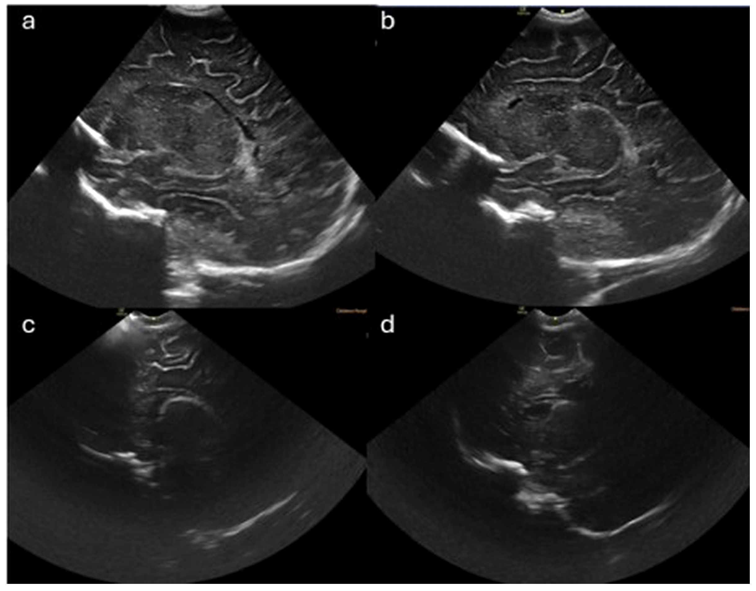
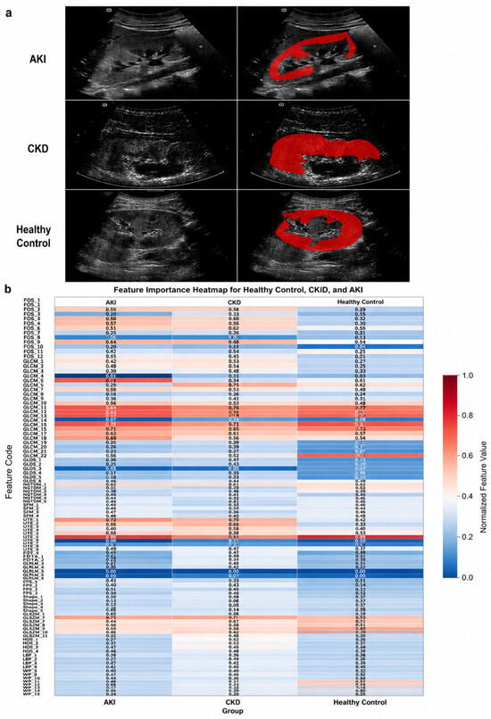
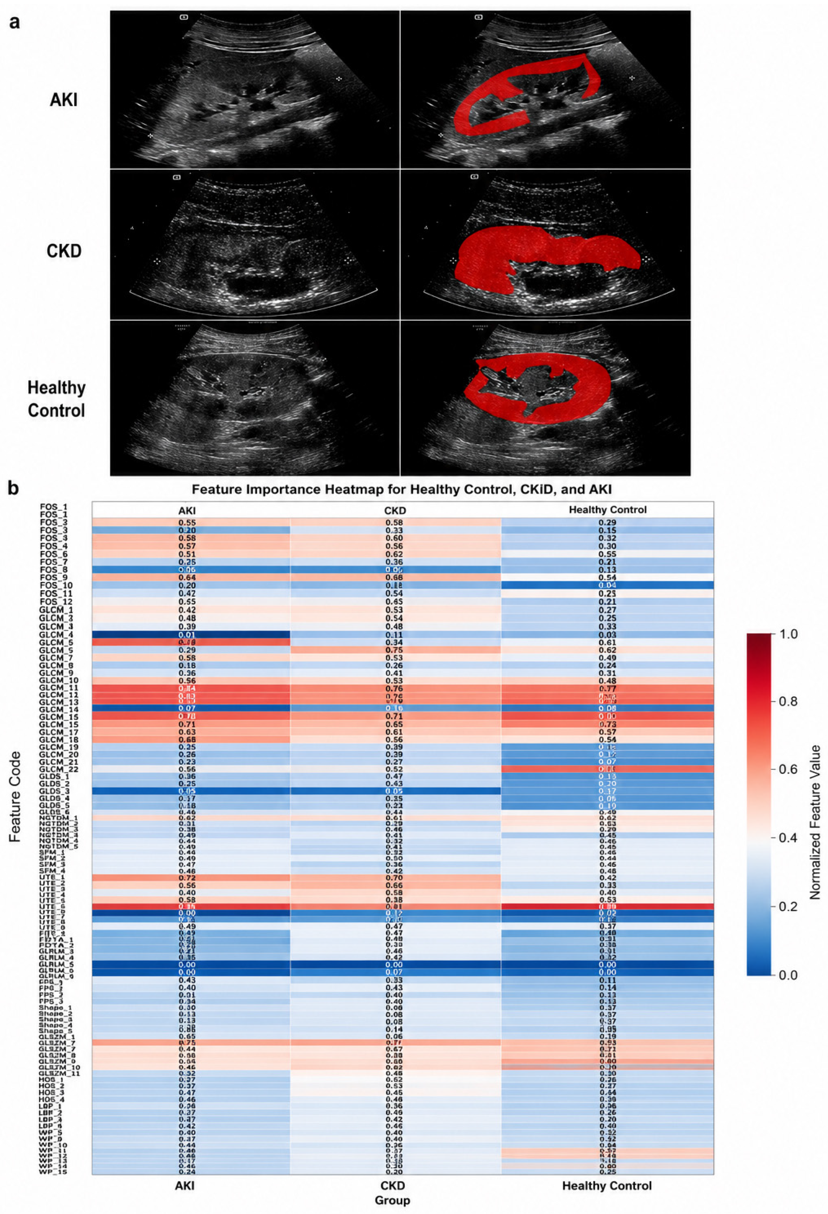
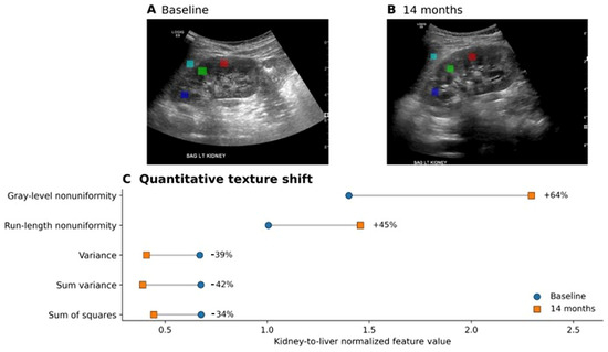
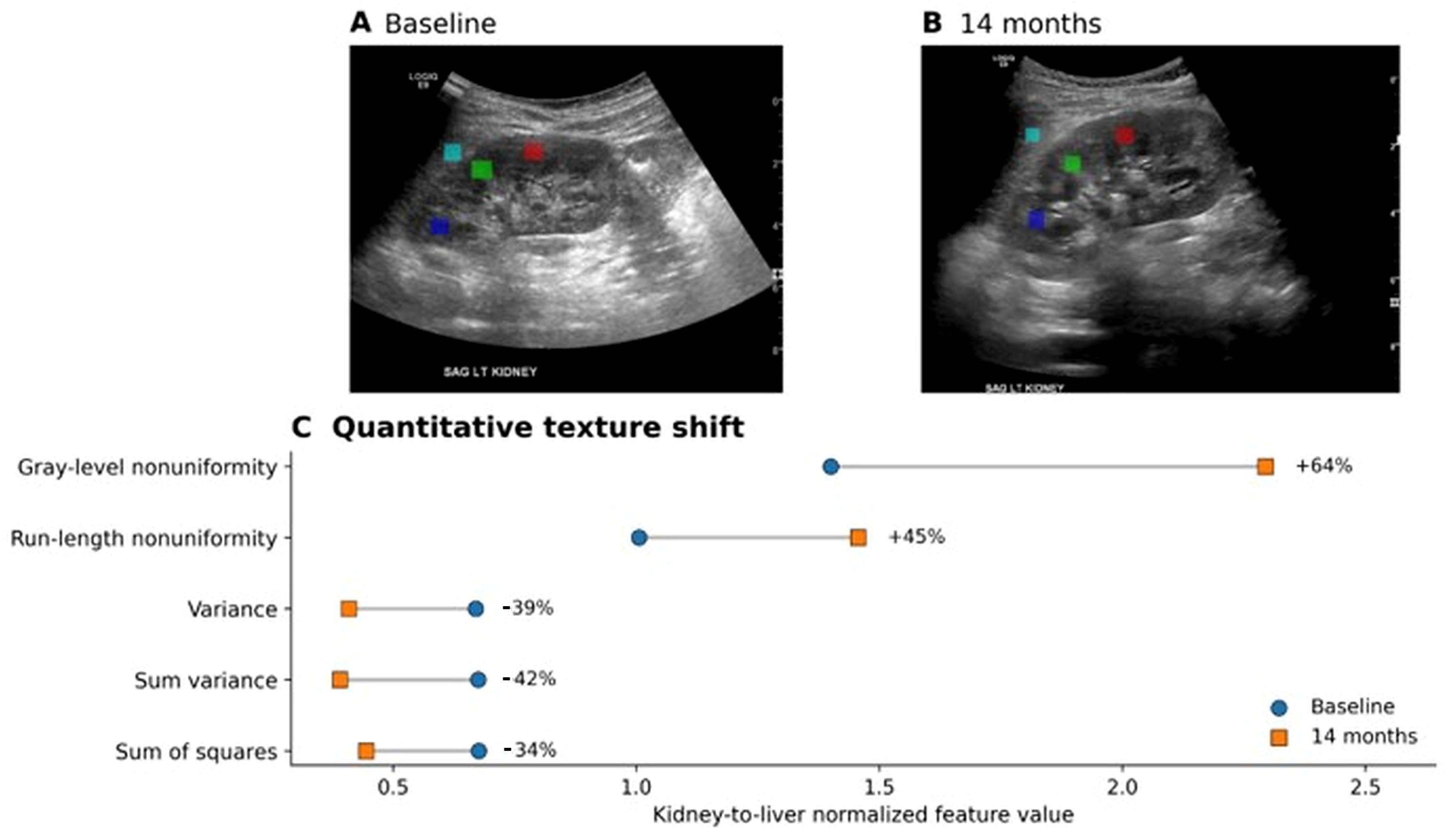
Disclaimer/Publisher’s Note: The statements, opinions and data contained in all publications are solely those of the individual author(s) and contributor(s) and not of MDPI and/or the editor(s). MDPI and/or the editor(s) disclaim responsibility for any injury to people or property resulting from any ideas, methods, instructions or products referred to in the content. |
© 2026 by the authors. Licensee MDPI, Basel, Switzerland. This article is an open access article distributed under the terms and conditions of the Creative Commons Attribution (CC BY) license.