


Abstract
Alcohol use disorder (AUD) is accompanied by persistent neuroadaptations, among which DNA methylation represents a mechanistically tractable, long-lasting epigenetic layer. Synthesizing evidence from human, non-human primate, and rodent studies alongside multi-omic datasets, this review establishes a cross-level framework of alcohol-induced 5-methylcytosine (5mC) and 5-hydroxymethylcytosine (5hmC) dysregulation across the three-stage addiction cycle. Chronic alcohol disrupts central one-carbon metabolism by lowering the SAM:SAH methylation index and impairing TET-dependent hydroxymethylation, producing a near-global hypomethylated backdrop with region-specific variations that coexists with locus-specific hypermethylation at plasticity genes. We map this dichotomy across five core circuit nodes (prefrontal cortex, nucleus accumbens, dorsal striatum, cerebellum, and amygdala), linking distinct 5mC and 5hmC signatures to executive failure, compulsive seeking, habit formation, loss of tonic inhibition, and negative reinforcement. Finally, to overcome key field challenges, including cellular heterogeneity, causal ambiguity versus exposure “scars”, and 5mC/5hmC array conflation, we evaluate single-nucleus multi-omics, CRISPR epigenome editing, methyl-donor interventions, and pharmacoepigenetics as translational axes toward precision medicine in AUD.
1. Introduction
Alcohol use disorder (AUD) is a chronic, relapsing brain disorder driven by complex interactions between genetic vulnerability and environmental exposures [1,2,3]. Epigenetic mechanisms, particularly DNA methylation, serve as a critical interface translating alcohol exposure into persistent neuroadaptations [4,5,6]. Canonically, methylation at the 5-carbon position of cytosine to form 5-methylcytosine (5mC) within promoter CpG islands represses gene transcription via steric hindrance and the recruitment of methyl-CpG-binding domain (MBD) proteins [6,7,8,9]. In mature post-mitotic neurons, however, 5mC is dynamically oxidized by Ten-Eleven Translocation (TET) enzymes into 5-hydroxymethylcytosine (5hmC), an active, transcriptionally permissive mark essential for activity-dependent synaptic plasticity (the functional mechanisms, 5mC/5hmC ratio dynamics, and regional detection caveats of which are detailed in Section 2 and Section 3) [10,11]. Beyond canonical CpG methylation, non-canonical cytosine modifications—such as non-CpG methylation (mCH), highly enriched in mature neurons—and adenine methylation (N6-methyladenine, 6mA) represent additional, emerging layers of neuroepigenetic regulation.
Alcohol exposure exerts a dual impact on the brain, driving genome-wide metabolic disruption alongside locus-specific target gene remodeling across the canonical three-stage addiction cycle (Binge/Intoxication, Withdrawal/Negative Affect, and Preoccupation/Anticipation) [1]. This review critically synthesizes molecular, metabolic, and multi-omic evidence of alcohol-induced methylomic dysregulation across five core neurocircuit nodes: the prefrontal cortex, nucleus accumbens, dorsal striatum, cerebellum, and amygdala. We integrate evidence from human postmortem and clinical cohorts, non-human primates, and rodent models, explicitly recognizing that these sources are not interchangeable and treating adult chronic AUD, acute or binge exposure, adolescent exposure, and developmental (prenatal) exposure as distinct conditions. Finally, we dissect key methodological bottlenecks—including cellular heterogeneity, causality versus exposure “scars”, and 5mC/5hmC array conflation—and outline translational frontiers in single-nucleus multi-omics, CRISPR epigenome editing, and pharmacoepigenetics for precision medicine in AUD.
Three features distinguish this review from prior syntheses of alcohol and DNA methylation [1,2,3,4,5,6]. First, rather than treating global hypomethylation and locus-specific hypermethylation as contradictory, this review integrates them into a compartmentalized model linking one-carbon biochemistry (SAM:SAH ratio) to enzymatic writing (DNMTs), active erasure (TETs), and downstream chromatin readers while defining the regional, cellular, and locus-dependent boundaries of this model. Second, it remaps methylation changes onto the three-stage addiction cycle across five functionally defined circuit nodes, cross-referencing human postmortem, rodent, and non-human primate evidence within an explicit hierarchy that separates observational human associations from interventional animal data. Third, it appraises key field bottlenecks—including bulk-tissue cellular heterogeneity, causal-versus-scar ambiguity, and 5mC/5hmC assay conflation—using single-nucleus expression data to illustrate cell-type limitations before evaluating nutritional/methyl-donor (e.g., choline) and epigenome-editing strategies that establish causal links in animal models.
2. Effects of Alcohol Exposure on DNA Methylation
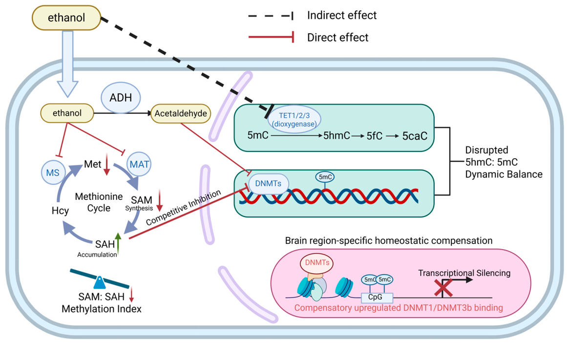
DNA methylation is a highly conserved, precisely regulated epigenetic modification catalyzed primarily by DNA methyltransferases (DNMTs), which utilize S-adenosylmethionine (SAM) as the universal methyl donor to covalently attach a methyl group to the 5-carbon position of cytosine, forming 5-methylcytosine (5mC) (Figure 1). Transcriptional consequences are strictly genomic-context-dependent: promoter CpG island hypermethylation typically enforces gene silencing by sterically blocking transcription factor binding or recruiting methyl-CpG-binding domain (MBD) proteins (e.g., MeCP2) and histone deacetylase (HDAC) co-repressors, whereas gene-body methylation frequently facilitates transcriptional elongation. After donating its methyl group, SAM is converted to S-adenosylhomocysteine (SAH), a potent competitive inhibitor of DNMTs; thus, the SAM:SAH ratio directly reflects cellular methylation capacity. Furthermore, active DNA demethylation is driven by Ten-Eleven Translocation dioxygenases (TET1, TET2, TET3), which oxidize 5mC to 5hmC (and sequentially to 5fC and 5caC). Crucially, TET isoforms exhibit functional divergence: TET1 acts as the primary enzyme driving locus-specific promoter hydroxymethylation in mature post-mitotic neurons, whereas TET2 and TET3 govern cell lineage commitment and rapid activity-dependent neural plasticity [12].
Figure 1.
Alcohol-associated disruption of one-carbon metabolism and DNA methylation dynamics. Ethanol is converted to acetaldehyde by alcohol dehydrogenase (ADH). In the methionine cycle, methionine synthase (MS) remethylates homocysteine (Hcy) to methionine (Met), and methionine adenosyltransferase (MAT) supports S-adenosylmethionine (SAM) synthesis. Ethanol and acetaldehyde directly suppress MS and MAT, reducing SAM synthesis, triggering S-adenosylhomocysteine (SAH) accumulation, and lowering the SAM:SAH ratio; elevated SAH competitively inhibits DNA methyltransferases (DNMTs). Acetaldehyde additionally acts as a direct inhibitor of DNMTs (solid maroon lines), whereas ethanol exerts an indirect inhibitory effect on Ten-Eleven Translocation (TET1/2/3) dioxygenases (dashed black line). DNMTs generate and maintain 5-methylcytosine (5mC) at CpG sites, while TET enzymes sequentially oxidize 5mC to 5-hydroxymethylcytosine (5hmC), 5-formylcytosine (5fC), and 5-carboxylcytosine (5caC), thereby shaping the dynamic 5hmC:5mC balance. The lower panel depicts proposed brain-region-specific homeostatic compensation involving increased DNMT1/DNMT3B binding, local CpG hypermethylation, and transcriptional silencing; this is not a universal response across regions or exposure conditions. For clarity, the oxidative pathway is truncated at 5caC; subsequent thymine DNA glycosylase (TDG)-dependent excision, base excision repair (BER), and replication-dependent passive dilution (negligible in postmitotic neurons) are distinguished in the main text. Arrowheads indicate transport or biochemical conversion, whereas blunt-ended lines indicate inhibition. Red downward arrows denote decreases, and the green upward arrow denotes SAH accumulation. The red cross indicates transcriptional silencing. Generated using BioRender (https://www.biorender.com/).
Rather than acting primarily through dietary vitamin depletion, chronic alcohol exposure directly compromises central one-carbon metabolic enzymatic machinery [7,13,14]. Ethanol and its primary metabolite, acetaldehyde, directly suppress the catalytic activity of key methyl-source enzymes—methionine synthase (MS) and methionine adenosyl-transferase (MAT)—blocking the remethylation of homocysteine and severely depressing de novo SAM synthesis [7,14,15]. Concurrently, toxic intracellular SAH accumulation depresses the SAM:SAH ratio, imposing direct competitive inhibition on the writers of DNA methylation (DNMT1, DNMT3A, DNMT3B) [7,16,17]. This catalytic suppression cascades downstream to the readers of the methylome: loss of precise 5mC/5hmC marks alters the chromatin recruitment kinetics of me-thyl-CpG-binding domain proteins (e.g., MeCP2, MBD2), leaving synaptic plasticity genes in aberrantly silenced or unstable transcriptional states [7,18]. Acetaldehyde can additionally bind and directly inactivate the maintenance methyltransferase DNMT1, whereas alcohol-induced oxidative stress and DNA damage repair further reduce 5mC abundance [7].
Within the nucleus, the dynamic writing and erasing of epigenetic marks exhibit a high degree of homeostatic compensation [9,17]. Reduced one-carbon metabolism is frequently accompanied by region-specific compensatory upregulation of epigenetic enzymes [19,20]. In rat models of chronic alcohol exposure, despite overall metabolic suppression, DNMT1 or DNMT3b expression in medial prefrontal cortex (mPFC) and amygdala neurons is compensatorily elevated, resulting in aberrant hypermethylation and transcriptional silencing at specific synaptic plasticity promoters [19,20].
Complementing writer dysregulation, active DNA demethylation is governed by Ten-Eleven Translocation dioxygenases (TET1, TET2, TET3), which sequentially oxidize 5mC to 5hmC, 5-formylcytosine (5fC), and 5-carboxylcytosine [9,17]. Active demethylation is completed via thymine DNA glycosylase (TDG)-mediated excision of 5fC/5caC coupled with base excision repair (BER). Because mature neurons are post-mitotic, passive replication-dependent dilution is negligible, leaving active oxidative demethylation as the primary route [21,22]. Crucially, 5hmC plays a dual biological role in neurons: beyond serving as a transient demethylation intermediate, high-density 5hmC functions as an independent, stable mark that blocks MeCP2 binding, maintains euchromatin, and recruits reader proteins to facilitate transcriptional elongation [21]. Alcohol exposure not only suppresses TET enzymatic activity but also severely disrupts the local 5hmC:5mC ratio across critical synaptic loci [23,24].
Importantly, these epigenetic responses exhibit notable cross-species and inter-strain divergence. While inbred rodent strains (e.g., C57BL/6J vs. DBA/2J) display distinct baseline methylomic architectures conferring differential vulnerability to alcohol-induced DNMT/TET dysregulation, non-human primates and human postmortem cohorts reveal broader, conserved cortico-limbic networks, highlighting the pivotal role of genetic background in modulating methylomic susceptibility.
Several key qualifications bound the global hypomethylation model. First, meta-bolic perturbation is not uniform across brain regions. In ethanol-treated rats, cerebellar SAH falls and the SAM:SAH ratio rises alongside Mat2a induction—a direction opposite to the hepatic response and consistent with reported regional hypermethylation in the cerebellum [25,26]. This indicates that regional one-carbon reserves, metabolic flux, and DNMT/TET ex-pression can buffer or invert systemic methylation shifts; thus, findings from liver or blood cannot be directly extrapolated to the whole brain. Second, “global” hypomethylation denotes a net bias at the bulk-tissue level rather than a uniform loss at every CpG. Within the same genome, loss of methylation at transcriptionally permissive loci coexists with focal hypermethylation at plasticity and stress-response genes. For example, post-mortem PFC profiles contain both hypo- and hypermethylated loci within individual brains (with approximately two-thirds of DMPs being hypermethylated) [27], the replicated blood marker SLC7A11 is hypomethylated [28], and KCNN3 is hypermethylated in the NAc core [29]. This bidirectionality reflects at least three non-mutually exclusive mechanisms: cell-type heterogeneity, local chromatin accessibility (preferential DNMT3B and TET binding at open promoters or poised enhancers), and transcription factor/lncRNA-guided locus recruitment.
Crucially, throughout this review, “upregulation” or “downregulation” of DNMTs and TETs denotes altered transcript or protein abundance unless catalytic activity is explicitly measured. Enzyme expression is not synonymous with enzymatic activity, and direct catalytic inhibition by alcohol is currently established for MS, MAT, and DNMT1 [7,25]. Finally, extending beyond neuronal circuits, ethanol persistently lowers 5mC and dysregulates DNMT1, DNMT3A, and DNMT3B in human brain microcapillary endothelial cells, demonstrating that alcohol-induced methylomic disruption impairs the cerebrovascular and blood–brain barrier compartment [30].
Although this review focuses on DNA methylation and hydroxymethylation, perturbations in methyl-donor availability also feed parallel methylation reactions sharing the one-carbon pool, including histone and RNA methylation. A functionally validated example is the histone methyltransferase PR-domain zinc-finger protein 2 (PRDM2), which selectively mono-methylates histone H3 at lysine 9 (H3K9me1). Chronic alcohol exposure persistently downregulates Prdm2 expression in the dorsomedial prefrontal cortex (dmPFC), and experimental manipulation of Prdm2 escalates alcohol self-administration and compulsive drinking, demonstrating that histone methyltransferases can directly drive alcohol-dependent behaviors [31]. This highlights that one-carbon perturbations should not be assumed to act solely through DNMTs; thus, DNA methylation signatures must be interpreted alongside, rather than conflated with parallel histone and RNA methylation mechanisms. Table 1 summarizes the key molecular machinery, biochemical targets, and alcohol-associated alterations discussed in this section.
Table 1.
Foundational Machinery, Biochemical Targets, and Alcohol-Induced Dysregulation of Neuroepigenetic Regulation.
3. DNA Methylation Changes and Their Effects in Major Brain Regions in the Context of Alcohol Use Disorder
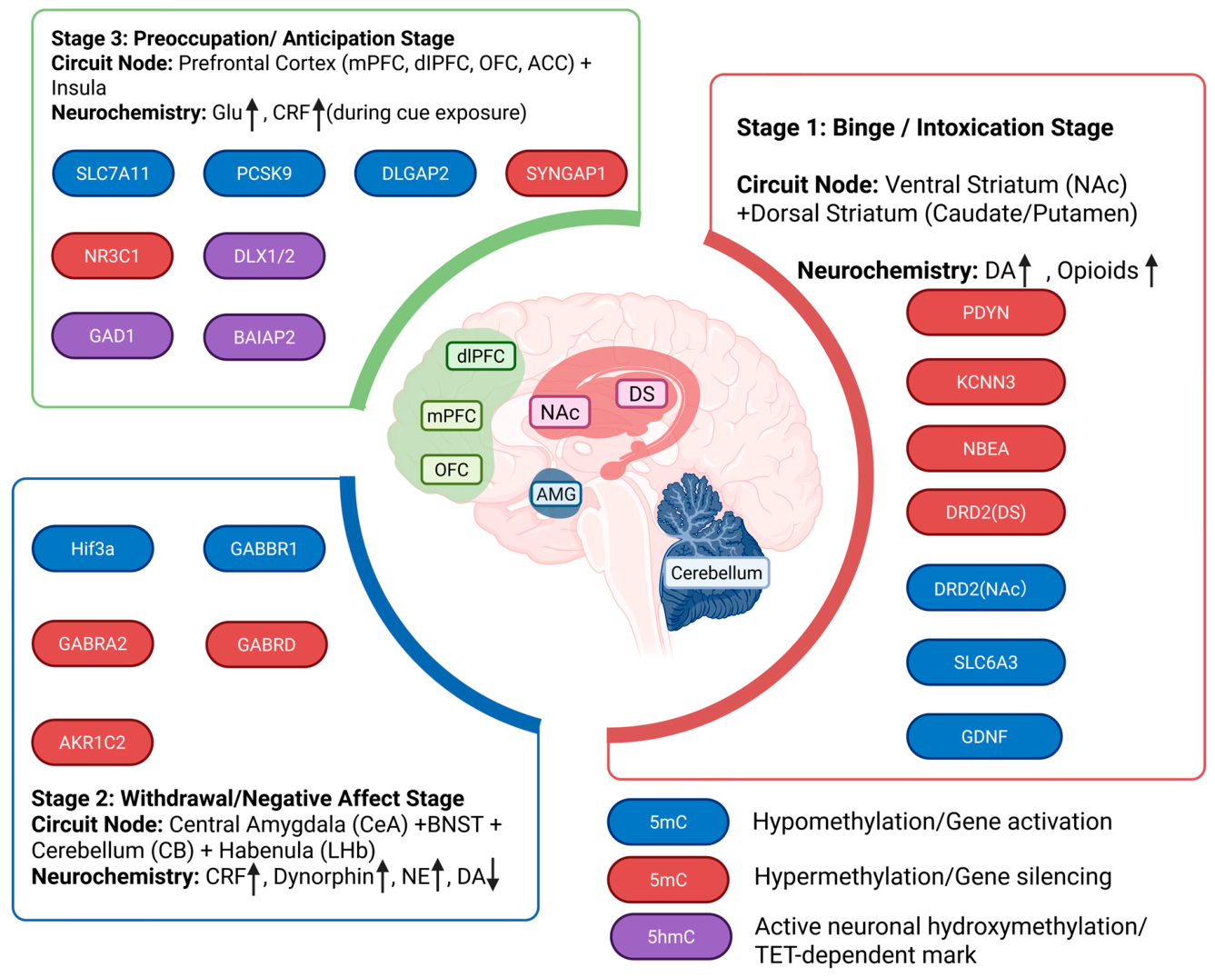
The neurobiology of alcohol addiction evolves through a three-stage cycle: Binge/Intoxication (incentive salience), Withdrawal/Negative Affect (anti-reward stress activation), and Preoccupation/Anticipation (executive dysfunction) (Figure 2) [32]. Rather than causing diffuse transcriptional noise, chronic alcohol exposure deposits locus-specific 5mC and 5hmC alterations across key circuit nodes to alter stage-specific molecular pathways. Below, we evaluate these methylomic landscapes across five core regions—the prefrontal cortex, nucleus accumbens, dorsal striatum, cerebellum, and amygdala—with full details on species, exposure paradigms, tissues, assays, and functional validation compiled in Table 2.
Figure 2.
Three-stage topographic epigenetic map of alcohol use disorder across neuroaddictive circuits. Schematic integration of locus-specific 5mC and 5hmC signatures within the canonical three-stage addiction cycle. Stage 1 (Binge/Intoxication) maps basal ganglia structures (NAc and DS) and associated reward/habit loci (PDYN, KCNN3, NBEA, DRD2, SLC6A3, GDNF). Stage 2 (Withdrawal/Negative Affect) details extended amygdala, habenular, and cerebellar adaptations under central stress states (Hif3a, GABBR1, GABRA2, GABRD, AKR1C2). Stage 3 (Preoccupation/Anticipation) highlights prefrontal-cortical executive control networks (SLC7A11, PCSK9, DLGAP2, SYNGAP1, NR3C1, DLX1/DLX2, GAD1, BAIAP2). Upward and downward arrows indicate schematic increases and decreases, respectively, in the neurochemical signals listed for each addiction stage. Generated using BioRender (https://www.biorender.com/).
Table 2.
Cross-species evidence map of locus-specific DNA methylation and hydroxymethylation findings across the three-stage addiction cycle, with the level of functional validation.
3.1. Prefrontal Cortex (PFC)
The prefrontal cortex (PFC) serves as the primary hub for executive control, impulse inhibition, and decision-making, making it a central target of alcohol-induced epigenetic remodeling [10,20,55]. Chronic alcohol exposure reshapes 5mC and 5hmC landscapes at specific loci, yielding neuroadaptations associated with impaired executive function and cycle perpetuation [27]. Bulk-tissue epigenome-wide association studies (EWAS) in postmortem PFC (Brodmann area 9, BA9) revealed that 66.3% of 1812 differentially methylated positions (DMPs) were hypermethylated, reflecting a predominant shift toward transcriptional silencing [27]. Neuron-specific (NeuN-sorted) methylome and hydroxymethylome profiling in the orbitofrontal cortex (OFC) further resolved cell-type heterogeneity, identifying 417 differentially methylated and 363 differentially hydroxymethylated loci, with nearly 60% enriched in promoter regions linked to PFC-dependent cognitive deficits [33].
Epigenetic remodeling within the PFC disrupts local neurotransmitter homeostasis. In OFC neurons, aberrant 5hmC enrichment at GAD1 (encoding glutamic acid decarboxylase) and DLX1/DLX2 (interneuron developmental regulators) may impair GABAergic inhibition, providing a candidate molecular correlate of impulse control dysfunction [33]. Concurrently, the cystine/glutamate antiporter gene SLC7A11 carries the strongest replicated whole-blood hypomethylation signal in AUD (cg06690548) [28]. Because its functional subunit xCT mediates glial glutamate–cystine exchange, this peripheral epigenetic mark is hypothesized—though not yet validated in central PFC tissue—to reflect an upregulated, glutamate-releasing state that could contribute to excitotoxicity and compulsive drinking.
Alcohol-induced methylomic shifts also target stress-regulatory machinery across PFC subregions (BA9, BA10) [23]. Chronic heavy drinking elevates 5mC and drives promoter 5hmC accumulation at NR3C1 (exon variant 1H) in BA10, secondary to diminished TET1-mediated 5hmC clearance [23]. This reduced TET1 abundance correlates with suppressed NR3C1 expression across cortico-limbic circuits, disrupting HPA axis negative feedback, upregulating stress mediators (CRF, FKBP5, POMC), and heightening relapse vulnerability [23]. Additionally, altered methylation at the glucocorticoid-regulatory long non-coding RNA GAS5 perturbs glucocorticoid receptor signaling [35], while hypomethylation of PCSK9 (cg01444643) links central prefrontal epigenetic changes to altered systemic lipid metabolism and cardiovascular risk [34].
Finally, locus-specific remodeling at postsynaptic scaffold genes destabilizes PFC synaptic architecture. Hypomethylation in the dorsolateral PFC (dlPFC) creates a functional enhancer (DMR-DLGAP2) that upregulates DLGAP2, altering postsynaptic density architecture in a manner correlated with adolescent binge drinking and addiction susceptibility [36]. Similarly, 5hmC alterations at BAIAP2 are associated with transcript variation [37]. Conversely, elevated methylation at SYNGAP1 correlates with impaired craving control in peripheral blood cohorts [38]; because this signal originates from peripheral blood rather than neural tissue, its proposed glutamatergic synaptic mechanism remains to be functionally validated in the central PFC. Together, these prefrontal methylomic and hydroxymethylomic shifts converge on a shared executive-control phenotype—impaired top-down regulation of subcortical drive—linking excitatory/inhibitory imbalance, glucocorticoid dysregulation, and postsynaptic scaffold instability to a common behavioral output.
3.2. Nucleus Accumbens (NAc)
In the pathological progression of AUD, the nucleus accumbens (NAc)—particularly its core subregion (NAcC)—undergoes pronounced locus-specific epigenetic remodeling. This remodeling regulates neuronal electrophysiological properties and synaptic scaffold function, driving the consolidation of alcohol-seeking behavior [29,40].
Ion channel dysfunction and electrophysiological adaptation represent core findings in NAc epigenetic research. Cross-species studies consistently demonstrate that KCNN3 (encoding the small-conductance calcium-activated potassium channel KCa2.3) undergoes critical epigenetic reprogramming in the NAcC. Excessive drinking induces hypermethylation of a DMR in KCNN3 exon 1A, promoting selective usage of an alternative promoter and increasing transcripts encoding a dominant-negative isoform that reduces functional KCa2.3 channel protein. Because KCa2.3 channels mediate the afterhyperpolarization that limits repetitive firing, reduced functional KCa2.3 would disinhibit NAcC medium spiny neurons and alter their excitability and firing patterns, thereby reshaping the reward circuitry that encodes alcohol value and providing a plausible mechanistic link to the tolerance and relapse that characterize the addiction cycle. This chain remains a proposed contributor whose direct effect on drinking behavior has not been fully tested [29].
Alterations in synaptic protein trafficking and reward sensitivity are likewise governed by NAc locus-specific methylation. NBEA (encoding Neurobeachin, a kinase-anchoring protein regulating membrane trafficking of glutamate and GABA receptors) exhibits robust hypermethylation in the NAcC of heavy drinkers and AUD patients, positively correlating with alcohol preference; knockdown of NBEA elevates alcohol preference by disrupting glutamatergic transmission [40]. Within the dopaminergic system, the DRD2 promoter (encoding the dopamine D2 receptor) is significantly hypomethylated in the NAcC of AUD patients in post-mortem tissue [41]; a positive association with alcohol-cue-induced ventral-striatal activation has been demonstrated only for peripheral blood methylation in living individuals [48]. Conversely, promoter hypomethylation of SLC6A3 (encoding the dopamine transporter) predicts NAc activation during loss aversion in healthy controls, but this predictive relationship is abolished in AUD, signaling an uncoupling between reward circuits and epigenetic marks [42]. Concurrently, promoter hypermethylation of the μ-opioid receptor gene (OPRM1, at CpG-71 and CpG-50) has been reported in peripheral blood from Han Chinese men with AUD [43]; whether this represses receptor transcription and blunts opioidergic signaling within the VTA–NAc axis has not been established in brain tissue.
Neurotrophic support and neuroinflammatory signaling also undergo dynamic, stage-dependent methylomic shifts. In animal models, GDNF (encoding glial cell line–derived neurotrophic factor) promoter methylation exhibits stage-dependent regulation: while core promoter hypomethylation and GDNF upregulation in the NAc serve as a compensatory protective barrier during early binge drinking, during chronic withdrawal this trophic support declines: in the NAc, GDNF upregulation is associated with core-promoter hypomethylation, whereas in the VTA, promoter negative regulatory element (NRE) demethylation is accompanied by reduced GDNF mRNA—changes proposed to contribute to compulsive relapse [44]. Weighted gene co-expression network analysis (WGCNA) of the ventral striatum further identified AUD-associated methylation modules enriched in immune pathways, with STAT3 acting as a key hub gene coordinating NAc neuroinflammation [56,57]. Because addiction is increasingly understood as a neuroinflammatory state, alcohol-induced methylation changes in microglia and astrocytes may reciprocally sustain immune activation; how this immune-epigenetic crosstalk feeds back onto neuronal excitability remains largely untested [56,57].
Finally, non-human primate models of voluntary self-administration reveal dose-dependent subcortical adaptations, where chronic alcohol intake induces distinct DMRs in the NAcC across key synaptic genes (LRP5, GPR39, and JAKMIP1), driving alternative transcript selection and biasing circuit function toward heightened excitation [45]. These NAc adaptations occur within the VTA-NAc mesolimbic dopaminergic pathway, where chronic alcohol remodels dopamine-driven reward signaling; as drinking progresses, the locus of behavioral control shifts along the ventral-to-dorsal striatal gradient—from reward-motivated, goal-directed actions in the NAc to habitual and ultimately compulsive behavior in the dorsal striatum, where related epigenetic mechanisms become entrenched within stimulus-response circuits.
3.3. Dorsal Striatum (DS)
The dorsal striatum (comprising the caudate nucleus [CN] and putamen [PUT]) serves as a primary circuit node mediating the transition of drinking behavior from goal-directed reward seeking to compulsive habit formation [58]. Epigenetic silencing within dorsal striatal opioid and dopamine systems has been proposed to contribute to this behavioral shift. In postmortem caudate and putamen from individuals with AUD, PDYN (encoding prodynorphin) mRNA and dynorphin peptide levels are markedly reduced, implicating diminished dorsal-striatal opioid output in compulsive habit formation [46]; whether promoter methylation underlies this reduction in brain is unknown, because a PDYN methylation association has to date been reported only in peripheral blood cells [47]. Furthermore, peripheral DRD2 promoter methylation significantly predicts the magnitude of alcohol-cue-induced fMRI activation in the putamen and accumbens, linking a blood-based mark to habit-circuit and craving responses [48].
3.4. Cerebellum (CB)
Beyond motor coordination, the cerebellum processes cognitive functions, sensory discrimination, and reward expectations, acting as a prominent target of alcohol-induced hypermethylation [25,26,49]. Alcohol exposure primarily disrupts cerebellar granule cell homeostasis by epigenetically suppressing inhibitory GABAergic transmission [26,49]. Multi-site promoter hypermethylation of GABRA2 (encoding the γ-aminobutyric acid type A receptor α2 subunit) in the postmortem cerebellum suppresses transcription, with protein loss negatively correlating with lifetime drinking history [49], by contrast, peripheral blood profiles lack this signature, highlighting tissue-specific epigenetic regulation [59].
For GABRD, promoter hypermethylation does not stem from DNMT upregulation, but rather from reduced TET1 abundance (implying impaired TET1-mediated demethylation) combined with one-carbon metabolic dysregulation, leading to local methyl accumulation [26]. Concurrently, the promoter of AKR1C2 (encoding 3α-HSD, the rate-limiting enzyme for neurosteroid synthesis) is hypermethylated, blocking transcription and lowering allopregnanolone levels [49]. Together, the downregulations of GABRD and AKR1C2 synergistically impair extrasynaptic GABAA receptor-mediated tonic inhibition [26,49]. Notably, the cerebellum also exhibits divergent one-carbon metabolic responses: in ethanol-treated rats, cerebellar SAH falls and the SAM:SAH ratio rises—the inverse of the hepatic response and consistent with regional hypermethylation [25,26]—reinforcing that the global hypomethylation model must be applied on a region-by-region basis.
3.5. Amygdala (AMG)
As the limbic hub for emotion, stress processing, and negative reinforcement, the amygdala undergoes dynamic epigenetic remodeling across addiction stages [50,51]. Multi-omic profiling (ATAC-seq and RNA-seq) demonstrates that acute alcohol exposure induces a global “open” chromatin state, associated with Hif3a (encoding hypoxia-inducible factor 3α) upregulation via promoter hypomethylation and increased H3K27ac [50]. Elevated Hif3a activates downstream Npy1r (neuropeptide Y receptor Y1) signaling implicated in acute anxiolysis. Upon chronic exposure and withdrawal, Hif3a expression collapses, coinciding with withdrawal-induced anxiety and negative affect [50].
Locus-specific rewiring of inhibitory transmission and chromatin architecture further consolidates amygdalar dysregulation. In male AUD patients, the GABBR1 promoter (encoding the GABAB1 receptor subunit) displays site-specific hypomethylation at CpG-4, which is proposed to elevate receptor expression and promote tolerance via presynaptic and postsynaptic over-inhibition—a mechanism exhibiting distinct sexual dimorphism absent in females [51]. Furthermore, adolescent binge drinking induces persistent enhancer repression in the central nucleus of the amygdala (CeA) by depleting H3K27ac and eRNAs at the Arc SARE enhancer. This loss recruits negative elongation factor (NELF) to the promoter, disrupts 3D chromatin looping, and silences Arc expression. Site-specific dCas9-based epigenome editing at this enhancer in the adult amygdala has causally implicated this locus in anxiety and excessive drinking [52], providing one of the few direct causal demonstrations linking a defined chromatin mark to alcohol-related behavior.
4. Methodological Bottlenecks and Knowledge Gaps in AUD Epigenomics
4.1. Cellular Heterogeneity and the Tissue Specificity Dilemma
One of the most persistent bottlenecks in current neuroepigenetic research is cellular heterogeneity [10,33]. The vast majority of human postmortem brain studies in alcohol use disorder (AUD) rely on bulk tissue homogenates, which inevitably average across diverse cell types and risk masking cell-type-specific epigenetic signatures unique to neuronal or glial subpopulations [10,33,57]. Crucially, while single-nucleus multi-omics can resolve cell-type heterogeneity, it completely disrupts tissue spatial architecture. The current lack of high-resolution spatial methylomics represents a major technological barrier; without spatial epigenomic mapping in intact tissue, present approaches fail to capture microenvironment-dependent glia–neuron crosstalk and laminar subregion-specific epigenetic remodeling in the alcoholic brain.
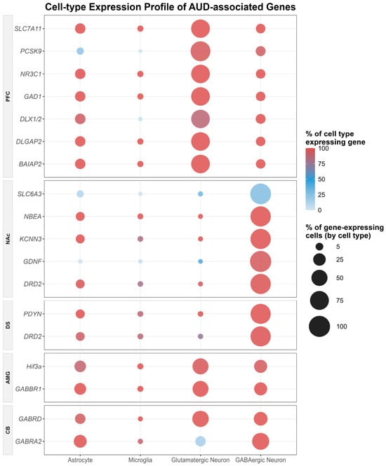
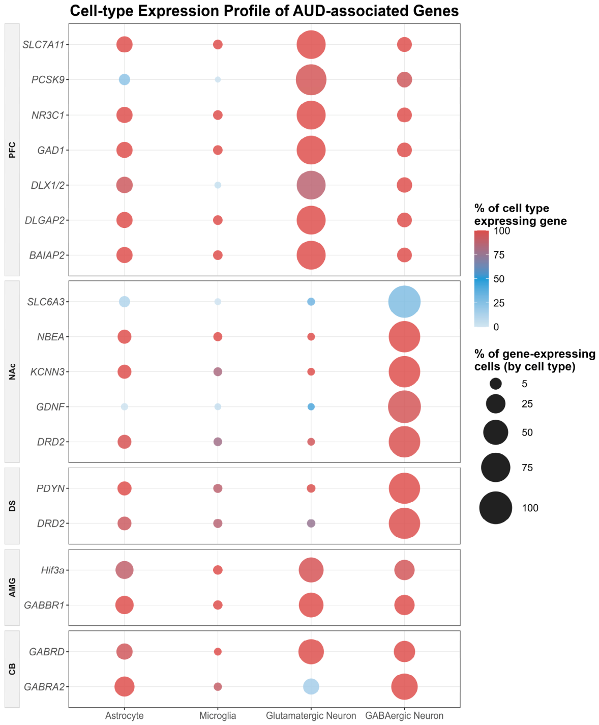
To illustrate cell-type distribution patterns of key AUD-associated genes across neurocircuits, we queried the Mouse Whole-Brain Transcriptomic Cell Type Atlas using the Allen Brain Cell Atlas (ABC Atlas) web interface. Mapping expression enrichment across the core brain regions of the three-stage addiction cycle (PFC, NAc, DS, AMG, and CB) reveals distinct cell-class specificity across glutamatergic neurons, GABAergic interneurons, astrocytes, and microglia (Figure 3) [60].
Figure 3.
Cell-type expression profiles of AUD-associated genes. Dot plot based on gene-expression data queried from the Mouse Whole-Brain Transcriptomic Cell Type Atlas through the Allen Brain Cell Atlas (ABC Atlas) web interface, showing the expression of selected AUD-associated genes across four major cell classes (astrocytes, microglia, glutamatergic neurons, and GABAergic neurons) in five brain regions: prefrontal cortex (PFC), nucleus accumbens (NAc), dorsal striatum (DS), amygdala (AMG), and cerebellum (CB). Color intensity encodes the fraction of cells within a cell class that express the gene (a continuous proportion independent of statistical significance). Dot size represents that cell class’s share of all gene-expressing cells within the region, where larger dots indicate that expression is predominantly concentrated within that specific cell population. Generated using R software (version 4.5.3; R Foundation for Statistical Computing, Vienna, Austria; https://www.r-project.org/), based on data from the open-access Allen Brain Cell Atlas.
To interrogate whether the bulk methylation changes reviewed above can be assigned to particular cell types, we queried the Mouse Whole-Brain Transcriptomic Cell Type Atlas through the Allen Brain Cell Atlas (ABC Atlas) web interface for core AUD-associated genes (DRD2, NR3C1, SLC7A11, GABRA2, GABRD, and DLGAP2) across the five addiction-stage regions (Figure 3). Two complementary statistics are shown: the percentage of cells within each cell class expressing a given gene, and the percentage of all gene-expressing cells in a region contributed by each cell class. Rather than being restricted to a single neuronal subtype, these core genes are co-expressed across multiple neuronal and glial classes within each region. This distribution has two important implications. First, bulk-tissue methylation signals represent population averages over these co-expressing classes, meaning a region-level methylation change cannot be assigned to a single causally operative cell class. Second, knowing the fraction of expressing cells—or their relative contribution to the expressing pool—does not reveal which cell type’s methylation event drives the phenotype. Expression ubiquity alone therefore cannot resolve cell-type specificity. Future causal studies must employ cell-type-resolved manipulations—such as Cre-driven or cell-class-enriched dCas9 epigenome editing, cell-type-specific methylome profiling, and chemo- or optogenetic readouts—to determine the specific cell population through which a methylation event alters circuit function.
Furthermore, although peripheral tissues (whole blood, saliva) are widely utilized as surrogates, their concordance with central nervous system (CNS) methylation is highly gene- and site-specific [61]. While certain genes like DLGAP2 exhibit moderate cross-tissue correlations, peripheral measurements for critical neurocircuitry receptors—such as DRD2—fail to reliably reflect or predict corresponding epigenetic landscapes in key subcortical nodes like the NAc or hippocampus [41,61]; indeed, peripheral DRD2 methylation changes dynamically over withdrawal and correlates with craving rather than providing a stable readout of brain methylation [62]. Nevertheless, specific loci demonstrate robust cross-tissue concordance; for instance, sorting neuronal and non-neuronal PFC nuclei revealed that PCSK9 promoter hypomethylation in AUD is conserved across central cell types and faithfully mirrored in peripheral blood and liver tissues [34].
Moreover, in peripheral blood EWAS, apparent disease-associated methylation shifts are frequently confounded by subtle alterations in cell-type composition (e.g., naive-to-memory T-cell differentiation, myeloid-to-lymphoid ratios). Standard six-cell in silico deconvolution methods fail to resolve fine-grained immune sub-populations, necessitating high-resolution immunophenotyping (e.g., 16-cell models or IDOL-ext) or single-cell approaches to distinguish direct intracellular epigenetic reprogramming from secondary immune proportion shifts [63]; purified CD3+ T-cell methylomes from patients undergoing alcohol treatment already carry chronic-dependence signatures [64]. Advancing this resolution, a 2026 multi-cell-type MWAS (n = 13,970) applied 12-cell-type deconvolution, revealing that while whole-blood hypomethylation of the cystine/glutamate transporter SLC7A11 dominates bulk signals, cell-type specific profiling uncovers opposing hypermethylation in CD8+ naive T-cells and implicates endoplasmic reticulum stress chaperone PDIA5 alongside Rho GTPase structural plasticity networks in alcohol-induced cellular pathology [65].
4.2. The Causality Dilemma: Epigenetic Predisposition Versus Exposure Scars
A fundamental challenge in human AUD epigenetics is resolving the “cause-versus-effect” question [10,35,37]. Because clinical biosamples are predominantly obtained post-acquisition or postmortem, researchers capture a static “epigenetic snapshot” that conflates two distinct biological phenomena: pre-existing susceptibility loci that genetically or developmentally predispose an individual to addiction, and acquired epigenetic “scars” resulting from chronic alcohol exposure and repeated withdrawal stress [10,23,27,41,43,66]. Disentangling these mechanisms is clinically critical, as predisposing risk markers and treatment-responsive, exposure-induced modifications require fundamentally different therapeutic interventions [10].
Providing human longitudinal evidence for pre-existing vulnerability, prospective tracking of over 1000 substance-naive adolescents demonstrated that baseline hypermethylation at the NR3C1 exon 1-F promoter (specifically at the NGFI-A binding site) in saliva significantly predicts future alcohol initiation and prolonged use duration, establishing HPA axis-related epigenetic marks as pre-existing risk signatures rather than mere exposure scars [67]. Conversely, longitudinal human cohorts (ages 14–19) demonstrate that adolescent binge drinking directly drives dynamic blood DNA methylation trajectories that mediate accelerated cortical thinning across the fronto-limbic-striatal axis [68]. Highlighting the dynamic reversibility of exposure-associated methylomic marks, longitudinal blood sampling in a large cohort showed that methylation at a substantial fraction of alcohol-associated CpGs tracks changes in alcohol intake over more than a decade, with former drinkers displaying profiles distinct from current drinkers, consistent with partial reversal after cessation [69].
Recent breakthroughs in CRISPR/dCas9 epigenomic editing in rodent models have begun to directly resolve this causality gap. By site-specifically depositing active (dCas9-p300) or repressive (dCas9-KRAB) chromatin marks at target enhancers in discrete brain regions, researchers can bidirectionally rescue or phenocopy AUD behaviors, providing locus-specific evidence that localized epigenetic remodeling can bidirectionally influence addiction-related phenotypes in animal models [52]. Furthermore, genotype-dependent epigenetic regulation (e.g., genetically controlled methylation clusters, or GeMes) at loci like DLGAP2 illustrates how inherited SNP variants constrain local methylomic flexibility, dictating baseline susceptibility to alcohol-induced reward processing deficits [36].
4.3. Methodological and Platform Constraints in Epigenomic Profiling
Traditional epigenome-wide association studies (EWAS) are technologically constrained by their inability to distinguish between different cytosine modifications [33,56]. Standard bisulfite conversion and array platforms (such as Illumina 450 K or EPIC arrays) chemically conflate 5-methylcytosine (5mC) with 5-hydroxymethylcytosine (5hmC) [33,56]. Given that 5hmC is highly enriched in mature post-mitotic neurons and functions as an active, transcriptionally permissive mark rather than a simple demethylation intermediate, this chemical resolution gap severely limits mechanistic interpretations of dynamic transcriptional regulation in the addicted brain [33]. Additionally, postmortem human brain cohorts are severely limited in sample size compared to blood-based cohorts, drastically compromising statistical power under stringent epigenome-wide multiple testing corrections. This power deficit is frequently exacerbated by demographic imbalances, particularly the historical underrepresentation of female cohorts and non-European ancestries [10,56].
To overcome sample size limitations of individual cohorts, the Psychiatric Genomics Consortium conducted a large-scale meta-analysis (n = 3775), identifying 118 robust AUD-associated CpGs enriched in GTPase signaling, stress response (FKBP5), transmembrane amino acid transport (SLC6A17, SLC7A5), and autophagy regulation (AMBRA1) [70]. Underscoring population-specific epigenetic divergence, targeted promoter profiling in alcohol–nicotine codependence identified loci such as HTR2B (cg27531267) with opposing methylation trajectories between African American (hypomethylated) and European American (hypermethylated) cohorts, emphasizing how underlying genetic ancestry modulates local methylomic responses to chronic substance abuse [71]. Additionally, non-heritable environmental exposures, such as chronic low-grade inflammation (e.g., elevated C-reactive protein) and systemic immune activation, heavily shape peripheral methylomic landscapes, highlighting the critical need to incorporate genetic (meQTL) and inflammatory covariates in medical epigenomics studies [65].
Paralleling central stress-response adaptations, promoter hypomethylation of the somatostatin receptor 4 gene (SSTR4) correlates with elevated life-event stress scores in AUD, further linking stress-system epigenetic dysregulation to withdrawal-induced negative affect [72]. Additionally, clinical comorbidity with nicotine dependence—present in the majority of AUD cohorts—profoundly reshapes peripheral promoter methylation across serotonergic (HTR2B), dopaminergic (DRD2, DRD4), GABAergic (GABRB1, GAD1), and opioidergic (PENK, OPRD1) gene networks, highlighting the imperative to control for smoking status as a primary co-exposure covariate [71].
5. Future Horizons: Single-Cell Multi-Omics, Longitudinal Modeling, and Pharmacoepigenetics
To propel the field forward, future investigations must transition toward single-cell and single-nucleus multi-omic sequencing (e.g., snmC-seq, snRNA-seq) to resolve spatial and cellular heterogeneity within complex brain subregions. Benchmarking peripheral signatures against central architecture, cross-referencing population-scale blood MWAS with single-nucleus postmortem brain transcriptomes has pinpointed deep-layer excitatory neurons and VIP interneurons in the PFC, alongside D1-expressing medium spiny neurons in the NAc, as key cellular hubs susceptible to alcohol-induced methylomic dysregulation [65]. It is equally imperative to pair 5mC profiling with high-resolution 5hmC mapping—utilizing platforms like reduced representation oxidative bisulfite sequencing (RRoxBS)—and three-dimensional chromatin architecture assays (Hi-C, ATAC-seq) to capture the full regulatory cascade governing chromatin accessibility.
Integrating longitudinal peripheral methylomic clusters with central neuroimaging endophenotypes provides a scalable framework to connect peripheral epigenetic biomarkers with circuit dysfunction. Combining structural MRI metrics (cortical thinning and subcortical volume loss in the mPFC, amygdala, and NAc) and functional connectivity networks (resting-state fMRI across salience and executive control networks) via mediation modeling offers a powerful strategy to map environmental exposures onto neurodevelopmental risk trajectories [68]. Concurrently, epigenetic estimators of biological aging—such as DNA methylation-based telomere length (DNAmTL)—reveal accelerated cellular aging in AUD that directly scales with drinking severity and mediates hippocampal atrophy through immunosenescent loci such as TESPA1 [73].
To definitively separate pre-existing vulnerability from acquired exposure scars, researchers must leverage longitudinal animal models—particularly non-human primates—by collecting pre-exposure brain biopsies to identify baseline epigenetic signatures that predict future drinking phenotypes. Translational non-human primate models provide an essential bridge to overcome human self-reporting bias, confirming that cumulative alcohol dose directly dictates the magnitude of epigenetic remodeling in subcortical reward structures [45]. Translating these insights to human medicine requires long-term prospective cohorts utilizing longitudinal peripheral blood mononuclear cell (PBMC) monitoring. Major international initiatives—such as the Psychiatric Genomics Consortium (PGC), Generation Scotland, and the Million Veteran Program—demonstrate that tracking dynamic PBMC methylomic trajectories across pre-exposure, heavy drinking, and recovery phases provides a scalable strategy to distinguish transient exposure scars from persistent relapse risk markers.
In parallel with discovery cohorts, salivary NR3C1 promoter methylation represents a scalable biosignature to screen for HPA axis dysregulation and identify high-risk adolescents before heavy drinking onset [67]. Machine learning models trained on large-scale EWAS cohorts have established blood-based CpG signatures—including loci within GABAergic receptor genes GABRD and GABBR1—that detect current heavy drinking with high accuracy (AUC = 0.90–0.99), outperforming standard liver enzymes (ALT, AST) [74]. Crucially, these high AUCs reflect current consumption status rather than prospective AUD risk prediction. Expanding polygenic concepts into epigenomics, a consensus Methylation Risk Score (MRS) derived from 204 lead CpGs explains ~10% of heavy drinking variance and predicts AUD status with modest discrimination (AUC = 0.657), functioning as a risk-stratification adjunct rather than a standalone diagnostic tool [70].
Finally, the ultimate clinical translation of AUD epigenetics lies in pharmacoepigenetics. Baseline methylation profiles at core reward genes (OPRM1, SLC6A3, and COMT) represent candidate biosignatures to forecast individual therapeutic responses to pharmacotherapies such as naltrexone, while loci such as DLGAP2 offer potential for monitoring relapse vulnerability during acamprosate treatment. Demonstrating the utility of combinatorial modeling, machine learning algorithms integrating patient age with multi-CpG promoter profiles at OPRM1 achieved 66% accuracy in predicting naltrexone relapse outcomes [75]. Although not yet sufficient to guide individual clinical decisions, this demonstrates that multivariate CpG signatures outperform single-locus assays in patient stratification. Furthermore, a longitudinal EWAS of psilocybin-assisted AUD therapy identified treatment-associated methylation changes at TLE4 and a DMR at RASGRP4, converging on neuroplasticity and immunomodulatory pathways. Whether baseline methylation at specific loci can prospectively predict treatment responsivity remains to be validated before peripheral blood methylation can serve as a biosignature for pharmacoepigenetic stratification. Across all pharmacoepigenetic applications, clinical translation remains constrained by tissue specificity (peripheral blood vs. central brain), limited cross-cohort reproducibility, heterogeneous assay platforms, and a lack of prospective validation. Consequently, peripheral methylation signatures must be treated as surrogate markers until validated against central neural mechanisms [70,74,75,76].
Restoring methyl-donor supply offers a mechanistically motivated route to reversing alcohol-induced methylation changes, and the most consistent causal evidence comes from prenatal alcohol exposure (PAE) models, where choline acts at four successive levels. At the metabolic-rescue level, prenatal choline supplementation reverses the placental SAM: SAH imbalance, which periconceptional ethanol lowers by 3- to 7.5-fold, raising the ratio by 3.5- to 14.5-fold in a dose-dependent manner [77]. At the epigenetic-mark level, gestational choline normalizes fetal alcohol-induced global DNA methylation and histone modifications in the hippocampus, prefrontal cortex, and hypothalamus, and choline supplementation reduces hypermethylation of POMC and PER2 in children with fetal alcohol spectrum disorders (FASD) [53,54]. At the gene-expression level, these interventions restore alcohol-suppressed transcription of stress- and plasticity-related genes such as Pomc [53,54]. At the behavioral level, methyl-donor support is hypothesized to improve PAE-related cognitive outcomes, but direct, methylation-linked behavioral reversal remains largely undocumented. Critically, these reversal data are almost entirely confined to developmental and PAE/FASD settings, in which the brain is undergoing rapid methylation-dependent programming; whether choline or methyl-donor supplementation can reverse established methylation marks or behavior in adult chronic AUD, and at which brain loci, remains untested and cannot be inferred from developmental models, which constitutes a major open question for adult translation.
Targeting the methylation machinery directly provides a complementary strategy. Pharmacological DNMT inhibition with the non-nucleoside inhibitor RG108, targeting the medial prefrontal cortex, prevented escalation of alcohol intake and restored synaptic plasticity while reversing alcohol-induced hypermethylation at the first exon of synaptotagmin 2 (Syt2); lentiviral knockdown of Syt2 in the medial prefrontal cortex increased aversion-resistant drinking, supporting a mechanistic role for this locus in compulsive-like behavior [39]. RG108 thus provides the strongest causal evidence that a defined DNA methylation state can control alcohol-related behavior. The other major causal demonstration—site-specific dCas9-based editing of the amygdalar Arc enhancer—manipulated histone acetylation (H3K27ac) rather than DNA methylation [52]; it establishes a causal role for an enhancer chromatin state in anxiety and excessive drinking but cannot be generalized as proof that DNA methylation drives AUD phenotypes across circuits. Several limitations nonetheless argue against premature therapeutic optimism. Existing DNMT inhibitors are largely systemic and non-selective, perturbing the methylome across the brain and peripheral tissues; blood–brain barrier delivery, cell-type specificity, and the distinction between the maintenance enzyme DNMT1 and the de novo enzymes DNMT3a/3b remain unresolved. Because alcohol concurrently produces global hypomethylation and focal hypermethylation, blanket inhibition could aggravate the former while correcting the latter. Causal evidence is presently confined to animal models with local or intracranial delivery, and no human data establishes that DNMT inhibition alters alcohol outcomes; peripheral and central methylation responses also diverge, so systemic delivery may not reproduce region-specific effects [39]. Cell-type-targeted, locus-specific epigenome editing rather than global enzymatic inhibition is therefore the more credible translational direction, although it remains preclinical.
Looking ahead, the field should move from cataloguing methylation changes toward establishing causality at cell-type- and locus-specific resolution: pairing single-nucleus multi-omics (which reveal where a mark resides) with cell-type-resolved epigenome editing (which tests whether that mark is necessary) within the same circuit, and reporting species, exposure paradigm, tissue, and assay within a standardized, cross-species framework (Table 2). Only then will convergent, conflicting, and untested links between DNA methylation and the three-stage addiction cycle become mechanistically distinguishable.
6. Conclusions
Alcohol-associated DNA methylomic disruption is best understood not as a uniform genome-wide loss of methylation, but as a region-, cell-type-, locus-, and exposure-dependent remodeling of 5mC and 5hmC. By perturbing one-carbon metabolism, SAM:SAH balance, DNMTs, and TET-mediated demethylation, ethanol can combine broad metabolic suppression with focal hyper- or hypomethylation at genes governing executive control, reward and habit learning, stress responsivity, inhibitory transmission, and neuroimmune signaling. The regional findings reviewed here link these molecular changes to the three-stage addiction cycle, while underscoring that human associations, peripheral biomarkers, and animal interventional evidence are not interchangeable. Progress now depends on establishing which marks are causal, in which cells, and at which stage of exposure. Bulk-tissue averaging, imperfect blood–brain concordance, bisulfite conflation of 5mC and 5hmC, and limited longitudinal and ancestrally diverse cohorts remain major constraints. Integrating cell-type- and spatially resolved methylomics with 5hmC-specific assays, longitudinal cross-species designs, and locus-specific epigenome editing should help separate pre-existing vulnerability from acquired exposure scars. Clinically, peripheral methylation signatures may ultimately support risk stratification and treatment selection, but require prospective replication and central validation. Likewise, methyl-donor supplementation and DNMT inhibition remain context-dependent preclinical strategies rather than established AUD treatments. Cell-type-targeted, locus-specific interventions therefore offer a more precise translational path than global manipulation of the methylome.
Author Contributions
Conceptualization, R.F. and Y.T.; investigation, J.G., Y.L., D.C., Y.T. and R.F.; writing—original draft preparation, J.G.; writing—review and editing, R.F., Y.T., Y.L. and D.C.; visualization, J.G. and D.C.; supervision, R.F.; project administration, R.F.; funding acquisition, R.F. All authors have read and agreed to the published version of the manuscript.
Funding
This work was supported by the National Natural Science Foundation of China (Grant Nos. 82571703 and 82071496 to R.F.); the Natural Science Foundation of Guangdong Province (Grant Nos. 2026A1515012927 and 2024A1515010970 to R.F.); the Shenzhen Science and Technology Program (Grant No. JCYJ20220530145401003 to R.F.).
Institutional Review Board Statement
Not applicable.
Informed Consent Statement
Not applicable.
Data Availability Statement
No new data were created or analyzed in this study. Data sharing is not applicable to this article.
Conflicts of Interest
The authors declare no conflicts of interest.
Abbreviations
The following abbreviations are used in this manuscript:
| 3α-HSD | 3α-hydroxysteroid dehydrogenase |
| 5caC | 5-carboxylcytosine |
| 5fC | 5-formylcytosine |
| 5hmC | 5-hydroxymethylcytosine |
| 5mC | 5-methylcytosine |
| ABC-Atlas | Allen Brain Cell Atlas |
| AMG | Amygdala |
| ATAC-seq | Assay for Transposase-Accessible Chromatin sequencing |
| AUD | Alcohol use disorder |
| BA9 | Brodmann area 9 |
| BER | Base excision repair |
| CB | Cerebellum |
| CCFv3 | Allen Mouse Common Coordinate Framework version 3 |
| CNS | Central nervous system |
| CN | Caudate nucleus |
| CpG | Cytosine–guanine dinucleotide |
| CRISPR | clustered regularly interspaced short palindromic repeats |
| dCas9 | deactivated (nuclease-dead) CRISPR-associated protein 9 |
| dl-PFC | Dorsolateral prefrontal cortex |
| DMP | Differentially methylated position |
| DMR | Differentially methylated region |
| DNAmTL | DNA methylation-based telomere length |
| DNMT | DNA methyltransferase |
| DS | Dorsal striatum |
| EWAS | Epigenome-wide association study |
| FASD | fetal alcohol spectrum disorders |
| GABA | γ-aminobutyric acid |
| GABAA | γ-aminobutyric acid type A receptor |
| GABRA | γ-aminobutyric acid type A receptor alpha subunit |
| H3K27ac | Histone H3 lysine 27 acetylation |
| Hcy | homocysteine |
| Hi-C | Chromosome conformation capture |
| HPA | Hypothalamic–pituitary–adrenal |
| KCa2.3 | Small-conductance calcium-activated potassium channel 2.3 |
| lncRNA | Long non-coding RNA |
| MAT | methionine adenosyltransferase |
| MBD | Methyl-CpG-binding domain |
| MeCP2 | Methyl CpG binding protein 2 |
| Met | methionine |
| mPFC | Medial prefrontal cortex |
| MRS | Methylation Risk Score |
| MS | Methionine synthase |
| NAc | Nucleus accumbens |
| NAcC | Nucleus accumbens core |
| NF-κB | Nuclear factor kappa-light-chain-enhancer of activated B cells |
| OFC | Orbitofrontal cortex |
| PAE | prenatal alcohol exposure |
| PFC | Prefrontal cortex |
| PUT | Putamen |
| RNA-seq | RNA sequencing |
| ROS | Reactive oxygen species |
| RRoxBS | Reduced representation oxidative bisulfite sequencing |
| SAH | S-adenosylhomocysteine |
| SAM | S-adenosylmethionine |
| snMethy-seq | Single-nucleus methylation sequencing |
| snRNA-seq | Single-nucleus RNA sequencing |
| Syt2 | synaptotagmin 2 |
| TDG | thymine DNA glycosylase |
| TET | Ten-eleven translocation dioxygenase |
| WGCNA | Weighted gene co-expression network analysis |
| WMB-10Xv3 | Whole Mouse Brain 10X version 3 single-nucleus RNA-sequencing dataset |
References
- Cruise, T.M.; Kotlo, K.; Malovic, E.; Pandey, S.C. Advances in DNA, histone, and RNA methylation mechanisms in the pathophysiology of alcohol use disorder. Adv. Drug Alcohol Res. 2023, 3, 10871. [Google Scholar] [CrossRef] [Scilit] [PubMed]
- Longley, M.J.; Lee, J.; Jung, J.; Lohoff, F.W. Epigenetics of alcohol use disorder—A review of recent advances in DNA methylation profiling. Addict. Biol. 2021, 26, e13006. [Google Scholar] [CrossRef] [Scilit] [PubMed]
- Berkel, T.D.M.; Pandey, S.C. Emerging Role of Epigenetic Mechanisms in Alcohol Addiction. Alcohol. Clin. Exp. Res. 2017, 41, 666–680. [Google Scholar] [CrossRef] [Scilit] [PubMed]
- Pandey, S.C.; Kyzar, E.J.; Zhang, H. Epigenetic basis of the dark side of alcohol addiction. Neuropharmacology 2017, 122, 74–84. [Google Scholar] [CrossRef] [Scilit] [PubMed]
- Kyzar, E.J.; Floreani, C.; Teppen, T.L.; Pandey, S.C. Adolescent Alcohol Exposure: Burden of Epigenetic Reprogramming, Synaptic Remodeling, and Adult Psychopathology. Front. Neurosci. 2016, 10, 222. [Google Scholar] [CrossRef] [Scilit] [PubMed]
- Zheng, Q.; Wang, H.; Yan, A.; Yin, F.; Qiao, X. DNA Methylation in Alcohol Use Disorder. Int. J. Mol. Sci. 2023, 24, 10130. [Google Scholar] [CrossRef] [Scilit] [PubMed]
- Ponomarev, I. Epigenetic Control of Gene Expression in the Alcoholic Brain. Alcohol Res. Curr. Rev. 2013, 35, 69–76. [Google Scholar] [CrossRef] [PubMed]
- Rodriguez, F.D. Targeting Epigenetic Mechanisms to Treat Alcohol Use Disorders (AUD). Curr. Pharm. Des. 2021, 27, 3252–3272. [Google Scholar] [CrossRef] [Scilit] [PubMed]
- Dulman, R.S.; Wandling, G.M.; Pandey, S.C. Epigenetic mechanisms underlying pathobiology of alcohol use disorder. Curr. Pathobiol. Rep. 2020, 8, 61–73. [Google Scholar] [CrossRef] [Scilit] [PubMed]
- White, J.D.; Minto, M.S.; Willis, C.; Quach, B.C.; Han, S.; Tao, R.; Deep-Soboslay, A.; Zillich, L.; Witt, S.H.; Spanagel, R.; et al. Alcohol Use Disorder—Associated DNA Methylation in the Nucleus Accumbens and Dorsolateral Prefrontal Cortex. Biol. Psychiatry Glob. Open Sci. 2024, 4, 100375. [Google Scholar] [CrossRef] [Scilit] [PubMed]
- Cherif, A.; Bourzam, A.; Fridhi, Z.; Boukhawiye, H.; Guillou, C.; Cosette, P.; Zekri, S.; Leprince, J.; Vaudry, D.; Masmoudi-Kouki, O. Regional and temporal dynamics of DNA methylation and epigenetic gene regulation in response to binge-like alcohol exposure in the adolescent mouse brain. Front. Mol. Neurosci. 2026, 18, 1716792. [Google Scholar] [CrossRef] [Scilit] [PubMed]
- Rasmussen, K.D.; Helin, K. Role of TET enzymes in DNA methylation, development, and cancer. Genes Dev. 2016, 30, 733–750. [Google Scholar] [CrossRef] [Scilit] [PubMed]
- Kruman, I.I.; Fowler, A.-K. Impaired one carbon metabolism and DNA methylation in alcohol toxicity. J. Neurochem. 2014, 129, 770–780. [Google Scholar] [CrossRef] [Scilit] [PubMed]
- Soundararajan, S.; Agrawal, A.; Purushottam, M.; Anand, S.D.; Shankarappa, B.; Sharma, P.; Jain, S.; Murthy, P. Changes in DNA methylation persist over time in males with severe alcohol use disorder—A longitudinal follow-up study. Am. J. Med. Genet. Part B Neuropsychiatr. Genet. 2021, 186, 183–192. [Google Scholar] [CrossRef] [Scilit] [PubMed]
- Zakhari, S. Alcohol metabolism and epigenetics changes. Alcohol Res. Curr. Rev. 2013, 35, 6. [Google Scholar] [CrossRef] [PubMed]
- Vadigepalli, R.; Hoek, J.B. Alcohol and epigenetic regulation: Do the products of alcohol metabolism drive epigenetic control of gene expression in alcohol-related disorders? Alcohol. Clin. Exp. Res. 2018, 42, 845–848. [Google Scholar] [CrossRef] [Scilit] [PubMed]
- Savarese, A.M.; Lasek, A.W. Transcriptional regulators as targets for alcohol pharmacotherapies. In The Neuropharmacology of Alcohol; Handbook of Experimental Pharmacology; Springer: Cham, Switzerland, 2019; Volume 250, pp. 131–146. [Google Scholar] [CrossRef] [Scilit] [PubMed]
- Gatta, E.; Saudagar, V.; Auta, J.; Grayson, D.R.; Guidotti, A. Epigenetic landscape of stress surfeit disorders: Key role for DNA methylation dynamics. Int. Rev. Neurobiol. 2020, 156, 127–183. [Google Scholar] [CrossRef] [Scilit] [PubMed]
- Sakharkar, A.J.; Kyzar, E.J.; Gavin, D.P.; Zhang, H.; Chen, Y.; Krishnan, H.R.; Grayson, D.R.; Pandey, S.C. Altered amygdala DNA methylation mechanisms after adolescent alcohol exposure contribute to adult anxiety and alcohol drinking. Neuropharmacology 2019, 157, 107679. [Google Scholar] [CrossRef] [Scilit] [PubMed]
- Hashimoto, J.G.; Gavin, D.P.; Wiren, K.M.; Crabbe, J.C.; Guizzetti, M. Prefrontal cortex expression of chromatin modifier genes in male WSP and WSR mice changes across ethanol dependence, withdrawal, and abstinence. Alcohol 2017, 60, 83–94. [Google Scholar] [CrossRef] [Scilit] [PubMed]
- Liu, X.S.; Wu, H.; Ji, X.; Stelzer, Y.; Wu, X.; Czauderna, S.; Shu, J.; Dadon, D.; Young, R.A.; Jaenisch, R. Editing DNA methylation in the mammalian genome. Cell 2016, 167, 233–247.e17. [Google Scholar] [CrossRef] [Scilit]
- Guo, J.U.; Su, Y.; Zhong, C.; Ming, G.-L.; Song, H. Hydroxylation of 5-methylcytosine by TET1 promotes active DNA demethylation in the adult brain. Cell 2011, 145, 423–434. [Google Scholar] [CrossRef] [Scilit] [PubMed]
- Gatta, E.; Grayson, D.R.; Auta, J.; Saudagar, V.; Dong, E.; Chen, Y.; Krishnan, H.R.; Drnevich, J.; Pandey, S.C.; Guidotti, A. Genome-wide methylation in alcohol use disorder subjects: Implications for an epigenetic regulation of the cortico-limbic glucocorticoid receptors (NR3C1). Mol. Psychiatry 2021, 26, 1029–1041. [Google Scholar] [CrossRef] [Scilit] [PubMed]
- Liyanage, V.R.; Zachariah, R.M.; Davie, J.R.; Rastegar, M. Ethanol deregulates Mecp2/MeCP2 in differentiating neural stem cells via interplay between 5-methylcytosine and 5-hydroxymethylcytosine at the Mecp2 regulatory elements. Exp. Neurol. 2015, 265, 102–117. [Google Scholar] [CrossRef] [Scilit] [PubMed]
- Auta, J.; Zhang, H.; Pandey, S.C.; Guidotti, A. Chronic Alcohol Exposure Differentially Alters One-Carbon Metabolism in Rat Liver and Brain. Alcohol. Clin. Exp. Res. 2017, 41, 1105–1111. [Google Scholar] [CrossRef] [Scilit] [PubMed]
- Gatta, E.; Auta, J.; Gavin, D.P.; Bhaumik, D.K.; Grayson, D.R.; Pandey, S.C.; Guidotti, A. Emerging Role of One-Carbon Metabolism and DNA Methylation Enrichment on δ-Containing GABAA Receptor Expression in the Cerebellum of Subjects with Alcohol Use Disorders (AUD). Int. J. Neuropsychopharmacol. 2017, 20, 1013–1026. [Google Scholar] [CrossRef] [Scilit] [PubMed]
- Wang, F.; Xu, H.; Zhao, H.; Gelernter, J.; Zhang, H. DNA co-methylation modules in postmortem prefrontal cortex tissues of European Australians with alcohol use disorders. Sci. Rep. 2016, 6, 19430. [Google Scholar] [CrossRef] [Scilit] [PubMed]
- Lohoff, F.W.; Clarke, T.-K.; Kaminsky, Z.A.; Walker, R.M.; Bermingham, M.L.; Jung, J.; Morris, S.W.; Rosoff, D.; Campbell, A.; Barbu, M.; et al. Epigenome-wide association study of alcohol consumption in N = 8161 individuals and relevance to alcohol use disorder pathophysiology: Identification of the cystine/glutamate transporter SLC7A11 as a top target. Mol. Psychiatry 2022, 27, 1754–1764. [Google Scholar] [CrossRef] [Scilit] [PubMed]
- Mulholland, P.J.; Padula, A.E.; Wilhelm, L.J.; Park, B.; Grant, K.A.; Ferguson, B.M.; Cervera-Juanes, R. Cross-species epigenetic regulation of nucleus accumbens KCNN3 transcripts by excessive ethanol drinking. Transl. Psychiatry 2023, 13, 364. [Google Scholar] [CrossRef] [Scilit] [PubMed]
- Siqueira, M.; Barros, M.; Almeida, P.L.; dos Santos Heringer, L.; Mendonça, H.R.; Gomes, F.C.A.; Stipursky, J. Ethanol Alters DNMT1/3a/3b Expression Profile, Promotes Persistent DNA Hypomethylation in Human Brain Endothelial Cells and Impairs Late Cortical Angiogenesis. J. Neurochem. 2026, 170, e70375. [Google Scholar] [CrossRef] [Scilit] [PubMed]
- Barbier, E.; Johnstone, A.L.; Khomtchouk, B.B.; Tapocik, J.D.; Pitcairn, C.; Rehman, F.; Augier, E.; Borich, A.; Schank, J.R.; Rienas, C.A.; et al. Dependence-induced increase of alcohol self-administration and compulsive drinking mediated by the histone methyltransferase PRDM2. Mol. Psychiatry 2017, 22, 1746–1758. [Google Scholar] [CrossRef] [Scilit] [PubMed]
- Uhl, G.R.; Koob, G.F.; Cable, J. The neurobiology of addiction. Ann. N. Y. Acad. Sci. 2019, 1451, 5–28. [Google Scholar] [CrossRef] [Scilit] [PubMed]
- Andrade-Brito, D.E.; Núñez-Ríos, D.L.; Martínez-Magaña, J.J.; Nagamatsu, S.T.; Rompala, G.; Zillich, L.; Witt, S.H.; Clark, S.L.; Lattig, M.C.; Montalvo-Ortiz, J.L. Neuronal-specific methylome and hydroxymethylome analysis reveal significant loci associated with alcohol use disorder. Front. Genet. 2024, 15, 1345410. [Google Scholar] [CrossRef] [Scilit] [PubMed]
- Lohoff, F.W.; Sorcher, J.L.; Rosen, A.D.; Mauro, K.L.; Fanelli, R.R.; Momenan, R.; Hodgkinson, C.A.; Vendruscolo, L.F.; Koob, G.F.; Schwandt, M.; et al. Methylomic profiling and replication implicates deregulation of PCSK9 in alcohol use disorder. Mol. Psychiatry 2018, 23, 1900–1910. [Google Scholar] [CrossRef] [Scilit] [PubMed]
- Lohoff, F.W.; Roy, A.; Jung, J.; Longley, M.; Rosoff, D.B.; Luo, A.; O’Connell, E.; Sorcher, J.L.; Sun, H.; Schwandt, M.; et al. Epigenome-wide association study and multi-tissue replication of individuals with alcohol use disorder: Evidence for abnormal glucocorticoid signaling pathway gene regulation. Mol. Psychiatry 2021, 26, 2224–2237. [Google Scholar] [CrossRef] [Scilit] [PubMed]
- Meng, W.; Sjöholm, L.K.; Kononenko, O.; Tay, N.; Zhang, D.; Sarkisyan, D.; Geske, J.R.; Ing, A.; Qiu, W.; Watanabe, H.; et al. Genotype-dependent epigenetic regulation of DLGAP2 in alcohol use and dependence. Mol. Psychiatry 2021, 26, 4367–4382. [Google Scholar] [CrossRef] [Scilit] [PubMed]
- Clark, S.L.; Chan, R.F.; Zhao, M.; Xie, L.Y.; Copeland, W.E.; Penninx, B.W.J.H.; Aberg, K.A.; van den Oord, E.J.C.G. Dual methylation and hydroxymethylation study of alcohol use disorder. Addict. Biol. 2022, 27, e13114. [Google Scholar] [CrossRef] [Scilit] [PubMed]
- Edelmann, S.; Kummer, C.; Pasche, S.; Zimmermann, M.; Nieratschker, V. Epigenetic regulation of SYNGAP1 in alcohol use disorder in whole blood and saliva. Front. Psychiatry 2025, 16, 1661760. [Google Scholar] [CrossRef] [Scilit] [PubMed]
- Barbier, E.; Tapocik, J.D.; Juergens, N.; Pitcairn, C.; Borich, A.; Schank, J.R.; Sun, H.; Schuebel, K.; Zhou, Z.; Yuan, Q.; et al. DNA methylation in the medial prefrontal cortex regulates alcohol-induced behavior and plasticity. J. Neurosci. 2015, 35, 6153–6164. [Google Scholar] [CrossRef] [Scilit] [PubMed]
- Cuzon Carlson, V.C.; Aylwin, C.F.; Carlson, T.L.; Ford, M.; Mesnaoui, H.; Lomniczi, A.; Ferguson, B.; Cervera-Juanes, R.P. Neurobeachin, a promising target for use in the treatment of alcohol use disorder. Addict. Biol. 2022, 27, e13107. [Google Scholar] [CrossRef] [Scilit] [PubMed]
- Arfmann, W.; Achenbach, J.; Meyer-Bockenkamp, F.; Proskynitopoulos, P.J.; Groh, A.; Muschler, M.A.N.; Glahn, A.; Hagemeier, L.; Preuss, V.; Klintschar, M.; et al. Comparing DRD2 Promoter Methylation Between Blood and Brain in Alcohol Dependence. Alcohol Alcohol. 2023, 58, 216–223. [Google Scholar] [CrossRef] [Scilit] [PubMed]
- Muench, C.; Wiers, C.E.; Cortes, C.R.; Momenan, R.; Lohoff, F.W. Dopamine Transporter Gene Methylation is Associated with Nucleus Accumbens Activation During Reward Processing in Healthy but not Alcohol-Dependent Individuals. Alcohol. Clin. Exp. Res. 2018, 42, 21–31. [Google Scholar] [CrossRef] [Scilit] [PubMed]
- Liu, L.; Yang, X.; Zhao, F.; Gao, C.; Zhang, N.; Bao, J.; Li, K.; Zhang, X.; Lu, X.; Ruan, Y.; et al. Hypermethylation of the OPRM1 and ALDH2 promoter regions in Chinese Han males with alcohol use disorder in Yunnan Province. Am. J. Drug Alcohol Abus. 2021, 47, 694–703. [Google Scholar] [CrossRef] [Scilit] [PubMed]
- Maier, H.B.; Neyazi, M.; Neyazi, A.; Hillemacher, T.; Pathak, H.; Rhein, M.; Bleich, S.; Goltseker, K.; Sadot-Sogrin, Y.; Even-Chen, O.; et al. Alcohol consumption alters Gdnf promoter methylation and expression in rats. J. Psychiatr. Res. 2020, 121, 1–9. [Google Scholar] [CrossRef] [Scilit] [PubMed]
- Cervera-Juanes, R.; Wilhelm, L.J.; Park, B.; Grant, K.A.; Ferguson, B. Alcohol-dose-dependent DNA methylation and expression in the nucleus accumbens identifies coordinated regulation of synaptic genes. Transl. Psychiatry 2017, 7, e994. [Google Scholar] [CrossRef] [Scilit] [PubMed]
- Sarkisyan, D.; Hussain, M.Z.; Watanabe, H.; Kononenko, O.; Bazov, I.; Zhou, X.; Yamskova, O.; Krishtal, O.; Karpyak, V.M.; Yakovleva, T.; et al. Downregulation of the endogenous opioid peptides in the dorsal striatum of human alcoholics. Front. Cell. Neurosci. 2015, 9, 187. [Google Scholar] [CrossRef] [Scilit] [PubMed]
- D’Addario, C.; Shchetynsky, K.; Pucci, M.; Cifani, C.; Gunnar, A.; Vukojević, V.; Padyukov, L.; Terenius, L. Genetic variation and epigenetic modification of the prodynorphin gene in peripheral blood cells in alcoholism. Prog. Neuropsychopharmacol. Biol. Psychiatry 2017, 76, 195–203. [Google Scholar] [CrossRef] [Scilit] [PubMed]
- Bidwell, L.C.; Karoly, H.C.; Thayer, R.E.; Claus, E.D.; Bryan, A.D.; Weiland, B.J.; YorkWilliams, S.; Hutchison, K.E. DRD2 promoter methylation and measures of alcohol reward: Functional activation of reward circuits and clinical severity. Addict. Biol. 2019, 24, 539–548. [Google Scholar] [CrossRef] [Scilit] [PubMed]
- Gatta, E.; Guidotti, A.; Saudagar, V.; Grayson, D.R.; Aspesi, D.; Pandey, S.C.; Pinna, G. Epigenetic Regulation of GABAergic Neurotransmission and Neurosteroid Biosynthesis in Alcohol Use Disorder. Int. J. Neuropsychopharmacol. 2021, 24, 130–141. [Google Scholar] [CrossRef] [Scilit] [PubMed]
- Krishnan, H.R.; Zhang, H.; Chen, Y.; Bohnsack, J.P.; Shieh, A.W.; Kusumo, H.; Drnevich, J.; Liu, C.; Grayson, D.R.; Maienschein-Cline, M.; et al. Unraveling the epigenomic and transcriptomic interplay during alcohol-induced anxiolysis. Mol. Psychiatry 2022, 27, 4624–4632. [Google Scholar] [CrossRef] [Scilit] [PubMed]
- Meyer-Bockenkamp, F.; Proskynitopoulos, P.J.; Glahn, A.; Muschler, M.; Hagemeier, L.; Preuss, V.; Klintschar, M.; Achenbach, J.; Frieling, H.; Rhein, M. Cytosine methylation in GABA B1 receptor identifies alcohol-related changes for men in blood and brain tissues. Alcohol Alcohol. 2023, 58, 308–314. [Google Scholar] [CrossRef] [Scilit] [PubMed]
- Bohnsack, J.P.; Zhang, H.; Wandling, G.M.; He, D.; Kyzar, E.J.; Lasek, A.W.; Pandey, S.C. Targeted epigenomic editing ameliorates adult anxiety and excessive drinking after adolescent alcohol exposure. Sci. Adv. 2022, 8, eabn2748. [Google Scholar] [CrossRef] [Scilit] [PubMed]
- Bekdash, R.A.; Zhang, C.; Sarkar, D.K. Gestational choline supplementation normalized fetal alcohol-induced alterations in histone modifications, DNA methylation, and proopiomelanocortin (POMC) gene expression in β-endorphin-producing POMC neurons of the hypothalamus. Alcohol Clin. Exp. Res. 2013, 37, 1133–1142. [Google Scholar] [CrossRef] [Scilit] [PubMed]
- Sarkar, D.K.; Gangisetty, O.; Wozniak, J.R.; Eckerle, J.K.; Georgieff, M.K.; Foroud, T.M.; Wetherill, L.; Wertelecki, W.; Chambers, C.D.; Riley, E.; et al. Persistent changes in stress-regulatory genes in pregnant woman or a child with prenatal alcohol exposure. Alcohol Clin. Exp. Res. 2019, 43, 1887–1897. [Google Scholar] [CrossRef] [Scilit] [PubMed]
- Dai, Y.; Hu, R.; Pei, G.; Zhang, H.; Zhao, Z.; Jia, P. Diverse types of genomic evidence converge on alcohol use disorder risk genes. J. Med. Genet. 2020, 57, 733–743. [Google Scholar] [CrossRef] [Scilit] [PubMed]
- Zillich, L.; Frank, J.; Streit, F.; Friske, M.M.; Foo, J.C.; Sirignano, L.; Heilmann-Heimbach, S.; Dukal, H.; Degenhardt, F.; Hoffmann, P.; et al. Epigenome-wide association study of alcohol use disorder in five brain regions. Neuropsychopharmacology 2022, 47, 832–839. [Google Scholar] [CrossRef] [Scilit] [PubMed]
- Zillich, L.; Poisel, E.; Frank, J.; Foo, J.C.; Friske, M.M.; Streit, F.; Sirignano, L.; Heilmann-Heimbach, S.; Heimbach, A.; Hoffmann, P.; et al. Multi-omics signatures of alcohol use disorder in the dorsal and ventral striatum. Transl. Psychiatry 2022, 12, 190. [Google Scholar] [CrossRef] [Scilit] [PubMed]
- Feltmann, K.; Borroto-Escuela, D.O.; Rüegg, J.; Pinton, L.; de Oliveira Sergio, T.; Narváez, M.; Jimenez-Beristain, A.; Ekström, T.J.; Fuxe, K.; Steensland, P. Effects of Long-Term Alcohol Drinking on the Dopamine D2 Receptor: Gene Expression and Heteroreceptor Complexes in the Striatum in Rats. Alcohol. Clin. Exp. Res. 2018, 42, 338–351. [Google Scholar] [CrossRef] [Scilit] [PubMed]
- Preuss, U.W.; Koller, G.; Zill, P. Prospective DNA Methylation Analysis of the CpG GABRA2 Receptor Subunit in Alcohol Dependence during Detoxification. Med. Kaunas. Lith. 2022, 58, 1653. [Google Scholar] [CrossRef] [Scilit] [PubMed]
- Yao, Z.; van Velthoven, C.T.J.; Kunst, M.; Zhang, M.; McMillen, D.; Lee, C.; Jung, W.; Goldy, J.; Abdelhak, A.; Aitken, M.; et al. A high-resolution transcriptomic and spatial atlas of cell types in the whole mouse brain. Nature 2023, 624, 317–332. [Google Scholar] [CrossRef] [Scilit] [PubMed]
- Özel, F.; Di Criscio, M.; Lupu, D.I.; Sarkisyan, D.; Hlady, R.A.; Robertson, K.D.; Bakalkin, G.; Liu, Y.; Biernacka, J.M.; Karpyak, V.M.; et al. DNA methylation at DLGAP2 and risk for relapse in alcohol dependence during acamprosate treatment. Drug Alcohol Depend. 2024, 256, 111116. [Google Scholar] [CrossRef] [Scilit] [PubMed]
- Hillemacher, T.; Rhein, M.; Burkert, A.; Heberlein, A.; Wilhelm, J.; Glahn, A.; Muschler, M.A.N.; Kahl, K.G.; Kornhuber, J.; Bleich, S.; et al. DNA-methylation of the dopamin receptor 2 gene is altered during alcohol withdrawal. Eur. Neuropsychopharmacol. 2019, 29, 1250–1257. [Google Scholar] [CrossRef] [Scilit] [PubMed]
- Bergstedt, J.; Azzou, S.A.K.; Tsuo, K.; Jaquaniello, A.; Urrutia, A.; Rotival, M.; Lin, D.T.S.; MacIsaac, J.L.; Kobor, M.S.; Albert, M.L.; et al. The immune factors driving DNA methylation variation in human blood. Nat. Commun. 2022, 13, 5895. [Google Scholar] [CrossRef] [Scilit] [PubMed]
- Brückmann, C.; Islam, S.A.; MacIsaac, J.L.; Morin, A.M.; Karle, K.N.; Di Santo, A.; Wüst, R.; Lang, I.; Batra, A.; Kobor, M.S.; et al. DNA methylation signatures of chronic alcohol dependence in purified CD3(+) T-cells of patients undergoing alcohol treatment. Sci. Rep. 2017, 7, 6605. [Google Scholar] [CrossRef] [Scilit] [PubMed]
- Clark, S.L.; Ramachandruni, S.; Schettini, G.P.; Carreras-Gallo, N.; Dwaraka, V.B.; Smith, R.; van den Oord, E.J. A large-scale DNA methylation study of alcohol use identified robust associations and cell-type specific insights. Mol. Psychiatry 2026, 31, 3506–3515. [Google Scholar] [CrossRef] [Scilit] [PubMed]
- Cervera-Juanes, R.P.; Zimmerman, K.D.; Wilhelm, L.J.; Lowe, C.C.; Gonzales, S.W.; Carlson, T.; Hitzemann, R.; Ferguson, B.M.; Grant, K.A. Pre-existing DNA methylation signatures in the prefrontal cortex of alcohol-naïve nonhuman primates define neural vulnerability for future risky ethanol consumption. Neurobiol. Dis. 2025, 209, 106886. [Google Scholar] [CrossRef] [Scilit] [PubMed]
- Raffetti, E.; Melas, P.A.; Landgren, A.J.; Andersson, F.; Forsell, Y.; Lavebratt, C.; Galanti, M.R. DNA methylation of the glucocorticoid receptor gene predicts substance use in adolescence: Longitudinal data from over 1000 young individuals. Transl. Psychiatry 2021, 11, 477. [Google Scholar] [CrossRef] [Scilit] [PubMed]
- Chen, D.; Yu, X.; Cheng, W.; Wang, L.; Xiang, S.; Zhang, W.; Banaschewski, T.; Bokde, A.L.W.; Flor, H.; Grigis, A.; et al. A longitudinal DNA methylation atlas and its link to brain structure and mental health. Mol. Psychiatry 2026, 31, 4535–4549. [Google Scholar] [CrossRef] [Scilit] [PubMed]
- Dugué, P.-A.; Wilson, R.; Lehne, B.; Jayasekara, H.; Wang, X.; Jung, C.; Joo, J.E.; Makalic, E.; Schmidt, D.F.; Baglietto, L.; et al. Alcohol consumption is associated with widespread changes in blood DNA methylation: Analysis of cross-sectional and longitudinal data. Addict. Biol. 2021, 26, e12855. [Google Scholar] [CrossRef] [Scilit] [PubMed]
- Zillich, L.; D’Augello, S.; Avetyan, D.; Delgado, G.E.; Gizer, I.R.; Jung, J.; Katrinli, S.; Kleber, M.E.; Merrill, N.; Moissl, A.P.; et al. DNA Methylation Signatures of Alcohol Use Disorder—A large-scale Meta-Analysis in the Psychiatric Genomics Consortium. medRxiv 2025. [Google Scholar] [CrossRef] [Scilit] [PubMed]
- Xu, H.; Wang, F.; Kranzler, H.R.; Gelernter, J.; Zhang, H. Alcohol and nicotine codependence-associated DNA methylation changes in promoter regions of addiction-related genes. Sci. Rep. 2017, 7, 41816. [Google Scholar] [CrossRef] [Scilit] [PubMed]
- Zhao, R.; Shi, H.; Yin, J.; Sun, Z.; Xu, Y. Promoter Specific Methylation of SSTR4 is Associated with Alcohol Dependence in Han Chinese Males. Front. Genet. 2022, 13, 915513. [Google Scholar] [CrossRef] [Scilit] [PubMed]
- Jung, J.; McCartney, D.L.; Wagner, J.; Rosoff, D.B.; Schwandt, M.; Sun, H.; Wiers, C.E.; de Carvalho, L.M.; Volkow, N.D.; Walker, R.M.; et al. Alcohol use disorder is associated with DNA methylation-based shortening of telomere length and regulated by TESPA1: Implications for aging. Mol. Psychiatry 2022, 27, 3875–3884. [Google Scholar] [CrossRef] [Scilit] [PubMed]
- Liu, C.; Marioni, R.E.; Hedman, Å.K.; Pfeiffer, L.; Tsai, P.-C.; Reynolds, L.M.; Just, A.C.; Duan, Q.; Boer, C.G.; Tanaka, T.; et al. A DNA methylation biomarker of alcohol consumption. Mol. Psychiatry 2018, 23, 422–433. [Google Scholar] [CrossRef] [Scilit] [PubMed]
- Lin, Y.; Kranzler, H.R.; Farrer, L.A.; Xu, H.; Henderson, D.C.; Zhang, H. An analysis of the effect of mu-opioid receptor gene (OPRM1) promoter region DNA methylation on the response of naltrexone treatment of alcohol dependence. Pharmacogenomics J. 2020, 20, 672–680. [Google Scholar] [CrossRef] [Scilit] [PubMed]
- Urban, M.M.; Zillich, L.; Rieser, N.M.; Herdener, M.; Spanagel, R.; Vollenweider, F.X.; Preller, K.H.; Meinhardt, M.W. Epigenome-wide association study of psilocybin-induced methylome changes in alcohol use disorder. Transl. Psychiatry 2026, 16, 283. [Google Scholar] [CrossRef] [Scilit] [PubMed]
- Steane, S.E.; Kumar, V.; Cuffe, J.S.M.; Moritz, K.M.; Akison, L.K. Prenatal choline supplementation alters one carbon metabolites in a rat model of periconceptional alcohol exposure. Nutrients 2022, 14, 1874. [Google Scholar] [CrossRef] [Scilit] [PubMed]

Disclaimer/Publisher’s Note: The statements, opinions and data contained in all publications are solely those of the individual author(s) and contributor(s) and not of MDPI and/or the editor(s). MDPI and/or the editor(s) disclaim responsibility for any injury to people or property resulting from any ideas, methods, instructions or products referred to in the content. |
© 2026 by the authors. Licensee MDPI, Basel, Switzerland. This article is an open access article distributed under the terms and conditions of the Creative Commons Attribution (CC BY) license.