


Abstract
Human erythropoietin (hEPO) is the principal regulator of erythropoiesis and has been used therapeutically as recombinant hEPO (rhEPO) since 1989. Beyond this hematopoietic role, hEPO is also a tissue-protective cytokine with neurotrophic and neuroprotective activities. Clinically, rhEPO is widely used to treat anemia, although even in that setting its approved labeling carries boxed warnings for an increased risk of death, myocardial infarction, stroke, venous thromboembolism, vascular access thrombosis, and tumor progression or recurrence. Neuroprotective applications, however, require substantially higher and more frequent doses to overcome its limited blood–brain barrier permeability, and at these doses rhEPO produces dose-related adverse effects. Some of these, such as polycythemia and thrombotic events, are attributable to its erythropoietic activity, whereas others, including the excess mortality reported in acute ischemic stroke, remain mechanistically unexplained. This safety profile, together with the absence of benefit on the primary endpoint of three large randomized trials, is the main reason why rhEPO has not been approved for neurodegenerative disorders or neuronal tissue injury, and it has motivated the search for non-erythropoietic hEPO analogues. This review examines the dual biological functions of hEPO and their implications for biopharmaceutical innovation through two complementary cases originating from the Universidad Nacional del Litoral (UNL), Argentina. Zelltek emerged from academic research on recombinant glycoprotein production and developed a manufacturing platform for rhEPO, subsequently commercializing the product and contributing to the development of Argentina’s biopharmaceutical industry. BioSynaptica arose from research into the non-erythropoietic activities of hEPO and is advancing engineered, hyperglycosylated hEPO variants designed to dissociate erythropoietic activity from neuroprotective and other tissue-protective effects. Considered together, these university spin-offs illustrate two distinct translational trajectories arising from the biology of a single molecule: the industrial production of an established therapeutic protein and the preclinical development of engineered candidates targeting unmet medical needs. Their experiences also highlight the complementary roles of academic research, technology transfer, intellectual property, and sustained development efforts in translating biological discoveries into biopharmaceutical innovation.
1. hEPO: History from Its Discovery to Its Commercialization
Regarding the history of the discovery of hEPO, we must look further back to 1836, when the scientist Richard Bright described anemia associated with kidney failure. However, the cause of such an effect remained unknown. Years later, in 1863, Denis Jourdanet, a French physician who spent many years in Mexico studying the effects of high altitude, discovered that there was a relation between the minimum partial pressure of blood oxygen and a rise in the number of erythrocytes when red blood cell counts were performed in patients who had lived in the highlands for long periods of time [1]. Therefore, he confirmed that their blood was more viscous than that of healthy patients. After three decades, in 1893, the Swiss biologist Friedrich Miescher—known for his isolation of nucleic acids—postulated that erythrocyte formation was triggered by a reduction in bone marrow oxygen [2]. In 1906, Frenchman Paul Carnot and his collaborator, Clotilde-Camille Deflandre, hypothesized the existence of an erythro-regulator humoral factor to explain their laboratory studies. They observed the induction of reticulocytes in healthy rabbits that had been injected with plasma derived from anemic rabbits. This factor was called ‘hemopoietin’ [3].
Efforts to reproduce and expand upon this hypothesis persisted for several decades. In 1948, Finnish physiologists Bonsdorff and Jalavisto coined the term erythropoietin, although their research did not provide definitive experimental proof of the humoral factor initially proposed by Carnot and Deflandre [4]. It was Allan Erslev who ultimately achieved conclusive experimental evidence for the existence of hEPO. After earning his medical degree from the University of Copenhagen in 1945 and moving to the United States, Erslev, during his residency at Yale University in New Haven, Connecticut, published a landmark study that offered clear evidence of a circulating hormone capable of stimulating red blood cell production [5].
A few years later, Eugene Goldwasser from the Department of Biochemistry at the University of Chicago (Chicago, IL, USA) demonstrated that erythropoiesis was regulated by the supply and demand relationship of oxygen and identified the kidney as the production site of the hormone [6]. These findings led Eugene Goldwasser and his colleagues to undertake an intense and prolonged effort to isolate hEPO. Initial attempts to obtain hEPO from kidneys were unsuccessful owing to the release of proteolytic enzymes during tissue homogenization.
At this point, the crucial and pioneering role played by Abraham Gutnisky led to the first successful concentration and partial purification of hEPO from human urine. Gutnisky worked as a clinician at the Hospital of Corrientes (Province of Corrientes, Argentina), as a professor at the Universidad Nacional del Nordeste (Corrientes, Province of Corrientes, Argentina), and as a researcher of the Consejo Nacional de Investigaciones Científicas y Técnicas (CONICET). His clinical practice gave him access to a large cohort of patients with hemolytic anemia caused by infestation with the hookworm Necator americanus. In collaboration with the chemical researcher Joaquín Espada, a substantial volume of urine was processed over nine months from 49 of these patients, as they thought that it would be a concentrated source of erythropoietin [7], allowing a partially purified hEPO preparation to be obtained [8].
Some years afterwards, the Japanese researcher Takaji Miyake from the Medical School in Kumamoto (Japan) worked in collaboration with Eugene Goldwasser to purify hEPO from the urine of Japanese patients with aplastic anemia. Through a seven-step process, 8 mg of pure hEPO was obtained. This yield enabled the determination of a partial amino acid sequence of hEPO, which in turn allowed the design of the oligonucleotide probes used to clone the gene [9]. Subsequently, Eugene Goldwasser provided a sample of this purified hEPO to researchers at Amgen (Applied Molecular Genetics, a biotech company from Thousand Oaks, CA, USA). There, a research team led by Fu-Kuen Lin from Taiwan identified and patented the gene encoding rhEPO, with a priority date of 13 December 1983. The assignee (holder) of this patent was Kirin-Amgen, Inc. (Thousan Oaks, CA, USA), a 50/50 joint venture between Amgen, Inc. and Kirin Brewery Company, Ltd. from Tokyo (Japan).
Initial cloning efforts used recombinant DNA technology across various expression systems. The first successful cloning and expression of rhEPO in a prokaryotic system (E. coli) was reported in 1984 by Sylvia Lee-Huang at the Department of Biochemistry, School of Medicine of New York University (New York, NY, USA) [10]. However, it was not until the following year that Fu Kuen Lin and collaborators from Amgen and the University of Chicago (Chicago, IL, USA) reported the cloning and expression of the hEPO gene in Chinese hamster ovary (CHO) cells [11]. This milestone in mammalian cell culture enabled the large-scale production of rhEPO, revolutionizing the therapeutic landscape for anemia.
In December 1985, the first administration of rhEPO to a patient with chronic kidney failure took place during a Phase 2 clinical trial led by Dr. Joseph Wetherill Eschbach of the Division of Hematology, Department of Medicine, University of Washington (Seattle, WA, USA). The trial’s success, subsequently published in 1987 [12], paved the way for the commercialization of epoetin alfa under the name of Epogen® (Amgen’s registered trademark). That same year, Kirin-Amgen, Inc. submitted a new application to the US Food and Drug Administration (FDA) for Epogen® authorization as an orphan drug, which was granted just two years later (1 June 1989) for the treatment of anemia in patients with renal insufficiency. It is important to note that the first batch of Kirin-Amgen Epogen® was launched the day after FDA approval at UCLA Medical Center, a hospital located on the campus of the University of California (Los Angeles, CA, USA). This initial commercialization of Epogen® became a financial success, and by the end of the same month, Amgen, Inc. had billed approximately USD 17 million in Epogen® sales.
2. rhEPO: Originator and Biosimilar Products
An originator biopharmaceutical is defined as a medicinal product that, worldwide, is authorized for the first time by the health authority of a particular country and is considered the reference medicinal product. Most originator biopharmaceuticals were introduced into the European and US markets, with their marketing protected by product patents.
The first biopharmaceutical products were approved in the early 1980s. Most of this first generation of biopharmaceuticals were primarily ‘simple replacement proteins’; that is, proteins with amino acid sequences identical to their native human counterparts, administered to supplement or restore endogenous levels [13]. This is the case of epoetin alfa, approved in 1989 by the FDA for Kirin-Amgen, Inc. Beginning in the mid-1990s, the industry shifted towards the innovative second generation of biopharmaceuticals designed to improve their therapeutic efficacy [13]. This generation encompasses bio-enhanced products (biobetters), me-too biopharmaceuticals, standalone biopharmaceuticals, and the more recently proposed spin-off biopharmaceuticals—the latter referring to derivatives that share structural homology with an approved biopharmaceutical but are designed for a distinct therapeutic indication [14]. In this review, terms such as biobetter, me-too, standalone biopharmaceutical, and spin-off biopharmaceutical are used as descriptive concepts, not as formal FDA, EMA, or WHO regulatory classifications. The term ‘biosimilar’ is reserved for products authorized under a regulatory comparability framework; products marketed under earlier or different legal frameworks are described according to their documented regulatory status. Regarding hEPO-based biopharmaceuticals, the following examples illustrate this evolution or categorization:
- -
- Darbepoetin alfa:Aranesp® represents a landmark biobetter of epoetin alfa, developed by Amgen, Inc. It was approved by the FDA in September 2001 for the treatment of anemia associated with chronic renal failure in dialysis and non-dialysis patients, and subsequently in July 2002 for anemia due to myelosuppressive chemotherapy. EMA authorization was granted in 2001 for the renal indication, with the oncology indication following in 2002. The patents for Aranesp® expired in 2016 in Europe and in 2024 in the US. Structurally, darbepoetin alfa incorporates five targeted amino acid substitutions that introduce two additional N-glycosylation consensus sequences at positions Asn30 and Asn88 of the hEPO polypeptide, resulting in the addition of two extra N-glycan chains when expressed in mammalian CHO cells. The concomitant increase in molecular mass and negative charge significantly reduces renal clearance, extending the circulating half-life approximately threefold compared to first-generation rhEPO, which results in greater in vivo erythropoietic biological activity. Therefore, for the treatment of patients with anemia (and depending on its severity), Aranesp® can be spaced out to a rate of one application per month, instead of the application of one to three times per week required for epoetin alfa [15].
- -
- Methoxy polyethylene glycol-epoetin beta (mPEG-epoetin beta):Mircera® is a second-generation biobetter of epoetin beta developed by F. Hoffmann-La Roche Ltd. (Basel, Switzerland), approved by both the EMA and the FDA in 2007, though its US commercialization was delayed until 2015 due to patent litigation with Amgen. The patents for Mircera® expired in 2021. The structure of mPEG-epoetin beta differs from hEPO in that it was chemically modified to contain a polyethylene glycol (PEG) chain. The PEGylation occurs primarily at the ε-amino groups of lysine residues, specifically Lys45 and Lys52 of hEPO [16]. Similar to the glycoengineered variant darbepoetin, mPEG-epoetin beta increased its hydrodynamic volume due to PEG addition and hence, the systemic clearance was considerably decreased, leading to a greater in vivo erythropoietic biological activity and enabling a similar frequency of administration to that of Aranesp®.
- -
- Epoetin theta:The term standalone biopharmaceutical, although etymologically applicable to any originator biopharmaceutical, is conventionally used to describe a product that is similar to an already-approved reference product, but whose marketing authorization was obtained through the full regulatory pathway for new drugs rather than through the biosimilar pathway. Epoetin theta exemplifies this category: originally developed as a biosimilar of rhEPO, it was ultimately registered and marketed as an independent product—Biopoin® (Ratiopharm GmbH, now part of Teva Pharmaceutical Industries, Petah Tikva, Israel)—owing to a distinct glycosylation profile that diverged sufficiently from the reference product to preclude a biosimilar designation [17].
3. rhEPO: Name of Epoetin According to WHO Guidelines
According to WHO guidelines, rhEPO molecules are classified by assigning a Greek letter after the word ‘epoetin’, to differentiate among products that share the same amino acid sequence, but that vary in glycosylation pattern. Therefore, the first product approved by Amgen, Inc. in 1989 was named epoetin alfa. The first non-alpha rhEPO, epoetin beta, was developed by the Genetics Institute, Inc. and initially manufactured by Boehringer Mannheim GmbH (acquired in 1997 by the holding company Roche Ltd., Basel, Switzerland) and approved in Europe under the brand name Recormon® in 1990, two years after the introduction of Eprex® in 1988. In 1997, NeoRecormon® (epoetin beta) was reintroduced in Europe by Roche as an optimized formulation of Recormon®, with minor changes to the excipient content to suit the lyophilized drug presentation and to facilitate a more convenient subcutaneous administration. Table 1 summarizes the erythropoiesis-stimulating agents available on the market [18].
Table 1.
Selected recombinant erythropoietin products and epoetin biosimilars: historical overview.
4. hEPO: Structural Characteristics
As shown in Figure 1, hEPO secondary structure has four α-helices and two folded β-structures. The two intracatenary disulfide bonds help maintain the molecule’s tertiary structure. One is between Cys7 of α-helix A and Cys161 of α-helix D, and the other disulfide bond is between Cys29 and Cys33 of Loop A–B. With respect to glycosylation sites, hEPO has an O-glycosylation site at Ser126 and three N-glycosylation sites at Asn24, Asn38, and Asn83 [19].
Figure 1.
Structural model of human erythropoietin (hEPO). Secondary structure (four α-helices, A–D, in gray) and disulfide bonds (Cys7–Cys161 and Cys29–Cys33 in yellow) are indicated. N-glycosylation sites (Asn24, Asn38, and Asn83) and the O-glycosylation site (Ser126) are shown with representative glycan structures (SNFG nomenclature) [19].
The molecular mass of hEPO without glycans (i.e., aglycoprotein) is around 18 kDa, which is the mass of the polypeptide backbone comprising 165 amino acids. However, the molecular mass of the fully glycosylated molecules ranges from 30 to 34 kDa. Consequently, the carbohydrate moiety accounts for approximately 40% of the total mass of the glycoprotein.
A representation of putative glycans to be found in rhEPO molecules is depicted in Figure 1 and Figure 2. When analyzing the carbohydrate branching (antennarity), a fully processed N-glycan may exhibit a tetra-antennary structure. In this configuration, each of the four branches typically ends up in a sialic acid residue (N-acetylneuraminic acid), which is critical for the molecule’s electronegativity and in vivo stability. The glycosylation profile of rhEPO is highly heterogeneous. In addition to tetra-antennary forms, the molecule exists in various degrees of branching, including bi-antennary and tri-antennary structures, determining a ‘glycan fingerprint’ of the biopharmaceutical. At the same time, each antenna may undergo elongation through the addition of up to three repeating N-acetyllactosamine (LacNAc) units. Regarding O-glycosylation, the structure at the Ser126 site typically consists of a single oligosaccharide chain (mainly Core 1) that can accommodate up to two units of sialic acid, further contributing to the overall charge of the rhEPO [20].
Figure 2.
N-glycan and O-glycan heterogeneity of rhEPO. Representative bi-, tri-, and tetra-antennary N-glycan structures and the Ser126 O-glycan (Core 1) are indicated, with corresponding sialic acid content per isoform [20].
Beyond glycan structural diversity (micro-heterogeneity), rhEPO exhibits macro-heterogeneity because each potential glycosylation site can be occupied differently. In summary, unlike protein synthesis, which follows a high-fidelity template to ensure a precise amino acid sequence, sugar addition to proteins is a complex process that involves numerous enzymes across different cellular organelles (e.g., the endoplasmic reticulum and the Golgi apparatus). That is why each recombinant cell can produce a great number of various molecules, all of them having the same amino acid sequence but differing in the occupancy degree and in the oligosaccharide structures linked to Asn and/or Ser (Figure 2). In the case of hEPO, a fully sialylated molecule can harbor a maximum of 14 sialic acid residues (12 residues from three N-glycosylation sites occupied by tetra-antennary structures, plus two residues from the O-glycosylation site).
Among oligosaccharides, sialic acid is the only glycan component that carries a negative charge at physiological pH, amply contributing to the molecule’s overall isoelectric point (pI). Each isoform represents a subset of molecules with identical net charge, resulting from their specific sialic acid distribution, and can be resolved using techniques such as capillary electrophoresis or isoelectric focusing.
Sialylation of glycoproteins is a determinant in pharmacokinetics. In our lab, we evaluated the plasma pharmacokinetics of rhEPO variants after subcutaneous administration of a single dose of 200 μg/animal in female rats (Figure 3). rhNEPO contains 3–9 sialic acid residues per molecule, corresponding to approximately 21–62% of the sialic acid content of the rhEPO reference preparation [21] determined by periodate-resorcinol method and CZE analysis, and rhAEPO is a rhEPO variant without sialic acid residues. In Figure 3, the inverse correlation—the higher the sialic acid content, the lower the clearance—is clearly illustrated [22]. The observed pharmacokinetic differences are consistent with the distinct physicochemical properties of the molecules, particularly their sialic acid content, which is known to influence EPO clearance. Although only female rats were used, the molecular interpretation of differences in sialic acid content has no sex variation, as it was also demonstrated by Erbayraktar et al. (2003) [23] using male rats. Two primary physiological mechanisms drive the correlation between sialylation and clearance:
Figure 3.
Plasma clearance of rhEPO variants differing in sialic acid content. rhEPO, rhNEPO, and rhAEPO plasma concentration–time profiles after a single subcutaneous dose (200 μg/animal) in female rats are indicated [22].
- Reduced glomerular filtration: through a rise in the molecule’s negative charge and the hydrodynamic volume [24].
- Avoidance of hepatic clearance: sialic acid masks underlying galactose residues. If it is absent, the exposed galactoses are rapidly recognized by asialoglycoprotein receptors in the liver, leading to immediate endocytosis and degradation [25].
The total sialic acid content per rhEPO molecule must be controlled and is a critical quality attribute (CQA) required by pharmacopoeias—e.g., the European Pharmacopoeia. While a recombinant cell may produce up to 15 isoforms (0–14 sialic acid residues), the pharmacopeial monograph requires that the isoform distribution of the final product be controlled and consistent from batch to batch, assessed against a suitable reference preparation [26]. It is important to note that not all isoforms produced by the recombinant cell may be present in the approved pharmaceutical form.
5. hEPO: In Vivo Production Sites
During pregnancy, hEPO is produced in the liver. However, kidneys are the primary site of production at birth or shortly thereafter and throughout adult life. Kidney cells are more sensitive to hypoxia than liver cells. When hypoxia is mild, the increase in hEPO expression is much higher in the kidneys than in the liver, and hepatic synthesis becomes important only when hypoxia is more severe [27].
Although these two organs are the main hEPO producers, hEPO has been detected in other tissues, including the brain [28,29], the spleen [30], the lungs [31], bone marrow, and reproductive organs [32,33,34,35]. As will be further described, in these non-hematopoietic tissues, hEPO often exerts a paracrine or autocrine role, including cytoprotection. In addition, in cases such as chronic kidney failure, those organs cannot substitute for kidney production, which represents 80–85% of total production.
6. Dual Biological Roles: Erythropoiesis and Cytoprotection
Regarding the biological role of hEPO, erythropoiesis stimulation in the bone marrow was the first described function of this hormone, as detailed in the history of its discovery.
This process is a highly regulated cascade involving multiple cytokines—including G-CSF, interleukins (IL-1, IL-3, IL-4, IL-6, IL-9, and IL-11), granulocyte–macrophage colony-stimulating factor (GM-CSF), and insulin-like growth factor (IGF-1)—which act synergistically at various stages of hematopoietic differentiation.
As shown in Figure 4, in its hormonal role, hEPO acts on specific membrane receptors presented on early erythroid precursor cells, BFU-E (Burst-Forming Unit-Erythroid) cells, and, in particular, on the most mature CFU-E (Colony-Forming Unit-Erythroid) cells, promoting their survival, proliferation, and differentiation. The result is the release of reticulocytes to peripheral blood. The number of receptors varies during erythroid lineage cell differentiation, from BFU-E to immature normoblasts, with the highest levels in CFU-E in later stages, and hEPO receptor expression decreases as erythroid cells differentiate and mature. Mature erythrocytes do not express hEPO receptors [36].
Figure 4.
hEPO signaling in erythropoiesis. A schematic of hEPO receptor engagement across erythroid differentiation stages, from BFU-E and CFU-E to reticulocyte release, is presented [36].
During erythropoiesis, the main functions of hEPO are as follows:
- Inducing CFU-E transformation into proerythroblasts.
- Increasing the mitotic capacity of all erythroid precursors.
- Starting and maintaining erythroid maturation through a constant stimulus of hemoglobinogenesis.
- Maintaining viability of erythroid precursors by inhibiting cell apoptosis.
hEPO was originally thought to function solely as a regulator of erythropoiesis. However, in 1991, it was recognized that rhEPO improves cognition in patients under dialysis treatment [37]. This result, together with the discovery of the expression of hEPO and its receptor in non-hematopoietic tissues, redefined hEPO as a pleiotropic cytokine with a dual role: the ‘classical’ erythropoietic function (endocrine role), and cytoprotective or, more specifically, neurobiological functions (autocrine/paracrine role).
In the central nervous system (CNS), hEPO is primarily expressed by astrocytes and neurons, whereas the erythropoietin receptor (EPOR) is expressed in neurons, astrocytes, microglia, oligodendrocytes, and vascular endothelial cells [38]. These findings are consistent with the idea that neuronal, glial, and vascular endothelial cells form an interactive network that promotes the formation of new blood vessels in the brain (neurovascular remodeling) under ischemic conditions to maintain oxygen and nutrient supply.
The receptor basis of this functional divergence remains incompletely resolved, and proposed candidate mechanisms rest on evidence of markedly unequal strength. They are therefore best considered at three levels.
The homodimeric erythropoietin receptor (EPOR)2 [39] is firmly established. It is expressed on erythroid progenitors, binds EPO with high affinity, and mediates erythropoiesis through JAK2-dependent activation of STAT5, PI3K/Akt, and MAPK/ERK signaling.
A second, more provisional level comprises alternative EPO-responsive complexes containing EPOR. The best characterized proposal is a heterodimeric assembly of EPOR with the β-common receptor subunit (βcR/CD131) [40], also termed the Innate Repair Receptor (IRR), advanced as a mediator of tissue-protective and immunomodulatory responses. This model is supported by functional and pharmacological observations, including the activity of peptides designed to engage it selectively, but direct biophysical demonstration of a stable EPOR–βcR complex has not been achieved, and its existence remains debated [41].
A third level comprises candidate receptors unrelated to EPOR in structure, proposed to confer EPO responsiveness independently of the canonical pathway. The ephrin type-B receptor 4 (EphB4), a receptor tyrosine kinase, has been reported to transduce EPO-initiated signaling in certain cell types [42], and the orphan cytokine receptor-like factor 3 (CRLF3) has been characterized as an EPOR-independent neuroprotective erythropoietin receptor, initially in invertebrate models [43,44]. These are not EPOR isoforms; they are distinct gene products for which EPO responsiveness has been proposed, and supporting evidence is currently limited in scope and in the range of systems examined.
Interpretation of this landscape is further constrained by a methodological problem affecting much of the underlying literature: the limited specificity of commercially available anti-EPOR antibodies, which has led to overestimation of EPOR expression in non-hematopoietic tissues, and the finding that functional EPOR is undetectable in several cell types in which it had been reported. Consequently, while the dissociation between the erythropoietic and tissue-protective responses to EPO is well documented at the functional level, the receptor configurations through which the latter are transduced remain hypotheses under investigation rather than an established structural explanation for that dissociation.
An additional methodological caveat applies to all non-canonical configurations: immunodetection of EPOR in non-hematopoietic tissue has historically been confounded by antibody cross-reactivity and by the low absolute abundance of the receptor outside erythroid progenitors. Several widely used commercial anti-EPOR antibodies were shown to recognize non-EPOR species, including a 66-kDa HSP70 protein, rather than the receptor itself [45], and EPOR transcript levels in non-hematopoietic cells have been reported to be 10- to 100-fold lower than in Epo-responsive cells [46]. Notably, studies employing antibodies validated by orthogonal approaches—mass spectrometry of immunoprecipitated protein, conditional knockout, and overexpression controls—have subsequently reported widespread EPOR expression in neurons, oligodendroglia, astrocytes, and microglia, together with injury-induced upregulation [47]. The discrepancy therefore reflects, at least in part, the performance of the detection reagents rather than the underlying biology, and conclusions drawn from antibody-based detection alone should be interpreted with this limitation in mind.
7. rhEPO as Neurotherapeutic Agent
An ideal neuroprotective agent for the treatment of CNS disorders should combine efficacy across relevant disease mechanisms with an acceptable safety profile, limited neurotoxicity, compatibility with concomitant therapies, and adequate exposure at the target site. Human safety and the absence of clinically relevant pharmacological interactions must be established for each intended application. Although hEPO has demonstrated cytoprotective, neurotrophic, and anti-inflammatory effects in preclinical models, these findings do not establish that it fulfills all the requirements of an ideal neuroprotective agent. In particular, the safety and therapeutic index of high-dose or repeated rhEPO administration remain indication- and regimen-dependent, and limited blood–brain barrier (BBB) permeability [48] may constrain CNS exposure after systemic administration.
It has been reported that hEPO satisfies the criterion of pleiotropic mechanisms of action, given its capacity to elicit cytoprotective and reparative responses across all neural tissues, including the brain [49]. Specifically, hEPO has been shown to attenuate neuroinflammation, exert neurotrophic effects that support the restoration of neuronal connectivity, and counteract both oxidative stress and neuronal apoptosis [39,50,51,52,53,54]. Collectively, these properties position hEPO as a multifunctional regulator of neuroprotection and neural regeneration, with documented activity across the nervous system and the retina.
Clinical studies have investigated the neurotherapeutic potential of rhEPO, ranging from small exploratory trials to large randomized controlled trials across different neurological and neurodevelopmental indications (Table 2). Two early-phase studies—in chronic schizophrenia and in chronic progressive multiple sclerosis—reported improvement on cognitive or motor measures. The schizophrenia study was a randomized, double-blind, placebo-controlled proof-of-principle trial in 39 men with a cognitive deficit (20 rhEPO, 19 placebo) and reported greater improvement in schizophrenia-relevant cognitive performance with rhEPO at week 12, without effects on psychopathology or social functioning [55]; a single primary outcome measure is not uniquely designated in that report. The multiple sclerosis study was open-label, uncontrolled, and limited to ten participants [56]. Two subsequent randomized, double-blind, placebo-controlled Phase 2 trials were conducted in treatment-resistant unipolar depression (40 patients randomized, 39 analyzed) and in bipolar disorder in remission (44 patients randomized, 43 analyzed), and their primary endpoints were not the same. In the depression trial, the pre-specified primary outcome was the change in depressive symptoms on the 17-item Hamilton Depression Rating Scale (HDRS-17); this outcome did not differ between rhEPO and placebo at week 9, while verbal recall and recognition on the Rey Auditory Verbal Learning Test (RAVLT) improved as tertiary outcomes [57]. In the bipolar trial, the pre-specified primary outcome was verbal memory, measured as the number of words recalled across RAVLT learning trials at week 9, which was not significantly improved (p = 0.10), whereas sustained attention (p = 0.001), recognition of happy faces (p = 0.03) and speed of complex information processing (p = 0.01) improved as secondary outcomes [58]. Critically, the only trial in this group conducted at pivotal scale—the German Multicenter EPO Stroke Trial (n = 522 enrolled, n = 460 per protocol), the largest randomized trial of rhEPO in adults with acute ischemic stroke—found no benefit on its primary functional endpoint (Barthel Index at day 90); this trial, instead, reported increased mortality in the EPO-treated group (16.4%, 42/256, versus 9.0%, 24/266; odds ratio 1.98, 95% CI 1.16–3.38; p = 0.01). In that trial, 63.4% of participants received recombinant tissue plasminogen activator, and the authors raised safety concerns, particularly for patients receiving systemic thrombolysis; they did not establish the mechanism of the excess mortality.
Pivotal-scale testing of rhEPO as a neuroprotective agent has not been confined to adult stroke, and two subsequent randomized trials in neonatal populations were larger than the German stroke trial. The PENUT trial randomized 941 infants born at 24 weeks 0 days to 27 weeks 6 days of gestation to high-dose rhEPO or placebo and found no reduction in its primary outcome of death or severe neurodevelopmental impairment at 22 to 26 months (26% in both groups; relative risk 1.03, 95% CI 0.81–1.32; p = 0.80), with no significant between-group difference in retinopathy of prematurity, intracranial hemorrhage, sepsis, necrotizing enterocolitis, bronchopulmonary dysplasia, death, or serious adverse events [59]. The HEAL trial randomized 501 neonates with moderate or severe hypoxic–ischemic encephalopathy, all of whom received therapeutic hypothermia, and likewise found no reduction in death or neurodevelopmental impairment at 22 to 36 months (52.5% versus 49.5%; relative risk 1.03, 95% CI 0.86–1.24; p = 0.74); in that trial the mean number of serious adverse events per child was higher in the rhEPO group (0.86 versus 0.67; relative risk 1.26, 95% CI 1.01–1.57) [60]. Considered together with the German Multicenter EPO Stroke Trial, these results indicate that the failure of rhEPO to reach approval as a neuroprotective agent rests on the absence of benefit on the primary endpoint of three adequately sized randomized trials spanning different populations, injury mechanisms, routes, and dosing regimens, rather than on insufficient clinical evaluation. They also constrain any claim that the neuroprotective potential of rhEPO remains untested at scale.
Table 2.
Randomized and exploratory clinical trials of rhEPO in neurological and psychiatric indications: pre-specified primary endpoints and outcomes.
Two principal factors account for this translational gap:
- Although rhEPO is widely used in the treatment of anemia, its approved uses carry important safety warnings, and its risk–benefit profile depends on the clinical indication, dose, treatment targets, and patient characteristics. This risk is compounded by the restricted BBB permeability of rhEPO, which requires the administration of high systemic doses to achieve therapeutically relevant CNS concentrations. High-dose rhEPO has been consistently associated with the above-mentioned hematopoietic effects [62,63,64], including polycythemia and thrombotic secondary stroke [65], as well as increased rates of adverse cardiovascular events [63,66]. These hematopoietic complications represent a major obstacle to the clinical development of rhEPO as a tissue-protective agent. Not every harm observed in neurological trials can, however, be ascribed to erythropoietic activity. The excess mortality in the German stroke trial occurred in a population in which 63.4% of participants received systemic thrombolysis, and the higher rate of serious adverse events in HEAL occurred in neonates undergoing therapeutic hypothermia [60]; in neither case was a mechanism established, and neither report attributes the finding to erythropoiesis. We therefore distinguish adverse effects that are plausibly erythropoiesis-dependent, such as polycythemia and thrombosis, from those whose origin remains undetermined.
- To a lesser extent, it has also been suggested that the expiry of rhEPO patents may have reduced sponsor incentives for large neuroprotection trials, although systematic evidence is lacking.
Against this background, the search for hEPO analogues capable of selectively activating its autocrine/paracrine cytoprotective functions while dissociating them from erythropoietic activity has gained considerable momentum across R&D centers worldwide. The therapeutic opportunity and its inherent limitation stem from the same molecule; the key, however, lies in the differential engagement of receptor signaling pathways.
8. rhEPO Analogues with Neuroprotective Effects, but with Reduced Erythropoietic Activity
To counteract the adverse effects associated with a high-dose rhEPO therapy, several molecular engineering strategies have been pursued to preserve neuroprotective activity while substantially eliminating hematopoietic capacity. These approaches can be broadly classified into different categories: chemical modifications, manipulation of sialic acid content, targeted modifications to the amino acid sequence of the hEPO molecule, fusion proteins, and peptides. Table 3 summarizes these rhEPO analogues.
8.1. Chemically Modified EPO
Multiple amino acid residues within the hEPO polypeptide—including lysines, arginines, tyrosines, and carboxyl groups—are amenable to chemical derivatization via carbamylation, amidation, trinitrophenylation, or acetylation. Of these, carbamylation has been the most thoroughly investigated. The nine available amino groups in hEPO—comprising eight lysine residues and the N-terminal alanine—can be fully carbamylated to yield carbamylated hEPO (CEPO) [67]. This derivative retains the native amino acid sequence while abolishing binding to the classical homodimeric hEPO receptor (EPOR). CEPO was developed by The Kenneth S. Warren Institute, Inc. (Ossining, NY, USA) and licensed to H. Lundbeck A/S (Copenhagen, Denmark). Preclinical studies demonstrated neuroprotective activity in multiple animal models, including ischemic stroke, sciatic nerve compression, spinal cord compression, and peripheral diabetic neuropathy [68]. In a multicenter, randomized, double-blind, placebo-controlled Phase 2 trial in Friedreich’s ataxia [69], CEPO was safe and well tolerated over the 2-week treatment period; however, secondary outcome measures, including frataxin and oxidative stress biomarkers and ataxia ratings, showed no differences compared with placebo. In acute ischemic stroke (AIS), CEPO was evaluated in two small Phase 1/2 studies (NCT00756249, n = 16; NCT00870844, n = 24), primarily designed to assess safety, tolerability, and pharmacokinetics. Although clinical measures such as the National Institutes of Health Stroke Scale (NIHSS) and modified Rankin Scale were included as study outcomes, no peer-reviewed efficacy results from these trials have been reported to date. These studies were discussed as early clinical safety investigations of CEPO in AIS by Minnerup et al. (2010) [70].
8.2. Fusion Proteins
Beyond direct chemical modification of the native hEPO polypeptide, two derivatives combine a sequence- or receptor-targeting rationale with a genetic or chemical fusion partner. Fused carbamylated EPO (CEPO-Fc) links two carbamylated EPO molecules to the Fc domain of a human IgG1 antibody, a construct intended to extend plasma half-life and reduce renal clearance relative to unconjugated CEPO. In neurorescue paradigms in rat models of Parkinson’s and Alzheimer’s disease, CEPO-Fc reduced dopaminergic neuron loss and improved behavioral scores relative to controls, effects attributed to prevention of caspase-3 activation and modulation of Akt/GSK-3β, ERK, and MMP-2 signaling [71,72,73,74]. As with CEPO itself, this evidence base is entirely preclinical, and the long-term safety and immunogenicity implications of Fc-fusion have not been established.
A structurally distinct fusion strategy is represented by cTfRMAb-EPO, in which hEPO is genetically fused to a monoclonal antibody directed against the transferrin receptor, exploiting transferrin-receptor-mediated transcytosis to improve BBB penetration. The increased systemic clearance conferred by the antibody moiety reduces the erythropoietic stimulus relative to unmodified hEPO. In preclinical models, cTfRMAb-EPO reduced infarct size and neurological deficit after stroke and improved neurobehavioral outcomes in models of Parkinson’s and Alzheimer’s disease [75,76,77]. Because the antibody component is directed against the mouse transferrin receptor, its translatability to humans has not been established and would require substitution with a human-reactive analogue.
8.3. EPO-Derived Peptides
A separate strategy dispenses with the full-length hEPO polypeptide altogether, reducing the molecule to short synthetic peptides designed to retain engagement with the receptor complex(es) proposed to mediate cytoprotection while eliminating the structural determinants of classical erythropoietic signaling. Cibinetide (ARA290), also known as the pyroglutamate helix-B surface peptide (pHBSP), is an 11-residue peptide corresponding to solvent-exposed residues of the hEPO helix-B region and the adjacent B–C loop, proposed to selectively engage the heteromeric EPOR–βcR complex without activating the classical homodimeric EPOR. Consistent with this proposed mechanism, no meaningful erythropoietic or hemodynamic effect has been observed, and its plasma half-life is very short. Cibinetide has advanced further into clinical testing than most other non-erythropoietic hEPO derivatives, with completed Phase 2 randomized controlled trials reporting improved corneal nerve-fiber regeneration and reduced pain in sarcoidosis-associated small-fiber neuropathy, improved HbA1c, neuropathy pain score, lipid profile, and corneal nerve fiber density in type 2 diabetes/pre-diabetes, and a pilot study in diabetic macular edema [78,79,80,81]. Nonetheless, its proposed receptor mechanism remains debated, although business-intelligence records (Crunchbase, CB Insights) still list its developer, Araim Pharmaceuticals, as an active company.
NL100 is a shorter, 12-residue dimeric peptide derived from the hEPO C-helix (residues 127–138), reported to bind EPOR with higher affinity than the earlier peptide EPO-bis and to promote neurotrophic activity through the classical EPOR homodimer—a route distinct from the EPOR–βcR heterodimer signaling proposed for CEPO and pHBSP. In vitro, NL100 promoted neurite outgrowth and neuronal survival; following peripheral administration, it was detectable in cerebrospinal fluid, consistent with blood–brain barrier penetration, and it improved social recognition and spatial memory in both healthy rats and a rat Aβ25–35 model of Alzheimer’s disease, reversing hippocampal neurodegeneration and gliosis [82]. This evidence currently derives from a single published study, and independent replication, together with more complete pharmacokinetic characterization, has not yet been reported.
8.4. Manipulation of Sialic Acid Content
This strategy exploits the well-established role of carbohydrate chains and terminal sialic acid residues in modulating the pharmacokinetics and plasma clearance of glycoproteins. While N-linked glycosylation is essential for proper biosynthesis, secretion, structural stability, and erythropoietic activity of hEPO, the sialic acid content is the primary determinant of in vivo half-life: the higher the sialic acid content, the slower the hepatic clearance and the longer the plasma residence time. Within the category of hEPO analogues that retain the native amino acid sequence but carry reduced sialic acid content, three molecules are relevant: asialoerythropoietin, neuroepoetin, and neuro-EPO.
Asialoerythropoietin (asialo-rhEPO) is produced by complete enzymatic desialylation of rhEPO. The resulting molecule has an extremely short plasma half-life (~1.4 min) and, at doses and dosing frequencies at which rhEPO stimulates erythropoiesis, does not increase hematocrit in mice or rats. Importantly, asialo-rhEPO retains a broad spectrum of neuroprotective activity, as demonstrated in animal models of cerebral ischemia, spinal cord compression, and sciatic nerve crush [23]. Cytoprotective effects have also been validated in non-neural tissues, including the kidney [83], heart [84], and lung [85]. Despite these promising preclinical data, the clinical efficacy of asialo-rhEPO as a neuroprotective agent has not yet been evaluated in human trials [86].
Neuroepoetin (rhNEPO) was isolated in our laboratory from the culture supernatant of a recombinant CHO cell clone producing rhEPO. It consists of a defined population of less acidic rhEPO glycoforms containing 3–9 sialic acid molecules per hEPO molecule, as mentioned above. The results were protected by an invention patent with a priority date of 20 June 2006 (Argentina), subsequently filed under the WIPO Patent Cooperation Treaty (PCT) and granted in several countries, including Israel, Morocco, Canada, the US, Mexico, Germany, Spain, France, and Italy [87]. Using a single physicochemical fractionation strategy, we demonstrated that rhEPO glycoforms could be resolved into two functionally distinct populations—a highly sialylated fraction with potent erythropoietic activity; a hyposialylated fraction (rhNEPO) with negligible in vivo hematopoietic activity (less than 4% of rhEPO) but preserved cytoprotective function—both in vitro and in vivo, including in a rat model of focal cerebral ischemia by middle cerebral artery occlusion, where neuroprotective efficacy was comparable to, or greater than, that of rhEPO [21,88]. The rhNEPO patents have been discontinued, and no clinical evaluation has been conducted.
Neuro-EPO consists of the intact hEPO polypeptide with a sialic acid content of 4–7 mol/mol, within the range produced endogenously in the brain under hypoxic conditions. As noted above, rhNEPO carries 3–9 sialic acid residues per molecule (~21–62% of the rhEPO reference content) [22], a range that overlaps that reported for neuro-EPO (4–7 residues per molecule) [89]. This overlap is expected, as both preparations are obtained by enrichment of hyposialylated rhEPO isoforms rather than by engineering of the polypeptide chain; the two entities are distinguished by their origin, manufacturing process, formulation and route of administration rather than by sialic acid content per se.
Because the liver rapidly clears low-sialic acid glycoforms, Neuro-EPO was designed for non-systemic delivery, specifically via the intranasal route, to bypass hepatic first-pass degradation. Intranasal neuro-EPO is safe in preclinical models; it reaches the brain rapidly following nasal administration, does not stimulate erythropoiesis after acute treatment, and has demonstrated efficacy in rodent models of cerebral ischemia and in non-human primates [90]. This product is marketed under the trade name NeuralCIM® and received regulatory approval from the Cuban national regulatory authority (CECMED) for mild-to-moderate Alzheimer’s clinical syndrome under Cuba’s own accelerated approval pathway (sanitary registration number B-22-016-N07-C [91]); this pathway is specific to the Cuban regulatory framework and is not equivalent to the accelerated approval, fast-track, or conditional marketing authorization pathways of the FDA or EMA. Approval was based on the ATHENEA trial: a double-blind, randomized, placebo-controlled, adaptive-randomization Phase 2–3 study (n = 174 enrolled, n = 170 treated, 1:1:1 across two dose levels and placebo) that met its pre-specified primary endpoint—change in ADAS-Cog11 from baseline to week 48—with a 7.0- and 8.0-point difference favoring the 0.5 mg and 1.0 mg groups, respectively, over placebo (p < 0.001 for both comparisons); imaging substudies were limited by small sample sizes (n = 28–98) [92]. A subsequent post-trial observational follow-up study, without a concurrent randomized control group, reported cognitive stabilization at 96 weeks in patients continuing or initiating treatment; therefore, it cannot establish the causal effect of continued treatment on long-term cognitive outcomes. These limitations do not negate the positive primary result of ATHENEA, but they support the need for independent confirmation and further evaluation of long-term efficacy and safety in broader populations [93]. There is no public evidence of approval by the FDA, EMA, or other regulatory agencies outside Cuba, which precludes commercialization and routine clinical use of NeuralCIM® in other countries.
The translational trajectories of hEPO-derived analogues differ in the extent and type of evidence available. Asialo-rhEPO has demonstrated neuroprotective activity in preclinical models, but its clinical efficacy has not been evaluated in humans; its very short plasma half-life represents a pharmacokinetic limitation that may complicate systemic development, although the reasons for the absence of clinical testing have not been established. CEPO has undergone clinical evaluation, including a Phase 2 trial in Friedreich’s ataxia that did not demonstrate significant differences from placebo in the reported secondary outcome measures; efficacy results from its early-phase acute ischemic stroke studies have not been published in the sources identified. Neuroepoetin (rhNEPO) has shown preclinical cytoprotective activity, but no clinical evaluation has been reported, and the discontinuation of its patent portfolio does not establish why clinical development did not proceed. Neuro-EPO/NeuralCIM represents a different trajectory: it received sanitary approval in Cuba following the ATHENEA Phase 2–3 trial, although the generalizability of these findings beyond the studied population and regulatory setting remains to be established. Overall, the available evidence does not support a single explanation for the translational outcomes of these candidates. Pharmacokinetic constraints, clinical efficacy, safety, regulatory requirements, development strategy, and the availability of independent confirmatory evidence should be considered separately for each molecule.
Specific modifications to the amino acid sequence of the hEPO molecule: Given that the principal barriers to clinical development of rhEPO as a neurotherapeutic agent are the hematopoietic activity-associated side effects linked with high-dose regimens, our group proposed a glycoengineering by hyperglycosylation approach based on the targeted introduction of additional N-glycosylation consensus sequences into the molecular region of hEPO responsible for erythropoietic activity. The design rationale—conceived in 2013—was not a random mutagenesis strategy but a receptor-based engineering approach aimed at four simultaneous objectives: blocking erythropoietic signaling through the homodimeric receptor; aiming to preserve neuroprotective and neuroplastic activity (hypothesized, based on structural site mapping, to be mediated by the heterodimeric receptor); extending systemic circulation time; and securing intellectual property protection (Figure 5).
Figure 5.
Proposed design hypothesis for var-hEPO analogues, based on structural site mapping.
At the time of design, the precise molecular determinants governing the selective engagement of the homodimeric versus heterodimeric hEPO receptor had not been fully established. To encompass all candidate interaction regions, twelve hEPO variants (var-hEPO) were generated by site-directed mutagenesis, each introducing an additional N-glycosylation consensus site at a different position. The resulting var-hEPO analogues were produced in recombinant CHO cells, purified by affinity chromatography, and functionally characterized for in vitro and in vivo erythropoietic activity, in vitro neuronal plasticity in hippocampal neurons, and neuroprotective capacity assessed by rescuing hippocampal neurons from staurosporine-induced apoptosis [94,95]. Several var-hEPO displayed in vitro ablated erythropoietic activity while retaining full neuroprotection and enhanced neuroplasticity relative to rhEPO [96]. The International Search Report and Written Opinion on patentability issued by WIPO for the PCT/WIPO-WO2020065576A1 application [95] rendered a positive assessment with respect to both absolute novelty and patentability of the proposed var-hEPO claims.
Additional N-glycosylation sites were introduced at Site 1/Site 2 with the intention of disrupting binding to the homodimeric receptor, while the region associated with heterodimeric-receptor engagement (Site 3/helix B) was left unmodified. Receptor engagement is depicted as hypothetical: receptor-selective binding and signaling have not been confirmed by binding or receptor-dependence assays [94,95,96].
Glycoengineering by targeted hyperglycosylation proved to be an effective strategy to functionally dissociate the biological activities of hEPO, selectively blocking hematopoietic signaling while preserving neurobiological function, both measured in vitro. This functional dissociation was also confirmed in vivo: abrogation of erythropoietic activity was demonstrated in normocythemic mice, in which the var-hEPO analogues failed to induce reticulocyte production, unlike rhEPO [96], while preservation of neurobiological activity was independently confirmed in vivo through Golgi-Cox staining and Sholl analysis of hippocampal CA1 pyramidal neurons: CHO-derived var-hEPO analogues (Mut 45_47 and Mut 104) significantly increased dendritic length and branching complexity compared with vehicle-treated mice, with dendritic length gains that even exceeded those induced by rhEPO itself [97]. Pharmacokinetic characterization of the CHO-derived analogues further supports their translational potential, although unevenly across variants: Mut 45_47 showed a longer elimination half-life and lower apparent plasma clearance than rhEPO (1.5-fold and 4.5-fold, respectively), whereas Mut 104 displayed a pharmacokinetic profile comparable to rhEPO [97], indicating that pharmacokinetic optimization remains candidate-specific rather than a generalizable feature of the hyperglycosylation strategy. Based on this in vitro and in vivo functional dissociation, the var-hEPO analogues represent a hypothesis-generating preclinical candidate class for further development as biotherapeutics for neurological diseases. Nevertheless, their translational potential—including reproducibility across independent laboratories, manufacturability and product heterogeneity at scale, comparative pharmacology against existing analogues, immunogenicity risk associated with a hyperglycosylated variant, and in vivo dose-selection and therapeutic-index data—remains to be established; no candidate has yet been evaluated for safety or efficacy in humans. Importantly, by dissociating erythropoietic from neurotrophic activity, these analogues are intended to address one recognized liability of rhEPO in neuroprotective indications, namely the hematological consequences of sustained erythropoietic stimulation. We emphasize that this does not in itself establish the safety of repeated or high-dose administration: immunogenicity—a particular consideration for a hyperglycosylated variant intended for chronic use—off-target toxicity, biodistribution, dose selection, and therapeutic index have not been evaluated for these candidates, and repeated-dose safety remains untested. It should be noted that direct evidence of EPOR/βcR receptor co-localization—including in hippocampal neurons specifically—and direct measurement of BBB permeability or brain/cerebrospinal fluid (CSF) exposure remain pending for the var-hEPO analogues. The neurotrophic effects reported above were obtained following peripheral (ip.) administration [97], which is consistent with, but does not by itself demonstrate, adequate CNS exposure.
From a regulatory and nomenclature standpoint, the var-hEPO analogues described herein are best characterized as originator candidates of a ‘derivative biopharmaceutical’ or ‘biopharmaceutical spin-off’—i.e., a product with a structure similar to another compound but with a different therapeutic objective [98]—rather than as me-too drugs in the classical sense. Aronson and Green define me-too drugs as compounds structurally related to a first-in-class molecule, belonging to the same therapeutic class, and intended for the same therapeutic indications [98]. The var-hEPO analogues, by contrast, share structural homology with rhEPO. However, they were designed, and are hypothesized, to preferentially engage non-erythropoietic EPO signaling—receptor-dependence studies confirming this preference have not yet been performed—exhibit a fundamentally different activity profile, and are intended for a therapeutic indication (neuroprotection) for which rhEPO itself has not received regulatory approval. This distinction is not merely semantic: it has implications for the regulatory pathway, the competitive landscape, and the intellectual property strategy applicable to these molecules.
Table 3.
Comparative overview of hEPO derivatives with reduced or absent erythropoietic activity and neuroprotective potential.
9. Case Study in LATAM: From Academic Research to Biopharmaceutical Innovation—Zelltek and BioSynaptica at the Universidad Nacional Del Litoral
The distinct biological activities of human erythropoietin (hEPO), discussed in the preceding sections, have motivated different approaches to biopharmaceutical development. The experience of two biotechnology companies originating from the Universidad Nacional del Litoral (UNL), Santa Fe, Argentina, illustrates two complementary translational pathways. Zelltek developed recombinant human erythropoietin (rhEPO) as a therapeutic protein, translating advances in mammalian cell culture and recombinant glycoprotein production into industrial manufacturing. BioSynaptica emerged from research on engineered hEPO variants (var-hEPO) designed to retain neurobiological activities while reducing or eliminating erythropoietic activity. Together, these cases illustrate how research on different functional properties of the same molecule can lead to distinct development strategies, from the industrial production of an established therapeutic protein to the preclinical evaluation of engineered candidates for unmet medical needs.
9.1. Zelltek: Translating rhEPO Research into Biopharmaceutical Manufacturing
Zelltek was established in 1992 through an initiative involving the Universidad Nacional del Litoral and the German research organization Gesellschaft für Biotechnologische Forschung (GBF). It became an early example of biotechnology company incubation within an Argentine public university, emerging from the development of local expertise in mammalian cell culture and recombinant therapeutic proteins [14,112,113].
The project focused on the production of recombinant glycoproteins using mammalian cells, with rhEPO as its first product. Development required sustained coordination between academic research, process development, manufacturing scale-up, institutional support, and commercial planning. The first rhEPO active pharmaceutical ingredient produced through this initiative reached the market in 2000, following several years of technological development. This trajectory illustrates the extended timelines and multidisciplinary capabilities required to translate recombinant protein research into a manufacturable biopharmaceutical.
A central component of Zelltek’s technological development was the establishment of high-density mammalian cell culture and perfusion-based production processes. These capabilities contributed to the development and scale-up of recombinant glycoprotein manufacturing and provided a technological foundation for subsequent biopharmaceutical activities. The experience highlights the importance of process engineering, product quality, and manufacturing scalability in the translation of glycoprotein biology into therapeutic products. However, comparative claims regarding productivity, manufacturing costs, or profitability require independent quantitative evidence and are not necessary to establish the translational significance of this case.
Zelltek was subsequently acquired by Amega Biotech, now associated with the Megalabs group (2008). The company’s trajectory illustrates how university-based research, technological development, and industrial investment can converge to establish local capacity for recombinant biopharmaceutical production. In the context of hEPO, this case represents the translation of its established erythropoietic activity into a therapeutic product, providing a historical counterpart to more recent efforts to develop engineered variants targeting non-erythropoietic biological functions.
9.2. BioSynaptica: Translating Non-Erythropoietic hEPO Biology into Therapeutic Candidates
BioSynaptica was established in 2020 as a biotechnology spin-off originating from research conducted within the UNL academic and research environment. The company is incubated at the Biotechnological Center of the Littoral of the Faculty of Biochemistry and Biological Sciences (FBCB-UNL) and was accredited as a technology-based company by CONICET in 2020. In 2021, it entered into an exclusive licensing agreement with the Argentine public institutions holding the relevant intellectual property rights. These institutional arrangements provided a framework for advancing the var-hEPO program toward further preclinical development.
The company’s scientific rationale derives from the potential to separate the erythropoietic activity of hEPO from its neuroprotective and other tissue-protective effects. As discussed in the preceding sections, the var-hEPO candidates were engineered to modify the molecule’s glycosylation profile to reduce erythropoietic activity while preserving relevant non-hematopoietic biological functions. The resulting development program addresses a central translational challenge in hEPO-based therapeutics: whether the biological activities of the native molecule can be differentiated sufficiently to support therapeutic applications beyond anemia.
The intellectual property strategy provided a framework for pursuing further development and investment. The relevant PCT application, WO2020065576A1 [95], describes the engineered variants and their intended applications. Subsequent national and regional patent proceedings have supported protection of technology in multiple jurisdictions. Patent status should be distinguished from evidence of therapeutic efficacy: patentability and grant do not establish clinical benefit, safety, or readiness for clinical development.
Aceleradora Litoral became an early investor in BioSynaptica in 2021, supporting further characterization and development of the var-hEPO candidates. The program included functional and biophysical characterization, candidate selection, and upstream and downstream process development, together with preclinical investigations in cellular and animal models. These activities address several key requirements for translation of an engineered glycoprotein: establishing a reproducible product profile, identifying a candidate for further development, developing a scalable manufacturing process, and generating evidence of biological activity in disease-relevant models.
The development program has prioritized candidates for which the available experimental evidence supports further investigation of retinal and neuroprotective applications. Preclinical studies have included oxygen-induced retinopathy and choroidal neovascularization models, as well as investigations of neurobiological activity in other experimental settings. However, results from ongoing, unpublished studies have not been independently peer-reviewed and are therefore not presented as established evidence in this review. The status of the var-hEPO candidates, including the available published evidence, remaining preclinical requirements, and principal uncertainties, is addressed in Section 8 alongside relevant comparators.
BioSynaptica therefore illustrates a different stage of translation from that represented by Zelltek. Whereas Zelltek’s experience culminated in the industrial production of rhEPO, BioSynaptica is pursuing the preclinical development of engineered hEPO variants whose therapeutic potential depends on demonstrating a sufficiently differentiated biological and safety profile. This distinction is important: the rationale for developing non-erythropoietic hEPO derivatives is biologically compelling, but their eventual therapeutic value will depend on reproducible evidence of efficacy, safety, manufacturability, and an acceptable development pathway.
10. Final Considerations
The discovery of hEPO exemplifies a classic trajectory of translational research: from Bright’s 1836 clinical observation linking anemia to renal failure, through Erslev’s demonstration of a humoral erythropoietic factor in 1953, to Miyake’s purification of hEPO in 1977, which enabled partial protein sequencing and, subsequently, cloning of the gene. These advances paved the way for pharmaceutical development, culminating in the FDA’s 1989 approval of epoetin alfa for anemias caused primarily by renal insufficiency.
The development of rhEPO also challenged the linear model of innovation. Its first clinical use in 1985 preceded the mechanistic discovery, made in 1988, that erythropoietin preserves the viability of erythroid precursor cells, aligning more closely with Stokes’ Pasteur’s Quadrant, where basic understanding and practical application advance together [114].
A clinical observation of unexpected cognitive benefit in anemic patients on dialysis, reported in 1991, motivated research that established two distinct biological functions of hEPO, proposed to be mediated by different receptor complexes: erythropoietic and neuroprotective. However, the composition and direct evidence for the non-canonical receptor configurations remain under active investigation. Subsequent clinical studies have produced heterogeneous results across neurological, psychiatric, and neurodevelopmental indications. Early exploratory studies reported improvements in selected secondary measures, whereas Phase 2 trials in treatment-resistant depression and bipolar disorder did not establish significant benefits on their respective primary endpoints. In addition, the German Multicenter EPO Stroke Trial found no improvement in its primary functional outcome and reported increased mortality, while the large PENUT and HEAL neonatal trials did not demonstrate significant benefits on their primary neurodevelopmental outcomes. HEAL also reported an increased frequency of serious adverse events. Taken together, these findings indicate that preclinical neuroprotective activity has not consistently translated into clinical benefit across the populations studied. They also underscore the need to evaluate efficacy and safety in an indication-specific manner rather than extrapolating results between distinct clinical settings. Adverse effects at high doses required for CNS exposure—some are plausibly erythropoiesis-dependent (e.g., polycythemia and thrombosis), while others are undetermined—together with the absence of benefit on the primary endpoints of these trials, have precluded regulatory approval of rhEPO for any neuroprotective indication and motivated the search for non-erythropoietic analogues. A distinction should be drawn at this point. Several non-erythropoietic EPO derivatives have entered clinical evaluation: carbamylated EPO was studied in Phase 1/2 trials, cibinetide in multiple randomized Phase 2 trials, and NeuroEPO plus has been evaluated in two completed randomized Phase 2–3 trials in Cuba: ATHENEA in Alzheimer’s clinical syndrome (RPCEC00000232), which met its primary endpoint and supported conditional national registration [92], and mAkEUP in Parkinson’s disease (RPCEC00000233), which did not meet its primary endpoint. None has so far generated efficacy evidence sufficient for marketing authorization by the EMA or the FDA, and the clinical neuroprotective benefit of this class therefore remains unproven by the standards of those agencies rather than untested. The var-hEPO analogues occupy an earlier position: they have not entered human testing, and no clinical inference about them can yet be drawn. In Argentina, the creation of Zelltek represented a shift in the university–industry interaction at a time when such engagement was rare, reflecting early theoretical frameworks like the Sabato–Botana model [115] and Varsavsky’s politicized science [116]. By contrast, BioSynaptica’s founding in 2020 occurred within a mature innovation ecosystem where knowledge-based startups are recognized as drivers of development.
11. Literature Search and Manuscript Preparation
This article is a selective expert narrative review rather than a systematic review. No pre-registered protocol was used, and the search was not designed to provide an exhaustive inventory of all available evidence. We identified sources through targeted literature searches, review of reference lists in relevant publications, and consultation of official clinical trial, regulatory, and patent records. The final search and status-verification date was 7 July 2026. The review covers historical and contemporary evidence relevant to erythropoietin (hEPO), biological functions, therapeutic applications, non-erythropoietic analogues, and university spin-off development.
For the scientific literature, PubMed and Google Scholar were used to identify publications on hEPO biology, erythropoietic and tissue-protective signaling, pharmacology, neuroprotection, clinical development, and the characterization of hEPO-derived analogues. Principal search terms included combinations of “erythropoietin”, “recombinant human erythropoietin”, “neuroprotection”, “neurodegeneration”, “erythropoietic activity”, “non-erythropoietic EPO”, “EPO analogues”, “carbamylated erythropoietin”, “CEPO”, “cibinetide”, “ARA290”, “asialoerythropoietin”, “neuroepoetin”, “Neuro-EPO”, “NeuralCIM”, “EPO-fusion proteins”, “glycoengineering”, “hyperglycosylation”, “clinical trial”, and the names of individual candidates and relevant indications. Search terms were adapted to the topic and the terminology used in the relevant literature.
Clinical studies were selected for their relevance to the biological activity, safety, pharmacokinetics, or therapeutic efficacy of rhEPO and its derivatives in neurological, neurodegenerative, psychiatric, and related tissue-protective indications. Randomized controlled trials, early-phase clinical studies, and relevant follow-up studies were considered, with particular attention to study design, sample size, pre-specified endpoints, reported outcomes, and limitations. Preclinical studies were included when they provided relevant mechanistic, pharmacological, or translational evidence. The analogues discussed were selected to represent distinct molecular strategies for reducing erythropoietic activity while retaining or targeting tissue-protective effects, as well as candidates with relevant clinical or regulatory development histories. The selection was therefore illustrative and based on scientific and translational relevance rather than on a claim of exhaustive coverage.
Clinical trial information was checked against ClinicalTrials.gov and, where applicable, other relevant national or regional trial registries and published trial reports. Registry identifiers, study status, phase, and reported results were cross-checked against available registry entries and primary publications whenever possible. Absence of a registry record or published result was not interpreted as proof that a study had never been conducted; where applicable, the text distinguishes between the absence of publicly identifiable evidence and evidence of non-occurrence.
Regulatory claims were checked against the relevant official sources, including the US Food and Drug Administration (FDA), the European Medicines Agency (EMA), and the Cuban regulatory authority (CECMED), as applicable. For NeuralCIM, the Cuban sanitary registration and the ATHENEA trial were considered separately from authorization by the FDA or EMA. Patent-related information was examined using available records from the United States Patent and Trademark Office (USPTO), the World Intellectual Property Organization (WIPO), and relevant national or regional patent offices, where applicable. Patent status and commercial-development claims were reported according to the information available in the consulted records; commercial databases and company-related sources were used as supplementary evidence where relevant and were not treated as substitutes for official regulatory or patent records.
Given the authors’ financial interest in BioSynaptica SA, particular care was taken to distinguish independently peer-reviewed findings from unpublished or company-generated data. The discussion of the company’s hEPO-derived candidates is interpreted considering this declared interest, and claims regarding their efficacy, safety, and translational potential are limited to the evidence available at the time of the final search.
During the preparation of this manuscript, the authors used Claude (Anthropic) for language editing and phrasing suggestions, and ChatGPT-4o (OpenAI) to assist in preparing Figure 2, Figure 4, and Figure 5. Accordingly, these figures contain AI-assisted graphical elements. No generative AI tool was used to verify, cross-check, or source factual, clinical, regulatory, or bibliographic claims; all such verification was performed by the authors using primary sources. The authors have reviewed and edited all AI-assisted output and take full responsibility for the content and accuracy of this publication.
Author Contributions
Conceptualization, M.d.l.M.B., M.R.O., M.A.D., M.E. and R.B.K.; methodology, M.d.l.M.B. and M.R.O.; writing—original draft preparation, M.E. and R.B.K.; writing—review and editing, M.d.l.M.B., M.R.O. and M.A.D.; visualization, M.d.l.M.B. and M.A.D.; supervision, M.R.O. and R.B.K.; project administration, R.B.K.; funding acquisition, M.R.O. and R.B.K. All authors have read and agreed to the published version of the manuscript.
Funding
This work was supported by funds from Project SAT No. 356.698/92 from the Universidad Nacional del Litoral and contributions from BioSynaptica SA.
Institutional Review Board Statement
Not applicable.
Informed Consent Statement
Not applicable.
Data Availability Statement
No new data were created or analyzed in this study. Data sharing is not applicable to this article.
Acknowledgments
The authors thank Julieta Pioli for her valuable contribution to the preparation of the figure illustrating the structural features of the studied molecule.
Conflicts of Interest
María de los Milagros Bürgi, Marcos Rafael Oggero, Matías Abel Depetris, and Ricardo Bertoldo Kratje conducted their research at UNL and CONICET as part of their academic studies, which led them to become founding partners of the spin-off BioSynaptica SA. Additionally, the authors re-port the patent applications mentioned in this review: 1. Application AR-20180102793 (Instituto Nacional de la Propiedad Industrial, INPI, Ciudad Autónoma de Buenos Aires, Argentina). 2. WIPO-WO2020065576A1 (World Intellectual Property Organization, WIPO). The authors confirm that these affiliations did not influence the study design, data collection, analysis, interpretation of results, or manuscript preparation. No financial or material support from BioSynaptica SA was provided for this research. The remaining author declares that the research was conducted in the absence of any commercial or financial relationships that could be construed as a potential conflict of interest.
Abbreviations
The following abbreviations are used in this manuscript:
| AIS | Acute ischemic stroke |
| Amgen | Applied Molecular Genetics |
| Asn | Asparagine |
| BBB | Blood–brain barrier |
| BCSFB | Blood–cerebrospinal fluid barrier |
| BDI-21 | 21-item Beck Depression Inventory |
| BFU-E | Burst-Forming Unit-Erythroid |
| CD131 | Cluster of differentiation Nr. 131 |
| CEPO | Carbamylated recombinant human erythropoietin |
| CFU-E | Colony-Forming Unit-Erythroid |
| CHO | Chinese hamster ovary |
| CNS | Central nervous system |
| CONICET | Consejo Nacional de Investigaciones Científicas y Técnicas |
| CQA | Critical quality attribute |
| CSF | Cerebrospinal fluid |
| Cys | Cysteine |
| DNA | Deoxyribonucleic acid |
| E. coli | Escherichia coli |
| EMA | European Medicines Agency |
| EPOR | Erythropoietin receptor |
| EU | European Union |
| FBCB | School of Biochemistry and Biological Sciences |
| FDA | Food and Drug Administration |
| Fuc | Fucose |
| Gal | Galactose |
| GalNAc | N-acetylgalactosamine |
| GlcNAc | N-acetylglucosamine |
| G-CSF | Granulocyte colony-stimulating factor |
| GM-CSF | Granulocyte–macrophage colony-stimulating factor |
| GMP | Good Manufacturing Practice |
| HDRS-17 | 17-item Hamilton Depression Rating Scale |
| hEPO | Human erythropoietin |
| IL | Interleukin |
| INPI | Instituto Nacional de la Propiedad Industrial |
| IRR | Innate Repair Receptor |
| LacNAc | N-acetyllactosamine |
| Lys | Lysine |
| Man | Mannose |
| MAPK/ERK | Mitogen-activated protein kinase/extracellular signal-regulated kinase |
| mPEG-epoetin beta | Methoxy polyethylene glycol-epoetin beta |
| NIHSS | National Institutes of Health Stroke Scale |
| PI3K/Akt | Phosphoinositide 3-kinase/protein kinase B |
| PCT | Patent Cooperation Treaty |
| PEG | Polyethylene glycol |
| pHBSP | Pyroglutamate helix-B surface peptide |
| PTLC | Parque Tecnológico del Litoral Centro |
| RBANS | Repeatable Battery for the Assessment of Neuropsychological Status |
| RAVLT | Rey Auditory Verbal Learning Test |
| R&D | Research and Development |
| rhAEPO | Recombinant human erythropoietin without sialic acid residues |
| rhEPO | Recombinant human erythropoietin |
| rhNEPO | Recombinant human erythropoietin with a content of 60% sialic acid |
| SA | Sialic acid |
| Ser | Serine |
| SNFG | Symbol Nomenclature for Glycans |
| UCLA | University of California, Los Angeles |
| UNL | Universidad Nacional del Litoral |
| US | United States |
| Var-hEPO | Human erythropoietin variants |
| WCST-64 | Wisconsin Card Sorting Test, 64-card version |
| WHO | World Health Organization |
| WIPO | World Intellectual Property Organization |
References
- Jourdanet, D. De l’anemie des Altitudes et de l’anemieen General Dans ses Rapports Avec la Pression del l’atmosphere; J.-B. Baillière: Paris, France, 1863. [Google Scholar]
- Miescher, F. Über die Beziehungen zwischen Meereshöhe und Beschaffenheit des Blutes. Corresp. Bl. Schweiz. Aerzte 1893, 3, 809–830. [Google Scholar]
- Carnot, P.; Deflandre, C.-C. Sur l’activité hémopoïétique du sérum au cours de la régénération du sang. CR Acad. Sci. 1906, 143, 384–386. [Google Scholar]
- Bonsdorff, E.; Jalavisto, E.A. A humoral mechanism in anoxic erythrocytosis. Acta Physiol. Scand. 1948, 16, 150–170. [Google Scholar] [CrossRef] [Scilit]
- Erslev, A. Humoral regulation of red cell production. Blood 1953, 8, 349–357. [Google Scholar] [CrossRef] [Scilit]
- Jacobson, L.O.; Goldwasser, E.; Fried, W.; Plzak, L. Role of the kidney in erythropoiesis. Nature 1957, 179, 633–634. [Google Scholar] [CrossRef] [Scilit] [PubMed]
- Espada, J.; Gutnisky, A. Purification of human urinary erythropoietin. Acta Physiol. Lat. Am. 1970, 20, 122–129. [Google Scholar] [PubMed]
- Gutnisky, A.; Nohr, M.L.; Malgor, L.; Van Dyke, D. Collection of erythropoietin from urine patients with anemia secondary to hookworm. Ann. N. Y. Acad. Sci. 1968, 149, 564–568. [Google Scholar] [CrossRef] [Scilit] [PubMed]
- Miyake, T.; Kung, C.K.; Goldwasser, E. Purification of human erythropoietin. J. Biol. Chem. 1977, 252, 5558–5564. [Google Scholar] [CrossRef] [Scilit]
- Lee-Huang, S. Cloning and expression of human erythropoietin cDNA in Escherichia coli. Proc. Natl. Acad. Sci. USA 1984, 81, 2708–2712. [Google Scholar] [CrossRef] [Scilit] [PubMed]
- Lin, F.-K.; Suggs, S.; Lin, C.H.; Browne, J.K.; Smalling, R.; Egrie, J.C.; Chen, K.K.; Fox, G.M.; Martin, F.; Stabinsky, Z. Cloning and expression of the human erythropoietin gene. Proc. Natl. Acad. Sci. USA 1985, 82, 7580–7584. [Google Scholar] [CrossRef] [Scilit] [PubMed]
- Eschbach, J.W.; Egrie, J.C.; Downing, M.R.; Browne, J.K.; Adamson, J.W. Correction of the anemia of end-stage renal disease with recombinant human erythropoietin. Results of a combined phase I and II clinical trial. N. Engl. J. Med. 1987, 316, 73–78. [Google Scholar] [CrossRef] [Scilit] [PubMed]
- Walsh, G. Second-generation biopharmaceuticals. Eur. J. Pharm. Biopharm. 2004, 58, 185–196. [Google Scholar] [CrossRef] [Scilit] [PubMed]
- Kratje, R. Investigación, desarrollo y producción de biofármacos. Un recorrido que desafía el vínculo entre lo público y lo privado. Rev. Farm. 2025, 167, 8–31. [Google Scholar]
- Amgen. Aranesp™ (Darbepoetin Alfa) for Injection. Available online: www.accessdata.fda.gov/drugsatfda_docs/label/2002/darbamg071902lb.pdf (accessed on 7 July 2026).
- MIRCERA. INN:Methoxy Polyethylene Glycol-Epoetin Beta, EMA. Available online: https://www.ema.europa.eu/en/documents/scientific-discussion/mircera-epar-scientific-discussion_en.pdf (accessed on 7 July 2026).
- Jelkmann, W. Biosimilar epoetins and other follow-on biologics: Update on the European experiences. Am. J. Hematol. 2010, 85, 771–780. [Google Scholar] [CrossRef] [Scilit] [PubMed]
- Kalantar-Zadeh, K. History of Erythropoiesis-Stimulating Agents, the Development of Biosimilars, and the Future of Anemia Treatment in Nephrology. Am. J. Nephrol. 2017, 45, 235–247. [Google Scholar] [CrossRef] [Scilit] [PubMed]
- Boissel, J.-P.; Lee, W.-R.; Presnell, S.R.; Cohen, F.E.; Bunn, H.F. Erythropoietin Structure-Function Relationships. Mutant proteins that test a model of tertiary structure. J. Biol. Chem. 1993, 268, 15983–15993. [Google Scholar] [CrossRef] [Scilit]
- Egrie, J.C.; Browne, J.K. Development and characterization of novel erythropoiesis stimulating protein (NESP). Br. J. Cancer 2001, 16, 3–13. [Google Scholar] [CrossRef] [Scilit]
- Mattio, M.; Ceaglio, N.; Oggero, M.; Perotti, N.; Amadeo, I.; Orozco, G.; Forno, G.; Kratje, R.; Etcheverrigaray, M. Isolation and characterization of a subset of erythropoietin glycoforms with cytoprotective but minimal erythropoietic activity. Biotechnol. Prog. 2011, 27, 1018–1028. [Google Scholar] [CrossRef] [Scilit] [PubMed]
- Mattio, M.; Oggero, M.; Perotti, N.; Forno, Á.G.; Kratje, R.; Etcheverrigaray, M. Análisis de la N-glicosilación de una variante de la eritropoyetina humana recombinante con reducida actividad eritropoyética y conservada acción neuroprotectora. Medicina 2011, 71, 573. [Google Scholar]
- Erbayraktar, S.; Grasso, G.; Sfacteria, A.; Xie, Q.-W.; Coleman, T.; Kreilgaard, M.; Torup, L.; Sager, T.; Erbayraktar, Z.; Gokmen, N.; et al. Asialoerythropoietin is a nonerythropoietic cytokine with broad neuroprotective activity in vivo. Proc. Natl. Acad. Sci. USA 2003, 100, 6741–6746. [Google Scholar] [CrossRef] [Scilit] [PubMed]
- Goochee, C.F.; Gramer, M.J.; Andersen, D.C.; Bahr, J.B.; Rasmussen, J.R. The oligosaccharides of glycoproteins: Bioprocess factors affecting oligosaccharide structure and their effect on glycoprotein properties. Biotechnology 1991, 9, 1347–1355. [Google Scholar] [CrossRef] [Scilit] [PubMed]
- Morell, A.G.; Gregoriadis, G.; Scheinberg, I.H.; Hickman, J.; Ashwell, G. The role of sialic acid in determining the survival of glycoproteins in the circulation. J. Biol. Chem. 1971, 246, 1461–1467. [Google Scholar] [CrossRef] [Scilit]
- European Directorate for the Quality of Medicines & HealthCare (EDQM). Erythropoietin concentrated solution. Monograph 1316. In European Pharmacopoeia, 12th ed.; Council of Europe: Strasbourg, France, 2025. [Google Scholar]
- Tilbrook, P.; Klinken, S. Erythropoietin and Erythropoietin Receptor. Growth Factors 1999, 17, 25–35. [Google Scholar] [CrossRef] [Scilit] [PubMed]
- Masuda, S.; Okano, M.; Yamagishi, K.; Nagao, M.; Ueda, M.; Sasaki, R. A novel site of erythropoietin production. Oxygen-dependent production in cultured rat astrocytes. J. Biol. Chem. 1994, 269, 19488–19493. [Google Scholar] [CrossRef] [Scilit]
- Bernaudin, M.; Bellail, A.; Marti, H.H.; Yvon, A.; Vivien, D.; Duchatelle, I.; Mackenzie, E.T.; Petit, E. Neurons and astrocytes express EPO mRNA: Oxygen-sensing mechanisms that involve the redox-state of the brain. Glia 2000, 30, 271–278. [Google Scholar] [CrossRef]
- De Franciscis, P.; De Bella, G.; Cifaldi, S.; Mastursi, B. Attività eritropoietica di un omogenato di milza dopo frazionamento su colonna di idrossilapatite. Boll. Soc. Ital. Biol. Sper. 1964, 40, 1771–1772. [Google Scholar] [PubMed]
- Fandrey, J.; Bunn, H.F. In vivo and in vitro regulation of erythropoietin mRNA: Measurement by competitive polymerase chain reaction. Blood 1993, 81, 617–623. [Google Scholar] [CrossRef] [Scilit]
- Ascensao, J.L.; Gaylis, F.; Bronson, D.; Fraley, E.E.; Zanjani, E.D. Erythropoietin production by a human testicular germ cell line. Blood 1983, 62, 1132–1134. [Google Scholar] [CrossRef] [Scilit]
- Kobayashi, T.; Yanase, H.; Iwanaga, T.; Sasaki, R.; Nagao, M. Epididymis is a novel site of erythropoietin production in mouse reproductive organs. Biochem. Biophys. Res. Commun. 2002, 296, 145–151. [Google Scholar] [CrossRef] [Scilit] [PubMed]
- Arcasoy, M.O. The non-haematopoietic biological effects of erythropoietin. Br. J. Haematol. 2008, 141, 14–31. [Google Scholar] [CrossRef] [Scilit] [PubMed]
- Haase, V.H. Regulation of erythropoiesis by hypoxia-inducible factors. Blood Rev. 2013, 27, 41–53. [Google Scholar] [CrossRef] [Scilit] [PubMed]
- Elliott, S.; Pham, E.; Macdougall, I.C. Erythropoietins: A common mechanism of action. Exp. Hematol. 2008, 36, 1573–1584. [Google Scholar] [CrossRef] [Scilit] [PubMed]
- Marsh, J.T.; Brown, W.S.; Wolcott, D.; Carr, C.R.; Harper, R.; Schweitzer, S.V.; Nissenson, A.R. rHuEPO treatment improves brain and cognitive function of anemic dialysis patients. Kidney Int. 1991, 39, 155–163. [Google Scholar] [CrossRef] [Scilit] [PubMed]
- Ogunshola, O.O.; Bogdanova, A.Y. Epo and non-hematopoietic cells: What do we know? Methods Mol. Biol. 2013, 982, 13–41. [Google Scholar] [CrossRef] [Scilit] [PubMed]
- Suresh, S.; Rajvanshi, P.K.; Noguchi, C.T. The many facets of erythropoietin physiologic and metabolic response. Front. Physiol. 2020, 10, 1534. [Google Scholar] [CrossRef] [Scilit] [PubMed]
- Cantarelli, C.; Angeletti, A.; Cravedi, P. Erythropoietin, a multifaceted protein with innate and adaptive immune modulatory activity. Am. J. Transplant. 2019, 19, 2407–2414. [Google Scholar] [CrossRef] [Scilit] [PubMed]
- Vats, T.; Saha, T.; Mehan, S. Erythropoietin mediated neuroprotection across neurological disorders through molecular signaling and cellular repair. Discov. Neurosci. 2026, 21, 90. [Google Scholar] [CrossRef] [Scilit]
- Kania, A.; Klein, R. Mechanisms of ephrin-Eph signalling in development, physiology and disease. Nat. Rev. Mol. Cell Biol. 2016, 17, 240–256. [Google Scholar] [CrossRef] [Scilit] [PubMed]
- Hahn, N.; Knorr, D.Y.; Liebig, J.; Wüstefeld, L.; Peters, K.; Büscher, M.; Bucher, G.; Ehrenreich, H.; Heinrich, R. The Insect Ortholog of the Human Orphan Cytokine Receptor CRLF3 Is a Neuroprotective Erythropoietin Receptor. Front. Mol. Neurosci. 2017, 10, 223. [Google Scholar] [CrossRef] [Scilit] [PubMed]
- Hahn, N.; Büschgens, L.; Schwedhelm-Domeyer, N.; Bank, S.; Geurten, B.R.H.; Neugebauer, P.; Massih, B.; Göpfert, M.C.; Heinrich, R. The Orphan Cytokine Receptor CRLF3 Emerged With the Origin of the Nervous System and Is a Neuroprotective Erythropoietin Receptor in Locusts. Front. Mol. Neurosci. 2019, 12, 251. [Google Scholar] [CrossRef] [Scilit] [PubMed]
- Elliott, S.; Busse, L.; Bass, M.B.; Lu, H.; Sarosi, I.; Sinclair, A.M.; Spahr, C.; Um, M.; Van, G.; Begley, C.G. Anti-Epo receptor antibodies do not predict Epo receptor expression. Blood 2006, 107, 1892–1895. [Google Scholar] [CrossRef] [Scilit] [PubMed]
- Sinclair, A.M.; Coxon, A.; McCaffery, I.; Kaufman, S.; Paweletz, K.; Liu, L.; Busse, L.; Swift, S.; Elliott, S.; Begley, C.G. Functional erythropoietin receptor is undetectable in endothelial, cardiac, neuronal, and renal cells. Blood 2010, 115, 4264–4272. [Google Scholar] [CrossRef] [Scilit] [PubMed]
- Ott, C.; Martens, H.; Hassouna, I.; Oliveira, B.; Erck, C.; Zafeiriou, M.-P.; Peteri, U.-K.; Hesse, D.; Gerhart, S.; Altas, B.; et al. Widespread Expression of Erythropoietin Receptor in Brain and Its Induction by Injury. Mol. Med. 2015, 21, 803–815. [Google Scholar] [CrossRef] [Scilit] [PubMed]
- Ehrenreich, H.; Hasselblatt, M.; Dembowski, C.; Cepek, L.; Lewczuk, P.; Stiefel, M.; Rustenbeck, H.-H.; Breiter, N.; Jacob, S.; Knerlich, F.; et al. Erythropoietin therapy for acute stroke is both safe and beneficial. Mol. Med. 2002, 8, 495–505. [Google Scholar] [CrossRef] [Scilit]
- Alnaeeli, M.; Wang, L.; Piknova, B.; Rogers, H.; Li, X.; Noguchi, C.T. Erythropoietin in brain development and beyond. Anat. Res. Int. 2012, 2012, 953264. [Google Scholar] [CrossRef] [Scilit] [PubMed]
- Genc, S.; Koroglu, T.F.; Genc, K. Erythropoietin as a novel neuroprotectant. Restor. Neurol. Neurosci. 2004, 22, 105–119. [Google Scholar] [PubMed]
- Liu, X.B.; Wang, J.A.; Yu, S.P.; Keogh, C.L.; Wei, L. Therapeutic strategy of erythropoietin in neurological disorders. CNS Neurol. Disord. Drug Targets 2008, 7, 227–234. [Google Scholar] [CrossRef] [Scilit] [PubMed]
- Sun, J.; Martin, J.M.; Vanderpoel, V.; Sumbria, R.K. The promises and challenges of erythropoietin for treatment of Alzheimer’s disease. Neuromol. Med. 2019, 21, 12–24. [Google Scholar] [CrossRef] [Scilit] [PubMed]
- Rey, F.; Balsari, A.; Giallongo, T.; Ottolenghi, S.; Di Giulio, A.M.; Samaja, M.; Carelli, S. Erythropoietin as a neuroprotective molecule: An overview of its therapeutic potential in neurodegenerative diseases. ASN Neuro 2019, 11, 1–18. [Google Scholar] [CrossRef] [Scilit] [PubMed]
- Bhushan, B.; Singh, N.K.; Singh, R. An overview of neuroprotective effects of erythropoietin against neurological disorders: Beyond erythropoiesis. J. Biochem. Mol. Toxicol. 2025, 39, e70442. [Google Scholar] [CrossRef] [Scilit] [PubMed]
- Ehrenreich, H.; Hinze-Selch, D.; Stawicki, S.; Aust, C.; Knolle-Veentjer, S.; Wilms, S.; Heinz, G.; Erdag, S.; Jahn, H.; Degner, D.; et al. Improvement of cognitive functions in chronic schizophrenic patients by recombinant human erythropoietin. Mol. Psychiatry 2007, 12, 206–220. [Google Scholar] [CrossRef] [Scilit] [PubMed]
- Ehrenreich, H.; Fischer, B.; Norra, C.; Schellenberger, F.; Stender, N.; Stiefel, M.; Sirén, A.-L.; Paulus, W.; Nave, K.-A.; Gold, R.; et al. Exploring recombinant human erythropoietin in chronic progressive multiple sclerosis. Brain 2007, 130, 2577–2588. [Google Scholar] [CrossRef] [Scilit] [PubMed]
- Miskowiak, K.W.; Vinberg, M.; Christensen, E.M.; Bukh, J.D.; Harmer, C.J.; Ehrenreich, H.; Kessing, L.V. Recombinant human erythropoietin for treating treatment-resistant depression: A double-blind, randomized, placebo-controlled phase 2 trial. Neuropsychopharmacology 2014, 39, 1399–1408. [Google Scholar] [CrossRef] [Scilit] [PubMed]
- Miskowiak, K.W.; Ehrenreich, H.; Christensen, E.M.; Kessing, L.V.; Vinberg, M. Recombinant human erythropoietin to target cognitive dysfunction in bipolar disorder: A double-blind, randomized, placebo-controlled phase 2 trial. J. Clin. Psychiatry 2014, 75, 1347–1355. [Google Scholar] [CrossRef] [Scilit] [PubMed]
- Juul, S.E.; Comstock, B.A.; Wadhawan, R.; Mayock, D.E.; Courtney, S.E.; Robinson, T.; Ahmad, K.A.; Bendel-Stenzel, E.; Baserga, M.; LaGamma, E.F.; et al. A Randomized Trial of Erythropoietin for Neuroprotection in Preterm Infants. N. Engl. J. Med. 2020, 382, 233–243. [Google Scholar] [CrossRef] [Scilit] [PubMed]
- Wu, Y.W.; Comstock, B.A.; Gonzalez, F.F.; Mayock, D.E.; Goodman, A.M.; Maitre, N.L.; Chang, T.; Van Meurs, K.P.; Lampland, A.L.; Bendel-Stenzel, E.; et al. Trial of Erythropoietin for Hypoxic-Ischemic Encephalopathy in Newborns. N. Engl. J. Med. 2022, 387, 148–159. [Google Scholar] [CrossRef] [Scilit] [PubMed]
- Ehrenreich, H.; Weissenborn, K.; Prange, H.; Schneider, D.; Weimar, C.; Wartenberg, K.; Schellinger, P.D.; Bohn, M.; Becker, H.; Wegrzyn, M.; et al. Recombinant human erythropoietin in the treatment of acute ischemic stroke. Stroke 2009, 40, e666–e674. [Google Scholar] [CrossRef] [Scilit] [PubMed]
- Sølling, C. Organ-protective and immunomodulatory effects of erythropoietin—An update on recent clinical trials. Basic Clin. Pharmacol. Toxicol. 2011, 110, 113–121. [Google Scholar] [CrossRef] [Scilit] [PubMed]
- Steppich, B.; Groha, P.; Ibrahim, T.; Schunkert, H.; Laugwitz, K.-L.; Hadamitzky, M.; Kastrati, A.; Ott, I. Effect of erythropoietin in patients with acute myocardial infarction: Five-year results of the REVIVAL-3 trial. BMC Cardiovasc. Disord. 2017, 17, 38. [Google Scholar] [CrossRef] [Scilit] [PubMed]
- Patel, A.M.R.; Apaiajai, N.; Chattipakorn, N.; Chattipakorn, S.C. The protective and reparative role of colony-stimulating factors in the brain with cerebral ischemia/reperfusion injury. Neuroendocrinology 2020, 111, 1029–1065. [Google Scholar] [CrossRef] [Scilit] [PubMed]
- Zhang, F.; Xing, J.; Liou, A.K.-F.; Wang, S.; Gan, Y.; Luo, Y.; Ji, X.; Stetler, R.A.; Chen, J.; Cao, G. Enhanced delivery of erythropoietin across the blood-brain barrier for neuroprotection against ischemic neuronal injury. Transl. Stroke Res. 2010, 1, 113–121. [Google Scholar] [CrossRef] [Scilit] [PubMed]
- Najjar, S.S.; Rao, S.V.; Melloni, C.; Raman, S.V.; Povsic, T.J.; Melton, L.; Barsness, G.W.; Prather, K.; Heitner, J.F.; Kilaru, R.; et al. Intravenous erythropoietin in patients with ST-segment elevation myocardial infarction: Primary results of the REVEAL randomized controlled trial. JAMA 2011, 305, 1863–1872. [Google Scholar] [PubMed]
- Leist, M.; Ghezzi, P.; Grasso, G.; Bianchi, R.; Villa, P.; Fratelli, M.; Savino, C.; Bianchi, M.; Nielsen, J.; Gerwien, J.; et al. Derivatives of erythropoietin that are tissue protective but not erythropoietic. Science 2004, 305, 239–242. [Google Scholar] [CrossRef] [Scilit] [PubMed]
- Chen, J.; Yang, Z.; Zhang, X. Erythropoietin: A prospective drug candidate for neuroprotection. Biochem. Insights 2015, 8, 25–29. [Google Scholar] [CrossRef] [Scilit] [PubMed]
- Boesch, S.; Nachbauer, W.; Mariotti, C.; Sacca, F.; Filla, A.; Klockgether, T.; Klopstock, T.; Schöls, L.; Jacobi, H.; Büchner, B.; et al. Safety and tolerability of carbamylated erythropoietin in Friedreich’s ataxia. Mov. Disord. 2014, 29, 935–939. [Google Scholar] [CrossRef] [Scilit] [PubMed]
- Minnerup, J.; Wersching, H.; Schäbitz, W.R. EPO for stroke therapy—Is there a future for further clinical development? Exp. Transl. Stroke Med. 2010, 2, 10. [Google Scholar] [CrossRef] [Scilit] [PubMed]
- Tayra, J.T.; Kameda, M.; Yasuhara, T.; Agari, T.; Kadota, T.; Wang, F.; Kikuchi, Y.; Liang, H.; Shinko, A.; Wakamori, T.; et al. The neuroprotective and neurorescue effects of carbamylated erythropoietin Fc fusion protein (CEPO-Fc) in a rat model of Parkinson’s disease. Brain Res. 2013, 1502, 55–70. [Google Scholar] [CrossRef] [Scilit] [PubMed]
- Moosavi, M.; Hooshmandi, E.; Javadpour, P.; Maghsoudi, N.; Katinger, H.; Ghasemi, R. Effect of carbamylated erythropoietin Fc fusion protein (CEPO-Fc) on learning and memory impairment and hippocampal apoptosis induced by intracerebroventricular administration of streptozotocin in rats. Behav. Brain Res. 2020, 384, 112554. [Google Scholar] [CrossRef] [Scilit] [PubMed]
- Hooshmandi, E.; Moosavi, M.; Katinger, H.; Sardab, S.; Ghasemi, R.; Maghsoudi, N. CEPO (carbamylated erythropoietin)-Fc protects hippocampal cells in culture against beta amyloid-induced apoptosis: Considering Akt/GSK-3β and ERK signaling pathways. Mol. Biol. Rep. 2020, 47, 2097–2108. [Google Scholar] [CrossRef] [Scilit] [PubMed]
- Hooshmandi, E.; Motamedi, F.; Moosavi, M.; Katinger, H.; Zakeri, Z.; Zaringhalam, J.; Maghsoudi, A.; Ghasemi, R.; Maghsoudi, N. CEPO-Fc (An EPO Derivative) Protects Hippocampus Against Aβ-induced Memory Deterioration: A Behavioral and Molecular Study in a Rat Model of Aβ Toxicity. Neuroscience 2018, 388, 405–417. [Google Scholar] [CrossRef] [Scilit] [PubMed]
- Zhou, Q.; Hui, E.K.; Zhiqiang, J.; Boado, R.J.; Pardridge, W.M. Brain penetrating IgG-erythropoietin fusion protein is neuroprotective following intravenous treatment in Parkinson’s disease in the mouse. Brain Res. 2011, 1382, 315–320. [Google Scholar] [CrossRef] [Scilit] [PubMed]
- Fu, A.; Hui, E.K.W.; Lu, J.Z.; Boado, R.J.; Pardridge, W.M. Neuroprotection in stroke in the mouse with intravenous erythropoietin-Trojan horse fusion protein. Brain Res. 2011, 1369, 203–207. [Google Scholar] [CrossRef] [Scilit] [PubMed]
- Chang, R.; Al Maghribi, A.; Vanderpoel, V.; Vasilevko, V.; Cribbs, D.H.; Boado, R.; Pardridge, W.M.; Sumbria, R.K. Brain Penetrating Bifunctional Erythropoietin-Transferrin Receptor Antibody Fusion Protein for Alzheimer’s Disease. Mol. Pharm. 2018, 15, 4963–4973. [Google Scholar] [CrossRef] [Scilit] [PubMed]
- Brines, M.; Cerami, A. Erythropoietin-mediated tissue protection: Reducing collateral damage from the primary injury response. J. Intern. Med. 2008, 264, 405–432. [Google Scholar] [CrossRef] [Scilit] [PubMed]
- Brines, M.; Dunne, A.N.; van Velzen, M.; Proto, P.L.; Ostenson, C.-G.; Kirk, R.I.; Petropoulos, I.N.; Javed, S.; Malik, R.A.; Cerami, A.; et al. ARA 290, a nonerythropoietic peptide engineered from erythropoietin, improves metabolic control and neuropathic symptoms in patients with type 2 diabetes. Mol. Med. 2015, 20, 658–666. [Google Scholar] [CrossRef] [Scilit] [PubMed]
- Culver, D.A.; Dahan, A.; Bajorunas, D.; Jeziorska, M.; van Velzen, M.; Aarts, L.P.H.J.; Tavee, J.; Tannemaat, M.R.; Dunne, A.N.; Kirk, R.I.; et al. Cibinetide Improves Corneal Nerve Fiber Abundance in Patients With Sarcoidosis-Associated Small Nerve Fiber Loss and Neuropathic Pain. Investig. Ophthalmol. Vis. Sci. 2017, 58, BIO52–BIO60. [Google Scholar] [CrossRef] [Scilit] [PubMed]
- Lois, N.; Gardner, E.; McFarland, M.; Armstrong, D.; McNally, C.; Lavery, N.J.; Campbell, C.; I Kirk, R.; Bajorunas, D.; Dunne, A.; et al. A Phase 2 Clinical Trial on the Use of Cibinetide for the Treatment of Diabetic Macular Edema. J. Clin. Med. 2020, 9, 2225. [Google Scholar] [CrossRef] [Scilit] [PubMed]
- Dmytriyeva, O.; Belmeguenai, A.; Bezin, L.; Soud, K.; Woldbye, D.P.D.; Gøtzsche, C.R.; Pankratova, S. Short erythropoietin-derived peptide enhances memory, improves long-term potentiation, and counteracts amyloid beta–induced pathology. Neurobiol. Aging 2019, 81, 88–101. [Google Scholar] [CrossRef] [Scilit] [PubMed]
- Nakazawa, J.; Isshiki, K.; Sugimoto, T.; Araki, S.; Kume, S.; Yokomaku, Y.; Chin-Kanasaki, M.; Sakaguchi, M.; Koya, D.; Haneda, M.; et al. Renoprotective effects of asialoerythropoietin in diabetic mice against ischaemia-reperfusion-induced acute kidney injury. Nephrology 2010, 15, 93–101. [Google Scholar] [CrossRef] [Scilit] [PubMed]
- Takeyama, T.; Takemura, G.; Kanamori, H.; Kawaguchi, T.; Ogino, A.; Watanabe, T.; Morishita, K.; Tsujimoto, A.; Goto, K.; Maruyama, R.; et al. Asialoerythropoietin, a nonerythropoietic derivative of erythropoietin, displays broad anti-heart failure activity. Circ. Heart Fail. 2012, 5, 274–285. [Google Scholar] [CrossRef] [Scilit] [PubMed]
- Sonoda, A.; Nitta, N.; Tsuchiya, K.; Otani, H.; Watanabe, S.; Mukaisho, K.; Tomozawa, Y.; Nagatani, Y.; Ohta, S.; Takahashi, M.; et al. Asialoerythropoietin ameliorates bleomycin-induced acute lung injury in rabbits by reducing inflammation. Exp. Ther. Med. 2014, 8, 1443–1446. [Google Scholar] [CrossRef] [Scilit] [PubMed]
- Kittur, F.S.; Hung, C.-Y.; Li, P.A.; Sane, D.C.; Xie, J. Asialo-rhuEPO as a potential neuroprotectant for ischemic stroke treatment. Pharmaceuticals 2023, 16, 610. [Google Scholar] [CrossRef] [Scilit] [PubMed]
- López, R.A.; Daelli, M.G.; Pereira Bacci, D.A.; Amadeo, G.I.; Pereiro, M.P.; Artana, C.N.; Maskin, N.; Pistillo, B.C.; Didier, C.; Etcheverrigaray, M.; et al. Combination of Erythropoietin Glycoisoforms. U.S. Patent 8343917 B2, 1 January 2013. [Google Scholar]
- Ceaglio, C.; Orozco, G.; Etcheverrigaray, M.; Mattio, M.; Kratje, R.; Perotti, N.; Oggero, M. High performance collection of cerebrospinal fluid in rats: Evaluation of erythropoietin penetration after osmotic opening of the blood-brain barrier. J. Neurosci. Methods 2013, 219, 70–75. [Google Scholar] [CrossRef] [Scilit] [PubMed]
- Gao, Y.; Mengana, Y.; Cruz, Y.R.; Muñoz, A.; Testé, I.S.; García, J.D.; Wu, Y.; Rodríguez, J.C.G.; Zhang, C. Different expression patterns of Ngb and EPOR in the cerebral cortex and hippocampus revealed distinctive therapeutic effects of intranasal delivery of Neuro-EPO for ischemic insults to the gerbil brain. J. Histochem. Cytochem. 2011, 59, 214–227. [Google Scholar] [CrossRef] [Scilit] [PubMed]
- García Rodríguez, J.C.; Teste, I. The nasal route as a potential pathway for delivery of erythropoietin in the treatment of acute ischemic stroke in humans. Sci. World J. 2009, 9, 970–981. [Google Scholar] [CrossRef] [Scilit] [PubMed]
- Centro para el Control Estatal de la Calidad de los Medicamentos (CECMED), Havana, Cuba. NeuralCIM®: Resumen de las Características del Producto. Available online: www.cecmed.cu (accessed on 3 July 2026).
- Sosa, S.; Bringas, G.; Urrutia, N.; Peñalver, A.I.; López, D.; González, E.; Fernández, A.; Hernández, Z.M.; Viña, A.; Peña, Y.; et al. NeuroEPO plus (NeuralCIM®) in mild-to-moderate Alzheimer’s clinical syndrome: The ATHENEA randomized clinical trial. Alzheimer’s Res. Ther. 2023, 15, 215. [Google Scholar] [CrossRef] [Scilit] [PubMed]
- Sosa Pérez, S.; Amable, N.U.; González, C.E.V.; Lorenzo-Luaces, P.; Valdés, L.S.; Ramos, T.C.; Monzón, K.L.; Obaya, T.d.J.R.; Ruiz, L.P. NeuroEPO plus (NeuralCIM®) in Alzheimer’s disease: Post-trial observational follow-up study. J. Alzheimers Dis. 2025, 108, 1422–1435. [Google Scholar] [CrossRef] [Scilit] [PubMed]
- Oggero, M.; Bürgi, M.; Dorella, A.; Aparicio, G.; Etcheverrigaray, M.; Kratje, R.; Scorticati, C. Eritropoyetina Humana Modificada. 2018. Available online: https://worldwide.espacenet.com/publicationDetails/originalDocument?FT=D&date=20200122&DB=EPODOC&locale=en_EP&CC=AR&NR=113091A1&KC=A1&ND=4 (accessed on 25 June 2026).
- Oggero, M.; Bürgi, M.; Dorella, A.; Aparicio, G.; Etcheverrigaray, M.; Kratje, R.; Scorticati, C. Modified Human Erythropoietin. WO2020065576 A1, 25 June 2026. [Google Scholar]
- Bürgi, M.; Aparicio, G.; Dorella, A.; Kratje, R.; Scorticati, C.; Oggero, M. Novel erythropoietin-based therapeutic candidates with extra N-glycan sites that block hematopoiesis but preserve neuroplasticity. Biotechnol. J. 2021, 16, 2000455. [Google Scholar] [CrossRef] [Scilit] [PubMed]
- Iturraspe, F.; Bürgi, M.d.L.M.; Fuster, R.G.; Kratje, R.; Scorticati, C.; Oggero, M. Host cell impact on pharmacokinetics and neurobiological activity of non-erythropoietic hyperglycosylated EPO variants. J. Pharm. Sci. 2025, 114, 103901. [Google Scholar] [CrossRef] [Scilit] [PubMed]
- Aronson, J.K.; Green, A.R. Me-too pharmaceutical products: History, definitions, examples, and relevance to drug shortages and essential medicines lists. Brit. J. Clin. Pharmacol. 2020, 86, 2114–2122. [Google Scholar] [CrossRef] [Scilit] [PubMed]
- Chamorro, M.E.; Maltaneri, R.E.; Vittori, D.C.; Nesse, A.B. Protein tyrosine phosphatase 1B (PTP1B) is involved in the defective erythropoietic function of carbamylated erythropoietin. Int. J. Biochem. Cell Biol. 2015, 61, 63–71. [Google Scholar] [CrossRef] [Scilit] [PubMed]
- Chamorro, M.E.; Wenker, S.D.; Vota, D.M.; Vittori, D.C.; Nesse, A.B. Signaling pathways of cell proliferation are involved in the differential effect of erythropoietin and its carbamylated derivative. Biochim. Biophys. Acta Mol. Cell Res. 2013, 1833, 1960–1968. [Google Scholar] [CrossRef] [Scilit] [PubMed]
- Choi, M.; Ko, S.Y.; Lee, I.Y.; Wang, S.E.; Lee, S.H.; Oh, D.H.; Kim, Y.-S.; Son, H. Carbamylated erythropoietin promotes neurite outgrowth and neuronal spine formation in association with CBP/p300. Biochem. Biophys. Res. Commun. 2014, 446, 79–84. [Google Scholar] [CrossRef] [Scilit] [PubMed]
- Tiwari, N.K.; Sathyanesan, M.; Schweinle, W.; Newton, S.S. Carbamoylated erythropoietin induces a neurotrophic gene profile in neuronal cells. Prog. Neuropsychopharmacol. Biol. Psychiatry 2019, 88, 132–141. [Google Scholar] [CrossRef] [Scilit] [PubMed]
- Ding, J.; Wang, J.; Li, Q.-Y.; Yu, J.-Z.; Ma, C.-G.; Wang, X.; Lu, C.-Z.; Xiao, B.-G. Neuroprotection and CD131/GDNF/AKT Pathway of Carbamylated Erythropoietin in Hypoxic Neurons. Mol. Neurobiol. 2017, 54, 5051–5060. [Google Scholar] [CrossRef] [Scilit] [PubMed]
- Diao, M.; Qu, Y.; Liu, H.; Ma, Y.; Lin, X. Effect of carbamylated erythropoietin on neuronal apoptosis in fetal rats during intrauterine hypoxic-ischemic encephalopathy. Biol. Res. 2019, 52, 28. [Google Scholar] [CrossRef] [Scilit] [PubMed]
- Wang, X.; Zhu, C.; Wang, X.; Gerwien, J.G.; Schrattenholz, A.; Sandberg, M.; Leist, M.; Blomgren, K. The nonerythropoietic asialoerythropoietin protects against neonatal hypoxia-ischemia as potently as erythropoietin. J. Neurochem. 2004, 91, 900–910. [Google Scholar] [CrossRef] [Scilit] [PubMed]
- Mennini, T.; De Paola, M.; Bigini, P.; Mastrotto, C.; Fumagalli, E.; Barbera, S.; Mengozzi, M.; Viviani, B.; Corsini, E.; Marinovich, M.; et al. Nonhematopoietic erythropoietin derivatives prevent motoneuron degeneration in vitro and in vivo. Mol. Med. 2006, 12, 153–160. [Google Scholar] [CrossRef] [Scilit] [PubMed]
- Yamashita, T.; Nonoguchi, N.; Ikemoto, T.; Miyatake, S.I.; Kuroiwa, T. Asialoerythropoietin attenuates neuronal cell death in the hippocampal CA1 region after transient forebrain ischemia in a gerbil model. Neurol. Res. 2010, 32, 957–962. [Google Scholar] [CrossRef] [Scilit] [PubMed]
- Rodríguez Cruz, Y.; Mengana, T.Y.; Cernuda, A.M.; Martines, N.S.; González-Quevedo, A.; Testé, I.S.; Rodríguez, J.C.G. Treatment with nasal neuro-EPO improves the neurological, cognitive, and histological state in a gerbil model of focal ischemia. Sci. World J. 2010, 10, 2288–2300. [Google Scholar] [CrossRef] [Scilit] [PubMed]
- Garzón, F.; Coimbra, D.; Parcerisas, A.; Rodriguez, Y.; García, J.C.; Soriano, E.; Rama, R. NeuroEPO Preserves Neurons from Glutamate-Induced Excitotoxicity. J. Alzheimers Dis. 2018, 65, 1469–1483. [Google Scholar] [CrossRef] [Scilit] [PubMed]
- Maurice, T.; Mustafa, M.-H.; Desrumaux, C.; Keller, E.; Naert, G.; García-Barceló, M.d.l.C.; Cruz, Y.R.; Rodríguez, J.C.G. Intranasal formulation of erythropoietin (EPO) showed potent protective activity against amyloid toxicity in the Aβ25−35 non-transgenic mouse model of Alzheimer’s disease. J. Psychopharmacol. 2013, 27, 1044–1057. [Google Scholar] [CrossRef] [Scilit] [PubMed]
- Cruz, Y.R.; Strehaiano, M.; Rodríguez Obaya, T.; Rodríguez, J.C.G.; Maurice, T. An Intranasal Formulation of Erythropoietin (Neuro-EPO) Prevents Memory Deficits and Amyloid Toxicity in the APPSwe Transgenic Mouse Model of Alzheimer’s Disease. J. Alzheimers Dis. 2017, 55, 231–248. [Google Scholar] [CrossRef] [Scilit] [PubMed]
- Gutman, G.E.; Robert, V. La trasferencia tecnológica en los orígenes de la moderna biotecnología en Argentina: El caso de la articulación de Zelltek con la Universidad Nacional del Litoral. In Vinculación de las Universidades con los Sectores Productivos. Casos en Iberoamérica; Garrido-Noguera, C., García Pérez de Lema, D., Eds.; Unión de Universidades de América Latina y el Caribe (UDUAL): Mexico City, Mexico, 2016; Volume 2, pp. 90–102. [Google Scholar]
- Etcheverrigaray, M.; Forno, G.; Zurbriggen, R.; Kratje, R. Incubación de Zelltek en la Universidad Nacional del Litoral. In Vinculación de Las Universidades Con Lossectoresproductivos. Casos enIberoamérica; Garrido-Noguera, C., García Pérez de Lema, D., Eds.; Unión de Universidades de América Latina y el Caribe (UDUAL): Mexico City, Mexico, 2016; Volume 1, pp. 111–123. [Google Scholar]
- Stokes, D. Transforming the Paradigm. In Pasteur’s Quadrant—Basic Science and Technological Innovation; Walker, T., Atlas, M., Adams Ragone, T., Lockwood, I., Petrakis, J., Armacost, M.H., Eds.; Brookings Institution Press: Washington, DC, USA, 1997; Volume 1, pp. 58–89. [Google Scholar]
- Sabato, J.; Botana, N. La Ciencia y la Tecnología en el Desarrollo Futuro de América Latina. Revista de la Integración. Available online: http://docs.politicascti.net/documents/Teoricos/Sabato_Botana.pdf (accessed on 25 June 2026).
- Varsavsky, O. Ciencia, Política y Cientificismo. Available online: https://repositorio.esocite.la/339/1/Varsavsky1969-CienciaPoliticaYCientificismo.pdf (accessed on 25 June 2026).
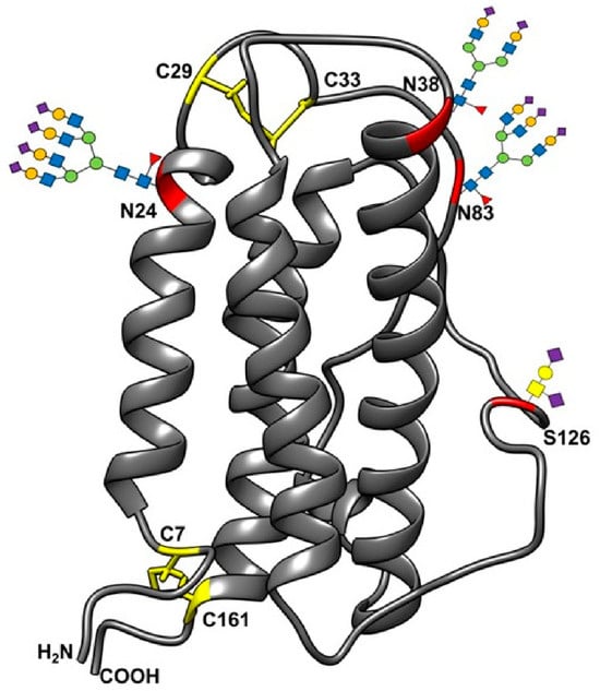
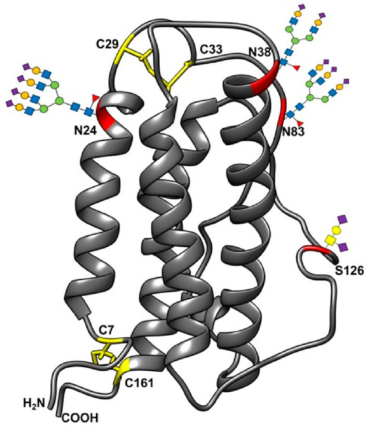
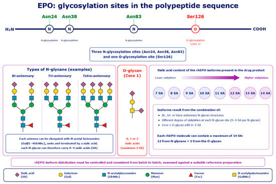
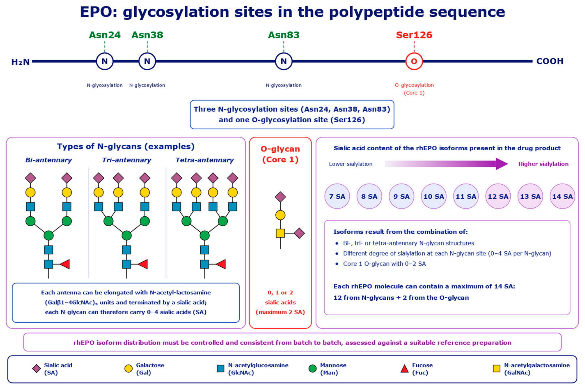
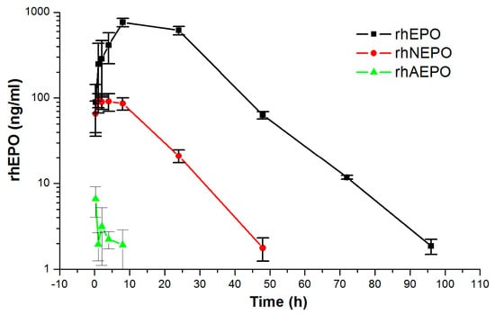
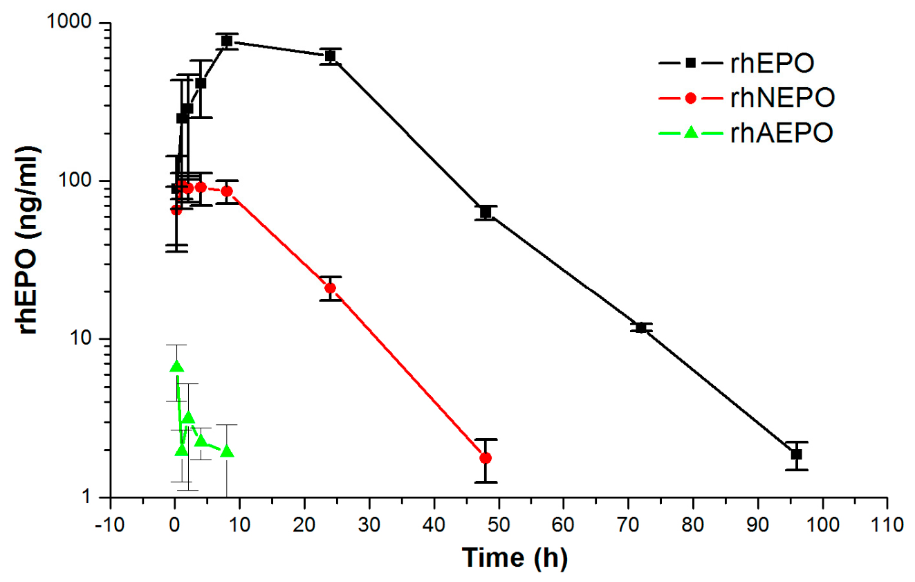
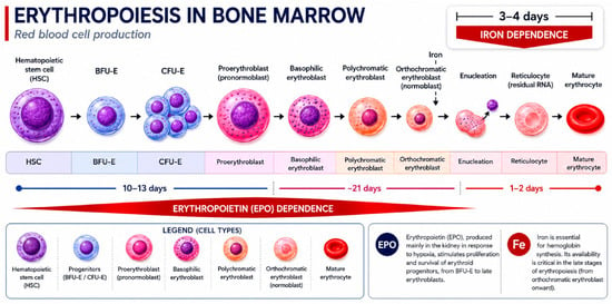
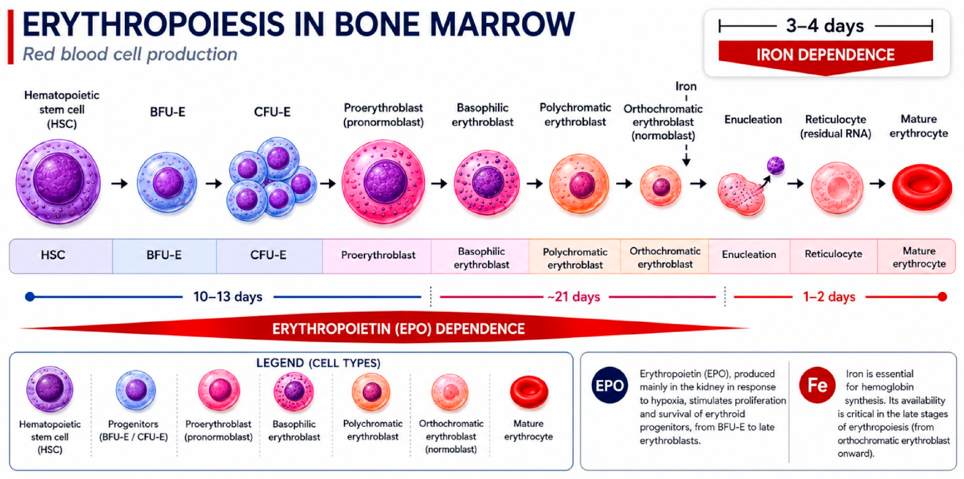
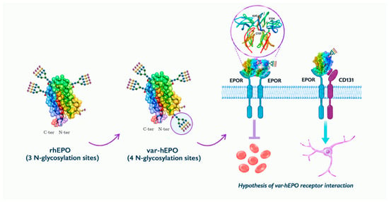
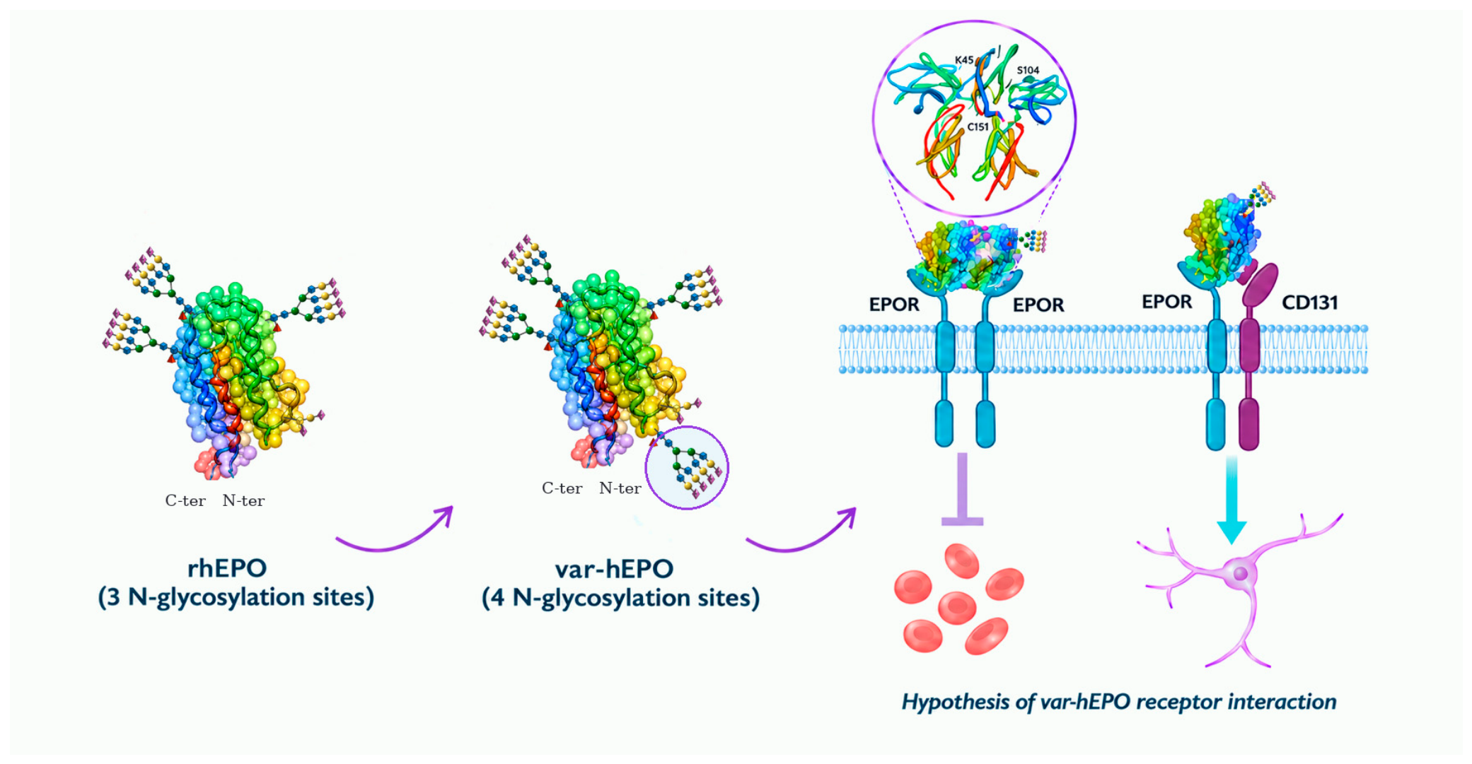

Disclaimer/Publisher’s Note: The statements, opinions and data contained in all publications are solely those of the individual author(s) and contributor(s) and not of MDPI and/or the editor(s). MDPI and/or the editor(s) disclaim responsibility for any injury to people or property resulting from any ideas, methods, instructions or products referred to in the content. |
© 2026 by the authors. Licensee MDPI, Basel, Switzerland. This article is an open access article distributed under the terms and conditions of the Creative Commons Attribution (CC BY) license.