


Abstract
Fowl adenoviruses (FAdVs) are key poultry pathogens associated with inclusion body hepatitis (IBH), causing severe hepatic injury and mortality in young chickens. This study investigated IBH outbreaks in commercial broiler flocks in the Kashmir Valley, India, during 2022–2023, performing field screening, pathological examination, molecular and phylogenetic characterization, and experimental pathogenicity assessment of the circulating strain. Across 25 suspected outbreaks comprising 27,353 birds, 3533 deaths were recorded (overall observed mortality rate: 12.92%). Polymerase chain reaction (PCR) screening of the 200 pooled tissue samples (liver, heart, kidney, and spleen) targeting the Hexon gene amplified an ~897-bp product, detecting FAdV in 144 samples (72%) across 18 of the 25 investigated outbreaks (72%). Gross pathological examination revealed hepatomegaly, hepatic mottling, pallor, friability, petechial haemorrhages in the liver and kidneys, and splenomegaly. Histopathologically, tissues exhibited hepatocellular degeneration and necrosis, heterophilic infiltration, congestion, basophilic intranuclear inclusion bodies, splenic lymphoid depletion, glomerular atrophy, and myocardial fiber disruption. Sequence analysis revealed 98.72% nucleotide identity with the FAdV-11 isolate (GenBank accession number HQ697595.1), while phylogenetic analysis supported classification of the virus as FAdV-11 (species D) and established its relationship with Asian isolates. Experimental pathogenicity evaluation in sixty 14-day-old specific-pathogen-free (SPF) chicks inoculated intramuscularly (IM) and/or (IP) with ( virus suspension, equivalent to per bird) resulted in 100% mortality by day 6 in IM-inoculated birds. IP-inoculated birds underwent scheduled serial euthanasia for pathological evaluation; due to differing termination schedules, mortality outcomes were not directly comparable between routes. Experimental infection induced clinical and pathological changes, haematological alterations, elevated serum liver enzyme activities, and hepatic viral DNA presence. To our knowledge, this study provides one of the first molecular and pathological documentations of FAdV-11-associated IBH in commercial broilers in this region.
1. Introduction
The poultry industry is one of the fastest-growing sectors of the agricultural economy and represents a major source of affordable animal protein worldwide [1,2]. Increasing consumer demand and the intensification of production systems continue to drive the expansion of poultry production, which is projected to reach approximately 131.6 million tonnes globally by 2026 [3]. In India, poultry production has expanded considerably, including in challenging geo-climatic regions such as the Kashmir Valley, where the sector contributes to livelihoods, employment generation, and regional food security [4,5,6]. However, intensive commercial production systems also increase the risk of infectious diseases through high stocking densities, rapid flock turnover, and complex production chains [7]. Viral diseases remain important constraints on poultry production, contributing to mortality, reduced productivity, carcass condemnation, and economic losses [8].
Fowl adenoviruses (FAdVs), members of the genus Aviadenovirus within the family Adenoviridae, are non-enveloped viruses with linear double-stranded DNA genomes and are associated with several diseases in poultry [9]. Traditionally, FAdVs have been classified into five species (A–E) and twelve serotypes based on their antigenic and genetic characteristics [10,11,12]. Under the current International Committee on Taxonomy of Viruses (ICTV) classification, the species previously referred to as Fowl Aviadenovirus D is named Aviadenovirus gallinae and includes FAdV-2, FAdV-3, FAdV-9, and FAdV-11 [13]. It is important to distinguish the serotype designation from the species name; accordingly, FAdV-11 is referred to as serotype 11 within the species Aviadenovirus gallinae. Different FAdV serotypes are associated with distinct clinical presentations. FAdV-1 is associated with adenoviral gizzard erosion and ulceration [14], whereas FAdV-4 is an important cause of hepatitis-hydropericardium syndrome [15]. Inclusion body hepatitis (IBH) in broiler chickens has been associated with several serotypes, particularly FAdV-2, FAdV-8a, FAdV-8b, and FAdV-11 [16,17,18]. Clinical disease is frequently reported in young broilers, especially between three and seven weeks of age, although susceptibility is influenced by several host and viral factors [19,20].
FAdVs can spread through both vertical and horizontal transmission. Vertical transmission may occur through infected breeder flocks and eggs, whereas horizontal transmission occurs through faecal contamination, fomites, and contaminated farm environments [19,20]. Their environmental stability further complicates disease control and creates persistent biosecurity challenges [21]. Recent studies have further highlighted the continued circulation and pathogenic significance of FAdV-11 (species D) in Asian poultry populations. In particular, Qiao et al. 2024 [20] characterized the genome of an FAdV-11 isolate obtained from chickens with IBH in China and demonstrated its pathogenicity in experimentally infected chicks, providing further evidence of the role of FAdV-11 in IBH.
IBH is characterized by acute mortality, hepatic degeneration and necrosis, and intranuclear inclusion bodies within hepatocytes [22,23]. Gross lesions commonly include enlarged, pale, friable, or mottled livers with haemorrhagic or necrotic foci; renal lesions and hydropericardium may also occur, depending on the virus and clinical presentation [24,25]. Mortality varies between outbreaks and is influenced by viral virulence, bird age, immune status, and concurrent infections [26,27,28,29]. Histopathologically, hepatocellular degeneration and necrosis accompanied by intranuclear inclusion bodies are characteristic findings in IBH [9,30]. Although these inclusions are highly suggestive of adenoviral infection, molecular or virological testing is required for etiological confirmation. IBH has historically been associated with immunosuppression and concurrent infections with infectious bursal disease virus (IBDV) or chicken anemia virus (CAV) [31,32]. Nevertheless, experimental and field studies have demonstrated that IBH may also occur in the absence of these infections [33,34,35].
IBH was first described in the United States in 1963 [36] and has subsequently been reported in poultry-producing regions worldwide. In India, IBH was first reported in 1994 [37], and subsequent investigations have documented the involvement of multiple FAdV serotypes in disease outbreaks [38,39,40]. Molecular investigations in India have identified FAdV-11 and FAdV-8b in field outbreaks, including investigations conducted in western India during 2019–2021 [39,41]. Recent research from China has also provided genomic and experimental pathogenicity data for an FAdV-11 isolate associated with IBH [20]. These studies demonstrate the continuing importance of molecular surveillance and the need to characterize field strains in different geographical settings. Despite the importance of commercial poultry production in the Kashmir Valley, published molecular and pathological information specifically characterizing FAdV strains associated with IBH outbreaks in this region remains limited [5]. The region’s distinctive temperate agro-climatic conditions and seasonal variations may influence disease occurrence and transmission dynamics in poultry; however, their specific association with FAdV epidemiology in the Kashmir Valley remains insufficiently characterized [39,42]. The extent to which FAdV-11 contributes to IBH outbreaks in local commercial broiler flocks therefore requires further investigation. Although antigenic differences among FAdV serotypes can influence vaccine protection, vaccine efficacy and cross-protection cannot be inferred from molecular detection or pathogenicity studies alone [43].
The present study was undertaken to investigate suspected IBH outbreaks in commercial broiler flocks in the Kashmir Valley, determine the occurrence of FAdV by polymerase chain reaction (PCR), and characterize selected PCR-positive samples through partial Hexon gene sequencing and phylogenetic analysis. In addition, the pathological findings in naturally affected birds were documented, and the pathogenicity of a field-derived FAdV isolate was evaluated experimentally in SPF chicks by assessing clinical signs, mortality, haematological and biochemical alterations, and tissue lesions. We hypothesized that FAdV would be detectable in a proportion of suspected IBH outbreaks and that experimental inoculation with the field-derived isolate would produce measurable clinicopathological changes in SPF chicks. This investigation was intended to provide regional molecular and pathological evidence of FAdV-associated IBH and improve understanding of the disease under field and experimental conditions.
2. Materials and Methods
2.1. Study Area and Sample Collection
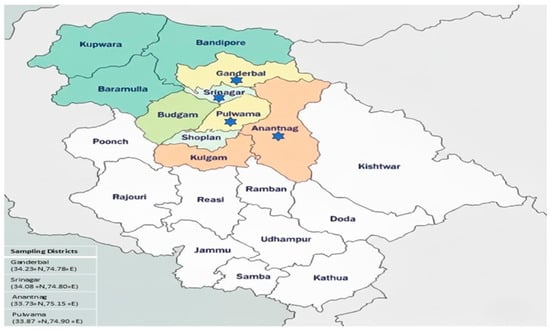
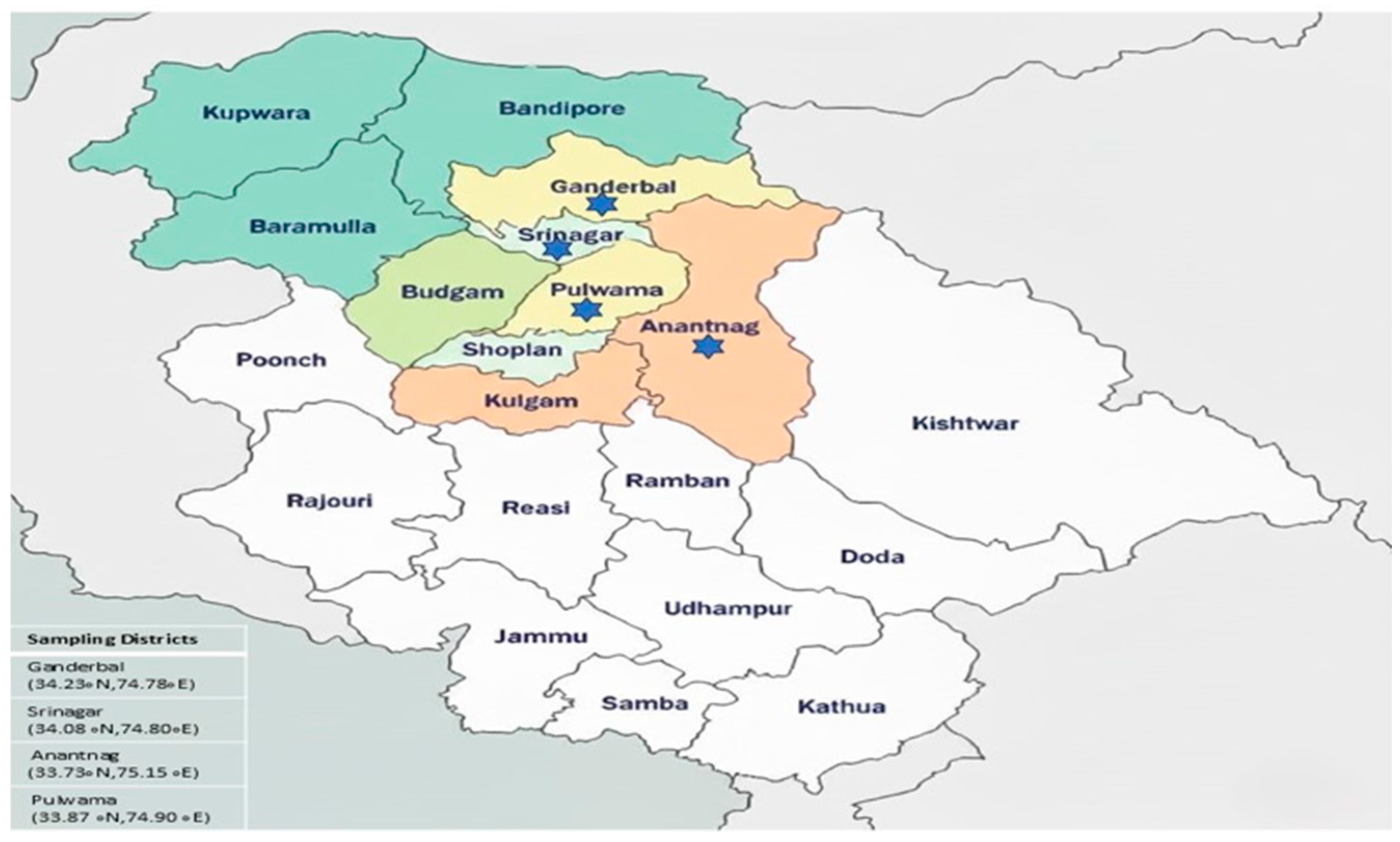
The study was conducted during 2022–2023 in the Kashmir Valley, India, and involved 25 suspected outbreaks of inclusion body hepatitis (IBH) in commercial broiler chicken flocks located in the districts of Ganderbal (34.23° N, 74.78° E), Srinagar (34.08° N, 74.80° E), Anantnag (33.73° N, 75.15° E), and Pulwama (33.87° N, 74.90° E) (Figure 1). The investigated flocks comprised a total population of 27,353 birds, among which 3533 deaths were recorded, corresponding to an overall observed mortality rate of 12.92%. A total of 200 pooled tissue samples comprising liver, heart, kidney, and spleen tissues were collected from the investigated outbreaks. Samples were obtained either directly from affected poultry farms or through post-mortem submissions received at the Division of Veterinary Pathology, Faculty of Veterinary Sciences and Animal Husbandry, Sher-e-Kashmir University of Agricultural Sciences and Technology of Kashmir (SKUAST-K), Shuhama, Srinagar. The outbreaks were investigated on the basis of suspected IBH, supported by clinical history and gross pathological findings. Representative tissue portions intended for histopathological examination were fixed in 10% neutral buffered formalin. Samples intended for molecular detection were transported in viral transport medium (HiMedia) and preserved in 50% buffered glycerol at −20 °C until further processing.
Figure 1.
Sampling districts of Kashmir Valley highlighted by a blue star.
2.2. Gross and Histopathological Examination of Natural Cases
Systematic post-mortem examinations were performed on broiler carcasses received during the field investigation, and gross lesions associated with inclusion body hepatitis (IBH) were recorded, with particular attention to changes in liver size, colour, consistency, and the presence and distribution of haemorrhages. Representative tissue samples from the liver, spleen, kidneys, and heart were collected and fixed in 10% neutral buffered formalin, processed using standard paraffin-embedding techniques, sectioned at 5–6 µm, and stained with haematoxylin and eosin (H&E) for light-microscopic examination [44]. In the experimental study, three birds each from Group I (control) and Group II (virus-inoculated) were euthanized on days 1, 3, 5, 7, and 9 post-inoculation (PI) for detailed pathological examination, whereas birds in Group III were monitored daily for mortality, and any carcasses were subjected to thorough post-mortem examination to record gross lesions associated with IBH. Group IV (I/M control) birds were kept until termination of the experiment or otherwise applicable. Representative tissues from experimental birds were fixed in 10% neutral buffered formalin, processed using standard paraffin-embedding techniques, sectioned at 4–5 µm using a microtome (Leica Microsystems GmbH, Wetzlar, Germany), and stained with H&E according to the protocol described by Luna (1968) [44]. The stained sections were examined under a light microscope to identify microscopic lesions characteristic of IBH.
2.3. Molecular Detection and Characterization of FAdVs in Natural Cases
A total of 200 pooled tissue samples preserved in 50% buffered glycerol at −20 °C were homogenized using a tissue homogenizer (Kinematica AG, Lucerne, Switzerland), and a 20% (w/v) tissue suspension was prepared. Viral DNA was extracted using the Wizard® Genomic DNA Purification Kit (A1120, Promega, Madison, WI, USA), following the manufacturer’s instructions. PCR amplification targeted the conserved pedestal 1 (P1) region flanking the variable loop 1 (L1) region of the FAdV Hexon gene, using the primers Hexon A (5′-CAARTTCAGRCAGACGGT-3′) and Hexon B (5′-TAGTGATGMCGSGACATCAT-3′), as described previously [45]. The expected amplicon size was approximately 897 bp. Each 25-µL PCR contained 12.5 µL of 2× Go Green Master Mix, 0.5 µL of each primer (10 pmol), 2 µL of extracted DNA, and nuclease-free water to the final volume. The thermal cycling conditions, based on previously published protocols [45] with an annealing temperature of 55 °C, consisted of initial denaturation at 95 °C for 5 min, followed by 35 cycles of denaturation at 94 °C for 30 s, annealing at 55 °C for 30 s, and extension at 72 °C for 30 s, with a final extension at 72 °C for 10 min. The PCR products were analyzed by electrophoresis on a 2% agarose gel prepared in 1× Tris-acetate-EDTA (TAE) buffer and visualized using a gel documentation system. A previously confirmed FAdV-positive DNA sample was used as the positive control, while apparently healthy commercial broiler birds as the negative control. The positive control was expected to yield the specific amplicon of approximately 897 bp, whereas no amplification was expected in the negative control.
2.4. Sequencing and Phylogenetic Analysis
Two PCR-positive amplicons representing different outbreaks were selected and submitted for Sanger sequencing to Unipath Specialty Laboratory, Ahmedabad, Gujarat, India. The amplicons were sequenced directly without cloning. Sequence quality was assessed using FinchTV, and low-quality regions were trimmed before analysis. The resulting FAdV Hexon gene sequence used for analysis was 851 bp in length after trimming. Multiple-sequence alignment was performed using Clustal Omega, version MEGA 11. The obtained sequence was compared with reference sequences of fowl aviadenovirus (FAdV) available in GenBank. The corresponding Hexon gene regions were used for sequence alignment and phylogenetic analysis. A phylogenetic tree was constructed using the Neighbor-Joining method in version 11 [46] with 1000 bootstrap replicates for testing the reliability of the inferred groupings. The evolutionary distances were computed using the Maximum Composite Likelihood method, and gaps and missing data were treated using the pairwise deletion option.
2.5. Experimental Study
The experimental infection study involved the use of 60 specific pathogen-free (SPF) 14-day-old chickens procured from Immunetic Life Sciences, Himachal Pradesh, India. The birds were kept in an isolated room in the animal facility of the Veterinary Pathology Division, provided with UV-treated feed and autoclaved water ad libitum. All experimental procedures were approved by the Institutional Animal Ethics Committee (IAEC), with approval number 1809/GO/ReBiS/Rel/15/CPCSEA, before initiation of the study.
2.6. Isolation and Propagation of the FAdV Field Strain in CEF Cells
The FAdV field strain used for experimental infection was obtained from liver tissue collected from broiler chickens affected during a natural outbreak. The field strain was molecularly characterized as FAdV-11, belonging to species D, and its sequence was deposited in GenBank under accession number PQ385598. Primary chicken embryo fibroblast (CEF) cells were prepared from 10- to 12-day-old specific-pathogen-free (SPF) embryonated chicken eggs following the procedure described by ref [47]. A liver-tissue homogenate was prepared, clarified, filtered, and inoculated onto confluent CEF monolayers. The inoculated cultures were maintained at 37 °C in a humidified incubator with 5% CO2 and monitored daily for cytopathic effects (CPE). CPE, characterized by cell rounding, detachment, and cellular aggregation, was first observed approximately 48 h post-infection during the second passage and became more pronounced by 72 h. During the third passage, CPE was observed within 24 h post-infection. Following the appearance of CPE, the cultures underwent three freeze–thaw cycles, and the harvested material was clarified, filtered, and stored at −80 °C. The presence of FAdV in the propagated culture was reconfirmed by PCR. Viral titres were determined in CEF cells using the tissue culture infectious dose 50% (TCID50) assay, involving tenfold serial dilutions and calculation according to the Reed–Muench method [48]. The resulting viral titre was 105 TCID50/mL.
2.7. Experimental Design
A total of 60 specific-pathogen-free (SPF) chickens, 14 days of age, were randomly allocated into four experimental groups, comprising 15 birds each. Group I served as the intraperitoneal (IP) control and received 0.2 mL of sterile phosphate-buffered saline (PBS) per bird. Group II received an IP inoculation of 0.2 mL of FAdV suspension containing 105 TCID50/mL, equivalent to a dose of 2 × 10⁴ TCID50 per bird. Group III received the same volume and concentration of the viral suspension via the intramuscular (IM) route, while Group IV served as the IM control and received 0.2 mL of sterile PBS per bird. Clinical observations were conducted daily throughout the experimental period. In Groups I and II, three birds from each group were scheduled for euthanasia on days 1, 3, 5, 7, and 9 post-inoculation (PI) for gross and histopathological examination. Birds selected for scheduled euthanasia were euthanized by decapitation. Humane endpoints included an inability to access feed and water and the development of a crouching posture. Birds exhibiting these signs were considered to have reached humane endpoints and were to be euthanized to prevent unnecessary suffering. Group III was monitored for clinical progression and mortality, with all 15 birds dying by day 6 PI. Birds in Group IV remained clinically normal throughout the experimental observation period and were euthanized at the planned termination of the experiment.
Birds that died during the observation period and those euthanized according to the scheduled sampling protocol underwent complete post-mortem examination. Representative tissue samples, including liver, spleen, heart, kidneys, and gizzard, were collected and fixed in 10% neutral buffered formalin for histopathological examination. Additional liver samples were preserved at −20 °C for molecular detection of FAdV. The IP and IM routes were used to investigate the clinical and pathological outcomes following experimental inoculation by different routes. However, because Group II birds underwent scheduled euthanasia at predefined time points, whereas Group III birds were monitored for mortality, the observation schedules differed between the two groups. Therefore, mortality outcomes were interpreted descriptively and were not considered directly comparable measures of route-specific mortality or survival.
2.8. Haematology and Biochemistry
Blood samples were collected from the wing vein of birds in Group I (intraperitoneal control) and Group II (intraperitoneal challenge group) on days 1, 3, 5, 7, and 9 post-inoculation (PI). Three birds per group were sampled at each time point. For haematological analysis, blood was collected into vacutainer tubes containing K3EDTA (tripotassium ethylenediaminetetraacetic acid). Haemoglobin (Hb, g/dL), total erythrocyte count (TEC, × 106/µL), and packed cell volume (PCV, %) were measured using an automated blood analyzer (Nihon Kohden Corporation, Tokyo, Japan) at the Division of Veterinary Medicine, Faculty of Veterinary Sciences and Animal Husbandry, Sher-e-Kashmir University of Agricultural Sciences and Technology of Kashmir (SKUAST-K), Jammu and Kashmir. For serum biochemical analysis, serum was separated from blood samples and stored at −20 °C until analysis. Creatinine and blood urea nitrogen (BUN), expressed in mg/dL, and the activities of alanine aminotransferase (ALT), aspartate aminotransferase (AST), and alkaline phosphatase (ALP), expressed in IU/L, were measured using a semi-automatic analyzer (Photometer 5010 V5+, ROBERT RIELE GmbH & Co. KG, Berlin, Germany) at the Division of Veterinary Biochemistry, Faculty of Veterinary Sciences and Animal Husbandry, SKUAST-K, Jammu and Kashmir. These analyses were performed in Groups I and II only; Groups III and IV were not included in the haematological or biochemical assessments. Accordingly, the findings were interpreted within the scope of the groups examined.
2.9. Statistical Analysis
Comparisons between the control (Group I) and FAdV-infected (Group II) groups at each sampling time point (days 1, 3, 5, 7 and 9 post-infection) were performed using Student’s t-test. Comparisons among the different sampling days within each group were performed using one-way analysis of variance (ANOVA). All statistical analyses were performed using SPSS Statistics, Version 27.0 (IBM Corp., Armonk, NY, USA), and differences were considered statistically significant at p < 0.05.
3. Results
3.1. Clinical Observations, Mortality and Gross Pathology
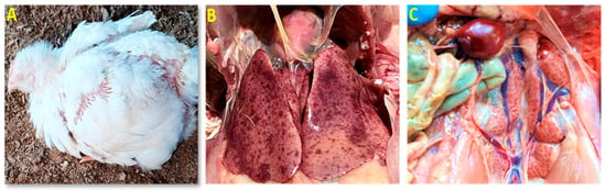
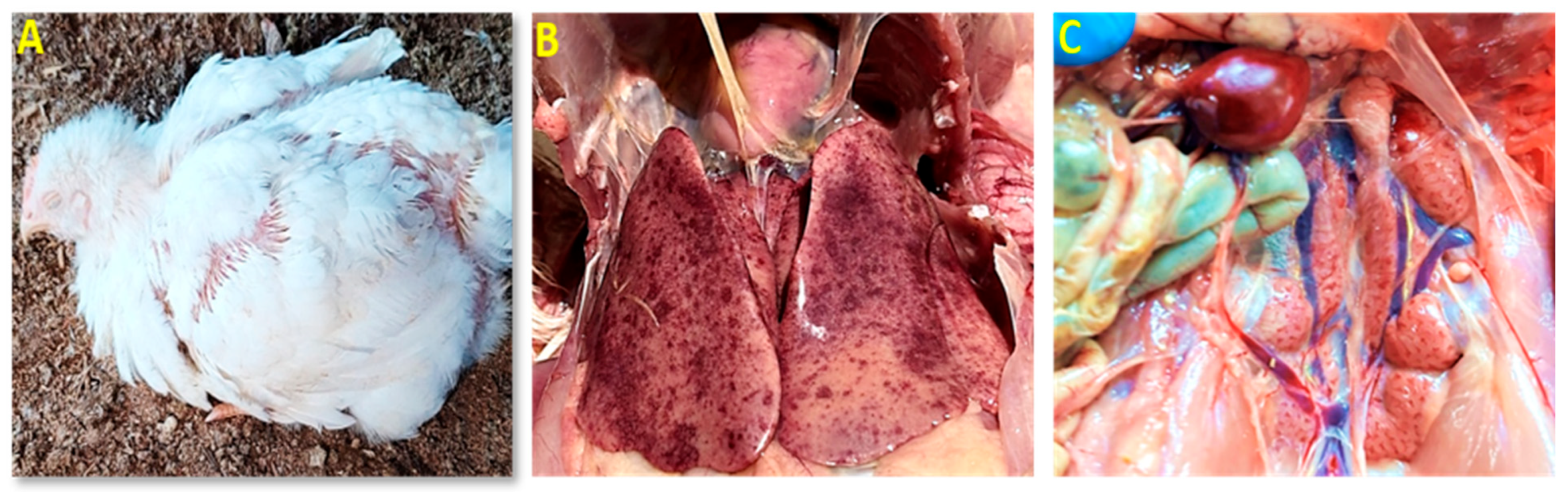
During 2022–2023, investigations were conducted in 25 outbreaks suspected to be associated with inclusion body hepatitis (IBH) in commercial broiler flocks in the Kashmir Valley. A total of 27,353 birds were present in the investigated flocks, and 3533 deaths were recorded, corresponding to an overall observed mortality of 12.92%. Of the 200 tissue samples tested by PCR, 144 (72%) were positive for FAdV, and 18 of the 25 investigated outbreaks (72%) were PCR-positive. In the PCR-positive outbreaks, mortality was most pronounced among birds aged 15–35 days. District-wise, the highest observed mortality was recorded in Pulwama (21.02%; 1194/5679), followed by Srinagar (14.00%; 873/6234), Ganderbal (10.01%; 757/7562), and Anantnag (8.99%; 709/7878). Clinically, affected flocks showed lethargy, depression, huddling, ruffled feathers, anorexia, rapid weight loss, and increased mortality (Figure 2A). Necropsy of affected broiler chickens revealed characteristic multisystemic lesions, with the liver being the most severely affected organ. Gross hepatic changes included hepatomegaly, pallor, friability, a characteristic reticular or mottled appearance, and petechial to ecchymotic haemorrhages with dark-red discoloration (Figure 2B). The kidneys were pale, swollen, congested, and showed cortical haemorrhages (Figure 2C). The spleen was mildly enlarged and congested in some birds. No gross lesions were apparent in the bursa of Fabricius, thymus, or other lymphoid organs.
Figure 2.
Clinical and gross pathological features of inclusion body hepatitis (IBH) in broiler chickens: (A) Bird showing crouching posture, ruffled feathers, and beak resting on the floor; (B) Liver exhibiting marked hepatomegaly with extensive petechial hemorrhages, mottling, and friable consistency; and (C) Kidney showing pallor, swelling, mottling, congestion, and occasional cortical hemorrhages.
3.2. Histopathological Findings
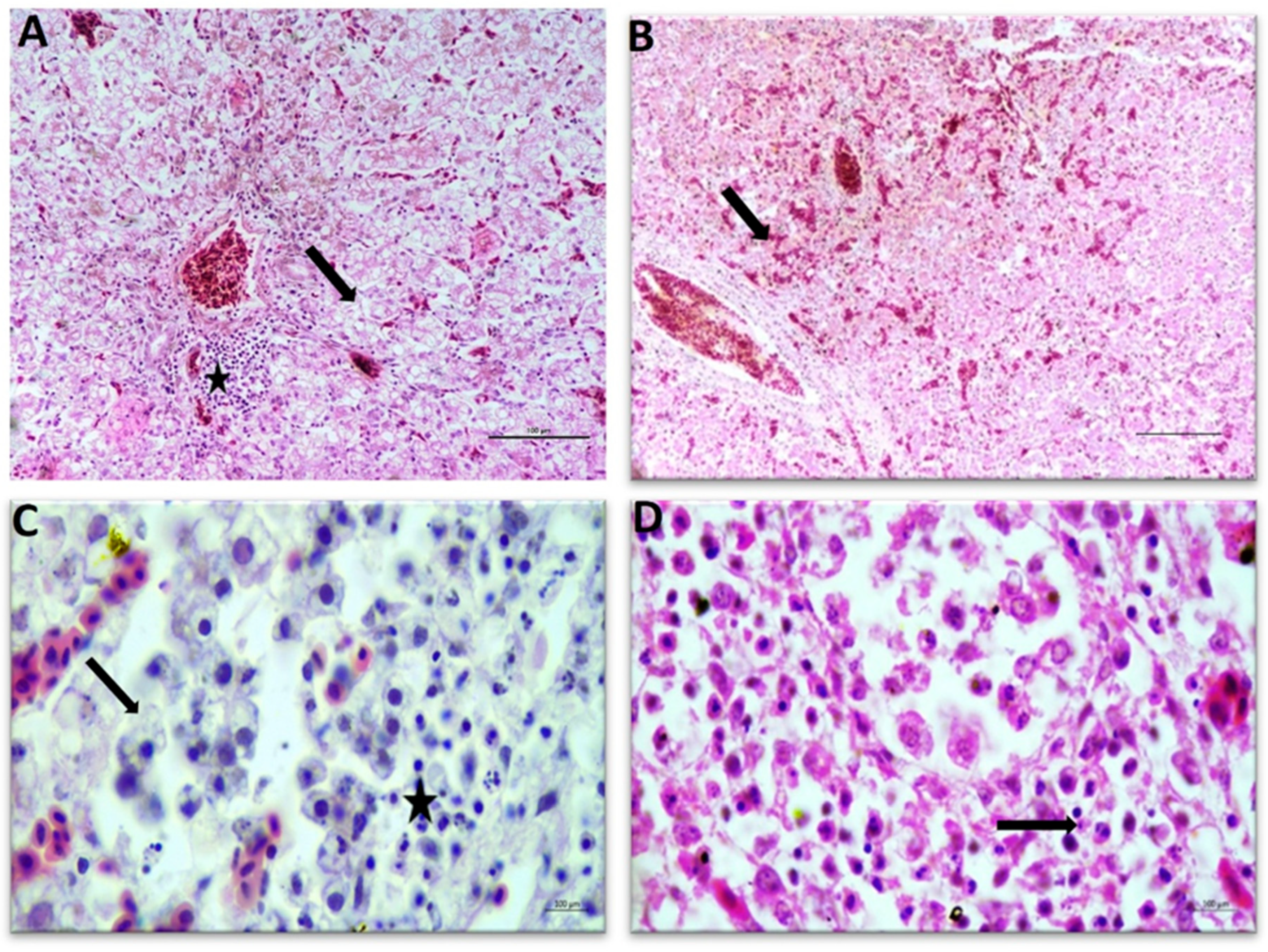
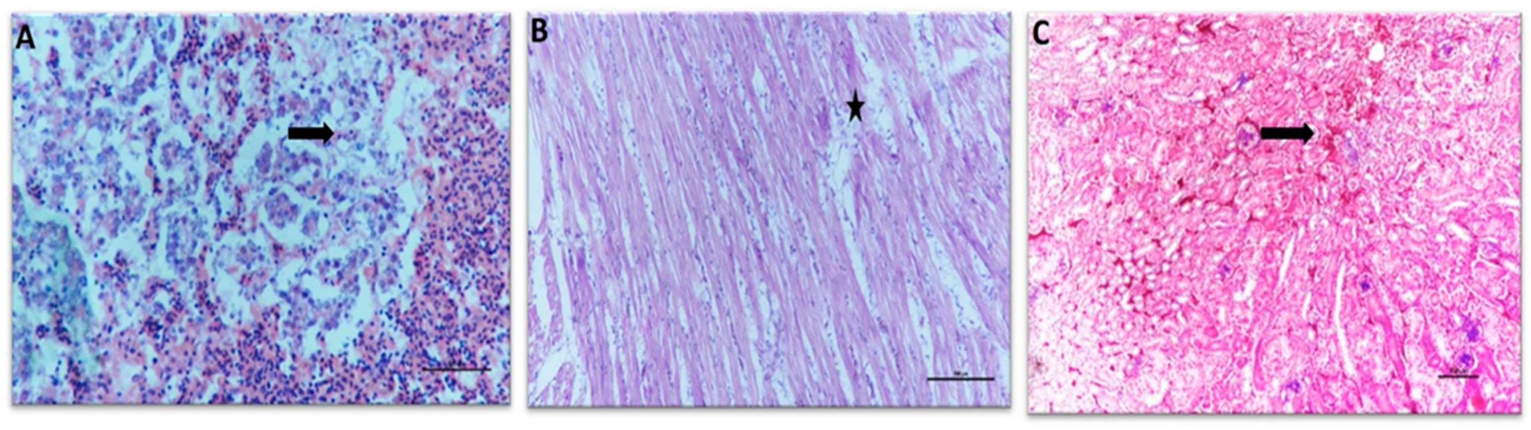
Histological examination demonstrated marked pathological changes, primarily in the liver. Hepatic lesions included vasodilation, sinusoidal congestion, hepatocellular degeneration, heterophilic infiltration, focal haemorrhages, and multifocal necrosis. Numerous basophilic intranuclear inclusion bodies of varying forms and sizes were observed in degenerating hepatocytes (Figure 3). Perihepatitis with mononuclear and heterophilic cell infiltration was also observed, together with severe degenerative changes, including karyolysis. The spleen was characterized by marked lymphoid depletion (Figure 4A), while the myocardium showed disruption of myocardial fibres accompanied by inflammatory cell infiltration (Figure 4B). Renal lesions included interstitial haemorrhages, glomerular atrophy, mononuclear cell infiltration, and tubular epithelial necrosis (Figure 4C).
Figure 3.
Histopathological changes in the liver of broiler chickens affected with inclusion body hepatitis (IBH): (A) hepatocellular degeneration (arrow), sinusoidal congestion, and heterophilic infiltration around the central vein (star); H.E. scale bar 100 µm; (B) severe congestion and parenchymal hemorrhage (arrow); H.E. scale bar 100 µm. (C) fatty change (arrow), karyolysis, and prominent basophilic intranuclear inclusion bodies (star). H.E. scale bar 10 µm. (D) Degenerated hepatocytes containing basophilic intranuclear inclusion bodies (arrow). H.E. scale bar 100 µm.
Figure 4.
Histopathological changes in broiler chickens affected with inclusion body hepatitis (IBH): (A) spleen showing severe lymphoid depletion (arrow), H.E. scale bar 100 µm. (B) heart exhibiting disruption of myocardial muscle fibers with mild inflammatory cell infiltration (star); H.E. scale bar 100 µm; and (C) kidney showing glomerular atrophy, renal tubular necrosis, interstitial hemorrhage, and cellular infiltration in the interstitium (arrow); H.E. scale bar 100 µm.
3.3. Molecular Detection and Genotypic Characterization
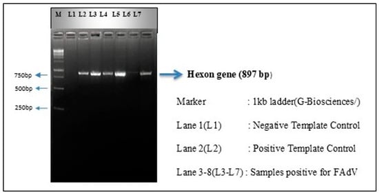
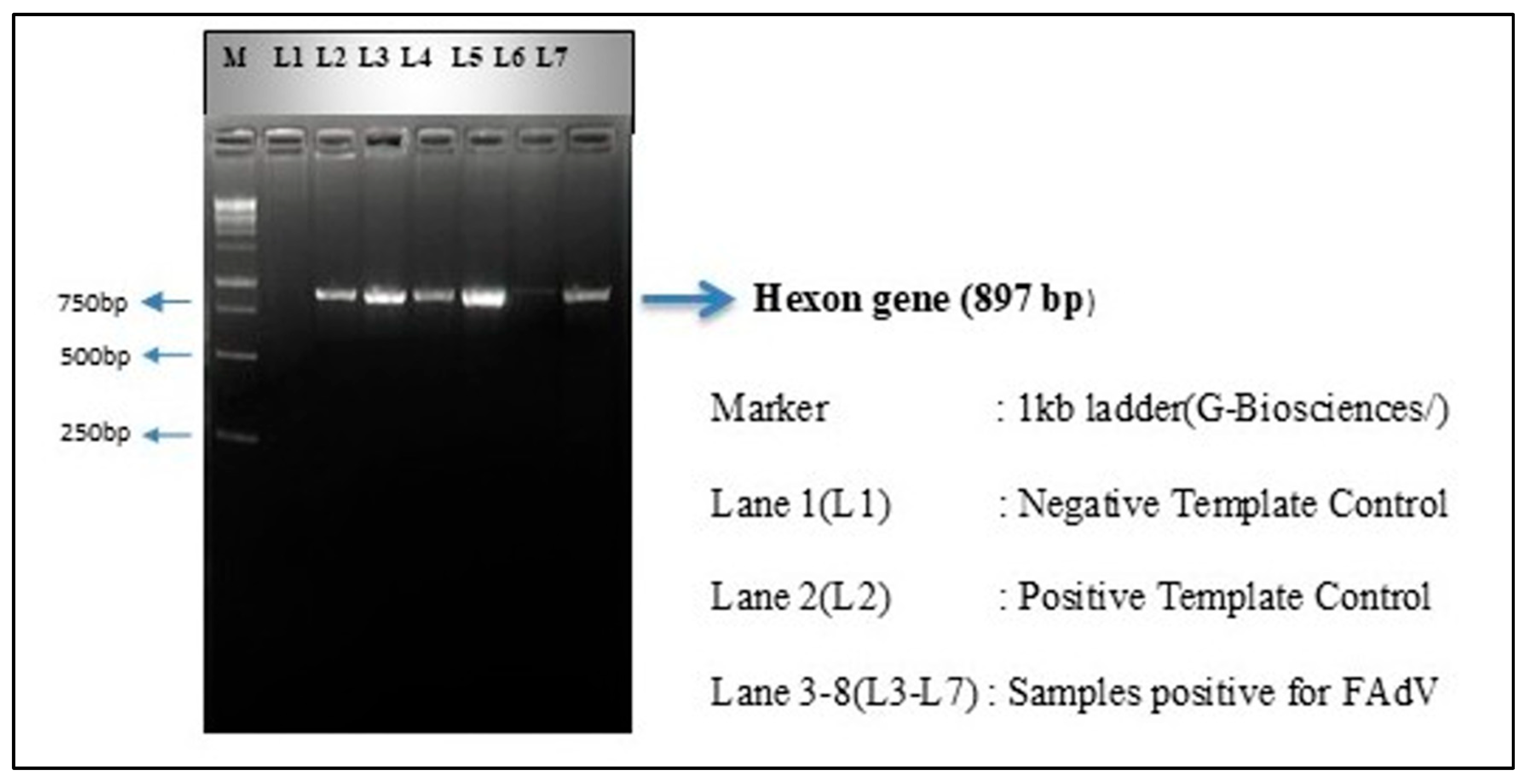
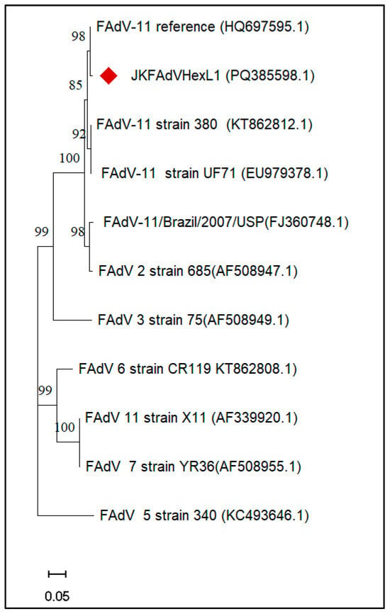
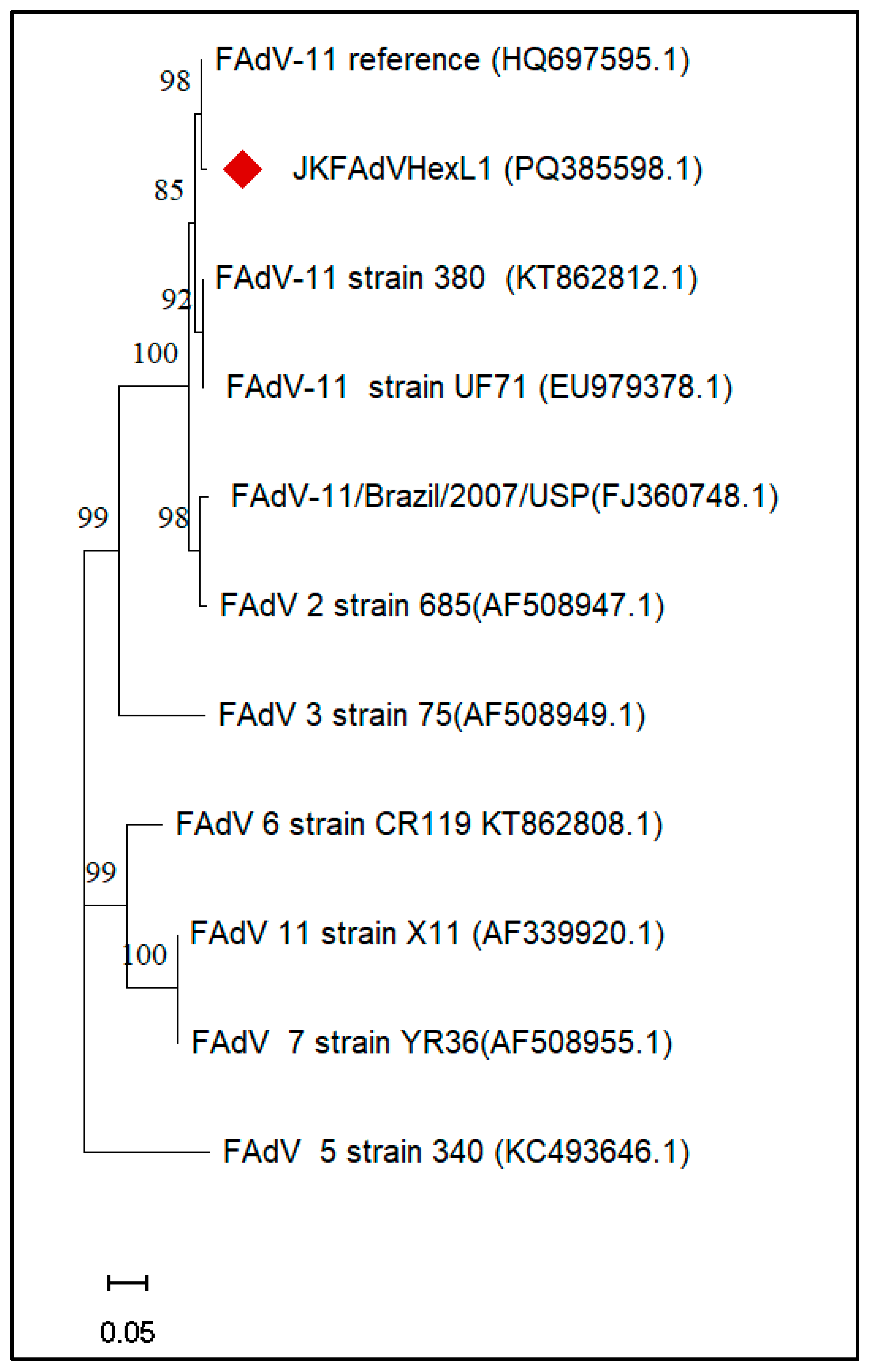
PCR amplification targeting the conserved pedestal 1 (P1) region flanking the variable loop 1 (L1) region of the FAdV Hexon gene yielded the expected amplicon of approximately 897 bp in positive samples (Figure 5). No amplification was observed in samples collected from apparently healthy commercial broiler birds serving as negative controls. Of the 200 tissue samples tested, 144 were PCR-positive. Two PCR-positive amplicons representing different outbreaks were selected for Sanger sequencing. The resulting sequences were compared and found to be identical or nearly identical; therefore, one representative sequence was selected for submission to GenBank and subsequent phylogenetic analysis (accession number: PQ385598). The sequence analyzed showed 98.72% nucleotide identity with the FAdV-11 reference isolate HQ697595.1. The corresponding sequence was deposited in GenBank under accession number PQ385598. Phylogenetic analysis placed the characterized isolate in a cluster with the FAdV-11 reference sequence HQ697595.1, supported by a bootstrap value of 98%. The isolate also grouped within the broader cluster containing other FAdV reference sequences (Figure 6). These relationships indicate a close phylogenetic association between JKFAdVHexL1 and the included FAdV-11 reference sequence based on the analyzed Hexon region. Following molecular characterization, the field isolate was propagated in vitro for the experimental study.
Figure 5.
Visualization of amplified PCR product of Hexon gene (~897 bp) in the Agarose gel (2%).
Figure 6.
Phylogenetic tree constructed using the Neighbor-Joining approach based on Hexon gene nucleotide sequences of fowl aviadenovirus (FAdV) reference strains and the isolate JKFAdVHexL1 (PQ385598.1) identified in the present study. Numbers at the nodes represent bootstrap support values from 1000 replicates. The Kashmir Isolate (JKFAdVHexL1) obtained in this study is marked with Red colour.
3.4. FAdV Isolation and Titration
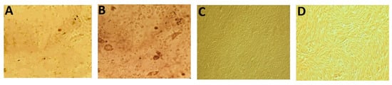
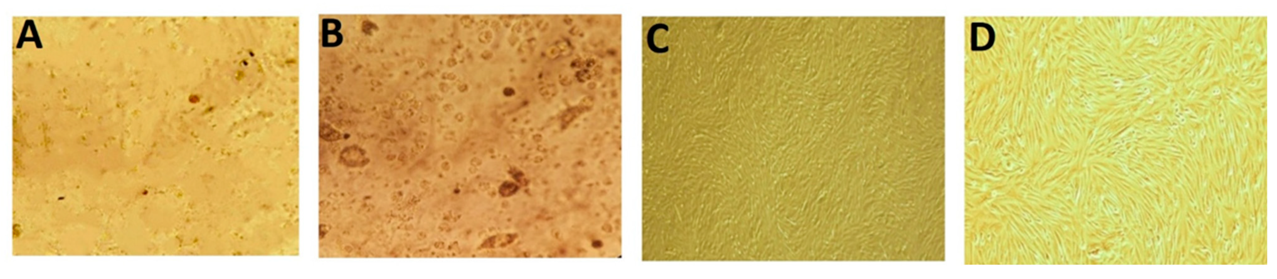
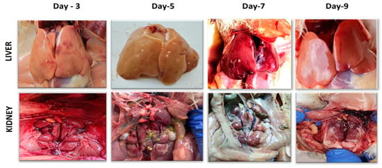
The molecularly characterized FAdV isolate was successfully propagated in chicken embryo fibroblast (CEF) cells. Characteristic cytopathic effects (CPE), including cell rounding, aggregation, and detachment from the monolayer, were first observed at 48 h post-infection during the second passage and became more pronounced by 72 h, whereas control cultures remained normal (Figure 7). By the third passage, CPE appeared within 24 h post-infection. The presence of FAdV in the propagated culture was confirmed by PCR. However, sequencing of the propagated virus was not performed to reconfirm its FAdV-11 identity. The propagated inoculum was not screened for other infectious agents The viral titre was determined to be 105 TCID50/mL using the Reed and Muench method.
Figure 7.
(A) Chicken Embryo Fibroblast Monolayer (48 h post infection) showing detachment and cell rounding. (B) Chicken Embryo Fibroblast Monolayer (72 h post infection) showing cell rounding, aggregation, and detachment from the monolayer. (C) Chicken Embryo Fibroblast Monolayer (10×, control). (D) Chicken Embryo Fibroblast Monolayer (40×, Control).
3.5. Clinical Signs, Mortality, Gross Pathology and Histopathological Findings in Experimental SPF Chicks
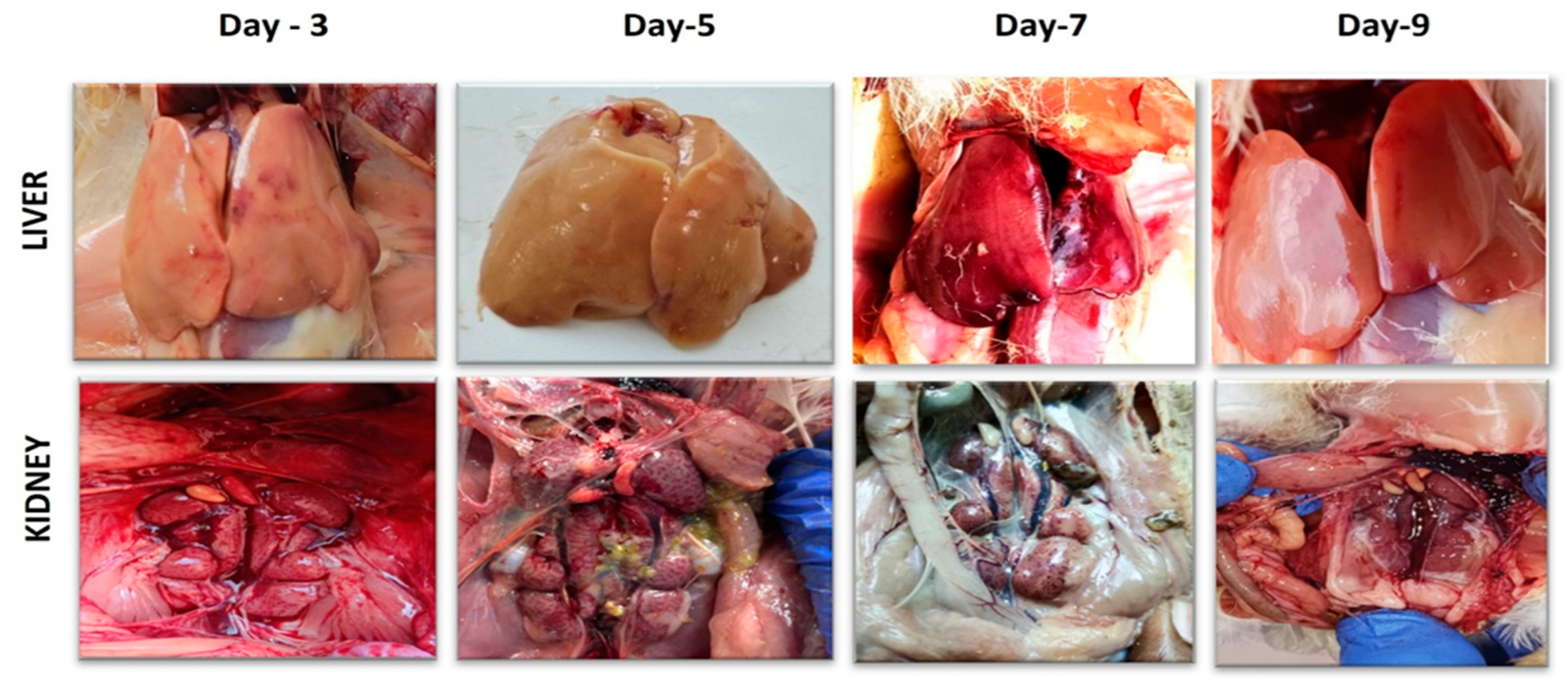
Birds in the control groups (Groups I and IV) remained clinically normal. In contrast, birds inoculated with FAdV (Groups II and III) developed clinical signs that included dullness, depression, lethargy, shrunken eyes, reduced appetite, ruffled feathers, stunted growth, huddled posture, and difficulty eating or drinking. In Group II, which received the virus by the intraperitoneal (IP) route, three birds were scheduled for euthanasia on each of days 1, 3, 5, 7, and 9 post-inoculation (PI) for pathological and laboratory examinations. No spontaneous mortality was recorded in this group before scheduled euthanasia. Group III, which received the virus by the intramuscular (IM) route, was monitored for clinical progression and mortality. Mortality began on day 3 PI, when four birds (26.67%) died, followed by eight birds (53.33%) on day 5 and the remaining three birds (20.00%) on day 6, resulting in 100% cumulative mortality by day 6 PI. Because the two inoculated groups were managed using different observation and sampling schedules, these mortality outcomes are descriptive and do not permit a direct comparison of route-specific mortality or survival. Post-mortem examination of experimentally infected SPF chicks revealed multisystemic lesions, with the liver consistently showing the most prominent gross changes. In Group II, hepatic petechial and diffuse haemorrhages, hepatomegaly, pallor, friability, mottling, congestion, dark-red discoloration, and multifocal necrosis were observed as early as day 1 PI. These lesions were most prominent on days 3–5 PI and decreased in apparent severity thereafter. In Group III, similar hepatic lesions were observed, with hepatic rupture additionally recorded on day 6 PI. In both inoculated groups, the kidneys showed enlargement, congestion, and haemorrhages, particularly on days 3 and 5 PI, with less prominent changes at later observations. The spleen showed mild swelling, pallor, and occasional discoloration, while no noticeable gross lesions were recorded in the heart or gizzard (Figure 8).
Figure 8.
Day-wise gross pathological changes in the liver and kidneys of SPF broiler chickens experimentally infected with FAdV. Liver: Day 3 post-infection (PI), showing a pale, swollen, friable, and hemorrhagic liver; Day 5 PI, showing marked hepatomegaly, pallor, and a friable hepatic surface; Day 7 PI, showing hepatomegaly and hepatic congestion; and Day 9 PI, showing no apparent gross lesions. Kidneys: Day 3 PI, showing pronounced renal congestion and dilation; Day 5 PI, showing marked renal congestion with dilated tubules; Day 7 PI, showing mild congestion and tubular dilation; and Day 9 PI, showing mild congestion without visible tubular dilation.
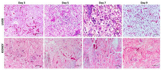
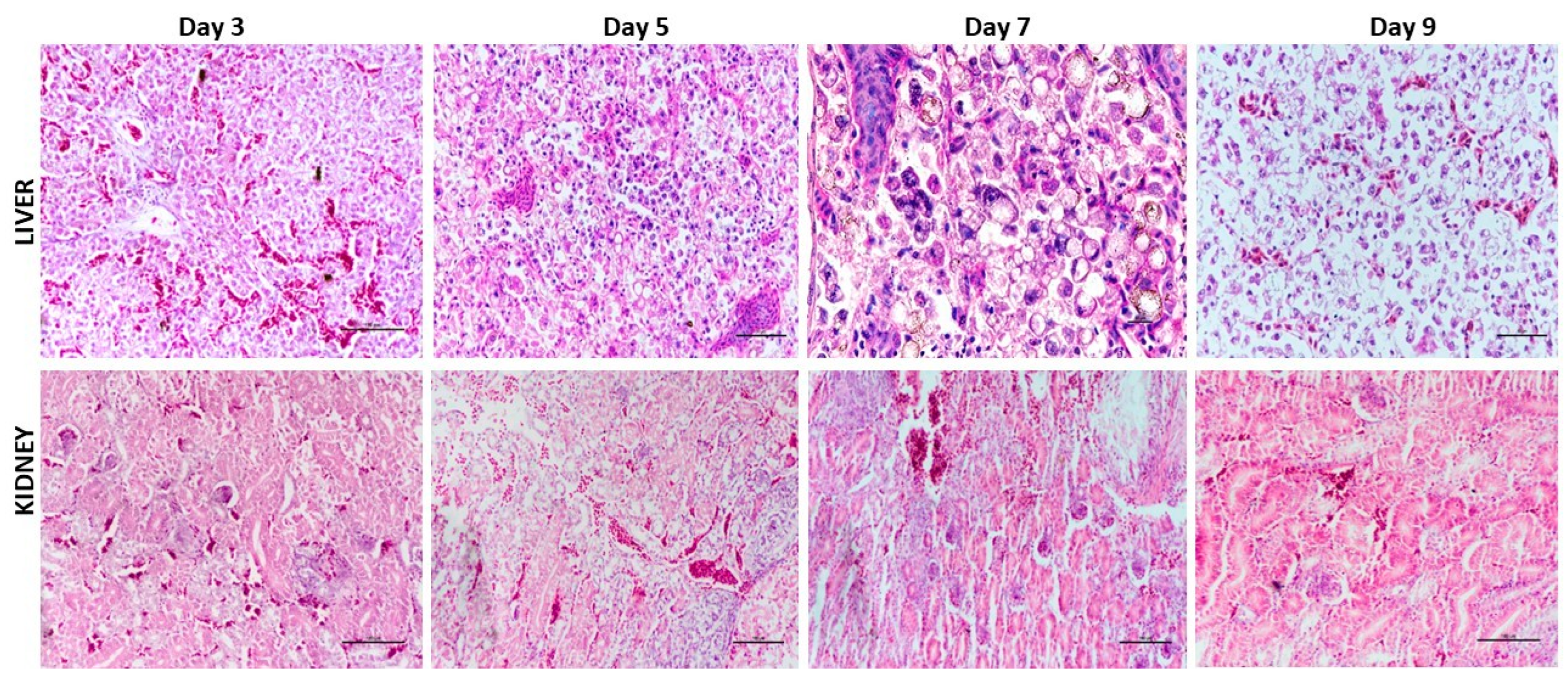
Microscopic examination of tissues from Group II revealed progressive hepatic and renal lesions. Hepatic changes were most prominent on days 3–5 PI and included sinusoidal congestion, hepatocellular degeneration, fatty change, mononuclear cell infiltration, multifocal necrosis, and basophilic intranuclear inclusion bodies. By days 7–9 PI, marked hepatocellular degeneration, karyorrhexis, and necrosis was evident. Renal lesions progressed from congestion to glomerular atrophy, tubular necrosis, and interstitial haemorrhage. In Group III, post-mortem tissues were also examined histologically, and comparable types of hepatic and renal lesions were observed. Cardiac lesions in Group II included myocardial fibre disruption and inflammatory infiltration, with focal necrosis at later observations; renal and splenic changes were also recorded. In Group III, cardiac haemorrhages and inflammatory infiltration, renal tubular degeneration and necrosis, and splenic lymphoid depletion with reticuloendothelial hyperplasia were described. Gizzard lesions were minimal in both inoculated groups. These observations were qualitative; a separate quantitative lesion score or statistical comparison of histopathological severity between Groups II and III was not performed (Figure 9). PCR amplification of the targeted Hexon gene from liver tissues of experimentally infected birds provided further evidence of FAdV infection.
Figure 9.
Day-wise histopathological changes in the liver and kidneys of SPF broiler chickens experimentally infected with FAdV. Liver: Day 3 post-infection (PI), showing marked sinusoidal congestion and hepatocellular degeneration; H.E. scale bar 100 µm; Day 5 PI, showing coagulative hepatocellular necrosis, sinusoidal congestion, and basophilic intranuclear inclusion bodies within hepatocytes; H.E. scale bar 50 µm.; Day 7 PI, showing severe hepatocellular degeneration with intranuclear inclusion bodies; H.E. scale bar 10 µm; and Day 9 PI, showing basophilic intranuclear inclusion bodies within some degenerated hepatocytes; H.E. scale bar 100 µm. Kidneys: Day 3 PI, showing marked interstitial congestion and glomerular atrophy; H.E. scale bar 100 µm; Day 5 PI, showing marked congestion and renal tubular necrosis; H.E. scale bar 100 µm; Day 7 PI, showing severe interstitial haemorrhage, glomerular atrophy, and mononuclear cell infiltration in the interstitium; H.E. scale bar 100 µm; and Day 9 PI, showing interstitial haemorrhage; H.E. scale bar 100 µm.
3.6. Hematological and Serum Biochemical Alterations in Experimentally Infected Birds
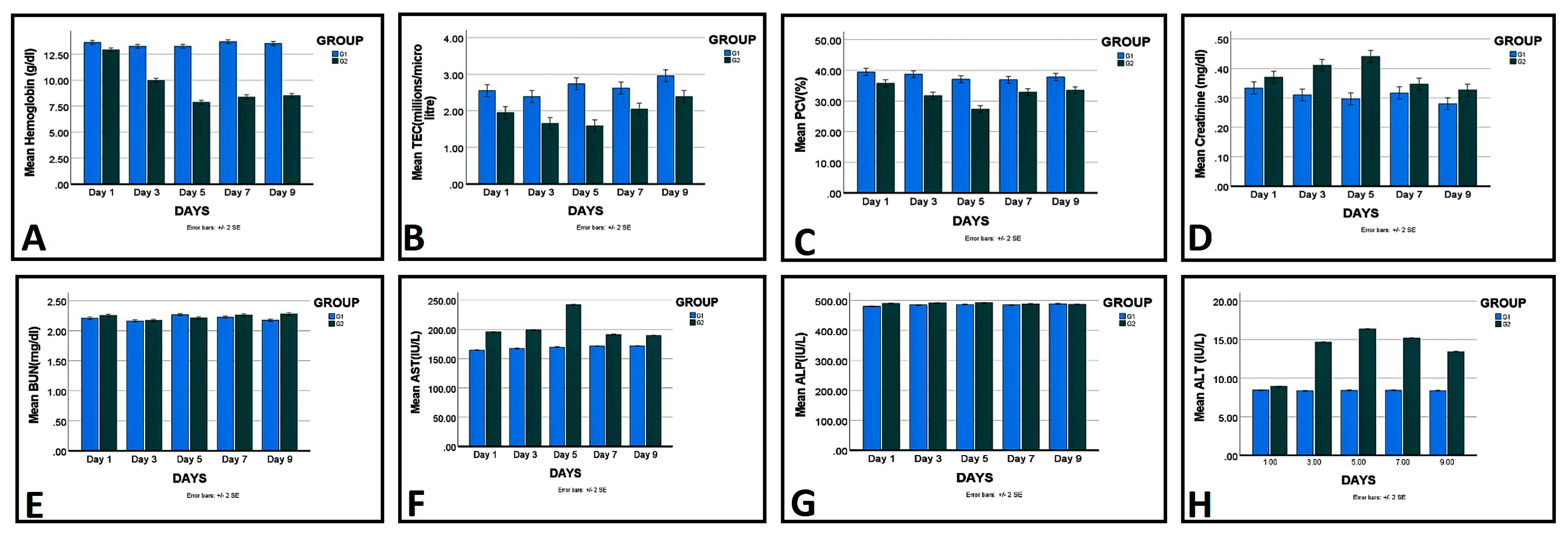
Haematological and serum biochemical parameters were evaluated in Group I (control) and Group II (IP-inoculated) birds over the 9-day post-inoculation period (Figure 10 and Table 1). In Group I, the recorded values showed relatively limited variation over the sampling period. In Group II, haemoglobin (Hb), total erythrocyte count (TEC), and packed cell volume (PCV) declined over the first five days. Mean Hb decreased from 12.91 ± 0.02 g/dL on day 1 to 9.97 ± 0.08 g/dL on day 3 and 7.87 ± 0.06 g/dL on day 5. TEC decreased from 1.95 ± 0.00 × 10⁶/µL on day 1 to 1.65 ± 0.02 × 10⁶/µL on day 3 and 1.59 ± 0.01 × 10⁶/µL on day 5. PCV decreased from 35.77 ± 0.13% on day 1 to 31.72 ± 0.03% on day 3 and 27.34 ± 0.23% on day 5. Partial increases in Hb, TEC, and PCV were observed on days 7 and 9; however, the recorded values remained below those of the control group.
Figure 10.
Day-wise mean values of (A) hemoglobin (g/dL), (B) total erythrocyte count (×106/µL), (C) packed cell volume (%), (D) creatinine (mg/dL), (E) blood urea nitrogen (mg/dL), (F) AST (IU/L), (G) ALT (IU/L), and (H) ALP (IU/L) in control Group I and infected Group II.
Table 1.
Data representing clinical parameters at different time points of the Experiment.
Serum creatinine in Group II increased from 0.37 ± 0.00 mg/dL on day 1 to 0.41 ± 0.00 mg/dL on day 3 and peaked at 0.44 ± 0.00 mg/dL on day 5, followed by a decline to 0.34 ± 0.00 mg/dL and 0.32 ± 0.00 mg/dL on days 7 and 9, respectively. BUN values in the infected group ranged from 2.17 to 2.28 mg/dL, compared with 2.16–2.26 mg/dL in the control group. ALT increased from 8.94 ± 0.01 IU/L on day 1 to 14.66 ± 0.01 IU/L on day 3 and peaked at 16.36 ± 0.04 IU/L on day 5, before declining to 15.19 ± 0.01 IU/L on day 7 and 13.43 ± 0.02 IU/L on day 9. AST increased from 195.63 ± 0.08 IU/L on day 1 to a peak of 242.07 ± 0.57 IU/L on day 5, followed by a decline to 191.16 ± 0.019 IU/L on day 7 and 189.35 ± 0.21 IU/L on day 9. ALP reached 492.99 ± 0.06 IU/L on day 5 and subsequently declined to 488.69 ± 0.35 IU/L and 487.40 ± 0.45 IU/L on days 7 and 9, respectively (Figure 10 and Table 1).
These results describe the temporal changes observed in the parameters measured in Groups I and II. Group IV, the IM control group, was not included in these haematological or biochemical assessments.
4. Discussion
In recent years, fowl adenovirus (FAdV) has emerged as an important pathogen of the poultry industry because of its economic impact, including mortality, poor weight gain, and reduced production performance [48]. Inclusion body hepatitis (IBH) has been reported in broiler chickens from different parts of India, including the Kashmir Valley, highlighting the importance of investigating its pathological and molecular characteristics [40,49]. The present study investigated the pathological findings, molecular characteristics, and experimental pathogenicity of an FAdV isolate identified as serotype 11 on the basis of Hexon gene sequencing. The overall observed mortality among the investigated flocks was 12.92%; however, this value represents mortality across the investigated flocks and should not be interpreted as mortality attributable exclusively to IBH. Deaths occurred predominantly between 15 and 35 days of age, and the disease course in affected flocks generally lasted 5–6 days. These observations are broadly consistent with previous reports describing mortality patterns during IBH outbreaks [50,51]. Mortality rates of 12–30% have been reported in some FAdV-positive flocks, whereas lower mortality rates have also been documented [25]. The acute onset of mortality and the gross and histopathological findings observed in the present study were consistent with the characteristic presentation of IBH described previously [52]. Differences in disease occurrence and severity among outbreaks may be associated with viral characteristics, host susceptibility, environmental conditions, management practices, and concurrent infections [53,54,55,56]. Nevertheless, the present field investigation was not designed to determine the independent contribution of these factors to mortality. Gross pathological examination of naturally affected broilers indicated multisystem involvement, with prominent changes in the liver and additional lesions in the kidneys, heart, and spleen. The liver was consistently affected and showed hepatomegaly, pallor, friability, petechial-to-ecchymotic haemorrhages, and mottling. Similar hepatic changes, including enlargement, friability, haemorrhages, yellowish-tan discoloration, and focal necrosis, have been reported in previous studies [51,57,58,59,60]. These findings are consistent with the hepatic injury commonly associated with FAdV-induced IBH. Renal lesions, including pallor, congestion, oedema, and cortical haemorrhages, were comparable to those described by [20,61], while similar changes have also been reported by [62,63]. Mild splenomegaly and congestion were observed in the present study, in agreement with previous descriptions of splenic alterations in IBH-affected flocks [20,51,63,64,65,66,67,68,69]. However, not all lesions reported in earlier studies were observed in the present cases. For example, lymphoid depletion, bursal atrophy, pancytopenia, and icteric discoloration were described by [69,70], whereas the bursa of Fabricius and thymus appeared grossly normal in the present study. Similarly, visceral gout, cholestasis, and gizzard wall lesions reported by [16] were not detected. Differences in disease stage, host factors, viral characteristics, concurrent infections, and environmental conditions may contribute to variation in the lesions observed across studies; however, these factors were not individually investigated here.
Histopathologically, the liver showed hepatocellular degeneration and necrosis, sinusoidal congestion, parenchymal haemorrhages, and basophilic intranuclear inclusion bodies. These findings are characteristic of IBH and are consistent with the hepatic lesions described by [70], including swollen hepatocytes containing basophilic intranuclear inclusions surrounded by a clear halo. Multifocal hepatocellular necrosis associated with intranuclear inclusion bodies has also been reported in IBH-affected broilers [71], with comparable microscopic changes described by [72,73]. Extrahepatic findings in the present study included renal tubular degeneration, glomerular atrophy, interstitial haemorrhages, myocardial fibre disruption, and splenic lymphoid depletion. Similar lesions have been reported in field cases of IBH [58,61,74], while myocardial degeneration and focal myocarditis associated with FAdV infection have also been described [61,75]. Collectively, these findings support the involvement of multiple organs during FAdV infection. However, the lesions should be interpreted alongside the clinical history and molecular findings, rather than being considered independently diagnostic of a particular FAdV serotype.
PCR amplification of the targeted Hexon gene region produced the expected amplicon of approximately 897 bp in positive samples, consistent with previous molecular investigations of FAdV in IBH outbreaks [25,45,62,76,77,78,79,80,81,82,83,84,85]. Sequencing and phylogenetic analysis of the selected amplicons supported the identification of the characterized virus as FAdV-11 within species D. The analyzed Hexon sequence showed 98.72% nucleotide identity with the reference isolate HQ697595.1. Similar identification of FAdV-11 using Hexon gene sequencing has been reported by [86]. FAdV-11 has also been reported in several Indian states, including Tamil Nadu, Karnataka, Andhra Pradesh, Telangana, West Bengal, Assam, Gujarat, Rajasthan, Haryana, Uttar Pradesh, and Maharashtra [49]. Furthermore, ref. [39] identified serotype 11 among FAdV strains associated with IBH outbreaks in India during 2019–2021. These reports provide epidemiological context for the present findings. Nevertheless, the current study does not establish FAdV-11 as the predominant FAdV serotype throughout the Kashmir Valley, because the molecular characterization was based on selected samples from the investigated outbreaks rather than comprehensive serotype characterization of all circulating FAdVs. Moreover, serotype identification in this study relied on Hexon gene sequencing; cross-neutralization testing and whole-genome analysis were not performed. These additional approaches could provide further evidence for confirming the serotype and assessing the genetic relationships of the virus.
To investigate the pathogenic potential of the characterized field isolate, experimental infection was conducted in specific-pathogen-free (SPF) chicks. Propagation in chicken embryo fibroblast (CEF) cultures produced cytopathic effects, including cell rounding, aggregation, and detachment, within approximately 48–72 h after infection. Comparable cytopathic changes have been described in previous FAdV studies using CEF cultures [48,87,88,89]. The appearance of cytopathic effects during serial passage indicated successful propagation of the virus in cell culture; however, increased replication efficiency or viral adaptation was not independently quantified in the present study. Furthermore, a limitation was that the propagated viral inoculum was not screened for other infectious agents. Although PCR was performed to reconfirm the presence of FAdV following cell culture propagation, sequencing was not carried out to reconfirm the FAdV-11 identity of the propagated virus. Therefore, additional pathogen screening and sequence-based confirmation following propagation would have provided further assurance regarding the purity and identity of the experimental inoculum. The challenge inoculum was prepared at a concentration of 105 TCID50/mL, and each bird received 0.2 mL, equivalent to approximately 2 × 10⁴ TCID50 per bird. Previous experimental studies have used FAdV inoculum at doses within the range of 104–105 TCID50 to induce IBH in SPF chickens [90,91], while infection and pathological changes have also been reported following administration of lower doses [92,93]. The experimentally inoculated birds developed clinical signs, including lethargy, anorexia, growth retardation, crouching, and depression, which were similar to those described in FAdV-11-infected chicks by [21]. In contrast, ref. [94] reported milder, transient clinical signs that subsided within 11 days post-infection. Differences among experimental outcomes may reflect variation in the virus isolate, age of birds, inoculum, route of administration, and experimental design.
No mortality was recorded in the intraperitoneally inoculated group during its observation and scheduled sampling period, although clinical signs and pathological lesions were observed. Similar findings of hepatic lesions and growth retardation without mortality have been reported in SPF chicks [95]. In contrast, all birds in the intramuscularly inoculated group died by day 6 post-inoculation, with mortality recorded on days 3, 5, and 6. Severe mortality following intramuscular inoculation has also been described by [21,94]. The intraperitoneally inoculated group underwent scheduled euthanasia at predefined time points, whereas the intramuscularly inoculated group was monitored for mortality. Consequently, inoculation route was confounded by differences in the observation and sampling schedules. The study therefore demonstrates that the tested isolate was capable of producing clinical disease, pathological lesions, and substantial mortality under the specific conditions of intramuscular challenge, but it does not establish that intramuscular inoculation is intrinsically more pathogenic than intraperitoneal inoculation. Likewise, the 100% mortality observed in the intramuscular group should not be generalized to natural infections or taken as evidence that all FAdV-11 isolates are highly pathogenic.
The haematological and serum biochemical findings provided additional evidence of changes associated with experimental infection. Reductions in haemoglobin, total erythrocyte count, and packed cell volume were observed during the earlier post-inoculation sampling points, particularly on days 3 and 5. These findings are consistent with alterations in haematological parameters during acute infection, although the present observations alone do not establish the underlying mechanism of these changes. Increases in ALT, AST, and ALP were also observed in the infected group, supporting an association between infection and hepatic tissue injury. Comparable biochemical alterations have been described in experimentally infected birds by [29], while similar changes have been reported by [21,94]. Interpretation of these parameters should nevertheless consider the sampling schedule and the fact that the measurements were obtained from the intraperitoneally inoculated group. The results do not establish that the magnitude of the biochemical changes was equivalent between inoculation routes.
The gross and histopathological lesions in experimentally infected birds resembled those observed in the naturally affected broilers, with the liver being the most prominently affected organ. Comparable hepatic lesions in SPF birds experimentally infected with FAdV-11 have been reported by [19,28,96]. Renal tubular degeneration, glomerular atrophy, and splenic lymphoid depletion were also consistent with previously reported systemic changes during FAdV infection [19,95]. PCR amplification of the targeted Hexon gene from liver tissues of experimentally infected birds provided further evidence of FAdV infection, in agreement with previous experimental studies [19,28,29,96]. These findings support the pathogenic potential of the tested isolate under the experimental conditions used. However, PCR detection confirms the presence of the targeted viral DNA and does not, by itself, quantify viral replication, establish viral load, or validate the diagnostic sensitivity and specificity of the assay. The experimental groups were relatively small, and the differing observation schedules between the intraperitoneal and intramuscular groups prevented an unbiased comparison of mortality and survival outcomes by route. The experimental observations were limited to the schedules used for each group, restricting conclusions about longer-term outcomes. Although Hexon gene sequencing supported the identification of the characterized isolate as FAdV-11, cross-neutralization and whole-genome sequencing were not performed. The study was not designed to validate the diagnostic sensitivity and specificity of the PCR assay, and these parameters should not be inferred from the proportion of positive samples. In addition, the field investigation did not establish that FAdV-11 was the dominant serotype across the entire Kashmir Valley or determine the contribution of other pathogens and management-related factors to the observed flock mortality. Further studies involving broader molecular surveillance, whole-genome characterization, standardized experimental designs, and vaccine efficacy and cross-protection trials are required to clarify the epidemiological significance of FAdV-11 in the region and to determine its relevance for future regional vaccination strategies.
5. Conclusions
The present study provides pathological and molecular evidence of FAdV-11 (species D) associated with inclusion body hepatitis (IBH) in commercial broiler flocks investigated in the Kashmir Valley. Experimental infection demonstrated the pathogenic potential of the characterized isolate, with 100% mortality in the intramuscularly inoculated group and no recorded mortality in the intraperitoneally inoculated group during the respective observation schedules. However, because the two groups differed in their sampling and observation schedules, these outcomes do not establish that the inoculation route alone accounted for the difference in mortality, nor can the findings be directly extrapolated to natural outbreaks. Furthermore, the present study does not establish FAdV-11 as the predominant FAdV serotype throughout the Kashmir Valley, and the observed overall flock mortality should not be attributed exclusively to IBH. The findings support the importance of strengthened biosecurity, continued molecular surveillance, and further investigation of FAdV-associated disease in the region. Future studies should include representative longitudinal surveillance of circulating FAdVs, whole-genome sequencing, cross-neutralization assays, and controlled vaccine challenge and cross-protection trials. These studies are necessary to determine the regional distribution and antigenic characteristics of FAdV-11 and to evaluate whether its inclusion in future vaccine formulations would improve protection and help reduce economic losses in the poultry industry.
Author Contributions
Conceptualization and study design, M.A.W. and A.B.B.; methodology and study design, M.A.W., A.B.B., M.S.M., N.S., P.A.D., and A.K.W.; data collection and investigation, M.A.W., A.B.B., S.A.S., M.S., and A.A.K.; formal data analysis, Y.M. and S.C.G.; data interpretation, M.A.W., A.B.B., Y.M., M.S.M., S.A.K., N.S., P.A.D., S.C.G., and A.K.W.; literature search and review, M.A.W., A.B.B., S.A.K., and A.K.W.; data curation, S.A.K., M.S. and A.A.K.; preparation of figures and visualization, M.A.W., A.B.B., S.A.K., and S.A.S.; scientific supervision, Y.M.; writing—original draft preparation, M.A.W., A.B.B., and A.K.W.; writing—review and editing and critical revision for important intellectual content, all authors. M.A.W. and A.B.B. contributed equally to this work and share first authorship. All authors have read and agreed to the published version of the manuscript.
Funding
The authors declare that this study received funding from the Holistic Agriculture Development Programme (HADP) Project 25 (“Roadmap for Poultry Development in J&K”).
Institutional Review Board Statement
The study was approved by the Institute Animal Ethics Committee under the approval number 1809/GO/ReBiS/Rel/15/CPCSEA, dated 29 January 2024.
Informed Consent Statement
Not Applicable.
Data Availability Statement
The original contributions presented in this study are included in the article. Further inquiries can be directed to the corresponding author.
Acknowledgments
The authors acknowledge the use of AI-assisted tools for language editing and improving the readability of the manuscript. The authors reviewed and approved the final content and take full responsibility for its accuracy.
Conflicts of Interest
The authors declare no conflicts of interest.
References
- FAO; IFAD; UNICEF; WFP; WHO. The State of Food Security and Nutrition in the World 2022: Repurposing Food and Agricultural Policies to Make Healthy Diets More Affordable; FAO: Rome, Italy, 2022. [Google Scholar]
- Sayed, G.I.; Hassanien, A.E.; Basha, S.H. Neutrosophic set and optimized deep learning for classification of chicken Eimeria species: A practical solution for poultry industry. Environ. Dev. Sustain. 2026, 28, 14941–14968. [Google Scholar]
- OECD-FAO. OECD-FAO Agricultural Outlook 2017–2026; OECD Publishing: Paris, France, 2017. [Google Scholar]
- Mehta, R.; Nambiar, R.G. The poultry industry in India. In Proceedings of the International Conference Poultry in the Twenty-First Century: Avian Influenza and Beyond, Bangkok, Thailand, 5–7 November 2007. [Google Scholar]
- Rather, M.A.; Shanaz, S.; Ganai, N.A.; Hamadani, A. Status of farm animal genetic resources of Jammu and Kashmir—A review. Int. J. Livest. Res. 2020, 10, 39–48. [Google Scholar] [CrossRef] [Scilit]
- Zargar, S.A.; Islam, T.; Magray, J.A. Agricultural crop diversity of Kashmir Valley. Biol. Life Sci. Forum 2021, 2, 30. [Google Scholar] [CrossRef] [Scilit]
- Khorajiya, J.H.; Pandey, S.; Ghodasara, P.D.; Joshi, B.P.; Prajapati, K.S.; Ghodasara, D.J.; Mathakiya, R.A. Patho-epidemiological study on Genotype-XIII Newcastle disease virus infection in commercial vaccinated layer farms. Vet. World 2015, 8, 372. [Google Scholar] [CrossRef] [Scilit] [PubMed]
- Yehia, N.; Salem, H.M.; Mahmmod, Y.; Said, D.; Samir, M.; Abdel Mawgod, S.; Sorour, H.K.; AbdelRahman, M.A.A.; Selim, S.; Saad, A.M.; et al. Common viral and bacterial avian respiratory infections: An updated review. Poult. Sci. 2023, 102, 102553. [Google Scholar] [CrossRef] [Scilit] [PubMed]
- Hess, M. Adenovirus infections. In Diseases of Poultry, 14th ed.; Swayne, D.E., Boulianne, M., Logue, C.M., McDougald, L.R., Nair, V., Suarez, D.L., Wit, S., Grimes, T., Johnson, D., Kromm, M., et al., Eds.; Wiley-Blackwell: Hoboken, NJ, USA, 2019. [Google Scholar]
- Zsák, L.; Kisary, J. Grouping of Fowl Adenoviruses Based upon the Restriction Patterns of DNA Generated by BamHI and HindIII. Intervirology 1984, 22, 110–114. [Google Scholar] [PubMed]
- Benkő, M.; Harrach, B.; Russell, W.C. Family Adenoviridae. In Virus Taxonomy: Classification and Nomenclature of Viruses; van Regenmortel, M.H.V., Ed.; Seventh Report of the International Committee on Taxonomy of Viruses; Academic Press: San Diego, CA, USA, 2000; pp. 227–238. [Google Scholar]
- Hess, M. Detection and differentiation of avian adenoviruses: A review. Avian Pathol. 2000, 29, 195–206. [Google Scholar] [CrossRef] [Scilit] [PubMed]
- International Committee on Taxonomy of Viruses (ICTV). Genus: Aviadenovirus. In ICTV Report on Virus Taxonomy, Family Adenoviridae; ICTV: Birmingham, AL, USA; Available online: https://ictv.global/report/chapter/adenoviridae/adenoviridae/aviadenovirus (accessed on 13 October 2026).
- Matczuk, A.K.; Niczyporuk, J.S.; Kuczkowski, M.; Woźniakowski, G.; Nowak, M.; Wieliczko, A. Whole genome sequencing of Fowl aviadenovirus A—A causative agent of gizzard erosion and ulceration, in adult laying hens. Infect. Genet. Evol. 2017, 48, 47–53. [Google Scholar] [CrossRef] [Scilit] [PubMed]
- Wang, Z.; Zhao, J. Pathogenesis of hypervirulent fowl adenovirus serotype 4: The contributions of viral and host factors. Viruses 2019, 11, 741. [Google Scholar] [CrossRef] [Scilit] [PubMed]
- Maartens, L.H.; Joubert, H.W.; Aitchison, H.; Venter, E.H. Inclusion body hepatitis associated with an outbreak of fowl adenovirus type 2 and type 8b in broiler flocks in South Africa. J. S. Afr. Vet. Assoc. 2014, 85, 1–5. [Google Scholar] [CrossRef] [Scilit] [PubMed]
- Hosseini, H.; Langeroudi, A.G.; Hossein, M.; Mehrabadi, F.; Kafi, Z.Z.; Reza, D.E.; Dizaji, E.Z.; Ghafouri, Z.A.; Hamadan, A.M.; Aghaiyan, L.; et al. The fowl adenovirus (FAdV-11) outbreak in Iranian broiler chicken farms: The first full genome characterization and phylogenetic analysis. Comp. Immunol. Microbiol. Infect. Dis. 2020, 70, 101365. [Google Scholar] [CrossRef] [Scilit] [PubMed]
- Schachner, A.; Matos, M.; Grafl, B.; Hess, M. Fowl adenovirus-induced diseases and strategies for their control—A review on the current global situation. Avian Pathol. 2018, 47, 111–126. [Google Scholar] [CrossRef] [Scilit] [PubMed]
- Abghour, S.; Mouahid, M.; Darkaoui, S.; Berrada, J.; Zro, K.; Kichou, F. Pathogenicity of field strain of fowl aviadenovirus serotype 11 isolated from chickens with inclusion body hepatitis in Morocco. PLoS ONE 2021, 16, e0261284. [Google Scholar] [CrossRef] [Scilit] [PubMed]
- Qiao, Q.; Xu, M.; Wang, X.; Tian, J.; Zhang, Y.; Song, C.; Liu, J.; Li, Y.; Li, X.; Yang, P.; et al. Genomic characterization and pathogenicity of a novel fowl adenovirus serotype 11 isolated from chickens with inclusion body hepatitis in China. Poult. Sci. 2024, 103, 103642. [Google Scholar] [CrossRef] [Scilit] [PubMed]
- McFerran, J.B.; Adair, B.M. Group I adenovirus infections. In Diseases of Poultry, 11th ed.; Saif, Y.M., Ed.; Iowa State Press: Ames, IA, USA, 2003; pp. 214–227. [Google Scholar]
- McFerran, J.B.; Smyth, J.A. Avian adenoviruses. Rev. Sci. Tech. Off. Int. Epiz. 2000, 19, 589–601. [Google Scholar]
- Hess, M. Aviadenovirus infections. In Diseases of Poultry, 13th ed.; Swayne, D., Glisson, J.R., McDougald, L.R., Nolan, L.K., Suarez, D.L., Nair, V., Eds.; Wiley-Blackwell: Ames, IA, USA, 2013; pp. 290–300. [Google Scholar]
- Li, G.; Yu, G.; Niu, Y.; Cai, Y.; Liu, S. Airborne transmission of a serotype 4 fowl adenovirus in chickens. Viruses 2019, 11, 262. [Google Scholar] [CrossRef] [Scilit] [PubMed]
- Mittal, D.; Jindal, N.; Khokhar, R.S. Epidemiological studies on inclusion body hepatitis-hydropericardium syndrome in broiler chicken in Haryana state. Haryana Vet. 2014, 53, 34–38. [Google Scholar]
- Schachner, A.; Grafl, B.; Hess, M. Spotlight on avian pathology: Fowl adenovirus (FAdV) in chickens and beyond—An unresolved host-pathogen interplay. Avian Pathol. 2021, 50, 2–5. [Google Scholar] [CrossRef] [Scilit] [PubMed]
- Grgić, H.; Yang, D.H.; Nagy, E. Pathogenicity and complete genome sequence of a fowl adenovirus serotype 8 isolate. Virus Res. 2011, 156, 91–97. [Google Scholar] [CrossRef] [Scilit] [PubMed]
- Steer, P.A.; Sandy, J.R.; O’Rourke, D.; Scott, P.C.; Browning, G.F.; Noormohammadi, A.H. Chronological analysis of gross and histological lesions induced by field strains of fowl adenovirus serotypes 1, 8b and 11 in one-day-old chickens. Avian Pathol. 2015, 44, 106–113. [Google Scholar] [CrossRef] [Scilit] [PubMed]
- Matos, M.; Grafl, B.; Liebhart, D.; Hess, M. The outcome of experimentally induced inclusion body hepatitis (IBH) by fowl aviadenoviruses (FAdVs) is crucially influenced by the genetic background of the host. Vet. Res. 2016, 47, 69. [Google Scholar] [CrossRef] [Scilit] [PubMed]
- Shalaby, S.; Awadin, W.; Karam, R.; Salem, S.; El-Shaieb, A. Pathological and phylogenetic characterization of a rare fowl adenovirus (FAdV-8b) associated with inclusion body hepatitis in naturally infected Meleagris gallopavo. Arch. Virol. 2024, 169, 146. [Google Scholar] [CrossRef] [Scilit] [PubMed]
- Fadly, A.M.; Winterfield, R.W.; Olander, H.J. Role of the bursa of Fabricius in the pathogenicity of inclusion body hepatitis and infectious bursal disease viruses. Avian Dis. 1976, 20, 467–477. [Google Scholar] [CrossRef] [Scilit] [PubMed]
- Saifuddin, M.; Wilks, C.R. Effects of fowl adenovirus infection on the immune system of chickens. J. Comp. Pathol. 1992, 107, 285–294. [Google Scholar] [CrossRef] [Scilit] [PubMed]
- Grimes, T.M.; King, D.J.; Kleven, S.H.; Fletcher, O.J. Involvement of type-8 avian adenovirus in the etiology of inclusion body hepatitis. Avian Dis. 1977, 21, 26–38. [Google Scholar] [CrossRef] [Scilit]
- Gomis, S.; Goodhope, R.; Ojkic, D.; Willson, P. Inclusion body hepatitis as a primary disease in broilers in Saskatchewan, Canada. Avian Dis. 2006, 50, 550–555. [Google Scholar] [CrossRef] [Scilit] [PubMed]
- Niczyporuk, J.S.; Kozdruń, W.; Czekaj, H.; Styś-Fijoł, N. Fowl adenovirus strains 1/A and 11/D isolated from birds with reovirus infection. PLoS ONE 2021, 16, e0256137. [Google Scholar] [CrossRef] [Scilit] [PubMed]
- Helmboldt, C.F.; Frazier, M.N. Avian hepatic inclusion bodies of unknown significance. Avian Dis. 1963, 7, 446–450. [Google Scholar] [CrossRef] [Scilit]
- Singh, A.; Oberoi, M.S.; Singh, S.K.; Singh, B. Epidemiology of inclusion body hepatitis in poultry in northern India from 1990 to 1994. Rev. Sci. Tech. Off. Int. Epiz. 1996, 15, 1053–1060. [Google Scholar] [CrossRef] [Scilit] [PubMed]
- Dutta, B.; Pathak, D.C.; Barman, N.N.; Hazarika, N.; Goswami, S. Temporo-spatial sero-epidemiology of Fowl Adenovirus (FAdV) infection causing inclusion body hepatitis-hydropericardium syndrome (IBH-HPS) in broiler population of Assam. Indian J. Anim. Res. 2023, 57, 102–107. [Google Scholar]
- Chavan, V.G.; Awandkar, S.P.; Kulkarni, R.C.; Chavhan, S.G.; Suryawanshi, R.D.; Jadhav, R.K.; Kulkarni, M.B.; Agnihotri, A.A. Molecular phylodynamics of fowl adenovirus serotype 11 and 8b from inclusion body hepatitis outbreaks. Virus Genes 2023, 59, 148–157. [Google Scholar] [CrossRef] [Scilit] [PubMed]
- Shankar, K.S.; Edigi, P.; Mathivanan, B.; Kumar, P.; Mukhopadhayay, S.K.; Mondal, S.; Kannaki, T.R. Molecular epidemiology of Fowl Adenovirus (FAdV) from Inclusion Body Hepatitis incidences from Indian broilers revealed the prevalence of serotypes of FAdV-D and E. Indian J. Anim. Res. 2023, 60, 885–890. [Google Scholar] [CrossRef] [Scilit]
- Chavan, V.G.; Awandkar, S.P.; Kulkarni, R.C.; Chavhan, S.G.; Suryawanshi, R.D.; Jadhav, R.K.; Kulkarni, M.B.; Agnihotri, A.A. Isolation and molecular characterization of fowl adenovirus from inclusion body hepatitis cases in Western India during 2019–2021. Iran. J. Vet. Res. 2024, 25, 62–68. [Google Scholar] [CrossRef] [Scilit] [PubMed]
- Ahad, S.; Tanveer, S.; Malik, T.A. Seasonal impact on the prevalence of coccidian infection in broiler chicks across poultry farms in the Kashmir Valley. J. Parasit. Dis. 2015, 39, 736–740. [Google Scholar] [CrossRef] [Scilit] [PubMed]
- Steer, P.A.; Sandy, J.R.; O’Rourke, D.; Scott, P.C.; Browning, G.F.; Noormohammadi, A.H. Vaccination with FAdV-8a induces protection against inclusion body hepatitis caused by homologous and heterologous strains. Avian Pathol. 2019, 48, 396–405. [Google Scholar] [CrossRef] [Scilit] [PubMed]
- Luna, L.G. Manual of Histologic Staining Methods of the Armed Forces Institute of Pathology, 3rd ed.; McGraw-Hill Book Company: New York, NY, USA, 1968. [Google Scholar]
- Meulemans, G.; Boschmans, M.; Van den Berg, T.P.; Decaesstecker, M. Polymerase chain reaction combined with restriction enzyme analysis for detection and differentiation of fowl adenoviruses. Avian Pathol. 2001, 30, 655–660. [Google Scholar] [CrossRef] [Scilit] [PubMed]
- Tamura, K.; Stecher, G.; Kumar, S. MEGA11: Molecular evolutionary genetics analysis version 11. Mol. Biol. Evol. 2021, 38, 3022–3027. [Google Scholar] [CrossRef] [Scilit]
- Abghour, S.; Khalil, Z.; Mohammed, M.; Fatima, T.; Meriam, T.; Jaouad, B.; Faouzi, K. Isolation and characterization of Fowl avian adenovirus serotype 11 from chickens with inclusion body hepatitis in Morocco. PLoS ONE 2019, 14, e0227004. [Google Scholar] [CrossRef] [Scilit] [PubMed]
- Reed, L.J.; Muench, H. A simple method of estimating fifty per cent endpoints. Am. J. Hyg. 1938, 27, 493–497. [Google Scholar] [CrossRef] [Scilit]
- Chitradevi, S.; Sukumar, K.; Suresh, P.; Balasubramaniam, G.A.; Kannan, D. Molecular typing and pathogenicity assessment of fowl adenovirus associated with inclusion body hepatitis in chicken from India. Trop. Anim. Health Prod. 2021, 53, 412. [Google Scholar] [CrossRef] [Scilit] [PubMed]
- McFerran, J.B. Adenovirus infections. In Diseases of Poultry, 10th ed.; Calnek, B.W., Barnes, H.J., Reid, W.M., Yoder, J.W., Eds.; Iowa State University Press: Ames, IA, USA, 1997; pp. 608–620. [Google Scholar]
- Islam, M.N.; Rahman, M.M.; Rahman, M.K.; Alam, J. First evidence of fowl adenovirus induced inclusion body hepatitis in chicken in Bangladesh. Can. J. Infect. Dis. Med. Microbiol. 2023, 2023, 7253433. [Google Scholar] [CrossRef] [Scilit] [PubMed]
- Gowda, R.S.; Satyanarayana, M.L. Hydropericardium syndrome in poultry. Indian J. Vet. Pathol. 1994, 18, 159–161. [Google Scholar]
- Dahiya, S.; Srivastava, R.N.; Hess, M.; Gulati, B.R. Fowl adenovirus serotype 4 associated with outbreaks of infectious hydropericardium in Haryana, India. Avian Dis. 2002, 46, 230–233. [Google Scholar] [CrossRef] [Scilit]
- Abdellatif, D.M.; El-Sawah, A.A.; Elkady, M.F.; Ali, A.; Abdelaziz, K.; Shany, S.A.S. Widespread detection of Fowl Adenovirus Serotype 2/11 Species D among cases of Inclusion Body Hepatitis–Hydropericardium Syndrome in chickens in Egypt. Microorganisms 2025, 13, 1107. [Google Scholar] [CrossRef] [Scilit] [PubMed]
- Guan, R.; Tian, Y.; Han, X.; Yang, X.; Wang, H. Complete genome sequence and pathogenicity of Fowl adenovirus serotype 4 involved in hydropericardium syndrome in Southwest China. Microb. Pathog. 2018, 117, 290–298. [Google Scholar] [CrossRef] [Scilit] [PubMed]
- Mohamed Sohaimi, N.; Bejo, M.H.; Omar, A.R.; Ideris, A.; Mat Isa, N. Molecular characterization of Fowl adenovirus isolate of Malaysia attenuated in chicken embryo liver cells and its pathogenicity and immunogenicity in chickens. PLoS ONE 2019, 14, e0225863. [Google Scholar] [CrossRef] [Scilit] [PubMed]
- Cizmecigil, U.Y.; Umar, S.; Yilmaz, A.; Bayraktar, E.; Turan, N.; Tali, B.; Aydin, O.; Tali, H.E.; Yaramanoglu, M.; Yilmaz, S.G.; et al. Characterisation of Fowl Adenovirus (FAdV-8b) strain concerning the geographic analysis and pathological lesions associated with inclusion body hepatitis in broiler flocks in Turkey. J. Vet. Res. 2020, 64, 231–237. [Google Scholar] [CrossRef] [Scilit] [PubMed]
- Tsiouris, V.; Mantzios, T.; Kiskinis, K.; Guérin, J.-L.; Croville, G.; Brellou, G.D.; Apostolopoulou, E.P.; Petridou, E.J.; Georgopoulou, I. First detection and identification of FAdV-8b as the causative agent of an outbreak of inclusion body hepatitis in a commercial broiler farm in Greece. Vet. Sci. 2022, 9, 160. [Google Scholar] [CrossRef] [Scilit] [PubMed]
- Ganesh, K.; Suryanarayana, V.V.; Raghavan, R. Detection of fowl adenovirus associated with hydropericardium hepatitis syndrome by a polymerase chain reaction. Vet. Res. Commun. 2002, 26, 73–80. [Google Scholar] [CrossRef] [Scilit] [PubMed]
- Kumar, V.; Kumar, R.; Chandra, R.; Bhatt, P.; Dhama, K. Outbreaks of inclusion body hepatitis (IBH) in chickens; pathological studies and isolation of fowl adenovirus. Adv. Anim. Vet. Sci. 2013, 1, 21–24. [Google Scholar]
- Mariappan, A.K.; Munusamy, P.; Latheef, S.K.; Singh, S.D.; Dhama, K. Hepato nephropathology associated with inclusion body hepatitis complicated with citrinin mycotoxicosis in a broiler farm. Vet. World 2018, 11, 112–117. [Google Scholar] [CrossRef] [Scilit] [PubMed]
- Dar, A.; Gomis, S.; Shirley, I.; Mutwiri, G.; Brownlie, R.; Potter, A.; Gerdts, V.; Tikoo, S.K. Pathotypic and molecular characterization of fowl adenovirus associated with inclusion body hepatitis in Saskatchewan chickens. Avian Dis. 2012, 56, 73–81. [Google Scholar] [CrossRef] [Scilit] [PubMed]
- Sharma, S.; Asrani, R.K.; Singh, G.; Gulati, B.R.; Patil, P.K.; Gupta, V.K. Outbreak of hydropericardium syndrome associated with ascites and liver rupture in caged broilers. Vet. Res. Int. 2014, 2, 33–45. [Google Scholar]
- Anjum, A.D. Study of the pathogenicity of the hydropericardium syndrome agent. In Proceedings of the National Seminar on Hydropericardium Syndrome in Chicken, Rawalpindi, Pakistan, 4 July 1988; pp. 111–115. [Google Scholar]
- Cheema, A.H.; Ahmad, J.; Afzal, M. An adenovirus infection of poultry in Pakistan. Rev. Sci. Tech. Off. Int. Epiz. 2000, 8, 789–795. [Google Scholar]
- Philippe, C.; Grgic, H.; Nagy, É. Inclusion body hepatitis in young broiler breeders associated with a serotype 2 adenovirus in Ontario, Canada. J. Appl. Poult. Res. 2005, 14, 588–593. [Google Scholar] [CrossRef] [Scilit]
- Thakor, K.B.; Dave, C.J.; Prajapati, K.S.; Fefar, D.T.; Jivani, B.M. Molecular characterization of avian adenovirus causing inclusion body hepatitis–hydropericardium syndrome in broiler chickens of Anand, Gujarat, India. Vet. World 2012, 5, 178–182. [Google Scholar] [CrossRef] [Scilit]
- Kumar, A.; Bhalerao, D.; Gupta, R.P.; Kumari, M. Pathological studies on natural cases of colibacillosis in Haryana State. Haryana Vet. 2013, 52, 118–120. [Google Scholar]
- Itakura, C.; Yasuba, M.; Goto, M. Histopathological studies on inclusion body hepatitis in broiler chickens. Jpn. J. Vet. Sci. 1974, 36, 329–340. [Google Scholar] [CrossRef] [Scilit] [PubMed]
- Abdularahman, N.; Saeed, N.; Othman, D.; Mohamad, S.; Sulaiman, R.; Abdulahad, E.; Rashid, P.A.; Mahmood, Z. Identification of Fowl Adenovirus as a Causative Agent of Inclusion Body Hepatitis (IBH) in Commercial Broilers. Authorea 2024, preprint. [Google Scholar] [CrossRef] [Scilit] [PubMed]
- Nakamura, K.; Mase, M.; Yamamoto, Y.; Takizawa, K.; Kabeya, M.; Wakuda, T.; Matsuda, M.; Chikuba, T.; Yamamoto, Y.; Ohyama, T.; et al. Inclusion body hepatitis caused by fowl adenovirus in broiler chickens in Japan, 2009–2010. Avian Dis. 2011, 55, 719–723. [Google Scholar] [CrossRef] [Scilit] [PubMed]
- Gaba, A.; Parmar, H.; Pal, J.K.; Prajapati, K.S. Isolation, identification and molecular characterization of inclusion body hepatitis virus. Vet. World 2010, 3, 415–417. [Google Scholar]
- Das, T.; Panda, S.K.; Panda, H.K.; Acharya, A.P.; Das, N. Pathology of inclusion body hepatitis and hydropericardium syndrome in broiler chicken in natural outbreaks in Odisha. Indian J. Vet. Pathol. 2015, 39, 46–49. [Google Scholar] [CrossRef] [Scilit]
- Pilkington, P.; Brown, T.; Villegas, P.; McMurray, B.; Page, R.K.; Rowland, G.N.; Thayer, S.G. Adenovirus-induced inclusion body hepatitis in four-day-old broiler breeders. Avian Dis. 1997, 41, 472–474. [Google Scholar] [CrossRef] [Scilit]
- Niu, Y.; Sun, Q.; Liu, X.; Liu, S. Mechanism of fowl adenovirus serotype 4-induced heart damage and formation of pericardial effusion. Poult. Sci. 2019, 98, 1134–1145. [Google Scholar] [CrossRef] [Scilit] [PubMed]
- Erny, K.M.; Barr, D.A.; Fahey, K.J. Molecular characterization of highly virulent fowl adenoviruses associated with outbreaks of inclusion body hepatitis. Avian Pathol. 1991, 20, 597–606. [Google Scholar] [CrossRef] [Scilit] [PubMed]
- Toro, H.; Prusas, C.; Raue, R.; Cerda, L.; Geisse, C.; Gonzalez, C.; Hess, M. Characterization of fowl adenoviruses from outbreaks of inclusion body hepatitis/hydropericardium syndrome in Chile. Avian Dis. 1999, 43, 262–270. [Google Scholar] [CrossRef] [Scilit] [PubMed]
- Memon, Z.N.; Gachal, G.S.; Yusuf, M.; Akbar, M. Incidence of hydropericardium syndrome disease in broilers of Hyderabad, Sindh. Int. J. Poult. Sci. 2006, 5, 673–676. [Google Scholar] [CrossRef] [Scilit]
- Gowthaman, V.; Singh, S.D.; Dhama, K.; Barathidasan, R.; Kumar, M.A.; Desingu, P.A.; Mahajan, N.K.; Ramakrishnan, M.A. Fowl adenovirus (FAdV) in India: Evidence for emerging role as primary respiratory pathogen in chickens. Pak. J. Biol. Sci. 2012, 15, 900–903. [Google Scholar] [CrossRef] [Scilit] [PubMed]
- Palanivelu, M.; Singh, S.D.; Kumar, M.A.; Barathidasan, R.; Varghese, B.P.; Prabhu, N.S.; Dhama, K. Investigation of hepatitis–hydropericardium syndrome caused by Fowl Adenovirus FAdV infection in broiler chicken flocks in northern parts of India. Res. J. Vet. Pract. 2014, 2, 58–62. [Google Scholar] [CrossRef] [Scilit]
- Mendelson, C.; Nothelfer, H.B.; Monreal, G. Identification and characterization of an avian adenovirus isolated from a ‘spiking mortality syndrome’ field outbreak in broilers on the Delmarva Peninsula, USA. Avian Pathol. 1995, 24, 693–706. [Google Scholar] [CrossRef] [Scilit] [PubMed]
- Ojkic, D.; Martin, E.; Swinton, J.; Vaillancourt, J.P.; Boulianne, M.; Gomis, S. Genotyping of Canadian isolates of Fowl adenoviruses. Avian Pathol. 2008, 37, 95–100. [Google Scholar] [CrossRef] [Scilit] [PubMed]
- Choi, K.S.; Kye, S.J.; Kim, J.Y.; Jeon, W.J.; Lee, E.K.; Park, K.Y.; Sung, H.W. Epidemiological investigation of outbreaks of fowl adenovirus infection in commercial chickens in Korea. Poult. Sci. 2012, 91, 2502–2506. [Google Scholar] [CrossRef] [Scilit] [PubMed]
- Mase, M.; Nakamura, K.; Minami, F. Fowl adenoviruses isolated from chickens with inclusion body hepatitis in Japan, 2009–2010. J. Vet. Med. Sci. 2012, 74, 1087–1089. [Google Scholar] [CrossRef] [Scilit] [PubMed]
- El-Tholoth, M.; Abou El-Azm, K.I. Molecular detection and characterization of fowl adenovirus associated with inclusion body hepatitis from broiler chickens in Egypt. Trop. Anim. Health Prod. 2019, 51, 1065–1071. [Google Scholar] [CrossRef] [Scilit] [PubMed]
- Cádiz, L.; Guzmán, M.; Navarrete, F.; Torres, P.; Hidalgo, H. First molecular detection and characterization of fowl aviadenovirus serotype 11 from broiler chickens in Chile. Microbiol. Res. 2024, 15, 626–633. [Google Scholar] [CrossRef] [Scilit]
- Domańska-Blicharz, K.; Tomczyk, G.; Śmietanka, K.; Kozruczyński, W.; Minta, Z. Molecular characterization of Fowl adenoviruses isolated from chickens with gizzard erosions. Poult. Sci. 2011, 90, 983–989. [Google Scholar] [CrossRef] [Scilit] [PubMed]
- Niczyporuk, J.S.; Woźniakowski, G.; Samorek-Salamonowicz, E. Application of cross-priming amplification (CPA) for detection of Fowl adenovirus (FAdV) strains. Arch. Virol. 2015, 160, 1005–1013. [Google Scholar] [CrossRef] [Scilit] [PubMed]
- Niczyporuk, J.S.; Czekaj, H. A comparative pathogenicity analysis of two adenovirus strains, 1/A and 8a/E, isolated from poultry in Poland. Arch. Virol. 2018, 163, 3005–3013. [Google Scholar] [CrossRef] [Scilit] [PubMed]
- Ruan, S.; Zhao, J.; He, Z.; Yang, H.; Zhang, G. Analysis of pathogenicity and immune efficacy of fowl adenovirus serotype 4 isolates. Poult. Sci. 2018, 97, 2647–2653. [Google Scholar] [CrossRef] [Scilit] [PubMed]
- Wei, Y.; Xie, Z.; Fan, Q.; Xie, Z.; Deng, X.; Luo, S.; Li, X.; Zhang, Y.; Zeng, T.; Huang, J.; et al. Pathogenicity and molecular characteristics of fowl adenovirus serotype 4 with moderate virulence in Guangxi Province, China. Front. Vet. Sci. 2023, 10, 1190126. [Google Scholar] [CrossRef] [Scilit] [PubMed]
- Niczyporuk, J.S. Phylogenetic and geographic analysis of Fowl adenovirus field strains isolated from poultry in Poland. Arch. Virol. 2017, 161, 33–42. [Google Scholar] [CrossRef] [Scilit] [PubMed]
- Ren, G.; Wang, H.; Yan, Y.; Liu, F.; Huang, M.; Chen, R. Pathogenicity of a fowl adenovirus serotype 4 isolated from chickens associated with hydropericardium-hepatitis syndrome in China. Poult. Sci. 2019, 98, 2765–2771. [Google Scholar] [CrossRef] [Scilit] [PubMed]
- Wang, T.; Meng, F.; Chen, C.; Shen, Y.; Li, P.; Xu, J.; Feng, Z.; Qu, X.; Wang, F.; Li, B.; et al. Pathogenicity and epidemiological survey of fowl adenovirus in Shandong Province from 2021 to 2022. Front. Microbiol. 2023, 14, 1166078. [Google Scholar] [CrossRef] [Scilit] [PubMed]
- Chitradevi, S.; Sukumar, K.; Balasubramaniam, G.A.; Arulmozhi, A. Sequential pathology of fowl adenovirus serotype 11 in experimentally infected chicken. Indian J. Anim. Sci. 2025, 95, 188–194. [Google Scholar] [CrossRef] [Scilit]
- Oliver-Ferrando, S.; Dolz, R.; Calderón, C.; Valle, R.; Rivas, R.; Pérez, M.; Biarnés, M.; Blanco, A.; Bertran, K.; Ramis, A.; et al. Epidemiological and pathological investigation of fowl aviadenovirus serotypes 8b and 11 isolated from chickens with inclusion body hepatitis in Spain (2011–2013). Avian Pathol. 2017, 46, 157–165. [Google Scholar] [CrossRef] [Scilit] [PubMed]

Disclaimer/Publisher’s Note: The statements, opinions and data contained in all publications are solely those of the individual author(s) and contributor(s) and not of MDPI and/or the editor(s). MDPI and/or the editor(s) disclaim responsibility for any injury to people or property resulting from any ideas, methods, instructions or products referred to in the content. |
© 2026 by the authors. Licensee MDPI, Basel, Switzerland. This article is an open access article distributed under the terms and conditions of the Creative Commons Attribution (CC BY) license.