


Abstract
Background/Objectives: Bacterial vaginosis (BV) is the leading cause of vaginal dysbiosis and remains a public health concern due to its high prevalence, recurrence, and association with adverse reproductive and infectious outcomes. Advances in next-generation sequencing (NGS) and microbiome characterization have redefined BV by revealing microbial composition profiles known as Community State Types (CSTs). Once regarded primarily as a polymicrobial infection, BV is now recognized as a multifactorial ecological disorder marked by microbiome instability, Lactobacillus depletion, anaerobic overgrowth, host–microbe interactions, and clinical heterogeneity. Methods: This narrative review critically analyzes recent evidence on vaginal microbial ecology, the transition from eubiosis to dysbiosis, and the mechanisms underlying BV development, persistence, and recurrence. This review focuses on interactions between BV-associated microorganisms and Trichomonas vaginalis, including microbial persistence, immune modulation, and susceptibility to Sexually Transmitted Infections (STIs). Results: BV involves ecological, metabolic, and immunological changes, including polymicrobial biofilm formation, alterations in the mucosal metabolome, and impaired resistance to colonization by other microorganisms. These changes may facilitate microbial persistence, recurrence, and T. vaginalis persistence. This is reflected in the association between BV and an increased risk of STIs, although the cause has not yet been fully established. Microbial resistance and recurrence underscore the need for new interventions, while probiotics, live biotherapeutic products, vaginal microbiota transplantation (VMT), targeted therapies, and vaccines have shown promising results. Conclusions: Integrating microbiological, immunological, and clinical evidence, this review provides a framework for BV pathophysiology and its association with T. vaginalis, supporting personalized therapeutic and prophylactic strategies.
1. Introduction
Bacterial vaginosis (BV) is recognized as the most prevalent form of vaginal dysbiosis among women of reproductive age. Global estimates range from 23–29%, corresponding to approximately one-quarter of women in this population, although prevalence varies substantially across populations and geographic regions [1,2,3]. In Brazil, official health information systems such as DataSUS do not provide consolidated national prevalence estimates, as BV is not a condition subject to mandatory notification. Available clinical and population-based studies report a mean prevalence of 18.1%, with estimates ranging from 15% to 20% across the populations investigated. These Brazilian estimates are lower than the reported global range; however, differences in study populations, geographic coverage, and diagnostic methods may limit direct comparisons [4,5]. BV recurrence is clinically significant because it affects quality of life and increases the risk of gynecological and obstetric complications [1,6]. However, the literature still lacks standardized epidemiological studies that allow consistent comparisons of recurrence rates across populations and geographic settings. This limitation highlights the need for multicenter studies to better characterize the condition’s epidemiology [1,7].
Unlike a classic infectious disease, BV is not caused by an external pathogen but by an imbalance in the bacteria naturally present in the host. The vaginal microbiota is a highly specialized and dynamic ecosystem that plays a fundamental role in maintaining women’s reproductive health [8]. Under physiological conditions, this environment is colonized, albeit in smaller numbers, by various bacterial genera in addition to the traditionally predominant Lactobacillus, including Prevotella spp., Sneathia spp., Leptotrichia spp., Megasphaera spp., and Mobiluncus spp. Other species include Gardnerella vaginalis, Fannyhessea vaginae (formerly Atopobium vaginae), and Hoylesella timonensis [9].
The state of vaginal eubiosis has traditionally been characterized by the presence of the genus Lactobacillus, which is considered a key protective component of the microbiota. In this microenvironment, the main species identified are Lactobacillus crispatus, Lactobacillus jensenii, Lactobacillus gasseri, and Lactobacillus iners. These bacteria exert protective effects by producing lactic acid (LA), hydrogen peroxide, and bacteriocins, which help maintain an acidic vaginal pH and limit colonization by opportunistic pathogens, thereby preserving microbial community balance [10]. Recent pan-genomic studies have shown that these mechanisms are associated with specific genes, such as ldh (lactate dehydrogenase), pox (pyruvate oxidase), and nox (NADH oxidase), which are involved in synthesizing these metabolites. In addition, genes related to epithelial adhesion, biofilm formation, and exopolysaccharide synthesis confer a competitive advantage against invading microorganisms and help modulate the vaginal mucosal immune response, promoting a balance between immune tolerance and protection against infection [11].
This classical understanding of vaginal eubiosis, centered on Lactobacillus predominance, has been challenged and expanded by recent studies, prompting a reassessment of the strict distinction between eubiosis and dysbiosis within the Community State Types (CSTs), which had long been regarded as an established consensus, without considering that the definition of CSTs reflects the dynamic nature of the vaginal ecosystem. Recent evidence has shown that the five CSTs (Figure 1) represent dynamic ecological states rather than fixed categories corresponding to health and disease [12]. Taxonomic composition alone is insufficient to explain the behavioral complexity of the vaginal microbiota, as factors such as genetic variability among species, the functional potential of microorganisms, interactions with viral components, and the community’s temporal dynamics can significantly influence vaginal health [13].
Figure 1.
Community State Types (CSTs) of the vaginal microbiome. Each CST represents a distinct microbial profile: CSTs I, II, and V are dominated by Lactobacillus species (L. crispatus, L. gasseri, and L. jensenii, respectively) and are associated with greater protection; CST III is characterized by L. iners, which provides partial protection and may be linked to the transition between eubiosis and dysbiosis; CST IV shows high microbial diversity, dominated by anaerobes such as Gardnerella vaginalis, Prevotella spp., and Mobiluncus spp., and is generally associated with dysbiosis and reduced protection. * However, there is ongoing debate regarding the direct classification of CST IV as a representative of dysbiosis.
Dysbiosis and BV have traditionally been associated with CST IV because of its high microbial diversity and reduced abundance of protective Lactobacillus. Recent studies indicate that this CST exhibits greater biological heterogeneity than previously recognized. However, according to the literature, it remains frequently associated with BV and other adverse gynecological outcomes [14]. There is evidence that some asymptomatic women may exhibit profiles consistent with CST IV without overt clinical manifestations, given the dynamic nature of the vaginal microbiome and its susceptibility to physiological and environmental fluctuations. These findings suggest that different physiological and ecological states can coexist within this same category, which encompasses heterogeneous microbial communities with distinct functional and immunological profiles, challenging the traditional interpretation that highly diverse communities necessarily represent a pathological state [15,16,17].
A microbiome study provided a high-resolution characterization, revealing that CST IV comprises distinct subgroups with specific bacterial taxa, as well as potential intermediate profiles between Lactobacillus-dominated communities and more diverse states. This also suggests that profiles classified as CST IV may represent different ecological states within the same category, reinforcing the dynamic nature of the vaginal microbiome [18]. Using 16S rRNA amplicon sequencing validated by RT-qPCR confirmed the heterogeneity of vaginal profiles classified by CST and demonstrated the ability to distinguish Lactobacillus-dominated communities from those associated with bacterial vaginosis. This reinforces that CST-based classification remains a useful tool for characterizing microbial patterns linked to various ecological and clinical conditions within the vaginal environment, even when accounting for the dynamic and transient nature of states within this ecosystem [19]. In addition, recent longitudinal analyses have demonstrated substantial temporal variability, with daily transitions between CSTs within this microbiome, influenced by factors such as menstruation, bacterial genetics, and interactions with bacteriophages, underscoring the importance of this temporal transience [20].
Similarly, the interpretation of CST III has also been refined. Although characterized by a predominance of L. iners, CST III is considered a dynamic microbial community that may remain relatively stable in some women or function as a transitional state, evolving toward a more diverse bacterial profile associated with dysbiosis [21,22]. Unlike species such as L. crispatus, which are more closely associated with greater ecological stability and sustained LA production, L. iners exhibits a high capacity to adapt to different environmental conditions, conferring ecological plasticity that is strongly associated with lower lactic acid production and reduced capacity for competitive exclusion of microorganisms associated with dysbiosis, which contributes to its ability to coexist in diverse microbial communities. These characteristics may have contributed to this transitional interpretation of CST III, which is often viewed as an intermediate state preceding dysbiosis [23,24]. Longitudinal analysis further supports the importance of considering the temporal dynamics of vaginal microbial communities, as different community states exhibit distinct probabilities of persistence and transition. In a cohort of South African women, L. iners-dominated communities were identified as an intermediate state between L. crispatus-dominated communities and more diverse bacterial communities associated with adverse reproductive outcomes [22]. Although this does not diminish the importance of more diverse CSTs in dysbiosis, these communities have also been associated with other health consequences. Studies have linked vaginal microbiome profiles characterized by Lactobacillus depletion to HPV infection and cervical cancer, although the mechanisms underlying these associations remain incompletely understood [24].
In summary, although BV remains strongly associated with reduced protective Lactobacillus species and increased bacterial diversity, recent evidence suggests that the presence of a specific CST alone may not be sufficient to characterize a pathological state. Thus, the interpretation of BV is shifting from a model based exclusively on microbial composition to an integrated approach that considers community stability, functional activity of microbiota, the host inflammatory response, and clinical manifestations. In this context, this review synthesizes current evidence on the microbiological and molecular mechanisms underlying the transition from a physiological to a dysbiotic state, with an emphasis on BV pathogenesis, including biofilm formation, antimicrobial resistance, and factors associated with disease recurrence. Furthermore, it addresses the association with the STI caused by Trichomonas vaginalis (Tv), a condition frequently linked to BV that presents with partially overlapping clinical manifestations, particularly in cases of coinfection. To this end, the review covers the composition of the vaginal microbiota, dysbiosis, available therapeutic strategies for both conditions, and clinical challenges such as treatment failure and recurrence, highlighting the clinical similarities between conditions that, in fact, have distinct pathogenic mechanisms.
2. Literature Search and Selection Criteria
The literature search was conducted primarily in PubMed and Google Scholar, complemented by searches in Scopus and Web of Science. The initial identification of articles was performed through these databases, and the organization and management of the references were supported by the Sciscape platform, which was used for citation tracking, thematic grouping, and avoiding duplicate records. In this initial search phase, a bibliographic search was conducted, primarily on PubMed, using the following keywords within the title/Abstract fields: (“vaginosis” [Title/Abstract]) AND (“bacterial vaginosis” [Title/Abstract]) AND (dysbiosis[Title/Abstract] OR pathogenic[Title/Abstract]). We evaluated studies for eligibility using the Sciscape tool, based on the predefined inclusion and exclusion criteria established earlier. The review included studies in human subjects that investigated bacterial vaginosis and provided information regarding dysbiosis, pathogenicity, epidemiology, microbiota, or prevalence. Duplicate studies unrelated to the review question and ineligible publication types were excluded. This process yielded 20 initial records, which served as the theoretical foundation for developing the outline of this narrative review. In the second search phase, additional searches addressed the specific topics covered in each section of the review. Search terms were combined according to the subject under investigation, including “vaginal” AND “healthy microbiota” AND “virome”; “bacterial vaginosis” AND “bacteriophages” AND “virome”; and “bacterial vaginosis” AND “coinfection” AND “Trichomonas vaginalis”, among others. Searches focused on publications from 2019 to 2026 in English and Portuguese. The final search was conducted in October 2026. This period was intended to capture recent advances in understanding bacterial vaginosis, Tv infection, and vaginal microbial ecology. The search timeframe was determined by the availability and relevance of publications in women’s health, particularly those related to bacterial vaginosis (BV) and Trichomonas vaginalis (Tv) infection, the association between these two conditions, and their interactions with the vaginal microbiome, resulting in the inclusion of 77 studies in the manuscript. Although research on the vaginal microbiome has progressed significantly since the characterization of community state types, important gaps remain regarding an integrated understanding of microbiological dynamics, host responses, and their clinical implications. As this was a narrative review, the literature search aimed to identify and critically discuss relevant publications rather than conduct an exhaustive systematic search. The selected publications were considered for their relevance to the review objectives and the consistency and limitations of the available evidence. No formal systematic-review selection process or quantitative risk-of-bias assessment was performed.
3. Healthy Vaginal Microbiota and States of Dysbiosis
3.1. Physiological State of the Vaginal Microbiota Associated with Other Microorganisms
BV is a complex condition resulting from profound alterations in the vaginal ecosystem. Although bacteria are the predominant components of the vaginal microbiome, current metagenomic studies are increasingly highlighting the importance of the virome and mycobiome in vaginal physiology, despite their lower relative abundance compared with bacteria [7,25]. The mechanisms by which virome and mycobiome components contribute to the maintenance of vaginal eubiosis remain poorly understood. Current hypotheses are primarily supported by observational evidence and ecological concepts drawn from studies of the gut microbiome, which is often used as a reference for understanding other complex microbial ecosystems [26]. In these ecosystems, ecological balance is shaped by a dynamic network of interactions, including selective predation, niche competition, and cross-kingdom interactions among bacteria, fungi, viruses, and the host [25,27].
Bacteriophages constitute a significant fraction of the vaginal virome and actively contribute to the dynamics of the microbial communities that comprise the vaginal microbiota [28]. These viruses may contribute to the ecological stability of these communities by regulating bacterial populations through selective predation on bacteria susceptible to bacteriophage infection [27]. They may also do so through superinfection exclusion, a mechanism in which prophages integrated into some bacterial genomes reduce susceptibility to subsequent viral infections [28].
However, it is important to emphasize that these bacteriophages should not be considered inherently beneficial or harmful; as such, a distinction is not applicable in this context. These microbial communities continuously interact within the ecosystem, and depending on the conditions and factors present, certain changes may occur in the microenvironment that may shift the community toward eubiosis or dysbiosis [29]. The ecological effects of prophages are thought to depend on the bacterium harboring the phage and the microbial context in which they occur (e.g., phages with a lysogenic cycle in protective bacteria or a lytic cycle in pathogenic bacteria). Thus, mechanisms such as superinfection exclusion may influence both eubiotic populations and bacteria associated with dysbiosis [28,29].
In addition, phages attached to vaginal mucus may provide a form of non-host-derived immunity, targeting pro-dysbiotic bacteria before they reach and damage the vaginal epithelium [30]. Another proposed mechanism involves pattern recognition receptors (PRRs) on vaginal mucosal cells, such as Toll-like receptor 9 (TLR9), recognizing bacteriophage DNA in the virome. Activation of these receptors triggers signaling pathways involved in the innate immune response, promoting the production of type I interferons (IFN-I) and inflammatory mediators that modulate the host antiviral response (Figure 2) [28,31]. These findings suggest that these mechanisms may play an important role in virome–microbiota–host interactions, contributing to vaginal mucosal immune homeostasis. However, these hypotheses still require experimental validation in the vaginal environment [28,32].
Figure 2.
Schematic representation of the biological processes underlying the transition of the vaginal microbiota from a physiological to a dysbiotic state during bacterial vaginosis (BV) and Trichomonas vaginalis coinfection. On the left, the physiological state (eubiosis–CST I) shows dominance of L. crispatus, which produces lactic acid (LA) and hydrogen peroxide (H2O2) via the lactate dehydrogenase (LDH)-dependent glycolytic pathway, maintaining acidic pH and mucosal integrity. Resident immune cells—macrophages, dendritic cells (DCs), NK cells, and CD4+/CD8+ T lymphocytes—recognize CpG motifs in bacterial DNA and induce type I interferons (IFN-1α/β). The virome and mycobiome regulate the ecosystem through selective bacteriophage predation and immune surveillance by commensal fungi such as Candida spp. At the center, the transition to dysbiosis involves increased pH, loss of Lactobacillus, activation of the lytic cycle, and proliferation of opportunists. On the right, the dysbiotic state (BV + Tv) shows epithelial damage by vaginolysin, increased epithelial permeability, and biofilm formation involving Gardnerella (a key contributor to BV). In cases of concomitant vulvovaginal candidiasis, Candida may also be present and, under specific conditions, contribute to inter-kingdom biofilm. Furthermore, anaerobic metabolism by P. bivia produces ATP and polyamines. L-arginine consumption through the arginine dehydrogenase (ADH) pathway limits nitric oxide (NO) synthesis, weakening antioxidant defense. The immune response includes reactive oxygen species (ROS), myeloperoxidase (MPO) activity, and expression of Gal-9 and Tim-3, leading to CD8+ T-cell exhaustion, dysregulated IFN-γ and IL-17, chronic inflammation, tissue damage, and persistence of coinfection. The dashed arrows help clarify which microorganisms participate in the indicated processes and how these processes may be interconnected, relationships that might not otherwise be readily apparent. The separate core blocks describe specific processes carried out by individual microorganisms, such as Tv (involved in scavenging), the Tv symbiont M. hominis (associated with L-arginine metabolism), and G. vaginalis (involved in mucin manipulation). The color transition in the background of the image represents the shift from a healthy vaginal microbiome state to a state of microbial imbalance (dysbiosis). Furthermore, in each case, the microorganism responsible for or involved in the described process is shown.
Similarly, the mechanisms of cross-kingdom interactions, which have been developed and explored most extensively within the gut microbiome, are gradually being applied to the female reproductive tract [25,26]. The mycobiome and the bacterial community establish a regulated coexistence that contributes to vaginal homeostasis. Under physiological conditions, Lactobacillus species regulate the fungal population, primarily by producing LA and, possibly, hydrogen peroxide [33], which help maintain an acidic vaginal pH and restrict the proliferation of opportunistic fungi such as Candida spp. They may also inhibit the fungus’s transition into its invasive filamentous form; suppress the expression of fungal virulence genes; produce biosurfactants and bacteriocins; compete for nutrients; and occupy adhesion sites on the vaginal epithelium, thereby limiting excessive fungal colonization (Figure 2) [34].
Among the components of the vaginal mycobiome, yeasts of the genus Candida are the predominant members, with Candida albicans as the dominant species. Other species, such as Candida glabrata, Candida tropicalis, Candida parapsilosis, Candida krusei, Candida dubliniensis, and Candida guilliermondii, may also be present at lower abundance [35]. In addition to Candida spp., sequencing and metagenomic studies of the vaginal microbiome have identified other fungal genera, also present in lower abundances, such as Saccharomyces and Cladosporium, among others. However, the ecological contribution of these taxa in vaginal homeostasis remains poorly understood [36].
Although eubiosis is predominantly associated with Lactobacillus species, these yeasts also form part of the vaginal ecological network and may persist as commensal organisms under conditions of microbial balance [37]. Evidence suggests that Candida colonization involves continuous interaction with the mucosa and remains under ongoing surveillance by the local immune system. Although this mechanism is not yet well established in the vaginal context because of a lack of experimental validation in this environment, this state of recognition and control can be hypothesized as a form of basal immune surveillance that may limit fungal expansion and prevent excessive inflammation [38]. Taken together, these interactions among fungi, bacteria, and the host also contribute to the dynamic balance of microbial communities within the vaginal ecosystem [1,39].
3.2. Bacterial Vaginosis Associated with Various Microorganisms
During the transition from a healthy vaginal microbiota to BV, bacteria play a central role, particularly species of the genus Gardnerella, which are recognized as key drivers of this condition [40]. Unlike other anaerobic bacteria associated with dysbiosis, G. vaginalis has the unique ability to adhere to the vaginal epithelium and initiate the formation of a structured biofilm that provides a physical and metabolic scaffold for the recruitment of secondary colonizers, such as F. vaginae and Prevotella bivia [1].
Concomitantly, G. vaginalis, the predominant species identified within the genus, secretes sialidases that degrade sialoglycans and mucins that protect the vaginal mucosa, exposing new adhesion sites and favoring the persistence and stability of the dysbiotic biofilm [41,42,43]. At the same time, G. vaginalis produces vaginolysin, which causes cytotoxic damage to the epithelium, while cooperative metabolic interactions among biofilm members increase nutrient availability and promote the stability of the dysbiotic community [42,43,44].
Thus, Gardnerella is not merely another component of the altered microbiota but acts as the keystone pathogen that initiates and shapes the pathogenic process of BV. In addition to biofilm formation, the genus expresses a broad repertoire of virulence factors involved in epithelial adhesion, mucin degradation, biogenic amine production, and immune modulation, thereby promoting a microenvironment conducive to persistent dysbiosis [45]. These microorganisms occur in different strains, whose characteristics may influence whether the vaginal microenvironment tends toward eubiosis or dysbiosis. While certain Lactobacillus taxa are associated with maintaining vaginal homeostasis, other microorganisms may contribute to the establishment and perpetuation of dysbiosis. In this context, unlike Gardnerella, which may act as a secondary keystone pathogen, remodeling the vaginal microenvironment in a way that favors the persistence of dysbiosis without triggering BV on its own [46,47]. Regarding the mechanisms of viral pathogenesis, bacteriophages may contribute to vaginal dysbiosis depending on the bacterium harboring the phage, the phage’s characteristics, and the ecological context in which they occur. This process can be initiated through two similar pathways that differ only in the bacteriophage’s origin. In the first, lysogenic phages integrated into Lactobacillus genomes may be induced to enter the lytic cycle by environmental and physiological stressors, such as chemical agents and mucosal disturbances associated with menstruation, sexual intercourse, and intimate hygiene practices. Second, exogenous bacteriophages, potentially introduced during sexual activity, can infect susceptible Lactobacillus strains [27,30,48]. Although their origins differ (endogenous or exogenous), both mechanisms promote lysis of protective Lactobacillus populations, reducing the production of LA and other metabolites that maintain acidic pH and mediate the competitive exclusion of pathogens. As a result, the microbiota’s chemical and ecological barriers weaken, creating conditions favorable for the establishment and persistence of dysbiotic communities [1,49]. Similarly, the protozoan T. vaginalis can disrupt the vaginal microenvironment by inducing simultaneous changes in the mucosa’s physical, immunological, and biochemical components. Infection with this protozoan creates a microenvironment conducive to the proliferation of anaerobic microorganisms [50]. In some cases, the presence of Trichomonas vaginalis virus (TVV) leads to galectin-mediated modulation and exacerbated activation of inflammatory pathways (Figure 2). These processes may reduce Lactobacillus abundance and weaken the microbial containment mechanisms characteristic of the physiological state. This review includes a section dedicated to the protozoan Trichomonas vaginalis [32].
Fungi, although rarely considered initiators of BV, may also disrupt homeostasis when environmental changes enable them to shift from a commensal state to pathogenic behavior. The loss of regulatory roles exerted by Lactobacillus and an increase in vaginal pH promote hyphal formation, virulence factor expression, and the establishment of inter-kingdom biofilms [7]. These processes intensify local inflammation, increase microbial resistance, and reinforce feedback loops that hinder the return to eubiosis [49]. Thus, viruses, protozoa, and fungi may weaken or disrupt the ecological, immunological, and biochemical barriers that maintain vaginal microbiota stability, creating conditions conducive to the onset and progression of dysbiosis [51].
4. Relationship Between Bacterial Vaginosis and Trichomonas vaginalis
According to the WHO, Tv is the causative agent of the world’s most common nonviral STI: trichomoniasis. More than an invader, this parasite modulates the vaginal ecosystem by increasing vaginal pH and reducing Lactobacillus dominance and its protective effects, creating a favorable niche for the overgrowth of anaerobic bacteria associated with BV [52]. In this context, trichomoniasis should not be viewed as an isolated infection, but rather as part of a complex network of interactions in which the protozoan and the dysbiotic microbiota reciprocally influence each other’s survival and persistence.
This endosymbiotic interaction is supported by complementary metabolic and structural mechanisms. Among these, the endosymbiotic relationship between the parasite and Mycoplasma hominis stands out, with the proportion of clinical isolates of Tv harboring the symbiont exceeding 80% [53]. In this context, the sharing of metabolic pathways, including arginine metabolism, may contribute to increased ATP production. While Tv benefits from enhanced proliferation, M. hominis receives protection against the host immune system’s effector mechanisms (Figure 2). Tv may act as a potential “Trojan horse” for M. hominis, functioning not only as a host but also as a vehicle for bacterial dissemination to niches more favorable for its colonization. For example, by remaining inside the protozoan, M. hominis can be transported while maintaining viability, facilitating dissemination to other compartments of the female genital tract [54]. However, beyond this potential for transport and dissemination, it is important to consider the biological consequences of endosymbiosis for both microorganisms.
This relationship is bidirectional. The intracellular presence of M. hominis significantly influences the biology and pathogenicity of Tv, promoting metabolic changes previously associated with pathway sharing, increased ATP production, and greater proliferative capacity in the protozoan [53]. In contrast, the bacterium occupies a stable intracellular niche within the parasite, gaining protection from the host’s immune effector mechanisms and, to some extent, from antimicrobial agents through a physical barrier [55].
Beyond effects on microbial survival and proliferation, this relationship is also linked to changes in host interactions. The presence of M. hominis has been associated with an amplified inflammatory response in experimental models, compared with strains lacking the bacterium, and with increased parasite cytotoxicity toward host cells [55,56]. Together, both microorganisms may interfere with the host immune response, promoting its modulation and evasion [57]. This interaction is also associated with increased adhesion to epithelial cells, greater cell-lysing capacity [56], and exacerbated stimulation of pro-inflammatory cytokine production, which may contribute to an environment of tissue damage and persistent inflammation [55].
These effects also relate to metabolic processes shared by the two microorganisms. Shared arginine metabolism increases the local consumption of this amino acid, potentially reducing its availability for nitric oxide (NO) synthesis, an important microbicidal mechanism of macrophages [53,58]. In parallel, infection with symbiont-harboring Tv activates interferon-ε (IFNε)- mediated responses, which have been associated with protection of the genital epithelium against parasite adhesion and cytolysis. However, Tv releases extracellular vesicles that can reduce this protective response, potentially favoring conditions associated with infection persistence and host colonization [57].
Tv interactions with a dysbiotic vaginal microbiota are not limited to endosymbiosis with M. hominis. Prevotella bivia also establishes a commensal relationship with Tv, providing metabolites such as polyamines that promote parasite growth and induce expression of the AP65 adhesion protein, thereby enhancing its ability to colonize the vaginal epithelium [52]. After adhesion, Tv secretes cysteine proteases and other virulence factors that degrade extracellular matrix components, compromising mucosal integrity [52,59]. Damage to host epithelial cells releases nutrients that become available to both the protozoa and the anaerobic bacteria associated with BV, reinforcing the dysbiotic community and hindering restoration of the protective vaginal microbiota [1,60]. Immune mechanisms represent another essential component of this cooperation. During infection, Tv releases extracellular vesicles (exosomes) containing proteins, small regulatory RNAs, and, in some strains, the TVV, which can alter communication among the parasite, the microbiota, and the host [59]. Recognition of the double-stranded RNA of TVV by the Toll-like receptor 3 (TLR3) triggers the production of pro-inflammatory cytokines and interferons that amplify the local inflammatory response. Although this is an innate antiviral immune response, its persistent activation may promote tissue damage and sustained inflammation, which also contributes to the persistence of the dysbiotic environment, benefiting both the protozoan and bacteria associated with BV [50].
This cooperation also affects the therapeutic response. The recommended treatment for trichomoniasis remains metronidazole (MTZ), a prodrug activated under highly reducing conditions, such as those present during the parasite’s anaerobic metabolism [61,62]. For BV, the treatment of choice also involves MTZ, although clindamycin is a widely used therapeutic alternative [7]. However, the efficacy of these antimicrobials may be compromised by the very cooperation that has been established. In the parasite, downregulation of enzymes such as flavin reductase 1 (FR1) reduces intracellular MTZ activation, representing one mechanism underlying clinical resistance [63]. In BV, polymicrobial biofilms produced by Gardnerella spp. and other anaerobes hinder the diffusion of MTZ and clindamycin, partially protecting biofilm-associated bacteria and the protozoan from antibiotic activity. Together, these mechanisms promote infection persistence, increase the likelihood of treatment failure, and contribute to higher recurrence rates of these STIs. In BV, recurrence rates can reach approximately 50% within the first 12 weeks after treatment [1,64,65]. In Tv infection, approximately 20% of women experience reinfection within three months after treatment, according to the Centers for Disease Control and Prevention (CDC) [66].
Although Tv has traditionally been described as anaerobic, it is microaerophilic. This metabolic versatility is crucial during the early stages of infection, when the pathogen is exposed to fluctuating oxygen (O2) concentrations in the urogenital tract, highlighting its adaptive mechanisms for coping with oxidative stress [67]. Nevertheless, Tv contains O2-sensitive organelles such as hydrogenosomes, which are the primary sites of ATP production, and therefore requires an integrated oxygen-scavenging system (Figure 2) to maintain energy metabolism and protect these sensitive structures [68].
This system consists of an enzymatic arsenal for removing molecular O2, thereby preserving the reducing environment that is essential for hydrogenosome function. Key components include flavin reductases (FRs), which comprise eight isoforms that catalyze the reduction of O2 to hydrogen peroxide (H2O2), and NADH oxidase, which directly reduces O2 to water (H2O) (Figure 2) [53,69]. Recent studies suggest that putative proteins of the Modulator of Drug Activity B (MdaB) family may be components of this redox network, as they possess conserved oxidoreductase domains and show increased expression under oxidative stress and after MTZ exposure, although their specific function still requires experimental validation [68]. Oxygen reduction generates reactive oxygen species (ROS), forming a second line of defense through an integrated antioxidant system [63,69]. This system includes thioredoxin reductase (TrxR), other thioredoxins, peroxiredoxins, osmotically inducible protein C (OsmC), and superoxide dismutase (TvFeSOD), which neutralize peroxides and superoxide radicals, thereby maintaining redox homeostasis and protozoan cellular viability [63,68].
From this perspective, adaptation to the microaerophilic environment is another survival mechanism of Tv. Its ability to maintain redox homeostasis despite fluctuations in oxygen availability enhances parasite viability across different stages of infection, prolonging persistence and facilitating colonization, transmission, and establishment of infection. Furthermore, by limiting damage from host-derived ROS (Figure 2), these mechanisms help the parasite evade the innate immune response, which also favors persistent infection [59,63]. However, this high metabolic plasticity suggests the parasite may function as an organism adapted to fluctuating atmospheric conditions that indirectly favor a microenvironment conducive to vaginal dysbiosis [60,67].
5. Clinical Implications, Recurrence, and Antimicrobial Resistance
The interaction between Tv and BV-associated dysbiosis may have important clinical implications. Persistent vaginal dysbiosis, which may develop into BV and involve pathogenic microorganisms, has been associated with a chronic inflammatory environment and increased susceptibility to the acquisition of other STIs, particularly HIV and HPV. Furthermore, BV and Tv infections have been associated with adverse reproductive outcomes, including pelvic inflammatory disease (PID), infertility, and preterm birth. However, these associations should not be interpreted as evidence that BV–T. vaginalis coinfection independently causes these outcomes [1,7]. In recent years, evidence has further highlighted the potential role of sexual partner reinoculation in the recurrence of BV and the reinfection of T. vaginalis. This issue is particularly relevant to trichomoniasis, for which treatment regimens differ between men and women. Consequently, the traditional understanding of these diseases is evolving, prompting ongoing discussions about partner treatment and updated therapeutic recommendations [70].
Although conventional treatments for BV have initial cure rates of about 80%, recurrence remains a major clinical challenge. Over a longer follow-up period, approximately 70% of women experience new episodes of abnormal vaginal flora (in some cases, BV) within one year [1]. The problem of recurrent BV is not simply the failure to eliminate bacteria associated with BV, but also the reintroduction of these bacteria from sexual partners and the inability to restore a stable vaginal microbial community [71]. However, recurrence should not be attributed solely to reinfection. Recent studies suggest that BV may also recur due to intrinsic biological and microbiological factors, even without new exposure to the initiating agents [7,65].
Antimicrobial resistance (AMR) is a major clinical challenge in urogenital infections, resulting from distinct biological mechanisms across different microorganisms. In Tv, reduced susceptibility to 5-nitroimidazoles, particularly MTZ, results from a coordinated multifactorial process involving remodeling of redox metabolism, altered expression of enzymes responsible for prodrug activation, hydrogenosome adaptation, and mutations and transcriptional changes associated with oxidative stress. This resistance cannot be explained solely by reduced FR1 expression [61,68]. In BV, treatment failures stem primarily from high heterogeneity among Gardnerella species [72], the organization of polymicrobial communities into biofilms, the presence of tolerant bacteria, and the limited penetration of MTZ and clindamycin into biofilm-associated communities [73,74].
The main factor associated with clinical treatment failure of BV is the persistence of an adherent polymicrobial biofilm, dominated by Gardnerella spp., which impedes antibiotic penetration, protects bacteria from the host immune system, and promotes the persistence of subpopulations in a metabolically tolerant state (persister cells), allowing them to survive after drug exposure. Beyond biofilm protection, recent studies have identified 11 genospecies within the genus Gardnerella, with the core genome comprising approximately 18% of the total pangenome across the 129 samples analyzed [72]. This high genomic diversity is reflected in significant differences among genospecies and strains (isolates), such as ATCC 14018, ATCC 14019, ATCC 49145, JCP7276, HMP9231, and UGent 25.49, which exhibit distinct profiles of biofilm formation, virulence factor production, and antimicrobial susceptibility [45]. In a separate taxonomic analysis based on 312 genomes, the authors proposed a revised classification of the genus Gardnerella, comprising 11 species and 15 subspecies [75]. Although these results highlight the importance of genomic diversity when investigating the biological and clinical characteristics of Gardnerella, they should be interpreted separately because they come from different genomic datasets and analytical approaches. At the same time, certain Gardnerella species have been detected in asymptomatic women whose vaginal microbiota tested negative for bacterial vaginosis (BV), suggesting that the mere presence of this genus is not sufficient to explain disease development [76]. These findings reinforce the idea that virulence potential varies among different species and strains [45]. Consequently, some genospecies show greater potential for clinical persistence, whereas others remain susceptible to clindamycin, underscoring the potential of genomics for personalized therapy and identifying predictive biomarkers of resistance [45,72].
In parallel, researchers have identified AMR genes in bacteria associated with BV, including nim (nitroimidazole) and ermG or ermTR (lincosamide) genes. However, current evidence indicates that these genetic determinants alone do not explain the high rates of treatment failure and clinical recurrence [77]. Another important factor is the possibility of reinfection from sexual partners, who may serve as reservoirs for bacteria associated with BV, reintroducing these microorganisms after the patient completes treatment [71]. However, treatment failure results from the interplay among antimicrobial resistance, biofilm persistence, the genetic heterogeneity of genospecies and their strains, the presence of persister cells, and the reintroduction of dysbiotic microbiota from sexual partners, thereby promoting the rapid reestablishment of the bacterial community after treatment.
6. Therapeutic and Immunoprophylactic Perspectives
Standard BV treatment, established decades ago, focuses primarily on symptom relief and reducing the anaerobic bacterial load through antimicrobial therapy [78]. Currently, MTZ and clindamycin remain the most used antibiotics for BV, available in both oral and intravaginal formulations [7]. More recently, secnidazole was introduced as a single-dose alternative, aimed at improving patient adherence to treatment [78].
The recommended treatment for trichomoniasis is MTZ, with tinidazole (TNZ) as an alternative. Secnidazole (SEC) has also been approved by the U.S. Food and Drug Administration (FDA) for the treatment of trichomoniasis in patients aged 12 years and older. However, MTZ and TNZ remain established treatment options [79]. Although these treatment options are available for both conditions, the current approach is often criticized for focusing solely on pathogen eradication, without ensuring the stable restoration of a healthy microbiome [71].
MTZ and its alternative, TNZ, belong to the nitroimidazole class and interfere with bacterial DNA synthesis and integrity; they are highly effective against free-living anaerobic bacteria when these organisms are not attached to the vaginal mucosa [1]. SEC has a similar mechanism of action but a longer half-life, allowing effective single-dose therapy. Clindamycin inhibits bacterial protein synthesis by binding to the 50S ribosomal subunit and has a broader spectrum of activity that includes both anaerobic bacteria and some aerobic species [7]. Adjuvant therapies such as LA also promote re-acidification of the vaginal environment, which the literature suggests inhibits pathogen growth and reduces the perception of unpleasant odors by protonating biogenic amines [80].
These limitations highlight the need for new therapeutic approaches and restoration of the native microbiota. In this context, emerging therapeutic strategies, still under investigation, are progressively moving away from an exclusively antimicrobial approach and toward restoring vaginal homeostasis, including live biotherapeutic products (LBPs), such as Lactobacillus crispatus-based formulations (LACTIN-V) in phase 2b studies, as well as vaginal microbiota transplantation (VMT), which is still in the experimental stage [71]. In parallel, more precise therapies, including targeted approaches such as metastable iron sulfides (mFeS) for resistant Gardnerella and bacteriophage endolysins (such as PM-477 and BNT331-EL) that penetrate biofilm through their cell wall-degrading activity, are being developed to selectively eliminate bacteria associated with BV while sparing beneficial microorganisms, which show promising results in preclinical studies [71,81].
This perspective becomes particularly relevant given the heterogeneity of the vaginal microbiota, which comprises bacteria, fungi, and other microorganisms that interact and coexist. C. albicans can adopt a pathogenic phenotype in response to microecological changes, while non-albicans species, such as C. glabrata and C. tropicalis, are gaining clinical relevance because of their lower susceptibility to antifungal agents and greater propensity for recurrence. In the context of emerging therapies for Candida associated with BV, researchers have also investigated adjunctive probiotic strategies to modulate the microbiota, with a focus on reducing recurrence of vulvovaginal candidiasis [37,82]. Furthermore, newer antifungal agents, such as ibrexafungerp and oteseconazole, expand the treatment options for recurrent infections and infections associated with azole resistance [83]. Therefore, the emerging trend is to develop precision interventions while promoting the restoration of a functional and resilient vaginal ecosystem.
In this context, women with BV are approximately twice as likely to acquire Tv infection, underscoring the clinical significance of coinfection [52,84]. From a therapeutic standpoint, the main challenge driving the development of new strategies for treating this STI is the continued reliance on the 5-nitroimidazole class, which is increasingly affected by resistance and limited alternatives [85]. Among the emerging approaches, the most promising ones involve mechanisms of action distinct from those of the drug class currently in use. One leading strategy is inhibition of the 20S proteasome of Tv, particularly the β5 subunit, which can be selectively targeted because it contains a threonine at the binding site that is absent from the human proteasome. This structural difference between the proteasomes may be exploited to achieve selective toxicity against the parasite. In a library of 373 compounds, proteasome inhibitors active against Tv were nearly 200-fold more active against the parasite than against mammalian cells, demonstrating high in vitro potency at concentrations ranging from 10–20 nM [86,87].
Synthetic antimicrobial peptides are another promising strategy because they remain active against metronidazole-resistant strains. Candidates for new therapies, such as D-TN1, D-TN3, and D-TN6, have shown in vitro activity against both susceptible and resistant isolates [88], while Epinecidin-1 has also shown activity in a murine model of trichomoniasis, achieving 100% inhibition at approximately 62.5 µg/mL [89]. These approaches aim to move beyond strategies focused solely on optimizing nitroimidazoles, instead seeking to identify new pharmacological vulnerabilities in the parasite and expand therapeutic options for treating resistant infections (Table 1) [68].
Although the current landscape of diseases associated with the vaginal ecosystem is driving a shift away from therapies aimed exclusively at eliminating the pathogen, this shift has not yet been fully realized for Tv. Therefore, future research should prioritize developing strategies that simultaneously address parasite resistance, interactions with BV-associated bacteria, and restoration of a vaginal microenvironment less conducive to persistent infection [90]. Indeed, this pathogen has received comparatively little attention; despite the growing number of experimental candidates, translating these findings into advanced therapeutic candidates remains limited [68].
Taken together, immunoprophylactic approaches are receiving increasing attention in global public health and are being extensively explored in STI-related research. Currently, no vaccines are approved for human use against BV and Tv infection, but advances in immunoinformatics and reverse vaccinology have expanded vaccine development research [91]. Recent studies have used computational tools to analyze the genomes of these microorganisms and identify highly conserved and immunogenic surface proteins as potential targets. One such study on G. vaginalis identified vaginolysin (VLY), a key cytotoxin involved in epithelial cell destruction, and adhesion proteins such as OMALP as candidate antigens for a multi-epitope vaccine designed to enhance antigen presentation to immune cells [92].
Another study on Tv highlighted and used various surface and secreted proteins as candidates for the development of a multi-epitope vaccine. These include adhesins (AP33-1, AP33-3, AP51-2, AP65-1, and AP65-3), which bind to the vaginal epithelium. We also included a cytotoxic cysteine protease involved in extracellular matrix degradation and immune evasion. In addition, P270, LP07818P1, and proteins with a calponin-homology (CH) domain exhibited high predicted antigenicity and lacked predicted allergenicity. However, the multi-epitope vaccine was specifically composed of epitopes derived from four proteins: AP33-1, AP33-3, LP07818P1, and the CH-domain-containing protein [93]. The goal of these multi-epitope vaccines is to induce a robust cellular and humoral immune response in the vaginal mucosa that can neutralize pathogen virulence factors and prevent initial colonization, potentially limiting biofilm establishment, in conditions such as BV associated with trichomoniasis [94]. This leads us to an emerging approach in vaccine development that has gained prominence in recent years. This strategy integrates multiple organisms into a single vaccine construct, enhancing immunological reach and the efficacy of protective responses. In the context of diseases caused by multiple infectious agents, this approach has shifted the traditional perspective on vaccine development, paving the way for more comprehensive and promising prophylactic solutions. Consequently, it emerges as a potential alternative for controlling polymicrobial infections and reducing the recurrence of complex diseases [95].
Looking ahead, medical research aims to break the cycle of reinfection and treatment failure. Recent studies suggest that simultaneous treatment of the sexual partner may be important to prevent recurrent infection, although clinical trials are still underway to determine the optimal treatment protocols [64]. From a technological standpoint, nanomedicine holds considerable promise; for example, metronidazole-coated nanoliposomes may enhance drug efficacy and induce parasite apoptosis more rapidly and directly [96]. Therefore, understanding Tv not merely as an isolated infection, but as a key driver of profound dysbiosis, is the path to therapies aimed at restoring the body’s natural balance [52,97].
Therefore, the need for new treatments and vaccines stems from BV’s serious public health burden. The persistence of dysbiosis is directly linked to an increased risk of acquiring HIV and other STIs, in addition to being associated with adverse obstetric outcomes such as preterm birth, spontaneous abortion, and infertility. Treatment with broad-spectrum antibiotics often results in unstable dominance by L. iners, a species considered less protective and more permissive to subsequent dysbiotic states [71]. Therefore, prophylactic and therapeutic vaccines represent a promising strategy for generating mucosal immune memory, preserving epithelial barrier integrity over the long term, and potentially protecting female fertility [92].
Table 1.
Therapeutic approaches for bacterial vaginosis and Trichomonas vaginalis infection: mechanisms, levels of evidence, and clinical applicability.
7. Limitations and Future Directions
The available literature still contains significant gaps in our understanding of the mechanisms governing the interactions between bacterial vaginosis, Tv infection, and the vaginal microbiota. Research on the vaginal microbiota has benefited from broader advances in microbiome research, including experimental approaches initially developed in other microbial systems. However, the distinctive features of the vaginal environment require adapting and validating these approaches for vaginal communities [102]. In addition, researchers have often investigated the gut and vaginal microbiomes separately, highlighting the need for integrated approaches to understand their potential physiological connections [103].
Another major limitation is our incomplete understanding of the complex interactions between vaginal microorganisms, the host immune response, and various environmental and behavioral factors. Although advances in molecular techniques have substantially expanded our knowledge of the vaginal microbiome, significant questions remain about the mechanisms underlying transitions within microbial communities, susceptibility to coinfections, and recurrence after treatment. Differences in study populations, diagnostic criteria, sampling strategies, and microbiome characterization methods may also limit direct comparisons across studies [104].
This study provides a critical synthesis of evidence from complementary research areas, rather than a systematic assessment of all available studies. The findings should therefore be interpreted in the context of the scope of the literature examined and the methodological differences among individual studies. Nevertheless, this approach supports an integrated discussion of BV, Tv infection, and vaginal microbiome dynamics, helping identify converging evidence, unresolved questions, and directions for future research.
Future research should prioritize longitudinal, multicenter studies that employ standardized methodologies for diagnosis and microbiome characterization. Such studies could clarify temporal changes in microbial communities and distinguish associations from the mechanisms that contribute to the onset, persistence, or recurrence of each disease. Further investigation into interactions among the host, the microbiome, and pathogens, as well as microbial metabolic pathways and biomarkers associated with disease progression, could contribute to a more comprehensive understanding of vaginal health. Furthermore, developing and evaluating microbiome-based therapies, including live biotherapeutic products, vaginal microbiota transplantation, and other precision medicine approaches, represents a promising avenue for improving prevention and treatment strategies. However, vaginal microbiota transplantation remains experimental, and its safety, efficacy, and clinical applicability require further investigation [105].
8. Conclusions
The interpretation of the vaginal microbiota should extend beyond static classifications. Recent evidence calls into question the direct association of CST III and, particularly, CST IV with dysbiosis, showing that these profiles are heterogeneous and transient, and that microbial composition alone does not necessarily define a disease state. The complexity of this topic increases further when considering the less-studied components of the vaginal microbiome. The mycobiome and virome appear to participate in ecological networks in which bacteria, fungi, viruses, and the host engage in interactions that can alter community stability and the mucosal immune response. The effects of bacteriophages on bacterial populations and the potential interactions between fungi and bacteria suggest that vaginal physiology cannot be explained solely by Lactobacillus abundance. These gaps therefore represent not only a limitation of the current literature but also an important frontier for understanding vaginal physiology and disease. In the context of BV and Tv coinfection, the transition from eubiosis to dysbiosis involves changes in the vaginal environment and interactions among microorganisms that promote adhesion, metabolic adaptation, immune evasion, and persistence. This involves a gradual reorganization of the microenvironment, particularly within commensal or opportunistic communities that establish mutually beneficial relationships when ecological balance is disrupted.
The relationship between BV and Tv warrants particular attention, especially given the parasite’s ability to exploit an environment already structured by the bacterial biofilm, contributing to coinfection persistence and limiting the effectiveness of available treatments. However, this association extends beyond the mere coexistence of distinct microorganisms. These agents share similar mechanisms that drive the transition toward dysbiosis, creating a synergistic interaction. The metabolism of other microorganisms, such as P. bivia, provides nutrients that support Tv energy metabolism, while G. vaginalis contributes to mucus degradation. This functional convergence enhances the persistence of coinfection and poses a significant challenge to women’s reproductive health. Given this, significant gaps remain in our understanding of vaginal microbiota and the parameters used to define what constitutes a “normal” state. Given the importance of female reproductive health, there is an urgent need to expand research on vaginal physiology and reevaluate long-established concepts. Our current, overly simplistic understanding of CSTs, vaginal pH, and vaginal microbiota composition reflects, in part, a history of limited scientific investment in studying the female body. This situation is rooted in social and historical contexts that have hindered the development of research focused on women’s health, thereby restricting the generation of knowledge about conditions fundamental to reproductive health.
Author Contributions
Conceptualization, A.F.M.d.O., C.R.G., A.K.J. and V.A.; formal analysis, A.F.M.d.O.; investigation, A.F.M.d.O., C.R.G. and A.K.J.; writing—original draft preparation, A.F.M.d.O., C.R.G. and L.G.C.; writing—review and editing, A.F.M.d.O., C.R.G., J.A.d.P., A.H.G.T., S.d.C.S., A.K.J. and V.A.; visualization, A.F.M.d.O.; supervision, A.K.J. and V.A.; project administration, V.A.; funding acquisition, V.A. All authors have read and agreed to the published version of the manuscript.
Funding
Aline Ferreira Maciel de Oliveira was supported by a CNPq master’s scholarship, number 132506/2025-4. This research was supported by CAPES and FAPEMIG, the Brazilian funding agencies that supported our research group.
Institutional Review Board Statement
Not applicable.
Informed Consent Statement
Not applicable.
Data Availability Statement
No new data were created or analyzed in this study. Data sharing is not applicable to this article.
Acknowledgments
The authors thank the administrative and technical support provided during the preparation of this manuscript.
Conflicts of Interest
The authors declare no conflicts of interest.
Abbreviations
The following abbreviations are used in this manuscript:
| STIs | Sexually Transmitted Infections |
| WHO | World Health Organization |
| CST | Community State Types |
| Tv | Trichomonas vaginalis |
| BV | Bacterial Vaginosis |
| PRRs | Pattern Recognition Receptors |
| VMT | Vaginal Microbiota Transplantation |
| MTZ | Metronidazole |
| TNZ | Tinidazole |
| SEC | Secnidazole |
| CDC | Centers for Disease Control and Prevention |
| DNA | Deoxyribonucleic Acid |
| TLR9 | Toll-Like Receptor 9 |
| IFN-I | Type I Interferons |
| TVV | Trichomonas vaginalis Virus |
| TLR3 | Toll-Like Receptor 3 |
| FR1 | Flavin Reductase 1 |
| FRs | Flavin Reductases |
| H2O2 | Hydrogen Peroxide |
| H2O | Water |
| O2 | Molecular Oxygen |
| LA | Lactic Acid |
| ROS | Reactive Oxygen Species |
| OsmC | Osmotically Inducible Protein C |
| TrxR | Thioredoxin Reductase |
| TvFeSOD | Superoxide Dismutase |
| LDH | Lactate Dehydrogenase |
| DCs | Dendritic Cells |
| MPO | Myeloperoxidase |
| ADH | Arginine Dehydrogenase |
| NO | Nitric Oxide |
| HIV | Human Immunodeficiency Virus |
| HPV | Human Papillomavirus |
| Gal-9 | Galectin-9 |
| Tim-3 | T-Cell Immunoglobulin and Mucin Domain-3 |
| IL-17 | Interleukin-17 |
| PID | Pelvic Inflammatory Disease |
| ATP | Adenosine Triphosphate |
| MdaB | Modulator of Drug Activity B |
| AMR | Antimicrobial Resistance |
| FDA | U.S. Food and Drug Administration |
| mFeS | Metastable Iron Sulfides |
| VLY | Vaginolysin |
| NGS | Next-Generation Sequencing |
| LBPs | Live Biotherapeutic Products |
References
- Bradshaw, C.S.; Plummer, E.L.; Muzny, C.A.; Mitchell, C.M.; Fredricks, D.N.; Herbst-Kralovetz, M.M.; Vodstrcil, L.A. Bacterial vaginosis. Nat. Rev. Dis. Primers 2025, 11, 43. [Google Scholar] [CrossRef] [Scilit] [PubMed]
- World Health Organization. Guidelines for the Management of Symptomatic Sexually Transmitted Infections, 1st ed.; World Health Organization: Geneva, Switzerland, 2021. [Google Scholar]
- World Health Organization. Recommendations for the Treatment of Trichomonas vaginalis, Mycoplasma Genitalium, Candida Albicans, Bacterial Vaginosis and Human Papillomavirus (Anogenital Warts); World Health Organization: Geneva, Switzerland, 2024. [Google Scholar]
- Camargo, K.C.D.; Alves, R.R.F.; Saddi, V.A. Prevalence and factors associated with bacterial vaginosis in women in Brazil: A systematic review. J. Bras. Doenças Sex. Transm. 2023, 35, e23351223. [Google Scholar] [CrossRef] [Scilit]
- Ministério da Saúde—Gabinete do Ministro. PORTARIA GM/MS No 10.175, DE 23 DE JANEIRO DE 2026; Ministério da Saúde: Brasília, Brazil, 2026.
- Chow, K.; Wooten, D.; Annepally, S.; Burke, L.; Edi, R.; Morris, S.R. Impact of (recurrent) bacterial vaginosis on quality of life and the need for accessible alternative treatments. BMC Women’s Health 2023, 23, 112. [Google Scholar] [CrossRef] [Scilit] [PubMed]
- Vodstrcil, L.A.; Muzny, C.A.; Plummer, E.L.; Sobel, J.D.; Bradshaw, C.S. Bacterial vaginosis: Drivers of recurrence and challenges and opportunities in partner treatment. BMC Med. 2021, 19, 194. [Google Scholar] [CrossRef] [Scilit] [PubMed]
- Lee, E.M.; Srinivasan, S.; Purvine, S.O.; Fiedler, T.L.; Leiser, O.P.; Proll, S.C.; Minot, S.S.; Djukovic, D.; Raftery, D.; Johnston, C.; et al. Syntrophic bacterial and host–microbe interactions in bacterial vaginosis. ISME J. 2025, 19, wraf055. [Google Scholar] [CrossRef] [Scilit] [PubMed]
- Sadeghpour Heravi, F. Host-vaginal microbiota interaction: Shaping the vaginal microenvironment and bacterial vaginosis. Curr. Clin. Microbiol. Rep. 2024, 11, 177–191. [Google Scholar] [CrossRef] [Scilit]
- Liu, F.; Zhou, Y.; Zhu, L.; Wang, Z.; Ma, L.; He, Y.; Fu, P. Comparative metagenomic analysis of the vaginal microbiome in healthy women. Synth. Syst. Biotechnol. 2021, 6, 77–84. [Google Scholar] [CrossRef] [Scilit] [PubMed]
- Bhattacharya, A.; Das, S.; Bhattacharjee, M.J.; Mukherjee, A.K.; Khan, M.R. Comparative pangenomic analysis of predominant human vaginal lactobacilli strains towards population-specific adaptation: Understanding the role in sustaining a balanced and healthy vaginal microenvironment. BMC Genom. 2023, 24, 565. [Google Scholar] [CrossRef] [Scilit] [PubMed]
- Massol, F. An ecological model of vaginal dysbiosis provides new research leads. PLoS Biol. 2026, 24, e3003604. [Google Scholar] [CrossRef] [Scilit] [PubMed]
- Young, R.B.; Correia, G.D.S.; MacIntyre, D.A. Host, microbial, and environmental drivers of vaginal microbiota composition. Fertil. Steril. 2026, 125, 548–557. [Google Scholar] [CrossRef] [Scilit] [PubMed]
- Brochu, H.N.; Zhang, Q.; Song, K.; Wang, L.; Deare, E.A.; Williams, J.D.; Icenhour, C.R.; Iyer, L.K. Characterization of vaginal microbiomes in clinician-collected bacterial vaginosis diagnosed samples. Microbiol. Spectr. 2025, 13, e02582-24. [Google Scholar] [CrossRef] [Scilit] [PubMed]
- Bautista, J.; Altamirano-Colina, A.; López-Cortés, A. The vaginal microbiome in HPV persistence and cervical cancer progression. Front. Cell. Infect. Microbiol. 2025, 15, 1634251. [Google Scholar] [CrossRef] [Scilit] [PubMed]
- De Seta, F.; Campisciano, G.; Zanotta, N.; Ricci, G.; Comar, M. The Vaginal Community State Types Microbiome-Immune Network as Key Factor for Bacterial Vaginosis and Aerobic Vaginitis. Front. Microbiol. 2019, 10, 2451. [Google Scholar] [CrossRef] [Scilit] [PubMed]
- Tsamir-Rimon, M.; Borenstein, E. A manifold-based framework for studying the dynamics of the vaginal microbiome. npj Biofilms Microbiomes 2023, 9, 102. [Google Scholar] [CrossRef] [Scilit] [PubMed]
- Molina, M.A.; Andralojc, K.M.; Huynen, M.A.; Leenders, W.P.J.; Melchers, W.J.G. In-depth insights into cervicovaginal microbial communities and hrHPV infections using high-resolution microbiome profiling. npj Biofilms Microbiomes 2022, 8, 75. [Google Scholar] [CrossRef] [Scilit] [PubMed]
- Lee, E.; Han, K.; Mun, S. CST-Based Molecular Profiling of Vaginal Microbiota Validated by qRT-PCR and 16S Amplicon Sequencing. Methods 2026, 251, 83–92. [Google Scholar] [CrossRef] [Scilit] [PubMed]
- Hugerth, L.W.; Krog, M.C.; Vomstein, K.; Du, J.; Bashir, Z.; Kaldhusdal, V.; Fransson, E.; Engstrand, L.; Nielsen, H.S.; Schuppe-Koistinen, I. Defining Vaginal Community Dynamics: Daily microbiome transitions, the role of menstruation, bacteriophages, and bacterial genes. Microbiome 2024, 12, 153. [Google Scholar] [CrossRef] [Scilit] [PubMed]
- Gajer, P.; Brotman, R.M.; Bai, G.; Sakamoto, J.; Schütte, U.M.E.; Zhong, X.; Koenig, S.S.; Fu, L.; Ma, Z.; Zhou, X.; et al. Temporal Dynamics of the Human Vaginal Microbiota. Sci. Transl. Med. 2012, 4, 132ra52. [Google Scholar] [CrossRef] [Scilit] [PubMed]
- Munoz, A.; Hayward, M.R.; Bloom, S.M.; Rocafort, M.; Ngcapu, S.; Mafunda, N.A.; Xu, J.; Xulu, N.; Dong, M.; Dong, K.L.; et al. Modeling the temporal dynamics of cervicovaginal microbiota identifies targets that may promote reproductive health. Microbiome 2021, 9, 163. [Google Scholar] [CrossRef] [Scilit] [PubMed]
- Petrova, M.I.; Reid, G.; Vaneechoutte, M.; Lebeer, S. Lactobacillus iners: Friend or Foe? Trends Microbiol. 2017, 25, 182–191. [Google Scholar] [CrossRef] [Scilit] [PubMed]
- Passarelli, G.V.; Whang, S.N.; Gilbert, N.M.; Hu, J. The vaginal microbiome, papillomavirus infection, and cervical cancer: Established associations in search of model systems and mechanistic answers. mBio 2026, 17, e02677-25. [Google Scholar] [CrossRef] [Scilit] [PubMed]
- Liu, Y.; Liao, X.; Chen, Q.; Wang, H.; Dai, H. What is the impact of the virome and mycobiome on female reproductive tract health? A systematic scoping review. Front. Immunol. 2026, 17, 1749584. [Google Scholar] [CrossRef] [Scilit] [PubMed]
- Eleutério Junior, J.; Giraldo, P.C.; Eleutério, R.M.N.; Teófilo, C.R. The Human Vaginal Virome: Composition, Microbiome Interactions, and Implications for Bacterial Vaginosis, HPV Persistence, and Preterm Birth. Microorganisms 2026, 14, 2055. [Google Scholar] [CrossRef] [Scilit] [PubMed]
- Lamont, R.F.; Ali, A.; Jørgensen, J.S. Recent Advances in the Role of Bacteriophages in the Aetiology and Therapy of Vaginal Dysbiosis in the Form of Bacterial Vaginosis and the Prevention of Preterm Birth. Microorganisms 2025, 13, 2410. [Google Scholar] [CrossRef] [Scilit] [PubMed]
- L’Cess, O.K.; Monaco, C.L. The Vaginal Virome in Women’s Health and Disease. Microorganisms 2025, 13, 431. [Google Scholar] [CrossRef] [Scilit] [PubMed]
- Landolt, E.F.; Da Conceição Mendonça, J.; Behler, A.E.; Lumsdaine, S.W.; Jafar, T.; Burcham, L.R. Exploring the vaginal ecosystem: Insights into host-microbe interactions and microbial community dynamics. Infect. Immun. 2025, 93, e00499-24. [Google Scholar] [CrossRef] [Scilit] [PubMed]
- Jakobsen, R.R.; Haahr, T.; Humaidan, P.; Jensen, J.S.; Kot, W.P.; Castro-Mejia, J.L.; Deng, L.; Leser, T.D.; Nielsen, D.S. Characterization of the Vaginal DNA Virome in Health and Dysbiosis. Viruses 2020, 12, 1143. [Google Scholar] [CrossRef] [Scilit] [PubMed]
- Popescu, M.; Van Belleghem, J.D.; Khosravi, A.; Bollyky, P.L. Bacteriophages and the Immune System. Annu. Rev. Virol. 2021, 8, 415–435. [Google Scholar] [CrossRef] [Scilit] [PubMed]
- Honorato, L.; Paião, H.G.O.; Da Costa, A.C.; Tozetto-Mendoza, T.R.; Mendes-Correa, M.C.; Witkin, S.S. Viruses in the female lower reproductive tract: A systematic descriptive review of metagenomic investigations. npj Biofilms Microbiomes 2024, 10, 137. [Google Scholar] [CrossRef] [Scilit] [PubMed]
- Miko, E.; Barakonyi, A. The Role of Hydrogen-Peroxide (H2O2) Produced by Vaginal Microbiota in Female Reproductive Health. Antioxidants 2023, 12, 1055. [Google Scholar] [CrossRef] [Scilit] [PubMed]
- Borrego-Ruiz, A.; Borrego, J.J. Microbial Pathogens Linked to Vaginal Microbiome Dysbiosis and Therapeutic Tools for Their Treatment. Acta Microbiol. Hell. 2025, 70, 19. [Google Scholar] [CrossRef] [Scilit]
- Esposito, M.M.; Patsakos, S.; Borruso, L. The Role of the Mycobiome in Women’s Health. J. Fungi 2023, 9, 348. [Google Scholar] [CrossRef] [Scilit] [PubMed]
- Gaziano, R.; Sabbatini, S.; Monari, C. The Interplay between Candida albicans, Vaginal Mucosa, Host Immunity and Resident Microbiota in Health and Disease: An Overview and Future Perspectives. Microorganisms 2023, 11, 1211. [Google Scholar] [CrossRef] [Scilit] [PubMed]
- Valentine, M.; Wilson, D.; Gresnigt, M.S.; Hube, B. Vaginal Candida albicans infections: Host–pathogen–microbiome interactions. FEMS Microbiol. Rev. 2025, 49, fuaf013. [Google Scholar] [CrossRef] [Scilit] [PubMed]
- MacAlpine, J.; Lionakis, M.S. Host-microbe interaction paradigms in acute and recurrent vulvovaginal candidiasis. Cell Host Microbe 2024, 32, 1654–1667. [Google Scholar] [CrossRef] [Scilit] [PubMed]
- Schille, T.B.; Sprague, J.L.; Naglik, J.R.; Brunke, S.; Hube, B. Commensalism and pathogenesis of Candida albicans at the mucosal interface. Nat. Rev. Microbiol. 2025, 23, 525–540. [Google Scholar] [CrossRef] [Scilit] [PubMed]
- Arroyo-Moreno, S.; Cummings, M.; Corcoran, D.B.; Coffey, A.; McCarthy, R.R. Identification and characterization of novel endolysins targeting Gardnerella vaginalis biofilms to treat bacterial vaginosis. npj Biofilms Microbiomes 2022, 8, 29. [Google Scholar] [CrossRef] [Scilit] [PubMed]
- Dillard, L.R.; Glass, E.M.; Lewis, A.L.; Thomas-White, K.; Papin, J.A. Metabolic Network Models of the Gardnerella Pangenome Identify Key Interactions with the Vaginal Environment. mSystems 2023, 8, e00689-22. [Google Scholar] [CrossRef] [Scilit] [PubMed]
- Ma, X.; Wang, X.; Ye, S.; Liu, J.; Yuan, H.; Wang, N. Biofilm and pathogenic factor analysis of Gardnerella vaginalis associated with bacterial vaginosis in Northeast China. Front. Microbiol. 2022, 13, 1033040. [Google Scholar] [CrossRef] [Scilit] [PubMed]
- Klimowicz, A.K.; Garcia, E.M.; Jefferson, K.K.; Dillard, J.P. Genetic transformation of Gardnerella species and characterization of vaginolysin and sialidase mutants. Infect. Immun. 2025, 93, e00299-25. [Google Scholar] [CrossRef] [Scilit] [PubMed]
- Li, J.; Zhu, X.; Peng, D.; Zhang, X.; Ba, L.; Wang, B.; Hong, X. Association Between Gardnerella vaginalis Vaginolysin Level and Clinical Symptoms of Bacterial Vaginosis. Microorganisms 2026, 14, 347. [Google Scholar] [CrossRef] [Scilit] [PubMed]
- Shvartsman, E.; Hill, J.E.; Sandstrom, P.; MacDonald, K.S. Gardnerella Revisited: Species Heterogeneity, Virulence Factors, Mucosal Immune Responses, and Contributions to Bacterial Vaginosis. Infect. Immun. 2023, 91, e00390-22. [Google Scholar] [CrossRef] [Scilit] [PubMed]
- Berard, A.R.; Brubaker, D.K.; Birse, K.; Lamont, A.; Mackelprang, R.D.; Noël-Romas, L.; Perner, M.; Hou, X.; Irungu, E.; Mugo, N.; et al. Vaginal epithelial dysfunction is mediated by the microbiome, metabolome, and mTOR signaling. Cell Rep. 2023, 42, 112474. [Google Scholar] [CrossRef] [Scilit] [PubMed]
- Swidsinski, A.; Amann, R.; Guschin, A.; Swidsinski, S.; Loening-Baucke, V.; Mendling, W.; Sobel, J.D.; Lamont, R.F.; Vaneechoutte, M.; Baptista, P.V.; et al. Polymicrobial consortia in the pathogenesis of biofilm vaginosis visualized by FISH. Historic review outlining the basic principles of the polymicrobial infection theory. Microbes Infect. 2024, 26, 105403. [Google Scholar] [CrossRef] [Scilit] [PubMed]
- Holdcroft, A.M.; Ireland, D.J.; Payne, M.S. The Vaginal Microbiome in Health and Disease—What Role Do Common Intimate Hygiene Practices Play? Microorganisms 2023, 11, 298. [Google Scholar] [CrossRef] [Scilit] [PubMed]
- Elnaggar, J.H.; Lammons, J.W.; Taylor, C.M.; Toh, E.; Ardizzone, C.M.; Dong, A.; Aaron, K.J.D.; Luo, M.; Tamhane, A.; Lefkowitz, E.J.; et al. Characterization of Vaginal Microbial Community Dynamics in the Pathogenesis of Incident Bacterial Vaginosis, a Pilot Study. Sex. Trans. Dis. 2023, 50, 523–530. [Google Scholar] [CrossRef] [Scilit] [PubMed]
- Fichorova, R.N.; DeLong, A.K.; Cu-Uvin, S.; King, C.C.; Jamieson, D.J.; Klein, R.S.; Sobel, J.D.; Vlahov, D.; Yamamoto, H.S.; Mayer, K.H. Protozoan-Viral-Bacterial Co-Infections Alter Galectin Levels and Associated Immunity Mediators in the Female Genital Tract. Front. Cell Infect. Microbiol. 2021, 11, 649940. [Google Scholar] [CrossRef] [Scilit] [PubMed]
- Xiao, B.; A, D.; Qin, H.; Mi, L.; Zhang, D. Correlation Analysis of Vaginal Microbiome Changes and Bacterial Vaginosis Plus Vulvovaginal Candidiasis Mixed Vaginitis Prognosis. Front. Cell. Infect. Microbiol. 2022, 12, 860589. [Google Scholar] [CrossRef] [Scilit] [PubMed]
- Chiu, S.-F.; Huang, C.-Y.; Chen, C.-Y.; Hsu, W.-J.; Yeh, Y.-M.; Shih, Y.-W.; Chu, L.J.; Lin, W.-N.; Huang, K.-Y. Interactions between bacterial vaginosis-associated microbiota and Trichomonas vaginalis modulate parasite-induced pathogenicity and host immune responses. Parasites Vectors 2025, 18, 346. [Google Scholar] [CrossRef] [Scilit] [PubMed]
- Leitsch, D. Recent advances in the molecular biology of the protist parasite Trichomonas vaginalis. Fac. Rev. 2021, 10, 26. [Google Scholar] [CrossRef] [Scilit] [PubMed]
- Thi Trung Thu, T.; Margarita, V.; Cocco, A.R.; Marongiu, A.; Dessì, D.; Rappelli, P.; Fiori, P.L. Trichomonas vaginalis Transports Virulent Mycoplasma hominis and Transmits the Infection to Human Cells after Metronidazole Treatment: A Potential Role in Bacterial Invasion of Fetal Membranes and Amniotic Fluid. J. Pregnancy 2018, 2018, 5037181. [Google Scholar] [CrossRef] [Scilit] [PubMed]
- Margarita, V.; Congiargiu, A.; Diaz, N.; Fiori, P.L.; Rappelli, P. Mycoplasma hominis and Candidatus Mycoplasma girerdii in Trichomonas vaginalis: Peaceful Cohabitants or Contentious Roommates? Pathogens 2023, 12, 1083. [Google Scholar] [CrossRef] [Scilit] [PubMed]
- Elikaee, S.; Mukherjee, S.K.; Johnson, P.J. Impact of pH and Mycoplasma hominis endosymbiosis on Trichomonas vaginalis pathogenesis. Proc. Natl. Acad. Sci. USA 2026, 123, e2610627123. [Google Scholar] [CrossRef] [Scilit] [PubMed]
- Kochanowsky, J.A.; Betts, E.L.; Encinas, G.; Amoah, J.; Johnson, P.J. Trichomonas vaginalis extracellular vesicles suppress IFNε-mediated responses driven by its intracellular bacterial symbiont Mycoplasma hominis. Proc. Natl. Acad. Sci. USA 2025, 122, e2508297122. [Google Scholar] [CrossRef] [Scilit] [PubMed]
- Margarita, V.; Rappelli, P.; Dessì, D.; Pintus, G.; Hirt, R.P.; Fiori, P.L. Symbiotic Association with Mycoplasma hominis Can Influence Growth Rate, ATP Production, Cytolysis and Inflammatory Response of Trichomonas vaginalis. Front. Microbiol. 2016, 7, 953. [Google Scholar] [CrossRef] [Scilit] [PubMed]
- Rada, P.; Hrdý, I.; Zdrha, A.; Narayanasamy, R.K.; Smutná, T.; Horáčková, J.; Harant, K.; Beneš, V.; Ong, S.-C.; Tsai, C.-Y.; et al. Double-Stranded RNA Viruses Are Released from Trichomonas vaginalis Inside Small Extracellular Vesicles and Modulate the Exosomal Cargo. Front. Microbiol. 2022, 13, 893692. [Google Scholar] [CrossRef] [Scilit] [PubMed]
- Mbulawa, Z.Z.A.; Mabunda, S.A. Bacterial vaginosis associated with high rates of sexually transmitted infections among South African adolescent girls and young women. Infection 2026, 54, 213–224. [Google Scholar] [CrossRef] [Scilit] [PubMed]
- Paunkov, A.; Strasser, D.; Huber, P.; Leitsch, D. Roles of efflux pumps and nitroreductases in metronidazole-resistant Trichomonas vaginalis. Parasitol. Res. 2025, 124, 21. [Google Scholar] [CrossRef] [Scilit] [PubMed]
- Patel, O.P.S.; Jesumoroti, O.J.; Legoabe, L.J.; Beteck, R.M. Metronidazole-conjugates: A comprehensive review of recent developments towards synthesis and medicinal perspective. Eur. J. Med. Chem. 2021, 210, 112994. [Google Scholar] [CrossRef] [Scilit] [PubMed]
- Gehl, V.; Paunkov, A.; Leitsch, D. A reassessment of the role of oxygen scavenging enzymes in the emergence of metronidazole resistance in trichomonads. Int. J. Parasitol. Drugs Drug Resist. 2021, 16, 38–44. [Google Scholar] [CrossRef] [Scilit] [PubMed]
- Plummer, E.L.; Vodstrcil, L.A.; Doyle, M.; Danielewski, J.A.; Murray, G.L.; Fehler, G.; Fairley, C.K.; Bulach, D.M.; Garland, S.M.; Chow, E.P.F.; et al. A Prospective, Open-Label Pilot Study of Concurrent Male Partner Treatment for Bacterial Vaginosis. mBio 2021, 12, e02323-21. [Google Scholar] [CrossRef] [Scilit] [PubMed]
- Muzny, C.A.; Sobel, J.D. Bacterial Vaginosis—Time to Treat Male Partners. N. Engl. J. Med. 2025, 392, 1026–1027. [Google Scholar] [CrossRef] [Scilit] [PubMed]
- Centers for Disease Control and Prevention (CDC). Sexually Transmitted Infections Treatment Guidelines, 2021; U.S. Department of Health and Human Services: Washington, DC, USA, 2021.
- Tachezy, J.; Makki, A.; Hrdý, I. The hydrogenosome of Trichomonas vaginalis. J. Eukaryot. Microbiol. 2022, 69, e12922. [Google Scholar] [CrossRef] [Scilit] [PubMed]
- Alves, M.S.D.; Sena-Lopes, Â.; Moron, L.D.; Fonseca, B.D.R.; Borsuk, S. Trichomonas vaginalis Drug Targets and Their Role in Drug Discovery and Development. Pharm. Res. 2026, 43, 1357–1378. [Google Scholar] [CrossRef] [Scilit] [PubMed]
- Lamien-Meda, A.; Leitsch, D. Identification of the NADH-oxidase gene in Trichomonas vaginalis. Parasitol. Res. 2020, 119, 683–686. [Google Scholar] [CrossRef] [Scilit] [PubMed]
- Vodstrcil, L.A.; Plummer, E.L.; Fairley, C.K.; Hocking, J.S.; Law, M.G.; Petoumenos, K.; eBateson, D.; Murray, G.L.; Donovan, B.; Chow, E.P.F.; et al. Male-Partner Treatment to Prevent Recurrence of Bacterial Vaginosis. N. Engl. J. Med. 2025, 392, 947–957. [Google Scholar] [CrossRef] [Scilit] [PubMed]
- Kim, E.S.; Waltmann, A.; Duncan, J.A.; Hood-Pishchany, I. Advances in treating bacterial vaginosis: Recognizing sexual transmission and pipeline of therapies. Curr. Opin. Infect. Dis. 2025, 39, 67. [Google Scholar] [CrossRef] [Scilit] [PubMed]
- Innamorati, K.A.; Earl, J.P.; Barrera, S.C.; Ehrlich, R.L.; Aiyeku, J.; Gordon, A.; Powell, E.; Retchless, A.C.; Ahmed, A.; Sen, B.; et al. Metronidazole response profiles of Gardnerella species are congruent with phylogenetic and comparative genomic analyses. Genome Med. 2025, 17, 28. [Google Scholar] [CrossRef] [Scilit] [PubMed]
- Qin, H.; Liu, Y.; Zhai, Z.; Xiao, B. Biofilm-Forming Capacity and Drug Resistance of Different Gardnerella Subgroups Associated with Bacterial Vaginosis. Microorganisms 2023, 11, 2186. [Google Scholar] [CrossRef] [Scilit] [PubMed]
- Amabebe, E.; Tatiparthy, M.; Kammala, A.K.; Richardson, L.S.; Taylor, B.D.; Sharma, S.; Menon, R. Vaginal pharmacomicrobiomics modulates risk of persistent and recurrent bacterial vaginosis. npj Biofilms Microbiomes 2025, 11, 115. [Google Scholar] [CrossRef] [Scilit] [PubMed]
- Bouzek, H.K.; Zepeda-Rivera, M.A.; Srinivasan, S.; Lee, E.M.; Strenk, S.M.; Jones, D.S.; McMahon, E.F.; Fiedler, T.L.; Kostovski, M.; France, M.T.; et al. A syntenic pangenome of Gardnerella reveals novel plasmids and phage, taxonomic boundaries, and species-level stratification of metabolic and virulence potential. Nat. Commun. 2026, 17, 8510. [Google Scholar] [CrossRef] [Scilit] [PubMed]
- Castro, J.; Jefferson, K.K.; Cerca, N. Genetic Heterogeneity and Taxonomic Diversity among Gardnerella Species. Trends Microbiol. 2020, 28, 202–211. [Google Scholar] [CrossRef] [Scilit] [PubMed]
- Muzny, C.A.; Sobel, J.D. The Role of Antimicrobial Resistance in Refractory and Recurrent Bacterial Vaginosis and Current Recommendations for Treatment. Antibiotics 2022, 11, 500. [Google Scholar] [CrossRef] [Scilit] [PubMed]
- Abou Chacra, L.; Fenollar, F.; Diop, K. Bacterial Vaginosis: What Do We Currently Know? Front. Cell Infect. Microbiol. 2022, 11, 672429. [Google Scholar] [CrossRef] [Scilit] [PubMed]
- U.S. Food and Drug Administration. NDA Multi-disciplinary Review and Evaluation: SOLOSEC (Secnidazole); U.S. Food and Drug Administration: Silver Spring, MD, USA, 2018.
- Pichailuck, C.; Kamolvit, W.; Phoonthong, S.; Aneklap, P.; Neungton, C.; Nitayanon, P.; Tansirichaiya, S. Adjunct vaginal lactic acid with single-dose metronidazole for bacterial vaginosis: A randomized controlled trial. BMC Women’s Health 2026, 26, 459. [Google Scholar] [CrossRef] [Scilit] [PubMed]
- Mazinani, S.; Arjmand, N.; Sheikh Soleimany Darany, Z.; Habibimoghadam, N.; Younis, N.K.; Kareem, R.A.; Sameer, H.N.; Adil, M.; Pour, A.S. Phage therapy for treatment of bacterial vaginosis. Arch. Microbiol. 2026, 208, 233. [Google Scholar] [CrossRef] [Scilit] [PubMed]
- Keikha, N.; Fouladi, B.; Yadegari, M.H. The efficacy and safety of current treatment of vulvovaginal candidiasis: An umbrella review of systematic reviews and meta-analyses. Naunyn-Schmiedeberg’s Arch. Pharmacol. 2025, 398, 7713–7720. [Google Scholar] [CrossRef] [Scilit] [PubMed]
- Quindós, G.; De-la-Pinta, I.; Marcos-Arias, C.; Jauregizar, N.; Sevillano, E.; Madariaga, L.; Eraso, E. Therapeutic Tools for Vulvovaginal Candidiasis: Current and Emerging Antifungal Agents. J. Fungi 2026, 12, 152. [Google Scholar] [CrossRef] [Scilit] [PubMed]
- Seña, A.C.; Goldstein, L.A.; Ramirez, G.; Parish, A.J.; McClelland, R.S. Bacterial Vaginosis and Its Association With Incident Trichomonas vaginalis Infections: A Systematic Review and Meta-Analysis. Sex. Trans. Dis. 2021, 48, e192–201. [Google Scholar] [CrossRef] [Scilit] [PubMed]
- Mayr, A.-L.; Paunkov, A.; Hummel, K.; Razzazi-Fazeli, E.; Leitsch, D. Comparative proteomic analysis of metronidazole-sensitive and resistant Trichomonas vaginalis suggests a novel mode of metronidazole action and resistance. Int. J. Parasitol. Drugs Drug Resist. 2024, 26, 100566. [Google Scholar] [CrossRef] [Scilit] [PubMed]
- Silhan, J.; Fajtova, P.; Bartosova, J.; Hurysz, B.M.; Almaliti, J.; Miyamoto, Y.; Eckmann, L.; Gerwick, W.H.; O’dOnoghue, A.J.; Boura, E. Structural elucidation of recombinant Trichomonas vaginalis 20S proteasome bound to covalent inhibitors. Nat. Commun. 2024, 15, 8621. [Google Scholar] [CrossRef] [Scilit] [PubMed]
- Miyamoto, Y.; Hurysz, B.M.; Fajtova, P.; Eckmann, P.; Serafim, M.S.M.; Kronenberger, T.; Al-Hindy, M.; Dekker, P.M.; Maurits, E.; Overkleeft, H.S.; et al. Inhibitors of the 20S proteasome β5 subunit as potent and selective agents against Trichomonas vaginalis. Antimicrob. Agents Chemother. 2025, 69, e00893-25. [Google Scholar] [CrossRef] [Scilit] [PubMed]
- Özden, Ö.; Polat, T.; Kocagöz, T.; Kurt, Ö. Antimicrobial Activity of D-Form Synthetic Peptides Against Metronidazole-Resistant and Susceptible Trichomonas vaginalis: A Comparative Transcriptomic Analysis. Int. J. Mol. Sci. 2026, 27, 3747. [Google Scholar] [CrossRef] [Scilit] [PubMed]
- Huang, H.-N.; Chuang, C.-M.; Chen, J.-Y.; Chieh-Yu, P. Epinecidin-1: A marine fish antimicrobial peptide with therapeutic potential against Trichomonas vaginalis infection in mice. Peptides 2019, 112, 139–148. [Google Scholar] [CrossRef] [Scilit] [PubMed]
- Cardoso, F.G.; Tasca, T. Advancements in vaginal microbiota, Trichomonas vaginalis, and vaginal cell interactions: Insights from co-culture assays. Microb. Cell 2025, 12, 109–118. [Google Scholar] [CrossRef] [Scilit] [PubMed]
- Gomes, L.G.R.; Dutra, J.C.F.; Profeta, R.; Dias, M.V.; García, G.J.Y.; Rodrigues, D.L.N.; Neto, A.G.; Aburjaile, F.F.; Tiwari, S.; Soares, S.C.; et al. Systematic review of reverse vaccinology and immunoinformatics data for non-viral sexually transmitted infections. An. Acad. Bras. Ciênc 2023, 95, e20230617. [Google Scholar] [CrossRef] [Scilit] [PubMed]
- Niknezhad, M.A.; Doosti, A.; Shakhsi-Niaei, M. Computational identification of antigens and development of a multi-epitope vaccine for bacterial vaginosis caused by Gardnerella vaginalis. Amino Acids 2026, 58, 44. [Google Scholar] [CrossRef] [Scilit] [PubMed]
- Ikram, E.; Yavas, C.; Akcali, N.; Batur, L.K.; Eslamkhah, S.; Koseoglu, A.E.; Aslan, E.S. In Silico Identification of Antigenic Peptides and multi-epitope Vaccine Design against Trichomonas vaginalis. Acta Parasit. 2025, 70, 174. [Google Scholar] [CrossRef] [Scilit] [PubMed]
- Ali, A.; Ali, N.; Saleem, A. In-silico design of a multi-epitope vaccine targeting conserved transmembrane proteins of Gardnerella vaginalis using immuno-informatics approach. J. Infect. Public Health 2026, 19, 103186. [Google Scholar] [CrossRef] [Scilit] [PubMed]
- Gomes, C.R.; Gomes, T.; de Oliveira, A.F.M.; Mirabal, B.; de Paula, J.A.; Gomes, G.C.; dos Santos Freitas, A.; e Santos, D.M.D.S.; de Araújo, M.R.B.; Reis, M.E.F.; et al. Current status of vaccine development for five curable bacterial STIs. Acad. Drug Dev. Pharmacother. 2026, 2. [Google Scholar] [CrossRef] [Scilit]
- Ebrahimi, M.; Montazeri, M.; Ahmadi, A.; Nami, S.; Hamishehkar, H.; Shahrivar, F.; Bakhtiar, N.M.; Nissapatorn, V.; Spotin, A.; Ahmadpour, E. Nanoliposomes increases Anti-Trichomonas vaginalis and apoptotic activities of metronidazole. Acta Trop. 2021, 224, 106156. [Google Scholar] [CrossRef] [Scilit] [PubMed]
- Gustin, A.T.; Thurman, A.R.; Chandra, N.; Schifanella, L.; Alcaide, M.; Fichorova, R.; Doncel, G.F.; Gale, M.; Klatt, N.R. Recurrent bacterial vaginosis following metronidazole treatment is associated with microbiota richness at diagnosis. Am. J. Obstet. Gynecol. 2022, 226, 225.e1–225.e15. [Google Scholar] [CrossRef] [Scilit] [PubMed]
- Shih, J.; Bloom, S.M.; Xu, J.; Mitchell, C.M.; Elsherbini, J.; Kwon, D.S. StrainFacts accurately quantifies both endogenous and live biotherapeutic product strain abundances in simulated and clinical vaginal microbiota samples. bioRxiv 2025. [Google Scholar] [CrossRef] [Scilit] [PubMed]
- Kroeger, E.; Malloy, C.; Chen, C.X.; Gondwe, K.W.; Kamp, K. Awareness of the Vaginal Microbiome and Willingness to Undergo Vaginal Microbiota Transplant. J. Obstet. Gynecol. Neonatal Nurs. 2026, 55, 183–190.e1. [Google Scholar] [CrossRef] [Scilit] [PubMed]
- Fang, L.; Ma, R.; Gao, X.J.; Chen, L.; Liu, Y.; Huo, Y.; Wei, T.; Wang, X.; Wang, Q.; Wang, H.; et al. Metastable Iron Sulfides Gram-Dependently Counteract Resistant Gardnerella Vaginalis for Bacterial Vaginosis Treatment. Adv. Sci. 2022, 9, 2104341. [Google Scholar] [CrossRef] [Scilit] [PubMed]
- Tisakova, L.P.; Schwebs, T.; Berdaguer, R.; Von Freyberg, M.; Foessleitner, P.; Kieninger, A.-K.; Poljak, A.; Corsini, L.; Farr, A. Endolysin selectively kills Gardnerella ex vivo in vaginal samples from women with bacterial vaginosis. npj Biofilms Microbiomes 2025, 11, 161. [Google Scholar] [CrossRef] [Scilit] [PubMed]
- Ottinger, S.; Robertson, C.M.; Branthoover, H.; Patras, K.A. The human vaginal microbiota: From clinical medicine to models to mechanisms. Curr. Opin. Microbiol. 2024, 77, 102422. [Google Scholar] [CrossRef] [Scilit] [PubMed]
- Rosenn, E.H.; Zuniga, M.; Lipshitz, M.; Fox, E.; Engel, M.; Pasinetti, G.M. The cervicovaginal gut microbiota axis as a key determinant of systemic physiology. Gut Microbes 2026, 18, 2726648. [Google Scholar] [CrossRef] [Scilit] [PubMed]
- Davidson, I.M.; Nikbakht, E.; Haupt, L.M.; Ashton, K.J.; Dunn, P.J. Methodological approaches in 16S sequencing of female reproductive tract in fertility patients: A review. J. Assist. Reprod. Genet. 2025, 42, 15–37. [Google Scholar] [CrossRef] [Scilit] [PubMed]
- Luo, H.; Zhou, C.; Zhou, L.; He, Y.; Xie, R. The effectiveness of vaginal microbiota transplantation for vaginal dysbiosis and bacterial vaginosis: A scoping review. Arch. Gynecol. Obstet. 2024, 310, 643–653. [Google Scholar] [CrossRef] [Scilit] [PubMed]
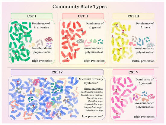
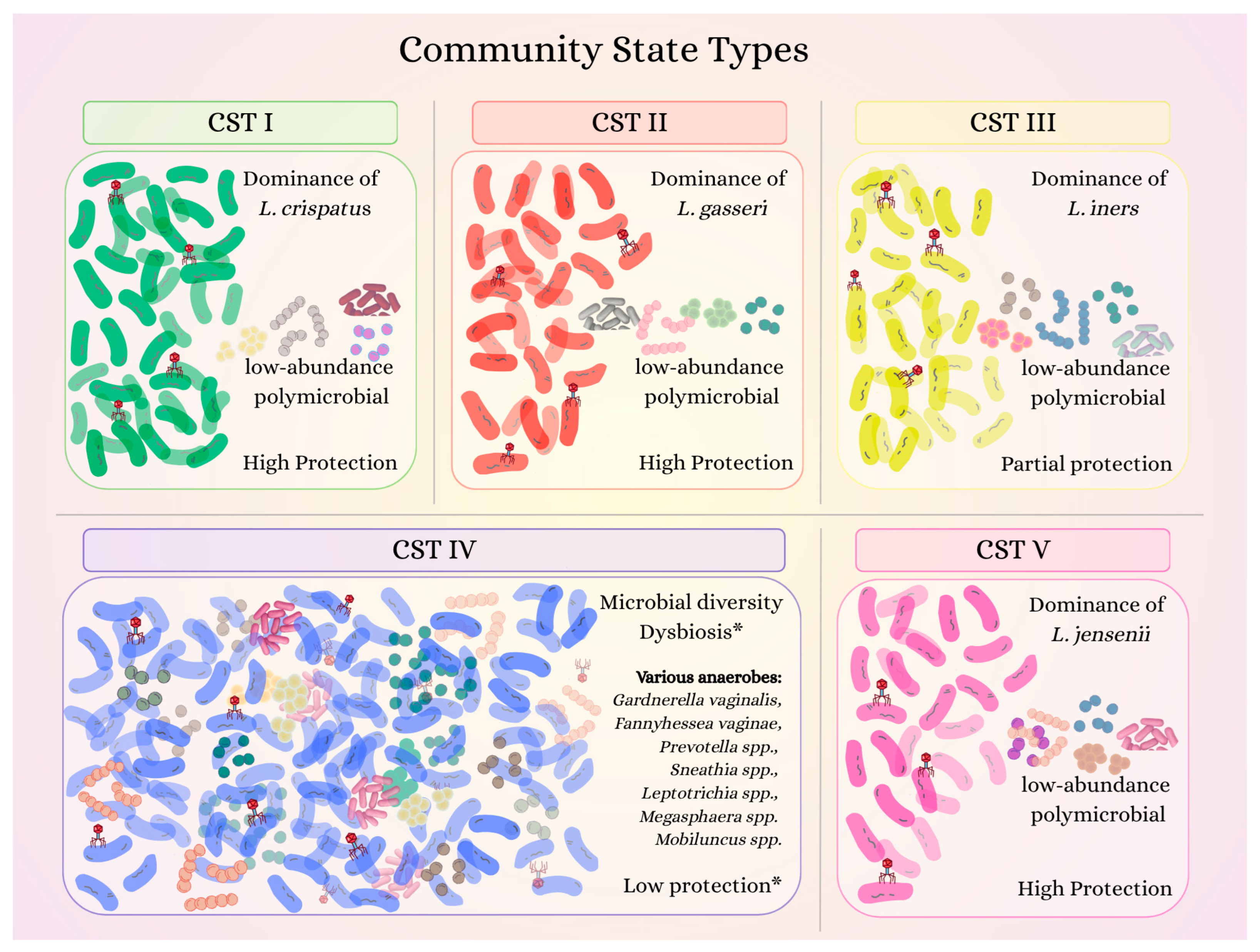
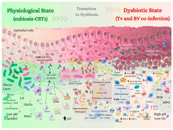
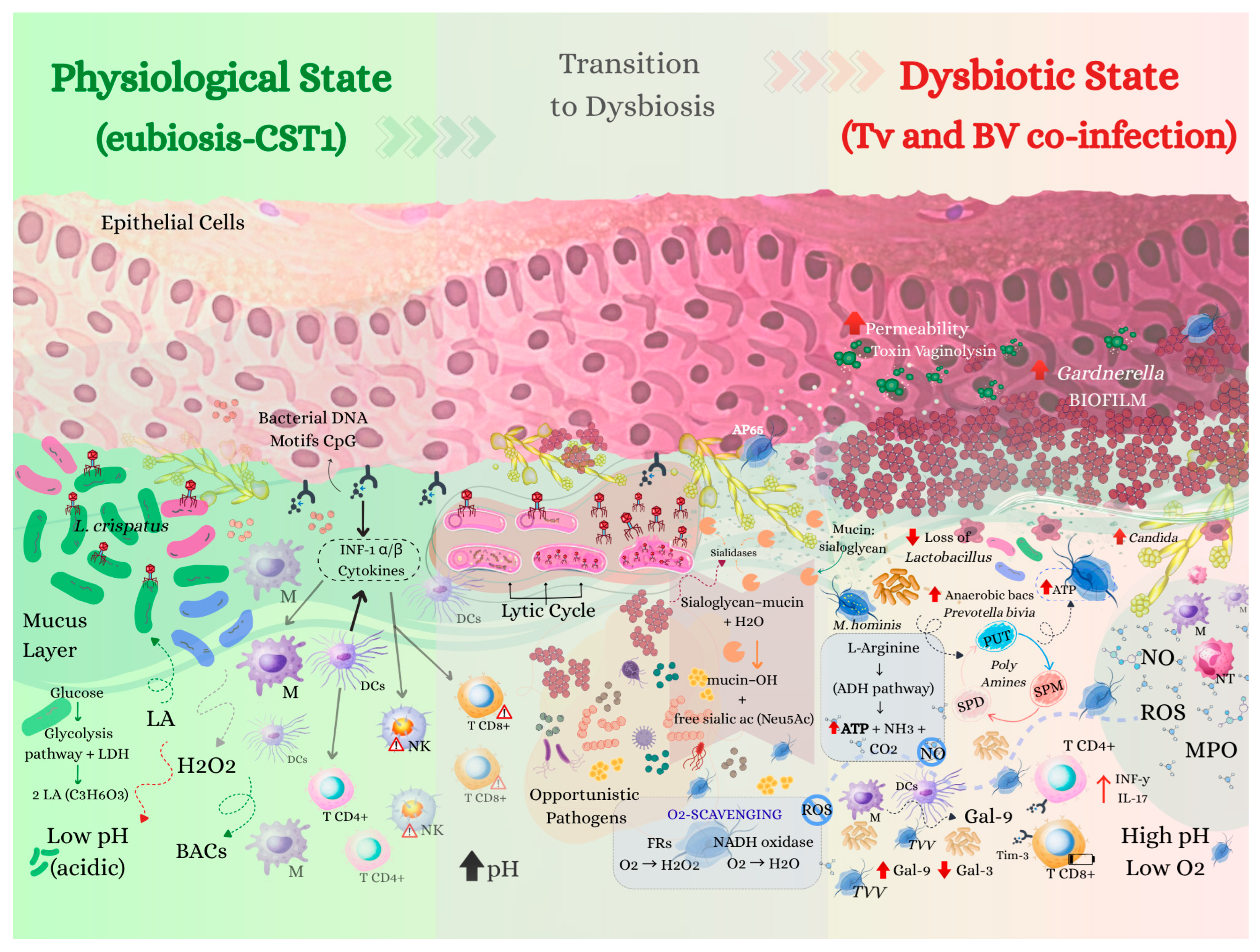
Disclaimer/Publisher’s Note: The statements, opinions and data contained in all publications are solely those of the individual author(s) and contributor(s) and not of MDPI and/or the editor(s). MDPI and/or the editor(s) disclaim responsibility for any injury to people or property resulting from any ideas, methods, instructions or products referred to in the content. |
© 2026 by the authors. Licensee MDPI, Basel, Switzerland. This article is an open access article distributed under the terms and conditions of the Creative Commons Attribution (CC BY) license.