


Abstract
MicroRNAs (miRNAs) are key post-transcriptional regulators of gene expression and play a fundamental role in host response to viral infections. SARS-CoV-2 has been shown to dysregulate host miRNA expression, potentially as a mechanism to modulate cellular pathways for its own replication or to evade immune responses. However, evidence from the literature related to the specific miRNA landscape in SARS-CoV-2 infection is conflicting and unclear. We employed an integrative multi-model approach using human nasal lung epithelial cell lines and pulmonary organoids to map miRNA expression dynamics after SARS-CoV-2 infection. Data obtained from the CALU-3 miRNAome were successively validated in other two cellular models (hAEC and hLORG), and from this comparative analysis, two miRNAs, miR-141-3p and miR-33a-5p, were found to be significantly dysregulated. These miRNAs are involved in critical pathways, including cytokine-mediated signalling, apoptotic processes and epithelial cell junction integrity. High-throughput miRNA profiling was followed by functional enrichment analysis of their predicted targets to delineate affected biological pathways. By highlighting candidate miRNAs and regulatory pathways that may contribute to disease pathogenesis, we identified robust, infection-associated hallmarks across cell models, establishing a foundation for future therapeutic strategies for COVID-19 based on the development of miRNA-guided approaches.
1. Introduction
Small non-coding RNAs, particularly microRNAs (miRNAs), are key post-transcriptional regulators of gene expression that control mRNA stability and translation. They orchestrate a wide range of cellular processes, including proliferation, differentiation, apoptosis, and immune responses, and their dysregulation has been implicated in numerous pathological conditions, such as cancer, cardiovascular, neurological, and autoimmune diseases. In the immune system, miRNAs play fundamental roles in immune cell differentiation, maturation, and activation, thereby shaping host responses to infectious agents [1].
Viruses have evolved multiple strategies to exploit host miRNA pathways to promote their replication and persistence. Viral infections can alter host miRNA expression profiles, thereby modulating immune signalling and influencing disease progression [2]. In addition, viral RNAs may function as competitive endogenous RNAs (ceRNAs), acting as “miRNA sponges” through miRNA response elements (MREs) that sequester host miRNAs and interfere with their regulatory activity [3]. This mechanism has been described for several viruses, including Hepatitis C virus and herpesviruses, where disruption of miRNA-mediated regulation contributes to immune evasion, inflammation, and viral pathogenesis [4].
In coronaviruses, both genomic and subgenomic SARS-CoV-2 RNAs have been predicted to contain numerous MREs with high affinity for human miRNAs, suggesting that viral transcripts may compete with endogenous mRNAs for miRNA binding. This interaction has been proposed as an additional mechanism contributing to the widespread transcriptomic alterations observed during infection by deregulating cellular pathways involved in inflammation, apoptosis, antiviral defence, and tissue homeostasis.
The COVID-19 pandemic has further highlighted the importance of miRNAs in viral diseases. Several studies have identified altered circulating and intracellular miRNA signatures in SARS-CoV-2-infected patients, with specific expression profiles correlating with disease severity, immune dysregulation, T-cell dysfunction, and modulation of viral entry factors such as ACE2 and TMPRSS2 [5,6,7,8,9,10,11,12,13,14,15,16,17,18,19]. Moreover, severe COVID-19 has been associated with a broad reduction in host miRNA abundance, leading to the hypothesis that SARS-CoV-2 may impair miRNA biogenesis through the downregulation of key components of the processing machinery, including DICER, DROSHA, and AGO2, a phenomenon also reported in other viral infections [20].
Despite the growing body of evidence demonstrating that SARS-CoV-2 infection is associated with altered miRNA expression, several key questions remain unresolved. Most studies have focused on patient samples or individual in vitro models, making it difficult to distinguish conserved virus-induced miRNA responses from model-specific changes. Moreover, the extent to which SARS-CoV-2 induces common or context-dependent alterations in the host miRNA across different lung epithelial systems remains poorly understood. Addressing this issue is essential for identifying robust miRNA signatures with potential biological and translational relevance.
To fill these knowledge gaps, we employed an integrative multi-model approach using three human lung epithelial models, including established human cell lines (i.e., CALU-3 and hAEC) and lung organoids, infected with a SARS-CoV-2 Spike pseudovirus. Because this replication-defective pseudovirus undergoes only a single round of infection, it provides a controlled system to investigate the early host response to viral entry while minimizing confounding effects associated with productive viral replication.
This study aimed to characterize the miRNA expression profile induced during the early phase of pseudovirus infection, identify miRNAs consistently or specifically modulated across different lung epithelial models, and integrate these data with transcriptomic analyses to infer the regulatory pathways potentially affected by miRNA dysregulation. To reach this goal, we first performed a comprehensive miRNAome analysis in CALU-3 cells and successively compared, validated and integrated the selected findings in two additional lung epithelial models. This approach allowed us to assess whether key miRNA changes identified in CALU-3 cells were conserved across different epithelial contexts (i.e., hLORG and hAEC).
This comparative approach enabled the identification of conserved and context-dependent miRNA-mediated regulatory networks involved in the host response to SARS-CoV-2 entry.
Our findings showed that pseudovirus infection induces selective rather than global remodelling of the host CALU-3 miRNAome. The analyses identified a limited number of reproducibly deregulated miRNAs with miR-141-3p representing the only miRNA consistently downregulated across the three cellular systems. Integration of our miRNA results and of transcriptomic datasets obtained previously in our work [21] highlighted TGFB2, ZEB2, and TIAM1 as candidate downstream targets involved in epithelial remodelling and TGF-β signalling. This data provided new insights into the post-transcriptional regulatory mechanisms activated during the early response to SARS-CoV-2 entry, identifying candidate miRNA–mRNA networks involved in epithelial remodelling during the initial stages of infection.
2. Materials and Methods
2.1. Differentiation into Human 3D Lung Organoids (hLORGs) and Cell Culture
Lung progenitor cells were derived from human induced pluripotent stem cells (hiPSCs) using the STEMdiff Lung Progenitor Kit (Stemcell Technologies, cat. #100-0230, Vancouver, BC, Canada), a serum-free medium optimized for the efficient and reproducible generation of lung progenitors, as previously described by [22]. The resulting 3D alveolospheres were matured and maintained through serial passaging, with re-embedding in Matrigel GFR (Corning, New York, NY, USA) every two weeks. Lung organoids (hLORGs) at 60 days of differentiation were then used for VSV_Pseudo SARS-CoV-2 infection studies. Prior to infection, successful differentiation of the human lung organoids (hLORGs) was confirmed by immunofluorescence analysis using established epithelial and alveolar type 2 (AT2) cell markers. Specifically, α-tubulin staining confirmed the characteristic epithelial morphology and the presence of ciliated cells. The epithelial identity of the differentiated organoids was further validated by the expression of the lung epithelial cell adhesion molecule (EPCAM). Differentiation toward the alveolar lineage was confirmed by the expression of surfactant proteins B and C (SFTPB and SFTPC), two canonical markers of alveolar type 2 pneumocytes. In addition, the expression of the viral entry receptors relevant to coronavirus infection was assessed. Immunofluorescence analysis demonstrated robust expression of ACE2, which was predominantly localized at the apical surface of AT2 cells, consistent with its physiological distribution. Likewise, DPP4 immunostaining confirmed strong positivity along the apical surface of the alveolar epithelial cells, in agreement with previous reports. The combined expression of EPCAM, SFTPB, SFTPC, ACE2, and DPP4, together with the characteristic epithelial morphology and ciliation revealed by α-tubulin staining, confirmed the successful differentiation and appropriate cellular identity of the organoids prior to infection.
Lung epithelial Calu-3 cells (ATCC HTB-55) and Human Airway Epithelial Cells (hAECs), (Epithelix Sarl, Geneva, Switzerland) were cultured in Minimum Essential Medium Eagle (SIGMA-Aldrich, Gillingham, UK) supplemented with 10% fetal bovine serum (FBS; Gibco, Thermo Fisher Scientific, Waltham, MA, USA), 1% L-Glutamine (The Cell Culture Company, Minneapolis, Minnesota, USA), and 1% Penicillin/Streptomycin ((The Cell Culture Company, Minneapolis, Minnesota, USA) ) and culture medium (Epithelix Sarl) respectively.
2.2. VSV_Pseudo SARS-CoV-2 Omicron BQ.1.1 and XBB.1.5 Strain Spike with Luciferase Reporter
The VSV_Pseudo SARS-CoV-2 S is a construct based on the recombinant vescicular stomatitis virus (rVSV) engineered to express the Spike protein of SARS-CoV-2 (GenBank: MN908947) with multiple mutations initially identified in the variants of Omicron BQ.1.1 and XBB.1.5. Pseudovirus infection is limited to a single replication cycle, which can be monitored via high-level luciferase activity.
2.3. VSV_Pseudotyped SARS-CoV-2 Infection
For VSV_pseudotyped SARS-CoV-2 infection, hLORGs were detached from the plate and briefly treated with trypsin for one minute to open the alveolospheres and facilitate viral entry. They were then infected with VSVpp.SARS-2-S virus and incubated at 37 °C with 5% CO2 for 2 h [21]. After infection, the alveolospheres were re-embedded in Matrigel GFR as 3D droplets at a cell density of approximately 1200–1600 cells/µL for 48 h before subsequent analysis. Calu-3 and hAEC cells were infected for 24 h, after which the medium was replaced, and cells were harvested 24 h later for both luciferase assays and RNA extraction.
2.4. miRNA Seq Analysis
RNA quality control was performed in infected and non-infected Calu-3 cell lines. RNA integrity was assessed using the RNA 6000 Nano Kit on a Bioanalyzer (Agilent Technologies, Santa Clara, California, USA). RNA samples were quantified using the Qubit RNA BR Assay Kit (Thermo Fisher Scientific). All samples met the requirements for RNA sequencing. Starting from raw FASTQ files, the quality of sequencing reads was assessed using the FastQC software (v.0.11.9) (http://www.bioinformatics.babraham.ac.uk/projects/fastqc/ accessed on 1 July 2025), adapters were trimmed and reads with length < 18bp or >27bp were filtered out with cutadapt (4.1). Filtered reads were aligned to miRBase v.22 using SHRiMP2 (2.2.3). Mature miRNAs were assigned to the fragments based on the mapping position on the hairpin miRNA precursors. The number of unique-UMI mapped fragments were counted for each mature miRNA. UMI counts were normalized as CPM (Counts Per Million), dividing them by the total mapped reads per sample and multiplying by one million. Differential miRNA expression analysis between samples was performed with DESeq2 (1.30.1). To generate more accurate Log2 FoldChange estimates for low-expressed genes, the shrinkage of the Log2 FoldChange was performed applying the “apeglm” algorithm or the “normal” algorithm. Log2 FoldChanges without any shrinkage are also provided, as well as Log2 FoldChanges derived from CPM values. Unless otherwise stated, the default parameters were utilized when applying the different software. Differential expression analysis was performed considering all samples.
2.5. Gene Expression Analysis
Trizol Reagent (Invitrogen Life Technologies Corporation, Carlsbad, CA, USA) was used to extract total RNA from cells, according to the manufacturer’s instructions. Total RNA samples were treated with DNase I-RNase-free (Ambion, Life Technologies Corporation, Foster City, CA, USA) to remove genomic DNA contamination. miRNA expression analysis was performed using TaqMan™ Advanced miRNA Assays for both cDNA synthesis and quantitative real-time PCR (RT-qPCR), in accordance with the manufacturer’s instructions. To analyze the gene expression of the miRNA target, 1 μg of RNA was reverse-transcribed and used in RT-qPCR using the Life Technologies Corporation’s High-Capacity cDNA Archive kit (Foster City, CA, USA). Candidate reference miRNAs were evaluated for expression stability in each cell line: miR-23a-5p was selected as the endogenous control for the Calu-3 cell line, while miR-24a-5p was identified as the most stable reference miRNA and was therefore used for normalization in the other two cell lines (i.e., hLORGs and hAECs). SYBR Green was used to assay mRNAs (Life Technologies Corporation, Foster City, CA, USA). GAPDH was employed as the reference gene. Primer sequences are described in Table S1. The 2−(ΔCt) and comparative ΔΔCt methods were used to quantify relative gene expression levels.
2.6. miRNA Target Identification
To investigate potential regulatory interactions between miRNA and its downstream targets, an integrative in silico approach was used. Predicted target genes were obtained from TargetScan, miRDB [23], and miRTarBase [24] and then merged into a non-redundant candidate list. This list was intersected with the genes found to be significantly upregulated in previously generated RNA-seq datasets from SARS-CoV-2-infected hLORGs [24].
2.7. Functional Enrichment
Functional interpretation of these candidate targets was performed through Gene Ontology (GO) and pathway enrichment analyses using cluster Profiler [25]. The entire human transcriptome was used as the background gene set, and enrichment was assessed across GO Biological Process, Molecular Function, and Cellular Component categories, as well as KEGG and Reactome pathways. Significance was determined using Benjamini–Hochberg FDR correction, and only terms with an adjusted p-value below 0.05 were retained.
2.8. Quantification and Statistical Analysis
miRNAoma analysis was performed on three independent Calu-3 biological replicates (infected and control) generated from three independent experiments. RT-qPCR validation experiments were performed in technical duplicates for each biological sample, and data are presented as mean ± SD of the technical replicates. Data were analyzed using GraphPad Prism 8 and the SPSS programme, version 25 (IBM Corp, Armonk, NY, USA). The difference between groups was tested by the two-tailed Student’s t-test for independent samples.
3. Results
3.1. SV_Pseudo SARS-CoV-2 Infection Triggers miRNA Deregulation in Calu-3 Cells
To investigate how SARS-CoV-2 infection alters post-transcriptional regulation in human lung and epithelium cell lines, we performed a comprehensive miRNAome analysis in Calu-3 cells, derived from human bronchial submucosal glands. Calu-3 cells are a well-characterized model suitable for modelling SARS-CoV-2 viral entry and innate immune activation due to their tight junctions, polarized architecture, and robust interferon response [26].
Cells were infected with SV_Pseudo SARS-CoV-2 S (Omicron BQ.1.1 variant) and the control for 24 h, followed by an additional 24 h post-infection before RNA extraction. Infection efficiency was evaluated via luciferase assay (Figure S1A).
For miRNA profiling, sequencing reads were analyzed using the DESeq2 package, which applies to a negative binomial model to account for biological variance in count data. Low-abundance miRNAs were filtered out, and normalization was performed using the median-of-ratios method to adjust for differences in sequencing depth and RNA composition. Statistically significant differences between infected and mock (control) samples were identified through Wald tests, with false discovery rate (FDR) correction applied using the Benjamini–Hochberg procedure. A threshold of adjusted p < 0.05 and |Log2 Fold Change| > 0.5 was used to define differentially expressed miRNAs.
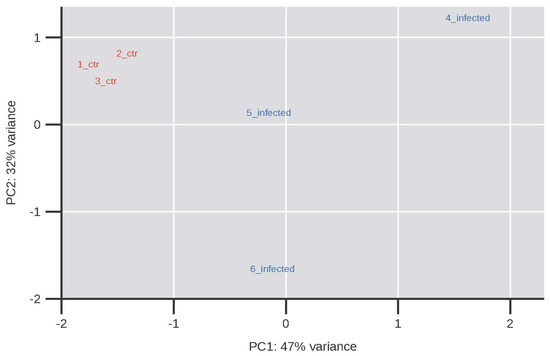
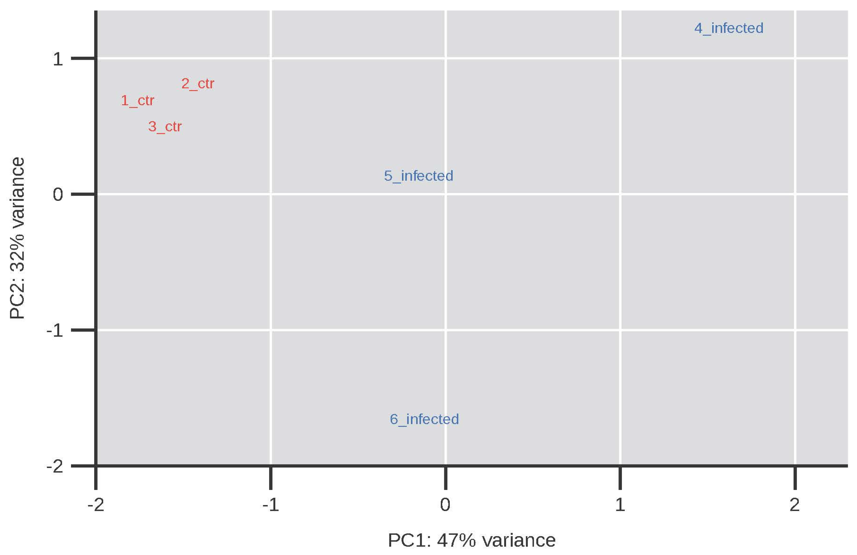
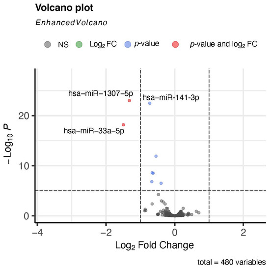
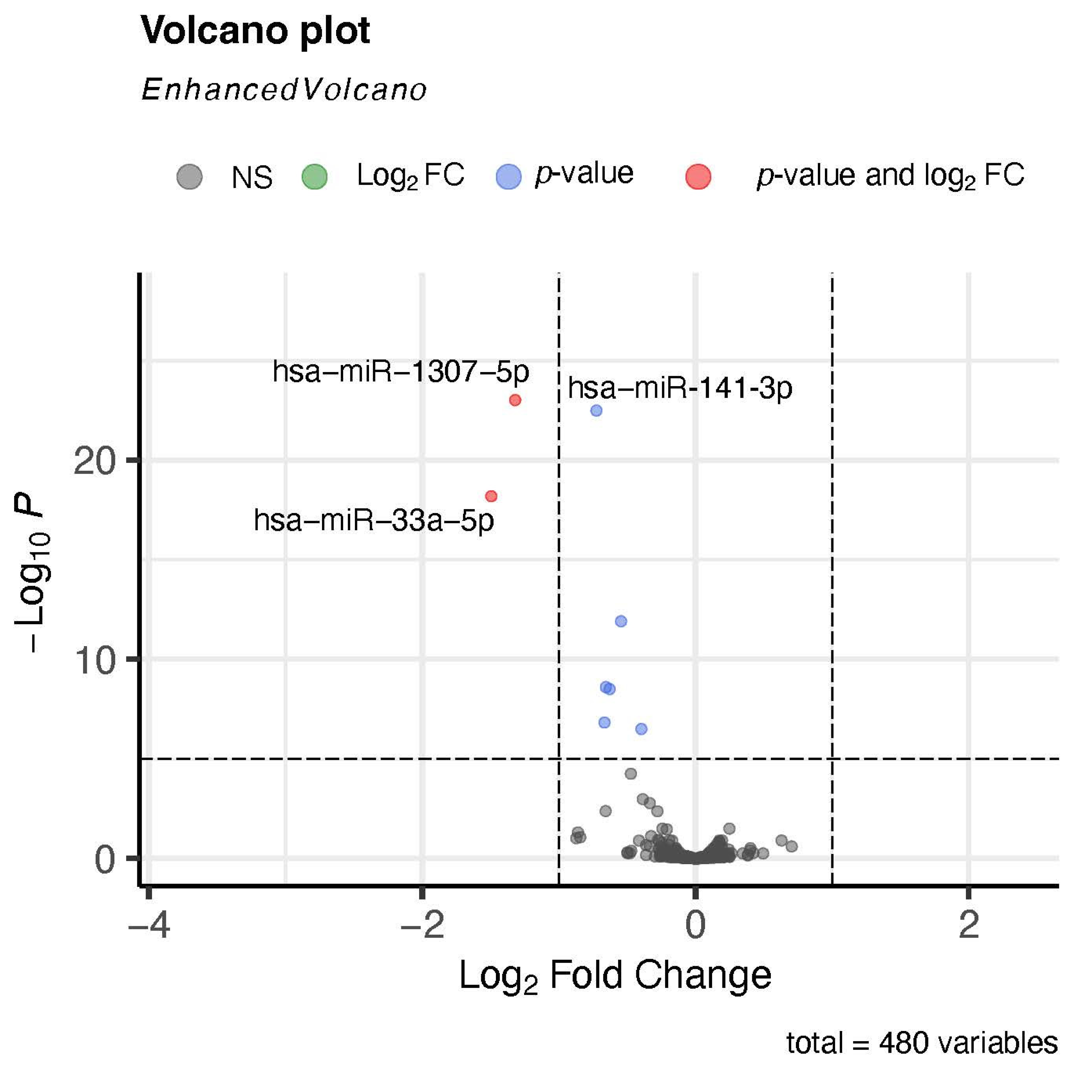
Principal Component Analysis (PCA) revealed a clear separation between infected and control Calu-3 cells, indicating robust SARS-CoV-2-driven remodelling of the miRNA expression pattern (Figure 1). Differential expression analysis identified a defined subset of significantly deregulated miRNAs in infected Calu-3 cells (Figure 2). The overall landscape was strongly asymmetric: of the 17 miRNAs (FDR < 0.05), 16 were downregulated upon infection and only one, miR-15b-5p, was significantly upregulated (log2FC = +0.25) (Table S2). This near-uniform loss of mature miRNA abundance is consistent with a global attenuation of host post-transcriptional control during SARS-CoV-2 replication, a pattern that has been attributed to viral sequestration of host miRNAs and broader perturbation of the miRNA pathway rather than to impaired miRNA biogenesis per se [27,28]. The single upregulated species, miR-15b-5p, is a recognized regulator of cell-cycle arrest and apoptosis through direct targeting of cyclin D1 (CCND1) [29], and its selective induction against a background of widespread repression may reflect a targeted host response rather than a passive expression shift. Among the most strongly repressed miRNAs were miR-33a-5p (log2FC = −1.49; FDR = 6.5 × 10⁻¹⁹) and miR-141-3p (log2FC = −0.54; FDR = 1.3 × 10⁻¹²). miR-33a-5p is an intronic miRNA of SREBF2 that constrains cholesterol efflux and fatty-acid oxidation by repressing ABCA1 and CPT1A [30]; its marked downregulation is notable given the well-documented dependence of coronavirus replication on host lipid and cholesterol metabolism, and may indicate a de-repression of lipid pathways exploited by the virus. miR-141-3p, a member of the miR-200 family, controls epithelial identity by targeting the E-cadherin repressors ZEB1 and ZEB2 [31,32], suggesting that its reduction could contribute to the epithelial remodelling observed in infected airway cells. A convergent autophagy-related signature also emerged: the miR-30 family (miR-30a-5p, miR-30e-5p), which represses the core autophagy effector BECN1 [33], and miR-101-3p, which represses the autophagy regulators ATG4D, RAB5A and STMN1 [34], were all coordinately downregulated, consistent with the extensive co-option of autophagy during SARS-CoV-2 infection. Finally, several PTEN/PI3K–AKT–FOXO-associated miRNAs (miR-32-5p, miR-301a-3p, miR-96-5p) were significantly reduced, further supporting a broad reconfiguration of proliferative and pro-survival signalling in the Calu-3 model. Together, these changes point to a coordinated post-transcriptional program affecting lipid metabolism, autophagy, epithelial architecture, and survival signalling during infection.
Figure 1.
PCA of miRNA expression profiles showing a clear separation between control (ctr) and infected Calu-3 cells.
Figure 2.
Volcano plot of differentially expressed miRNAs in Calu-3 cells. The plot displays 480 miRNAs according to their log2 fold change (x-axis) and –log10 (p-value) (y-axis). Grey points indicate non-significant miRNAs, while blue points mark those significant by p-value or fold change. Red points represent miRNA meeting both thresholds, hsa-miR-33a-5p. Dashed lines denote the significance cutoffs.
3.2. RT-qPCR Validation Confirms the Deregulation of miR-141-3p and miR-33a-5p Expression
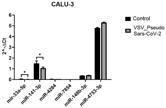
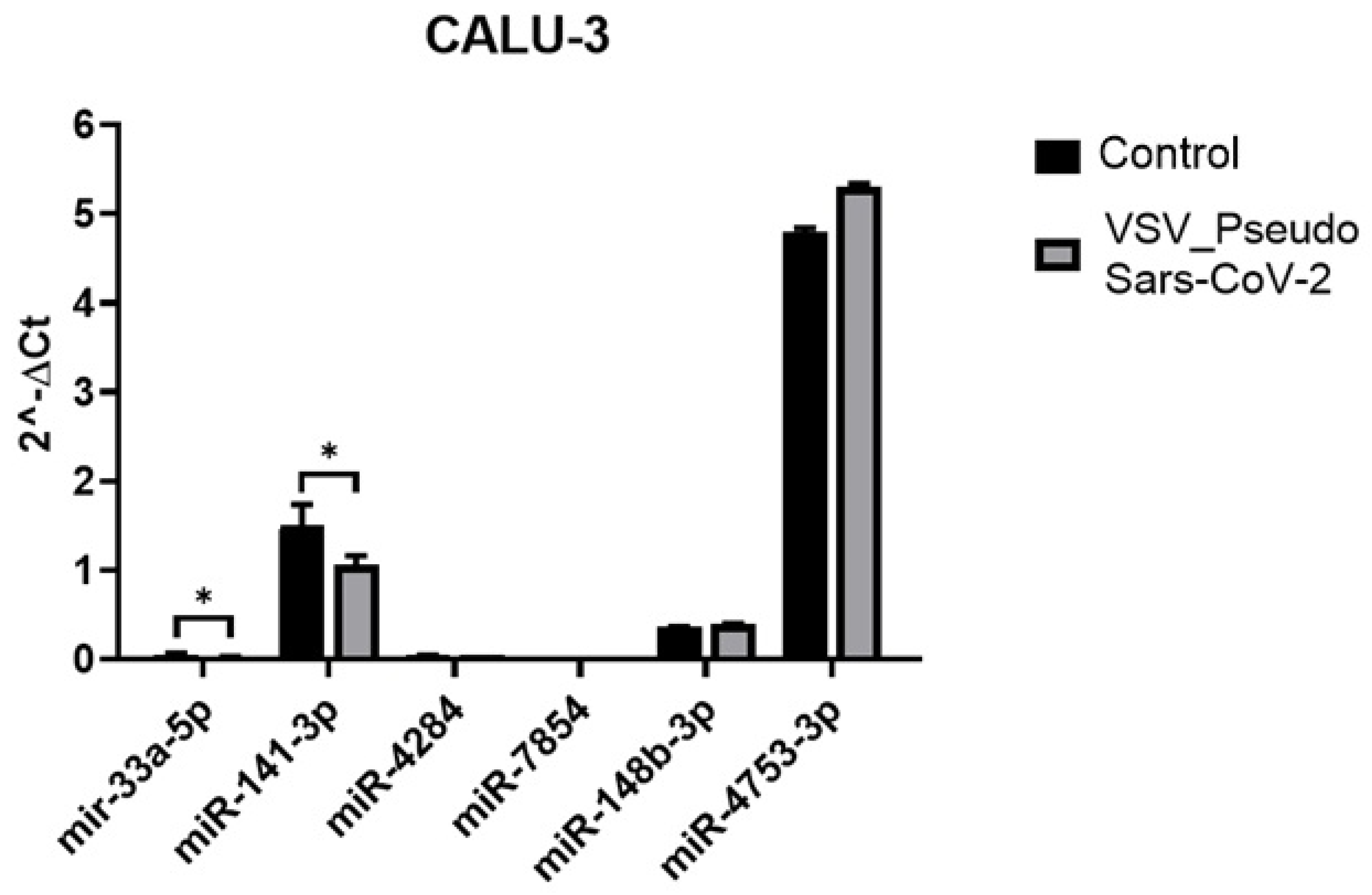
To validate the miRNA-seq results in CALU-3 cells, a subset of miRNAs showing nominal p-value < 0.05 and |log2FC| > 0 was selected for RT-qPCR analysis. Particular attention was given to miR-141-3p and miR-33a-5p, due to their substantial deregulation and known biological relevance to lung epithelial homeostasis and viral response pathways. Additional miRNAs were included for comparison, miR-148b-3p and miR-4753-3p, both displaying mild or no variation in the sequencing dataset, and two non-deregulated miRNAs (4284 and 7854) serving as negative controls (Figure 3).
Figure 3.
Real-time RT-qPCR analyses of miR33a-5p, -141-3p, -4284, -7854, -148b-3p, and -4753-3p in CALU-3 control and after BQ1.1 SV_Pseudo infection. The data were normalized to internal control small nucleolar 23a-5p. Data are from two independent experiments and represented as mean ± SD; (* p < 0.05).
RT-qPCR results were consistent with sequencing data: both miR-141-3p and miR-33a-5p were significantly downregulated in infected Calu-3 cells compared to controls. The reduction in miR-141-3p expression was particularly marked, reinforcing its role as a robust and reproducible marker of SARS-CoV-2-induced transcriptional reprogramming. miR-33a-5p also displayed significant downregulation, albeit with more modest fold changes, possibly due to its lower baseline expression in Calu-3 cells or to post-transcriptional buffering mechanisms limiting its dynamic range. Control miRNAs (i.e., miR-148b-3p and miR-4753-3p) showed no significant variation.
3.3. miRNA RT-qPCR Further Highlights the Deregulation of miR-141-3p and miR-33a-5p Expression in Human Lung Organoids (hLORGs) and in hAECs
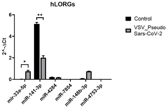
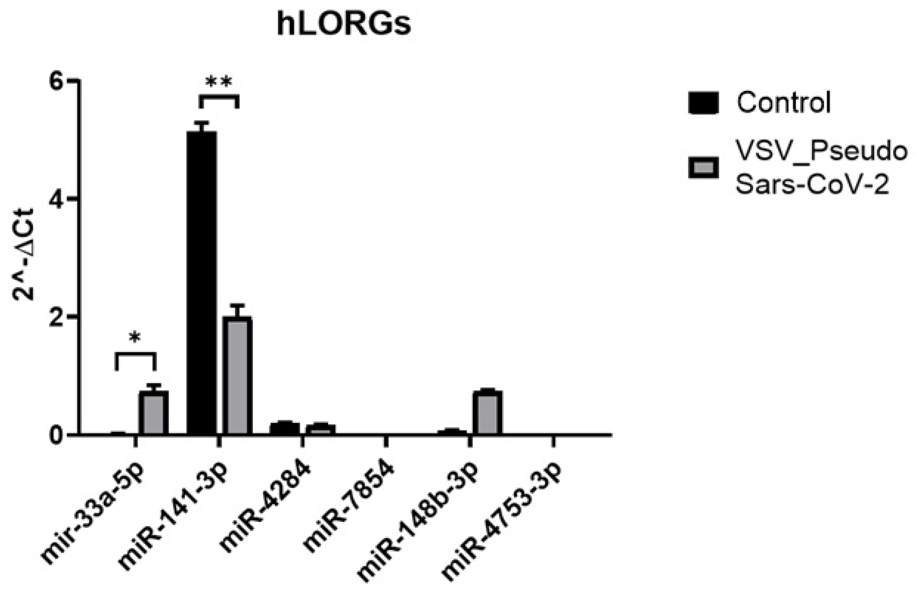
The same subset of miRNAs was evaluated in human lung organoids (hLORGs) derived from human induced pluripotent stem cells (hiPSCs). The organoid structures, described in detail in our previous work [21], were exposed for 24 h to SV_Pseudo SARS-CoV-2 (Omicron BQ.1.1 variant) and control. After viral removal, cells were maintained for an additional 24 h before collection. Infection efficiency was evaluated via luciferase assay (Figure S1B). The same subset of miRNA was validated by RT-qPCR analysis (Figure 4) in hLORGs, which confirmed the upregulation of miR-33a-5p but revealed a marked downregulation of miR-141-3p, suggesting a discrepancy between the two cell lines.
Figure 4.
Real-time RT-PCR analyses of miR-4284, -7854, -33a-5p, -141-3p, -148b-3p, and -4753-3p in hLORG control and after BQ1.1 pseudovirus infection. The data were normalized to internal control 24a-5p. Data are from two independent experiments and represented as mean ± SD; (* p < 0.05), (** p < 0.01).
Other miRNAs tested—miR-148b-3p and miR-4753-3p, which showed minimal or inconsistent changes in sequencing—did not display significant differences in RT-qPCR.
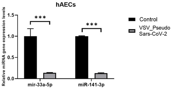
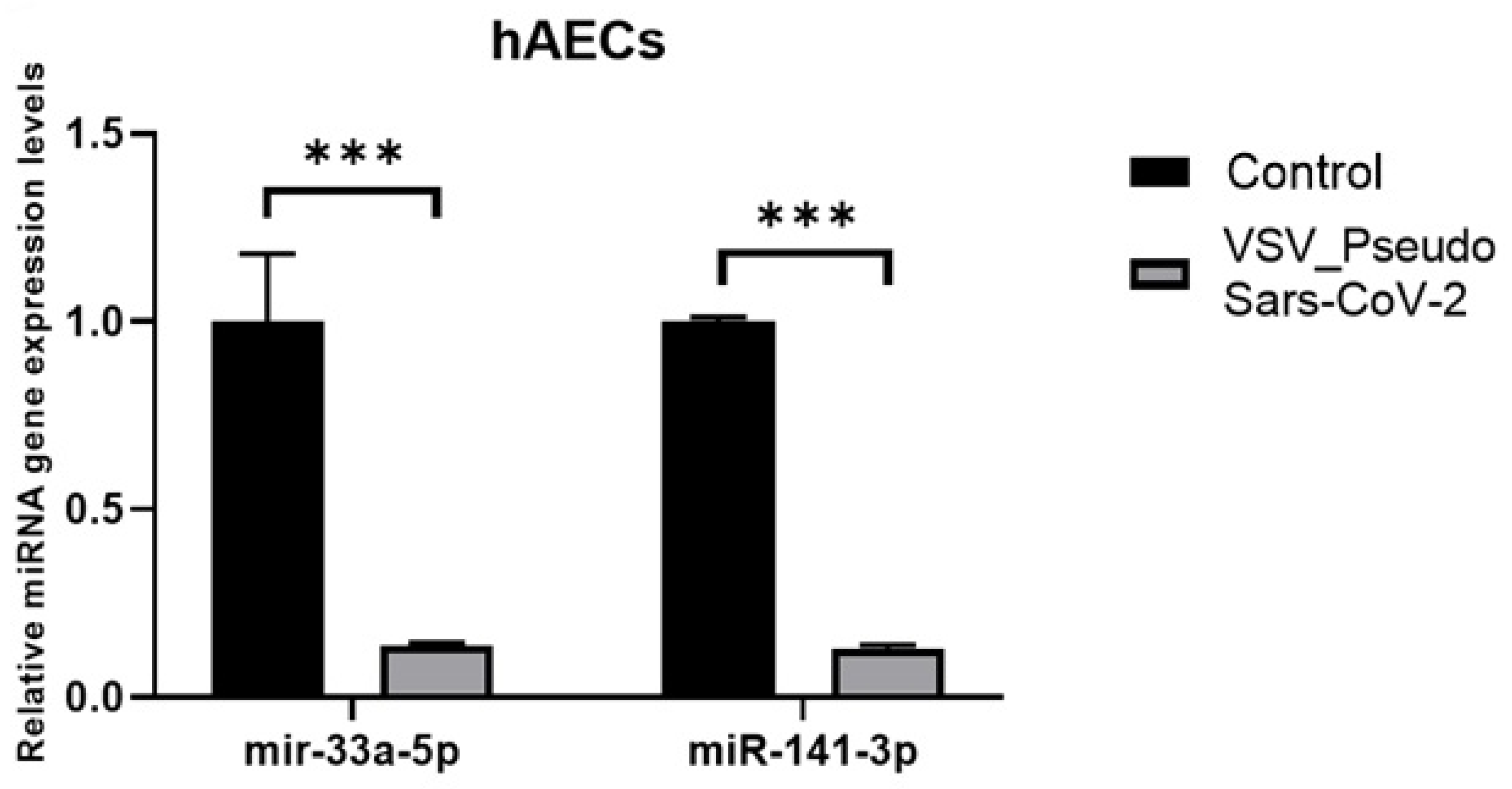
These experiments were further assessed in Human Airway Epithelial Cell (hAEC) cultures, which morphologically and functionally resemble the upper conducting airways in vivo and represent the primary site of viral entry. In addition to validating the miRNA expression data using the BQ.1 strain, the same experimental procedures were replicated in hAECs using the VSV_Pseudo SARS-CoV-2 Omicron XBB.1.5 variant, which subsequently replaced BQ.1.1 as the predominant circulating variant. Infection efficiency was evaluated via luciferase assay (Figure S1C). RT-qPCR analyses confirmed a statistically significant downregulation of the two principal miRNAs under investigation, showing an approximately 90% reduction compared to control cells (p *** < 0.001) (Figure 5).
Figure 5.
Real-time RT-qPCR analyses of miR-33a-5p and miR-141-3P in hAEC control and after XBB.1.5 pseudovirus infection. The data were normalized to internal control 24a-5p. Data are from two independent experiments and represented as mean ± SD; (*** p < 0.001).
Importantly, the identification of the miR-141-3p downregulation in all three respiratory models infected by SV_Pseudo SARS-CoV-2 allowed us to define this miRNA as the most consistent infection-associated hallmark.
3.4. Gene Expression Analyses of Innate Immunity and Inflammation Markers
Although we are using SV_Pseudo SARS-CoV-2 [21], we have already demonstrated that pseudovirus infection significantly increases antiviral and inflammatory markers at the gene expression level, including IFN-β, IFN-λ1, IFIT1, MX2, CXCL10, IL-6, and TNF-α, indicating activation of key innate immune pathways [21].
Parallelly to miRNA differential expression, we determined whether infection by SV_Pseudo SARS-CoV-2 triggered a similar immune response at the transcriptional level across different cell lines (i.e., hLORG, CALU3, hAEC) by evaluating the expression of the two most significantly key inflammatory and antiviral genes, TNF-α and interferon-β (IFN-β). Transcript levels were quantified by RT-qPCR both prior to and following infection with VSV-pseudo-SARS-CoV-2. In all three cellular models, we observed a significant upregulation of TNF-α and IFN-β post-infection. As shown in Figure S1D, while control cells displayed basal levels of expression, infection with SV_Pseudo SARS-CoV-2 induced a significant increase in both transcripts. Notably, the increase in expression for both genes was comparable across models, suggesting a reproducible innate immune activation in response to the pseudovirus.
3.5. Identification of miR-141 Target Genes Downregulated in SARS-CoV-2-Infected hLORGs
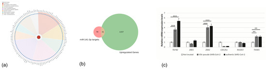
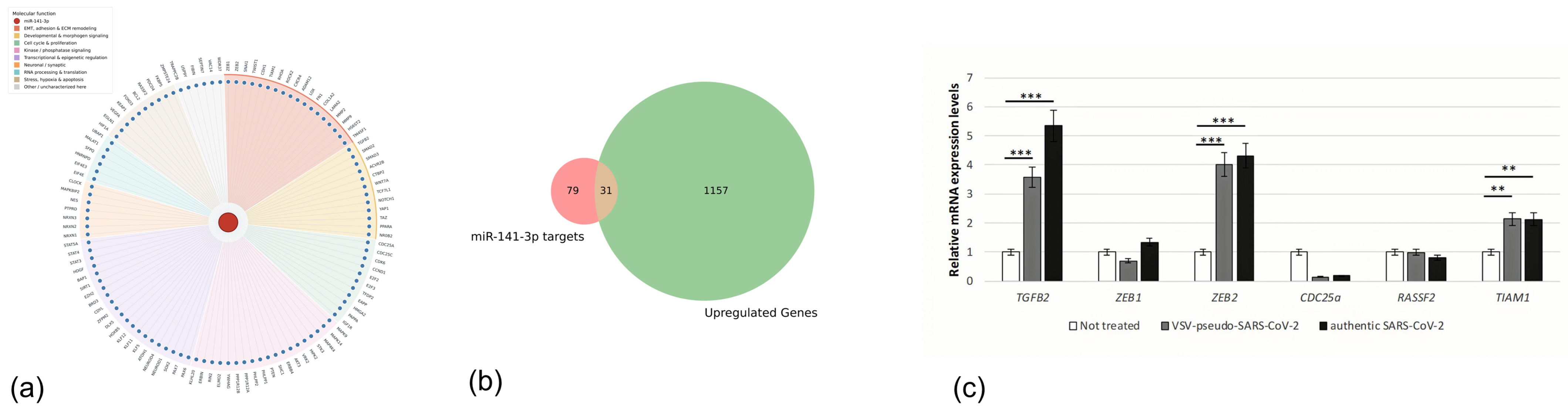
Among the analyzed targets, one miRNA, miR141-3p, consistently emerged as the most stable and robustly downregulated across all three cellular models. Its expression levels were significantly reduced in infected cells compared to their respective controls, regardless of the viral strain or cell type. This consistent pattern suggests a potential function for this miRNA as a reliable biomarker of the molecular response to infection. To elucidate the possible role of miR-141-3p in the host cellular response to SARS-CoV-2 infection, we performed an integrative analysis combining in silico miRNA target prediction with transcriptomic data from RNA sequencing of hLORGs infected with SV_Pseudo SARS-CoV-2 (Omicron XBB.1.5) in our previous study [24]. Figure 6a shows the complete network of predicted miR-141-3p target genes, with nodes coloured according to their molecular function. To pinpoint transcripts potentially under direct miR-141-3p regulation and concurrently modulated during the host response, we intersected this target set with the genes significantly upregulated in pseudovirus-infected hLORGs, yielding the intersection shown in Figure 6b. This approach identified 31 candidate target genes, the complete list of which is provided in Table S3. Among these, TGFB2, RASSF2, ZEB1, TIAM1, CDC25A, and ZEB2 were selected for experimental validation because of Fold Changes > 1.5. Subsequent Gene Ontology (GO) and pathway enrichment analyses of the identified targets highlighted a significant enrichment in the transforming growth factor beta receptor signalling pathway, regulation of cell differentiation, and epithelial-to-mesenchymal transition (EMT), suggesting a possible role for miR-141-3p in modulating key signalling cascades relevant to lung epithelial cell function and integrity during infection.
Figure 6.
Predicted miR-141-3p target network and overlap with the host transcriptional response. (a) Network of the predicted target genes of hsa-miR-141-3p (central red node). Each surrounding blue node represents an individual predicted target gene, and edges denote predicted regulatory interactions inferred from target prediction analyses. Target genes are arranged into sectors according to their primary molecular function, indicated by the shaded background and the accompanying legend. (b) Venn diagram showing the overlap between the predicted miR-141-3p target genes and the genes significantly upregulated in pseudovirus-infected hLORGs. Of the two sets, 31 genes were shared, representing candidate transcripts that may be directly regulated by miR-141-3p while being concurrently modulated during the host response. (c) RT-qPCR analyses of miR-141 target genes in hLORGs before and after infection with VSV-Pseudo-SARS-CoV-2 and authentic SARS-CoV-2. Data are from three independent experiments and represented as mean ± SD. ** p < 0.01 and *** p < 0.001 by one-way ANOVA test.
To validate the biological relevance of the miR-141-3p regulatory network identified, we performed RT-qPCR analyses in hLORGs infected with either SV_Pseudo SARS-CoV-2 or authentic SARS-CoV-2 (Figure 6c). The authentic infection model was previously established and extensively characterized by our group, demonstrating productive viral replication together with robust activation of antiviral and inflammatory pathways; moreover, the live virus was already available and being used in parallel for other SARS-CoV-2 studies [24]. Thus, we first examined the expression of candidate miR-141-3p target genes identified through the integration of target prediction analyses and transcriptomic data. Among the selected candidates, TGFB2, ZEB2, and TIAM1 were consistently upregulated following both pseudovirus and authentic SARS-CoV-2 infection (Figure 6c). Notably, TGFB2 expression was induced under both conditions but showed a stronger increase in cells infected with authentic SARS-CoV-2, suggesting that additional viral replication-dependent mechanisms may contribute to its regulation. In contrast, TIAM1 and ZEB2 displayed comparable upregulation in both infection models, indicating that their induction is largely associated with virus-triggered host responses reproduced by the pseudovirus system. Conversely, CDC25A and RASSF2 expression changes could not be confirmed by RT-qPCR. Overall, these findings validate the transcriptomic predictions and demonstrate that key components of the miR-141-3p regulatory network are consistently activated in both pseudovirus and authentic SARS-CoV-2 infection models. The concordant upregulation of TGFB2, ZEB2, and TIAM1 further supports their involvement as biologically relevant downstream targets associated with miR-141-3p repression during SARS-CoV-2 infection.
4. Discussion
While proteomic and transcriptomic studies have clarified the mechanisms of SARS-CoV-2 pathogenesis, the contribution of post-transcriptional regulatory mechanisms remains less clearly defined. Given that miRNAs fine-tune gene expression networks involved in antiviral defence, inflammation and tissue homeostasis, understanding their modulation during infection is essential for a holistic view of host–virus interactions and could hold significant promise for developing novel antiviral strategies, such as therapies based on antisense oligonucleotides.
To dissect the expression regulatory mechanisms of miRNAs and compare/validate the obtained data across distinct biological models, we adopted a stepwise approach, starting from a more simple and experimentally tractable model like a human lung epithelial cell line (CALU-3), moving to a complex cellular system such as human lung organoids (hLORGs) and progressing toward airway epithelial cells (hAECs), the primary route of viral entry into the host. This multi-model strategy allowed us to distinguish a common, conserved, infection-driven miRNA hallmark from context-specific variations.
hLORGs, Calu-3 cells, and hAECs are widely used and physiologically relevant models for studying SARS-CoV-2 infection. hLORGs closely reproduce the architecture and function of human lung tissue [21], while Calu-3 cells mimic bronchial epithelial cells and serve as a sensitive preclinical model for respiratory diseases [26]. hAECs, representing the first cells encountered by the virus, support SARS-CoV-2 replication and are valuable for investigating early stages of infection.
The VSV-based pseudovirus system faithfully reproduces Spike-mediated viral entry and the early host responses triggered by virus–receptor interactions [35]. Consistent with these observations, we previously demonstrated that the same pseudovirus platform induces robust activation of antiviral and inflammatory pathways in human lung organoids [21]. Furthermore, studies performed in our laboratory using replication-competent (authentic) SARS-CoV-2 in the same organoid model validated many of the host responses identified after pseudovirus infection [24]. This supports the biological relevance of this experimental system for investigating conserved early host responses to SARS-CoV-2 infection.
Although SARS-CoV-2 infection led to minimal global changes in miRNA expression—affecting less than 10% of the CALU-3 miRNAome—a discrete subset of miRNAs exhibited virus-specific modulation. This study examines these regulatory changes and their implication in different cell lines to better comprehend the context-dependent nature of host responses and to pinpoint candidate pathways perturbed during infection. While miRNAs hold considerable promise as therapeutic agents and biomarkers, their clinical translation remains limited by the difficulty of identifying stably expressed candidates across diverse cellular systems.
Among all deregulated miRNAs, miR-141-3p emerged as the only transcript consistently downregulated across Calu-3 cells, hLORGs and primary airway epithelial cells. The reproducibility of this finding across distinct respiratory systems suggests that miR-141-3p repression represents a conserved component of the epithelial response to SARS-CoV-2 exposure. This observation is particularly meaningful because miR-141-3p is a well-known regulator of epithelial identity, cytoskeletal stability, and innate immunity, all processes that viruses frequently manipulate during infection. Although numerous studies have identified circulating miRNA profiles associated with COVID-19 severity and clinical outcome, miR-141-3p has not consistently emerged among the most widely reported circulating miRNAs associated with COVID-19. Alterations in tissue-specific miRNAs are not necessarily reflected in the circulation; therefore, we proposed miR-141-3p as a potential marker of epithelial responses to SARS-CoV-2. However, a recent clinical study described significantly reduced circulating levels of miR-141-3p in SARS-CoV-2-positive patients, suggesting that its dysregulation may also occur in vivo. Future studies directly comparing respiratory tissues with corresponding blood samples will be needed to determine whether the conserved epithelial signature identified here is also reflected in the systemic compartment and may therefore have potential as a clinical biomarker [36].
Previous studies have shown that miR-141-3p modulates type I interferon signalling and epithelial-to-mesenchymal transition (EMT) [37,38], suggesting that its suppression may weaken antiviral defences and alter epithelial homeostasis.
To understand the functional implications of this downregulation, we reconstructed the miR-141-3p target network by integrating bioinformatic predictions with expression data. The predicted miR-141-3p target network was enriched for genes involved in epithelial differentiation, TGF-β signalling and tissue remodelling, suggesting that reduced miR-141-3p expression may contribute to transcriptional programmes activated during infection.
The first cluster of targets converged on cytoskeletal dynamics, a critical interface for viral entry and intracellular trafficking. Among these, RHOA, a master regulator of actin organization, emerged as a central node. Its de-repression may facilitate membrane rearrangements required for viral internalization and spread, particularly in Calu-3 cells where these processes are highly active. Similarly, increased expression of TIAM1, a RAC1 activator, reinforces this remodelling landscape, potentially supporting both viral movement and EMT-related transitions [39]. A second functional module involves immune regulation. Targets such as IFI35 and BAG4, known negative regulators of interferon responses and TNF-mediated apoptosis [40], were suppressed following infection.
A third coherent axis linked miR-141-3p suppression to EMT and tissue remodelling, featuring well-established mediators such as TGFB2, ZEB1 and ZEB2. Interestingly, TGFB2 expression appeared more strongly induced following infection with authentic SARS-CoV-2 than with the pseudovirus system. This observation suggests that while viral entry is sufficient to activate part of the miR-141-3p-associated regulatory network, additional replication-dependent mechanisms may further amplify TGF-β-related responses. Their upregulation points toward a shift toward pro-fibrotic transcriptional reprogramming, reminiscent of the fibroproliferative remodelling described in severe COVID-19 phenotypes [21,35]. This suggests that transient miRNA perturbation may prime epithelial cells for maladaptive repair responses.
An important aspect of this study is the validation of selected components of the miR-141-3p regulatory network in hLORGs infected with authentic SARS-CoV-2. Although the majority of experiments were performed using a pseudotyped virus system, which primarily reproduces viral entry events, the consistent induction of TGFB2, ZEB2 and TIAM1 following infection with replication-competent SARS-CoV-2 supports the biological relevance of the pathways identified through our integrative analyses. The concordance observed between pseudovirus and authentic virus models suggests that key elements of the miR-141-3p-associated transcriptional response are not merely artefacts of the experimental system but may represent conserved features of SARS-CoV-2-induced epithelial reprogramming.
Interestingly, these findings parallel strategies employed by large DNA viruses such as EBV and HCMV, which encode their own miRNAs to manipulate TGF-β and EMT pathways. Although SARS-CoV-2 does not encode viral miRNAs, our data suggest that it may achieve a comparable outcome by downregulating host regulatory miRNAs, such as miR-141-3p, thus indirectly reshaping the TGF-β axis and EMT programmes. This indicates a convergent evolutionary strategy among unrelated viral families to modulate epithelial state and immune tone.
This study has some limitations, including the absence of functional validation of the identified miRNAs and protein-level characterization of their target transcripts. Nevertheless, these aspects fall outside the scope of the present work, which primarily aims to emphasize the potential relevance of the identified miRNAs as biomarkers of infection, as they are differentially expressed in the same way in distinct human cell types functionally associated with SARS-CoV-2 infection and are also consistently modulated by both pseudo/authentic SARS-CoV-2 viruses.
5. Conclusions
Overall, our results indicate that SARS-CoV-2 infection is associated with selective rather than widespread remodelling of the host miRNAome. Across three independent respiratory epithelial models, miR-141-3p emerged as the most reproducible infection-associated miRNA, displaying consistent downregulation following viral exposure. Its coordinated effects on cytoskeletal remodelling, immune evasion and EMT position it as a potential biomarker of infection and a promising therapeutic entry point. Although additional functional studies are required to establish causality, these results identify miR-141-3p as a promising candidate biomarker and provide further insight into post-transcriptional regulatory mechanisms associated with SARS-CoV-2 infection.
These findings lay the groundwork for future in vivo validation and support the development of miRNA-based antiviral strategies, including synthetic genetic circuits or oligonucleotide therapies designed to restore or exploit host regulatory networks.
In conclusion, this study elucidates novel aspects of SARS-CoV-2 pathogenesis, with the overarching goal of advancing current clinical and molecular understanding and providing a foundation for the design and synthesis of an inducible plasmid circuit in which gene expression is modulated by miRNA activity, as a proof of principle for specific therapeutic intervention in viral and non-viral infections.
Supplementary Materials
The following supporting information can be downloaded at https://www.mdpi.com/article/10.3390/covid6080142/s1, Figure S1: Transduction efficiency was quantified by measuring virus-encoded luciferase activity in hLORG (A), Calu-3 (B) and hAEC (C) infected with VSV-pseudotyped SARS-CoV-2 Omicron BQ.1.1. Data are expressed as the percentage of infection, and the average data from two biological replicates are presented. (D) RT-qPCR analyses of TNF-α and IFN-β in hLORGs, Calu-3 cells and hAEC cells before and after VSV_Pseudo SARS-CoV-2 Omicron XBB.1.5 infection. Data are from three independent experiments and represented as mean ± SD. **** p < 0.0001 by one-way ANOVA test; Table S1: Primer sequences; Table S2: list of differentially expressed miRNA in infected Calu-3 cells; TableS3: candidate target genes in hLORG.
Author Contributions
Conceptualization, M.M.; methodology, M.M., G.P., A.L. and P.S.; software, G.P.; investigation, M.M., A.L. and P.S.; writing—original draft preparation, M.M., A.L. and G.P.; review and editing, F.S., M.H.-C. and G.N.; supervision, G.N.; funding acquisition, G.N. and M.H.-C. All authors have read and agreed to the published version of the manuscript.
Funding
This research was supported by PNRR Next Generation EU, M4-C2-I1.4, National Center for Gene Therapy and Drugs based on RNA Technology, Spoke 5 (CUP E83C22003200001) to G.N. and M.M., and PNRR Next Generation EU, M4-C2-I1.4: National Center for Gene Therapy and Drugs based on RNA Technology, CN3—Spoke 7 (code: CN00000041) to MHC.
Institutional Review Board Statement
The study was conducted in accordance with the Declaration of Helsinki and approved by the Ethics Committee CET Lazio Area 2 Tor Vergata Hospital (50.26 CET2 ptv_utv, 4 June 2026) for studies involving humans.
Informed Consent Statement
Informed consent was obtained from all subjects involved in the study.
Data Availability Statement
Data will be made available on request. To request data, please write to the corresponding author.
Conflicts of Interest
The authors declare no conflicts of interest.
Abbreviations
The following abbreviations are used in this manuscript:
| miRNAs | microRNAs |
| ceRNA | Competitive Endogenous RNA |
| MREs | miRNA Response Elements |
| miRBase | microRNA Database |
| miRDB | microRNA Database |
| miRTarBase | microRNA Target Database |
| IBM | International Business Machines |
| hLORG | Human Lung Organoid |
| hiPSCs | Human Induced Pluripotent Stem Cells |
| hAECs | Human Airway Epithelial Cells |
| hAEC | Human Airway Epithelial Cell |
| ATCC | American Type Culture Collection |
| GFR | Growth Factor Reduced |
| FBS | Fetal Bovine Serum |
| HCV | Hepatitis C Virus |
| AGO2 | Argonaute 2 |
| DICER | Endoribonuclease Dicer |
| DROSHA | Drosha Ribonuclease III |
| ACE2 | Angiotensin-Converting Enzyme 2 |
| TMPRSS2 | Transmembrane Serine Protease 2 |
| TGFB2 | Transforming Growth Factor Beta 2 |
| ZEB1 | Zinc Finger E-Box Binding Homeobox 1 |
| ZEB2 | Zinc Finger E-Box Binding Homeobox 2 |
| TIAM1 | T-Cell Lymphoma Invasion and Metastasis 1 |
| RASSF2 | Ras Association Domain Family Member 2 |
| CDC25A | Cell Division Cycle 25A |
| RHOA | Ras Homologue Family Member A |
| RAC1 | Rac Family Small GTPase 1 |
| IFI35 | Interferon-Induced Protein 35 |
| BAG4 | BCL2-Associated Athanogene 4 |
| IFN-β | Interferon Beta |
| IFN-λ1 | Interferon Lambda 1 |
| IFIT1 | Interferon-Induced Protein with Tetratricopeptide Repeats 1 |
| MX2 | MX Dynamin Like GTPase 2 |
| CXCL10 | C-X-C Motif Chemokine Ligand 10 |
| IL-6 | Interleukin 6 |
| TNF-α | Tumour Necrosis Factor Alpha |
| EMT | Epithelial-to-Mesenchymal Transition |
| GO | Gene Ontology |
| FDR | False Discovery Rate |
| KEGG | Kyoto Encyclopedia of Genes and Genomes |
References
- Ebert, M.S.; Sharp, P.A. Roles for MicroRNAs in Conferring Robustness to Biological Processes. Cell 2012, 149, 515–524. [Google Scholar] [CrossRef] [Scilit] [PubMed]
- Arghiani, N.; Nissan, T.; Matin, M.M. Role of MicroRNAs in COVID-19 with Implications for Therapeutics. Biomed. Pharmacother. 2021, 144, 112247. [Google Scholar] [CrossRef] [Scilit] [PubMed]
- Pepe, G.; Guarracino, A.; Ballesio, F.; Parca, L.; Ausiello, G.; Helmer-Citterich, M. Evaluation of Potential miRNA Sponge Effects of SARS Genomes in Human. Non-Coding RNA Res. 2022, 7, 48–53. [Google Scholar] [CrossRef] [Scilit] [PubMed]
- Jopling, C.L.; Yi, M.; Lancaster, A.M.; Lemon, S.M.; Sarnow, P. Modulation of Hepatitis C Virus RNA Abundance by a Liver-Specific MicroRNA. Science 2005, 309, 1577–1581. [Google Scholar] [CrossRef] [Scilit] [PubMed]
- Zhou, H.; Ni, W.J.; Meng, X.M.; Tang, L.Q. MicroRNAs as Regulators of Immune and Inflammatory Responses: Potential Therapeutic Targets in Diabetic Nephropathy. Front. Cell Dev. Biol. 2021, 8, 618536. [Google Scholar] [CrossRef] [Scilit] [PubMed]
- de Gonzalo-Calvo, D.; Benítez, I.D.; Pinilla, L.; Carratalá, A.; Moncusí-Moix, A.; Gort-Paniello, C.; Molinero, M.; González, J.; Torres, G.; Bernal, M.; et al. Circulating microRNA Profiles Predict the Severity of COVID-19 in Hospitalized Patients. Transl. Res. 2021, 236, 147–159. [Google Scholar] [CrossRef] [Scilit] [PubMed]
- Donyavi, T.; Bokharaei-Salim, F.; Baghi, H.B.; Khanaliha, K.; Janat-Makan, M.A.; Karimi, B.; Nahand, J.S.; Mirzaei, H.; Khatami, A.; Garshasbi, S.; et al. Acute and Post-Acute Phase of COVID-19: Analyzing Expression Patterns of miRNA-29a-3p, miRNA-146a-3p, miRNA-155-5p and let-7b-3p in PBMCs. Int. Immunopharmacol. 2021, 97, 107641. [Google Scholar] [CrossRef] [Scilit] [PubMed]
- Duecker, R.P.; Adam, E.H.; Wirtz, S.; Gronau, L.; Khodamoradi, Y.; Eberhardt, F.J.; Donath, H.; Gutmann, D.; Vehreschild, M.J.G.T.; Zacharowski, K.; et al. The miR-320 Family Is Strongly Downregulated in Patients with COVID-19-Induced Severe Respiratory Failure. Int. J. Mol. Sci. 2021, 22, 10351. [Google Scholar] [CrossRef] [Scilit] [PubMed]
- Farr, R.J.; Rootes, C.L.; Rowntree, L.C.; Nguyen, T.H.O.; Hensen, L.; Kedzierski, L.; Cheng, A.C.; Kedzierska, K.; Au, G.G.; Marsh, G.A.; et al. Altered microRNA Expression in COVID-19 Patients Enables Identification of SARS-CoV-2 Infection. PLoS Pathog. 2021, 17, e1009759. [Google Scholar] [CrossRef] [Scilit] [PubMed]
- Fayyad-Kazan, M.; Makki, R.; Skafi, N.; Homsi, M.; Hamade, A.; Majzoub-Hamade, E.R.; Hamade, E.; Fayyad-Kazan, H.; Badran, B. Circulating miRNAs: Potential Diagnostic Role for Coronavirus Disease 2019 (COVID-19). Infect. Genet. Evol. 2021, 94, 105020. [Google Scholar] [CrossRef] [Scilit] [PubMed]
- Garg, A.; Seeliger, B.; Derda, A.A.; Xiao, K.; Gietz, A.; Scherf, K.; Sonnenschein, K.; Pink, I.; Hoeper, M.M.; Welte, T.; et al. Circulating Cardiovascular microRNAs in Critically Ill COVID-19 Patients. Eur. J. Heart Fail. 2021, 23, 468–475. [Google Scholar] [CrossRef] [Scilit] [PubMed]
- Garnier, N.; Pollet, K.; Fourcot, M.; Caplan, M.; Marot, G.; Goutay, J.; Goutay, J.; Labreuche, J.; Soncin, F.; Boukherroub, R.; et al. Altered microRNA Expression in Severe COVID-19: Potential Prognostic and Pathophysiological Role. Clin. Transl. Med. 2022, 12, e899. [Google Scholar] [CrossRef] [Scilit] [PubMed]
- Li, C.; Hu, X.; Li, L.; Li, J. Differential microRNA Expression in the Peripheral Blood from Human Patients with COVID-19. J. Clin. Lab. Anal. 2020, 34, e23590. [Google Scholar] [CrossRef] [Scilit] [PubMed]
- Li, C.X.; Chen, J.; Lv, S.K.; Li, J.H.; Li, L.L.; Hu, X. Whole-Transcriptome RNA Sequencing Reveals Significant Differentially Expressed mRNAs, miRNAs and lncRNAs and Related Regulating Biological Pathways in the Peripheral Blood of COVID-19 Patients. Mediat. Inflamm. 2021, 2021, 6635925. [Google Scholar] [CrossRef] [Scilit] [PubMed]
- Parray, A.; Mir, F.A.; Doudin, A.; Iskandarani, A.; Danjuma, I.M.M.; Kuni, R.A.T.; Abdelmajid, A.; Abdelhafez, I.; Arif, R.; Mulhim, M.; et al. SnoRNAs and miRNAs Networks Underlying COVID-19 Disease Severity. Vaccines 2021, 9, 1056. [Google Scholar] [CrossRef] [Scilit] [PubMed]
- Sabbatinelli, J.; Giuliani, A.; Matacchione, G.; Latini, S.; Laprovitera, N.; Pomponio, G.; Ferrarini, A.; Baroni, S.S.; Pavani, M.; Moretti, M.; et al. Decreased Serum Levels of the Inflammaging Marker miR-146a Are Associated with Clinical Non-Response to Tocilizumab in COVID-19 Patients. Mech. Ageing Dev. 2020, 193, 111413. [Google Scholar] [PubMed]
- Tang, H.; Gao, Y.; Li, Z.; Miao, Y.; Huang, Z.; Liu, X.; Xie, L.; Li, H.; Wen, W.; Zheng, Y.; et al. The Noncoding and Coding Transcriptional Landscape of the Peripheral Immune Response in Patients with COVID-19. Clin. Transl. Med. 2020, 10, e200. [Google Scholar] [CrossRef] [Scilit] [PubMed]
- Wilson, J.C.; Kealy, D.; James, S.R.; Plowman, T.; Newling, K.; Jagger, C.; Filbey, K.; Mann, E.R.; Konkel, J.E.; Menon, M.; et al. Integrated miRNA/Cytokine/Chemokine Profiling Reveals Severity-Associated Step Changes and Principal Correlates of Fatality in COVID-19. iScience 2022, 25, 103672. [Google Scholar] [CrossRef] [Scilit] [PubMed]
- Kakumani, P.K.; Ponia, S.S.; Rajgokul, K.S.; Sood, V.; Chinnappan, M.; Banerjea, A.C.; Medigeshi, G.R.; Malhotra, P.; Mukherjee, S.K.; Bhatnagar, R.K. Role of RNA Interference (RNAi) in Dengue Virus Replication and Identification of NS4B as an RNAi Suppressor. J. Virol. 2013, 87, 8870–8883. [Google Scholar] [CrossRef] [Scilit] [PubMed]
- Mousavi, S.R.; Sajjadi, M.S.; Khosravian, F.; Feizbakhshan, S.; Salmanizadeh, S.; Esfahani, Z.T.; Beni, F.A.; Arab, A.; Kazemi, M.; Shahzamani, K.; et al. Dysregulation of RNA Interference Components in COVID-19 Patients. BMC Res. Notes 2021, 14, 401. [Google Scholar] [CrossRef] [Scilit] [PubMed]
- Latini, A.; Spitalieri, P.; Centofanti, F.; Rizzacasa, B.; Amatore, D.; Grilli, G.; de Santis, R.; Vaccaro, L.; Colona, V.L.; Puleri, G.; et al. Modelling Severe COVID-19 in TLR3-Mutated hiPSCs-Derived Lung Organoids. Cell Death Discov. 2026, 12, 74. [Google Scholar] [CrossRef] [Scilit] [PubMed]
- Spitalieri, P.; Centofanti, F.; Murdocca, M.; Scioli, M.G.; Latini, A.; Di Cesare, S.; Citro, G.; Rossi, A.; Orlandi, A.; Miersch, S.; et al. Two Different Therapeutic Approaches for SARS-CoV-2 in hiPSC-Derived Lung Organoids. Cells 2022, 11, 1235. [Google Scholar] [CrossRef] [Scilit] [PubMed]
- Chen, Y.; Wang, X. miRDB: An Online Database for Prediction of Functional microRNA Targets. Nucleic Acids Res. 2020, 48, D127–D131. [Google Scholar] [CrossRef] [Scilit] [PubMed]
- Cui, S.; Yu, S.; Huang, H.Y.; Lin, Y.C.D.; Huang, Y.; Zhang, B.; Xiao, J.; Zuo, H.; Wang, J.; Li, Z.; et al. miRTarBase 2025: Updates to the Collection of Experimentally Validated microRNA–Target Interactions. Nucleic Acids Res. 2025, 53, D147–D156. [Google Scholar] [CrossRef] [Scilit] [PubMed]
- Yu, G.; Wang, L.G.; Han, Y.; He, Q.Y. clusterProfiler: An R Package for Comparing Biological Themes Among Gene Clusters. OMICS 2012, 16, 284–287. [Google Scholar] [CrossRef] [Scilit] [PubMed]
- Murdocca, M.; Santos-Filho, O.A.; De Masi, C.; dos Santos Rodrigues, E.; Campos de Souza, C.V.; de Santis, R.; Amatore, D.; Latini, A.; Schipani, R.; Businco, L.d.R.; et al. Characterization of the Symmetrical Benzimidazole Twin Drug TL1228: The Role as Viral Entry Inhibitor for Fighting COVID-19. Biol. Direct 2024, 19, 93. [Google Scholar] [CrossRef] [Scilit] [PubMed]
- Lin, Y.; Sun, Q.; Zhang, B.; Zhao, W.; Shen, C. The regulation of lncRNAs and miRNAs in SARS-CoV-2 infection. Front. Cell Dev. Biol. 2023, 11, 1229393. [Google Scholar] [CrossRef] [Scilit] [PubMed]
- Garnier, N.; Sane, F.; Massara, L.; Soncin, F.; Gosset, P.; Hober, D.; Szunerits, S.; Engelmann, I. Genes Involved in miRNA Biogenesis Are Not Downregulated in SARS-CoV-2 Infection. Viruses 2023, 15, 1177. [Google Scholar] [CrossRef] [Scilit] [PubMed]
- Sun, G.; Shi, L.; Yan, S.; Wan, Z.; Jiang, N.; Fu, L.; Li, M.; Guo, J. MiR-15b Targets Cyclin D1 to Regulate Proliferation and Apoptosis in Glioma Cells. BioMed Res. Int. 2014, 2014, 687826. [Google Scholar] [CrossRef] [Scilit] [PubMed]
- Gerin, I.; Clerbaux, L.-A.; Haumont, O.; Lanthier, N.; Das, A.K.; Burant, C.F.; Leclercq, I.A.; MacDougald, O.A.; Bommer, G.T. Expression of miR-33 from an SREBP2 Intron Inhibits Cholesterol Export and Fatty Acid Oxidation. J. Biol. Chem. 2010, 285, 33652–33661. [Google Scholar] [CrossRef] [Scilit] [PubMed]
- Gregory, P.A.; Bert, A.G.; Paterson, E.L.; Barry, S.C.; Tsykin, A.; Farshid, G.; Vadas, M.A.; Khew-Goodall, Y.; Goodall, G.J. The miR-200 Family and miR-205 Regulate Epithelial to Mesenchymal Transition by Targeting ZEB1 and SIP1. Nat. Cell Biol. 2008, 10, 593–601. [Google Scholar] [CrossRef] [Scilit] [PubMed]
- Park, S.M.; Gaur, A.B.; Lengyel, E.; Peter, M.E. The miR-200 Family Determines the Epithelial Phenotype of Cancer Cells by Targeting the E-Cadherin Repressors ZEB1 and ZEB2. Genes Dev. 2008, 22, 894–907. [Google Scholar] [CrossRef] [Scilit] [PubMed]
- Zhu, H.; Wu, H.; Liu, X.; Li, B.; Chen, Y.; Ren, X.; Liu, C.-G.; Yang, J.-M. Regulation of Autophagy by a Beclin 1-Targeted MicroRNA, miR-30a, in Cancer Cells. Autophagy 2009, 5, 816–823. [Google Scholar] [CrossRef] [Scilit] [PubMed]
- Frankel, L.B.; Wen, J.; Lees, M.; Høyer-Hansen, M.; Farkas, T.; Krogh, A.; Jäättelä, M.; Lund, A.H. MicroRNA-101 Is a Potent Inhibitor of Autophagy. EMBO J. 2011, 30, 4628–4641. [Google Scholar] [CrossRef] [Scilit] [PubMed]
- Schmidt, F.; Weisblum, Y.; Muecksch, F.; Hoffmann, H.-H.; Michailidis, E.; Lorenzi, J.C.C.; Mendoza, P.; Rutkowska, M.; Bednarski, E.; Gaebler, C.; et al. Measuring SARS-CoV-2 Neutralizing Antibody Activity Using Pseudotyped and Chimeric Viruses. J. Exp. Med. 2020, 217, e20201181. [Google Scholar] [CrossRef] [Scilit] [PubMed]
- Kakavandi, E.; Sadeghi, K.; Shayestehpour, M.; Mirhendi, H.; Rahimi Foroushani, A.; Mokhtari-Azad, T.; Shafiei Jandaghi, N.Z.; Yavarian, J. Evaluation of Angiotensin Converting Enzyme 2 (ACE2), Angiotensin II (Ang II), miR-141-3p, and miR-421 Levels in SARS-CoV-2 Patients: A Case-Control Study. BMC Infect. Dis. 2024, 24, 429. [Google Scholar] [CrossRef] [Scilit] [PubMed]
- Fardi, M.; Alivand, M.R.; Baradaran, B.; Farshdousti Hagh, M.; Solali, S. The Crucial Role of ZEB2: From Development to Epithelial-to-Mesenchymal Transition and Cancer Complexity. J. Cell. Physiol. 2019, 234, 14783–14799. [Google Scholar] [CrossRef] [Scilit] [PubMed]
- Takada, H.; Chen, N.J.; Mirtsos, C.; Suzuki, S.; Suzuki, N.; Wakeham, A.; Mak, T.W.; Yeh, W.-C. Role of SODD in Regulation of Tumor Necrosis Factor Responses. Mol. Cell. Biol. 2003, 23, 4026–4033. [Google Scholar] [CrossRef] [Scilit] [PubMed]
- George, P.M.; Wells, A.U.; Jenkins, R.G. Pulmonary Fibrosis and COVID-19: The Potential Role for Antifibrotic Therapy. Lancet Respir. Med. 2020, 8, 807–815. [Google Scholar] [CrossRef] [Scilit] [PubMed]
- McDonald, L.T. Healing After COVID-19: Are Survivors at Risk for Pulmonary Fibrosis? Am. J. Physiol. Lung Cell. Mol. Physiol. 2021, 320, L257–L265. [Google Scholar] [CrossRef] [Scilit] [PubMed]
Disclaimer/Publisher’s Note: The statements, opinions and data contained in all publications are solely those of the individual author(s) and contributor(s) and not of MDPI and/or the editor(s). MDPI and/or the editor(s) disclaim responsibility for any injury to people or property resulting from any ideas, methods, instructions or products referred to in the content. |
© 2026 by the authors. Licensee MDPI, Basel, Switzerland. This article is an open access article distributed under the terms and conditions of the Creative Commons Attribution (CC BY) license.