


Abstract
Stress can reshape the epigenome through changes in DNA methylation, histone modifications, chromatin accessibility, and non-coding RNA regulation. Much is known about how these changes arise during stress, yet their fate once the initiating stimulus has subsided is less clear. Some alterations are rapidly reversed, whereas others recover only partially or at selected loci, and a subset persists long after the acute response has ended. This review examines the mechanisms and contextual factors that shape post-stress recovery and persistence. We consider how stress interacts with cell identity, chromatin context, metabolic state, DNA damage and repair, and the machinery responsible for rebuilding epigenetic organization. We use epigenetic recovery capacity as a conceptual framework describing the extent and rate at which altered epigenetic features return toward a defined pre-stress or homeostatic state. We propose that this capacity, together with stress burden and locus susceptibility, may shape post-stress fate. Persistence itself does not necessarily constitute epigenetic memory. Some persistent states may remain functionally neutral, whereas others may be engaged during adaptive or maladaptive responses. We further outline an evidence framework that distinguishes epigenetic persistence from functional and causal epigenetic memory and discuss the potential for redirecting maladaptive post-stress trajectories.
1. Introduction
Cells experience environmental, metabolic, inflammatory, and genotoxic stresses constantly. These stresses can modify gene regulation and epigenetic features such as DNA methylation, histone modifications, chromatin accessibility, transcription factor occupancy, and non-coding RNA regulation, enabling rapid adaptation to changing conditions [1,2]. Signaling induced by stress, metabolic perturbation, and DNA damage can further modify the activity or recruitment of transcription factors, chromatin regulators, and epigenetic enzymes, providing multiple routes through which transient signals reshape the regulatory landscape [3,4].
However, what happens after the stress has been removed remains less clear. Stress removal does not necessarily result in an immediate or complete return of the epigenome to its original state. Some changes may reverse rapidly, whereas others recover only partially or remain altered at selected loci, and a subset may persist [5,6,7,8]. These alterations raise a central question: What determines whether the post-stress epigenome recovers or retains molecular traces of prior exposure?
Persistence raises a related question: when does a retained epigenetic state become biologically meaningful memory? Persistence alone does not establish epigenetic memory; a molecular alteration may remain detectable without influencing subsequent behavior. A persistent epigenetic state found together with a later phenotype represents an association and does not, by itself, establish functional memory. By contrast, functional epigenetic memory requires further evidence that the retained state is engaged during the later response or sustained phenotype, whereas causal memory requires direct manipulation of the retained state and evidence that this changes the response. Distinguishing molecular persistence from functional and causal memory is therefore essential.
Post-stress fate is likely shaped by interactions among stress intensity, duration, and recurrence; cell identity and proliferative state; genomic and chromatin context; metabolism; DNA damage and repair; and chromatin restoration mechanisms [3,4,8,9,10]. We use epigenetic recovery capacity to describe the extent and rate at which altered epigenetic features return toward a defined pre-stress or homeostatic state after stress resolution. We propose that this capacity may interact with stress burden and locus susceptibility to influence whether stress-associated states recover, remain incompletely restored, or persist. This relationship is presented as a conceptual framework rather than an experimentally established model.
In this review, we examine the post-stress epigenome, including the establishment of states responsive to stress, their recovery and persistence, the determinants of these trajectories, and their adaptive or maladaptive consequences. We further distinguish epigenetic alteration associated with stress, epigenetic persistence, functional epigenetic memory, and causal epigenetic memory, and consider their experimental and translational implications. This review focuses primarily on mammalian cellular and tissue contexts, from which most of the evidence discussed here is derived, although some principles may extend to other biological systems. By shifting attention from the induction of epigenetic change to its fate after stress, this framework places recovery alongside responsiveness as a central dimension of cellular adaptation.
2. Establishment of the Epigenome Responsive to Stress
Before considering what remains after stress has subsided, it is necessary to understand how epigenetic states responsive to stress are established. Signaling and metabolic pathways activated by stress converge on transcription factors, chromatin regulators, and epigenetic enzymes, altering DNA methylation, histone modifications, nucleosome organization, chromatin remodeling, and non-coding RNA regulation [2,3,11,12,13]. These layers are interconnected, and the mechanisms through which a state responsive to stress is established may influence how readily it can later be dismantled.
2.1. DNA Methylation and Demethylation
DNA methylation contributes to maintaining genome stability; however, it is dynamic and can change in response to stress and changes in gene regulation. DNMT1 maintains existing methylation patterns, while DNMT3A and DNMT3B create new methylation patterns. These activities are counterbalanced by TET proteins, which can oxidize 5-methylcytosine and promote demethylation [14,15]. Stress can alter this balance through inflammatory or oxidative signaling, hypoxia, or metabolic changes. Since the activities of DNMT and TET depend on metabolites and cofactors such as S-adenosylmethionine, α-ketoglutarate, Fe2+, and oxygen, changes in metabolism, mitochondrial function, or redox status can influence the dynamics of DNA methylation [2,3,15].
Outcomes depend on the context. DNA methylation changes at promoters or enhancers can alter regulatory activity. Changes in repetitive element methylation have also been associated with altered genomic regulation and states related to disease [14,16,17,18]. Importantly, DNA methylation alterations detected during stress do not by themselves provide evidence of memory. Their significance in post-stress regulation depends on whether they return to normal, remain selectively altered, or are maintained after the initiating stress has subsided.
2.2. Histone Modifications and Chromatin Remodeling
Histone modifications and nucleosome remodeling provide a more rapidly responsive interface between stress signaling and transcription. Pathways responsive to stress alter the recruitment or activity of enzymes that modify histones, histone chaperones, and chromatin remodeling complexes dependent on ATP, thereby changing the accessibility and regulatory potential of promoters and enhancers [19,20]. Activating states such as H3K27ac and H3K4 methylation, repressive states including H3K9me3 and H3K27me3, and changes in nucleosome positioning or composition can all participate in this remodeling [4,19,20].
These features differ in their kinetics and stability. Some histone modifications and accessibility changes reverse rapidly when signaling or metabolic conditions normalize, whereas selected chromatin configurations may persist. An accessible enhancer or altered histone mark during acute stress therefore represents a state responsive to stress, not memory. Functional memory requires retention beyond the recovery period together with evidence that the retained state is engaged during a later response or sustained phenotype.
2.3. Transcription Factors, Non-Coding RNAs, and Regulatory Integration
Transcription factors responsive to stress such as NF-κB, AP-1, STATs, HIFs, and p53 bridge signaling pathways to specific regions of the genome by recruiting chromatin remodelers, enzymes that modify histones and other regulatory machinery [2,11]. Pioneer factors or factors with pioneer properties may further facilitate access to previously constrained chromatin, while regulatory feedback may reinforce selected states induced by stress.
Non-coding RNAs are part of the same interconnected network. MicroRNAs responsive to stress can modulate transcription factors and epigenetic enzymes, while long non-coding RNAs can recruit or scaffold chromatin regulators and affect transcriptional or nuclear organization [12,13]. Reciprocal feedback between non-coding RNAs and chromatin regulation may provide one mechanism for stabilizing transient responses.
DNA methylation, histone modifications, chromatin accessibility, transcription factors, and non-coding RNAs are interconnected but differ in how they are maintained and reversed. DNA methylation may be propagated through cell division and remain stable at some loci, whereas histone modifications and chromatin accessibility often respond more directly to changes in signaling, metabolism, or transcriptional activity. Transcription factor occupancy and non-coding RNA regulation may depend on continued signaling or regulatory feedback. These patterns are not fixed, however, and their stability varies among genomic regions, cell types, and biological contexts [11,12,13,14,15,19,20,21]. The different layers may therefore recover together, at different times, or independently after stress. Recovery of one layer should not be taken as evidence that the others have returned to baseline.
3. The Post-Stress Epigenome: From Recovery to Persistent Remodeling
Resolution of the initiating stress does not necessarily restore the epigenome immediately to its pre-stress configuration. Instead, states established by stress may undergo substantial recovery, partial or locus-selective recovery, or persistence. These trajectories can coexist because individual loci, regulatory features, and cellular subpopulations may recover at different rates and to different extents. Persistent states support functional epigenetic memory only when there is evidence that they are engaged during an altered subsequent response or sustained phenotype.
3.1. Epigenetic Recovery After Stress Resolution
Epigenetic recovery refers to the substantial restoration of regulatory organization toward a defined pre-stress or homeostatic state. Recovery does not require every molecular feature to return completely to baseline, because regulatory balance may be restored while some alterations remain. However, removal of the initiating stressor does not by itself establish that the system has entered a post-stress state. Changes in chromatin states and regulatory structures may continue after acute stress signals have ceased [4,5], and downstream biological processes initiated during exposure may remain active. Evidence of resolution should therefore include assessment of the processes most relevant to the experimental model, such as inflammatory signaling, metabolic disturbance, oxidative stress, DNA damage, or tissue injury [3,4,7,22].
Rebuilding chromatin after DNA damage provides an example of active recovery. Once the DNA lesion has been repaired, chromatin surrounding the damaged site must still be reconstructed. HIRA deposits histone H3.3 at damaged chromatin and contributes to chromatin restoration [4]. However, subsequent work showed that HIRA supports transcription recovery through a mechanism that does not require newly deposited H3.3 at the damaged locus [23]. Chromatin restoration and recovery of transcriptional activity should therefore be considered related but distinct processes. Recovery from heat shock similarly requires restoration of epigenetic regulators and chromatin organization [5]. Together, these examples show that post-stress recovery involves active regulatory processes.
3.2. Partial or Locus-Selective Recovery
Recovery may take different forms. Partial recovery occurs when a particular alteration returns toward, but not fully to, its previous state. In locus-selective recovery, some regions of the genome or regulatory features are restored while others remain altered. Hypoxia followed by reoxygenation provides an example. In MCF7 breast cancer cells exposed to hypoxia for 24 h, many histone changes associated with hypoxia reversed after reoxygenation, whereas bivalent H3K4me3–H3K27me3 marking remained at selected loci 8 h later [6]. However, the short observation period does not clearly distinguish incomplete recovery from persistence over a longer period. This finding provides Level 2 evidence of locus-selective retention over the measured recovery interval but does not establish functional or causal epigenetic memory. Epigenetic recovery can therefore be incomplete and may differ among loci.
Several factors can influence the extent of recovery. These include local DNA sequence, CpG pattern, chromatin state, transcription factor occupancy, and nucleosome organization. Recovery may also differ among cellular subpopulations, even when these differences are obscured in bulk measurements. Repeated stress may further consolidate certain regulatory configurations.
3.3. Epigenetic Persistence
Persistence refers to an epigenetic alteration that arose during stress and remains detectable after the initiating exposure has ended and its relevant downstream biological effects have substantially resolved. The recovery interval and the measures used to assess biological resolution should be reported. Persistent remodeling can involve DNA methylation, histone modifications, chromatin remodeling, or regulatory networks and has been observed in inflammatory, metabolic, hypoxic, and treatment-related contexts [6,7,24,25,26].
As a concept, persistence pertains to biological time and should not be confused with functional epigenetic memory. Retention of a particular molecular state does not mean that it will influence subsequent behavior. Moreover, persistence should not be interpreted as permanent. Its interpretation depends on the relevant biological time scale and evidence that the alteration arose during stress and remained detectable after the stress had resolved.
Retention through cell division can provide evidence of stable propagation, but cell division is not required for epigenetic persistence. Maintenance of DNA methylation, recycling of histones, transcription factor bookmarking, and reconstruction of chromatin may contribute to persistence in dividing cells. In cells that do not divide, slow molecular turnover or regulatory feedback may allow altered states to remain detectable. Persistence can therefore be achieved through different mechanisms [4,14,19,27]. Similar patterns of persistence do not necessarily imply the same underlying mechanisms.
3.4. From Persistence to Functional Memory
Post-stress epigenetic fate spans a continuum from substantial recovery through partial or locus-selective recovery to persistence. A persistent epigenetic state found together with a later phenotype represents an association and does not, by itself, establish functional memory. Functional epigenetic memory requires further evidence that the retained state is engaged during the later response or sustained phenotype. For example, retained regulatory regions may be accompanied by altered transcriptional activity at the same loci during rechallenge [7,28,29]. This supports functional involvement but does not establish that the retained state causes the response.
Causal epigenetic memory requires direct manipulation of the retained state and evidence that this changes the subsequent response [30]. The evidence required for each interpretation is summarized in Table 1 and Table 2.
Table 1.
Interpretive criteria for classifying post-stress epigenetic fates and functional memory.
Table 2.
Proposed evidence hierarchy for evaluating post-stress epigenetic persistence and memory.
4. Determinants of Post-Stress Epigenetic Fate
The coexistence of substantial recovery, partial or locus-selective recovery, and persistence raises a central mechanistic question: what determines the fate of an epigenetic alteration induced by stress after the initiating stress has subsided? Post-stress fate is unlikely to be dictated by a single pathway or epigenetic mark. Instead, it emerges from interactions among stress history, cellular context, local genomic and chromatin properties, and the mechanisms required to restore regulatory homeostasis.
4.1. Stress History and Cellular Context
Stress intensity, duration, recurrence, and recovery interval together create the overall stress burden [2,6,7]. Prolonged stress can cause significant changes in regulation, and repeated exposure to stress without complete recovery can lead to the persistence of alterations [6,7]. Thus, post-stress fate reflects cumulative stress history rather than the severity of a single exposure alone. The cellular context also matters because cellular identity provides the framework for stress responses.
Furthermore, the proliferative state governs whether effects induced by stress are diluted or propagated through cell division. Stem and progenitor cells with long lifespans are of particular interest because they can transmit effects to differentiated progeny [7,29,36]. Aging may also impair recovery through epigenetic drift, mitochondrial dysfunction, accumulated DNA damage, altered proteostasis, and disruption of chromatin regulation [9,10]. Thus, the same stress burden can lead to different trajectories depending on the capacity of the affected cell to resolve it.
4.2. Genomic and Chromatin Determinants
Post-stress fate also varies among regulatory regions within the same genome. DNA sequence and CpG island structure can influence methylation patterns and transcription factor binding, while nucleosome positioning can affect chromatin accessibility and reconstruction [8]. The state of chromatin before the onset of stress determines whether stress affects regions that are already active or poised, or regions that require extensive remodeling.
Other factors contributing to chromatin dynamics include patterns of transcription factor access to chromatin, histone variants, the composition and structure of nucleosomes, local chromatin interactions, and genome organization at higher levels [8,11,37]. These properties can influence whether altered states are readily reversed or retained. Collectively, they define locus susceptibility, which refers to the propensity of particular genomic regions to retain regulatory information associated with stress.
4.3. Metabolic State, DNA Repair, and Chromatin Restoration
Successful recovery requires restoration of both the biochemical and structural conditions that support epigenetic regulation. Enzymes that modify chromatin depend on metabolites and cofactors such as S-adenosylmethionine, acetyl-CoA, α-ketoglutarate, NAD+, oxygen, and iron [3]. Mitochondrial dysfunction, redox imbalance, or altered metabolite availability can persist after removal of the initiating stress and thereby constrain enzymes required for regulatory resetting [3,9].
DNA damage creates a complementary structural challenge. Repair involves local chromatin disruption followed by nucleosome reassembly and restoration of histone modifications, transcription factor occupancy, and regulatory interactions [4]. Repair of the DNA lesion therefore does not guarantee exact reconstruction of the original epigenetic state. Repeated cycles of damage and imperfect restoration may further increase divergence from the pre-stress configuration.
Metabolic restoration and chromatin reconstruction are mechanistically distinct but converge on the same requirement. Recovery depends on recreating the biochemical and structural conditions necessary for regulatory homeostasis.
4.4. Epigenetic Recovery Capacity Within the Proposed Framework
The determinants discussed above operate at different levels. Stress history defines the burden imposed on the system, genomic and chromatin features influence the susceptibility of individual loci, and cellular resetting processes influence the recovery trajectory. We use epigenetic recovery capacity to describe the extent and rate at which altered epigenetic features return toward a defined pre-stress or homeostatic state after the stressor and its relevant downstream effects have resolved.
Epigenetic recovery capacity has a narrower scope than cellular resilience, which refers broadly to the ability of cells or tissues to maintain or regain function after disturbance. It also differs from chromatin plasticity, which describes the ability of chromatin to change between regulatory states, and from stress adaptation, which includes the wider molecular and physiological responses that help a biological system cope with stress. Recovery of cellular function or successful adaptation does not necessarily indicate restoration of the pre-stress epigenome [38,39,40].
At present, epigenetic recovery capacity should be viewed as a conceptual framework rather than a validated biological quantity. Within a defined experimental system, it could be assessed by measuring selected epigenetic features before stress, during or immediately after exposure, and at several points during recovery. These measurements could show the rate and extent of return toward baseline and identify loci that recover or remain altered. Because different epigenetic layers may follow different trajectories, DNA methylation, histone modifications, and chromatin accessibility should be evaluated separately [14,19,20].
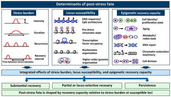
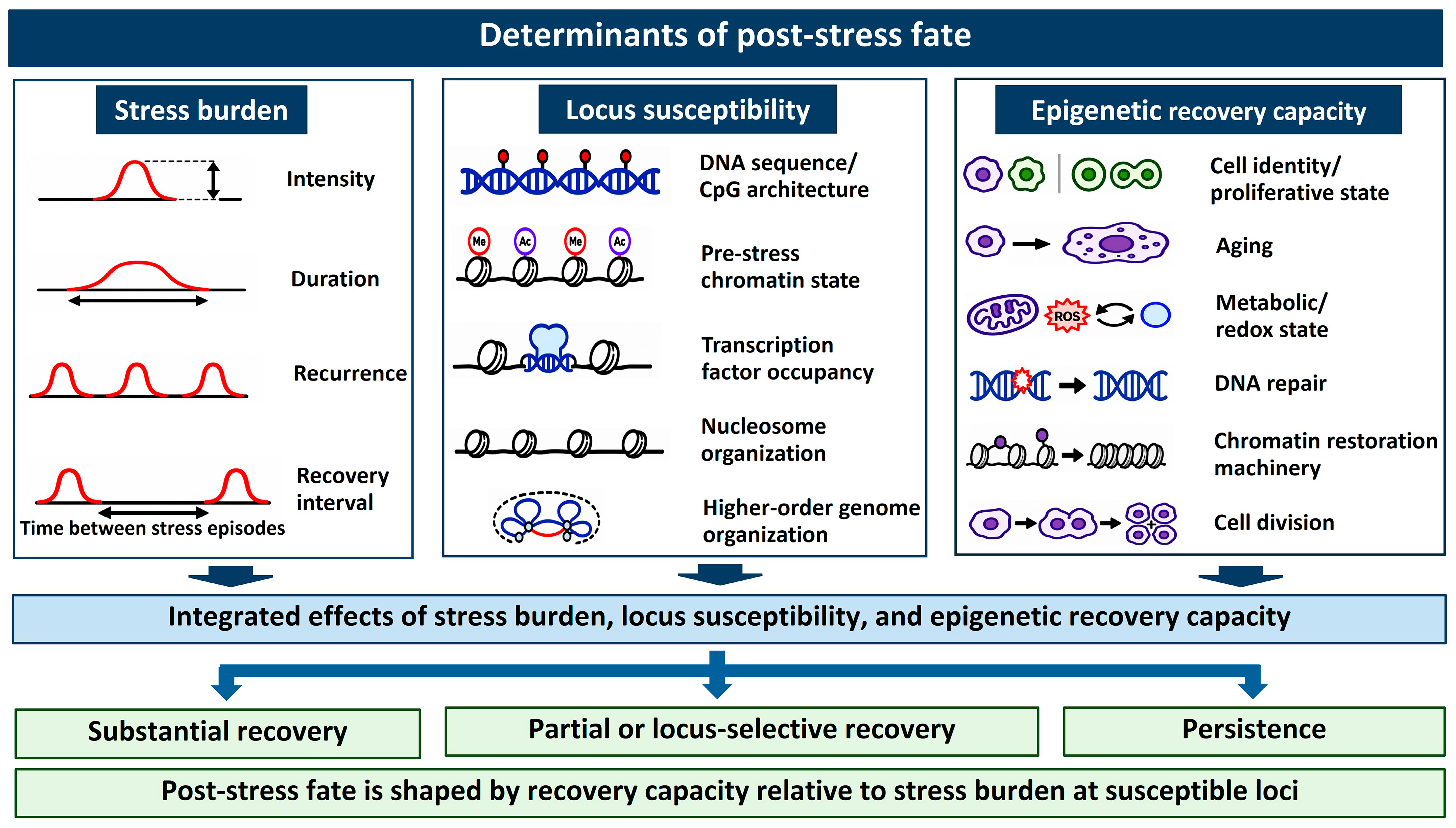
We propose that epigenetic recovery capacity may interact with stress burden and locus susceptibility to shape post-stress epigenetic fate. This relationship provides a basis for testable hypotheses but should not be regarded as an experimentally established model. Current evidence for its individual components comes from different biological systems, and the three components have not yet been examined together within a single experimental design. Figure 1 summarizes this proposed framework.
Figure 1.
Proposed conceptual framework for post-stress epigenetic fate. Stress burden, locus susceptibility, and epigenetic recovery capacity are proposed to interact in shaping whether epigenetic alterations show substantial recovery, partial or locus-selective recovery, or persistence. The framework integrates evidence from different biological systems and has not yet been validated by examining all three components within a single experimental design.
5. Biological Consequences of Epigenetic Persistence
Epigenetic alterations that persist after stress may remain functionally neutral or may be engaged during an altered subsequent response. Only in the latter case does the retained regulatory information constitute functional epigenetic memory. The consequences of such memory may be adaptive or maladaptive and depend on how the retained regulatory information is subsequently engaged. The same persistent state may have little detectable functional consequence in one context but be engaged during adaptive or maladaptive responses in another.
5.1. Adaptive Persistence
Persistence after stress is not necessarily evidence of failed recovery. In some settings, selected regulatory features remain after broader resolution and are associated with altered responses to subsequent challenge. We refer to this outcome as adaptive persistence when the later response improves cellular or tissue performance. Long-lived stem and progenitor cells provide an important substrate for adaptive persistence. In epidermal stem cells, selected regulatory regions remain accessible after inflammatory challenge and are accompanied by more rapid activation of wound response programs during subsequent injury [7]. The retained accessibility and altered wound response are consistent with Level 3 functional memory, although the study does not establish that the retained chromatin state is necessary or sufficient for the response. The consequences of such retention are context-dependent. In pancreatic epithelial models, inflammatory exposure is followed by long-lasting chromatin states, altered responses to further injury, and increased susceptibility to oncogenic transformation [28]. Innate immune memory provides another important example. Monocytes, macrophages, and their precursors can show altered responsiveness to subsequent challenge after exposure to certain infectious, inflammatory, or metabolic stimuli, a phenomenon generally called trained immunity. This altered transcriptional potential is accompanied by enduring changes in chromatin organization and cellular metabolism [29,41,42]. These findings support Level 3 functional memory. Direct manipulation of the candidate retained state would be required to establish Level 4 causal memory.
Adaptive persistence may involve selective retention. In this case, selected regulatory features remain while broader homeostatic restoration occurs. However, the benefit of this strategy depends on the biological context. Repeated engagement, prolonged retention or activation in an inappropriate biological context may turn a regulatory state that initially promotes protection and repair into a detrimental outcome. Hence, the difference between adaptive and maladaptive persistence is not the epigenetic mark itself, but the functional consequences of retaining and reusing that regulatory information.
5.2. Maladaptive Persistence
Regulatory information retained after stress may be associated with beneficial or harmful outcomes, depending on the context. We use the term maladaptive persistence when a retained state is engaged during sustained dysfunction, inappropriate responsiveness, impaired tissue restoration, or increased susceptibility to subsequent disease. Persistent regulatory features may be observed together with exaggerated or recurrent inflammatory responses [7,8,28]. Exposure to high glucose may lead to regulatory changes that remain detectable after glucose levels return to normal; this phenomenon is usually called metabolic memory [24]. The retention of these changes provides Level 2 evidence of persistence. Functional memory requires additional evidence that the retained state shows regulatory activity during the sustained phenotype or a later response. In paired human adipose tissue samples collected at the time of bariatric surgery and two years later, RNA sequencing of individual nuclei showed retained transcriptional changes after weight loss. The epigenomic findings came from mouse adipocytes: after obesity induced by diet followed by eight weeks of chow feeding, histone modifications and chromatin accessibility remained altered. Formerly obese mice also showed an accelerated response to four weeks of renewed feeding with a high fat diet [25]. Thus, the human findings demonstrate transcriptional persistence, whereas the evidence of epigenomic persistence and an altered response to a later metabolic challenge comes from the mouse model. The level of evidence should therefore be determined from the findings in each experimental model rather than from the term metabolic memory alone. These findings illustrate that physiological recovery and epigenetic recovery can occur on different timescales.
Maladaptive persistence may also arise when programs associated with stress constrain restoration of normal cell identity or regulatory flexibility. Epigenetic regulation of fibroblast activation during tissue repair can contribute to fibrotic remodeling [43]. Retained states in epithelial or progenitor cells may show regulatory activity during subsequent responses [7,36]. Radiotherapy can also leave persistent chromatin and transcriptional alterations in surviving cells [26]. However, clonal selection and stable genetic changes can produce a similar pattern without showing that the same epigenetic state was retained in the originally affected cells. Such observations provide evidence of persistence, but functional or causal memory requires the additional evidence defined in Table 2.
Persistent remodeling need not cause disease directly. Its biological relevance may become apparent when a previously exposed cell encounters another challenge and the retained regulatory state shows activity during an altered subsequent response [28,36]. Persistent states may therefore be associated with context-dependent responses rather than acting as autonomous causes of disease. The same regulatory history can be beneficial in some instances but detrimental in others, depending on the subsequent context in which the retained state is engaged.
6. Studying the Post-Stress Epigenome: From Persistence to Causal Memory
Distinguishing recovery, persistence, and epigenetic memory requires experimental designs that capture the transition from acute stress to the post-stress state. Three dimensions are particularly important: time, resolution, and function. Time distinguishes acute remodeling from recovery and persistence. Spatial and cellular resolution identifies where and in which cells retained states occur. Functional evidence determines whether a retained state is engaged during a later response, whereas causal evidence determines whether the retained state contributes directly to that response.
6.1. Tracking Recovery and Persistence over Time
A single post-stress measurement is generally insufficient to distinguish recovery from persistence. Studies should include a pre-stress or control reference, a measurement during or immediately after exposure, and more than one recovery time point when feasible. The duration of exposure and the interval after stressor removal should also be reported. In repeated-exposure studies, the timing of each exposure should be considered because a second challenge may occur before recovery from the first is complete [7,44,45].
Removal of the stressor must be distinguished from resolution of its downstream effects [22]. Investigators should identify the biological processes most relevant to their model and follow appropriate measures during recovery. Depending on the experimental context, these may include inflammatory mediators [22,45], metabolic or redox indicators [3,46,47], evidence of DNA damage [4], and measures of tissue injury or function [7,28]. Complete normalization of every measure is not required, but persistent biological activity should be reported and considered as a possible explanation for continued epigenetic remodeling. Without this information, an alteration observed after stressor removal cannot be clearly distinguished from remodeling maintained by unresolved stress.
6.2. Resolving Trajectories Specific to Loci and Cells
Bulk measurements can obscure differences among genomic regions and cell populations. A signal that remains detectable after stress may reflect persistence within the originally affected cells, but it may also arise because the cellular composition of the sample has changed. Stress may selectively eliminate susceptible cells, allow pre-existing subpopulations to expand, favor particular clones, or alter differentiation trajectories. Each of these processes can change the epigenetic profile of a tissue without retention of the altered state within the same cells.
Cell sorting, computational deconvolution, and single-cell or spatial profiling can help identify or account for differences in cellular composition [48,49]. However, these approaches do not necessarily show that the alteration is retained in the same cells over time. Where feasible, lineage tracing or clonal analysis can provide stronger evidence that an altered state is maintained within previously exposed cells or their descendants [48,50]. A claim of cell-intrinsic persistence should therefore be supported at the cellular or lineage level rather than inferred from bulk tissue alone.
6.3. Testing Functional and Causal Epigenetic Memory
Temporal persistence is necessary but not sufficient for functional epigenetic memory. A persistent epigenetic state found together with a later phenotype represents an association and should not, by itself, be classified as functional memory. Additional evidence is needed to show that the retained state is engaged during the later response or sustained phenotype. In a rechallenge experiment, this may be shown when retained regulatory regions are accompanied by altered transcriptional activity at the same loci during the subsequent response [7,44,45]. This supports functional involvement but does not show that the retained state is necessary or sufficient for the response. Causal epigenetic memory requires direct manipulation of the candidate retained state. If disrupting the state changes the subsequent response, or recreating it in a naive system reproduces that response, the evidence supports a causal role [30,31].
These criteria form the basis of the evidence framework with four levels summarized in Table 2. At a minimum, epigenetic persistence requires evidence that an alteration arose during stress and remained over a defined recovery interval after the stressor and its relevant downstream effects had resolved. Functional memory additionally requires evidence that the retained state is engaged during an altered later response or sustained phenotype. Causal memory requires direct manipulation of the retained state and evidence that this changes the response. In bulk tissue studies, changes in cellular composition and cellular selection should also be assessed before persistence within the same cells is claimed. The levels represent increasing experimental support rather than separate biological stages.
7. Translational Implications: Biomarkers and Therapeutic Opportunities
The evidence framework outlined above also has translational implications because it shifts attention from static epigenetic abnormalities toward trajectories of recovery and persistence. A single measurement cannot distinguish an actively resolving state from one retained after previous stress or maintained by an unresolved stimulus. Similarly, therapeutic strategies should distinguish maladaptive persistence from retained states that are neutral or adaptive.
7.1. Epigenetic Recovery as a Dynamic Biomarker
Most epigenetic biomarkers describe molecular state at a particular time. The post-stress framework introduces a complementary concept: the trajectory of change after stress resolution. Individuals with similar alterations during stress may subsequently show rapid, delayed, or incomplete recovery, or recovery at selected loci, potentially providing information that is not captured by a single endpoint.
Candidate measures include DNA methylation at specific loci, chromatin accessibility, histone modifications, non-coding RNAs, and composite signatures. Global or repetitive element measures such as LINE-1 and Alu methylation may provide scalable indicators of broader methylomic disturbance in selected settings, but they should currently be regarded as candidate state indicators rather than established biomarkers of epigenetic recovery [51,52,53,54,55]. Establishing recovery biomarkers will require longitudinal evidence that their trajectories reproducibly reflect stress resolution and predict biologically or clinically relevant outcomes.
Dynamic epigenetic measurements must also be interpreted alongside evidence that the relevant stressor has diminished. Continued inflammation, metabolic dysfunction, oxidative stress, treatment exposure, or tissue injury can maintain regulatory alterations and mimic persistence [45,46,47]. The translational value of a recovery biomarker will therefore depend on whether its trajectory provides information beyond conventional measures of stress or disease activity.
7.2. Targeting Maladaptive Epigenetic Persistence
Epigenetic reversibility allows persistent regulatory states to serve as potential therapeutic targets, but persistence itself does not justify intervention. Functional and, if possible, causal evidence should be used to identify persistent states whose retention contributes to pathological behavior. Such considerations follow directly from the proposed evidence hierarchy discussed in Section 6.
Many existing drugs target DNA methylation, histone deacetylases, bromodomain proteins, and other chromatin regulators, demonstrating that epigenetic states are subject to pharmacological manipulation [56]. However, broad manipulation may disrupt physiologically necessary or adaptively retained regulatory properties. Thus, it may be better to consider the therapeutic goal as selective reprogramming rather than global epigenetic erasure.
Targeted epigenome editing can be used to remove or recreate a candidate retained state at a specific locus [30,32,33]. If this manipulation changes the subsequent response or sustained phenotype, it provides Level 4 evidence of causal epigenetic memory. Despite this potential, translation into practice will require control of delivery, cell specificity, durability, and off-target effects, as well as careful assessment of safety [31,34,35,57]. Hence, the main question is not simply whether a persistent state is reversible, but which particular state should be targeted, in which cells, and when after stress.
7.3. Restoring Epigenetic Recovery Capacity
Another option could be to focus on the processes involved in appropriate resetting rather than on specific retained marks. If persistence is indeed a result of disrupted metabolic, redox, mitochondrial, DNA repair, or chromatin restoration processes, then improving these systems could, in principle, shift post-stress outcomes toward recovery [3,4]. However, at present, this idea should be treated as a mechanistic hypothesis rather than a confirmed therapeutic approach.
Looking at this through the lens of epigenetic recovery capacity changes the question from “Which persistent mark do we need to erase?” to “What has gone wrong in resetting, and can we restore appropriate regulatory processes?” Targeted interventions could be preferable when a specific retained state is known to play a causal role, while attempts to enhance recovery may be more appropriate when persistence stems from a more generalized disruption of cellular homeostasis.
We use the term recovery window to describe a hypothetical period after stress during which altered epigenetic states may remain relatively plastic before becoming stabilized through feedback, repeated exposure, cellular selection, mitotic propagation, or tissue remodeling. It has not yet been established whether such a window exists across biological systems or whether intervention during this period is more effective than intervention after a persistent state has stabilized. Testing this hypothesis will require longitudinal studies that compare interventions delivered at different stages of recovery and assess their effects on epigenetic recovery and relevant biological outcomes.
8. Outstanding Questions and Future Directions
Understanding the post-stress epigenome requires moving beyond cataloging alterations induced by stress toward explaining and predicting their fate. Key unresolved questions concern whether recovery follows generalizable rules, how genuine memory can be distinguished from residual stress or cellular selection, and whether post-stress trajectories can ultimately be predicted and controlled.
8.1. Defining the Rules of Epigenetic Recovery
Differences among epigenetic layers have direct implications for the study of recovery. A major challenge is to determine whether DNA methylation, histone modifications, chromatin accessibility, transcription factor occupancy, and non-coding RNA regulation recover together, sequentially, or independently. Whenever possible, these layers and the associated transcriptional output should be measured in the same samples and at matched recovery time points.
Recovery should be described separately for each layer. Its rate, extent, and completeness may not change together, and measurements obtained from different epigenetic assays should not be combined into a single recovery score without experimental validation. Reporting these features separately would allow clearer comparison across cell types, stressors, and interventions.
Another priority is identifying features that predict persistence before it becomes established. DNA sequence, chromatin state before stress, regulatory architecture, cellular state, and stress history may distinguish rapidly recovering loci from loci prone to persistence [8]. Such prediction would move the field from retrospective description toward prospective models of post-stress fate.
8.2. Distinguishing Memory from Residual Stress and Cellular Selection
Epigenetic alterations observed after stressor removal may still reflect ongoing inflammation, metabolic disturbance, oxidative stress, DNA damage, or tissue injury. Studies should therefore report both the recovery interval and the evidence used to establish biological resolution. Otherwise, continued remodeling cannot be distinguished from post-stress epigenetic persistence. Changes in tissue composition provide another explanation for apparent persistence. A bulk epigenetic profile may remain altered because the relative abundance of cell populations has changed, even when the alteration is not retained within the originally affected cells. Selective survival, expansion of pre-existing subpopulations, clonal selection, and altered differentiation may all produce this pattern [49]. Studies based on bulk tissue should therefore account for cellular composition and, where possible, confirm persistence within previously exposed cells or their descendants using single-cell, lineage-tracing, or clonal approaches [48,50].
Even genuine persistence may be a marker rather than a driver of altered behavior. A further priority is therefore to determine when a retained state is engaged during a subsequent response and when it contributes causally to that response. Establishing functional memory will require linking the retained information to a subsequent response, whereas causal attribution will require direct perturbation or recreation of the candidate state, as outlined in Section 6 [7,30].
8.3. Predicting and Redirecting Post-Stress Epigenetic Trajectories
A long-term goal is to predict post-stress trajectories before persistent states become firmly established. Achieving this goal will require models that integrate stress history, cellular state before stress, locus susceptibility, and recovery dynamics. Longitudinal data and measurements from individual cells may help identify predictors of recovery or persistence, whereas perturbation studies will be required to distinguish predictors from mechanisms.
Such predictive capacity may help identify points of divergence during recovery and determine when retained states remain sufficiently plastic for intervention. Future studies should test whether maladaptive trajectories can be redirected toward regulatory homeostasis without disrupting adaptive or neutral states and distinguish genuine changes in recovery from the suppression of ongoing stress.
9. Conclusions
Cellular stress can cause considerable alterations in the epigenome, but the consequences of those alterations following the removal of stress are not uniform. Some changes are substantially reversed, others recover only partially or at selected loci, whereas a subset persists. Persistence alone does not indicate epigenetic memory. Functional memory requires evidence that a retained state is engaged during a later response or sustained phenotype, whereas causal memory requires direct evidence that manipulating the retained state changes that response. Distinguishing recovery from persistence and persistence from functional memory therefore requires longitudinal data with sufficient resolution at the locus and cellular levels.
We propose that post-stress epigenetic fate may be shaped by the interaction among stress burden, locus susceptibility, and epigenetic recovery capacity. Variation in these factors may explain why similar stresses produce different trajectories across loci, cells, and biological contexts. Persistent states may remain functionally neutral or be engaged during adaptive or maladaptive responses, depending on the subsequent context.
Thus, a post-stress epigenome perspective shifts the focus from the strength of the initial stress response to the ability to regain regulatory homeostasis after stress exposure has ended. Understanding why some states induced by stress recover while others persist may help link stress history to resilience, adaptation, and disease susceptibility.
Author Contributions
Conceptualization, S.C., S.S., and J.T.; writing—original draft preparation, S.C., S.S., and J.T.; writing—review and editing, S.C., S.S., and J.T.; visualization, S.C., S.S., and J.T.; supervision, S.C., and J.T. All authors have read and agreed to the published version of the manuscript.
Funding
This research received no external funding.
Institutional Review Board Statement
Not applicable.
Informed Consent Statement
Not applicable.
Data Availability Statement
No new data were created or analyzed in this study. Data sharing is not applicable to this article.
Acknowledgments
The Center for Library Resources and Educational Media, Walailak University.
Conflicts of Interest
The authors declare no conflicts of interest.
Abbreviations
The following abbreviations are used in this manuscript:
| AP-1 | Activator protein 1 |
| CpG | Cytosine–phosphate–guanine dinucleotide |
| CRISPR | Clustered regularly interspaced short palindromic repeats |
| DNMT | DNA methyltransferase |
| H3K4me3 | Histone H3 lysine 4 trimethylation |
| H3K9me3 | Histone H3 lysine 9 trimethylation |
| H3K27ac | Histone H3 lysine 27 acetylation |
| H3K27me3 | Histone H3 lysine 27 trimethylation |
| HIF | Hypoxia-inducible factor |
| HIRA | Histone cell cycle regulator |
| LINE-1 | Long interspersed nuclear element-1 |
| mTOR | Mechanistic target of rapamycin |
| NAD+ | Nicotinamide adenine dinucleotide |
| NF-κB | Nuclear factor kappa B |
| STAT | Signal transducer and activator of transcription |
| TET | Ten-eleven translocation |
References
- Allis, C.D.; Jenuwein, T. The molecular hallmarks of epigenetic control. Nat. Rev. Genet. 2016, 17, 487–500. [Google Scholar] [CrossRef] [Scilit] [PubMed]
- Wu, Z.; Qu, J.; Zhang, W.; Liu, G.-H. Stress, epigenetics, and aging: Unraveling the intricate crosstalk. Mol. Cell 2024, 84, 34–54. [Google Scholar] [CrossRef] [Scilit] [PubMed]
- Etchegaray, J.-P.; Mostoslavsky, R. Interplay between metabolism and epigenetics: A nuclear adaptation to environmental changes. Mol. Cell 2016, 62, 695–711. [Google Scholar] [CrossRef] [Scilit] [PubMed]
- Adam, S.; Polo, S.E.; Almouzni, G. Transcription recovery after DNA damage requires chromatin priming by the H3.3 histone chaperone HIRA. Cell 2013, 155, 94–106, Correction in Cell 2013, 155, 963. https://doi.org/10.1016/j.cell.2013.10.039. [Google Scholar] [CrossRef] [Scilit] [PubMed]
- Azkanaz, M.; Rodríguez López, A.; de Boer, B.; Huiting, W.; Angrand, P.-O.; Vellenga, E.; Kampinga, H.H.; Bergink, S.; Martens, J.H.; Schuringa, J.J.; et al. Protein quality control in the nucleolus safeguards recovery of epigenetic regulators after heat shock. eLife 2019, 8, e45205. [Google Scholar] [CrossRef] [Scilit] [PubMed]
- Prickaerts, P.; Adriaens, M.E.; van den Beucken, T.; Koch, E.; Dubois, L.; Dahlmans, V.E.; Gits, C.; Evelo, C.T.; Chan-Seng-Yue, M.; Wouters, B.G.; et al. Hypoxia increases genome-wide bivalent epigenetic marking by specific gain of H3K27me3. Epigenet. Chromatin 2016, 9, 46. [Google Scholar] [CrossRef] [Scilit] [PubMed]
- Naik, S.; Larsen, S.B.; Gomez, N.C.; Alaverdyan, K.; Sendoel, A.; Yuan, S.; Polak, L.; Kulukian, A.; Chai, S.; Fuchs, E. Inflammatory memory sensitizes skin epithelial stem cells to tissue damage. Nature 2017, 550, 475–480, Correction in Nature 2018, 560, E2. https://doi.org/10.1038/s41586-018-0229-5. [Google Scholar] [CrossRef] [Scilit] [PubMed]
- Cowley, C.J.; Sajjath, S.M.; Soto-Ugaldi, L.F.; Steiger, M.; Larsen, S.B.; Carroll, T.; Barrows, D.; Mattei, A.; Gonzales, K.A.; Wang, W.; et al. Distinctive DNA sequence features define epigenetic longevity of inflammatory memory. Science 2026, 391, eadz6830. [Google Scholar] [CrossRef] [Scilit] [PubMed]
- López-Otín, C.; Blasco, M.A.; Partridge, L.; Serrano, M.; Kroemer, G. Hallmarks of aging: An expanding universe. Cell 2023, 186, 243–278. [Google Scholar] [CrossRef] [Scilit] [PubMed]
- Bertucci-Richter, E.M.; Parrott, B.B. The rate of epigenetic drift scales with maximum lifespan across mammals. Nat. Commun. 2023, 14, 7731. [Google Scholar] [CrossRef] [Scilit] [PubMed]
- Zaret, K.S.; Carroll, J.S. Pioneer transcription factors: Establishing competence for gene expression. Genes Dev. 2011, 25, 2227–2241. [Google Scholar] [CrossRef] [Scilit] [PubMed]
- Statello, L.; Guo, C.-J.; Chen, L.-L.; Huarte, M. Gene regulation by long non-coding RNAs and its biological functions. Nat. Rev. Mol. Cell Biol. 2021, 22, 96–118, Correction in Nat. Rev. Mol. Cell Biol. 2021, 22, 159. https://doi.org/10.1038/s41580-021-00330-4. [Google Scholar] [CrossRef] [Scilit] [PubMed]
- Sato, F.; Tsuchiya, S.; Meltzer, S.J.; Shimizu, K. MicroRNAs and epigenetics. FEBS J. 2011, 278, 1598–1609. [Google Scholar] [CrossRef] [Scilit] [PubMed]
- Du, J.; Johnson, L.M.; Jacobsen, S.E.; Patel, D.J. DNA methylation pathways and their crosstalk with histone methylation. Nat. Rev. Mol. Cell Biol. 2015, 16, 519–532. [Google Scholar] [CrossRef] [Scilit] [PubMed]
- Zhang, X.; Zhang, Y.; Wang, C.; Wang, X. TET (Ten-eleven translocation) family proteins: Structure, biological functions and applications. Signal Transduct. Target. Ther. 2023, 8, 297. [Google Scholar] [CrossRef] [Scilit] [PubMed]
- Thongsroy, J.; Mutirangura, A. The association between Alu hypomethylation and the severity of hypertension. PLoS ONE 2022, 17, e0270004. [Google Scholar] [CrossRef] [Scilit] [PubMed]
- Thongsroy, J.; Mutirangura, A. Decreased Alu methylation in type 2 diabetes mellitus patients increases HbA1c levels. J. Clin. Lab. Anal. 2023, 37, e24966. [Google Scholar] [CrossRef] [Scilit] [PubMed]
- Thongsroy, J.; Patchsung, M.; Mutirangura, A. The association between Alu hypomethylation and severity of type 2 diabetes mellitus. Clin. Epigenet. 2017, 9, 93. [Google Scholar] [CrossRef] [Scilit] [PubMed]
- Eustermann, S.; Patel, A.B.; Hopfner, K.-P.; He, Y.; Korber, P. Energy-driven genome regulation by ATP-dependent chromatin remodellers. Nat. Rev. Mol. Cell Biol. 2024, 25, 309–332. [Google Scholar] [CrossRef] [Scilit] [PubMed]
- Millán-Zambrano, G.; Burton, A.; Bannister, A.J.; Schneider, R. Histone post-translational modifications—Cause and consequence of genome function. Nat. Rev. Genet. 2022, 23, 563–580. [Google Scholar] [CrossRef] [Scilit] [PubMed]
- Klemm, S.L.; Shipony, Z.; Greenleaf, W.J. Chromatin accessibility and the regulatory epigenome. Nat. Rev. Genet. 2019, 20, 207–220. [Google Scholar] [CrossRef] [Scilit] [PubMed]
- Nathan, C.; Ding, A. Nonresolving inflammation. Cell 2010, 140, 871–882. [Google Scholar] [CrossRef] [Scilit] [PubMed]
- Bouvier, D.; Ferrand, J.; Chevallier, O.; Paulsen, M.T.; Ljungman, M.; Polo, S.E. Dissecting regulatory pathways for transcription recovery following DNA damage reveals a non-canonical function of the histone chaperone HIRA. Nat. Commun. 2021, 12, 3835. [Google Scholar] [CrossRef] [Scilit] [PubMed]
- El-Osta, A.; Brasacchio, D.; Yao, D.; Pocai, A.; Jones, P.L.; Roeder, R.G.; Cooper, M.E.; Brownlee, M. Transient high glucose causes persistent epigenetic changes and altered gene expression during subsequent normoglycemia. J. Exp. Med. 2008, 205, 2409–2417, Correction in J. Exp. Med. 2008, 205, 2683. https://doi.org/10.1084/jem.20081188092608c. [Google Scholar] [CrossRef] [Scilit] [PubMed]
- Hinte, L.C.; Castellano-Castillo, D.; Ghosh, A.; Melrose, K.; Gasser, E.; Noé, F.; Massier, L.; Dong, H.; Sun, W.; Hoffmann, A.; et al. Adipose tissue retains an epigenetic memory of obesity after weight loss. Nature 2024, 636, 457–465, Correction in Nature 2025, 643, E24. https://doi.org/10.1038/s41586-025-08909-z. [Google Scholar] [CrossRef] [Scilit] [PubMed]
- Bian, X.; Piipponen, M.; Liu, Z.; Luo, L.; Geara, J.; Chen, Y.; Sangsuwan, T.; Maselli, M.; Diaz, C.; Bain, C.; et al. Epigenetic memory of radiotherapy in dermal fibroblasts impairs wound repair capacity in cancer survivors. Nat. Commun. 2024, 15, 9286. [Google Scholar] [CrossRef] [Scilit] [PubMed]
- Kadauke, S.; Blobel, G.A. Mitotic bookmarking by transcription factors. Epigenet. Chromatin 2013, 6, 6. [Google Scholar] [CrossRef] [Scilit] [PubMed]
- Del Poggetto, E.; Ho, I.-L.; Balestrieri, C.; Yen, E.-Y.; Zhang, S.; Citron, F.; Shah, R.; Corti, D.; Diaferia, G.R.; Li, C.-Y.; et al. Epithelial memory of inflammation limits tissue damage while promoting pancreatic tumorigenesis. Science 2021, 373, eabj0486. [Google Scholar] [CrossRef] [Scilit] [PubMed]
- Mitroulis, I.; Ruppova, K.; Wang, B.; Chen, L.-S.; Grzybek, M.; Grinenko, T.; Eugster, A.; Troullinaki, M.; Palladini, A.; Kourtzelis, I.; et al. Modulation of myelopoiesis progenitors is an integral component of trained immunity. Cell 2018, 172, 147–161.e12. [Google Scholar] [CrossRef] [Scilit] [PubMed]
- Coda, D.M.; Watt, L.; Glauser, L.; Batiuk, M.Y.; Burns, A.M.; Stahl, C.L.; Wong, L.Y.; Gräff, J. Cell-type- and locus-specific epigenetic editing of memory expression. Nat. Genet. 2025, 57, 2661–2668. [Google Scholar] [CrossRef] [Scilit] [PubMed]
- Nuñez, J.K.; Chen, J.; Pommier, G.C.; Cogan, J.Z.; Replogle, J.M.; Adriaens, C.; Ramadoss, G.N.; Shi, Q.; Hung, K.L.; Samelson, A.J.; et al. Genome-wide programmable transcriptional memory by CRISPR-based epigenome editing. Cell 2021, 184, 2503–2519.e17. [Google Scholar] [CrossRef] [Scilit] [PubMed]
- Cappelluti, M.A.; Mollica Poeta, V.; Valsoni, S.; Quarato, P.; Merlin, S.; Merelli, I.; Lombardo, A. Durable and efficient gene silencing in vivo by hit-and-run epigenome editing. Nature 2024, 627, 416–423. [Google Scholar] [CrossRef] [Scilit] [PubMed]
- Tremblay, F.; Xiong, Q.; Shah, S.S.; Ko, C.-W.; Kelly, K.; Morrison, M.S.; Giancarlo, C.; Ramirez, R.N.; Hildebrand, E.M.; Voytek, S.B.; et al. A potent epigenetic editor targeting human PCSK9 for durable reduction of low-density lipoprotein cholesterol levels. Nat. Med. 2025, 31, 1329–1338. [Google Scholar] [CrossRef] [Scilit] [PubMed]
- Nakamura, M.; Gao, Y.; Dominguez, A.A.; Qi, L.S. CRISPR technologies for precise epigenome editing. Nat. Cell Biol. 2021, 23, 11–22. [Google Scholar] [CrossRef] [Scilit] [PubMed]
- Xu, D.; Besselink, S.; Ramadoss, G.N.; Dierks, P.H.; Lubin, J.P.; Pattali, R.K.; Brim, J.I.; Christenson, A.E.; Colias, P.J.; Ornelas, I.J.; et al. Programmable epigenome editing by transient delivery of CRISPR epigenome editor ribonucleoproteins. Nat. Commun. 2025, 16, 7948. [Google Scholar] [CrossRef] [Scilit] [PubMed]
- Nagaraja, S.; Ojeda-Miron, L.; Zhang, R.; Oreskovic, E.; Hock, C.; Hu, Y.; Zeve, D.; Sharma, K.; Hyman, R.R.; Zhang, Q.; et al. Epigenetic memory of colitis promotes tumour growth. Nature 2026, 652, 774–783. [Google Scholar] [CrossRef] [Scilit] [PubMed]
- Jerkovic, I.; Cavalli, G. Understanding 3D genome organization by multidisciplinary methods. Nat. Rev. Mol. Cell Biol. 2021, 22, 511–528. [Google Scholar] [CrossRef] [Scilit] [PubMed]
- Kitano, H. Biological robustness. Nat. Rev. Genet. 2004, 5, 826–837. [Google Scholar] [CrossRef] [Scilit] [PubMed]
- Flavahan, W.A.; Gaskell, E.; Bernstein, B.E. Epigenetic plasticity and the hallmarks of cancer. Science 2017, 357, eaal2380. [Google Scholar] [CrossRef] [Scilit] [PubMed]
- Kültz, D. Molecular and evolutionary basis of the cellular stress response. Annu. Rev. Physiol. 2005, 67, 225–257. [Google Scholar] [CrossRef] [Scilit] [PubMed]
- Saeed, S.; Quintin, J.; Kerstens, H.H.; Rao, N.A.; Aghajanirefah, A.; Matarese, F.; Cheng, S.-C.; Ratter, J.; Berentsen, K.; van der Ent, M.A.; et al. Epigenetic programming of monocyte-to-macrophage differentiation and trained innate immunity. Science 2014, 345, 1251086. [Google Scholar] [CrossRef] [Scilit] [PubMed]
- Cheng, S.-C.; Quintin, J.; Cramer, R.A.; Shepardson, K.M.; Saeed, S.; Kumar, V.; Giamarellos-Bourboulis, E.J.; Martens, J.H.; Rao, N.A.; Aghajanirefah, A.; et al. mTOR- and HIF-1α-mediated aerobic glycolysis as metabolic basis for trained immunity. Science 2014, 345, 1250684, Erratum in Science 2014, 346, aaa1503. https://doi.org/10.1126/science.aaa1503. [Google Scholar] [CrossRef] [Scilit] [PubMed]
- Chu, L.; Xie, D.; Xu, D. Epigenetic regulation of fibroblasts and crosstalk between cardiomyocytes and non-myocyte cells in cardiac fibrosis. Biomolecules 2023, 13, 1382. [Google Scholar] [CrossRef] [Scilit] [PubMed]
- Vihervaara, A.; Mahat, D.B.; Himanen, S.V.; Blom, M.A.; Lis, J.T.; Sistonen, L. Stress-induced transcriptional memory accelerates promoter-proximal pause release and decelerates termination over mitotic divisions. Mol. Cell 2021, 81, 1715–1731.e6. [Google Scholar] [CrossRef] [Scilit] [PubMed]
- Larsen, S.B.; Cowley, C.J.; Sajjath, S.M.; Barrows, D.; Yang, Y.; Carroll, T.S.; Fuchs, E. Establishment, maintenance, and recall of inflammatory memory. Cell Stem Cell 2021, 28, 1758–1774.e8. [Google Scholar] [CrossRef] [Scilit] [PubMed]
- Chen, Z.; Natarajan, R. Epigenetic modifications in metabolic memory: What are the memories, and can we erase them? Am. J. Physiol. Cell Physiol. 2022, 323, C570–C582. [Google Scholar] [CrossRef] [Scilit] [PubMed]
- Reddy, M.A.; Zhang, E.; Natarajan, R. Epigenetic mechanisms in diabetic complications and metabolic memory. Diabetologia 2015, 58, 443–455. [Google Scholar] [CrossRef] [Scilit] [PubMed]
- Jindal, K.; Adil, M.T.; Yamaguchi, N.; Yang, X.; Wang, H.C.; Kamimoto, K.; Rivera-Gonzalez, G.C.; Morris, S.A. Single-cell lineage capture across genomic modalities with CellTag-multi reveals fate-specific gene regulatory changes. Nat. Biotechnol. 2024, 42, 946–959. [Google Scholar] [CrossRef] [Scilit] [PubMed]
- Guo, X.; Teschendorff, A.E. Guidelines on optimizing DNA methylation reference panels for cell-type deconvolution. Commun. Biol. 2026, 9, 454. [Google Scholar] [CrossRef] [Scilit] [PubMed]
- Zhu, H.; Liu, X.; Tang, M.; Lui, K.O.; Zhou, B. Lineage tracing from cellular heritage to disease destiny. Nat. Genet. 2026, 58, 1490–1504. [Google Scholar] [CrossRef] [Scilit] [PubMed]
- de Oliveira, N.F.P.; Persuhn, D.C.; Dos Santos, M.C.L.G. Can global DNA methylation be influenced by polymorphisms in genes involved in epigenetic mechanisms? A review. Genes 2024, 15, 1504. [Google Scholar] [CrossRef] [Scilit] [PubMed]
- Nelson, H.H.; Marsit, C.J.; Kelsey, K.T. Global methylation in exposure biology and translational medical science. Environ. Health Perspect. 2011, 119, 1528–1533. [Google Scholar] [CrossRef] [Scilit] [PubMed]
- Udomsinprasert, W.; Kitkumthorn, N.; Mutirangura, A.; Chongsrisawat, V.; Poovorawan, Y.; Honsawek, S. Global methylation, oxidative stress and relative telomere length in biliary atresia patients. Sci. Rep. 2016, 6, 26969. [Google Scholar] [CrossRef] [Scilit] [PubMed]
- Patchsung, M.; Settayanon, S.; Pongpanich, M.; Mutirangura, D.; Jintarith, P.; Mutirangura, A. Alu siRNA to increase Alu element methylation and prevent DNA damage. Epigenomics 2018, 10, 175–185. [Google Scholar] [CrossRef] [Scilit] [PubMed]
- Pobsook, T.; Subbalekha, K.; Sannikorn, P.; Mutirangura, A. Improved measurement of LINE-1 sequence methylation for cancer detection. Clin. Chim. Acta 2011, 412, 314–321. [Google Scholar] [CrossRef] [Scilit] [PubMed]
- Dai, W.; Qiao, X.; Fang, Y.; Guo, R.; Bai, P.; Liu, S.; Li, T.; Jiang, Y.; Wei, S.; Na, Z.; et al. Epigenetics-targeted drugs: Current paradigms and future challenges. Signal Transduct. Target. Ther. 2024, 9, 332. [Google Scholar] [CrossRef] [Scilit] [PubMed]
- Heller, E.A.; Bintu, L.; Rots, M.G. Epigenetic editing: From concept to clinic. Nat. Rev. Drug Discov. 2026, 25, 227–248. [Google Scholar] [CrossRef] [Scilit] [PubMed]

Disclaimer/Publisher’s Note: The statements, opinions and data contained in all publications are solely those of the individual author(s) and contributor(s) and not of MDPI and/or the editor(s). MDPI and/or the editor(s) disclaim responsibility for any injury to people or property resulting from any ideas, methods, instructions or products referred to in the content. |
© 2026 by the authors. Licensee MDPI, Basel, Switzerland. This article is an open access article distributed under the terms and conditions of the Creative Commons Attribution (CC BY) license.