


Abstract
Background: Breast cancer (BC) is a very critical health concern, as it is the leading cause of global cancer incidence and female cancer-related deaths worldwide. Despite the clinically successful outcome of immunotherapy, there are always considerable concerns about the initial or eventual failure to control the disease. Adenosine 2A receptors (A2AR) play an essential role in protecting tissues against excessively indulgent immune reactions. The scope of this study is to evaluate the immunohistochemical expression of adenosine receptor A2A in invasive breast carcinoma and its association with clinicopathological parameters. Materials and Methods: A retrospective cross-sectional tissue-biomarker study of 50 cases of invasive breast carcinoma of no special type (NST), archived as formalin-fixed paraffin-embedded blocks stained immunohistochemically for A2AR. Of the 50 cases, 26 were obtained following neoadjuvant chemotherapy. Cases were divided according to A2AR expression using a cutoff of 100 to low (<100) and high (≥100) A2AR expression. Results: High A2AR was detected in 30% of tumor cells (TCs) in the studied cases. High A2AR expression in TCs was significantly associated with high A2AR expression in tumor-infiltrating lymphocytes (TILs) and with a high Ki-67 proliferation index. Conclusions: In conclusion, this exploratory study showed that A2AR is expressed in both TCs and TILs, with a positive association between A2AR expression in TCs and in TILs. High A2AR expression in TCs was also associated with high Ki-67 proliferative activity. These associations persisted after adjustment for neoadjuvant chemotherapy, but they do not establish causality. They support further investigation of A2AR as a potential immunotherapeutic target in larger, independent studies.
1. Introduction
Breast cancer is the second most common type of cancer among women. In 2022, there were approximately 20 million new cases and 9.7 million deaths from cancer worldwide. Female breast cancer accounted for 2.3 million new cases and 665,684 deaths globally, making it the most frequently diagnosed cancer and the leading cause of cancer death among women. Given this substantial burden among women, precise breast cancer biological stratification remains a priority [1]. The incidence rates of breast cancer have been steadily increasing since the mid-2000s. This progress over recent years can be ascribed to advancements in earlier detection, targeted therapies, and surgical approaches. Despite these advances, it still affects millions of people worldwide. Research into novel treatment strategies is still ongoing to significantly enhance the prognosis and reduce the incidence of recurrences and metastases in breast cancer patients [2]. Emerging cancer therapeutic approaches increasingly focus on targeting malignant cells and stimulating antitumor immune responses and reprogramming the immunosuppressive TME to improve treatment effectiveness [3]. TCs apply numerous immunosuppressive mechanisms, including the buildup of extracellular adenosine, which is an effective technique used by tumors to escape immunosurveillance [4].
Adenosine is increasingly recognized as a major immune response mediator; it is one of the most important immunological checkpoints in the tumor microenvironment (TME). The primary route for extracellular synthesis of the adenosine in the TME is represented by the sequential action of CD39 and CD73 which are expressed on tumor stromal cells and some cancer cells, which enable the transformation of extracellular ATP into adenosine [5]. By activating adenosine receptors expressed on tumor cells, endothelial cells, or immune cells, adenosine promotes tumor growth in both paracrine and autocrine ways [5]. However, A2AR expression only indicates receptor availability but not necessarily receptor activation or suppression effect [6,7]. Functional A2AR signaling occurs when extracellular adenosine binds A2AR and activates intracellular signaling pathways, typically increasing cAMP and engaging PKA-dependent immunosuppressive programs [8,9]. Target cells’ G-protein-coupled cell-surface receptors (GPCRs) A1, A2A, A2B, and A3 which recognize and transduce signals sent by extracellular adenosine. All aspects of inflammatory and immunological responses are modulated by these receptors [10]. Adenosine 2A and 2B receptors activation results in a crucial regulatory mechanism that insulates tissues from exaggerated immune reactions [11].
A2AR has been identified as the mediator that suppresses adaptive immunity [12]. Tumors can use the production of adenosine to suppress the antitumor immunological reactions, as it inhibits the proinflammatory effects of adenosine triphosphate (ATP) generated by injured or dying cells [13]. Blocking immune checkpoint pathways has emerged as one of the most favorable therapeutic approaches for cancer treatment [14]. Notably, the biological significance of A2AR expression may differ according to the cellular compartment in which it is detected. In TILs, A2AR activation often reduces antitumor immunity by suppressing T-cell proliferation cytotoxic activity and cytokine production. In TCs, A2AR expression may reflect tumor-intrinsic signaling implicated in proliferation and survival, although these effects are less well-established. Tumor-cell A2AR expression should therefore not be equated with lymphocyte expression or interpreted as direct evidence of immune suppression [15,16]. Many studies only measure the expression of tumor-cell markers or stromal TILs, not both. As a result, they are unable to ascertain whether an immune-high tumor truly contains lymphocytes that engage tumor nests or lymphocytes confined outside of them [17,18]. This is crucial because stromal and epithelial compartments can exhibit disconnected immunological states, as opposed to one unified biology across the entire tumor [19]. The clinical and immunological effects of A2ARs in invasive breast carcinoma are still unclear [20]. In our study, our objective is to evaluate the immunohistochemical expression of A2AR in invasive breast carcinoma NST not only in TCs but also in stromal TILs, and to study its correlation with different clinical and pathological parameters.
2. Materials and Methods
2.1. Study Design
This retrospective cross-sectional tissue-biomarker study included surgically excised breast carcinoma specimens obtained from the Pathology Department Laboratory of Ain Shams University Hospitals, Cairo, Egypt, during 2024. A total of 90 cases were screened for eligibility, of which 40 were excluded for the following reasons: male breast carcinoma (n = 3), complete pathological response following neoadjuvant chemotherapy, with no residual tumor cells (n = 15), and invasive breast carcinoma histological types other than invasive breast carcinoma NST (n = 22). The remaining 50 eligible cases, all diagnosed as invasive breast carcinoma NST, were consecutively included in the study.
One specimen per patient was analyzed to avoid duplicate patient representation. The sample size was determined by pragmatic availability rather than a formal sample size calculation. Complete clinicopathological data were available for all 50 included cases; therefore, no missing data required management.
2.2. Materials and Data Collection
An electronic database system was used to obtain clinicopathological data, and cases were collected anonymously. Hematoxylin and eosin-stained slides were re-examined according to the World Health Organization (WHO) classification of breast tumors, 5th edition, published in 2019, to confirm the final diagnosis [21].
Selected cases were chosen according to these criteria: Female patients with healthcare data used to assign sex, diagnosed with invasive breast carcinoma NST, who were surgically treated by modified radical mastectomy (MRM) or wide local excision (WLE); the availability of formalin-fixed, paraffin-embedded tissue blocks; and patients who have distinct molecular subtypes (luminal A-like, luminal B-like, human epidermal growth factor receptor 2 (HER2)-enriched, or triple-negative breast carcinoma (TNBC).
Molecular subtypes were defined using immunohistochemical surrogates: Luminal A-like (ER and/or PR-positive, HER2-negative, Ki-67 < 20%), Luminal B-like (ER and/or PR-positive with either HER2-negative and Ki-67 ≥ 20%, or HER2-positive), HER2-positive (ER- and PR-negative, HER2-positive), and Triple-negative (ER-, PR-, and HER2-negative). The 20% Ki-67 threshold was applied for subtype classification [22,23,24,25]. HER2 status was assessed according to ASCO/CAP guidelines [26].
Additionally, by examining the tumor tissues, the following factors were confirmed: tumor type, Nottingham grade, TNM (it was recorded as pTNM in untreated cases and ypTNM in post-neoadjuvant cases) and 0-IV staging according to Breast Cancer Staging AJCC Cancer Staging Manual 8th Edition [27], lymphovascular invasion, presence or absence of in situ tumor component, and considering the patient’s response to neoadjuvant chemotherapy (NAC) if it was administered before the surgery.
Furthermore, the percentage of stromal TILs was evaluated using the standards put forward by the International TILs Working Group that recommended assessing all stromal mononuclear cells (plasma cells and lymphocytes) inside the borders of the invasive tumor and reported as a continuous percentage value of the stromal area without concentrating on hot spots [28,29]. Areas of necrosis, fibrosis, DCIS, and treatment-related changes were excluded from the evaluation area [29,30]. In post-NAC specimens, stromal TILs were assessed within the residual invasive tumor bed [29,31].
In keeping with international recommendations, stromal TILs were analyzed primarily as a continuous variable [29,32]. Stromal TILs percentages were classified as low infiltration level (≤60%) and high infiltration (>60%) for descriptive categorical analysis. Although the International TILs Working Group does not validate a universal clinically meaningful threshold, the 60% cut-off was used only for descriptive grouping, corresponding to the upper range of the thresholds used to define lymphocyte-predominant breast cancer (LPBC) (50–60%), and it has not been clinically validated [31,32,33,34].
2.3. Immunohistochemistry Procedures
All 50 paraffin sections were cut at 4 µm thickness. Sections were oven-dried at 60 °C for one hour and then left at 37 °C overnight. After deparaffinization and rehydration, heat-induced epitope retrieval was performed using Cell Conditioning 1 (CC1, Tris-EDTA buffer, pH 8.5) for 64 min. Staining was carried out on a BenchMark XT autostainer (Ventana Medical Systems, Tucson, AZ, USA), with hematoxylin as counterstain and DAB as chromogen. Sections were incubated with mouse monoclonal anti-A2AR antibody (clone 7F6-G5-A2, Cat. No. sc-32261; Santa Cruz Biotechnology, Dallas, TX, USA) at 1:50 dilution for 32 min at 37 °C. Detection was performed using the ultraView Universal DAB Detection Kit (Ventana Medical Systems). Staining-positive stromal lymphocytes were used for the positive control [2].
2.4. Immunohistochemistry Interpretation
ER, PR, HER2 and Ki-67 had been performed on the surgical resection specimens as part of routine diagnostic work-up. The corresponding slides were retrieved from the archive, reviewed and re-scored by the authors following ASCO/CAP guidelines [26]. A2AR staining was evaluated on surgical resection specimen.
We assessed the staining intensity, localization (cytoplasm and membrane) but not incorporated into scoring, and labeled tumor and TILs percentage. Immunostaining was evaluated semi-quantitatively and expressed as a Histoscore (H-score). This method combines the percentage of tagged cells with the staining intensity There were four levels of staining intensity: weak (1), intermediate (2), strong (3), and negative (0). (1 × % of weak positive cells) + (2 × % of moderate positive cells) + (3 × % of strong positive cells) was an equation used for calculating the H-score. Consequently, the expression level was between 0 and 300 [20,35,36].
Using an H-score of 100 as a cut-off value, the cases were divided according to A2AR expression using a cutoff of 100 into low (<100) and high (≥100) A2AR expression; this was an operational, study-specific threshold. An H-score of 100—one-third of the 0–300 maximum—is a pragmatic point where staining shifts from predominantly weak baseline coverage to a more pronounced combination of intensity and extent. The hot spot method was used to assess Ki-67 proliferation index score [37]. For the association analysis with A2AR, Ki-67 proliferation index was categorized as high (≥30%) [38,39] and low (<30%); this 30% threshold was used as an operational study-specific cut-off value [39,40]. Using Olympus BX41 light microscopy (Olympus Corporation, Tokyo, Japan) in at least five locations (×400 magnification), two pathologists blinded to clinical and histological data independently analyzed each IHC-stained section, and discrepancies were resolved through discussion to reach a consensus diagnosis.
2.5. Data Management and Analysis
The Statistical Package for Social Science (SPSS 27) (IBM, Armonk, NY, USA) was used to revise, code, tabulate, and present the collected data on a PC. For each parameter, data was given and appropriate analysis was conducted based on the type of data collected. Fisher–Freeman–Halton exact tests were used for contingency tables larger than 2 × 2 with small expected counts. Binary logistic regression was used to adjust for NAC status. Confidence intervals for proportions were calculated using the Jeffreys method.
2.6. Descriptive Statistics
Mean, standard deviation (±SD), and range were used to describe parametric numerical data, whereas median and interquartile range (IQR) were used to express non-parametric numerical data. Frequency and percentage were used to express non-numerical data.
Odds ratios (ORs) with corresponding 95% confidence intervals (95% CIs) were calculated and reported for the relevant comparisons.
2.7. Analytical Statistics
The statistical significance of the difference between two study group means was assessed using Student’s t-test. Examination of the relationship between two qualitative variables was done using the chi-square test. When the expected count was less than 5 in more than 20% of cells, the association between two qualitative variables was examined using Fisher’s exact test. A p-value < 0.05 was considered statistically significant, and a p-value ≥ 0.05 was considered non-significant.
3. Results
3.1. Clinicopathological Parameters
The mean age of all included cases was 50.76 ± 12.8 and ranged from 26 to 76 years old. All cases were diagnosed as invasive breast carcinoma NST. The right breast represented 28 cases (56%), while the left breast represented 22 cases (44%). Grade 2 was the most common histologic grade, represented in 43 cases (86%), followed by grade 3, represented in five cases (10%), and the least common was grade 1, represented in two cases (4%). Regarding (TNM), tumor size (T): 11 cases were classified as T1 (22%), 27 cases (54%) were related to T2, seven cases (14%) were related to T3, and five cases (10%) were related to T4. Seventeen cases (34%) were classified as N0 (with no lymph node metastasis), while positive lymph node metastasis was detected in 33 cases (66%). Forty-eight cases (96%) had no distant metastasis (M0), and two cases (4%) had distant metastasis (M1).
Regarding 0-IV staging, three cases (6%) were related to stage I, 22 cases (44%) were related to stage II, 23 cases (46%) were related to stage III, and two cases (4%) were related to stage IV. In situ components were identified in 30 cases (60%). Lymphovascular invasion was detected in 28 cases (56%). Tumors were molecularly subclassified into Luminal A-like (25 cases, 50%), Luminal B-like (16 cases, 32%), HER2-enriched (four cases, 8%), and TNBC (five cases, 10%). Regarding the history of receiving NAC: 24 cases (48%) did not receive NAC, and 26 cases (52%) received NAC. Regarding stromal TILs, 35 cases (70%) showed low levels of stromal infiltration, and high stromal infiltration levels were detected in 15 cases (30%). The mean stromal TIL percentage in all cases was 36.8% and ranged from 5 to 90%.
3.2. Immunohistochemical Expression of A2AR in the Studied Cases
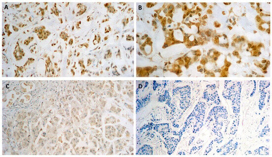
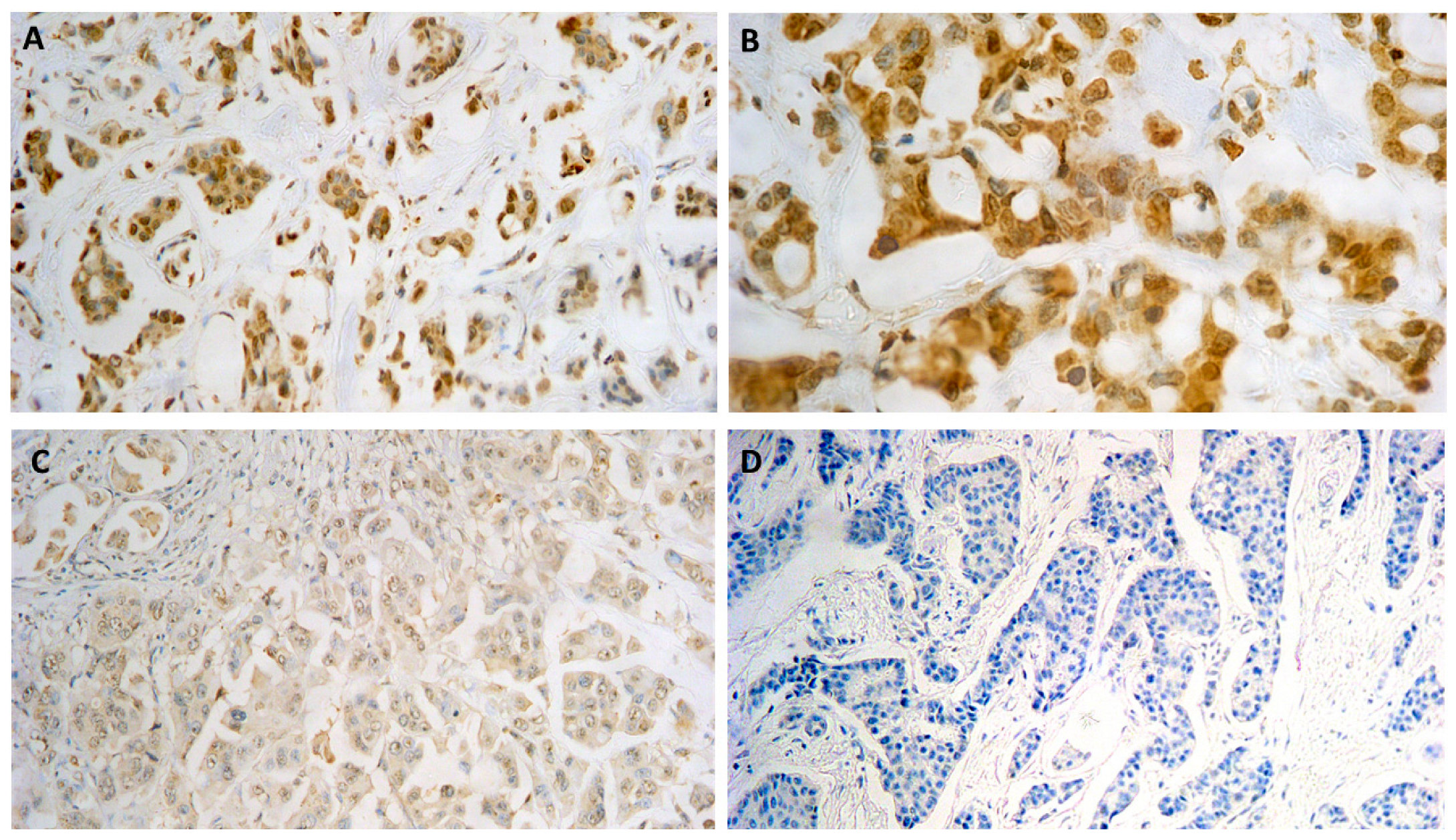
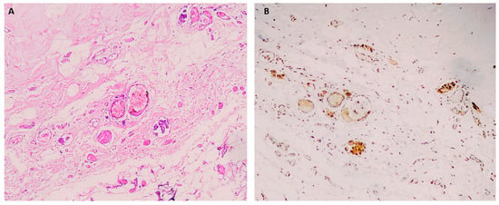
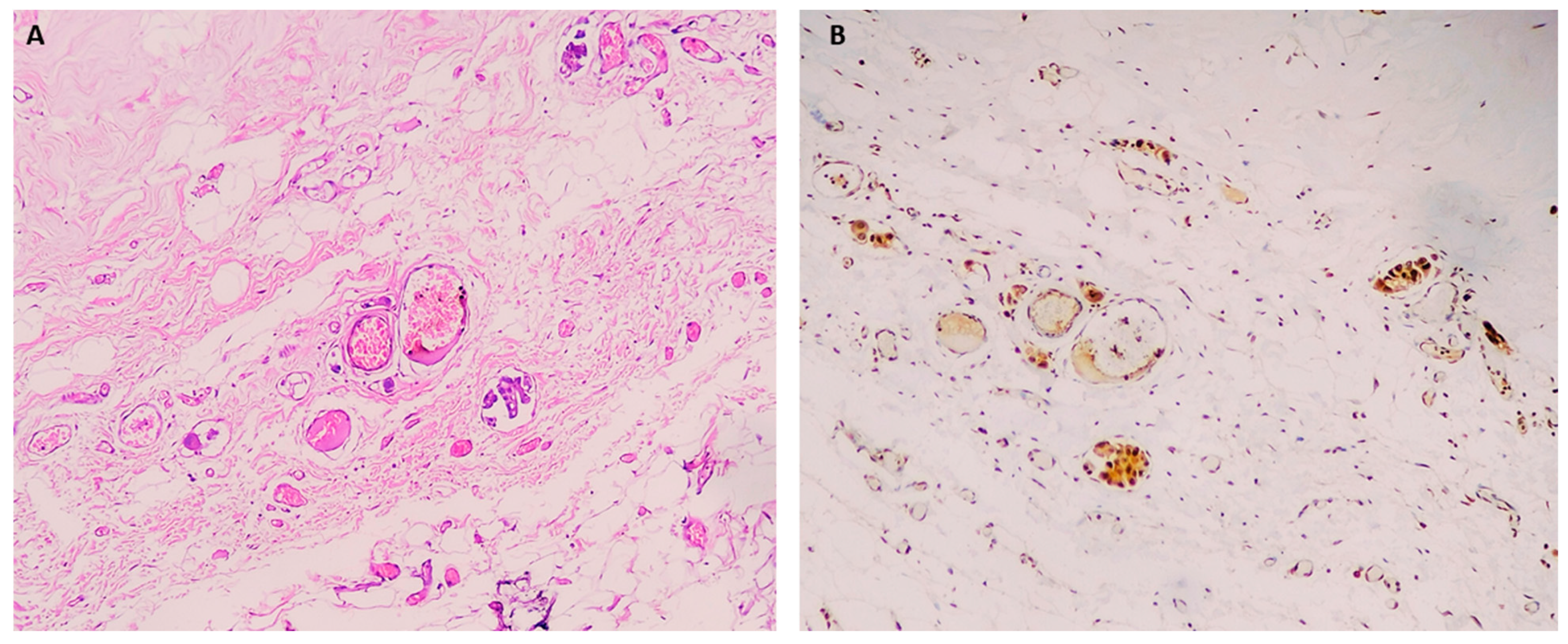
Low A2AR expression in TCs was detected in 35 cases (70%; 95% CI: 56.4–81.3%), whereas high A2AR expression was observed in 15 cases (30%; 95% CI: 18.7–43.6%). Low A2AR expression in TILs (H-score < 100) was found in 31 cases (62%; 95% CI: 48.2–74.5%), while 19 cases (38%; 95% CI: 25.5–51.8%) showed high A2AR expression (H-score ≥ 100) (Figure 1 and Figure 2, and Table 1).
Figure 1.
(A) Invasive breast carcinoma NST showing high A2AR cytoplasmic expression in TCs and TILs, H-score 300 (IHC × 200). (B) Same case (IHC × 400). (C) Low cytoplasmic expression of A2AR in TCs and TILs, H-score 50 (IHC × 200). (D) Negative A2AR expression, H-score 0 (IHC × 200).
Figure 2.
Invasive breast carcinoma NST: (A) showing tumor vascular emboli (H&E × 100); (B) showing high A2AR cytoplasmic expression in TCs of tumor vascular emboli in the same spot, H-score 300 (IHC × 100).
Table 1.
A2AR expression in TCs and TILs for the studied cases.
3.3. A2AR Expression in Association with the Clinicopathological Parameter
Statistically significant results were obtained between high A2AR expression in TCs and high proliferation index (Ki-67) (p = 0.011) with an OR of 5.27 (95% CI: 1.38–20.14) (Table 2), and there was also a significant association between high expression of A2AR in TILs and high expression of A2AR in TCs (p = 0.006) with an OR of 5.78 (95% CI: 1.55–21.50) (Table 3).
Table 2.
Correlation between A2AR expression in TCs and molecular subtypes and percentage of Ki-67 (proliferation index) in studied cases.
Table 3.
Correlation between expression of A2AR in TCs and expression of A2AR in TILs.
No statistically significant difference was detected between A2AR expression in TCs and age (p = 0.783), tumor laterality (p = 0.106) (Supplementary Table S1), tumor grade (p = 1.00), tumor size (T) (p = 0.699), lymph node status (N) (p = 0.216), history of metastasis (M) (p = 0.514), stage of the tumor (p = 0.582), lymphovascular invasion (p = 0.384), in situ component (p = 1.00) (Supplementary Table S2), stromal levels of TILs (p = 1.00), stromal TILs continuous percentage (p = 1.00) (Supplementary Table S3), the history of NAC prior to the surgical treatment (p = 0.266) (Supplementary Table S4), and the molecular subtype (p = 0.739) (Table 2).
3.4. NAC Status in Correlation with Clinicopathological Parameters and A2AR Expression in TCs and TILs
Among patients who received NAC, a statistically significant association with low Ki-67 was observed (p = 0.025), with an OR of 0.27 (95% CI: 0.08–0.86) (Table 4). No statistically significant association was detected between NAC status and age (p = 0.168), tumor laterality (p = 0.412) (Supplementary Table S5), tumor grade (p = 0.827), tumor size (T) (p = 0.28), lymph node status (N) (p = 0.272), history of metastasis (M) (p = 0.225), stage of the tumor (p = 0.106), lymphovascular invasion (p = 0.164), in situ component (p = 0.355) (Supplementary Table S6), molecular subtypes (p = 0.409) (Supplementary Table S7), stromal levels of TILs (p = 0.902), stromal TILs continuous percentage (p = 0.512) (Supplementary Table S8), A2AR expression in TCs (p = 0.266) or A2AR expression in TILs (p = 0.273) (Supplementary Table S9).
Table 4.
Correlation between NAC status and Ki-67 levels.
3.5. A2AR Expression in TILs in Correlation with the Clinicopathological Parameters
A2AR expression in TILs differed across molecular subtypes (p = 0.039) (supplementary Table S14); however, the HER2-positive (n = 4) and TNBC (n = 5) groups were very small, so this finding is exploratory and should be interpreted with caution. No significant association was found between A2AR expression in TILs and Ki-67 (p = 0.057) (supplementary Table S14), age (p = 0.696), tumor laterality (p = 0.425) (supplementary Table S10), tumor grade (p = 1.00), tumor size (T) (p = 0.339), lymph node status (N) (p = 0.344), history of metastasis (M) (p = 1.00), stage of the tumor (p = 0.665), in situ component (p = 0.122), lymphovascular invasion (p = 0.336) (supplementary Table S11), stromal levels of TILs (p = 0.656), stromal TILs continuous percentage (p = 0.864) (supplementary Table S12), or the history of NAC prior to the surgical treatment (p = 0.273) (supplementary Table S13).
3.6. Logistic Regression Analysis for High Ki-67 Expression
In binary logistic regression for high Ki-67 expression (Supplementary Table S15), high A2AR expression in TCs was associated with higher odds of high Ki-67 (crude OR 5.27, 95% CI 1.38–20.14, p = 0.015), whereas NAC was associated with lower odds (crude OR 0.27, 95% CI 0.08–0.86, p = 0.027).
After mutual adjustment, A2AR expression in TCs remained significantly associated with high Ki-67 (adjusted OR 4.91, 95% CI 1.21–19.82, p = 0.026), indicating that the association was not explained by NAC status, while the adjusted effect of NAC was not significant (adjusted OR 0.29, 95% CI 0.08–1.00, p = 0.050). Stratified analyses by NAC status or molecular subtype were not performed because of the small size of the subgroups (NAC, n = 26; no NAC, n = 24; HER2-positive, n = 4; TNBC, n = 5).
4. Discussion
Over the past decade, the treatment of breast cancer has progressed significantly [41]. In breast carcinoma, immune checkpoint inhibitors (ICIs) have been studied as single agents and paired with chemotherapy and targeted therapies [42]. In the past, poorly immunogenic breast cancer was not investigated for its vulnerability to immunotherapy. However, ICIs have recently been used as an achievable immunotherapeutic strategy in breast cancer patients [43].
Immunotherapy’s efficacy depends on its capability to generate long-lasting T cell responses [44]. TILs demonstrate the local immune reaction directed against the progression of the tumor [45]. Immune infiltrate range and composition are highly different between breast cancer subtypes and within every subtype; this variation means that not all breast cancer patients would have the same immunotherapeutic approach to provoke an anti-tumor immune response [46]. Framing A2AR as a component of an adenosine-driven system that connects hypoxia, immune suppression, and tumor growth in breast cancer and other solid tumors is also supported by the research [8].
In the present study, we selected all of our cases to belong to invasive breast carcinoma NST, to prevent fragmentation of specimens in order to yield a high statistically significant result. In contrast to other special subtypes of breast carcinoma, invasive breast carcinoma NST expresses a broad range of molecular subtypes: Luminal A-like in low and intermediate grades, Luminal B-like in intermediate to high grades, HER2-enriched subtype, and finally TNBC in high grades. Different clinical presentations and behaviors, as well as responses to therapy, arise as a result of this molecular diversity [47]. LPBC arises within invasive breast carcinoma NST represents a substantial immunogenic subgroup marked by dense stromal immune infiltration [48].
In our study, high expression of A2AR in TCs was reported in 15 (30%) out of 50 cases; these results were lower than the percentage documented before [2]. The sample size, differences in tissue preparation, and the use of various antibody clones are all responsible for the observed diversity in A2AR expression; this can also be attributed to the use of an H-Score. Even among professional pathologists, the results of the manually determined H-score are not always repeatable, and there is also no validated or universally accepted H-score cutoff point [36,49].
Since TILs are a sign of an anti-tumor immune response, they have been linked to improved prognosis and responsiveness to chemotherapy [43]. Recently, attention has been shifted to the advantages of TILs for the host and to different treatment strategies that maximize the benefits by decreasing immune suppression in the TME [50]. It has been widely accepted that TILs are linked to better survival in different cancer types. TILs in breast carcinoma are a promising predictor of complete remission after preoperative chemotherapy [51].
It was interesting to detect a statistically significant association between high expression of A2AR in TCs and high expression of A2AR in TILs (p = 0.006). Similarly, a positive correlation between high A2AR expression and high stromal and immune scores, calculated from transcriptomic data, has been documented [20]. However, no statistically significant association between A2AR expression and TILs has been reported where expression of A2AR in intratumoral TILs was studied [2]. In contrast, A2AR expression in stromal TILs was assessed in our study, whereas Zohair et al. [20] assessed immune cells as a single group, without distinguishing stromal from intratumoral lymphocytes. Recent studies have reported that stromal TILs are a more accurate and superior parameter, as they are not affected by the carcinoma nest growth pattern [28]. Furthermore, TIL levels might vary significantly within each subtype of breast cancer [52].
Moreover, a statistically significant association was detected between high A2AR expression in TCs and high proliferation index (Ki-67) (p = 0.011). This was in accordance with the findings of previous studies, where a positive association between A2AR expression in TCs and high proliferation index (Ki-67) was reported [2,20]. The expression of Ki-67 is closely linked to cancer proliferation and is a predictor of prognosis and outcome; in certain situations, this can help guide therapy choices [53].
In our study, the identified positive association between high A2AR expression and a high proliferation index may suggest a possible relationship between A2AR expression and tumor proliferative activity. However, further functional and clinical studies are required to clarify the role of this association in tumor proliferation.
Our findings are consistent with previous studies [54,55,56] that have reported a statistically significant association between a prior history of NAC and a lower Ki-67 proliferation index in invasive breast carcinoma, which may greatly affect the survival outcome, although the present study cannot establish a causal relationship.
Importantly, high A2AR expression in tumor cells remained significantly associated with high Ki-67 expression after adjustment for NAC status (adjusted OR = 4.91, 95% CI: 1.21–19.82, p = 0.026). This suggests that the observed association between A2AR expression and tumor proliferation persisted after accounting for NAC status. However, the wide confidence interval indicates limited precision, and residual confounding cannot be excluded.
In the present study, we found no statistically significant difference between A2AR expression in TCs and the different breast carcinoma molecular subtypes. In contrast to our findings, a significant association with Luminal B-like and TNBC subtypes was reported by Guimei and Eladl [2]; a significant association with HER2-positive and TNBC subtypes in immune cells was reported by Zohair et al. [20]. This may be due to the sample size of our study, as subgroups are too small for reliable subtype-specific stratification and for differences in antibody clone and scoring technique. A2AR expression in TILs differed across molecular subtypes (p = 0.039); however, because of the very small HER2-positive and TNBC groups in our study, this finding is exploratory and should be interpreted with caution.
Our study was innovative in evaluating the association between A2AR expression and the history of receiving NAC, but our results did not show a statistical significance. Unfortunately, the previous literature lacks any data concerning the association between A2AR expression and the history of receiving NAC. Additionally, no statistically significant association was found between A2AR expression and other clinicopathological parameters; this was in accordance with the previously reported literature [2,20].
Recently, A2AR and A2BR have been used in many clinical trials to assess their potential and efficacy in different cancers as chemotherapeutic or immunotherapeutic anti-cancer approaches. A2AR has great relevance to activated immune cells, so it may have a great role in immunotherapy [57]. A previous study found that A2AR expression was higher in TNBC tumor tissues compared to normal epithelial tissues. Within this study, it was proposed that A2AR may be of greater significance and relevance to clinical tumor progression than A2BR. Interestingly, A2AR expression was documented to play a role in the growth and spread of radiotherapy-resistant breast carcinoma [58].
The currently utilized ICIs in breast carcinoma are PD-1 and PDL-1 inhibitors and HER2-directed monoclonal antibodies [42]. Immunotherapy with anti-PD-L1 has been underwhelming in monotherapy for breast carcinoma metastasis, spurring the use of synergistic treatment combinations combining immunotherapy with ICIs [59]. When compared to PD-1, A2AR is the most prominently expressed protein in breast carcinoma tumor tissues [20]. A2AR antagonists’ anti-tumor effect indicates that they are suitable for cancer therapies, particularly in patients who are not responsive to anti-PD-L1 therapy or in combination with PD-L1 blockage to prevent treatment resistance [60].
This study has several limitations. It is a retrospective, single-center study with a small sample size (n = 50), and the HER2-positive (n = 4) and TNBC (n = 5) subgroups were too small for reliable subtype-specific conclusions; stratified analyses were therefore not feasible. Our study also combined untreated and post-neoadjuvant chemotherapy specimens, which are biologically non-equivalent; although NAC status was adjusted for regression analysis, residual confounding cannot be excluded. The observed associations are correlative and do not imply causality, and no adjustment for multiple testing was applied. We recommend future research with multicenter integration to widen sample representativeness and result reliability.
5. Conclusions
Breast carcinoma treatment strategies have been a challenging issue over the last few decades. The use of ICIs in immunotherapy has sparked a lot of attention. Our exploratory study revealed a positive association between high expression of A2AR in TCs and high expression of A2AR in TILs; TILs have a major role in the antitumor immune responses in TME.
Furthermore, a statistically significant association was detected between high A2AR expression in TCs and a high proliferation index (Ki-67). We suggest that larger studies be conducted in order to clarify the role of this association in breast carcinoma immunotherapy.
Supplementary Materials
The following supporting information can be downloaded at: https://www.mdpi.com/article/10.3390/immuno6040071/s1, Table S1: Correlation between A2AR expression in TCs and the clinical data. Table S2: Correlation between expression of A2AR in TCs and Pathological parameters of studied cases. Table S3: Correlation between expression of A2AR in TCs and stromal levels of TILs. Table S4: Correlation between expression of A2AR in TCs and history of receiving NAC in the studied cases. Table S5: Correlation between NAC status and the clinical data. Table S6: Correlation between NAC status and Pathological parameters of studied cases. Table S7: Correlation between NAC status and Molecular subtypes. Table S8: Correlation between NAC status and stromal levels of TILs. Table S9: Correlation between NAC status and expression of A2AR in TCs and TILs. Table S10: Correlation between A2AR expression in TILs and the clinical data. Table S11: Correlation between expression of A2AR in TILs and Pathological parameters of studied cases. Table S12: Correlation between expression of A2AR in TILs and stromal levels of TILs. Table S13: Correlation between expression of A2AR in TILs and history of receiving NAC in the studied cases. Table S14: Association between A2AR expression in TILs and molecular subtypes and percentage of Ki-67 (proliferation index) of studied cases. Table S15: Binary Logistic Regression Analysis for High Ki-67 Expression.
Author Contributions
M.I.H. and Z.U.A. contributed to conceptualization, investigation, and writing of the original draft, as well as review and editing of the manuscript. M.F.G. and Z.A.K. contributed to validation, supervision, review and editing of the manuscript. All authors have read and agreed to the published version of the manuscript.
Funding
This research received no external funding.
Institutional Review Board Statement
All procedures performed in the current study were approved by the National Research Ethics Committee for Ain Shams University with approval number (FMASU MS 256/2023) according to the revised tenets of the Declaration of Helsinki.
Informed Consent Statement
Patient consent was waived due to the retrospective nature of the study, which used archived, anonymized tissue samples with no direct patient contact; this waiver was granted by FMASU REC (approval code MS 256/2023).
Data Availability Statement
The availability of data and materials supporting the findings of this study are available within the article.
Acknowledgments
The authors acknowledge the anatomic Pathology Department, Faculty of Medicine, Ain Shams University, for the great help, and support in the current study. The authors also gratefully acknowledge Mahmoud K. Senna for his valuable help with the statistical analysis.
Conflicts of Interest
The authors declare no conflicts of interest.
Abbreviations
| BC | Breast cancer |
| A2AR | Adenosine 2A receptor |
| ICIs | Immune Checkpoint Inhibitors |
| NST | No special type |
| ER | Estrogen receptors |
| PR | Progesterone receptors |
| HER2 | Human epidermal growth factor receptor 2 |
| LPBC | Lymphocyte-predominant breast cancer |
| TCs | Tumor cells |
| TILs | Tumor infiltrating lymphocytes |
| GPCRs | G-protein-coupled cell-surface receptors |
| ATP | Adenosine triphosphate |
| H-score | Histoscore |
| NAC | Neoadjuvant chemotherapy |
| T | Tumor size |
| N | Lymph node metastasis |
| M | Distant metastasis |
| TNBC | Triple-negative breast carcinoma |
| PD-1 | Programmed Cell Death 1 |
| PD-(L)1 | Programmed Cell Death ligand 1 |
| TME | Tumor microenvironment |
| AJCC | American Joint Committee on Cancer |
References
- Bray, F.; Laversanne, M.; Sung, H.; Ferlay, J.; Siegel, R.L.; Soerjomataram, I.; Jemal, A. Global cancer statistics 2022: GLOBOCAN estimates of incidence and mortality worldwide for 36 cancers in 185 countries. CA Cancer J. Clin. 2024, 74, 229–263. [Google Scholar] [CrossRef] [Scilit] [PubMed]
- Guimei, M.; Eladl, M.A. Expression of Adenosine Receptors in Young Patients with Breast Cancer and its Association with High Proliferative Index. Int. J. Clin. Biochem. Res. 2021, 5, 57–66. [Google Scholar] [CrossRef] [Scilit]
- Wang, L.; Zhang, L.; Zhang, Z.; Wu, P.; Zhang, Y.; Chen, X. Advances in targeting tumor microenvironment for immunotherapy. Front. Immunol. 2024, 15, 1472772. [Google Scholar] [CrossRef] [Scilit] [PubMed]
- Allard, B.; Beavis, P.A.; Darcy, P.K.; Stagg, J. Immunosuppressive activities of adenosine in cancer. Curr. Opin. Pharmacol. 2016, 29, 7–16. [Google Scholar] [CrossRef] [Scilit] [PubMed]
- Allard, B.; Turcotte, M.; Stagg, J. CD73-Generated Adenosine: Orchestrating the Tumor-Stroma Interplay to Promote Cancer Growth. BioMed Res. Int. 2012, 2012, 485156. [Google Scholar] [CrossRef] [Scilit] [PubMed]
- Koshiba, M.; Rosin, D.; Hayashi, N.; Linden, J.; Sitkovsky, M. Patterns of A2A extracellular adenosine receptor expression in different functional subsets of human peripheral T cells. Flow cytometry studies with anti-A2A receptor monoclonal antibodies. Mol. Pharmacol. 1999, 55, 614–624. [Google Scholar] [CrossRef] [Scilit]
- Moreira-De-Sá, A.; Lourenço, V.; Canas, P.; Cunha, R. Adenosine A2A Receptors as Biomarkers of Brain Diseases. Front. Neurosci. 2021, 15, 702581. [Google Scholar] [CrossRef] [Scilit] [PubMed]
- Sun, C.; Wang, B.; Hao, S.-L. Adenosine-A2A Receptor Pathway in Cancer Immunotherapy. Front. Immunol. 2022, 13, 837230. [Google Scholar] [CrossRef] [Scilit] [PubMed]
- Moreno, E.; Canet, J.; Gracia, E.; Lluis, C.; Mallol, J.; Canela, E.; Cortés, A.; Casadó, V. Molecular Evidence of Adenosine Deaminase Linking Adenosine A2A Receptor and CD26 Proteins. Front. Pharmacol. 2018, 9, 106. [Google Scholar] [CrossRef] [Scilit] [PubMed]
- Antonioli, L.; Fornai, M.; Blandizzi, C.; Pacher, P.; Haskó, G. Adenosine signaling and the immune system: When a lot could be too much. Immunol. Lett. 2019, 205, 9–15. [Google Scholar] [CrossRef] [Scilit] [PubMed]
- Bahreyni, A.; Samani, S.S.; Rahmani, F.; Behnam-Rassouli, R.; Khazaei, M.; Ryzhikov, M.; Parizadeh, M.R.; Avan, A.; Hassanian, S.M. Role of adenosine signaling in the pathogenesis of breast cancer. J. Cell. Physiol. 2018, 233, 1836–1843. [Google Scholar] [CrossRef] [Scilit] [PubMed]
- IJzerman, A.P.; Jacobson, K.A.; Müller, C.E.; Cronstein, B.N.; Cunha, R.A. International Union of Basic and Clinical Pharmacology. CXII: Adenosine Receptors: A Further Update. Pharmacol. Rev. 2022, 74, 340–372. [Google Scholar] [CrossRef] [Scilit] [PubMed]
- Sharma, P.; Hu-Lieskovan, S.; Wargo, J.A.; Ribas, A. Primary, Adaptive, and Acquired Resistance to Cancer Immunotherapy. Cell 2017, 168, 707–723. [Google Scholar] [CrossRef] [Scilit] [PubMed]
- Yu, F.; Zhu, C.; Xie, Q.; Wang, Y. Adenosine A2A Receptor Antagonists for Cancer Immunotherapy. J. Med. Chem. 2020, 63, 12196–12212. [Google Scholar] [CrossRef] [Scilit] [PubMed]
- Venugopala, K.N.; Buccioni, M. Current Understanding of the Role of Adenosine Receptors in Cancer. Molecules 2024, 29, 3501. [Google Scholar] [CrossRef] [Scilit] [PubMed]
- Han, Y.; Dong, C.; Hu, M.; Wang, X.; Wang, G. Unlocking the adenosine receptor mechanism of the tumour immune microenvironment. Front. Immunol. 2024, 15, 1434118. [Google Scholar] [CrossRef] [Scilit] [PubMed]
- Yeung, T.; Zhang, Y.; Zhou, Q.-H.; Burack, R. Neighborhood clustering analysis to define epithelial–stromal interface for tumor infiltrating lymphocyte evaluation. J. Pathol. Inform. 2025, 19, 100465. [Google Scholar] [CrossRef] [Scilit] [PubMed]
- Hamada, K.; Murakami, R.; Ueda, A.; Kashima, Y.; Miyagawa, C.; Taki, M.; Yamanoi, K.; Yamaguchi, K.; Hamanishi, J.; Minamiguchi, S.; et al. A deep learning-based assessment pipeline for intraepithelial and stromal tumor-infiltrating lymphocytes in high-grade serous ovarian carcinoma. Am. J. Pathol. 2024, 194, 1272–1284. [Google Scholar] [CrossRef] [Scilit] [PubMed]
- Kinsella, Z.; Jahangir, C.A.; Nyarko, H.N.; Kalinska-Lysiak, D.; Gonzalez, C.A.; Murphy, V.; O’Grady, T.; Fay, J.; Sheehan, K.; Rahman, A.; et al. Spatial analyses implicate high stromal tumour-infiltrating CD8+ lymphocytes as a negative predictive marker for chemotherapy in estrogen receptor-positive breast cancer. Nat. Commun. 2026, 17, 4863. [Google Scholar] [CrossRef] [Scilit] [PubMed]
- Zohair, B.; Chraa, D.; Rezouki, I.; Benthami, H.; Razzouki, I.; Elkarroumi, M.; Olive, D.; Karkouri, M.; Badou, A. The immune checkpoint adenosine 2A receptor is associated with aggressive clinical outcomes and reflects an immunosuppressive tumor microenvironment in human breast cancer. Front. Immunol. 2023, 14, 1201632. [Google Scholar] [CrossRef] [Scilit] [PubMed]
- Tan, P.H.; Ellis, I.; Allison, K.; Brogi, E.; Fox, S.B.; Lakhani, S.; Lazar, A.J.; Morris, E.A.; Sahin, A.; Salgado, R.; et al. The 2019 World Health Organization classification of tumours of the breast. Histopathology 2020, 77, 181–185. [Google Scholar] [CrossRef] [Scilit] [PubMed]
- Goldhirsch, A.; Winer, E.P.; Coates, A.S.; Gelber, R.D.; Piccart-Gebhart, M.; Thürlimann, B.; Senn, H.J. Personalizing the treatment of women with early breast cancer: Highlights of the St Gallen International Expert Consensus on the Primary Therapy of Early Breast Cancer 2013. Ann. Oncol. 2013, 24, 2206–2223. [Google Scholar] [CrossRef] [Scilit] [PubMed]
- Bustreo, S.; Osella-Abate, S.; Cassoni, P.; Donadio, M.; Airoldi, M.; Pedani, F.; Papotti, M.; Sapino, A.; Castellano, I. Optimal Ki67 cut-off for luminal breast cancer prognostic evaluation: A large case series study with a long-term follow-up. Breast Cancer Res. Treat. 2016, 157, 363–371. [Google Scholar] [CrossRef] [Scilit] [PubMed]
- Lombardi, A.; Lazzeroni, R.; Bersigotti, L.; Vitale, V.; Amanti, C. The Proper Ki-67 Cut-Off in Hormone Responsive Breast Cancer: A Monoinstitutional Analysis with Long-Term Follow-Up. Breast Cancer Targets Ther. (Dove Press) 2021, 13, 213–217. [Google Scholar] [CrossRef] [Scilit] [PubMed]
- Goldhirsch, A.; Wood, W.; Coates, A.; Gelber, R.; Thürlimann, B.; Senn, H. Strategies for subtypes—Dealing with the diversity of breast cancer: Highlights of the St Gallen International Expert Consensus on the Primary Therapy of Early Breast Cancer 2011. Ann. Oncol. 2011, 22, 1736–1747. [Google Scholar] [CrossRef] [Scilit] [PubMed]
- Wolff, A.C.; Somerfield, M.R.; Dowsett, M.; Hammond, M.E.H.; Hayes, D.F.; McShane, L.M.; Saphner, T.J.; Spears, P.A.; Allison, K.H. Human Epidermal Growth Factor Receptor 2 Testing in Breast Cancer. Arch. Pathol. Lab. Med. 2023, 147, 993–1000. [Google Scholar] [CrossRef] [Scilit] [PubMed]
- Young, J.; Asaoka, M.; Ghasemi, F.; Chida, K.; Roy, A.M.; Yan, L.; Hakamada, K.; Takabe, K. The American Joint Committee on Cancer (AJCC) Breast Cancer Staging, Eighth Edition, is more Reflective of Cancer Biology than the Seventh Edition. Ann. Surg. Oncol. 2025, 32, 3268–3277. [Google Scholar] [CrossRef] [Scilit] [PubMed]
- Salgado, R.; Denkert, C.; Demaria, S.; Sirtaine, N.; Klauschen, F.; Pruneri, G.; Wienert, S.; van den Eynden, G.; Baehner, F.L.; Penault-Llorca, F.; et al. The evaluation of tumor-infiltrating lymphocytes (TILs) in breast cancer: Recommendations by an International TILs Working Group 2014. Ann. Oncol. 2015, 26, 259–271. [Google Scholar] [CrossRef] [Scilit] [PubMed]
- Dieci, M.V.; Radosevic-Robin, N.; Fineberg, S.; van den Eynden, G.; Ternes, N.; Penault-Llorca, F.; Pruneri, G.; D’Alfonso, T.M.; Demaria, S.; Castaneda, C.; et al. Update on tumor-infiltrating lymphocytes (TILs) in breast cancer, including recommendations to assess TILs in residual disease after neoadjuvant therapy and in carcinoma in situ: A report of the International Immuno-Oncology Biomarker Working Group on Breast Cancer. Semin. Cancer Biol. 2018, 52, 16–25. [Google Scholar] [CrossRef] [Scilit] [PubMed]
- Symonds, L.; Reiter, D.; Hippe, D.S.; Kazerouni, A.; Mittal, S.; Hunter, N.; Yung, R.; Gwin, W.R., III; Nair, V.; Davidson, N.; et al. Abstract PS4-10-13: Clinical Feasibility and Reproducibility of Tumor Infiltrating Lymphocyte (TIL) Reporting for Early-Stage Triple Negative Breast Cancer Patients at the University of Washington. Clin. Cancer Res. 2026, 32, PS4-10-13. [Google Scholar] [CrossRef] [Scilit]
- Denkert, C.; von Minckwitz, G.; Darb-Esfahani, S.; Lederer, B.; Heppner, B.I.; Weber, K.E.; Budczies, J.; Huober, J.; Klauschen, F.; Furlanetto, J.; et al. Tumour-infiltrating lymphocytes and prognosis in different subtypes of breast cancer: A pooled analysis of 3771 patients treated with neoadjuvant therapy. Lancet Oncol. 2017, 19, 40–50. [Google Scholar] [CrossRef] [Scilit] [PubMed]
- Li, S.-Q.; Zhang, Y.; Zhang, P.; Xue, S.; Chen, Y.; Sun, L.; Yang, R. Predictive and prognostic values of tumor infiltrating lymphocytes in breast cancers treated with neoadjuvant chemotherapy: A meta-analysis. Breast 2022, 66, 97–109. [Google Scholar] [CrossRef] [Scilit] [PubMed]
- Loi, S.; Michiels, S.; Adams, S.; Loibl, S.; Budczies, J.; Denkert, C.; Salgado, R. The journey of tumor infiltrating lymphocytes (TIL) as a biomarker in breast cancer: Clinical utility in an era of checkpoint inhibition. Ann. Oncol. 2021, 32, 1236–1244. [Google Scholar] [CrossRef] [Scilit] [PubMed]
- Schüler, K.; Bethmann, D.; Kaufhold, S.; Hartung, C.; Stückrath, K.; Lantzsch, T.; Uleer, C.; Hanf, V.; Peschel, S.; John, J.; et al. Prognostic Value of Tumour-Infiltrating Lymphocytes in an Unselected Cohort of Breast Cancer Patients. Diagnostics 2022, 12, 2527. [Google Scholar] [CrossRef] [Scilit] [PubMed]
- Ram, S.; Vizcarra, P.; Whalen, P.; Deng, S.-B.; Painter, C.; Jackson-Fisher, A.; Pirie-Shepherd, S.; Xia, X.; Powell, E. Pixelwise H-score: A novel digital image analysis-based metric to quantify membrane biomarker expression from immunohistochemistry images. PLoS ONE 2021, 16, e0245638. [Google Scholar] [CrossRef] [Scilit] [PubMed]
- Price, P.; Ganugapati, U.; Gatalica, Z.; Kakadekar, A.; Macpherson, J.; Quenneville, L.; Rees, H.; Slodkowska, E.; Suresh, J.; Yu, D.; et al. Reinventing Nuclear Histo-score Utilizing Inherent Morphologic Cutoffs: Blue-brown Color H-score (BBC-HS). Appl. Immunohistochem. Mol. Morphol. 2023, 31, 500–506. [Google Scholar] [CrossRef] [Scilit] [PubMed]
- Jang, M.H.; Kim, H.J.; Chung, Y.R.; Lee, Y.; Park, S.Y. A comparison of Ki-67 counting methods in luminal Breast Cancer: The Average Method vs. the Hot Spot Method. PLoS ONE 2017, 12, e0172031. [Google Scholar] [CrossRef] [Scilit] [PubMed]
- Nielsen, T.O.; Leung, S.C.Y.; Rimm, D.L.; Dodson, A.; Acs, B.; Badve, S.; Denkert, C.; Ellis, M.J.; Fineberg, S.; Flowers, M.; et al. Assessment of Ki67 in Breast Cancer: Updated Recommendations From the International Ki67 in Breast Cancer Working Group. J. Natl. Cancer Inst. 2020, 113, 808–819. [Google Scholar] [CrossRef] [Scilit] [PubMed]
- Shim, V.C.; Baker, R.J.; Jing, W.; Puentes, R.; Agersborg, S.S.; Lee, T.K.; Gorea, W.; Achacoso, N.; Lee, C.; Villasenor, M.; et al. Evaluation of the international Ki67 working group cut point recommendations for early breast cancer: Comparison with 21-gene assay results in a large integrated health care system. Breast Cancer Res. Treat. 2023, 203, 281–289. [Google Scholar] [CrossRef] [Scilit] [PubMed]
- Tashima, R.; Nishimura, R.; Osako, T.; Nishiyama, Y.; Okumura, Y.; Nakano, M.; Fujisue, M.; Toyozumi, Y.; Arima, N. Evaluation of an Optimal Cut-Off Point for the Ki-67 Index as a Prognostic Factor in Primary Breast Cancer: A Retrospective Study. PLoS ONE 2015, 10, e0119565. [Google Scholar] [CrossRef] [Scilit] [PubMed]
- Akpinar, R.; Demir, H.; Ilvan, Ş. Prognostic Importance of PD-L1 Expression in Breast Carcinomas. Turk. Onkol. Derg. 2024, 39, 153–166. [Google Scholar] [CrossRef] [Scilit]
- Dvir, K.; Giordano, S.; Leone, J.P. Immunotherapy in Breast Cancer. Int. J. Mol. Sci. 2024, 25, 7517. [Google Scholar] [CrossRef] [Scilit] [PubMed]
- Debien, V.; De Caluwé, A.; Wang, X.; Piccart-Gebhart, M.; Tuohy, V.K.; Romano, E.; Buisseret, L. Immunotherapy in breast cancer: An overview of current strategies and perspectives. npj Breast Cancer 2023, 9, 7. [Google Scholar] [CrossRef] [Scilit] [PubMed]
- Barbari, C.; Fontaine, T.; Parajuli, P.; Lamichhane, N.; Jakubski, S.; Lamichhane, P.; Deshmukh, R.R. Immunotherapies and Combination Strategies for Immuno-Oncology. Int. J. Mol. Sci. 2020, 21, 5009. [Google Scholar] [CrossRef] [Scilit] [PubMed]
- Pujani, M.; Jain, H.; Chauhan, V.; Agarwal, C.; Singh, K.; Singh, M. Evaluation of Tumor infiltrating lymphocytes in breast carcinoma and their correlation with molecular subtypes, tumor grade and stage. Breast Dis. 2020, 39, 61–69. [Google Scholar] [CrossRef] [Scilit] [PubMed]
- Bareche, Y.; Buisseret, L.; Gruosso, T.; Girard, E.; Venet, D.; Dupont, F.; Desmedt, C.; Larsimont, D.; Park, M.; Rothé, F.; et al. Unraveling Triple-Negative Breast Cancer Tumor Microenvironment Heterogeneity: Towards an Optimized Treatment Approach. J. Natl. Cancer Inst. 2020, 112, 708–719. [Google Scholar] [CrossRef] [Scilit] [PubMed]
- Ronchi, A.; Pagliuca, F.; Zito Marino, F.; Accardo, M.; Cozzolino, I.; Franco, R. Current and potential immunohistochemical biomarkers for prognosis and therapeutic stratification of breast carcinoma. Semin. Cancer Biol. 2021, 72, 114–122. [Google Scholar] [CrossRef] [Scilit] [PubMed]
- Zhao, Y.; Huang, T.; Jin, X.; Gong, X.-M.; Lu, Y. Clinicopathologic Features and Immune Cell Subtypes Analysis of Tumor-infiltrating Lymphocytes Rich Invasive Breast Carcinoma of No Special Type. Appl. Immunohistochem. Mol. Morphol. 2023, 31, 354–362. [Google Scholar] [CrossRef] [Scilit] [PubMed]
- Wen, Z.; Luo, D.; Wang, S.; Rong, R.; Evers, B.M.; Jia, L.; Fang, Y.; Daoud, E.V.; Yang, S.; Gu, Z.; et al. Deep Learning–Based H-Score Quantification of Immunohistochemistry-Stained Images. Mod. Pathol. 2024, 37, 100398. [Google Scholar] [CrossRef] [Scilit] [PubMed]
- Whiteside, T.L. Tumor-Infiltrating Lymphocytes and Their Role in Solid Tumor Progression. Exp. Suppl. 2022, 113, 89–106. [Google Scholar] [CrossRef] [Scilit] [PubMed]
- Li, L.; Yang, P.; Hong, C.; Chen, D.; Lian, W. Predictive value of tumor-infiltrating lymphocytes for neoadjuvant therapy response and prognosis in breast cancer: A multicenter retrospective study based on Chinese population. BMC Cancer 2025, 25, 1585. [Google Scholar] [CrossRef] [Scilit] [PubMed]
- Nelson, M.A.; Ngamcherdtrakul, W.; Luoh, S.W.; Yantasee, W. Prognostic and therapeutic role of tumor-infiltrating lymphocyte subtypes in breast cancer. Cancer Metastasis Rev. 2021, 40, 519–536. [Google Scholar] [CrossRef] [Scilit] [PubMed]
- Davey, M.G.; Hynes, S.O.; Kerin, M.J.; Miller, N.; Lowery, A.J. Ki-67 as a Prognostic Biomarker in Invasive Breast Cancer. Cancers 2021, 13, 4455. [Google Scholar] [CrossRef] [Scilit] [PubMed]
- Chen, Y.; Liu, X.; Yu, K.; Sun, X.; Xu, S.; Qiu, P.; Lv, Z.; Zhang, X.; Guo, A.; Xu, Y. Impact of hormone receptor, HER2, and Ki-67 status conversions on survival after neoadjuvant chemotherapy in breast cancer patients: A retrospective study. Ann. Transl. Med. 2022, 10, 93. [Google Scholar] [CrossRef] [Scilit] [PubMed]
- Devi, D.G.A.P.; Purwanto, H.; Sandhika, W. Changes in Ki67 expression and clinical response to neoadjuvant chemotherapy in locally advanced breast cancer (LABC). Bali Med. J. 2021, 10, 2700. [Google Scholar] [CrossRef] [Scilit]
- Fang, Y.; Zhang, Q.; Wu, Y.; Wu, J. HER2-positive is an independent indicator for predicting pathological complete response to neoadjuvant therapy and Ki67-changed after neoadjuvant chemotherapy predicts favorable prognosis in Chinese women with locally advanced breast cancer. Medicine 2024, 103, e37170. [Google Scholar] [CrossRef] [Scilit] [PubMed]
- Franco, R.; Rivas-Santisteban, R.; Navarro, G.; Reyes-Resina, I. Adenosine Receptor Antagonists to Combat Cancer and to Boost Anti-Cancer Chemotherapy and Immunotherapy. Cells 2021, 10, 2831. [Google Scholar] [CrossRef] [Scilit] [PubMed]
- Jin, H.; Lee, J.-S.; Kim, D.-C.; Ko, Y.-S.; Lee, G.-W.; Kim, H.-J. Increased Extracellular Adenosine in Radiotherapy-Resistant Breast Cancer Cells Enhances Tumor Progression through A2AR-Akt-β-Catenin Signaling. Cancers 2021, 13, 2105. [Google Scholar] [CrossRef] [Scilit] [PubMed]
- Ledys, F.; Kalfeist, L.; Galland, L.; Limagne, E.; Ladoire, S. Therapeutic Associations Comprising Anti-PD-1/PD-L1 in Breast Cancer: Clinical Challenges and Perspectives. Cancers 2021, 13, 5999. [Google Scholar] [CrossRef] [Scilit] [PubMed]
- Willingham, S.B.; Hotson, A.N.; Miller, R.A. Targeting the A2AR in cancer; early lessons from the clinic. Curr. Opin. Pharmacol. 2020, 53, 126–133. [Google Scholar] [CrossRef] [Scilit] [PubMed]

Disclaimer/Publisher’s Note: The statements, opinions and data contained in all publications are solely those of the individual author(s) and contributor(s) and not of MDPI and/or the editor(s). MDPI and/or the editor(s) disclaim responsibility for any injury to people or property resulting from any ideas, methods, instructions or products referred to in the content. |
© 2026 by the authors. Licensee MDPI, Basel, Switzerland. This article is an open access article distributed under the terms and conditions of the Creative Commons Attribution (CC BY) license.