


Abstract
Neutrophils were long regarded as a uniform population of short-lived phagocytes that execute a stereotyped antimicrobial program before dying at the site of infection. Single-cell transcriptomics, mass cytometry, and spatial imaging have replaced this view with a picture of neutrophils as a developmental continuum, beginning with committed bone marrow precursors and extending through circulating, aged, suppressive, and tissue-imprinted states, each carrying a distinct transcriptional and functional signature. This review traces how granulopoiesis generates that continuum, how chemokine axes such as CXCR4-CXCL12 and CXCR2 govern bone marrow release and peripheral trafficking, and how infection reshapes the balance between protective and pathogenic neutrophil programs, including neutrophil extracellular trap formation, immunometabolic reprogramming, and tissue-specific adaptation in lung, gut, liver, and central nervous system compartments. Particular attention is given to the eye, where neutrophils confer early antiviral protection against herpes simplex virus type 1 keratitis yet also drive corneal neovascularization and nerve damage that contribute to vision loss, illustrating how a single tissue can showcase both faces of neutrophil biology. We also examine the comparatively underappreciated role of neutrophils and neutrophil extracellular traps in helminth and protozoan infection, where they can both restrict parasite burden and drive collateral tissue injury. We conclude by evaluating strategies to modulate specific neutrophil states, rather than neutrophils as a whole, as an emerging therapeutic direction across bacterial, viral, fungal, and parasitic diseases, and by identifying gaps that single-cell and spatial approaches are positioned to close.
1. Introduction
Neutrophils are the most abundant leukocyte in human blood and the innate immune system’s foot soldiers, the first responders mobilized within minutes to sites of infection or tissue injury to contain invading pathogens before adaptive immunity engages [1]. For much of the twentieth century, the neutrophil occupied a modest place in immunology textbooks: a terminally differentiated phagocyte, produced in vast numbers, deployed rapidly to sites of injury or infection, and cleared by apoptosis within hours to days [1]. That portrait was built on bulk assays and morphological criteria that could not distinguish one neutrophil from another, so uniformity was assumed by default rather than demonstrated [2]. The last ten years have dismantled that assumption. High-dimensional cytometry, single-cell RNA sequencing, and intravital imaging have resolved circulating and tissue neutrophils into discrete transcriptional and functional states, some tied to a fixed developmental stage and others induced by the local microenvironment [3,4,5].
The revised picture is one of a continuum rather than a single population. Hematopoietic progenitors give rise to a graded series of neutrophil precursors that acquire distinct chemokine receptor profiles, granule contents, and effector capacities as they mature in the bone marrow, egress into blood, and enter inflamed or infected tissue [6,7]. Along this continuum sit cells that specialize in expansion and self-renewal, others built for rapid trafficking, and still others equipped for terminal effector functions such as phagocytosis, degranulation, and the release of neutrophil extracellular traps, or NETs [3,8]. Superimposed on the developmental stage is a second axis of variation driven by tissue context: a neutrophil recruited to the lung parenchyma during viral pneumonia acquires a transcriptional signature distinct from that of one infiltrating the gut mucosa or the cornea, even when both derive from the same circulating pool [9,10].
Why does this matter for infectious diseases specifically? Because outcome in bacterial sepsis, viral pneumonia, fungal invasion, and ocular herpesvirus infection tracks less with the absolute number of neutrophils recruited than with which neutrophil states dominate the response, and for how long [11,12,13,14,15]. An early wave of appropriately activated neutrophils can be decisive for pathogen containment, while a later accumulation of hyperinflammatory, exhausted, or NET-prone cells frequently marks the transition from protective inflammation to tissue injury, organ failure, or chronic disease [13,14]. This duality is not a minor caveat; it is the central organizing principle of neutrophil biology in infection, and it explains why simply depleting or boosting neutrophil numbers has so often failed as a therapeutic strategy while more selective interventions are beginning to show promise [16,17].
This review synthesizes the current understanding of neutrophil heterogeneity across four axes: developmental origin, surface and transcriptional phenotypes, functional programs, and tissue imprinting. It examines how these axes converge during bacterial, viral, fungal, and parasitic infection, with particular emphasis on NETosis as a functional node that can be either protective or pathogenic depending on context. The eye, specifically herpes simplex virus type 1 (HSV-1) keratitis, is used as an extended case study because it illustrates, with unusual clarity, how the same cell type defends a tissue against a pathogen in one time window and irreversibly damages that tissue in the next; where relevant, we also note how findings in HSV-2 infection diverge from this HSV-1 picture. The review closes by considering how single-cell and spatial technologies are beginning to define a shared neutrophil ontology across species and disease states, and how this ontology might be exploited therapeutically.
2. Neutrophil Development and Ontogeny
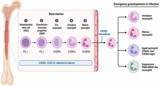
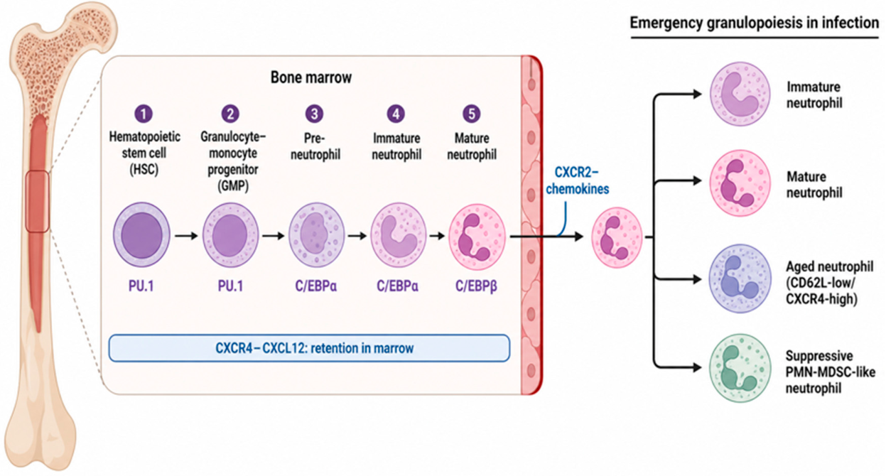
Granulopoiesis supplies the raw material from which neutrophil heterogeneity is built. Hematopoietic stem cells generate granulocyte-monocyte progenitors, which commit to the neutrophil lineage and pass through a series of morphologically and transcriptionally defined stages—pre-neutrophils, immature neutrophils, and mature neutrophils—before release into the circulation [3,6]. Coordinated activity of the transcription factors PU.1, C/EBPα, C/EBPβ, and GFI1 drives this progression, controlling proliferation, granule biogenesis, and the sequential acquisition of surface receptors that define maturity [3,6]. In a steady state, the bone marrow produces in the order of 10^11 neutrophils per day in an adult human, a rate that must expand rapidly during infection without sacrificing quality control over which cells leave the marrow (Figure 1).
Figure 1.
Neutrophil developmental hierarchy and infectious perturbation. Schematic of granulopoiesis from hematopoietic stem cells (HSC) through granulocyte-monocyte progenitors (GMP), pre-neutrophils, and immature neutrophils to mature, egress-competent neutrophils, driven sequentially by PU.1, C/EBPα, and C/EBPβ. Marrow retention is enforced by CXCR4-CXCL12 signaling; downregulation of CXCR4 paired with CXCR2 upregulation licenses release into circulation. Under infectious or inflammatory challenge, emergency granulopoiesis accelerates this pipeline and can additionally divert output toward aged and suppressive polymorphonuclear myeloid-derived suppressor cell (PMN-MDSC) branch states, shown at right.
Two chemokine axes govern this release. CXCR4 engagement by its ligand CXCL12, expressed by marrow stromal cells, retains developing and newly matured neutrophils within marrow niches and also recalls aged neutrophils home for clearance [7,18]. CXCR2 and its ELR+ CXC chemokine ligands, CXCL1, CXCL2, and CXCL8 in humans, drive the opposing process: downregulation of CXCR4 paired with upregulation of CXCR2 licenses egress into the bloodstream [18,19]. During infection, this balance shifts sharply. Emergency granulopoiesis, driven by G-CSF, IL-6, IL-1 family cytokines, and direct microbial sensing, accelerates precursor proliferation and relaxes marrow retention, so that immature neutrophils appear in blood at frequencies rarely seen at baseline [6]. The clinical correlate is the left shift long recognized in bacterial sepsis, but transcriptomic profiling now shows that this shift is not simply an increase in immature cell number; it reflects a genuine reprogramming of the entire granulopoietic output toward states with altered chemotactic responsiveness, oxidative burst capacity, and cytokine secretion [11,12].
The populations generated by this process are functionally distinct rather than interchangeable. Immature and low-density neutrophils, CD10-low/CD16-low in humans and Ly6G-intermediate/CD101-negative in mice, emerge preferentially under emergency granulopoiesis and often show blunted respiratory burst alongside distinct cytokine output compared with their mature counterparts [3,20]. Mature circulating neutrophils, CD10-positive/CD16-bright in humans and Ly6G-high/CD101-positive/CXCR2-positive in mice, carry the full antimicrobial toolkit and migrate efficiently into inflamed tissue [3,14]. A separate aged state arises through the circadian shedding of CD62L and the upregulation of CXCR4, which redirects these cells back toward the marrow, liver, and spleen for clearance and paradoxically primes them for enhanced vascular adhesion and NETosis en route [7,9]. Chronic infection, sepsis, and cancer additionally generate suppressive neutrophils and polymorphonuclear myeloid-derived suppressor cells (PMN-MDSC), which restrain T-cell proliferation through arginase-1, reactive nitrogen and oxygen species, and checkpoint ligand expression [21,22,23].
Whether these states represent fixed lineages or transient positions along a single trajectory remains contested. Pseudotime analyses of bone marrow and blood neutrophils describe a largely continuous transcriptional gradient—termed neutrotime—that spans early precursors to fully mature cells, arguing against sharp lineage boundaries [24]. Yet functional studies in healthy human cohorts followed longitudinally show that an individual’s neutrophil degranulation capacity, NET propensity, and killing efficiency against Staphylococcus aureus, Escherichia coli, and Candida albicans are stable, donor-specific traits linked to reproducible gene expression signatures, present even before any infectious challenge [25]. Reconciling a continuous developmental trajectory with a fixed, heritable component of neutrophil function remains an open problem and likely accounts for part of the substantial patient-to-patient variability in infection severity, ranging from bacterial sepsis to herpetic keratitis.
3. Defining Neutrophil Heterogeneity: Markers, States, and Controversies
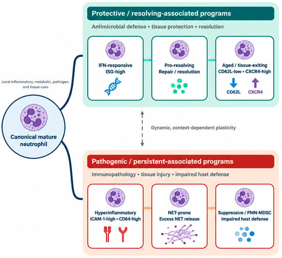
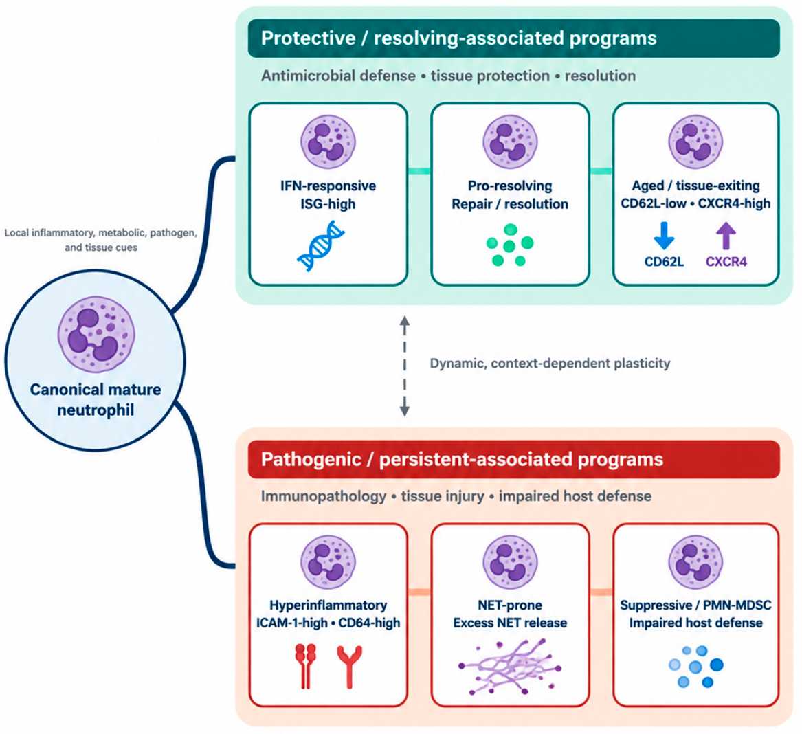
Attempts to classify neutrophil subsets have converged on a working vocabulary built from surface markers, buoyant density, transcriptional signatures, and functional readouts, though no single scheme has achieved universal acceptance [2,13]. Canonical mature neutrophils are typically defined as Ly6G-high, CD62L-positive, CD101-positive, CXCR2-positive, and CD64-low in mice, with CD10-positive/CD16-bright/CD62L-positive as the closest human equivalents [3,10]. Immature neutrophils carry the inverse profile—CD101-negative, CXCR2-low, and frequently CD64-positive—and segregate with the low-density fraction on density gradient separation [3,20]. Beyond these two poles, infection and inflammation drive the mature neutrophil toward several additional states branching from this common node: a hyperinflammatory phenotype marked by high ICAM-1 (CD54), CD64, and CD11b with a strong degranulation signature; an interferon-responsive state defined by elevated interferon-stimulated genes; a NET-prone state; an aged, CD62L-low/CXCR4-high state; a pro-resolving state; and suppressive or PMN-MDSC-like cells identified by arginase-1 expression and low-density co-sedimentation with peripheral blood mononuclear cells (Figure 2) [8,12,21]. Whether a given branch point favors pathogen clearance or bystander injury, indicated schematically by the protective and pathogenic shading of these states, depends on the cues described in the sections that follow.
Figure 2.
Infection-induced neutrophil state transitions. A mature neutrophil transitions, depending on local cytokine and metabolic cues, into interferon-responsive, pro-resolving states, aged, hyperinflammatory, NET-prone, or suppressive. These trajectories are not agent-specific: bacterial sepsis, viral pneumonia, and chronic infection can each drive a neutrophil toward either the protective or the pathogenic branch depending on pathogen burden, tissue context, and timing, rather than each infection type being restricted to a single fixed path.
Epigenetic and immunometabolic profiling reinforces the view that these categories reflect more than just surface phenotype. Chromatin accessibility at loci encoding effector genes, transcription factor occupancy, and metabolic wiring—glycolytic versus mitochondrial dependence—differ systematically between immature, interferon-driven, and suppressive states, indicating durable regulatory differences rather than superficial marker shifts [4,26]. Classification is nonetheless complicated by several practical realities. Many markers vary continuously rather than in discrete steps, individual cells can transition between states over hours, and species differences limit direct translation: Ly6G and CD101 have no human orthologue in routine clinical use, and Siglec-F-positive neutrophils, prominent in some mouse models of chronic lung inflammation, lack a clear human counterpart [9,13].
The greater conceptual difficulty is separating fixed, fate-committed lineages from transient activation states imposed by the local environment. A harmonized nomenclature that links surface phenotype, transcriptional program, and function across tissues and species, validated in parallel human and mouse infection models, remains an unmet need in the field and would materially improve cross-study comparison [24,27].
Metabolic wiring is emerging as a further, still incompletely mapped, axis of this heterogeneity. The long-standing view that neutrophils rely almost exclusively on aerobic glycolysis has been revised by evidence that immature and nutrient-restricted neutrophils can engage mitochondrial fatty-acid oxidation to sustain NADPH oxidase-dependent reactive oxygen species production when glucose is limiting, and that pentose-phosphate-pathway flux, rather than glycolysis alone, shapes oxidative burst capacity in specific contexts [28,29]. Hypoxia-inducible factor-1α integrates these metabolic inputs with local oxygen tension and inflammatory cues, acting as a node that links the tissue microenvironment to neutrophil activation state and survival [30]. Whether a given metabolic profile marks a fixed developmental identity or a reversible adaptation to local nutrient and oxygen availability remains unresolved, and it sharpens the broader controversy over neutrophil heterogeneity described above: transcriptional and metabolic states co-vary so closely in current datasets that a purely maturation-driven explanation cannot yet be distinguished from one in which the same cell adopts different programs as it moves through distinct microenvironments [28].
4. Functional Programs of Neutrophils During Infection
4.1. Core Effector Functions and Immunometabolic Control
Neutrophils execute several partially overlapping programs whose relative weighting shifts with developmental state, tissue context, and time after infection: phagocytosis and intracellular killing of bacteria, fungi, and opsonized virions; degranulation, releasing myeloperoxidase, elastase, and antimicrobial peptides from primary, secondary, and tertiary granules; a respiratory burst generating reactive oxygen species through NADPH oxidase; NETosis; cytokine and chemokine secretion that recruits and instructs other leukocytes; direct modulation of T-cell responses; and, in the resolution phase, efferocytosis and pro-repair signaling [1,31,32].
Against parasites, this repertoire operates differently than against bacteria, fungi, or viruses, because most helminths are too large to be phagocytosed, and many protozoa actively subvert phagocytic killing. Neutrophils instead rely heavily on NETosis and degranulation to immobilize and damage extracellular parasites: neutrophil-derived NETs entrap larval and adult helminths and, together with granule proteins, contribute to parasite killing, though the same NET response can also injure host tissue at the site of larval migration [33]. This pattern extends beyond tissue-migrating larvae to systemic helminth infection more broadly. In Toxocara canis infection, neutrophils act as key modulators of immunity rather than purely antimicrobial effectors, deploying effector functions against the parasite while also being subject to parasite-driven immune evasion strategies and crosstalk with the type 2 immune response that shapes overall disease outcome [34]. In protozoan infections, neutrophil NETosis is similarly double-edged; in malaria, extracellular heme released during erythrocyte rupture triggers NET formation, and NET components, in turn, promote endothelial ICAM-1 upregulation, emergency granulopoiesis, and parasite sequestration, linking a nominally antimicrobial program directly to severe malaria pathology [35]. Enteric protozoa illustrate the same tension from the pathogen’s perspective: live Giardia intestinalis trophozoites activate human neutrophils and drive NET formation, yet simultaneously dampen neutrophil ROS production, indicating that the parasite actively reshapes the neutrophil response rather than simply triggering it wholesale [36]. Entamoeba histolytica similarly induces human neutrophils to undergo NETosis during the early inflammatory phase of amoebiasis [37], and follow-up work shows this response can be pharmacologically tuned by pre-incubating neutrophil–amoeba interactions with the carbohydrate GalNAc, which suppresses NET formation while paradoxically increasing myeloperoxidase enzyme activity, underscoring that NET release and granule-derived killing are separable, independently regulated arms of the neutrophil antiparasitic response [37,38]. NETosis therefore functions as a genuinely cross-pathogen effector mechanism, operating against bacteria, viruses, and parasites alike through the same core machinery dependent on citrullination and chromatin decondensation, with the balance of benefit and harm set by pathogen size, tissue location, and the efficiency of NET clearance rather than by pathogen class itself [33,34,39].
Most of these functions run on aerobic glycolysis, which sustains rapid ATP production even in the hypoxic, poorly perfused microenvironments typical of infected or necrotic tissue [13,40]. Hypoxia-inducible factor signaling reinforces this glycolytic commitment and extends neutrophil survival at inflamed sites, while mitochondrial metabolism, though quantitatively modest, regulates redox balance, apoptotic timing, and the propensity to undergo NETosis [13,26,40]. Fatty acid oxidation, once assumed to be dispensable in a cell dominated by glycolysis, has recently been shown to be required for efficient neutrophil trafficking to infected tissue in a Cpt1a-dependent manner, adding a further layer of metabolic control over recruitment that had not been anticipated from studies of neutrophil metabolism at rest [41]. Lipid metabolism more broadly shapes neutrophil phenotype in sepsis, viral pneumonia, and cancer, linking the systemic metabolic state to local neutrophil function in ways that are still being mapped [40,42].
4.2. Temporal Transitions from Protective to Pathogenic Programs
Neutrophil programs are not static across the course of an infection; they typically transition from an early, appropriately protective response to a later phase in which persistence of activated or exhausted states drives collateral damage [11,43]. In bacterial sepsis, an early neutrophil signature dominated by antimicrobial and phagocytic genes gives way, in non-survivors, to a signature enriched for immature and suppressive transcriptional programs, suggesting that failure to resolve this transition contributes directly to mortality [11,44]. Comparable dynamics appear in the gut, where chronic neutrophil infiltration transitions from a barrier-protective, microbiota-regulating function into a driver of epithelial injury and, over years, colitis-associated tumorigenesis [17,45]. The recurring lesson is that duration and resolution kinetics, not peak neutrophil number, determine whether a given infection ends in clearance or in chronic tissue damage. Duration and resolution kinetics are not the only variables governing this dichotomy. Pathogen-intrinsic factors, including virulence factor repertoire and the capacity to actively subvert or delay neutrophil recruitment, set the baseline intensity of the response before host factors come into play. Host genetic background contributes independently: donor-specific, heritable differences in neutrophil degranulation and killing capacity, present even before infection, predict how far a given individual’s response drifts toward the pathogenic end of the spectrum [25]. Comorbid metabolic and inflammatory states, such as diabetes and chronic kidney disease, further shift this balance by altering baseline granulopoiesis and priming circulating neutrophils toward a more activated, NET-prone phenotype before an infectious trigger arrives, so that the same pathogen can produce a qualitatively different neutrophil response in an otherwise healthy host than in one with pre-existing systemic inflammation [46,47,48].
5. The Protective–Pathogenic Balance Across Infectious Disease
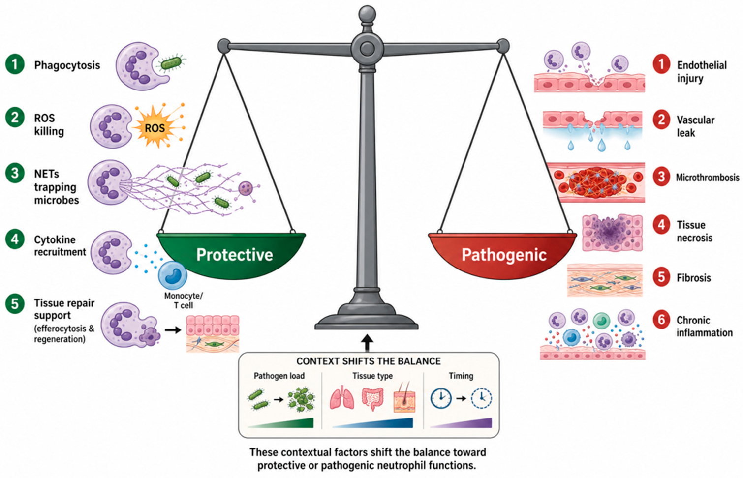
The ocular case study generalizes to a broader principle that recurs across every infection considered in this review (Figure 3). On the protective side of the balance, neutrophils contribute phagocytic and oxidative killing of microbes, NET-mediated entrapment of pathogens too large for phagocytosis, cytokine-driven recruitment of complementary immune populations, and, in the resolution phase, efferocytosis of apoptotic cells and secretion of pro-repair mediators [1,32]. On the pathogenic side sit endothelial injury from sustained protease release, vascular leak, NET-associated microthrombosis, tissue necrosis, and, if inflammation fails to resolve, progression to fibrosis [49,50].
Figure 3.
Protective versus pathogenic neutrophil functions in infection. Balance-scale representation contrasting protective neutrophil outputs, phagocytosis, oxidative and NET-mediated microbial killing, cytokine-driven immune recruitment, and pro-resolving efferocytosis against pathogenic outputs, protease- and reactive-oxygen-mediated tissue injury, NET-associated microvascular thrombosis, and unresolved chronic inflammation. Pathogen burden, tissue type, and timing of neutrophil recruitment determine which side of the balance predominates in a given infection.
Three variables determine where a given infection sits along this spectrum: pathogen load, tissue type, and timing.
Pathogen load: High pathogen burden generally favors the protective side of the ledger in the short term, since uncontrolled replication carries its own severe cost, but sustained high burden eventually exhausts the resolution machinery and tips the balance toward pathology. This pattern is well documented in severe bacterial sepsis and, separately, in fulminant viral pneumonia, where dysregulated or immature neutrophil states track with worse outcomes independent of absolute neutrophil count [11,42,44].
Tissue type: Tissue type sets the threshold at which a given level of neutrophil activity becomes damaging: immune-privileged tissues such as the cornea and central nervous system tolerate far less neutrophil-mediated injury than skin or gut before permanent damage results, because these tissues combine limited regenerative capacity with an intrinsic structural or functional reliance on being left undisturbed, such as corneal transparency or the blood–brain barrier [15,45].
Timing: Timing determines whether a neutrophil response arrives during a window when pathogen control still outweighs collateral risk, or after that window has closed, at which point continued neutrophil activity offers diminishing antimicrobial return for escalating tissue cost, exactly the pattern documented for the early-versus-late corneal neutrophil waves described above [51,52].
6. NETosis: Mechanisms and Dual Consequences
6.1. Triggers and Mechanisms of NET Formation
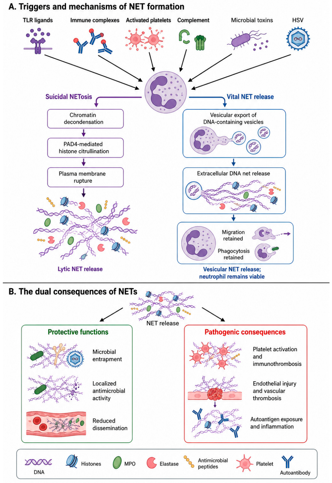
NETosis illustrates the double-edged nature of neutrophil biology more clearly than any other single program, and it merits close examination given how frequently it recurs across the tissue contexts and disease states already discussed (Figure 4). Toll-like receptor ligands, immune complexes, activated platelets, complement components, and microbial toxins can each trigger the classical suicidal pathway, in which chromatin decondenses, PAD4-mediated citrullination of histones loosens nucleosome packing, and the plasma membrane ruptures to extrude a web of DNA decorated with myeloperoxidase, elastase, and antimicrobial peptides [53,54]. A separate vital pathway enables vesicular release of nuclear or mitochondrial DNA without compromising cell viability, thereby allowing the neutrophil to continue phagocytosis after NET extrusion [53,54]. Recent work indicates that at least part of the NETotic program is a default epigenetic outcome of PAD4 activity during neutrophil apoptosis rather than an exclusively stimulus-driven event, which may explain why low-level NET formation is detectable even in the absence of overt infection [54].
Figure 4.
Triggers, mechanisms, and dual consequences of neutrophil extracellular trap formation. (A) Neutrophil extracellular trap (NET) formation can be induced by Toll-like receptor ligands, immune complexes, activated platelets, complement components, microbial toxins, and HSV. In suicidal NETosis, chromatin decondensation and PAD4-mediated histone citrullination culminate in plasma membrane rupture and the lytic release of extracellular DNA decorated with histones, MPO, elastase, and antimicrobial peptides. In the vital NET release, DNA-containing vesicles are released without loss of neutrophil viability, allowing continued migration and phagocytosis. (B) NETs have context-dependent effects during infection. Protective NET functions include entrapment of bacteria, fungi, and viruses, local concentration of antimicrobial effectors, and limitation of microbial dissemination. Conversely, excessive or insufficiently cleared NETs can promote platelet activation, immunothrombosis, endothelial injury, vascular thrombosis, autoantigen exposure, persistent inflammation, and bystander tissue damage.
6.2. The Dichotomous Nature of NETs
The antimicrobial rationale for NETosis is straightforward: entrapment restricts the spread of bacteria and fungi too large to be phagocytosed, exposes them to a locally concentrated antimicrobial payload of histones, myeloperoxidase, elastase, and defensins, and limits pathogen dissemination while slower clearance mechanisms are recruited [8,39]. NET-bound elastase and myeloperoxidase degrade bacterial virulence factors and outer-membrane proteins directly, and histone components exhibit independent bactericidal and fungicidal activity, so antimicrobial killing occurs through both physical entrapment and the biochemical activity of the trapped components themselves, not through entrapment alone. The cost of this strategy becomes apparent when NET formation is excessive or poorly cleared. NETs provide a scaffold for platelet adhesion and fibrin deposition, directly promoting microvascular thrombosis, and their histone and DNA content can serve as autoantigens that provoke autoantibody formation [49,55]. In severe viral pneumonia, including COVID-19, uncontrolled NETosis has been linked to pulmonary endothelial injury, immunothrombosis, and multi-organ dysfunction, and elevated circulating NET markers track with disease severity across viral and bacterial sepsis alike [49,50,55]. The rapid, protective corneal NETosis described in Section 8 and the pathogenic pulmonary and systemic NETosis described here are mechanistically the same program; what differs is the speed of resolution and the tissue’s tolerance to its byproducts, reinforcing the framework introduced in the preceding section. The same molecular machinery that clears a localized bacterial infection can therefore become the proximate driver of pathology once its containment fails, a pattern that recurs across nearly every organ system considered in this review.
7. Tissue-Specific Neutrophil Programming
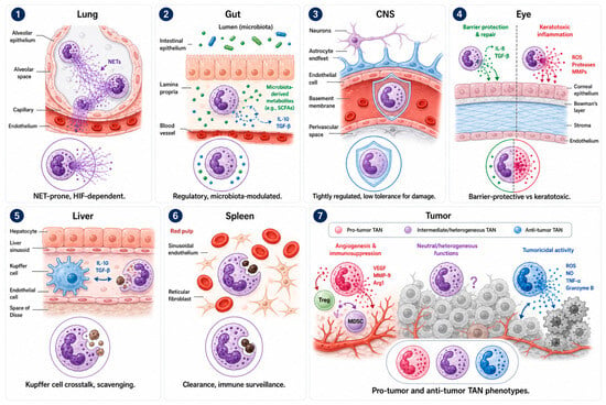
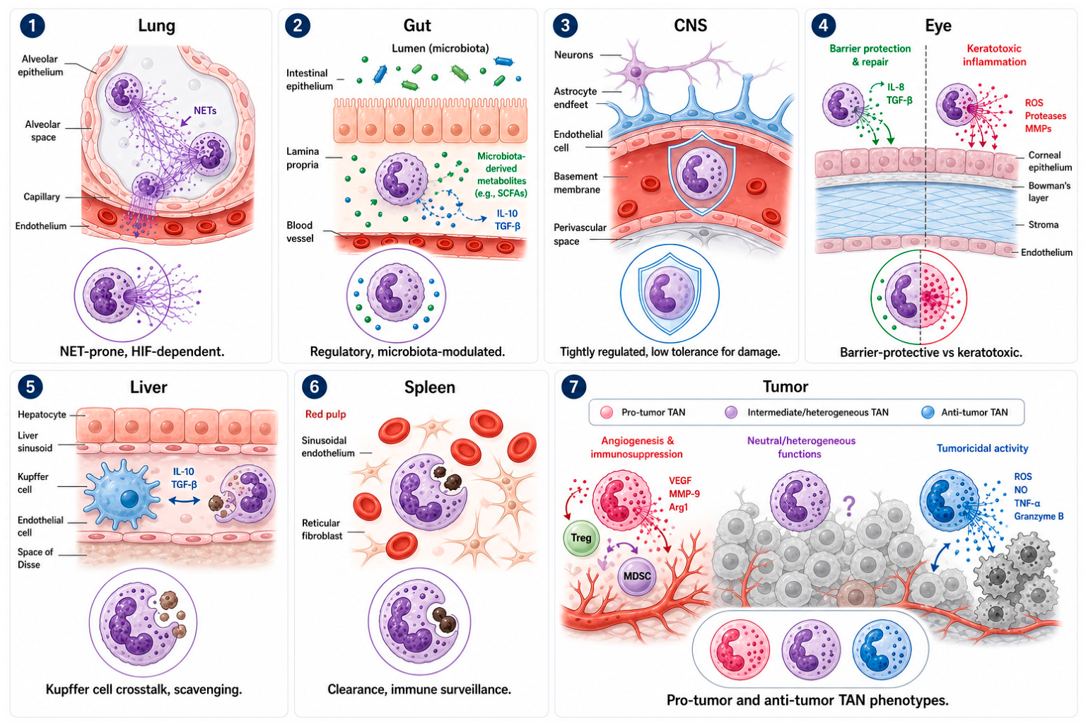
Neutrophils that enter a tissue do not simply execute a generic program; they are reprogrammed by local oxygen tension, resident microbiota, stromal architecture, and neighboring immune populations, thereby generating organ-specific functional signatures layered atop the developmental and infection-induced states outlined above (Figure 5) [13,56]. Mapping these signatures across organs clarifies why the same pathogen can provoke very different neutrophil-driven outcomes depending on the site of infection.
Figure 5.
Tissue-specific neutrophil phenotypes and functions across barrier organs, reticuloendothelial tissues, and tumors. Neutrophils adopt distinct functional states according to local tissue architecture, stromal and immune-cell interactions, microbial signals, and inflammatory cues. (1) Lung. Within the alveolar compartment, neutrophils can undergo robust NET formation in response to hypoxia and inflammatory activation. Although NETs may contribute to antimicrobial defense, excessive or persistent NETosis can damage the alveolar–capillary barrier and promote pulmonary inflammation. (2) Gut. Intestinal neutrophil activity is shaped by epithelial and microbial signals, including microbiota-derived metabolites such as short-chain fatty acids (SCFAs). In this setting, regulatory neutrophil programs and IL-10/TGF-β-associated signals can support barrier homeostasis and limit excessive mucosal inflammation. (3) Central nervous system. Neutrophil entry into the CNS is tightly controlled by the neurovascular unit, including endothelial cells, astrocytes, and the basement membrane. Because of the CNS’s limited tolerance for inflammatory injury, neutrophil responses require stringent regulation to balance host defense with protection of neural tissue. (4) Eye. In the cornea, neutrophils can support epithelial barrier protection and repair through regulated inflammatory signaling, including responses mediated by IL-8 and TGF-β. Conversely, excessive neutrophil activation, reactive oxygen species (ROS), protease release, and matrix metalloproteinase (MMP) activity can cause keratotoxic inflammation and stromal tissue damage. (5) Liver. In the hepatic sinusoidal microenvironment, neutrophils interact with Kupffer cells and endothelial cells to coordinate pathogen clearance, debris removal, and inflammatory resolution. IL-10 and TGF-β-associated crosstalk may restrain excessive activation and help preserve hepatic homeostasis. (6) Spleen. Splenic neutrophils reside within the red-pulp environment, where they interact with sinusoidal endothelial cells, reticular fibroblasts, and circulating blood cells. These interactions support clearance of blood-borne material, immune surveillance, and regulation of systemic inflammatory responses. (7) Tumor. Tumor-associated neutrophils (TANs) display marked functional heterogeneity. Pro-tumor TAN programs can promote angiogenesis and immunosuppression through mediators such as VEGF, MMP-9, Arg1, and interactions with regulatory T cells and myeloid-derived suppressor cells (MDSCs), whereas anti-tumor TAN programs can support tumoricidal activity through ROS, nitric oxide (NO), TNF-α, and granzyme B. Intermediate TAN states (shown as ‘?’) likely represent dynamic and context-dependent phenotypes within the tumor microenvironment.
In the lung, neutrophils recruited to alveolar and interstitial compartments acquire a NET-prone, hypoxia-adapted phenotype distinct from their circulating precursors, reflecting both the low oxygen tension of inflamed alveoli and dense local chemokine gradients [56,57]. Flow-cytometric phenotyping in murine models of sterile and infectious lung inflammation shows that neutrophils entering the lung parenchyma versus the bronchoalveolar space carry different maturity markers and cytokine profiles, and that NADPH oxidase-deficient neutrophils persist abnormally long in this compartment because of impaired reactive-oxygen-mediated clearance [57]. In viral respiratory infection, influenza and SARS-CoV-2 alike, dysregulated or immature neutrophil states, marked by defective reactive oxygen species production and altered NET-release gene signatures, correlate with disease severity, and neutrophil depletion experiments across multiple respiratory viral models, including an intriguing cross-reference to ocular HSV-1 depletion studies, point to an underappreciated protective role for early neutrophil influx that is easily obscured once inflammation becomes chronic [50,58].
The gut imposes different constraints. Constant exposure to a dense commensal microbiota selects for a more regulatory neutrophil phenotype that ordinarily limits bacterial translocation across the epithelium while contributing mediators needed for mucosal healing [45]. When this balance fails, whether through dysbiosis, barrier breach, or persistent inflammation, neutrophils shift toward a pathogenic phenotype implicated in inflammatory bowel disease and, over a longer time course, colitis-associated colorectal cancer, in part through interleukin-1β-driven amplification of local inflammation and reactive oxygen-mediated genomic damage to epithelial cells [45].
The central nervous system and the eye occupy opposite ends of the tolerance spectrum. Both are immune-privileged, structurally delicate, and populated by post-mitotic cells with limited regenerative capacity, so neutrophil infiltration that would be inconsequential in the skin or gut can cause irreversible damage in the cornea or brain parenchyma [15,59]. This constraint sets up the central paradox explored in detail in Section 8: tissues with the least tolerance for neutrophil-mediated collateral damage are often the tissues in which neutrophils are most essential for early pathogen containment.
The liver adds a further variation on the theme. Kupffer cells lining hepatic sinusoids capture circulating pathogens and recruit neutrophils through a coordinated intravascular surveillance mechanism, so that hepatic neutrophil function is inseparable from crosstalk with resident macrophages, a relationship less prominent in most other organs [60]. In the spleen, neutrophils participate in the filtration of aged and opsonized cells and in surveillance against blood-borne pathogens, contributing to the broader network of neutrophil trafficking that links marrow output, aging, and clearance across the reticuloendothelial system [7,9].
Tumors represent an extreme case of tissue-imprinted plasticity. Tumor-associated neutrophils can adopt either an N1, broadly anti-tumor phenotype capable of direct cytotoxicity and CD8+ T-cell activation, or an N2, pro-tumor phenotype that suppresses cytotoxic lymphocytes and supports angiogenesis and metastasis, with the balance between these states shaped by transforming growth factor-β and the surrounding cytokine milieu [27,61]. This N1/N2 framework, first developed in oncology, has proven useful well beyond cancer, offering a conceptual vocabulary for describing protective-versus-pathogenic neutrophil polarization in infectious contexts more generally.
8. Neutrophils in Ocular Herpes Simplex Virus Keratitis: A Tissue Case Study
Herpes simplex virus type 1 keratitis is among the most instructive settings in which to examine neutrophil duality because the tissue involved, the cornea, is avascular, densely innervated, and dependent on transparency for function, so that even modest neutrophil-driven injury has outsized clinical consequences. HSV-1 keratitis remains the leading infectious cause of corneal blindness in industrialized countries, and its pathology unfolds in two temporally and mechanistically distinct neutrophil-dominated phases [15,62].
8.1. The Early Antiviral Wave
Corneal epithelial cells sense HSV-1 infection and rapidly secrete CXCL1, the dominant early neutrophil chemoattractant in this tissue, producing a first wave of neutrophil infiltration within the initial 48 to 72 h after inoculation [15,63]. This wave is genuinely protective. Neutrophils recruited during this window restrict viral replication, and their depletion using anti-Ly6G antibodies increases both corneal viral titers and disease severity in mouse models, a finding consistent with neutrophil-depletion phenotypes reported across other viral infections, including influenza [58,64]. Recent work from our own group demonstrates that HSV-1 triggers an unusually rapid form of NETosis in corneal neutrophils, completed within 60 to 120 min of exposure through a caspase-1/pyroptosis-linked pathway coupled to myeloperoxidase release, far faster than the NETosis kinetics typically reported for bacterial stimuli [64]. This rapid antiviral NETosis was confirmed in both C57BL/6 and genetically diverse Diversity Outbred mice, and in tear samples from patients with human HSV-1 keratitis; neutrophil depletion in this model increased viral load and worsened ocular pathology, directly supporting a protective role for this pathway rather than a purely incidental one [64]. Corneal transcriptomes from the same patient cohort showed coordinated upregulation of CXCR2, heparanase, and PADI4, linking chemokine-driven recruitment to the molecular machinery of citrullination-dependent NET formation in this tissue specifically [64,65].
Type I and type III interferon signaling calibrate the intensity of this early response. IFN-λ, the dominant interferon induced by corneal HSV-1 infection, suppresses excessive neutrophil and macrophage infiltration and dampens CXCL1, IL-6, and IL-1β production without compromising the reactive-oxygen-dependent antiviral function of neutrophils that do arrive, indicating that interferon signaling reshapes neutrophil behavior rather than simply restraining neutrophil number [51]. In genital mucosal HSV models, a parallel but distinct axis operates: transient type I interferon signaling within the neutrophil compartment is protective, whereas sustained IFNAR1 engagement drives pathogenic IL-18 production, illustrating that duration of interferon exposure, not its presence or absence, determines whether the neutrophil–interferon axis protects the tissue or damages it [66]. As Lausch and colleagues first described, neutrophils themselves amplify this loop by producing CXCL10 (IP-10), the T-cell-recruiting chemokine more commonly attributed to epithelial or dendritic sources, providing an early bridge between innate neutrophil recruitment and the adaptive T-cell response that follows [67].
8.2. The Late, CD4+ T-Cell-Driven Pathogenic Wave
A second neutrophil influx begins roughly one week after infection and is driven not by direct viral sensing but by CD4+ T-cell-derived cytokines, principally interferon-γ, acting together with IL-2, which sustains the corneal neutrophil chemotactic gradient and antagonizes neutrophil apoptosis at a point in the disease course when viral replication has typically already been controlled [15,68]. This second wave is responsible for the immunopathology that defines herpes stromal keratitis as a clinical entity: neutrophils recruited during this phase release matrix metalloproteinase-9, which degrades corneal stromal collagen and promotes corneal neovascularization, thereby driving progressive vision loss [69,70]. Genetic ablation of CXCR2 might be expected to protect the cornea by blunting this neutrophil influx, but the opposite occurs: CXCR2-deficient mice actually develop worse herpetic stromal keratitis because diminished early neutrophil recruitment permits higher viral titers and triggers compensatory IL-6-driven neovascularization, a result first reported by Yan, Lausch, and colleagues over two decades ago that has stood since and that remains one of the clearest demonstrations that neutrophil number and disease severity are not simply proportional [63,71].
Whether this early-protective/late-pathogenic architecture generalizes to HSV-2 is less certain. In a mucosal genital model directly comparing the two serotypes, HSV-1 infection produced comparatively mild inflammation without a sustained neutrophil interferon-stimulated-gene program, whereas HSV-2 drove a persistent type I interferon–neutrophil–IL-18 axis that was itself responsible for much of the resulting genital pathology, and blocking IFNAR1 or neutralizing IL-18 reduced disease in the HSV-2 setting specifically [66]. This raises the possibility that HSV-2 keratitis, though clinically less common than HSV-1 keratitis, could involve a more protracted or more IL-18-dependent neutrophil pathogenic phase than the corneal model described above, a comparison that has not yet been tested directly in the eye and that would be a natural extension of the corneal HSV-1 framework presented here.
Neutrophils recruited during this late phase also intersect directly with corneal nerve integrity. HVEM-expressing myeloid populations shift from a monocyte/macrophage-dominated profile at three days post-infection to a neutrophil-dominated profile by two weeks, and this shift correlates with progressive loss of corneal mechanosensitivity; therapy with immune-modifying nanoparticles that blunt this late neutrophil and CD4+ T-cell infiltration preserves corneal sensation, directly linking the timing of neutrophil recruitment to a specific, clinically meaningful outcome [72]. A related mechanism operates through VEGF-A: CD4+ T cells and myeloid cells, including neutrophils, produce VEGF-A, which disrupts the corneal subbasal nerve plexus and drives the vascular ingrowth characteristic of stromal keratitis; depleting CD4+ T cells permits corneal reinnervation even after established disease [52]. Neutrophils further participate in a Th17-linked amplification loop, secreting factors that support IL-17-driven regulation of vascular endothelial growth factor receptor signaling, thereby connecting the adaptive Th17 axis to the innate neutrophil-driven neovascularization pathway described above [63].
8.3. Reconciling the Two Faces of the Corneal Neutrophil
The apparent contradiction, that neutrophils protect the cornea early and damage it later, resolves once developmental and functional heterogeneity are taken into account rather than treating “the neutrophil” as a single actor. The early antiviral wave is dominated by mature, virus-responsive neutrophils executing a rapid, interferon-calibrated NETosis program with a favorable benefit-to-cost ratio, given the low viral burden and intact stromal architecture at that stage [51,64]. The late wave recruits neutrophils into a tissue already primed by CD4+ T-cell cytokines, sustained chemokine gradients, and ongoing matrix remodeling, conditions that favor a hyperinflammatory, MMP-9-high, pro-angiogenic phenotype whose antimicrobial value has largely been exhausted while its capacity for collateral damage has not [69,70]. This framework generates a clear, testable prediction: therapeutic strategies that preserve or even enhance the early NETotic, antiviral neutrophil program while selectively restraining the late CD4+ T-cell-dependent, MMP-9-driven neutrophil program should outperform blunt neutrophil depletion or blanket immunosuppression, an approach that broad clinical experience with corticosteroids in herpetic keratitis has already shown to be a blunt and imperfect tool. Comparison with other ocular surface infections reinforces this model: neutrophil extracellular traps in Pseudomonas aeruginosa keratitis show a similarly dual protective-and-pathogenic role depending on bacterial virulence and NET clearance efficiency, and sterile corneal inflammation driven by NLRP3 inflammasome activation produces comparable MMP-9-associated neutrophil injury in the complete absence of any pathogen, indicating that the corneal tissue microenvironment itself, more than any single pathogen-specific signal, sets the threshold at which neutrophil recruitment tips from protective to pathogenic [59,73,74].
Open questions remain. Whether the fixed, donor-specific component of neutrophil functional heterogeneity described in healthy cohorts [25] contributes to the substantial variability in human HSV keratitis severity and recurrence risk has not been tested directly, despite being a plausible and readily testable hypothesis. Similarly, essentially all mechanistic dissections of corneal neutrophil subsets to date have relied on Ly6G/Gr-1 immunostaining or bulk antibody-mediated depletion rather than the deep single-cell phenotyping now standard in lung and blood neutrophil studies, leaving the transcriptional landscape of corneal neutrophils comparatively uncharacterized [57,64].
9. Therapeutic Targeting of Neutrophil Programs
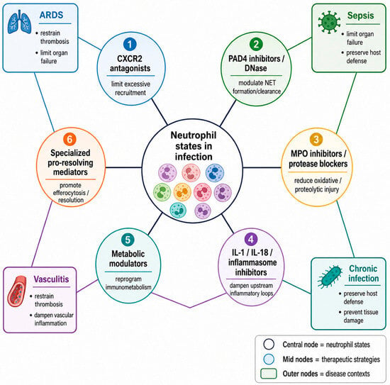
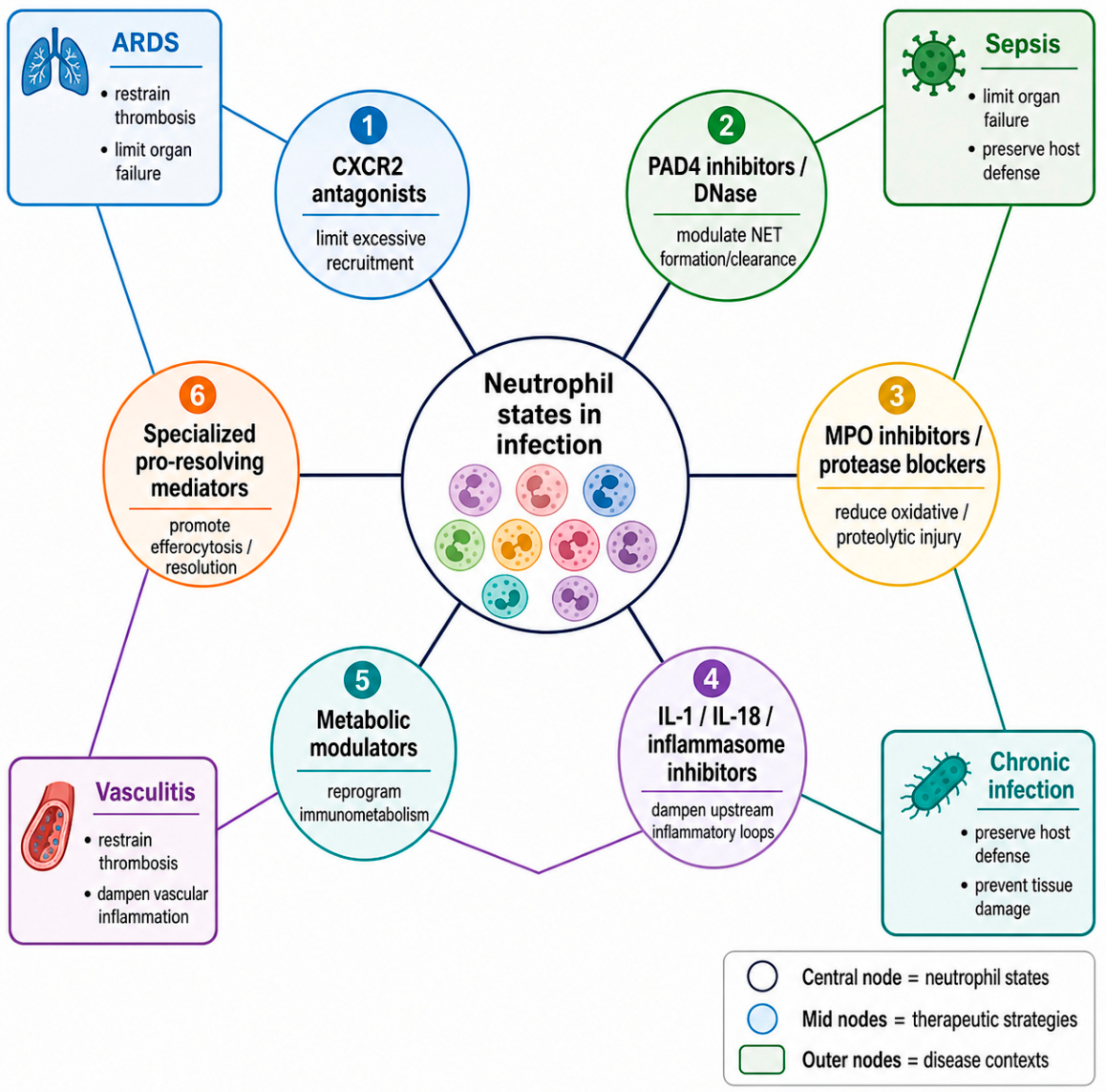
Recognition that neutrophil heterogeneity, rather than neutrophil number, drives infection outcomes has redirected therapeutic strategies away from global neutropenia-inducing approaches and toward interventions targeting specific programs (Figure 6). CXCR2 antagonists can limit excessive recruitment in acute respiratory distress syndrome and chronic inflammatory disease, though the CXCR2-knockout keratitis phenotype described above is a cautionary reminder that broadly blocking recruitment can worsen outcomes if it also blunts a genuinely protective early response [19,71]. In oncology, CXCR1/2 antagonism has been shown to reprogram tumor-associated neutrophil function, blunting immunosuppressive polarization and reactive-oxygen release without eliminating neutrophils from the tumor microenvironment altogether, a selectivity principle that may translate to infectious settings where the goal is to preserve antimicrobial capacity while curbing pathological byproducts [75].
Figure 6.
Therapeutic targeting of neutrophil programs. Six candidate intervention strategies mapped onto the neutrophil functional program they modulate: CXCR2 antagonists (recruitment), PAD4 inhibitors and DNase therapy (NET formation and clearance), myeloperoxidase inhibitors (oxidative injury), IL-1/IL-18 pathway inhibitors (inflammasome-driven amplification), metabolic modulators (trafficking and survival), and specialized pro-resolving mediators (resolution and efferocytosis).
PAD4 inhibitors and DNase-based therapies directly target NET formation and clearance, offering a route to reduce NET-associated immunothrombosis and autoantigen exposure in severe sepsis and viral pneumonia without abolishing neutrophil phagocytic function [53,54]. Myeloperoxidase inhibitors and protease blockers address the oxidative and proteolytic arms of neutrophil-mediated injury, relevant to both sepsis-associated organ damage and matrix metalloproteinase-9-driven corneal destruction in herpetic keratitis [70]. Inflammasome and IL-1/IL-18 pathway inhibitors dampen the upstream inflammatory loops that sustain pathological neutrophil recruitment, an approach with particular relevance to the sustained type I interferon–IL-18 axis identified in mucosal HSV infection [66]. Metabolic modulators that target glycolytic or fatty-acid-oxidation-dependent neutrophil trafficking represent an earlier-stage but conceptually attractive strategy, since interfering with Cpt1a-dependent trafficking could, in principle, blunt pathological neutrophil recruitment to a specific tissue while sparing systemic neutrophil function [26,41]. Specialized pro-resolving mediators promote efferocytosis and active resolution rather than simple suppression, offering a complementary strategy to accelerate the transition out of the pathogenic phase rather than prevent entry into the protective phase in the first place [17].
A shared lesson from these efforts, and from the corneal case study in particular, is that timing and tissue selectivity matter as much as target choice. An intervention applied during the early antiviral neutrophil wave in HSV-1 keratitis risks the same paradoxical worsening seen with global CXCR2 deletion, whereas the identical intervention applied selectively during the late CD4+ T-cell-driven wave might substantially reduce corneal scarring and neovascularization without compromising viral control [71,72].
10. Single-Cell and Spatial Approaches: Toward a Unified Neutrophil Ontology
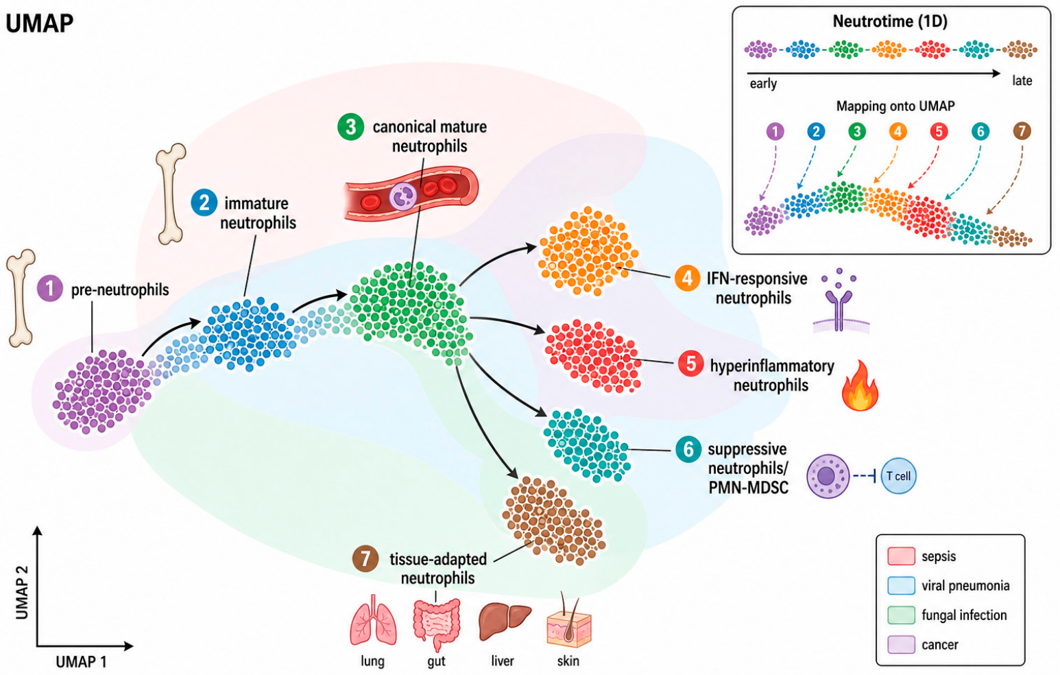
Single-cell RNA sequencing, mass cytometry, and spatial transcriptomics are converging on a shared description of neutrophil states that spans bone marrow, blood, and infected tissue, building on the state-transition framework introduced above (Figure 2 and Figure 7). Pseudotime trajectory analyses place pre-neutrophils, immature neutrophils, and canonical mature neutrophils along a single continuum, with interferon-responsive, hyperinflammatory, suppressive/PMN-MDSC, and tissue-adapted states branching from this trajectory under specific disease conditions such as sepsis, viral pneumonia, fungal infection, and cancer [4,24]. This neutrotime framework has already been extended across tissue compartments and species, suggesting the beginnings of a genuinely portable classification scheme rather than one specific to a single organ or model system [5,24].
Figure 7.
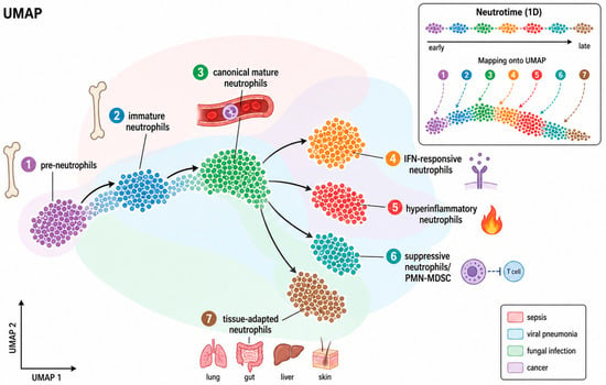
Single-cell atlas of neutrophil heterogeneity. Uniform manifold approximation and projection (UMAP)-style representation of neutrophil transcriptional states positioned along the neutrotime developmental trajectory, with disease-associated states, sepsis, viral infection, and cancer, overlaid to illustrate how distinct infectious and inflammatory contexts redistribute cells across the same underlying developmental continuum.
Two methodological gaps limit further progress. First, most single-cell neutrophil atlases remain concentrated in blood, bone marrow, lung, and tumor tissue; immune-privileged sites such as cornea and central nervous system, where the protective–pathogenic balance is most consequential clinically, have barely been profiled at single-cell resolution, and the flow-cytometric protocols now standard for lung neutrophil phenotyping have not yet been widely adapted to the smaller, more fragile corneal neutrophil population [57,64]. Second, spatial context is frequently lost in dissociation-based single-cell approaches; recent spatial transcriptomic mapping of neutrophil and macrophage heterogeneity in inflammatory bowel disease demonstrates that positional information relative to the epithelial barrier substantially refines functional interpretation of a given transcriptional state, a level of resolution not yet applied to ocular or CNS infection [27,61].
11. Future Perspectives
Closing these gaps will require coordinated effort along several fronts. Single-cell and spatial atlases of neutrophils in immune-privileged tissues during active infection, generated in parallel across mouse models and human clinical samples, would establish whether the corneal neutrophil states described functionally in Section 8 correspond to the transcriptional states already defined in blood and lung, or whether the cornea and CNS harbor distinct, previously undescribed neutrophil programs. Longitudinal studies that track the fixed, donor-specific component of neutrophil function identified in healthy cohorts through the course of an actual infection, rather than only at baseline, would clarify whether pre-existing neutrophil functional set-points predict susceptibility to severe keratitis, sepsis, or pneumonia. On the therapeutic side, the field would benefit from moving beyond binary neutrophil depletion or global suppression toward state-selective interventions, timed to specific phases of infection, that can first be tested in tractable models such as murine HSV-1 keratitis, where the early-versus-late neutrophil dichotomy is unusually well-defined and directly measurable. Finally, given how strongly interferon signaling calibrates neutrophil behavior in the eye and at mucosal surfaces, systematic comparison of type I, II, and III interferon effects on neutrophil programs across infection sites could identify a shared regulatory node amenable to therapeutic modulation across multiple organ systems simultaneously.
12. Conclusions
Neutrophils no longer fit the model of a uniform, disposable phagocyte. They constitute a developmental and functional continuum shaped by granulopoietic origin, chemokine-driven trafficking, and tissue-specific reprogramming, which determines whether a given infection resolves cleanly or progresses to tissue injury. Herpes simplex virus type 1 keratitis distills this principle into an unusually sharp form: an early, interferon-calibrated, NETosis-dependent neutrophil wave protects the cornea, while a later, CD4+ T-cell-driven wave, dominated by matrix metalloproteinase-9-high, pro-angiogenic neutrophils, causes the scarring and neovascularization that lead to vision loss. Extending single-cell and spatial profiling into this and other immune-privileged tissues, and translating the resulting state-level understanding into therapies that preserve protective neutrophil programs while restraining pathogenic ones, represents the clearest path toward turning three decades of heterogeneity research into meaningful clinical benefit.
Funding
This research was supported by grants from the National Institutes of Health (NIH) to D.S. (R01EY036253, R01EY029426, R01EY033622, R01EY024710, P30EY001792, and R24EY033598).
Institutional Review Board Statement
Not applicable.
Informed Consent Statement
Not applicable.
Data Availability Statement
No new data were created or analyzed in this study. Data sharing is not applicable to this article.
Acknowledgments
During the preparation of this manuscript, the authors used AI-assisted tools for literature organization support, schematic figure drafting, and language editing. The authors have reviewed and edited the output and take full responsibility for the content of this publication.
Conflicts of Interest
The authors declare no conflicts of interest.
Abbreviations
The following abbreviations are used in this manuscript:
| ARDS | acute respiratory distress syndrome |
| CNS | central nervous system |
| CXCL | C-X-C motif chemokine ligand |
| CXCR | C-X-C motif chemokine receptor |
| DNase | deoxyribonuclease |
| ELR+ | glutamate-leucine-arginine motif-positive |
| G-CSF | granulocyte colony-stimulating factor |
| HSK | herpes stromal keratitis |
| HSV-1 | herpes simplex virus type 1 |
| ICAM-1 | intercellular adhesion molecule 1 |
| IFN | interferon |
| IFNAR1 | interferon-alpha/beta receptor subunit 1 |
| IL | interleukin |
| MDSC | myeloid-derived suppressor cell |
| MMP-9 | matrix metalloproteinase 9 |
| MPO | myeloperoxidase |
| NET | neutrophil extracellular trap |
| NLRP3 | NOD-, LRR- and pyrin domain-containing protein 3 |
| PAD4/PADI4 | peptidylarginine deiminase 4 |
| PMN | polymorphonuclear neutrophil |
| ROS | reactive oxygen species |
| SPM | specialized pro-resolving mediator |
| TGF-β | transforming growth factor beta |
| Th17 | T helper 17 cell |
| UMAP | uniform manifold approximation and projection |
| VEGF-A | vascular endothelial growth factor A |
References
- Kolaczkowska, E.; Kubes, P. Neutrophil recruitment and function in health and inflammation. Nat. Rev. Immunol. 2013, 13, 159–175. [Google Scholar] [CrossRef] [Scilit]
- Rosales, C. Neutrophil: A Cell with Many Roles in Inflammation or Several Cell Types? Front. Physiol. 2018, 9, 113. [Google Scholar] [CrossRef] [Scilit]
- Evrard, M.; Kwok, I.W.H.; Chong, S.Z.; Teng, K.W.W.; Becht, E.; Chen, J.; Sieow, J.L.; Penny, H.L.; Ching, G.C.; Devi, S.; et al. Developmental Analysis of Bone Marrow Neutrophils Reveals Populations Specialized in Expansion, Trafficking, and Effector Functions. Immunity 2018, 48, 364–379.e8. [Google Scholar] [CrossRef] [Scilit]
- Xie, X.; Shi, Q.; Wu, P.; Zhang, X.; Kambara, H.; Su, J.; Yu, H.; Park, S.; Guo, R.; Ren, Q.; et al. Single-cell transcriptome profiling reveals neutrophil heterogeneity in homeostasis and infection. Nat. Immunol. 2020, 21, 1119–1133. [Google Scholar] [CrossRef] [Scilit]
- McLaren, A.S.; Fetit, R.; Wood, C.S.; Falconer, J.; Steele, C.W. Single cell sequencing of neutrophils demonstrates phenotypic heterogeneity and functional plasticity in health, disease, and cancer. Chin. Clin. Oncol. 2023, 12, 18. [Google Scholar] [CrossRef] [Scilit]
- Manz, M.G.; Boettcher, S. Emergency granulopoiesis. Nat. Rev. Immunol. 2014, 14, 302–314. [Google Scholar] [CrossRef] [Scilit]
- Adrover, J.M.; del Fresno, C.; Crainiciuc, G.; Cuartero, M.I.; Casanova-Acebes, M.; Weiss, L.A.; Huerga-Encabo, H.; Silvestre-Roig, C.; Rossaint, J.; Cossío, I.; et al. A Neutrophil Timer Coordinates Immune Defense and Vascular Protection. Immunity 2019, 50, 390–402.e10. [Google Scholar] [CrossRef] [Scilit]
- Papayannopoulos, V. Neutrophil extracellular traps in immunity and disease. Nat. Rev. Immunol. 2018, 18, 134–147. [Google Scholar] [CrossRef] [Scilit]
- Ng, L.G.; Ostuni, R.; Hidalgo, A. Heterogeneity of neutrophils. Nat. Rev. Immunol. 2019, 19, 255–265. [Google Scholar] [CrossRef] [Scilit]
- Zilionis, R.; Engblom, C.; Pfirschke, C.; Savova, V.; Zemmour, D.; Saatcioglu, H.D.; Krishnan, I.; Maroni, G.; Meyerovitz, C.V.; Kerwin, C.M.; et al. Single-Cell Transcriptomics of Human and Mouse Lung Cancers Reveals Conserved Myeloid Populations across Individuals and Species. Immunity 2019, 50, 1317–1334.e10. [Google Scholar] [CrossRef] [Scilit]
- Reyes, M.; Filbin, M.R.; Bhattacharyya, R.P.; Billman, K.; Eisenhaure, T.; Hung, D.T.; Levy, B.D.; Baron, R.M.; Blainey, P.C.; Goldberg, M.B.; et al. An immune-cell signature of bacterial sepsis. Nat. Med. 2020, 26, 333–340. [Google Scholar] [CrossRef] [Scilit]
- Schulte-Schrepping, J.; Reusch, N.; Paclik, D.; Baßler, K.; Schlickeiser, S.; Zhang, B.; Krämer, B.; Krammer, T.; Brumhard, S.; Bonaguro, L.; et al. Severe COVID-19 Is Marked by a Dysregulated Myeloid Cell Compartment. Cell 2020, 182, 1419. [Google Scholar] [CrossRef] [Scilit]
- Silvestre-Roig, C.; Hidalgo, A.; Soehnlein, O. Neutrophil heterogeneity: Implications for homeostasis and pathogenesis. Blood 2016, 127, 2173–2181. [Google Scholar] [CrossRef] [Scilit]
- Gazendam, R.P.; van de Geer, A.; Roos, D.; van den Berg, T.K.; Kuijpers, T.W. How neutrophils kill fungi. Immunol. Rev. 2016, 273, 299–311. [Google Scholar] [CrossRef] [Scilit]
- Lobo, A.; Agelidis, A.M.; Shukla, D. Pathogenesis of herpes simplex keratitis: The host cell response and ocular surface sequelae to infection and inflammation. Ocul. Surf. 2019, 17, 40–49. [Google Scholar] [CrossRef] [Scilit]
- Rawat, K.; Shrivastava, A. Neutrophils as emerging protagonists and targets in chronic inflammatory diseases. Inflamm. Res. 2022, 71, 1477. [Google Scholar] [CrossRef] [Scilit]
- Herrero-Cervera, A.; Soehnlein, O.; Kenne, E. Neutrophils in chronic inflammatory diseases. Cell Mol. Immunol. 2022, 19, 177. [Google Scholar] [CrossRef] [Scilit]
- Eash, K.J.; Greenbaum, A.M.; Gopalan, P.K.; Link, D.C. CXCR2 and CXCR4 antagonistically regulate neutrophil trafficking from murine bone marrow. J. Clin. Investig. 2010, 120, 2423–2431. [Google Scholar] [CrossRef] [Scilit]
- Zhang, X.; Guo, R.; Kambara, H.; Ma, F.; Luo, H.R. The role of CXCR2 in acute inflammatory responses and its antagonists as anti-inflammatory therapeutics. Curr. Opin. Hematol. 2019, 26, 28. [Google Scholar] [CrossRef] [Scilit]
- Pillay, J.; den Braber, I.; Vrisekoop, N.; Kwast, L.M.; de Boer, R.J.; Borghans, J.A.M.; Tesselaar, K.; Koenderman, L. In vivo labeling with 2H2O reveals a human neutrophil lifespan of 5.4 days. Blood 2010, 116, 625–627. [Google Scholar] [CrossRef] [Scilit]
- Gabrilovich, D.I. Myeloid-Derived Suppressor Cells. Cancer Immunol. Res. 2017, 5, 3–8. [Google Scholar] [CrossRef] [Scilit]
- Veglia, F.; Sanseviero, E.; Gabrilovich, D.I. Myeloid-derived suppressor cells in the era of increasing myeloid cell diversity. Nat. Rev. Immunol. 2021, 21, 485. [Google Scholar] [CrossRef] [Scilit]
- Dorhoi, A.; Glaría, E.; Garcia-Tellez, T.; Nieuwenhuizen, N.E.; Zelinskyy, G.; Favier, B.; Singh, A.; Ehrchen, J.; Gujer, C.; Münz, C.; et al. MDSCs in infectious diseases: Regulation, roles, and readjustment. Cancer Immunol. Immunother. 2019, 68, 673–685. [Google Scholar] [CrossRef] [Scilit]
- Grieshaber-Bouyer, R.; Radtke, F.A.; Cunin, P.; Stifano, G.; Levescot, A.; Vijaykumar, B.; Nelson-Maney, N.; Blaustein, R.B.; Monach, P.A.; Nigrovic, P.A.; et al. The neutrotime transcriptional signature defines a single continuum of neutrophils across biological compartments. Nat. Commun. 2021, 12, 2856. [Google Scholar] [CrossRef] [Scilit]
- Maskarinec, S.A.; McKelvy, M.; Boyle, K.; Hotchkiss, H.; Duarte, M.E.; Addison, B.; Amato, N.; Khandelwal, S.; Arepally, G.M.; Lee, G.M. Neutrophil functional heterogeneity is a fixed phenotype and is associated with distinct gene expression profiles. J. Leukoc. Biol. 2022, 112, 1485–1495. [Google Scholar] [CrossRef] [Scilit]
- Toller-Kawahisa, J.E.; O’neill, L.A.J. How neutrophil metabolism affects bacterial killing. Open Biol. 2022, 12, 220248. [Google Scholar] [CrossRef] [Scilit]
- Antuamwine, B.B.; Bosnjakovic, R.; Hofmann-Vega, F.; Wang, X.; Theodosiou, T.; Iliopoulos, I.; Brandau, S. N1 versus N2 and PMN-MDSC: A critical appraisal of current concepts on tumor-associated neutrophils and new directions for human oncology. Immunol. Rev. 2023, 314, 250–279. [Google Scholar] [CrossRef] [Scilit]
- Yipeng, Z.; Chao, C.; Ranran, L.; Tingting, P.; Hongping, Q. Metabolism: A potential regulator of neutrophil fate. Front. Immunol. 2024, 15, 1500676. [Google Scholar] [CrossRef] [Scilit]
- Rice, C.M.; Davies, L.C.; Subleski, J.J.; Maio, N.; Gonzalez-Cotto, M.; Andrews, C.; Patel, N.L.; Palmieri, E.M.; Weiss, J.M.; Lee, J.; et al. Tumour-elicited neutrophils engage mitochondrial metabolism to circumvent nutrient limitations and maintain immune suppression. Nat. Commun. 2018, 9, 5099. [Google Scholar] [CrossRef] [Scilit]
- Stothers, C.L.; Luan, L.; Fensterheim, B.A.; Bohannon, J.K. Hypoxia-inducible factor-1α regulation of myeloid cells. J. Mol. Med. 2018, 96, 1293–1306. [Google Scholar] [CrossRef] [Scilit]
- Scapini, P.; Cassatella, M.A. Social networking of human neutrophils within the immune system. Blood 2014, 124, 710–719. [Google Scholar] [CrossRef] [Scilit]
- Segal, A.W. How Neutrophils Kill Microbes. Annu. Rev. Immunol. 2005, 23, 197–223. [Google Scholar] [CrossRef] [Scilit]
- Bouchery, T.; Moyat, M.; Sotillo, J.; Silverstein, S.; Volpe, B.; Coakley, G.; Tsourouktsoglou, T.; Becker, L.; Shah, K.; Kulagin, M.; et al. Hookworms Evade Host Immunity by Secreting a Deoxyribonuclease to Degrade Neutrophil Extracellular Traps. Cell Host Microbe 2020, 27, 277–289.e6. [Google Scholar] [CrossRef] [Scilit]
- Abou-El-Naga, I.F. Neutrophils in Toxocara canis infection: Effector functions, immune evasion and crosstalk with type 2 immunity. Microb. Pathog. 2026, 211, 108267. [Google Scholar] [CrossRef] [Scilit]
- Knackstedt, S.L.; Georgiadou, A.; Apel, F.; Abu-Abed, U.; Moxon, C.A.; Cunnington, A.J.; Raupach, B.; Cunningham, D.; Langhorne, J.; Krüger, R.; et al. Neutrophil extracellular traps drive inflammatory pathogenesis in malaria. Sci. Immunol. 2019, 4, eaaw0336. [Google Scholar] [CrossRef] [Scilit]
- Salinas-Varas, C.; Bezerra, T.L.; Rojas-Barón, L.; Gondim, L.F.P.; Wagenlehner, F.; Gärtner, U.; Taubert, A.; Hermosilla, C.; Conejeros, I. Giardia intestinalis trophozoites activate human PMN and induce NET formation but dampen neutrophil ROS production. Front. Immunol. 2026, 17, 1724948. [Google Scholar] [CrossRef] [Scilit]
- Ventura-Juarez, J.; Campos-Esparza, M.; Pacheco-Yepez, J.; López-Blanco, J.A.; Adabache-Ortíz, A.; Silva-Briano, M.; Campos-Rodríguez, R. Entamoeba histolytica induces human neutrophils to form NETs. Parasite Immunol. 2016, 38, 503–509. [Google Scholar] [CrossRef] [Scilit]
- Levaro-Loquio, D.; Cruz-Baquero, A.; Higuera-Martínez, G.; Serrano-Luna, J.D.J.; Contis-Montes de Oca, A.; Abarca-Rojano, E.; Arciniega-Martínez, I.M.; Reséndiz-Albor, A.A.; Pacheco-Yépez, J. GalNAc Carbohydrate Prevents the Formation of Neutrophil Extracellular Traps and Increase Myeloperoxidase Enzyme Activity in Interactions of Neutrophils and Entamoeba histolytica Preincubated with GalNAc. BioMed Res. Int. 2026, 2026, e8280585. [Google Scholar] [CrossRef] [Scilit]
- Brinkmann, V.; Reichard, U.; Goosmann, C.; Fauler, B.; Uhlemann, Y.; Weiss, D.S.; Weinrauch, Y.; Zychlinsky, A. Neutrophil Extracellular Traps Kill Bacteria. Science 2004, 303, 1532–1535. [Google Scholar] [CrossRef] [Scilit]
- Leblanc, P.; Bourgoin, S.G.; Poubelle, P.E.; Tessier, P.A.; Pelletier, M. Metabolic regulation of neutrophil functions in homeostasis and diseases. J. Leukoc. Biol. 2024, 116, 456–468. [Google Scholar] [CrossRef] [Scilit]
- Pham, L.; Komalavilas, P.; Eddie, A.M.; Thayer, T.E.; Greenwood, D.L.; Liu, K.H.; Weinberg, J.; Patterson, A.; Fessel, J.P.; Boyd, K.L.; et al. Neutrophil trafficking to the site of infection requires Cpt1a-dependent fatty acid β-oxidation. Commun. Biol. 2022, 5, 1366. [Google Scholar] [CrossRef] [Scilit]
- Camp, J.V.; Jonsson, C.B. A Role for Neutrophils in Viral Respiratory Disease. Front. Immunol. 2017, 8, 550. [Google Scholar] [CrossRef] [Scilit]
- Denning, N.; Aziz, M.; Gurien, S.D.; Wang, P. DAMPs and NETs in Sepsis. Front. Immunol. 2019, 10, 2536. [Google Scholar] [CrossRef] [Scilit]
- Zhang, J.; Shao, Y.; Wu, J.; Zhang, J.; Xiong, X.; Mao, J.; Wei, Y.; Miao, C.; Zhang, H. Dysregulation of neutrophil in sepsis: Recent insights and advances. Cell Commun. Signal. 2025, 23, 87. [Google Scholar] [CrossRef] [Scilit]
- Chen, T.; Liu, J.; Hang, R.; Chen, Q.; Wang, D. Neutrophils: From Inflammatory Bowel Disease to Colitis-Associated Colorectal Cancer. J. Inflamm. Res. 2025, 18, 925. [Google Scholar] [CrossRef] [Scilit]
- Wong, S.L.; Demers, M.; Martinod, K.; Gallant, M.; Wang, Y.; Goldfine, A.B.; Kahn, C.R.; Wagner, D.D. Diabetes primes neutrophils to undergo NETosis, which impairs wound healing. Nat. Med. 2015, 21, 815–819. [Google Scholar] [CrossRef] [Scilit]
- Thakur, M.; Junho, C.V.C.; Bernhard, S.M.; Schindewolf, M.; Noels, H.; Döring, Y. NETs-Induced Thrombosis Impacts on Cardiovascular and Chronic Kidney Disease. Circ. Res. 2023, 132, 933. [Google Scholar] [CrossRef] [Scilit]
- Nakazawa, D.; Masuda, S.; Nishibata, Y.; Watanabe-Kusunoki, K.; Tomaru, U.; Ishizu, A. Neutrophils and NETs in kidney disease. Nat. Rev. Nephrol. 2025, 21, 383–398. [Google Scholar] [CrossRef] [Scilit]
- Middleton, E.A.; He, X.; Denorme, F.; Campbell, R.A.; Ng, D.; Salvatore, S.P.; Mostyka, M.; Baxter-Stoltzfus, A.; Borczuk, A.C.; Loda, M.; et al. Neutrophil extracellular traps contribute to immunothrombosis in COVID-19 acute respiratory distress syndrome. Blood 2020, 136, 1169–1179. [Google Scholar] [CrossRef] [Scilit]
- Behzadifard, M.; Soleimani, M. NETosis and SARS-COV-2 infection related thrombosis: A narrative review. Thromb. J. 2022, 20, 13. [Google Scholar] [CrossRef] [Scilit]
- Antony, F.; Pundkar, C.; Sandey, M.; Jaiswal, A.K.; Mishra, A.; Kumar, A.; Channappanavar, R.; Suryawanshi, A. IFN-λ Regulates Neutrophil Biology to Suppress Inflammation in Herpes Simplex Virus-1-Induced Corneal Immunopathology. J. Immunol. 2021, 206, 1866–1877. [Google Scholar] [CrossRef] [Scilit]
- Yun, H.; Yee, M.B.; Lathrop, K.L.; Kinchington, P.R.; Hendricks, R.L.; St Leger, A.J. Production of the Cytokine VEGF-A by CD4+ T and Myeloid Cells Disrupts the Corneal Nerve Landscape and Promotes Herpes Stromal Keratitis. Immunity 2020, 53, 1050–1062.e5. [Google Scholar] [CrossRef] [Scilit]
- Thiam, H.R.; Wong, S.L.; Wagner, D.D.; Waterman, C.M. Cellular Mechanisms of NETosis. Annu. Rev. Cell Dev. Biol. 2020, 36, 191–218. [Google Scholar] [CrossRef] [Scilit]
- Zhu, Y.P.; Speir, M.; Tan, Z.H.; Lee, J.C.; Nowell, C.J.; Chen, A.A.; Amatullah, H.; Salinger, A.J.; Huang, C.J.; Wu, G.; et al. NET formation is a default epigenetic program controlled by PAD4 in apoptotic neutrophils. Sci. Adv. 2023, 9, eadj1397. [Google Scholar] [CrossRef] [Scilit]
- Barnes, B.J.; Adrover, J.M.; Baxter-Stoltzfus, A.; Borczuk, A.; Cools-Lartigue, J.; Crawford, J.M.; Daßler-Plenker, J.; Guerci, P.; Huynh, C.; Knight, J.S.; et al. Targeting potential drivers of COVID-19: Neutrophil extracellular traps. J. Exp. Med. 2020, 217, e20200652. [Google Scholar] [CrossRef] [Scilit]
- Giacalone, V.D.; Margaroli, C.; Mall, M.A.; Tirouvanziam, R. Neutrophil Adaptations upon Recruitment to the Lung: New Concepts and Implications for Homeostasis and Disease. Int. J. Mol. Sci. 2020, 21, 851. [Google Scholar] [CrossRef] [Scilit]
- Song, Z.; Clemens, R.A.; Zhang, Y.; Chen, J.; Wang, Y.; Dinauer, M.C.; Meng, S. Investigating pulmonary neutrophil responses to inflammation in mice via flow cytometry. J. Leukoc. Biol. 2025, 117, qiae189. [Google Scholar] [CrossRef] [Scilit]
- Zhang, Y.; Wang, Q.; Mackay, C.R.; Ng, L.G.; Kwok, I. Neutrophil subsets and their differential roles in viral respiratory diseases. J. Leukoc. Biol. 2022, 111, 1159–1173. [Google Scholar] [CrossRef] [Scilit]
- Mun, Y.; Hwang, J.S.; Shin, Y.J. Role of Neutrophils on the Ocular Surface. Int. J. Mol. Sci. 2021, 22, 10386. [Google Scholar] [CrossRef] [Scilit]
- Jenne, C.N.; Kubes, P. Immune surveillance by the liver. Nat. Immunol. 2013, 14, 996–1006. [Google Scholar] [CrossRef] [Scilit]
- Qin, J.; Wei, F.; Ren, X. Neutrophils in the era of single-cell RNA sequencing: Functions and targeted therapies in cancer. Cancer Biol. Med. 2024, 20, 903. [Google Scholar] [CrossRef] [Scilit]
- Antony, F.; Kinha, D.; Nowińska, A.; Rouse, B.T.; Suryawanshi, A. The immunobiology of corneal HSV-1 infection and herpetic stromal keratitis. Clin. Microbiol. Rev. 2024, 37, e00006-24. [Google Scholar] [CrossRef] [Scilit]
- Wang, L.; Wang, R.; Xu, C.; Zhou, H. Pathogenesis of Herpes Stromal Keratitis: Immune Inflammatory Response Mediated by Inflammatory Regulators. Front. Immunol. 2020, 11, 766. [Google Scholar] [CrossRef] [Scilit]
- Patil, C.D.; Borase, H.; Gagan, S.; Sharma, P.; Kapoor, D.; Yadavalli, T.; Jain, S.; Joseph, J.; Bagga, B.; Shukla, D. Rapid NETosis Is an Effector Mechanism to Combat Ocular Herpes Infection. Investig. Ophthalmol. Vis. Sci. 2024, 65, 36. [Google Scholar] [CrossRef] [Scilit]
- Kapoor, D.; Shukla, D. Neutrophil Extracellular Traps and Their Possible Implications in Ocular Herpes Infection. Pathogens 2023, 12, 209. [Google Scholar] [CrossRef] [Scilit]
- Lebratti, T.; Lim, Y.S.; Cofie, A.; Andhey, P.; Jiang, X.; Scott, J.; Fabbrizi, M.R.; Ozantürk, A.N.; Pham, C.; Clemens, R.; et al. A sustained type I IFN-neutrophil-IL-18 axis drives pathology during mucosal viral infection. eLife 2021, 10, e65762. [Google Scholar] [CrossRef] [Scilit]
- Molesworth-Kenyon, S.J.; Popham, N.; Milam, A.; Oakes, J.E.; Lausch, R.N. Resident Corneal Cells Communicate with Neutrophils Leading to the Production of IP-10 during the Primary Inflammatory Response to HSV-1 Infection. Int. J. Inflamm. 2012, 2012, 810359. [Google Scholar] [CrossRef] [Scilit]
- Tang, Q.; Chen, W.; Hendricks, R.L. Proinflammatory functions of IL-2 in herpes simplex virus corneal infection. J. Immunol. 1997, 158, 1275–1283. [Google Scholar] [CrossRef] [Scilit]
- Giménez, F.; Suryawanshi, A.; Rouse, B.T. Pathogenesis of herpes stromal keratitis—A focus on corneal neovascularization. Prog. Retin. Eye Res. 2013, 33, 1–9. [Google Scholar] [CrossRef] [Scilit]
- Akinsiku, S.; Shukla, D. Molecular Pathways Driving Corneal Neovascularization in Herpes Simplex Keratitis. Pathogens 2026, 15, 186. [Google Scholar] [CrossRef] [Scilit]
- Banerjee, K.; Biswas, P.S.; Kim, B.; Lee, S.; Rouse, B.T. CXCR2-/- mice show enhanced susceptibility to herpetic stromal keratitis: A role for IL-6-induced neovascularization. J. Immunol. 2004, 172, 1237–1245. [Google Scholar] [CrossRef] [Scilit]
- Edwards, R.G.; Kopp, S.J.; Ifergan, I.; Shui, J.; Kronenberg, M.; Miller, S.D.; Longnecker, R. Murine Corneal Inflammation and Nerve Damage After Infection With HSV-1 Are Promoted by HVEM and Ameliorated by Immune-Modifying Nanoparticle Therapy. Investig. Ophthalmol. Vis. Sci. 2017, 58, 282–291. [Google Scholar] [CrossRef] [Scilit]
- Zhu, B.; Zhang, L.; Yuan, K.; Huang, X.; Hu, R.; Jin, X. Neutrophil extracellular traps may have a dual role in Pseudomonas aeruginosa keratitis. Eur. J. Clin. Microbiol. Infect. Dis. 2021, 40, 169–180. [Google Scholar] [CrossRef] [Scilit]
- Shimizu, H.; Sakimoto, T.; Yamagami, S. Pro-inflammatory role of NLRP3 inflammasome in experimental sterile corneal inflammation. Sci. Rep. 2019, 9, 9596. [Google Scholar] [CrossRef] [Scilit]
- Kwak, J.W.; Nguyen, H.Q.; Camai, A.; Huffman, G.M.; Mekvanich, S.; Kenney, N.N.; Zhu, X.; Randolph, T.W.; Houghton, A.M. CXCR1/2 antagonism inhibits neutrophil function and not recruitment in cancer. OncoImmunology 2024, 13, 2384674. [Google Scholar] [CrossRef] [Scilit]
Disclaimer/Publisher’s Note: The statements, opinions and data contained in all publications are solely those of the individual author(s) and contributor(s) and not of MDPI and/or the editor(s). MDPI and/or the editor(s) disclaim responsibility for any injury to people or property resulting from any ideas, methods, instructions or products referred to in the content. |
© 2026 by the authors. Licensee MDPI, Basel, Switzerland. This article is an open access article distributed under the terms and conditions of the Creative Commons Attribution (CC BY) license.