


Abstract
Oral lichen planus (OLP) is a chronic T cell-mediated inflammatory disorder characterized by recurrent epithelial injury and incomplete inflammatory resolution. Cellular senescence and the senescence-associated secretory phenotype may help explain how stromal cells sustain this inflammatory circuit. This narrative review critically evaluates direct and indirect evidence linking senescence to OLP. A focused search of PubMed/MEDLINE, Scopus, and Web of Science through 31 July 2026 identified evidence localizing senescence-associated signatures predominantly to PDGFRα-positive mesenchymal cells. The available multimodal study also reported enhanced CXCL12–CXCR4 and extracellular matrix–CD44 communication toward CD8-positive T cells and showed that conditioned medium from experimentally induced senescent fibroblasts increased lymphoid-cell activation and keratinocyte injury in non-oral and immortalized cell models. Earlier studies of p16, p21, p53, altered cyclin-dependent kinases, oxidative/nitrative DNA damage, and SASP-compatible cytokines provide supportive evidence but are insufficient to establish cellular senescence. We hypothesize a feed-forward model in which persistent oxidative and cytokine stress induces senescence in mesenchymal and possibly epithelial cells. A context-dependent SASP may recruit and activate cytotoxic lymphocytes, promote basal keratinocyte damage, and propagate further cellular stress. Further progress will require spatially resolved multi-marker validation and functional testing in primary oral-cell models before biomarker-guided clinical trials can be considered. These advances may ultimately support the development of novel targeted therapies for OLP.
1. Introduction
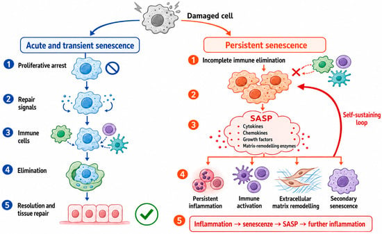
Oral lichen planus (OLP) is a chronic autoimmune inflammatory disease characterized by papular, reticular, plaque-like, atrophic, erosive or bullous lesions and histologically by interface mucositis, basal keratinocyte degeneration and a band-like lymphocytic infiltrate. OLP is classified as an oral potentially malignant disorder (OPMD), although its malignant transformation rate is relatively low. As a chronic disease, its main clinical issue is the persistence and recurrence of symptomatic lesions. Pathological alterations typically follow a relapsing–remitting course and may recur after local anti-inflammatory or immunosuppressive therapy is discontinued [1,2]. The canonical pathogenetic model emphasizes antigen-dependent activation of CD8-positive T cells, helper T cells, keratinocyte apoptosis, basement-membrane disruption, and recruitment of additional immune cells [3]. A recent study identified activated tissue-resident memory CD8-positive T cells near the epithelial–stromal interface and a cytokine network enriched in erosive OLP [4]. These observations clarify the mechanisms related to CD8-positive T cells during the active phases of OLP, but they do not fully explain how the lesional microenvironment sustains a pro-inflammatory state over time that can reactivate the cytotoxic response and promote disease recurrence. Cellular senescence could contribute to the maintenance of chronic inflammation. Senescence is not synonymous with biological ageing or cellular quiescence. It is a stress-adaptive cell state involving durable proliferative withdrawal, macromolecular damage, metabolic and structural remodelling, and, in some context, a bioactive secretome termed the senescence-associated secretory phenotype (SASP) [5,6,7]. In chronic inflammatory lesions, T cell anergy, exhaustion and senescence should not be used interchangeably. Anergy is an antigen-specific hyporesponsive state, typically induced by T cell receptor stimulation without adequate co-stimulation [8]. Exhaustion develops during persistent antigenic stimulation and is characterized by progressive impairment of proliferation and effector functions and sustained co-expression of multiple inhibitory receptors. No single marker is sufficient to define an exhausted phenotype [9,10]. By contrast, senescence involves durable stress-associated proliferative arrest, potentially accompanied by preserved secretory or cytotoxic activity [5]. Therefore, neither reduced proliferation nor the expression of inhibitory receptors such as killer cell lectin-like receptor G1 (KLRG1) is sufficient, on its own, to distinguish among these states [11,12]. Acute and transient senescence may protect tissues by arresting the proliferation of damaged cells and releasing signals that coordinate tissue repair and promote immune-mediated clearance of senescent cells (Figure 1) [13,14]. However, when immune-mediated elimination is incomplete, senescent cells may persist and continue to release SASP components, including pro-inflammatory cytokines, chemokines, growth factors and matrix-remodelling enzymes [13,14]. These mediators could sustain local inflammation, alter immune-cell recruitment and activation, remodel the extracellular matrix and induce secondary senescence in neighbouring cells through paracrine signalling [15].
Figure 1.
Schematic representation of transient senescence, which promotes immune-mediated elimination and tissue repair, versus persistent senescence, which sustains a SASP-driven inflammatory feedback loop.
In 2023, Ijima and colleagues provided the first integrated evidence suggesting that mesenchymal cells with senescence-associated features may contribute to cytotoxic immune activation in OLP [16]. This finding supports a shift from viewing the subepithelial stroma as passive scaffolding to considering it an active inflammatory organizer. Recent reviews have discussed cellular senescence in various oral tissues and diseases, with OLP considered within this broader context rather than as a specific focus [17,18]. Given the limited evidence, we do not present senescence and the SASP as established pathogenic mechanisms. Instead, we critically assess the two available primary studies, distinguish direct findings from indirect or extrapolated evidence, and identify hypotheses that require further testing.
2. Narrative Review Methods
A focused narrative literature search was last conducted on 31 July 2026. PubMed/MEDLINE, Scopus and Web of Science were searched for English-language publications addressing cellular senescence or senescence-adjacent mechanisms in OLP. The search strategy combined “oral lichen planus” or “oral lichenoid” with “senescence”, “SASP”, “p16”, “p21”, “cell-cycle arrest”, “fibroblast”, “mesenchymal”, “single-cell”, “CXCL12”, “oxidative stress”, “cytokine”, “KLRG1”, “malignant transformation”, “senolytic”, “senomorphic” and “JAK inhibitor”. Reference lists of key primary studies and consensus documents were hand-searched. General senescence literature was selected to define accepted concepts, minimum biomarker requirements and therapeutic classes. Evidence was interpreted according to its direct relevance to cellular senescence in OLP. OLP-specific studies integrating complementary molecular, spatial and/or functional approaches were distinguished from studies reporting isolated senescence-compatible findings, such as cell-cycle regulators, oxidative damage or inflammatory mediators. Evidence derived from non-oral forms of lichen planus, other tissues, or experimental systems was used only for mechanistic contextualization and hypothesis generation. This approach was adopted to distinguish direct evidence from supportive or extrapolated observations and to minimize causal overinterpretation. The review is narrative rather than systematic: it does not claim exhaustive retrieval, protocol registration, formal risk-of-bias scoring or quantitative pooling. Reporting was informed by SANRA principles [19].
3. Cellular Senescence and the SASP: A Working Framework
3.1. A Dynamic Cell State, Not a Single Marker
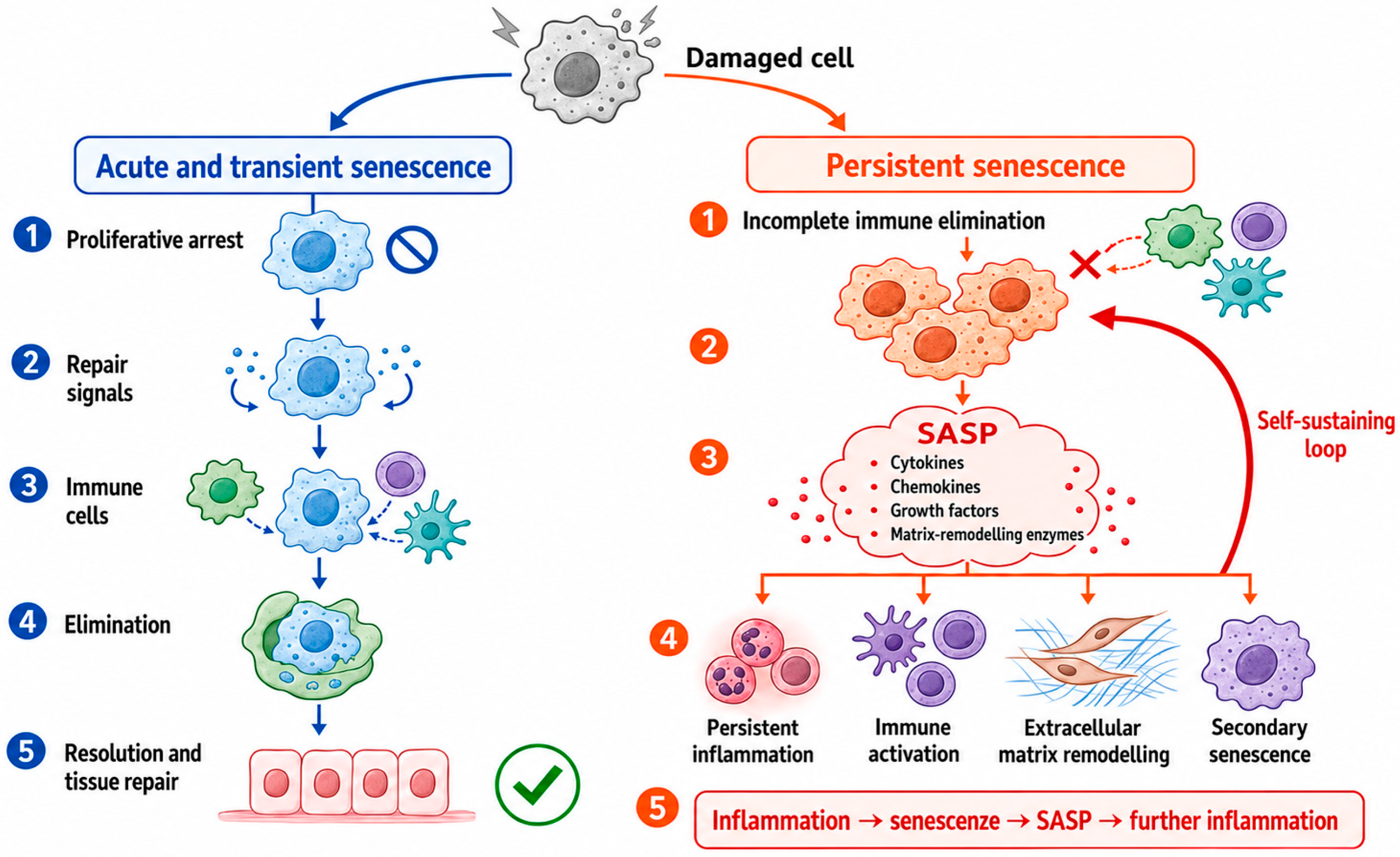
Contemporary definitions treat cellular senescence as a family of stress-induced states rather than a uniform endpoint (Figure 2). Common features include persistent engagement of the p53–p21 and/or p16–RB pathways, resistance to mitogenic stimulation, chromatin and lysosomal alterations, macromolecular damage and metabolic reprogramming [5,20].
Figure 2.
Cellular senescence comprises a heterogeneous spectrum of stress-induced states whose reliable identification requires multiple complementary markers, cell-type localization, and functional validation.
However, no individual marker is specific: p16 may accompany differentiation or cellular stress, p21 may be transiently induced during DNA repair, SA-β-gal activity can occur outside senescence, and putative SASP mediators may originate from non-senescent cells [21,22,23]. Current recommendations therefore favour a multi-domain panel integrating evidence of stable cell-cycle arrest, cellular damage or structural change, and secretory activity, together with cell-type localization and, where possible, functional validation [20,24]. Marker combinations may need to be adapted for fibroblasts, keratinocytes, and immune cells, and no validated OLP-specific panel is currently available.
3.2. Composition and Regulation of the SASP
The SASP should be regarded as a dynamic and context-dependent secretory program rather than as a fixed panel of mediators. Its composition varies according to the cell type, the senescence-inducing stimulus, the surrounding tissue microenvironment and the time elapsed since senescence induction. Accordingly, the SASP may include IL-6, CXCL8/IL-8, IL-1 family members, TNF-related signals, CXCL and CCL chemokines, growth factors, matrix metalloproteinases, extracellular-matrix proteins, bioactive lipids and extracellular vesicles [6,7,25]. Its biological effects may also change over time: early and late SASP programs can differ substantially, and their overall activity may be pro-inflammatory, immunosuppressive or profibrotic. This variability reflects the combined activity of multiple regulatory pathways. NF-κB and C/EBPβ act as major transcriptional regulators. More specifically, activation of the canonical IKK–IκBα–NF-κB pathway promotes the nuclear translocation of RelA/p65–p50 dimers, which induce inflammatory SASP mediators such as IL-6 and IL-8/CXCL8 [26]. Other pathways—including p38 MAPK, mTOR, cGAS–STING, DNA-damage signalling and JAK/STAT—influence the timing and composition of specific SASP programs [6,7]. Among these pathways, JAK/STAT can amplify selected SASP programs through autocrine and paracrine cytokine signalling. In senescent human adipocyte progenitors, JAK1/2 inhibition reduced several inflammatory SASP mediators, supporting a potential senomorphic effect. However, this mechanism appears cell- and context-dependent and should not be considered a universal feature of senescence [27]. The heterogeneity of SASP composition and regulation is particularly relevant to OLP, where isolated soluble mediators cannot identify which cells produced them or establish that the source cells are senescent [28]. Conversely, the absence of an IL-6/IL-8-dominant profile does not exclude alternative SASP programs involving CXCL12, matrix-remodelling factors or extracellular-matrix ligands [16,20,25].
4. Evidence for Cellular Senescence in OLP
A series of immunohistochemical studies conducted before the SASP hypothesis reported increased expression of p21, p53 and p16 in the OLP epithelium (Table 1). Bascones et al. found that terminal deoxynucleotidyl transferase dUTP nick-end labelling (TUNEL), caspase-3, and p21 positivity were more frequent in OLP than in normal mucosa, suggesting the concomitant activation of apoptosis and a p21-associated cell-cycle checkpoint response [29]. González-Moles et al. identified basal p53, p21 and Ki-67 expression in substantial proportions of OLP specimens, revealing a mosaic in which stress arrest and compensatory proliferation coexist [30]. Safadi and colleagues likewise reported greater p53 and p21 labelling in OLP than in normal mucosa, focal keratosis and nonspecific mucositis [31].
Table 1.
Human evidence connecting cellular senescence or senescence-adjacent biology to OLP.
Studies assessing the expression of p16INK4A provide indirect evidence of altered cell-cycle control in OLP. Montebugnoli et al. detected p16INK4A immunopositivity in a substantial proportion of OLP specimens [32]. Poomsawat et al. reported increased expression of p16 together with cyclin-dependent kinases (CDK) 4 and 6, whereas Goel et al. observed cytoplasmic p16 and CDK4 staining in erosive and non-erosive lesions [33,34]. Danielsson et al. found significantly higher p16 expression in oral and genital lichen planus than in site-matched healthy controls [35]. Collectively, these studies demonstrate altered cell-cycle regulation and cellular stress in OLP epithelium, but they do not establish stable arrest, persistence, or a functional SASP within the same cells.
Senescence marker protein-30/regucalcin was evaluated by Peter et al. in OLP, oral lichenoid reactions and nonspecific inflammation. Expression was somewhat greater in OLP than in oral lichenoid reactions, but the difference was not statistically significant, and only a small proportion of cases showed positivity [36]. Regucalcin has pleiotropic calcium-regulatory and metabolic functions and is not a canonical, senescence-specific tissue marker; this study should therefore be regarded as exploratory rather than confirmatory [36].
Ijima et al. investigated whether cellular senescence contributes to OLP using four complementary approaches [16]. First, they reanalysed two independent gene-expression datasets (GSE38616 and GSE52130; each including seven OLP samples and seven control samples). In both datasets, gene-expression patterns associated with senescence and the SASP were enriched in OLP. Second, the analysis of a single-cell RNA-sequencing dataset (GSE211630; two erosive OLP, three non-erosive OLP and one healthy control) indicated that mesenchymal cells had the highest senescence-associated scores. Third, immunohistochemistry supported these computational findings by showing an increased number of p21-positive/PDGFRα-positive mesenchymal cells and greater subepithelial p16 expression in OLP. Finally, computational analysis of cell–cell communication predicted that the senescent-like mesenchymal-cell cluster was a major source of signals directed to CD8-positive T cells. The predicted interactions included increased CXCL12–CXCR4 signalling and collagen/laminin–CD44 communication [16]. The authors then examined whether factors released by senescent fibroblasts could affect immune and epithelial cells. Conditioned medium from doxorubicin-induced senescent TIG-118 fibroblasts increased the proliferation and activation of Jurkat T cells, enhanced cytotoxic-gene expression in KHYG-1 natural killer cells, and promoted cellular injury and secondary senescence-like changes in HaCaT keratinocytes [16]. These experiments suggest that the secretome of experimentally induced senescent fibroblasts may amplify immune activation and increase epithelial vulnerability [16]. However, these findings should be interpreted cautiously. The single-cell analysis included only six samples, with a single healthy control, and some histological controls consisted of clinically healthy mucosa from patients with leukoplakia rather than tissue from healthy volunteers [16]. Moreover, the predicted cell–cell interactions were inferred from gene-expression data and therefore do not demonstrate direct or causal signalling. The functional experiments also used skin-derived fibroblasts and immortalized cell lines rather than primary oral cells obtained from matched OLP lesions. Finally, senescence induced by doxorubicin may differ from that produced by chronic inflammation in OLP [16]. Overall, this study provides important multimodal proof-of-concept evidence that senescent-like mesenchymal cells may influence immune activation and epithelial damage in OLP, but it does not yet demonstrate causality in vivo, long-term persistence, or therapeutic relevance.
Several studies have reported elevated levels of TNF, IL-1, IL-6, IL-8/CXCL8 and other inflammatory mediators in saliva, serum or lesional tissue of patients with OLP [28,37,38]. In a salivary multiplex study, levels of TNF, granulocyte colony-stimulating factor, IL-1α, IL-1β and IL-8 were elevated, and selected cytokine levels correlated with clinical severity [28]. These mediators are commonly found in SASP profiles, but they may also be produced by activated T cells, macrophages and keratinocytes. Therefore, their presence alone does not demonstrate a senescence-associated secretory phenotype. Accordingly, fluid biomarkers should be described as “SASP-compatible” until their cellular origin is established.
Oxidative and nitrative stressors provide plausible senescence-inducing stimuli. OLP epithelium accumulates inducible nitric oxide synthase, 3-nitrotyrosine, 8-nitroguanine and 8-oxo-7,8-dihydro-2′-deoxyguanosine, indicating chronic nucleic-acid damage [39]. Broader studies and reviews describe redox imbalance, reduced antioxidant capacity and lipid or DNA oxidation in OLP [40,41,42]. These observations are biologically coherent with a DNA-damage response capable of inducing p21 or p16, but they do not identify which cells enter a durable arrest.
5. A Proposed Senescence–SASP Mechanism in OLP
5.1. Induction: Repeated Stress at the Epithelial–Stromal Interface
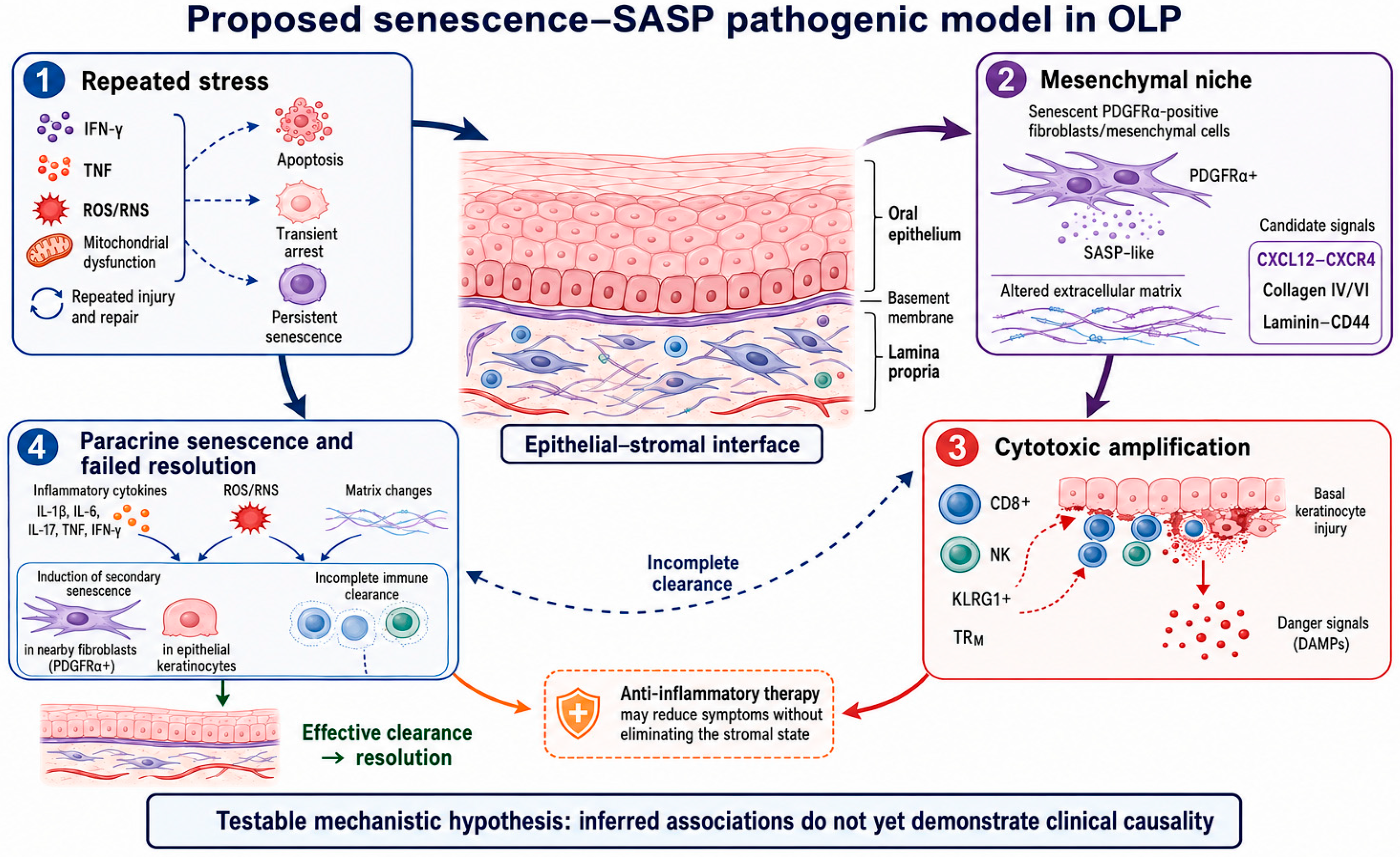
Persistent IFN-γ and TNF signalling, reactive oxygen and nitrogen species, mitochondrial dysfunction and repeated cycles of epithelial destruction and repair can activate DNA-damage response and stress-kinase pathways in oral fibroblasts and keratinocytes (Figure 3). Cells that cannot restore homeostasis may undergo apoptosis, remain transiently arrested or enter a longer-lived senescent-like state. The balance among these outcomes is likely lesion- and cell-type-specific.
Figure 3.
Conceptual senescence–SASP model in OLP. Solid elements summarize OLP-specific observations, including CD8-positive cytotoxic inflammation and senescence-associated mesenchymal signatures; dashed arrows represent proposed induction, paracrine spread, incomplete clearance and recurrence mechanisms for which causality has not been demonstrated.
5.2. The Mesenchymal Niche as an Inflammatory Organizer
Senescent-like PDGFRα-positive mesenchymal cells may contribute to a persistent stromal memory of previous tissue injury. Their secretome and altered matrix production may establish chemokine gradients, alter leukocyte adhesion, and modify basement-membrane composition. The CXCL12–CXCR4 axis is the most concrete candidate because signalling from the senescent-like mesenchymal cluster to CD8-positive T cells was selectively strengthened in OLP [16]. Immunologically, CXCL12–CXCR4 regulates lymphocyte chemotaxis, positioning and retention. It promotes T cell recruitment and may modulate TCR signalling, whereas in B-lineage cells it contributes to germinal-centre organization and plasma-cell trafficking; however, these B-cell functions remain unproven in OLP [43,44,45]. Interactions of collagen IV/VI and laminin with CD44 may complement CXCL12–CXCR4-directed chemotaxis by stabilizing recruited lymphocytes within the altered matrix. The causal relevance of these inferred pathways remains to be established through targeted perturbation experiments in biologically relevant oral models [16].
5.3. Amplification by Cytotoxic Lymphocytes
A stromal SASP-like program could facilitate the recruitment and activation of CD8-positive T cells and natural killer cells. OLP lesions already contain highly differentiated KLRG1-positive cytotoxic cells and, in erosive lesions, cytokine-rich tissue-resident memory cells [4,12]. Conditioned medium from senescent-fibroblast can increase T cell activation and NK-cell cytotoxic-gene expression in vitro [16]. These mechanisms may converge on basal keratinocyte killing, barrier failure and new danger signals.
5.4. Paracrine Senescence and Failed Resolution
Inflammatory cytokines, reactive oxygen species (ROS) and matrix changes generated by the lesion could induce secondary senescence-like changes in adjacent fibroblasts or epithelium [15]. If immune surveillance clears these putative senescent cells, the response may resolve. If clearance is incomplete because of chronic antigenic stimulation, immune exhaustion/dysfunction, local immunosuppression, or an inaccessible stromal niche, the lesion may enter a feed-forward loop. This loop offers a mechanistic explanation for recurrence after anti-inflammatory therapy: suppressing lymphocyte activity may reduce symptoms without removing the stromal state that re-initiates recruitment. This remains a testable hypothesis rather than a demonstrated clinical sequence [13].
6. Explaining Clinical Heterogeneity
The senescence model predicts heterogeneous rather than uniform marker expression. Reticular, plaque-like, and erosive lesions may differ in the abundance of senescent-like cells, the cell types involved, and the stage of the senescence response. Erosive lesions, with greater cytokine output from CD8-positive tissue-resident memory cells [4], may generate a stronger senescence-associated response or a more inflammatory SASP. Conversely, a fibrotic or matrix-dominant SASP could be present in clinically less inflamed lesions. Accordingly, sampling a single site at a single time point may fail to capture the relevant senescence-associated state. Secondary humoral findings, including anti-desmoglein 1/3 antibodies in erosive OLP and BP180/BP230 reactivity in rare cases of lichen planus pemphigoides overlap, may reflect chronic epithelial or basement-membrane injury but do not provide direct evidence for the senescence–SASP model [46,47]. These observations do not demonstrate cellular senescence, but they illustrate how persistent epithelial and basement-membrane damage may broaden the local immune response.
Treatment may also confound interpretation. In patients with OLP, topical dexamethasone and calcineurin inhibitors have been associated with reduced salivary or circulating levels of pro-inflammatory cytokines, including IL-1α, IL-6, IL-8, and TNF [48,49]. However, these changes may reflect suppression of immune activation rather than clearance of putative senescent stromal cells. Age, smoking and periodontal inflammation can independently influence local or systemic senescence-associated markers [50,51]. Diabetes, candidal colonization and concomitant medications may further modify inflammatory or tissue-stress readouts and should therefore be recorded as potential confounders. Future cohorts should document these variables and include disease controls such as oral lichenoid reactions, chronic graft-versus-host disease, lupus mucositis and nonspecific ulceration.
7. Senescence and Malignant Transformation: Brake, Accelerator or Bystander?
The malignant potential of OLP is real but modest and is highly sensitive to diagnostic criteria, exclusion of baseline dysplasia and duration of follow-up. A recent meta-analysis estimated an overall malignant transformation proportion of approximately 1.43%, while another large meta-analysis reported approximately 0.94% for OLP and higher rates for oral lichenoid lesions or lesions with dysplasia [52,53]. Red/erosive phenotype, tobacco, alcohol, and hepatitis C infection have been associated with greater risk in pooled analyses [53].
Cellular senescence may have opposing roles in carcinogenesis: stable p16–RB or p53–p21 arrest can restrain damaged cells, whereas persistent SASP signalling may promote inflammation, matrix remodelling, angiogenesis and epithelial plasticity [6,7]. In OLP, oxidative/nitrative DNA damage and chronic immune injury create a plausible setting for both effects [39]. The senescence–SASP framework may help explain the emergence of OSCC at OLP sites previously sampled without dysplasia. A senescent stromal niche could favour the selection of altered epithelial clones while early molecular changes remain histologically undetectable. This does not imply that dysplasia is biologically bypassed, as it may be focal, missed during sampling or develop between biopsies. However, current evidence is indirect, and no longitudinal study has shown that senescent-like cell burden predicts malignant transformation independently of established clinicopathological factors. Accordingly, p16 or p21 positivity should not be regarded as a cancer biomarker, and the prognostic relevance of stromal senescence requires prospective validation.
8. Future Therapeutic Perspectives
8.1. Senolytics
Senolytics eliminate senescent cells by targeting their pro-survival pathways. Agents such as dasatinib plus quercetin, fisetin and BCL-2-family inhibitors have shown activity in non-oral experimental or early clinical studies, but none has been validated in OLP [54,55,56]. Systemic administration would be difficult to justify in the absence of a predictive biomarker, the physiological role of senescent cells in tissue repair and potential toxicities, such as navitoclax-associated thrombocytopenia [14,57]. Local mucoadhesive delivery may reduce exposure, but selectivity, tissue penetration and wound-healing effects require testing.
8.2. Senomorphics
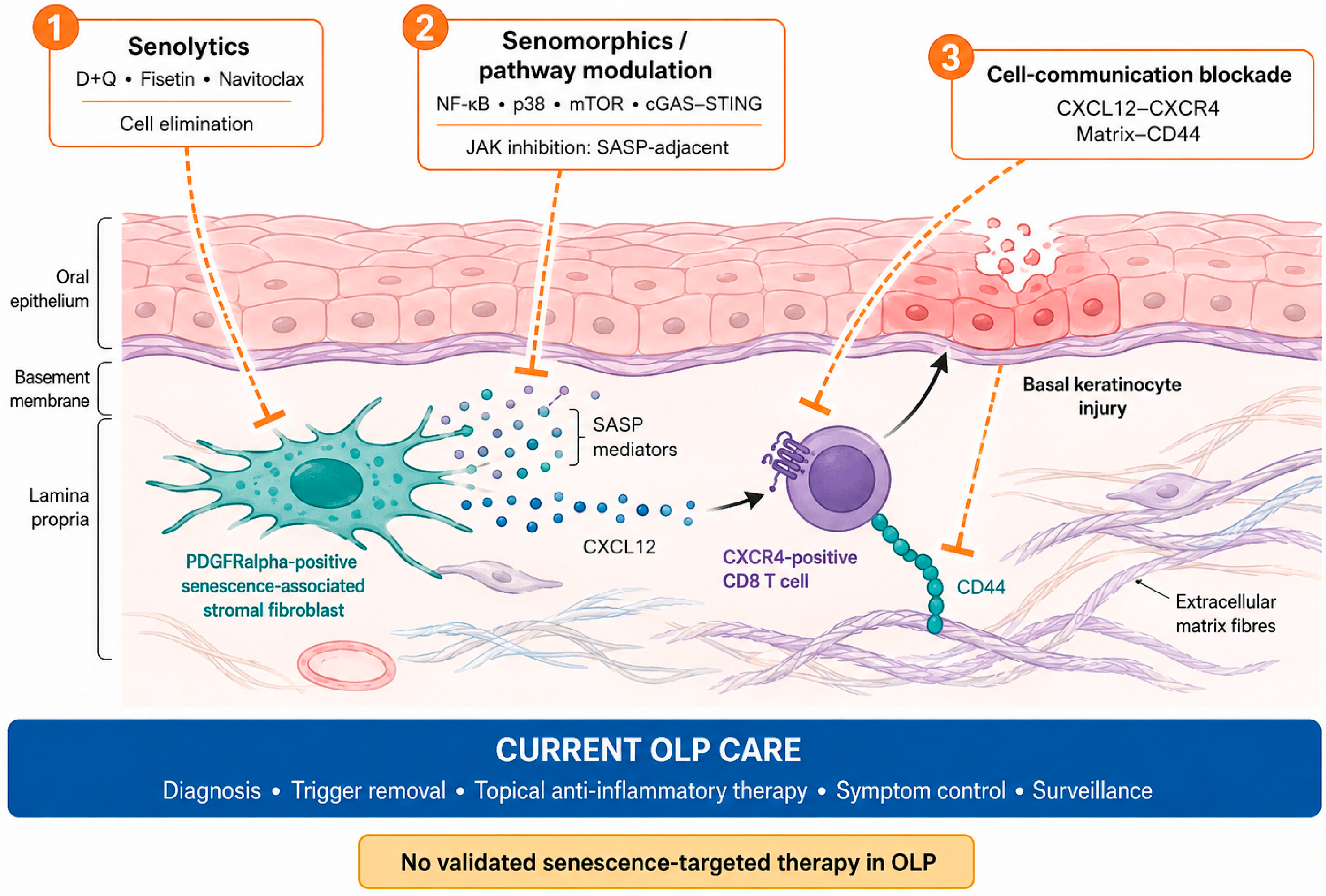
Senomorphics suppress components of the SASP without eliminating senescent cells (Figure 4). Potential targets include NF-κB, p38 MAPK, mTOR, cGAS–STING and JAK/STAT [6,7]. However, these pathways also regulate conventional inflammatory responses [26,58,59].
Figure 4.
Hypothetical therapeutic strategies targeting the proposed senescence-associated stromal niche in OLP. Senolytics would eliminate putative PDGFRα-positive senescence-associated fibroblasts. Senomorphics or pathway modulators would suppress SASP activity. Cell-communication blockade would interfere with CXCL12–CXCR4-mediated T cell recruitment or matrix–CD44-dependent retention, potentially reducing basal keratinocyte injury. These approaches remain experimental, and no senescence-targeted therapy has been validated in OLP.
Reports of improvement in refractory OLP with upadacitinib are preliminary and uncontrolled and do not demonstrate that the response results from modulation of a cell-specific SASP [60,61,62,63].
8.3. Targeting Cell Communication
Blocking the CXCL12–CXCR4 axis is a pathway-specific hypothesis derived directly from the mesenchymal-cell study. A useful preclinical experiment would test whether CXCR4 antagonism reduces CD8-positive T cell migration and keratinocyte injury in a primary oral fibroblast–immune–epithelial model [64]. Systemic CXCR4 antagonists mobilize hematopoietic cells and may disrupt normal immunity, so local delivery or ligand-neutralization strategies may be preferable. Similarly, interference with CD44 or matrix ligands could alter immune retention but may impair epithelial repair and matrix integrity [65,66,67].
8.4. Positioning Within Current OLP Care
Senescence-directed concepts should complement, not replace, accurate diagnosis, elimination of lichenoid triggers where applicable, use of topical corticosteroids or calcineurin inhibitors, symptom control, and long-term surveillance. The first translational objective is not an off-label senolytic trial; it is a biomarker-defined mechanistic study showing that a cell population meeting multi-domain criteria persists, predicts a clinically relevant phenotype and can be safely perturbed.
9. Conclusions
Current evidence for cellular senescence and the SASP in OLP is limited and largely hypothesis-generating. The strongest findings suggest the presence of a senescent-like PDGFRα-positive mesenchymal population that communicates with CD8-positive T cells through CXCL12–CXCR4 and matrix-associated pathways, whereas isolated p16/p21 expression or SASP-compatible mediators do not prove senescence. Spatial multi-marker and functional studies are required before biomarkers or senescence-targeted therapies can be considered.
Author Contributions
Conceptualization, D.D.F. and M.P.; methodology, D.D.F. and M.P.; validation, D.D.F., M.P., A.L. and G.P.; investigation, D.D.F. and M.P.; data curation, D.D.F.; writing—original draft preparation, D.D.F.; writing—review and editing, D.D.F., M.P., A.L. and G.P.; visualization, D.D.F.; supervision, M.P.; project administration, D.D.F. and M.P. All authors have read and agreed to the published version of the manuscript.
Funding
This research received no external funding.
Informed Consent Statement
Not applicable.
Data Availability Statement
No new data were created or analyzed in this study. Data sharing is not applicable to this article.
Conflicts of Interest
The authors declares no conflicts of interest.
References
- Cheng, Y.S.L.; Gould, A.; Kurago, Z.; Fantasia, J.; Muller, S. Diagnosis of oral lichen planus: A position paper of the American Academy of Oral and Maxillofacial Pathology. Oral Surg. Oral Med. Oral Pathol. Oral Radiol. 2016, 122, 332–354. [Google Scholar] [CrossRef] [Scilit] [PubMed]
- Warnakulasuriya, S.; Kujan, O.; Aguirre-Urizar, J.M.; Bagan, J.V.; González-Moles, M.Á.; Kerr, A.R.; Lodi, G.; Mello, F.W.; Monteiro, L.; Ogden, G.R.; et al. Oral potentially malignant disorders: A consensus report from an international seminar on nomenclature and classification, convened by the WHO Collaborating Centre for Oral Cancer. Oral Dis. 2021, 27, 1862–1880. [Google Scholar] [CrossRef] [Scilit] [PubMed]
- Sugerman, P.B.; Savage, N.W.; Walsh, L.J.; Zhao, Z.Z.; Zhou, X.J.; Khan, A.; Seymour, G.J.; Bigby, M. The P athogenesis of Oral Lichen Planus. Crit. Rev. Oral Biol. Med. 2002, 13, 350–365. [Google Scholar] [CrossRef] [Scilit] [PubMed]
- Qing, M.; Yang, D.; Shang, Q.; Peng, J.; Deng, J.; Lu, J.; Li, J.; Dan, H.; Zhou, Y.; Xu, H.; et al. CD8+ tissue-resident memory T cells induce oral lichen planus erosion via cytokine network. eLife 2023, 12, e83981. [Google Scholar] [CrossRef] [Scilit] [PubMed]
- Gorgoulis, V.; Adams, P.D.; Alimonti, A.; Bennett, D.C.; Bischof, O.; Bishop, C.; Campisi, J.; Collado, M.; Evangelou, K.; Ferbeyre, G.; et al. Cellular Senescence: Defining a Path Forward. Cell 2019, 179, 813–827. [Google Scholar] [CrossRef] [Scilit] [PubMed]
- Wang, B.; Han, J.; Elisseeff, J.H.; Demaria, M. The senescence-associated secretory phenotype and its physiological and pathological implications. Nat. Rev. Mol. Cell Biol. 2024, 25, 958–978. [Google Scholar] [CrossRef] [Scilit] [PubMed]
- Coppé, J.P.; Desprez, P.Y.; Krtolica, A.; Campisi, J. The Senescence-Associated Secretory Phenotype: The Dark Side of Tumor Suppression. Annu. Rev. Pathol. Mech. Dis. 2010, 5, 99–118. [Google Scholar] [CrossRef] [Scilit] [PubMed]
- Harding, F.A.; McArthur, J.G.; Gross, J.A.; Raulet, D.H.; Allison, J.P. CD28-mediated signalling co-stimulates murine T cells and prevents induction of anergy in T-cell clones. Nature 1992, 356, 607–609. [Google Scholar] [CrossRef] [Scilit] [PubMed]
- Wherry, E.J.; Ha, S.J.; Kaech, S.M.; Haining, W.N.; Sarkar, S.; Kalia, V.; Subramaniam, S.; Blattman, J.N.; Barber, D.L.; Ahmed, R. Molecular Signature of CD8+ T Cell Exhaustion during Chronic Viral Infection. Immunity 2007, 27, 670–684. [Google Scholar] [CrossRef] [Scilit] [PubMed]
- Blackburn, S.D.; Shin, H.; Haining, W.N.; Zou, T.; Workman, C.J.; Polley, A.; Betts, M.R.; Freeman, G.J.; Vignali, D.A.A.; Wherry, E.J. Coregulation of CD8+ T cell exhaustion by multiple inhibitory receptors during chronic viral infection. Nat. Immunol. 2009, 10, 29–37. [Google Scholar] [CrossRef] [Scilit] [PubMed]
- Henson, S.M.; Akbar, A.N. KLRG1—More than a marker for T cell senescence. Age 2009, 31, 285–291. [Google Scholar] [CrossRef] [Scilit] [PubMed]
- Soler-Ferran, D.; Louis, F.; Woo, S.B.; Greenberg, S.A. Infiltration of Mature KLRG1 Expressing Cytotoxic T Cells in Oral Lichen Planus. Head Neck Pathol. 2022, 16, 1124–1129. [Google Scholar] [CrossRef] [Scilit] [PubMed]
- Kale, A.; Sharma, A.; Stolzing, A.; Desprez, P.Y.; Campisi, J. Role of immune cells in the removal of deleterious senescent cells. Immun. Ageing 2020, 17, 16. [Google Scholar] [CrossRef] [Scilit] [PubMed]
- Demaria, M.; Ohtani, N.; Youssef, S.A.; Rodier, F.; Toussaint, W.; Mitchell, J.R.; Laberge, R.-M.; Vijg, J.; Van Steeg, H.; Dollé, M.E.; et al. An Essential Role for Senescent Cells in Optimal Wound Healing through Secretion of PDGF-AA. Dev. Cell 2014, 31, 722–733. [Google Scholar] [CrossRef] [Scilit] [PubMed]
- Acosta, J.C.; Banito, A.; Wuestefeld, T.; Georgilis, A.; Janich, P.; Morton, J.P.; Athineos, D.; Kang, T.-W.; Lasitschka, F.; Andrulis, M.; et al. A complex secretory program orchestrated by the inflammasome controls paracrine senescence. Nat. Cell Biol. 2013, 15, 978–990. [Google Scholar] [CrossRef] [Scilit] [PubMed]
- Ijima, S.; Saito, Y.; Yamamoto, S.; Nagaoka, K.; Iwamoto, T.; Kita, A.; Miyajima, M.; Sato, T.; Miyazaki, A.; Chikenji, T.S. Senescence-associated secretory phenotypes in mesenchymal cells contribute to cytotoxic immune response in oral lichen planus. Immun. Ageing 2023, 20, 72. [Google Scholar] [CrossRef] [Scilit] [PubMed]
- Elashiry, M.; Cornelius Timothius, C.J.; Zaman, R.; Elliott, M.; Crosby, B.; Bhat, K.; Saad, K.M.; Elsayed, R. The Role of Cellular Senescence in Oral Health and Disease. Int. J. Mol. Sci. 2026, 27, 2269. [Google Scholar] [CrossRef] [Scilit] [PubMed]
- Parkinson, E.K.; Prime, S.S. Oral Senescence: From Molecular Biology to Clinical Research. Front. Dent. Med. 2022, 3, 822397. [Google Scholar] [CrossRef] [Scilit]
- Baethge, C.; Goldbeck-Wood, S.; Mertens, S. SANRA—A scale for the quality assessment of narrative review articles. Res. Integr. Peer Rev. 2019, 4, 5. [Google Scholar] [CrossRef] [Scilit] [PubMed]
- Ogrodnik, M.; Carlos Acosta, J.; Adams, P.D.; d’Adda Di Fagagna, F.; Baker, D.J.; Bishop, C.L.; Chandra, T.; Collado, M.; Gil, J.; Gorgoulis, V.; et al. Guidelines for minimal information on cellular senescence experimentation in vivo. Cell 2024, 187, 4150–4175. [Google Scholar] [CrossRef] [Scilit] [PubMed]
- González-Gualda, E.; Baker, A.G.; Fruk, L.; Muñoz-Espín, D. A guide to assessing cellular senescence in vitro and in vivo. FEBS J. 2021, 288, 56–80. [Google Scholar] [CrossRef] [Scilit] [PubMed]
- Kohli, J.; Wang, B.; Brandenburg, S.M.; Basisty, N.; Evangelou, K.; Varela-Eirin, M.; Campisi, J.; Schilling, B.; Gorgoulis, V.; Demaria, M. Algorithmic assessment of cellular senescence in experimental and clinical specimens. Nat. Protoc. 2021, 16, 2471–2498. [Google Scholar] [CrossRef] [Scilit] [PubMed]
- Hewitt, G.; Jurk, D.; Marques, F.D.M.; Correia-Melo, C.; Hardy, T.; Gackowska, A.; Anderson, R.; Taschuk, M.; Mann, J.; Passos, J.F. Telomeres are favoured targets of a persistent DNA damage response in ageing and stress-induced senescence. Nat. Commun. 2012, 3, 708. [Google Scholar] [CrossRef] [Scilit] [PubMed]
- Suryadevara, V.; Hudgins, A.D.; Rajesh, A.; Pappalardo, A.; Karpova, A.; Dey, A.K.; Hertzel, A.; Agudelo, A.; Rocha, A.; Soygur, B.; et al. SenNet recommendations for detecting senescent cells in different tissues. Nat. Rev. Mol. Cell Biol. 2024, 25, 1001–1023. [Google Scholar] [CrossRef] [Scilit] [PubMed]
- Basisty, N.; Kale, A.; Jeon, O.H.; Kuehnemann, C.; Payne, T.; Rao, C.; Holtz, A.; Shah, S.; Sharma, V.; Ferrucci, L.; et al. A proteomic atlas of senescence-associated secretomes for aging biomarker development. PLoS Biol. 2020, 18, e3000599. [Google Scholar] [CrossRef] [Scilit] [PubMed]
- Chien, Y.; Scuoppo, C.; Wang, X.; Fang, X.; Balgley, B.; Bolden, J.E.; Premsrirut, P.; Luo, W.; Chicas, A.; Lee, C.S.; et al. Control of the senescence-associated secretory phenotype by NF-κB promotes senescence and enhances chemosensitivity. Genes Dev. 2011, 25, 2125–2136. [Google Scholar] [CrossRef] [Scilit] [PubMed]
- Xu, M.; Tchkonia, T.; Ding, H.; Ogrodnik, M.; Lubbers, E.R.; Pirtskhalava, T.; White, T.A.; Johnson, K.O.; Stout, M.B.; Mezera, V.; et al. JAK inhibition alleviates the cellular senescence-associated secretory phenotype and frailty in old age. Proc. Natl. Acad. Sci. USA 2015, 112, E6301–E6310. [Google Scholar] [CrossRef] [Scilit] [PubMed]
- Zhu, Z.D.; Ren, X.M.; Zhou, M.M.; Chen, Q.M.; Hua, H.; Li, C.L. Salivary cytokine profile in patients with oral lichen planus. J. Dent. Sci. 2022, 17, 100–105. [Google Scholar] [CrossRef] [Scilit] [PubMed]
- Bascones, C.; Gonzalez-Moles, M.A.; Esparza, G.; Bravo, M.; Acevedo, A.; Gil-Montoya, J.A.; Bascones, A. Apoptosis and cell cycle arrest in oral lichen planus. Arch. Oral Biol. 2005, 50, 873–881. [Google Scholar] [CrossRef] [Scilit] [PubMed]
- González-Moles, M.A.; Bascones-Ilundain, C.; Gil Montoya, J.A.; Ruiz-Avila, I.; Delgado-Rodríguez, M.; Bascones-Martínez, A. Cell cycle regulating mechanisms in oral lichen planus: Molecular bases in epithelium predisposed to malignant transformation. Arch. Oral Biol. 2006, 51, 1093–1103. [Google Scholar] [CrossRef] [Scilit] [PubMed]
- Safadi, R.A.; Jaber, S.Z.A.; Hammad, H.M.; Hamasha, A.A.H. Oral lichen planus shows higher expressions of tumor suppressor gene products of p53 and p21 compared to oral mucositis. An immunohistochemical study. Arch. Oral Biol. 2010, 55, 454–461. [Google Scholar] [CrossRef] [Scilit] [PubMed]
- Montebugnoli, L.; Venturi, M.; Gissi, D.B.; Leonardi, E.; Farnedi, A.; Foschini, M.P. Immunohistochemical expression of p16INK4A protein in oral lichen planus. Oral Surg. Oral Med. Oral Pathol. Oral Radiol. Endodontology 2011, 112, 222–227. [Google Scholar] [CrossRef] [Scilit] [PubMed]
- Poomsawat, S.; Buajeeb, W.; Khovidhunkit, S.O.P.; Punyasingh, J. Overexpression of cdk4 and p16 in oral lichen planus supports the concept of premalignancy: Overexpression of cdk4 and p16 in oral lichen planus. J. Oral Pathol. Med. 2011, 40, 294–299. [Google Scholar] [CrossRef] [Scilit] [PubMed]
- Goel, S.; Khurana, N.; Marvah, A.; Gupta, S. Expression of cdk4 and p16 in Oral Lichen Planus. J. Oral Maxillofac. Res. 2015, 6, e4. [Google Scholar] [CrossRef] [Scilit] [PubMed][Green Version]
- Danielsson, K.; Olah, J.; Zohori-Zangeneh, R.; Nylander, E.; Ebrahimi, M. Increased expression of p16 in both oral and genital lichen planus. Med. Oral Patol. Oral Cir. Bucal 2018, 23, e449. [Google Scholar] [CrossRef] [Scilit] [PubMed]
- Peter, C.D.; Shashidara, R.; Jain, V.; Haragannavar, V.C.; Samuel, P.; Nayak, S.R. Senescence in oral lichen planus as assessed by the immunohistochemical evaluation of senescence marker protein-30 (Regucalcin). Indian J. Pathol. Microbiol. 2023, 66, 9–13. [Google Scholar] [CrossRef] [Scilit] [PubMed]
- Rhodus, N.L.; Cheng, B.; Myers, S.; Bowles, W.; Ho, V.; Ondrey, F. A comparison of the pro-inflammatory, NF-κB-dependent cytokines: TNF-alpha, IL-1-alpha, IL-6, and IL-8 in different oral fluids from oral lichen planus patients. Clin. Immunol. 2005, 114, 278–283. [Google Scholar] [CrossRef] [Scilit] [PubMed]
- Zhang, Y.; Liu, W.; Zhang, S.; Dan, H.; Lu, R.; Wang, F.; Wang, J.; Zhou, M.; Zeng, X.; Lin, M.; et al. Salivary and Serum Interleukin-18 in Patients with Oral Lichen Planus: A Study in an Ethnic Chinese Population. Inflammation 2012, 35, 399–404. [Google Scholar] [CrossRef] [Scilit] [PubMed]
- Chaiyarit, P.; Ma, N.; Hiraku, Y.; Pinlaor, S.; Yongvanit, P.; Jintakanon, D.; Murata, M.; Oikawa, S.; Kawanishi, S. Nitrative and oxidative DNA damage in oral lichen planus in relation to human oral carcinogenesis. Cancer Sci. 2005, 96, 553–559. [Google Scholar] [CrossRef] [Scilit] [PubMed]
- Sardaro, N.; Della Vella, F.; Incalza, M.A.; Di Stasio, D.; Lucchese, A.; Contaldo, M.; Laudadio, C.; Petruzzi, M. Oxidative Stress and Oral Mucosal Diseases: An Overview. In Vivo 2019, 33, 289–296. [Google Scholar] [CrossRef] [Scilit] [PubMed]
- Battino, M.; Greabu, M.; Totan, A.; Mohora, M.; Bullon, P.; Bucur, A.; Totan, C.; Tovaru, S.; Parlatescu, I.; Didilescu, A.; et al. Oxidative stress markers in oral lichen planus. BioFactors 2008, 33, 301–310. [Google Scholar] [CrossRef] [Scilit] [PubMed]
- Banerjee, S.; Mukherjee, S.; Mitra, S.; Singhal, P. Comparative Evaluation of Mitochondrial Antioxidants in Oral Potentially Malignant Disorders. Kurume Med. J. 2019, 66, 15–27. [Google Scholar] [CrossRef] [Scilit] [PubMed]
- Allen, C.D.C.; Ansel, K.M.; Low, C.; Lesley, R.; Tamamura, H.; Fujii, N.; Cyster, J.G. Germinal center dark and light zone organization is mediated by CXCR4 and CXCR5. Nat. Immunol. 2004, 5, 943–952. [Google Scholar] [CrossRef] [Scilit] [PubMed]
- Kumar, A.; Humphreys, T.D.; Kremer, K.N.; Bramati, P.S.; Bradfield, L.; Edgar, C.E.; Hedin, K.E. CXCR4 Physically Associates with the T Cell Receptor to Signal in T Cells. Immunity 2006, 25, 213–224. [Google Scholar] [CrossRef] [Scilit] [PubMed]
- Hargreaves, D.C.; Hyman, P.L.; Lu, T.T.; Ngo, V.N.; Bidgol, A.; Suzuki, G.; Zou, Y.-R.; Littman, D.R.; Cyster, J.G. A Coordinated Change in Chemokine Responsiveness Guides Plasma Cell Movements. J. Exp. Med. 2001, 194, 45–56. [Google Scholar] [CrossRef] [Scilit] [PubMed]
- De Falco, D.; Di Stasio, D.; Caggiula, A.; Lajolo, C.; Lucchese, A.; Petruzzi, M. Pemphigoid Antibodies in Patients with Oral Lichen Planus: A Systematic Review. Pathophysiology 2025, 32, 51. [Google Scholar] [CrossRef] [Scilit] [PubMed]
- De Falco, D.; Iaquinta, F.; Pedone, D.; Lucchese, A.; Di Stasio, D.; Petruzzi, M. Circulating Antibodies Against DSG1 and DSG3 in Patients with Oral Lichen Planus: A Scoping Review. Antibodies 2025, 14, 51. [Google Scholar] [CrossRef] [Scilit] [PubMed]
- Vohra, S.; Singal, A.; Sharma, S.B. Clinical and serological efficacy of topical calcineurin inhibitors in oral lichen planus: A prospective randomized controlled trial. Int. J. Dermatol. 2016, 55, 101–105. [Google Scholar] [CrossRef] [Scilit] [PubMed]
- Rhodus, N.; Cheng, B.; Bowles, W.; Myers, S.; Miller, L.; Ondrey, F. Proinflammatory cytokine levels in saliva before and after treatment of (erosive) oral lichen planus with dexamethasone. Oral Dis. 2006, 12, 112–116. [Google Scholar] [CrossRef] [Scilit] [PubMed]
- Pérez-Leal, M.; Estornut, C.; Alfaro-Ochoa, L.; Sánchez-Herrera, G.; Roger, I.; Montero, P.; Arias-Herrera, S.; Flacco, N. Cigarette smoke induces expression of senescence markers and pro-inflammatory SASP in oral mucosa: Potential implications for early carcinogenic processes. Int. J. Cancer 2026, 158, 752–762. [Google Scholar] [CrossRef] [Scilit] [PubMed]
- Rattanaprukskul, K.; Xia, X.J.; Jiang, M.; Albuquerque-Souza, E.; Bandyopadhyay, D.; Sahingur, S.E. Molecular Signatures of Senescence in Periodontitis: Clinical Insights. J. Dent. Res. 2024, 103, 800–808. [Google Scholar] [CrossRef] [Scilit] [PubMed]
- González-Moles, M.Á.; Ramos-García, P. An Evidence-Based Update on the Potential for Malignancy of Oral Lichen Planus and Related Conditions: A Systematic Review and Meta-Analysis. Cancers 2024, 16, 608. [Google Scholar] [CrossRef] [Scilit] [PubMed]
- Li, J.W.; Li, K.Y.; Chan, B.W.A.; McGrath, C.P.; Zheng, L.W. Rate of Malignant Transformation Differs Based on Diagnostic Criteria for Oral Lichenoid Conditions: A Systematic Review and Meta-Analysis of 24,277 Patients. Cancers 2023, 15, 2537. [Google Scholar] [CrossRef] [Scilit] [PubMed]
- Kirkland, J.L.; Tchkonia, T. Cellular Senescence: A Translational Perspective. eBioMedicine 2017, 21, 21–28. [Google Scholar] [CrossRef] [Scilit] [PubMed]
- Hickson, L.J.; Langhi Prata, L.G.P.; Bobart, S.A.; Evans, T.K.; Giorgadze, N.; Hashmi, S.K.; Herrmann, S.M.; Jensen, M.D.; Jia, Q.; Jordan, K.L.; et al. Senolytics decrease senescent cells in humans: Preliminary report from a clinical trial of Dasatinib plus Quercetin in individuals with diabetic kidney disease. eBioMedicine 2019, 47, 446–456. [Google Scholar] [CrossRef] [Scilit] [PubMed]
- Yousefzadeh, M.J.; Zhu, Y.; McGowan, S.J.; Angelini, L.; Fuhrmann-Stroissnigg, H.; Xu, M.; Ling, Y.Y.; Melos, K.I.; Pirtskhalava, T.; Inman, C.L.; et al. Fisetin is a senotherapeutic that extends health and lifespan. eBioMedicine 2018, 36, 18–28. [Google Scholar] [CrossRef] [Scilit] [PubMed]
- Rudin, C.M.; Hann, C.L.; Garon, E.B.; Ribeiro De Oliveira, M.; Bonomi, P.D.; Camidge, D.R.; Chu, Q.; Giaccone, G.; Khaira, D.; Ramalingam, S.S.; et al. Phase II Study of Single-Agent Navitoclax (ABT-263) and Biomarker Correlates in Patients with Relapsed Small Cell Lung Cancer. Clin. Cancer Res. 2012, 18, 3163–3169. [Google Scholar] [CrossRef] [Scilit] [PubMed]
- Freund, A.; Patil, C.K.; Campisi, J. P38MAPK is a novel DNA damage response-independent regulator of the senescence-associated secretory phenotype. EMBO J. 2011, 30, 1536–1548. [Google Scholar] [CrossRef] [Scilit] [PubMed]
- Laberge, R.M.; Sun, Y.; Orjalo, A.V.; Patil, C.K.; Freund, A.; Zhou, L.; Curran, S.C.; Davalos, A.R.; Wilson-Edell, K.A.; Liu, S.; et al. MTOR regulates the pro-tumorigenic senescence-associated secretory phenotype by promoting IL1A translation. Nat. Cell Biol. 2015, 17, 1049–1061. [Google Scholar] [CrossRef] [Scilit] [PubMed]
- Noot, C.H.; Hansen, A.M.; Frost, Z.; Rhoads, J.L.W.; Hull, C.M.; Zone, J.J.; Hopkins, Z.H. Oral lichen planus treated with upadacitinib: A case series. JAAD Case Rep. 2025, 63, 125–127. [Google Scholar] [CrossRef] [Scilit] [PubMed]
- Sheehan, C.A.; Brownstone, N.D.; Lee, J.B.; Hsu, S. Use of upadacitinib for recalcitrant oral erosive lichen planus. JAAD Case Rep. 2025, 60, 167–169. [Google Scholar] [CrossRef] [Scilit] [PubMed]
- Ames, E.; Sanders, M.S.; Cotter, D. Erosive Oral Lichen Planus Treated with Upadacitinib Amid Systemic Psoriatic Therapy. Cureus 2025, 17, e98831. [Google Scholar] [CrossRef] [Scilit] [PubMed]
- Nwogu, O.; Krivda, K.; Montebello, A.; Green, J.; Gelman, A. Cutaneous and erosive oral lichen planus treated with upadacitinib. JAAD Case Rep. 2026, 71, 89–92. [Google Scholar] [CrossRef] [Scilit] [PubMed]
- Bradfield, P.F.; Amft, N.; Vernon-Wilson, E.; Exley, A.E.; Parsonage, G.; Rainger, G.E.; Nash, G.B.; Thomas, A.M.C.; Simmons, D.L.; Salmon, M.; et al. Rheumatoid fibroblast-like synoviocytes overexpress the chemokine stromal cell–derived factor 1 (CXCL12), which supports distinct patterns and rates of CD4+ and CD8+ T cell migration within synovial tissue. Arthritis Rheum. 2003, 48, 2472–2482. [Google Scholar] [CrossRef] [Scilit] [PubMed]
- Oksala, O.; Salo, T.; Tammi, R.; Häkkinen, L.; Jalkanen, M.; Inki, P.; Larjava, H. Expression of proteoglycans and hyaluronan during wound healing. J. Histochem. Cytochem. 1995, 43, 125–135. [Google Scholar] [CrossRef] [Scilit] [PubMed]
- Liu, Q.; Li, Z.; Gao, J.; Wan, W.; Ganesan, S.; McDermott, D.H.; Murphy, P.M. CXCR4 antagonist AMD3100 redistributes leukocytes from primary immune organs to secondary immune organs, lung, and blood in mice. Eur. J. Immunol. 2015, 45, 1855–1867. [Google Scholar] [CrossRef] [Scilit] [PubMed]
- Liles, W.C.; Broxmeyer, H.E.; Rodger, E.; Wood, B.; Hübel, K.; Cooper, S.; Hangoc, G.; Bridger, G.J.; Henson, G.W.; Calandra, G.; et al. Mobilization of hematopoietic progenitor cells in healthy volunteers by AMD3100, a CXCR4 antagonist. Blood 2003, 102, 2728–2730. [Google Scholar] [CrossRef] [Scilit] [PubMed]

Disclaimer/Publisher’s Note: The statements, opinions and data contained in all publications are solely those of the individual author(s) and contributor(s) and not of MDPI and/or the editor(s). MDPI and/or the editor(s) disclaim responsibility for any injury to people or property resulting from any ideas, methods, instructions or products referred to in the content. |
© 2026 by the authors. Licensee MDPI, Basel, Switzerland. This article is an open access article distributed under the terms and conditions of the Creative Commons Attribution (CC BY) license.