


Abstract
Background and aim: The influence of fabrication techniques, cement type, and cyclic loading on the marginal adaptation of lithium disilicate crowns remains a clinical concern that may affect their long-term performance. This study aimed to evaluate the effects of cyclic loading and cement type on the marginal fit of milled and pressed lithium disilicate crowns. Methods: Twenty lithium disilicate crowns were fabricated and divided into two groups based on the manufacturing technique: milled and pressed (n = 10 each). Each group was further subdivided according to the cement type: resin or resin-modified glass ionomer (n = 5 per group). Crowns were cemented on standardized epoxy resin dies, and the marginal gap was measured using a stereomicroscope before and after cyclic loading. Cyclic loading was performed at 50 N for 37,000 cycles. Data were statistically analyzed using a three-way ANOVA (α = 0.05). Results: Milled crowns showed marginal gaps ranging from 52 to 57 µm, whereas the pressed crowns exhibited smaller gaps ranging from 39 to 47 µm. Neither the cement type nor the cyclic loading produced a significant difference in the marginal gap values (p > 0.05). Conclusions: Pressed lithium disilicate crowns exhibited superior marginal adaptation compared with the milled crowns. Neither the type of cement nor the cyclic loading had a significant effect on the marginal gap. Both fabrication techniques yielded clinically acceptable fits (<100 µm).
1. Introduction
Marginal fit is a critical determinant of the long-term success of fixed dental prostheses (FDPs). Inadequate marginal adaptation can lead to plaque accumulation, recurrent caries, and periodontal inflammation, all of which compromise the biological and mechanical integrity of restorations [1].
All-ceramic restorations are among the most commonly used options for restoring extensively damaged or fractured teeth, providing both function and esthetics [2]. Their optical properties, such as translucency and fluorescence, along with their chemical inertness, biocompatibility, and corrosion resistance, make them the preferred choice for esthetic prosthodontic applications [3,4]. Among the fabrication techniques available for all-ceramic restorations, computer-aided milling and heat-pressing are the two most widely adopted methods [5].
Marginal discrepancies have been described using various terms, including marginal gaps, deficient margins, and improper seating. Several techniques are available for evaluating marginal fit, such as stereomicroscopy, digital microscopy, and scanning electron microscopy after sectioning cemented specimens [6].
Resin-based cements and resin-modified glass ionomer cements are commonly recommended for cementing all-ceramic restorations. Resin cements are particularly preferred with lithium disilicate ceramics because they can be etched and silanized, producing strong adhesive bonds and improved marginal adaptation [7]. Ensuring high marginal quality is crucial, as exposure of the cement line may facilitate plaque accumulation and recurrent caries, thereby threatening the longevity of the restoration [8].
It has been continuously reported in the literature that traditional glass ionomer cements (GICs) are only appropriate for preparations with high mechanical retention, since they have the lowest retentive values and are prone to deterioration under cyclic loading, even if they provide a streamlined workflow and fluoride release. It is important to investigate resin-modified glass ionomer cement in its initial application. Resin-modified glass ionomer (RMGI) cement offers a versatile “middle-ground,” combining the fluoride release and moisture tolerance of classic GICs with the enhanced mechanical qualities and reduced solubility of resin. Compared to multi-step adhesive resins, RMGIs have better fracture toughness and less technique sensitivity, but they usually do not have the strong chemical bond needed to support lithium disilicate in high-stress or minimally retentive preparations. Research shows that although RMGIs exhibit constant marginal integrity over time, they frequently fail at lower cyclic loads and do not offer the same level of fatigue resistance as self-adhesive resin cements. To maximize the structural durability of CAD/CAM ceramics in high-load regions, pure resin-based methods continue to be superior. RMGI is a great compromise for conventional retentive preparations where moisture control is difficult [9,10].
On the other hand, by using a cross-linked polymer matrix that more effectively absorbs occlusal stress, self-adhesive resin cements (SARCs) offer noticeably greater fatigue resistance and bond stability. SARCs typically outperform GICs in high-stress locations by shifting the failure mode from adhesive debonding to a more stable bulk-fracture resistance, even though their viscosity may affect marginal fit. In the end, resin-based methods continue to be the best option for reducing microleakage in lithium disilicate restorations [11].
In view of its exceptional blend of strong mechanical qualities and excellent translucency, lithium disilicate (LD) has become a key component of restorative dentistry. The performance of these restorations under functional stresses has become the focus of current research as dentists increasingly use monolithic CAD/CAM technology to avoid the hazards of material chipping. While initial retentive strength is well-documented, the combination of variables, such as the capacity of the fabrication techniques and cement interface to sustain cycle fatigue in high-stress posterior locations, remains a critical field of inquiry.
Because lithium disilicate (LD) crowns primarily rely on chemical bonding to endure fatigue, long-term performance in high-stress locations depends on the synergy between precision production and strong adhesive luting.
Previous studies have explored the marginal adaptation of lithium disilicate crowns produced by heat-pressing or CAD/CAM milling, with many authors reporting a finer marginal fit for pressed restorations [5,9,10]. Other investigations, however, have shown no significant difference—or even slightly better adaptation—for milled crowns, likely reflecting differences in milling system calibration, spacer thickness, or crystallization procedures used during fabrication [11,12]. The influence of cyclic loading on the marginal integrity of all-ceramic restorations has also been examined; however, the reported findings remain inconsistent across studies [13,14]. Most of this work has focused on a single factor at a time, such as the fabrication method, cement type, or fatigue loading.
Current literature often focuses on the initial retention of CAD/CAM crowns, but limited data exists on the long-term, fatigue-resistant, retentive, and luting materials used. This study distinctly determines the fabrication-cement combination that exhibits the greatest fatigue resistance, offering recommendations for selecting a resin-based cement in high-stress areas and suggesting specific fabrication techniques to reduce gap development.
Therefore, this study was designed to examine all three variables simultaneously. By assessing the combined effects of fabrication technique, cement type, and cyclic loading on the marginal fit of lithium disilicate crowns, this study provides a broader, more clinically relevant understanding of how these factors interact to influence precision and clinical performance.
The null hypothesis tested was that there is no significant variation that will exist in the fatigue failure loads of the crowns, irrespective of the construction method, cement type, or the implementation of cyclic loading.
2. Materials and Methods
Ethical approval was not applicable since the study was conducted on acrylic typodont teeth. The materials used in this study are lithium disilicate ceramics as shown in Table 1, and the cements used for luting the crowns listed in Table 2.
Table 1.
Materials evaluated in this study: ceramics.
Table 2.
Materials evaluated in this study: luting cements.
2.1. Study Design and Sample Size Determination
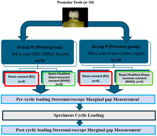
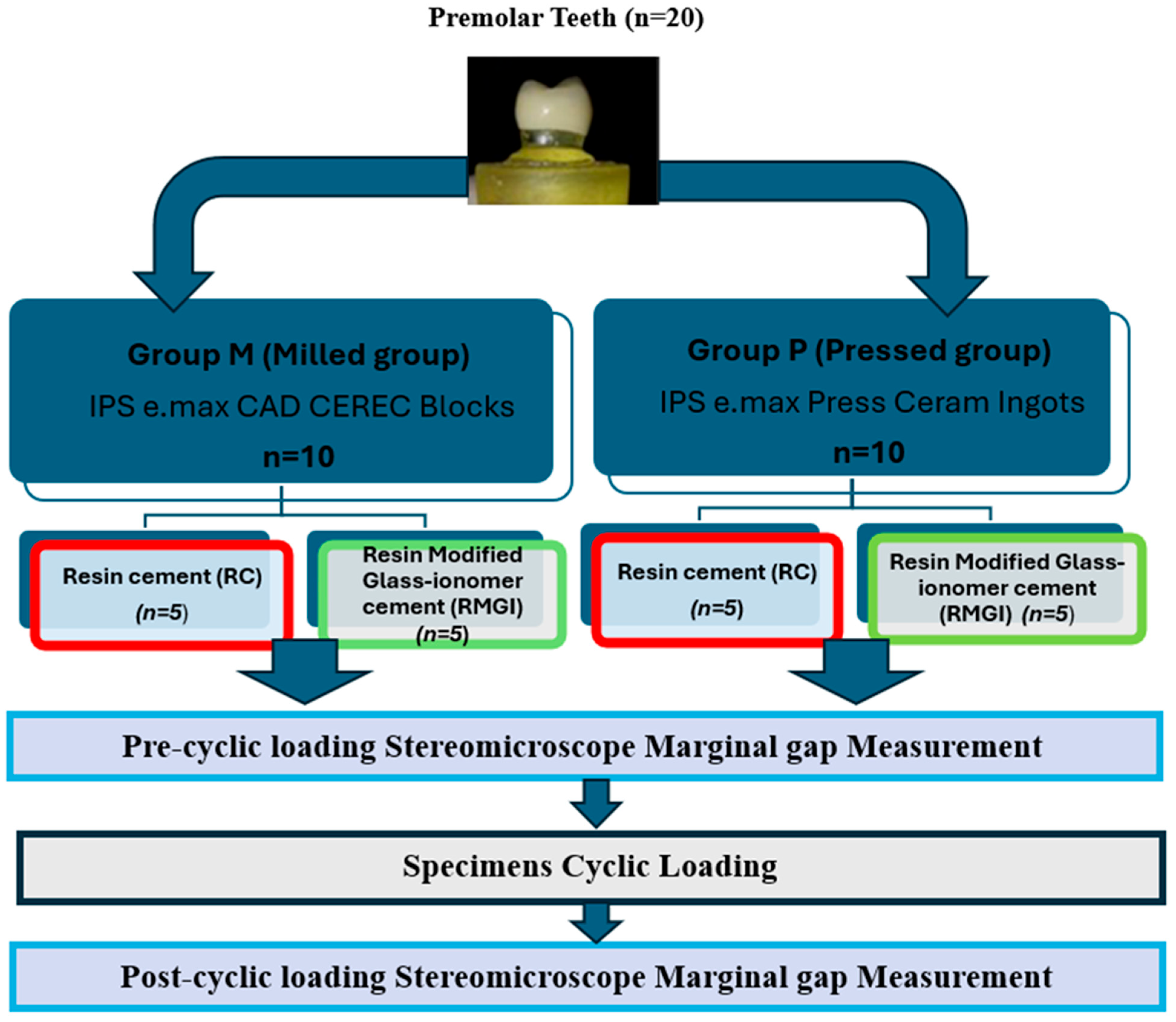
This in vitro experimental study was conducted to evaluate the effect of fabrication technique, cement type, and cyclic loading on the marginal accuracy of lithium disilicate crowns. A total of 20 crowns were fabricated and analyzed as summarized in Figure 1.
Figure 1.
Diagram illustrates sample grouping and study design.
The sample size for this in vitro study was determined based on previous studies assessing the marginal adaptation of lithium disilicate restorations, which typically reported similar subgroup sizes (n = 5–10) [15,16]. The number of samples in each subgroup was small, but rigorous standardization of the experimental methods was used to minimize variability. This included consistent tooth preparation, standardized die production, controlled fabrication processes [17], and uniform cementation with a constant load of 50 N. Additionally, several measurements were taken for each specimen (12 measurement points for each crown), which enhanced the reliability of the data collected. The notable differences found between fabrication techniques in this study reinforce the appropriateness of the chosen sample size for identifying significant differences in a controlled laboratory setting.
2.2. Sample Grouping
Twenty epoxy resin dies were prepared and divided according to fabrication technique into two main groups:
- Group M (Milled crowns): IPS E.max CAD blocks (n = 10).
- Group P (Pressed crowns): IPS E.max Press ingots (n = 10).
Each group was further subdivided according to the type of cement used:
- Resin cement subgroup (n = 5).
- Resin-modified glass ionomer cement subgroup (n = 5).
2.3. Tooth Preparation and Die Fabrication
A maxillary premolar acrylic tooth was prepared for a full-coverage crown restoration, with an occlusal reduction of approximately 2 mm and a total convergence angle of approximately 12°. A rubber base index was used to standardize the preparation.
The prepared tooth was duplicated to produce 20 epoxy resin dies using a standardized mold. All dies were mounted vertically in a custom mold, and alignment was verified using a milling surveyor to ensure parallel positioning and reproducibility.
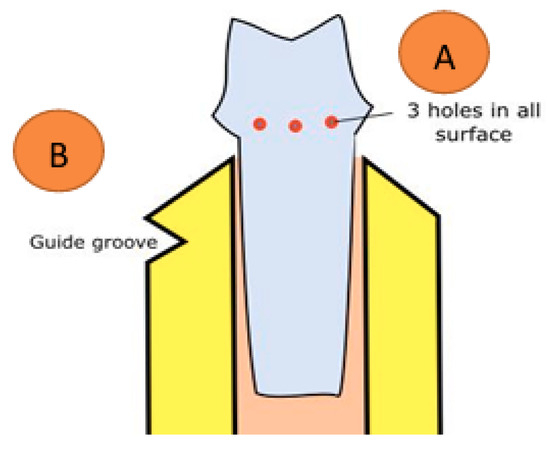
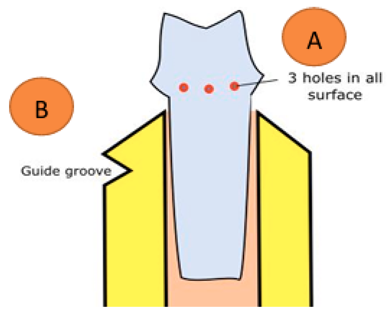
Reference points (Figure 2) were created on the axial surfaces of the dies to facilitate standardized marginal gap measurements.
Figure 2.
The diagram shows reference points added to the die before duplication.
Three small holes were created on each axial surface of the acrylic tooth (mesial, distal, buccal, and lingual) at uniform intervals of approximately 1 mm beneath the finish line. These holes were replicated on each die to facilitate the assessment of marginal fit at identical places across all samples (Figure 2A). A small line was marked on the upper section of the mold’s mounting area, aligning with a corresponding line created in the chewing chamber of the chewing simulator. This alignment ensures that cyclic loading is consistently applied at the same place throughout all samples (Figure 2B).
All preparation and duplication procedures were performed by the same operator to minimize variability.
2.4. Crown Fabrication
2.4.1. Milled Crowns
Ten crowns were fabricated using the CAD/CAM system (CEREC inLab MC XL, Dentsply Sirona). The restorations were designed using CEREC in the Lab 3D software following the standard workflow of scanning, designing, and milling.
The crowns were milled from IPS E.max CAD blocks and subsequently crystallized in a Programat furnace according to the manufacturer’s instructions.
2.4.2. Pressed Crowns
Ten pressed lithium disilicate crowns were fabricated using the heat-press technique. A silicone index was used to standardize the anatomical form of the wax patterns. Three layers of die spacer (total thickness of 60 μm) were applied to the epoxy dies.
Wax patterns were invested, and the pressing procedure was carried out using a pressing furnace according to the manufacturer’s instructions. After pressing, the crowns were finished and cleaned before cementation.
A silicone index was obtained from a reference CAD/CAM crown and utilized to direct the manual waxing by the same operator in order to standardize the exterior morphology between groups. Three layers of Renfert die spacer were used to provide a controlled 60 μm cement spacing on epoxy dies, guaranteeing a perfect fit for subsequent cementation. The crowns were fired in an EP 600 furnace using a conventional investment and 800 °C burnout process. To reduce surface damage, airborne particle abrasion, 50 μm glass particle abrasion was used for recovery from the investment. The literature supports this strict fabrication-press process to provide higher mechanical reliability and better marginal adaptation than typical milling.
2.4.3. Marginal Gap Measurement
Marginal gap measurements were performed using a stereomicroscope (Leica EZ4 ND, Leica Microsystems GmbH, Wetzlar, Germany) at ×35 magnification with digital image analysis software (Leica LAS EZ software, version 3.4.0).
The marginal gap was evaluated by measuring the vertical gap between the crown margin and the finish line of the epoxy dies using a stereomicroscope device and an integrated digital camera.
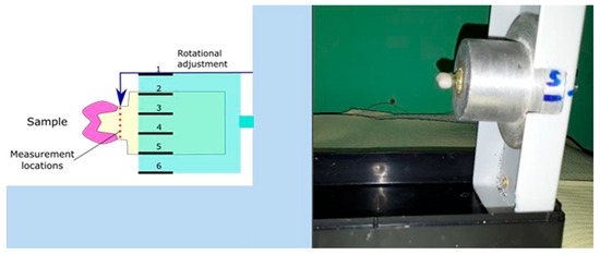
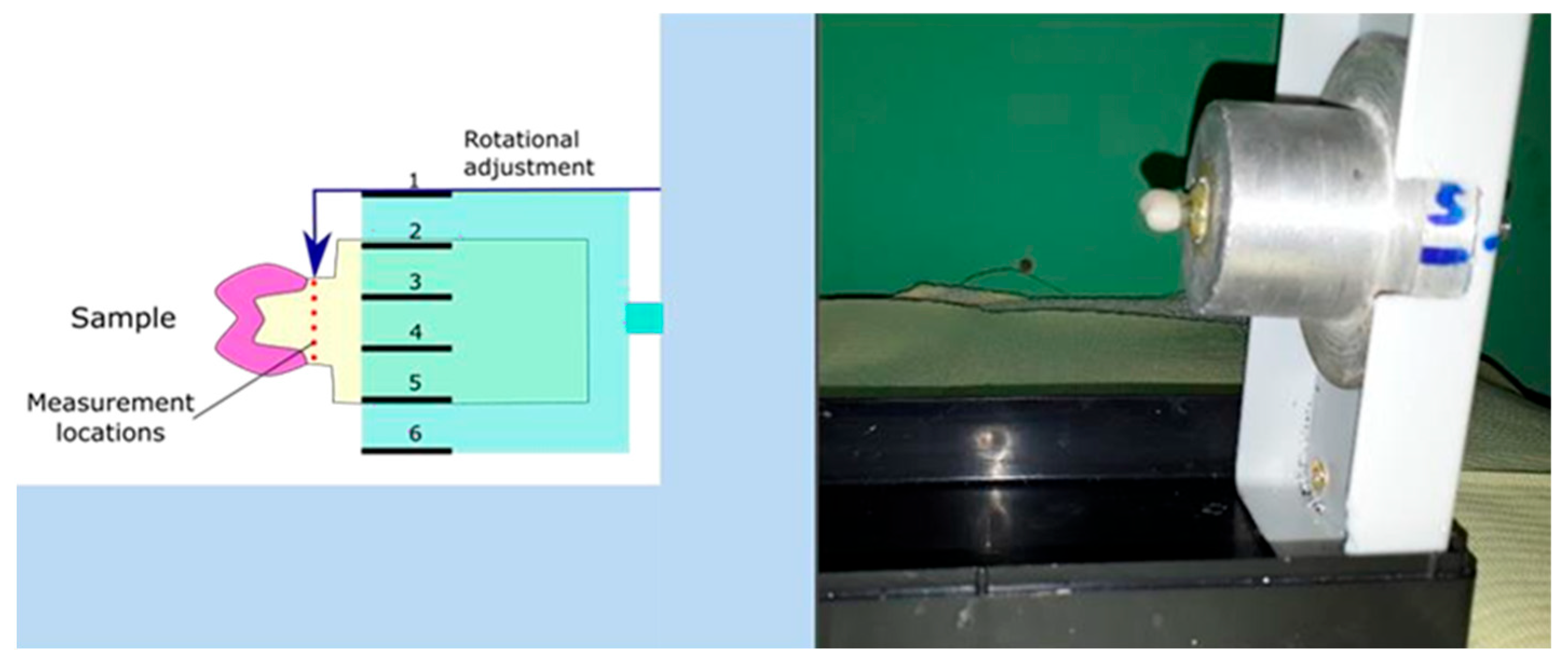
Measurements were obtained at three points on each surface of the four axial walls, resulting in a total of 12 measurement points per crown. The mean marginal gap value was calculated for each specimen. A custom alignment device was used to ensure that measurements were taken at the same locations before and after cyclic loading. A special holding device (SHD) is a simple tool that consists of a calibrated gauge, a fixable base, and reading marks. The samples were inserted in the corresponding housing created in the holding device till the guidance groove created previously in the sample was parallel to the groove created in the holding device (Figure 3).
Figure 3.
Special holding device for gap measurement standardization.
2.4.4. Cyclic Loading Procedure
All cemented crowns were subjected to cyclic loading using a dual-axis chewing simulator. A total of 37,000 cycles were applied at a load of 50 N [17,18,19,20] with a frequency of 1–1.6 Hz, simulating approximately three months of clinical function.
After cyclic loading, marginal gap measurements were repeated using the same protocol.
2.4.5. Statistical Analysis
The data were tabulated and statistically analyzed using IBM SPSS Statistics version 19.0. (Armonk, NY, USA: IBM Corp). Quantitative data were expressed as mean, standard deviation, minimum, and maximum values.
A three-way analysis of variance (ANOVA) [21,22] was performed to evaluate the effect of:
- Ceramic type;
- Cement type;
- Cyclic loading.
In addition, their potential interactions on marginal gap values were evaluated.
2.4.6. Cementation Protocol
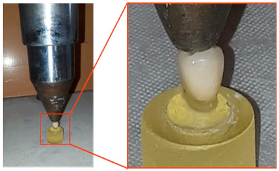
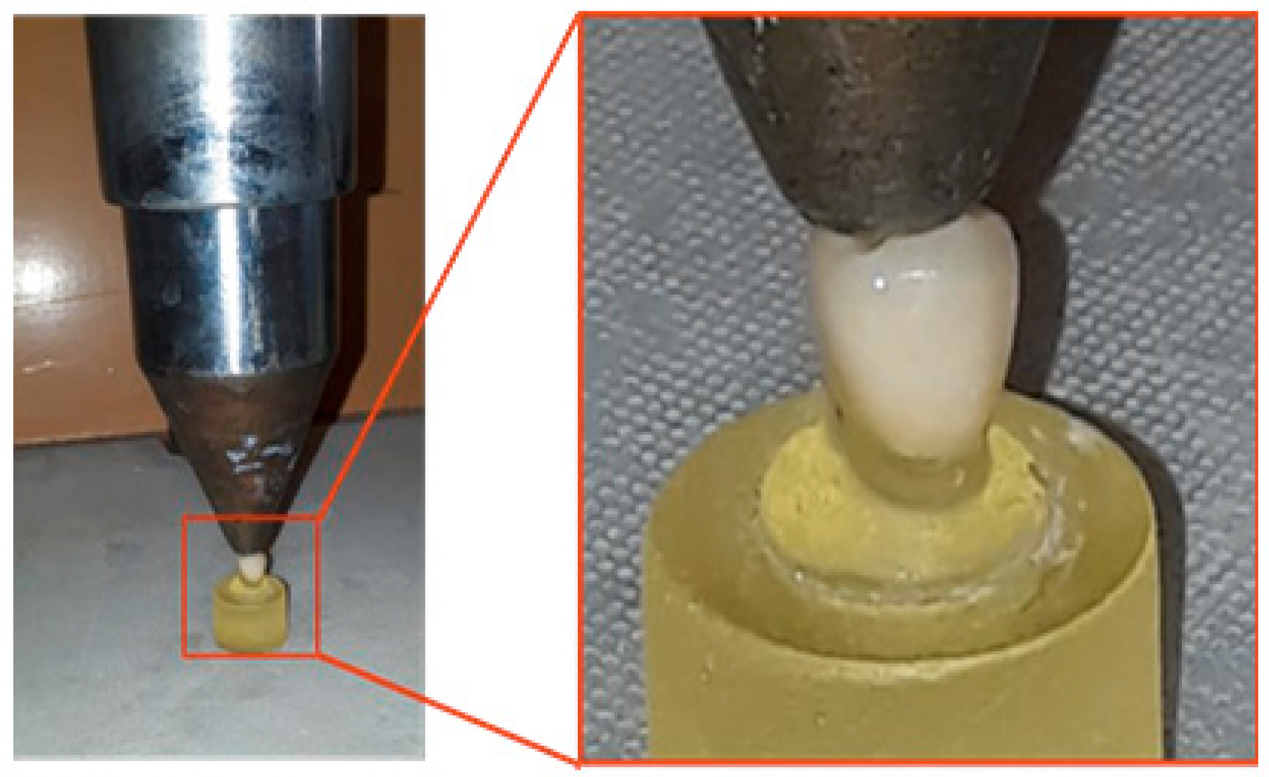
Regarding luting cements, self-adhesive resin cement and resin-modified glass ionomer cement were used for cementation in two subgroups for each fabrication technique (Table 2). For the resin cement subgroups, the internal crown surfaces were etched with 9% hydrofluoric acid gel for 20 s, rinsed thoroughly, and dried. A silane coupling agent was applied before cementation using a self-adhesive resin cement (seT PP, SDI Australia). The cement was mixed according to the manufacturer’s instructions, ensuring a uniform consistency. A freshly mixed resin cement was extruded evenly onto all bonding surfaces of the restoration, then seated on the corresponding epoxy dies for 30 s before any light curing was carried out according to the manufacturing instructions. Light curing was then carried out for 20 s on each surface. The excess material was trimmed away after light curing for 2 s, and the sample was cemented under a static loading force (40 N) for 10 min using a custom-made device (Figure 4).
Figure 4.
Static loading force (40 N).
For the resin-modified glass ionomer subgroups, SDI Reva, Bayswater, VIC, and Australia, luting capsules were triturated for 10 s and injected into the crown-fitting surfaces. The crowns were seated on the dies under a static load of 40 N for 10 min using a custom loading device (Figure 3). After cementation, all samples were ready for the marginal evaluation test.
Restoration had a specific positioning on the tooth preparation. Checking points must be well-defined and precise. The direct view technique was used by many studies for measuring marginal distortion of ceramic restorations for determining marginal fidelity of all-ceramic crown systems. To minimize variability, seating procedures were performed by the same operator.
3. Results
Quantitative data are expressed as minimum (Min), maximum (Max), mean, and standard deviation (±SD). The significance of the results was determined at a 5% probability level (α = 0.05). A three-way analysis of variance (ANOVA) was conducted to assess the effects of the ceramic type, cement type, and cyclic loading, as well as their potential interactions. Post hoc pairwise comparisons were performed using Tukey’s test.
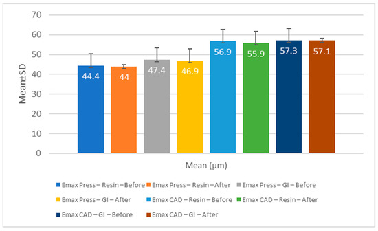
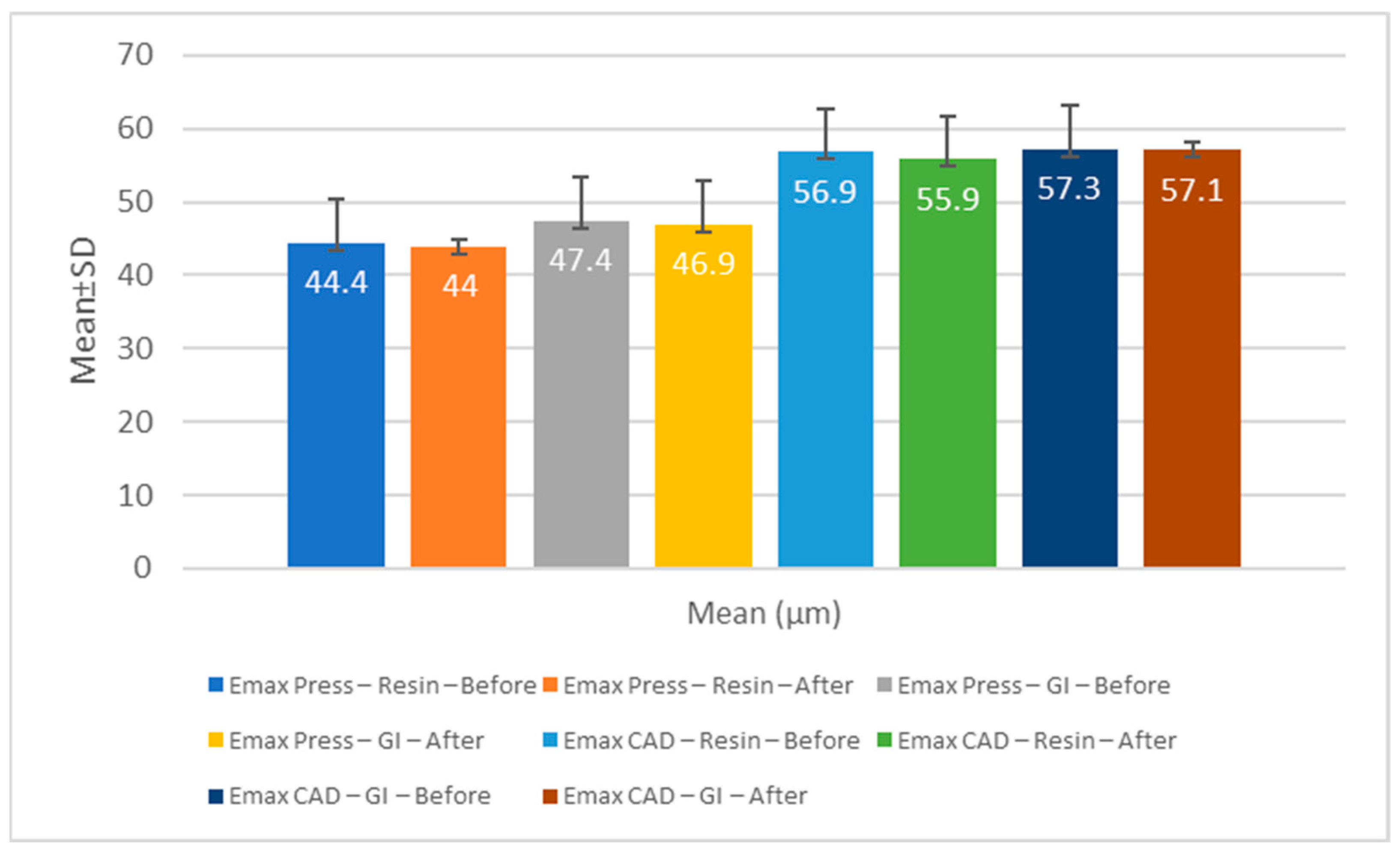
The Max, Min, mean, and standard deviation values of the marginal gap (μm) for the material fabrication technique—EMAX PRES, which stands for pressed technique, and EMAX CA, which stands for milling technique—cement type, and cyclic loading are summarized in Table 3 and graphically represented in Figure 5.
Table 3.
Comparison between the measurements of the studied groups represented by (Min, Max, mean, and Standard deviation) according to the material used, type of cement, and cyclic loading.
Figure 5.
Marginal gap before and after cyclic loading between the pressed and milled groups.
Regarding the type of cement, there was no statistically significant difference (p > 0.05) between press groups cemented with resin cement (44.22 μm) or glass ionomer cement (47.17 μm). In addition, there was no statistically significant difference (p > 0.05) between the CAD groups cemented with resin cement (56.46 μm) and glass ionomer cement (57.22 μm).
The results of the three-way ANOVA (Table 4) revealed that the ceramic type had a statistically significant effect on marginal gap values (p = 0.001). In contrast, the cement type and cyclic loading showed no significant influence (p > 0.05). No significant interactions were observed between the tested variables.
Table 4.
Comparison of the results of the study variables.
Regarding the effect of the fabrication method, a statistically significant difference (p ≤ 0.05) was observed between the two materials, with the E.max Press crowns showing lower mean marginal gap values (45.6 ± 5.6 µm) than the E.max CAD crowns (56.8 ± 4.2 µm).
Regarding the effect of cyclic loading, no statistically significant difference (p > 0.05) was found between the measurements obtained before and after loading in either group. The E.max Press group recorded mean values of 45.64 µm before and 45.47 µm after loading, whereas the E.max CAD group recorded values of 56.7 µm before and 56.56 µm after loading.
The three-way ANOVA demonstrated that the ceramic type had a highly statistically significant effect on the marginal gap values, as summarized in Table 4 (F (1, 32) = 44.65, p < 0.001). This suggests that the fabrication method had a considerable impact on the margin fit. Crowns fabricated using the press technique had smaller marginal gaps than those produced by CAD/CAM milling, indicating that the pressed ceramic restorations had greater marginal accuracy.
In contrast, the cement type did not have a statistically significant effect on the marginal gap values (F (1, 32) = 1.27, p = 0.268). This suggests that the choice between resin and glass ionomer cements did not significantly influence the marginal fit of the restorations within the conditions of this study.
Similarly, cyclic loading showed no statistically significant effect on marginal gap values (F (1, 32) = 0.10, p = 0.755). This indicates that mechanical fatigue simulation did not significantly alter the marginal adaptation of the crowns before and after loading.
4. Discussion
Marginal accuracy is a critical criterion for evaluating the success of fixed prosthodontic treatment. It is defined as the space between the restoration margin and the tooth’s finish line. Optimal marginal adaptation is essential for both the biological and mechanical success of restorations. When the marginal fit is inadequate, bacterial accumulation may occur, increasing the risk of secondary caries and periodontal inflammation [1].
In the present study, strict standardization was achieved using a parallelometer and a custom mounting device to ensure reproducible tooth positioning. A chamfer finish line was selected according to the recommendation of Pera et al. [23]. The prepared tooth was duplicated using a resin material with a modulus of elasticity comparable to that of dentin, providing adequate strength, abrasion resistance, and detailed reproduction [24] The polyether impression material was selected because of its excellent dimensional stability and fine detail accuracy [25]. Proper die spacing was maintained, as it has been reported to significantly influence marginal adaptation [26,27].
Marginal gaps in this study were evaluated using digital microscopy at 35× magnification, which is a non-destructive and reliable method for assessing marginal quality [6]. Cyclic loading was applied using a custom-made dual-axis chewing simulator that reproduced pure vertical movements. A vertical load of 50 N was selected to represent the average masticatory force in non-bruxers [16].
In the present study, the results partially supported the null hypothesis. The pressed lithium disilicate crowns exhibited significantly smaller marginal gaps (mean 44.4 ± 5.8 µm) than the milled crowns (mean 56.9 ± 3.9 µm). These results indicate that the fabrication technique directly affects marginal adaptation, whereas the type of cement and cyclic loading do not.
Regarding the pressed fabrication technique, the characteristics of the heat-pressing process may explain the superior adaptation observed in the pressed restorations. The ceramic ingot is softened and pushed into a refractory mold by an alumina plunger under controlled pressure and temperature, thereby minimizing internal defects and producing dense margins with minimal porosity [28,29]. Conversely, CAD/CAM milling removes material from a partially crystallized block, and the subsequent crystallization step can induce dimensional changes that slightly increase the marginal discrepancies [30].
Our findings are consistent with those of Shadur et al. (2024) [31], Pachiou et al. (2024) [32], and Ispas et al. (2023) [33], who reported smaller marginal gaps in pressed lithium disilicate crowns than in milled crowns. In addition, the crystallization firing process of CAD/CAM-milled ceramics can contribute to marginal inaccuracy [34]. Similarly, Velho et al. (2023) demonstrated superior marginal fit and fatigue resistance in pressed ceramics compared with those in milled ceramics [35].
In contrast, Attia et al. (2024) [16] and Elsayed et al. (2023) [17] found better marginal adaptation in milled restorations, suggesting that differences in milling machine calibration, bur wear, and spacer settings may have accounted for this discrepancy.
Vasiliu et al. (2020) also reported no significant difference between pressed and milled zirconia-reinforced lithium disilicate crowns [15]. The divergence among studies highlights the influence of multiple variables, such as ceramic composition, manufacturing precision, laboratory standardization, and sample size on the results.
Cyclic loading simulates functional fatigue and is a crucial variable for assessing restoration longevity. In the current study, cyclic loading (37,000 cycles at 50 N) did not significantly affect the marginal adaptation. These findings are consistent with those of Attia et al. (2024) [16], who reported no measurable changes in the marginal fit after thermomechanical aging of lithium disilicate restorations. In contrast, Kızılırmak et al. (2025) [36] found that prolonged mechanical aging affected the fit of metal frameworks, possibly because of differences in material stiffness and elastic modulus. The relatively short simulated loading period in our study, equivalent to approximately 3 months of clinical function, may explain the absence of measurable changes [37].
An ongoing debate exists regarding clinically acceptable marginal gaps. McLean and von Fraunhofer in 1971 [38] considered gaps below 120 µm to be sufficient, whereas Alghauli et al. [39] recommended a maximum limit of 100 µm. Therefore, the mean marginal gaps obtained in this study for both pressed and milled lithium disilicate crowns fell within the range of clinical acceptability.
For the luting agents, no statistically significant difference was detected between the resin and resin-modified glass ionomer cement. Resin cements produced slightly smaller gaps (50.3 ± 5.9 µm) than resin-modified glass ionomer cements (54.8 ± 0.8 µm), but the difference was not clinically relevant. The slight improvement observed with resin cements may be attributed to micromechanical interlocking after etching and salinization of lithium disilicate, which enhances adhesive strength and marginal integrity (Jeng-Fen Liu et al., 2022) [40].
These results align with those of Vishnu and Jeevanandan (2024) [41] and Benli et al. (2022) [42], who reported that resin and glass ionomer cements can achieve satisfactory marginal sealing when proper surface treatment is performed. Regarding the lithium disilicate material, both machine-milled and pressed versions exhibited good performances. However, they need to be tested under variable clinical conditions of acidic exposure, as reported by El Sayed et al. (2025), who suggested that systemic conditions might affect the performance of ceramic restorations [43].
For force simulation limitation, a 50 N load was considered appropriate; the study investigators might defend and justify the decision by presenting it as a simulation of typical physiological chewing rather than maximal effort. The 50 N procedure can be supported by the following points. Maximum bite power is not reached in most everyday masticatory cycles. According to research, the average force used to grind soft-to-medium food boluses frequently fluctuates between 30 and 70 N. Numerous significant fatigue investigations, such as those conducted by Guess et al. in 2010 [44], advocated cyclic loading in “step-stress” patterns at 50 N. This was also supported by Magne et al. in 2010 [45], who focused on the material’s performance under “normal” rather than “extreme” conditions, accurately representing the recurrent, low-level functional loads encountered during most of a patient’s daily cycles. Prospective studies could better follow greater loading forces to simulate longer stimulation periods to focus on the long-term clinical simulations for expected survival rates. Moreover, the use of a fixed cement space (60 µm) may have influenced the marginal adaptation results. Future studies should assess longer fatigue durations, various cement space settings, and thermomechanical aging to better simulate clinical conditions.
5. Conclusions
Pressed lithium disilicate crowns demonstrated better marginal adaptation than milled crowns, whereas cement type and cyclic loading showed no measurable influence. Despite these differences, all tested restorations exhibited marginal gaps within clinically acceptable limits, confirming that both fabrication methods are viable for esthetic full-coverage restoration. In addition, both cement types can achieve clinically feasible performance.
Clinical implication: Both pressing and milling techniques can produce clinically acceptable lithium disilicate crowns, but the press technique may be preferred when optimal marginal accuracy is required. This can be correlated with adhesive resin cement, and resin-modified glass ionomer cement shows no clinically significant difference in outcome, despite the need for long-term survival rate investigations with the incorporation of more clinically oriented variables, such as saliva and acidic exposure.
Author Contributions
Conceptualization, H.M.E., S.A.Y. and A.M.F.; methodology, R.M.Y.A., H.M.E., S.A.Y., A.M.S.A. and M.A.A.; software, S.Y.A., S.S.G. and K.D.A.; validation, A.M.F., M.A.A., A.I.M. and E.I.A.; formal analysis, M.D., H.M.E., S.A.Y. and A.I.M.; investigation, M.D., H.M.E., S.A.Y. and A.I.M.; resources, R.M.Y.A., A.M.S.A. and K.D.A.; data curation, H.M.E., S.A.Y., M.D. and M.A.A.; writing—original draft preparation, S.Y.A., S.S.G. and K.D.A.; writing—review and editing, R.M.Y.A., A.M.F., H.M.E. and A.I.M.; visualization, S.Y.A., S.S.G., K.D.A. and H.M.E.; supervision, H.M.E., S.A.Y., A.I.M. and A.M.F.; project administration, A.M.S.A., H.M.E., S.A.Y., A.M.F. and M.D.; funding acquisition, R.M.Y.A., A.M.S.A., S.Y.A., S.S.G., K.D.A. and E.I.A. All authors have read and agreed to the published version of the manuscript.
Funding
This research was funded by the General Directorate of Scientific Research & Innovation, Dar Al Uloom University, through the Scientific Publishing Funding Program.
Institutional Review Board Statement
Not applicable.
Informed Consent Statement
Not applicable.
Data Availability Statement
The data presented in this study are available upon request from the corresponding author, as they are part of an ongoing study.
Conflicts of Interest
The authors declare no conflict of interest.
References
- Lee, K.B.; Park, C.W.; Kim, K.H.; Kwon, T.Y. Marginal and internal fit of all-ceramic crowns fabricated with two different CAD/CAM systems. Dent. Mater. J. 2008, 27, 422–426. [Google Scholar] [CrossRef] [Scilit] [PubMed]
- Kelly, J.R.; Benetti, P. Ceramic materials dentistry: Historical evolution and current practice. Aust. Dent. J. 2011, 56, 84–96. [Google Scholar] [CrossRef] [Scilit]
- Denry, I.; Kelly, J.R. State of the art of zirconia for dental applications. Dent. Mater. 2008, 24, 299–307. [Google Scholar] [CrossRef] [Scilit]
- Elsaka, S.E.; Elnaghy, A.M. Mechanical Properties of Zirconia-Reinforced Lithium Silicate Glass-Ceramic. Dent. Mater. 2016, 32, 908–914. [Google Scholar]
- Neves, F.D.; Prado, C.J.; Prudente, M.S.; Carneiro, T.A.; Zancope, K.; Davi, L.R.; Mendonca, G.; Cooper, L.F.; Soares, C.J. Micro-computed tomography evaluation of marginal fit of lithium disilicate crowns fabricated by using chairside CAD/CAM systems or the heat-pressing technique. J. Prosthet. Dent. 2014, 112, 1134–1140. [Google Scholar]
- Dudley, J.; Farook, T.H. Marginal Gap of Pre-Cemented Endocrowns: A Systematic Review of Measurement Methods and the Influence of Fabrication Method and Crown Material. Clin. Exp. Dent. Res. 2025, 11, e70152. [Google Scholar]
- Blatz, M.B.; Sadan, A.; Kern, M. Resin-ceramic bonding: A review of the literature. J. Prosthet. Dent. 2003, 89, 268–274. [Google Scholar] [CrossRef] [Scilit]
- Sorensen, J.A. A standardized method for the determination of crown margin fidelity. J. Prosthet. Dent. 1990, 64, 18–24. [Google Scholar] [CrossRef] [Scilit]
- Dapieve, K.S.; Velho, H.C.; da Rosa, L.S.; Pivetta, J.P.; Maidana, F.C.; Venturini, A.B.; Kleverlaan, C.J.; Pereira, G.K.R.; Valandro, L.F. Ceramic surface conditioning, resin cement viscosity, and aging relationships affect the load-bearing capacity under fatigue of bonded glass-ceramics. J. Mech. Behav. Biomed. Mater. 2023, 139, 105667. [Google Scholar] [CrossRef] [Scilit]
- Schestatsky, R.; Zucuni, C.P.; Venturini, A.B.; Burgo, T.A.d.L.; Bacchi, A.; Valandro, L.F.; Pereira, G.K.R. CAD-CAM milled versus pressed lithium-disilicate monolithic crowns adhesively cemented after distinct surface treatments: Fatigue performance and ceramic surface characteristics. J. Mech. Behav. Biomed. Mater. 2019, 94, 144–154. [Google Scholar] [CrossRef] [Scilit]
- Al-Obaidi, M.Y.N.; Gholam, M.K. Post fatigue fracture resistance of lithium disilicate and zirconia crowns: Vertical versus horizontal preparations (an in vitro study). Saudi Dent. J. 2025, 37, 57. [Google Scholar] [CrossRef] [Scilit] [PubMed]
- Azar, B.; Eckert, S.; Kunkela, J.; Ingr, T.; Mounajjed, R. The marginal fit of lithium disilicate crowns: Press vs. CAD/CAM. Braz. Oral Res. 2018, 32, e001. [Google Scholar] [CrossRef] [Scilit] [PubMed]
- Mounajjed, R.; Layton, D.M.; Azar, B. The marginal fit of E.max Press and E.max CAD lithium disilicate restorations: A critical review. Dent. Mater. J. 2016, 35, 835–844. [Google Scholar] [CrossRef] [Scilit]
- Elrashid, A.H.; AlKahtani, A.H.; Alqahtani, S.J.; Alajmi, N.B.; Alsultan, F.H. Stereomicroscopic Evaluation of Marginal Fit of E.Max Press and E.Max Computer-Aided Design and Computer-Assisted Manufacturing Lithium Disilicate Ceramic Crowns: An In vitro Study. J. Int. Soc. Prev. Community Dent. 2019, 9, 178–184. [Google Scholar]
- Vasiliu, R.D.; Porojan, S.D.; Porojan, L. In Vitro Study of Comparative Evaluation of Marginal and Internal Fit between Heat-Pressed and CAD-CAM Monolithic Glass-Ceramic Restorations After Thermal Aging. Materials 2020, 13, 4239. [Google Scholar] [CrossRef] [Scilit]
- Attia, R.E.; Mohammed, H.R.; Haggag, K.M. Effect of thermomechanical aging on marginal fit of three CAD-CAM restorative materials: An in vitro study. J. Dent. Res. Dent. Clin. Dent. Prospect. 2024, 18, 189–195. [Google Scholar] [CrossRef] [Scilit]
- Elsayed, S.M.; Emam, Z.N.; Abu-Nawareg, M.; Zidan, A.Z.; Elsisi, H.A.; Abuelroos, E.M.; Fansa, H.A.; Shokier, H.M.R.; Elbanna, K.A. Marginal gap and cyclic fatigue loading for different all-ceramic endocrowns. Eur. Rev. Med. Pharmacol. Sci. 2023, 27, 879–887. [Google Scholar]
- Akhlaghian, M.; Khaledi, A.A.; Mosaddad, S.A.; Dabiri, S.; Giti, R.; Kadkhodae, F.; Gholami, S. The internal and marginal adaptation of lithium disilicate endocrowns fabricated using intra and extraoral scanners: An in-vitro study. PLoS ONE 2024, 19, e0301361. [Google Scholar] [CrossRef] [Scilit]
- Mohamed, H.A.; Azer, A.; AboElHassan, R.G. Accuracy of Marginal and Internal Adaptation of Advanced Lithium Disilicate Crowns Using Different Margin Designs (In Vitro Study). Int. J. Dent. 2026, 2026, 5550877. [Google Scholar] [CrossRef] [Scilit]
- Bataineh, K.; Al Janaideh, M. Fatigue behavior of three thin CAD/CAM all-ceramic crown materials. Int. Biomech. 2023, 10, 29–36. [Google Scholar] [CrossRef] [Scilit]
- Da Silva, B.C.D.; da Silva, S.E.G.; da Silva, N.R.; de Moreira, F.G.G.; Souza, K.B.; Zhang, Y.; de Souza, R.O.A. Effect of different surface treatments and adhesive cementation on the surface topography and flexural strength of translucent and ultra-translucent monolithic zirconia. J. Prosthodont. 2024, 33, 863–871. [Google Scholar] [CrossRef] [Scilit]
- Lu, Y.; Bierman, T.E.; Dal Piva, A.M.O.; Tribst, J.P.M.; Feilzer, A.J.; Kleverlaan, C.J. Effect of Surface Treatment and Resin Cement on the Bond Strength of an Advanced Lithium Disilicate. Eur. J. Dent. 2024, 18, 869–876. [Google Scholar] [CrossRef] [Scilit] [PubMed]
- Pera, P.; Gilodi, S.; Bassi, F.; Carossa, S. In vitro marginal adaptation of alumina porcelain ceramic crowns. J. Prosthet. Dent. 1994, 72, 585–590. [Google Scholar] [CrossRef] [Scilit] [PubMed]
- Ribeiro, I.L.; Campos, F.; Sousa, R.S.; Souza, R.A.; Alves, M.L.L.; Rodrigues, D.M.; Bottino, M.A. Marginal and internal discrepancies of zirconia copings: Effects of milling system and finish line design. Indian J. Dent. Res. 2015, 26, 15–20. [Google Scholar] [CrossRef] [Scilit]
- Martinez, Y.S.; Moreira, J.C. Major clinical findings on the types of impressions used in implant-supported prostheses: A systematic review. MedNEXT J. Med. Health Sci. 2025, 6, 1–7. [Google Scholar] [CrossRef] [Scilit]
- Sripairojn, S.; Juntavee, N.; Juntavee, A. Marginal Accuracy of Ceramic Veneer Alloy Related to Different Alloy Fabrication Techniques, Ceramic Veneering Methods, Stages, and Sites of Fabrication. Eur. J. Dent. 2025, 19, 966–978. [Google Scholar]
- Charoenporn, W.; Sornsuwan, T.; Sae-Lee, D.; Amornvit, P.; Chaijareenont, P.; Rungsiyakull, P. Evaluating fatigue resistance in occlusal veneers: A comparative study of processing techniques and material thickness of lithium disilicate (IPS e.max Press vs. IPS e.max CAD). BMC Oral Health 2024, 24, 1542. [Google Scholar] [CrossRef] [Scilit]
- Reich, S.; Gozdowski, S.; Trentzsch, L.; Frankenberger, R.; Lohbauer, U. Marginal fit of heat-pressed vs. CAD/CAM processed all-ceramic onlays using a milling unit prototype. Oper. Dent. 2008, 33, 644–650. [Google Scholar]
- Kim, J.H.; Oh, S.; Uhm, S.H. Effect of the Crystallization Process on the Marginal and Internal Gaps of Lithium Disilicate CAD/CAM Crowns. BioMed Res. Int. 2016, 2016, 8635483. [Google Scholar] [CrossRef] [Scilit]
- Rekow, E.D.; Silva, N.R.; Coelho, P.G.; Zhang, Y.; Guess, P.; Thompson, V.P. Performance of dental ceramics: Challenges for improvements. J. Dent. Res. 2011, 90, 937–952. [Google Scholar] [CrossRef] [Scilit]
- Shadur, A.; Nissan, J.; Lugassy, D.; Umansky, A.; Zenziper, E.; Ben-Izhack, G. Effects of Different Crystallization Protocols on Marginal Gap of Lithium Disilicate Single Crowns: SEM Analysis. Dent. J. 2024, 12, 416. [Google Scholar] [CrossRef] [Scilit]
- Pachiou, A.; Zervou, E.; Tsirogiannis, P.; Sykaras, N. Evaluation of the marginal fit of lithium disilicate single crowns fabricated with the conventional (IPS e.max Press) and CAD/CAM (IPS e.max CAD) methods: A systematic review and meta-analysis. Int. J. Comput. Dent. 2024, 27, 365–377. [Google Scholar]
- Ispas, A.; Moldovan, M.; Cuc, S.; Prodan, D.; Bacali, C.; Petean, I.; Buduru, S.; Manziuc, M.; Iosif, L. SEM Evaluation of Marginal Adaptation E-Max Crowns Manufactured by Printing-Pressed and Milling. Diagnostics 2023, 13, 3518. [Google Scholar] [CrossRef] [Scilit]
- Hassan, S.M.; Montaser, A.G.; Sharkawy, Z.R.E.; Gad, N.A.; Alrafee, S.A.; Gabal, Z.A. Marginal fit and fracture resistance of CAD/CAM glass ceramic occlusal veneers with different preparation designs. BMC Oral Health 2025, 25, 793. [Google Scholar] [CrossRef] [Scilit]
- Velho, H.C.; Dapieve, K.S.; Grassi, E.D.A.; Borges, A.L.S.; Marinho, R.M.d.M.; Pereira, G.K.R.; Venturini, A.B.; Valandro, L.F. Fatigue behavior, failure mode, and stress distribution of occlusal veneers: Influence of the prosthetic preparation cusp inclinations and the type of restorative material. Clin. Oral Investig. 2023, 27, 5539–5548. [Google Scholar] [CrossRef] [Scilit] [PubMed]
- Kızılırmak, K.T.; Yılmaz, B.K.; Orhan, K.; Tamam, E. Effect of manufacturing type and aging on the fit of metal frameworks: An in vitro Micro-CT study. Clin. Oral Investig. 2025, 29, 271. [Google Scholar] [CrossRef] [Scilit] [PubMed]
- Dudley, J.; Farook, T.H. Measuring the Marginal Gap of Pre-Cemented All-Metal Single Crowns: A Systematic Review of In Vitro Studies. Dent. J. 2025, 13, 204. [Google Scholar] [CrossRef] [Scilit] [PubMed]
- McLean, J.W.; von Fraunhofer, J.A. The estimation of cement film thickness by an in vivo technique. Br. Dent. J. 1971, 131, 107–111. [Google Scholar] [CrossRef] [Scilit]
- Alghauli, M.A.; Al-Gabri, R.S.; Keshk, A.; Mirah, M.A.; Alqutaibi, A.Y. Clinical Complications and Survival Rate of Resin Matrix Ceramic Restorations: A Systematic Review and Meta-Analysis. J. Esthet. Restor. Dent. 2025, 37, 1721–1739. [Google Scholar] [CrossRef] [Scilit]
- Liu, J.F.; Yang, C.C.; Luo, J.L.; Liu, Y.C.; Yan, M.; Ding, S.J. Bond strength of self-adhesive resin cements to a high transparency zirconia crown and dentin. J. Dent. Sci. 2022, 17, 973–983. [Google Scholar] [CrossRef] [Scilit]
- Vishnu, G.; Jeevanandan, G. Evaluation of Microleakage Using Different Luting Cements in Kedo Zirconia Crowns: An In Vitro Assessment. Cureus 2024, 16, e66237. [Google Scholar] [CrossRef] [Scilit]
- Benli, M.; Turkyilmaz, I.; Martinez, J.L.; Schwartz, S. Clinical performance of lithium disilicate and zirconia CAD/CAM crowns using digital impressions: A systematic review. Prim. Dent. J. 2022, 11, 71–76. [Google Scholar] [CrossRef] [Scilit]
- Elsayed, H.M.; Elmarakby, A.M.; Yousief, S.A.; Abd Elwahab, H.A.; Alturki, M.W.; Tawwash, E.M.; Albahkaly, H.S.; Rayes, K.A.; Bawazir, H.A.; Samran, H.A.; et al. An In Vitro Acidic Media Simulation of GERD and Its Effect on Machine-Milled Ceramics’ Optical Properties. Prosthesis 2025, 7, 156. [Google Scholar] [CrossRef] [Scilit]
- Guess, P.C.; Zhang, Y.; Kim, J.W.; Rekow, E.D.; Thompson, V.P. Damage and reliability of Y-TZP after cementation surface treatment. J. Dent. Res. 2010, 89, 592–596. [Google Scholar] [CrossRef] [Scilit]
- Magne, P.; Schlichting, L.H.; Maia, H.P.; Baratieri, L.N. In vitro fatigue resistance of CAD/CAM composite resin and ceramic posterior occlusal veneers. J. Prosthet. Dent. 2010, 104, 149–157. [Google Scholar] [CrossRef] [Scilit]

Disclaimer/Publisher’s Note: The statements, opinions and data contained in all publications are solely those of the individual author(s) and contributor(s) and not of MDPI and/or the editor(s). MDPI and/or the editor(s) disclaim responsibility for any injury to people or property resulting from any ideas, methods, instructions or products referred to in the content. |
© 2026 by the authors. Licensee MDPI, Basel, Switzerland. This article is an open access article distributed under the terms and conditions of the Creative Commons Attribution (CC BY) license.