


Abstract
Tussah silk fibroin is a promising material for drug release because of protein component, biocompatibility, aqueous processability, and biodegradability. Tussah silk fibroin nanoparticles were self-assembled and prepared in this paper. Their appearances and sizes were modified by altering the volume ratios between tussah silk fibroin and ethanol. When 9 mL of ethanol was added, the average microsphere size was 184.52 nm and the scattered index was 0.2903. The tussah silk fibroin nanoparticles for controlled drug release was studied and the result showed that the time of drug release was significantly longer. The process of drug release was divided into rapid release in early stage and slow release in late stage. The target drug delivery can reduce the negative effects and improve the efficiency of the drug. As a result, it favors deeper investigation of tussah silk fibroin nanoparticles as drug carriers for controlled release of sensitive biologicals.
1. Introduction
Silk fibroin is a fibrous protein manufactured by insects or spiders. With its unique properties like processability, mechanical strength, long-term degradability and biocompatibility, it is currently being used in several biomedical fields [1]. Foremost, silk fibroin is used in great potential fields, such as target drug delivery and controlled release system, because of strong mechanical stability, unique protein component and slow degradability, compared with other synthetic and natural polymers, such as alginate, polyanhydrides, polyorthoesters, chitosan and polyesters etc [2]. Therefore, design and characteristic of devices composed by silk fibroin have been reported, including microspheres [3,4,5] and scaffolds [6,7,8]. For biomedical purposes, the silk fibroin is a successful material that fulfills most necessary requirements.
The applications of silk fibroin are mostly about the silkworm fibroin. The reports about the developments and applications of tussah silk are few except some studies of tussah silk fibroin membrane [9,10]. Compared with silkworm fibroin, Tussah Silk Fibroin (TSF) with special three peptide sequence of Arginine-Glycine-Aspartate acid (RGD) [11], contributes to cell adhesion which was studied by Minoura et al. [12]. The property of affinity of tussah silk fibroin was better than silkworm fibroin because the tussah silk fibroin contained a certain amount of amino acids with positive charges. Cells adhesion property and value-added rate in tussah silk fibroin membrane were much higher than silkworm fibroin membrane which was investigated by Altman et al. [13]. However, there was no investigation of tussah silk fibroin self-assembly nanoparticles for drug release.
Above all, tussah silk fibroin is a natural protein, which can be used as biomaterials, e.g., surgical suture, scaffold for tissue engineering, wound covering material, soft contact lens and controlled release carrier, because it has good biocompatibility and biodegradability. Tussah silk fibroin contains 18 kinds of essential amino acids, which can be decomposed by specific enzyme to non-toxic products for the body, not easy to cause inflammation and immune rejection. In view of these characteristics, the nanoparticles as drug carriers and controlled release have good application in medicine. Therefore, the preparation and properties of self-assembly tussah silk fibroin nanoparticles were characterized and the application in controlled release was investigated in this paper.
2. Materials and Methods
2.1. Materials
Cocoons of tussah was provided by Nanyang, Henan Province, China. Sodium carbonate, lithium hydroxide, n-butanol, ammonium thiocyanate, polyethylene glycol (20000 Da), dialysis bag, 5-Fu and other chemicals were purchased from Sinopharm Chemical Reagent Co., Ltd.
2.2. Tussah Silk Purification
Tussah cocoons were boiled 4 times for 30 min in aqueous solution of 0.02 M Na2CO3, and thoroughly rinsed with distilled water to remove the sericin completely. After air drying, the extracted tussah silk fibroin was dissolved in 9 M LiSCN solution at 55 °C for 2 h, yielding a 20% (w/v) solution. The solution was dialyzed against distilled water using dialysis bags (MWCO 8000-14000 Da) for 4 days. Silk aggregates as well as debris were removed by filter papers. The solution was concentrated by polyethylene glycol. The final concentration of tussah silk fibroin aqueous solution was approximately 3% (w/v). The 3% tussah silk fibroin solution was kept at 4 °C for futher application.
2.3. Preparation of Tussah Silk Nanoparticles
The concentration of anhydrous ethanol and tussah silk were adjusted to different concentrations (w/v), typically a 3% solution was used for this experiment. 10 mL tussah silk fibroin solution and 0.5 mL, 1 mL, 1.5 mL, 2 mL, 2.5 mL, 3 mL, 3.5 mL, 4 mL, 4.5 mL, 5 mL anhydrous ethanol were mixed separately, and fully blended on a magnetic stirrer at low temperature and low speed for 5 min. Briefly the volume ratio of ethanol to tussah silk fibroin solution was adjusted from 1:20 to 10:20, to investigate the best proportion of ethanol in total solution. Before thawing and centrifugation, the whole liquid solution was frozen at −20 °C for 24 h. Supernatant was discarded after centrifugation, and nanoparticles were collected, washed 4 times by ultrapure water prior to lyophilization.
2.4. Release of Drugs from TSF Particles
The concentration of tussah silk solutions used for this experiment were adjusted to concentration of 3% (w/v), typically 20 mL solution were used here. 100 mg 5-Fu was added, and then 9 mL ethanol was mixed, and fully blended with the solution on a magnetic stirrer at 20 °C and low speed for 5 min. Before thawing and centrifugation, this liquid solution was kept at −20 °C for 24h. Supernatant was discarded after centrifugation, and TSF particles with 5-Fu were collected, washed 4 times by ultrapure water prior to lyophilization. Dynamic dialysis method was used to obtain the TSF particles release property of drugs.
3g 5-Fu loaded TSF nanoparticles were encased in dialysis bag and suspended in 5 mL phosphate (pH 7.4), and the dialysis bas was put in beaker with 1000 mL phosphate (pH 7.4), then incubation at 37 °C with constant shaking. The solvent was removed from each 5 mL sample periodically and replaced with 5 mL fresh phosphate (pH 7.4). Using UV-Vis-spectrometry, in the medium, 5-Fu content was analyzed. The percentage of cumulative model 5-Fu release (% w/w) was investigated as a function of incubation time. In this study, every experiment was processed in triplicate.
2.5. Characterization
2.5.1. Field Emission Electron Microscopy
TSF nanoparticles were collected after washed by water, ultrasonic dispersed in ethanol solution. The suspension was dropped on the silicon wafer, dried at room temperature and investigated using a JSM-6700F scanning electron microscopy (JEOL Company of Japan) at an operating voltage of 5.0 kV.
2.5.2. Dynamic/Static Light Scattering Instrument
The DLS/SLS experiments were processed using a Brookhaven Instrument BI200-SM goniometer equipped with a diode laster operated at a wavelength, λ = 532 nm. The temperature was kept at 25 °C with 0.05 °C accuracy with a temperature-control re-circulating bath.
2.5.3. Laser Partical Sizer
The particle sizes of TSF nanopartiles were measured by a laser partical sizer (Winner 801). The testing temperature was 10 °C, the dispersion medium was water, the viscosity of medium was 1.307E-03 Pa, the refractive index of medium was 1.333, the angle was 90.0 Deg, and the delay unit time was 20 µs.
2.5.4. Fourier Transform Infrared (FTIR) Spectroscopy
TSF nanoparticles were centrifugated, and washed by water for 4 times. Then nanoparticles were collected, freeze-dried and analyzed by FTIR (NEXUS670), from 500 to 4000 cm−1. The specimen was mixed with KBr the mixture was compressed to KBr disks. The spectrum of each sample was achieved via accumulation of 100 scans with a nominal resolution of 2 cm−1.
3. Results and Discussion
3.1. Tussah Silk Precipitation Efficiency
The impact of ethanol on tussah silk sedimentation was researched by the addition of pure ethanol to aqueous tussah silk fibroin solution, then was frozen for 24 h. Finally, tussah silk nanoparticles were collected by centrifugation. The efficiency of tussah silk precipitation could be calculated via Eq. (1).
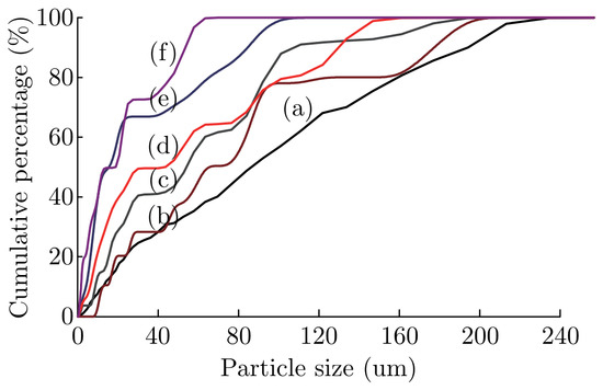
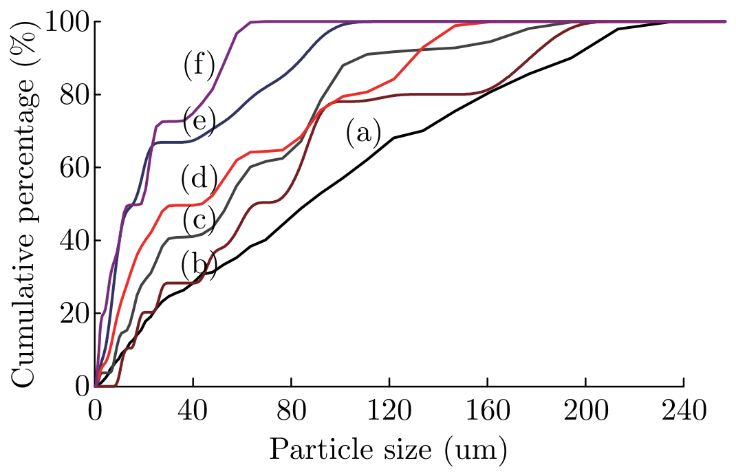
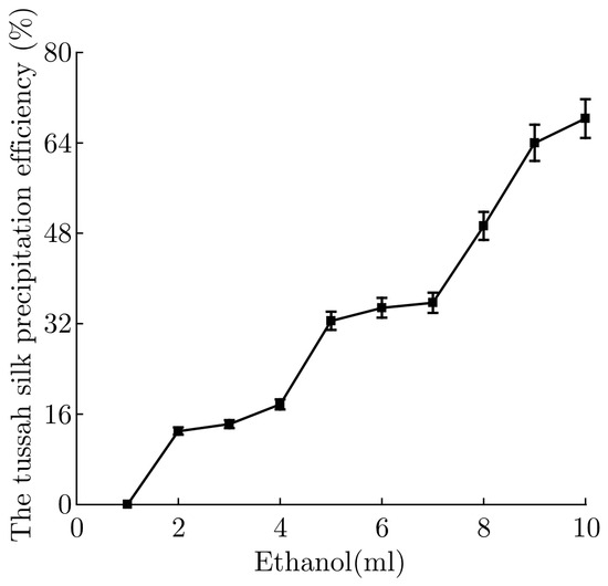
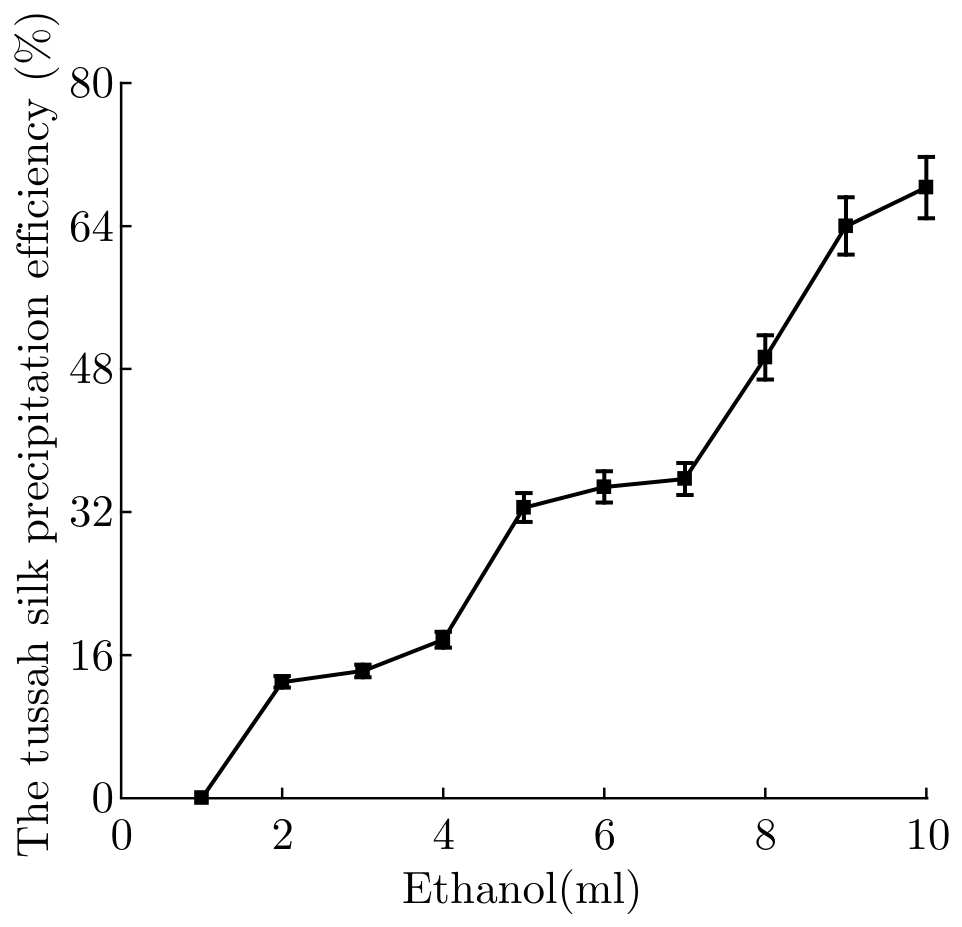
The silk molecular would be induced to form β-sheet structure by addition of ethanol to tussah silk solution and self-assemble to form nucleation (micellar like structure), confirmed by FTIR results. Then, more silk molecular would change to β-sheet structure, and attached to the nucleation during the freezing process, under situation of the ethanol concentration going up as water was slowly solidified. Finally, the nucleation would form tussah silk nanoparticles. Therefore, well-formed silk particles were controllable, as it was indicated in Figure 1. Along with volume ratio of tussah silk solution to ethanol from 20:1 to 20:6, sizes of silk particles were changed from micro to nano (Figure 1), and the tussah silk precipitation efficiency increased (Figure 2). However, neither the total addition of ethanol is to be increased excessively, nor is there massive aggregation instead of particle formation. This phenomenon was probably caused by aggregated and tabular tussah particles obtained due to excessive ethanol.
Figure 1.
Cumulative frequency of TSF particles with different amount of ethanol added when the tussah silk fibroin solution of 20 mL (a) 1 mL, (b) 2 mL, (c) 3 mL, (d) 4 mL, (e) 5 mL, (f) 6 mL.
Figure 2.
The tussah silk precipitation efficiency with different amount of ethanol added when the tussah silk fibroin solution of 20 mL.
3.2. TSF Particles Morphology
TSF particles volume ratios of tussah silk solution to ethanol were 20:2, 20:4, 20:6, 20:8, 20:10 and 20:12 in the experiments. While the volume ratio of tussah silk to ethanol decreased from 20:2 to 20:12, the average diameter of particles were decreased. As mentioned above, optimal addition of ethanol was necessary for the formation of regular tussah silk particles and reduction of aggregation. Moreover, the amount of ethanol also affected the particle morphology significantly. The particles shape was changed when compared with particles with less ethanol. Too many similar stick substance and tiny silk nucleation were induced by an increase in excessive ethanol. Therefore, massive silk aggregation without uniform appearance were be obtained when more ethanol was input into the reaction of tussah silk particles fabrication. The prime volume was about 2:1, further, there is intermediate situation. When the volume ratio of tussah silk solution to ethanol was between 20:2 to 20:12, there were both irregular and regular particles.
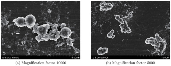
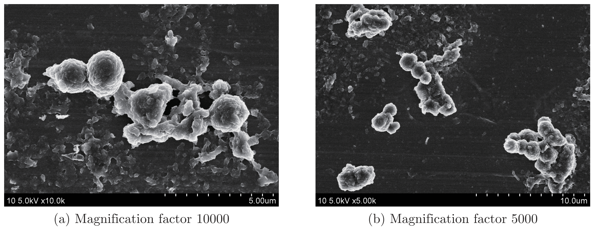
The field emission electron microscope photographs of TSF particles volume ratios of tussah silk solution to ethanol for was shown in Figure 3. The shapes of the TSF particles were spherical. As shown in Figure 3, nanoparticles with a spherical shaper were simply prepared.
Figure 3.
The field emission electron microscope photographs of TSF particles with 10 mL ethanol added when the tussah silk fibroin solution of 20 mL.
3.3. TSF Particle Size Analysis
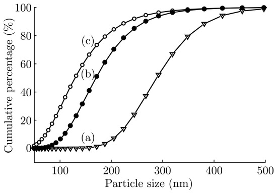
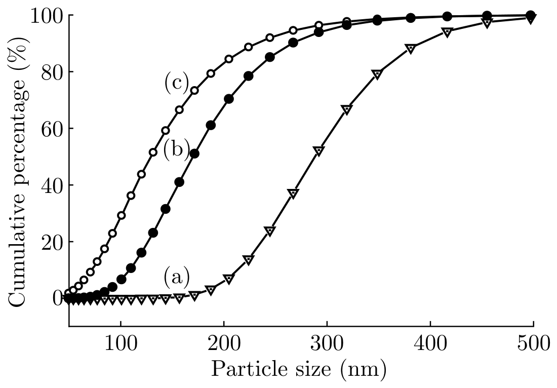
The different volume ratios of tussah silk solution to ethanol significantly influenced the size of the TSF particles which can be shown in Figure 4. As volume ratio of tussah silk solution to ethanol was 20:3, TSF particles with an average diameter of 326.98 nm and the scattered index was 0.0564. TSF particles had an average diameter of 213.44 nm and the scattered index was 0.1304 with the volume ratio of 20:6, whereas TSF particles had an average diameter of 184.52 nm and the scattered index was 0.2903 with the volume ratio of 20:9.
Figure 4.
Cumulative frequency of TSF particles with different volume ratios of tussah silk solution to ethanol (a) 20:3, (b) 20:6, (c) 20:9.
At the same time, the different volume ratios of tussah silk solution to ethanol influenced the size distributions of the TSF particles (Figure 4). When the cumulative frequency of TSF particles was 80%, the diameters of the particles were from 50 nm to 175 nm with the volume ratio of 20:9, and the diameters of the particles were from 50 nm to 225 nm with the volume ratio of 20:6, whereas the diameters of the particles were from 50 nm to 350 nm with the volume ratio of 20:3. The size distribution of TSF particles decreased with the volume ratios of tussah silk solution to ethanol from 20:9 to 20:3, whereas the concentration of TSF particles increased.
3.4. FTIR Analysis
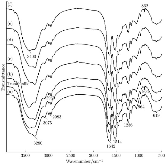
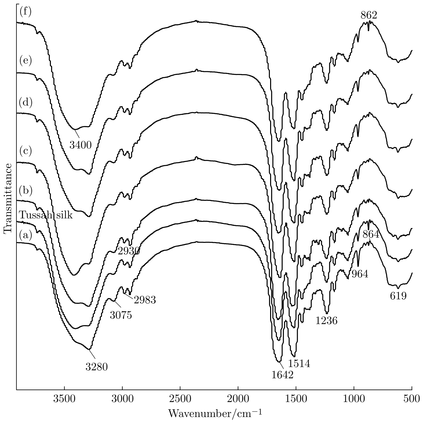
The silk molecules were induced to form β-sheet structure and self-assemble to form nucleation (micellar like structure) by addition of ethanol to tussah silk solution, it was confirmed by FTIR results which is shown in Figure 5. Figure 5 shows FTIR spectras of tusah silk and TSF particles. The characteristic absorption bands of β-sheet structure were around 1640 cm−1 were strengthened with the addition of ethanol. FTIR analysis of tussah fibroin particles with different volume ratios of tussah silk solution to ethanol shows that there are not much differences among their FTIR spectras, and characteristic absorption bands were basic consistent, which can be seen in Figure 5.
Figure 5.
FTIR spectras of tussah silk and TSF particles with different amount of ethanol added when the tussah silk fibroin solution of 20 mL (a) 2 mL, (b) 4 mL, (c) 6 mL, (d) 8 mL, (e) 9 mL, (f) 10 mL.
3.5. In Vitro 5-Fu Release Studies
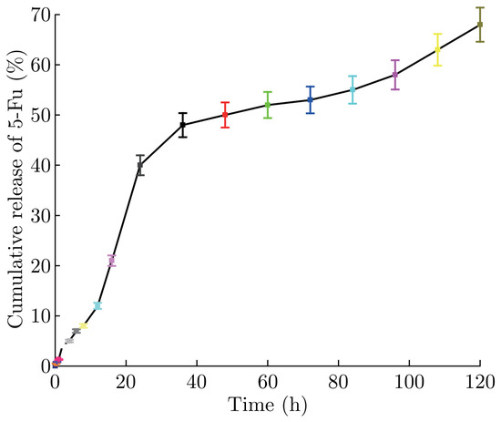
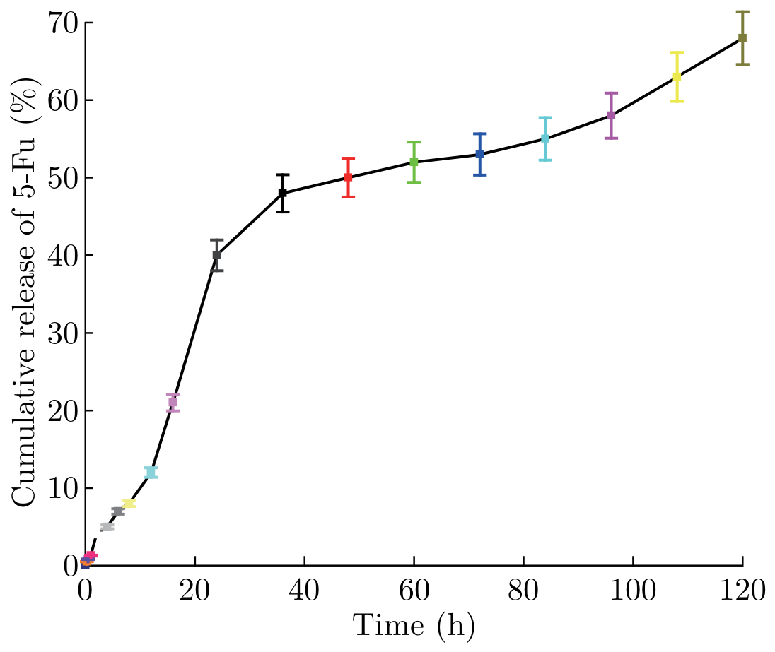
The cumulative release profiles of model 5-Fu from the TSF particles were studied in phosphate of pH 7.4. The results are shown in Figure 6. The TSF particles for controlled drug release shows that the time of drug release are significantly longer. The process of drug release are divided into rapid release in early stage and slow release in late stage. The speed of the drug release was reduced and the drug reacted in a more awkward way in order to reduce the harm to the human body, then the target drug delivery can reduce the negative effects and improve the efficiency of the drug. TSF particles prepared from a 3% TSF solution and loaded with 5-Fu showed a release of 4% with the first 2 h. After the initial burst of 10 h, the release speed of 5-Fu was much slowly. The release of TSF particles loaded with 5-Fu was 68% after 120 h (Figure 6). The release of 5-Fu in solution was quickly without TSF particles.
Figure 6.
The cumulative release of 5-Fu from the TSF particles.
4. Conclusion
A method of self-assembly producing tussah silk fibroin nanoparticles using ethanol was investigated in this paper. The appearance and size distribution of TSF particles can be controlled by changing the ratios of ethanol used in the producing process, as long as the preparation amount of TSF particles can be also controlled by changing the ratios of ethanol. The prime volume ratio of tussah silk to ethanol was about 2:1, evenmore, there is intermediate situation, and both irregular and regular particles were obtained. The experiments of tussah silk fibroin nanoparticles for controlled drug release showed that the time of drug release was significantly longer. The process of drug release was divided into rapid release in early stage and slow release in late stage. The speed of the drug release was slowed down in order to reduce the harm to human body, so, the drug delivery can reduce the negative effects and improve the efficiency of the drug. The fabrication method of tussah silk particles investigated in this paper was convenient, mild, and compromised to loading drugs, hence should be helpful for TSF-based drug delivery systems.
References
- Wenk, E.J.; Wandrey, A.P.; Merkle, H.; Meinel, L. Silk fibroin spheres as a platform for controlled drug delivery. J. Control. Release 2008, 132, 26–34. [Google Scholar] [CrossRef] [Scilit]
- Kundu, J.; Chung, Y.; Ha, K.K.; Tae, G.; Kundu, S.C. Silk fibroin nanoparticles for cellular uptake and control release. Int. J. Pharm. 2010, 388, 242–250. [Google Scholar] [CrossRef] [Scilit]
- Shi, P.J.; James, C.H.G. Self-assembled silk fibroin particles: Tunable size and appearance. Powder Technol. 2012, 215-216, 85–90. [Google Scholar] [CrossRef] [Scilit]
- Wen, X.G.; Peng, X.S.; Fu, H.; et al. Preparation and in vitro evaluation of silk fibroin microspheres produced by a novel ultra-fine particle processing system. Int. J. Pharm. 2011, 416, 195–201. [Google Scholar] [CrossRef] [Scilit]
- Yeo, J.H.; Lee, K.G.; Lee, Y.W.; Kim, S.Y. Simple preparation and characteristics of silk fibroin microsphere. Eur. Polym. Journal. 2003, 39, 1195–1199. [Google Scholar] [CrossRef] [Scilit]
- Nagano, A.; Tanioka, Y.; Sakurai, N.; et al. Regeneration of the femoral epicondyle on calciumbinding silk scaffolds developed using transgenic silk fibroin produced by transgenic silkworm. Acta Biomater. 2011, 7, 1192–1201. [Google Scholar] [CrossRef] [Scilit]
- Lu, Q.; Wang, X.L.; Lu, S.Z.; et al. Nanofibrous architecture of silk fibroin scaffolds prepared with a mild self-assembly process. Biomaterials 2011, 32, 1059–1067. [Google Scholar] [CrossRef] [Scilit]
- Mandal, B.B.; Kundu, S.C. Calcium alginate beads embedded in silk fibroin as 3D dual drug releasing scaffolds. Biomaterials 2009, 30, 5170–5177. [Google Scholar] [CrossRef] [Scilit]
- Kweon, H.; Park, Y.H. Dissolution and characterization of regenerated antheraea pernyi silk fibroin. Appl. Polym. Sci. 2001, 82, 750–758. [Google Scholar] [CrossRef] [Scilit]
- Freddi, G.; Monti, P.; Nagura, M.; et al. Struture and molecular conformation of tussah silk fibroin films: effect of heat treatment. Polym. Sci. Part B PolymPhys 1997, 35, 841–847. [Google Scholar] [CrossRef] [Scilit]
- Houseman, B.T. The microenvironment of immobilized Arg-Gly-Asp peptides is an important determinant of cell adhesion. Biomaterials 2001, 22(9), 943. [Google Scholar] [CrossRef] [Scilit]
- Minoura, N.; Aiba, S.I.; Higuchi, M.; et al. Attachment and Growth of Fibroblast Cells on Silk Fibroin. Biochem. Biophys. Res. Commun. 1995, 208, 511–516. [Google Scholar] [CrossRef] [Scilit]
- Altman, G.H.; Diaz, F.; Jakuba, C.; et al. Silk-based biomaterials. Biomaterials 2003, 24, 401–416. [Google Scholar] [CrossRef] [Scilit] [PubMed]
© 2014 Textile Bioengineering and Informatics Society. This article is an open access article distributed under the terms and conditions of the Creative Commons Attribution (CC BY) license (https://creativecommons.org/licenses/by/4.0/).