


Abstract
In order to provide the basic data for the development of medical guide-wire and a simulator for the PTCA (Percutaneous Transluminal Coronary Angioplasty), we have developed the blood vessel model made of PVA hydrogel and investigated the mechanical characteristics of guide-wire in the blood vessel model. In the present study, we discussed the characteristics of torque transmission and rotational response of the guide-wire in curved blood vessel model with change in the PVA concentration of the hydrogel and thickness of blood vessel model.
1. Introduction
Medical guide-wires used for Interventional Cardiology have a function to guide the catheter inserting to the site of coronary artery disease. In order to provide the basic data for the development of medical guide-wire and a simulator for the PTCA (percutaneous transluminal coronary angioplasty), measurements of the guide-wire’s mechanical properties were conducted. Schröder measured stiffness and torsional strength of guidewires and friction of guide-wires in catheter [1,2]. Zako et al. measured the sliding force and torque of guide-wire in guiding catheter [3]. Ceschinski et al. measured the torsional rigidity of micro guide-wires [4]. However, these measurements were only for the mechanical properties of guide-wire. The mechanical interaction between guide-wire and arteries are important. In vivo experiment for the measurement of mechanical interaction between guide-wire and arteries are difficult. If we conduct in vitro experiment for the measurement of mechanical interaction between guide-wires and isolated real arteries, there are many problems of handling real arteries, such as individual difference, few availability of artery, risk of infection disease, etc. Thus, the blood vessel models instead of real arteries are needed.
Some of blood vessel models were used for the simulators of PTCA, but these models were simple [5]. For the in vitro study of atherosclerosis, we have developed the stenosis model made of PVA hydrogel closer to artery [6]. This model can be applied to the measurement of mechanical interaction between guide-wires and blood vessel model. In the present study, we have measured torque and rotational angle of the guide-wire in curved blood vessel model, and discussed the effect of the PVA concentration of the hydrogel and thickness of blood vessel model on torque transmission and rotational response of the guide-wire.
2. Methods
2.1. Blood Vessel Model
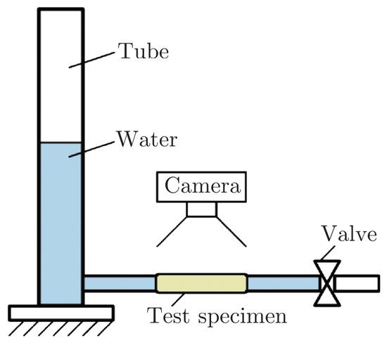
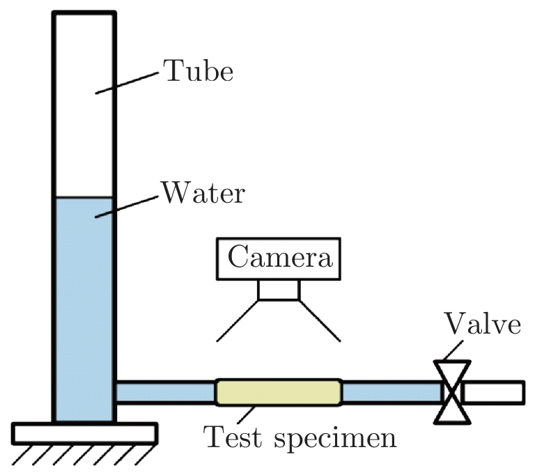
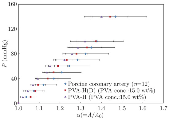
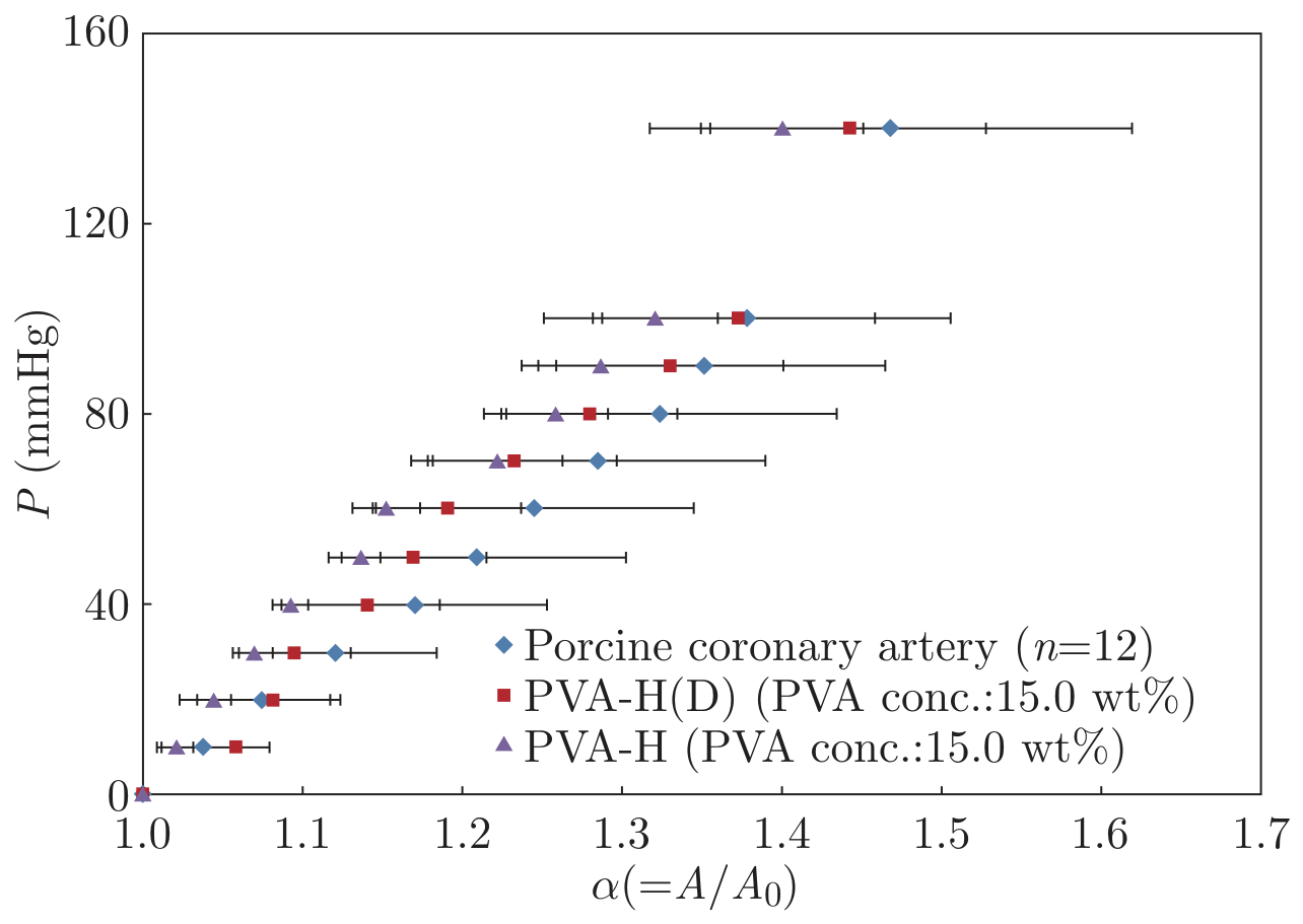
Cylinder shape of the blood vessel models were used (length: 20 mm, inner diameter di: 0.75 mm). To discuss the effect of the thickness of the blood vessel model, the outer diameter do was changed from 2.0 mm to 4.0 mm. Two types of hydrogel used for the blood vessel models (PVA-H and PVA-H (D)) were made; (1) PVA-H polyvinyl alcohol aqueous solution, (2) PVA-H (D), polyvinyl alcohol aqueous solution, a transparent hydrogel, and dimethyl sulfioxide (DMSO); all of which were used for gelation. Figure 1 shows the device of pressurization to the blood vessel model and porcine coronary artery. Figure 2 shows the relationships between pressure and cross sectional area of blood vessel model and porcine coronary artery. Since the mechanical properties of the blood vessel model and artery are non-linear, we used the stiffness parameter β. This parameter is adequate for arteries [7].
where P0 is internal (luminal) diastlic pressure, PS is internal (luminal) systolic pressure, D0 is the outside diameter for P0, and DS is the outside diameter for PS. We have changed the concentration of PVA from 10 wt% to 20 wt%. As the result of mechanical properties of the blood vessel models, stiffness parameter and Young’s modulus of the PVA-H and PVA-H (D) are shown in Table 1. The stiffness parameters of the blood vessel models are closer to that of porcine coronary artery. But the stiffness parameter of human coronary artery is greater than that of porcine coronary artery.
β = ln(P0/PS)/(D0/DS − 1)
Figure 1.
Device for measurement of cross sectional area A with change in pressure P.
Figure 2.
Relationship between pressure and cross sectional area of blood vessel model and porcine coronary artery. A0: cross sectional area when P=0 mmHg.
Table 1.
Stiffness parameter and Young’s modulus.
2.2. Method of Torque Transmission Measurement
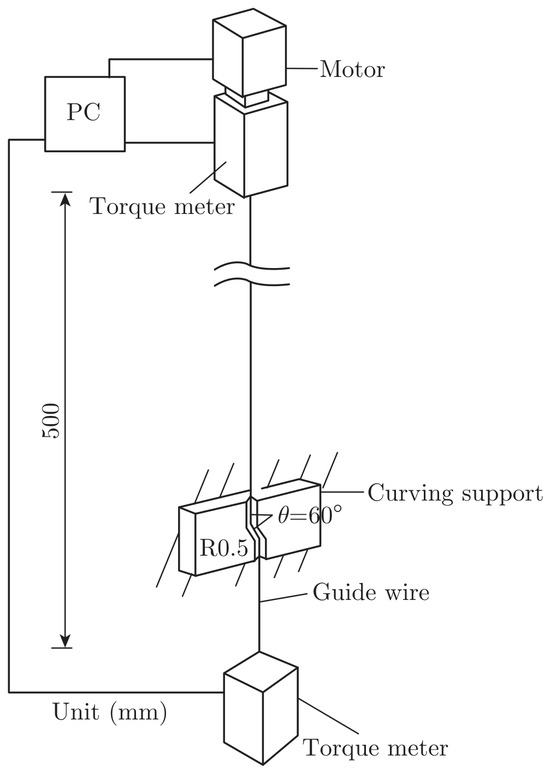
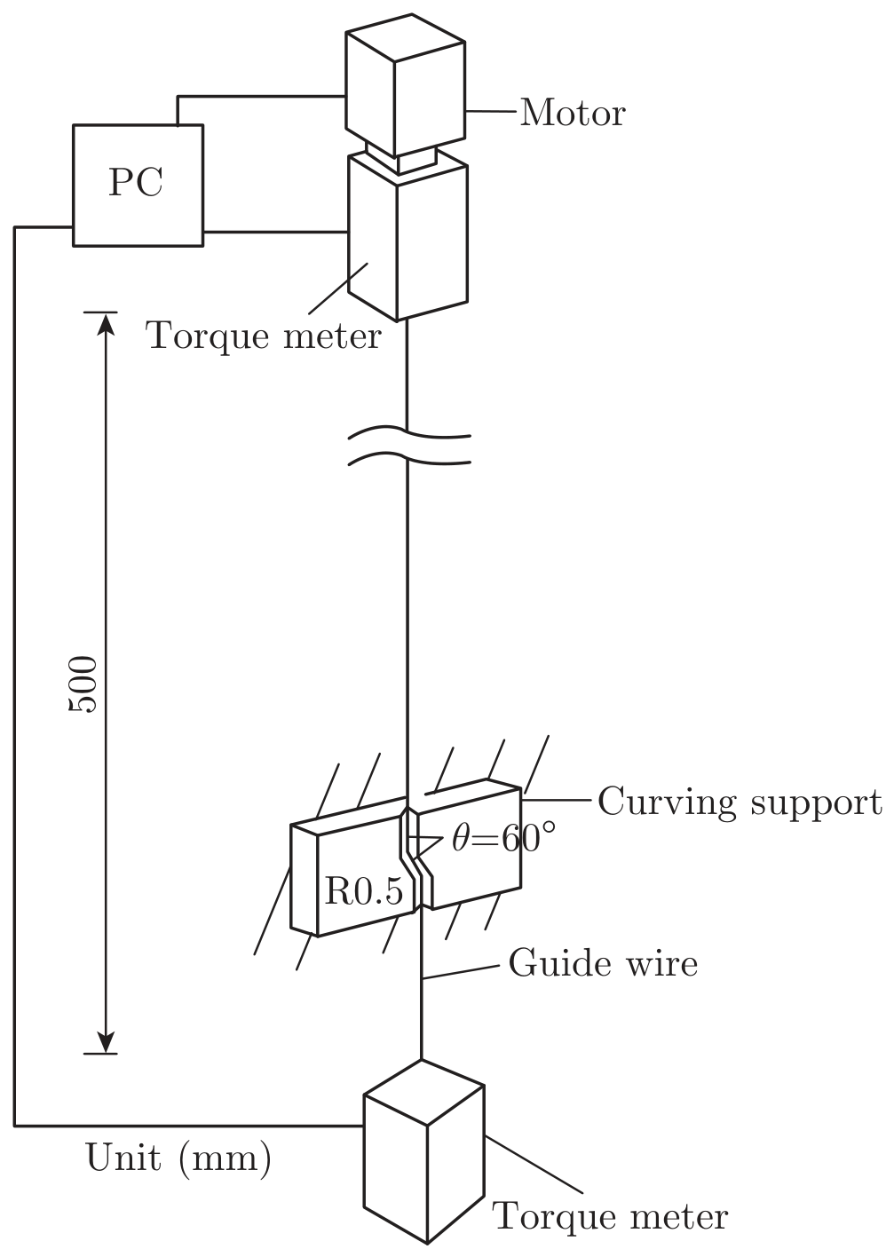
The device used for the measurement of torque transmission is shown in Figure 3. We used a guidewire for PTCA (Outer diameter of distal side=0.36 mm). Proximal and distal tips of guide-wire were connected to the shafts of torque transducers. The shaft in the torque transducer of proximal side was rotated by a stepping motor. The shaft of torque transducer of proximal side was fixed. Distal side of the guide-wire was inserted to the blood vessel model and bent by the curving support. The bending angle of the blood vessel model θ was set as 60 degrees. The proximal side of guide-wire was rotated one revolution in the measurement. The rotational speed of proximal side ω was changed from 0.05 to 0.25 rps (Rotation period T was changed from 20 to 4 s). The torque was measured from the beginning of rotation t=0 to two rotation period t=2T (rotation of proximal side was stopped from t = T to t=2T). Time was normalized by t/T.
Figure 3.
Device for the measurement of torque transmission.
3.3. Method of Rotational Response Measurement
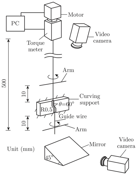
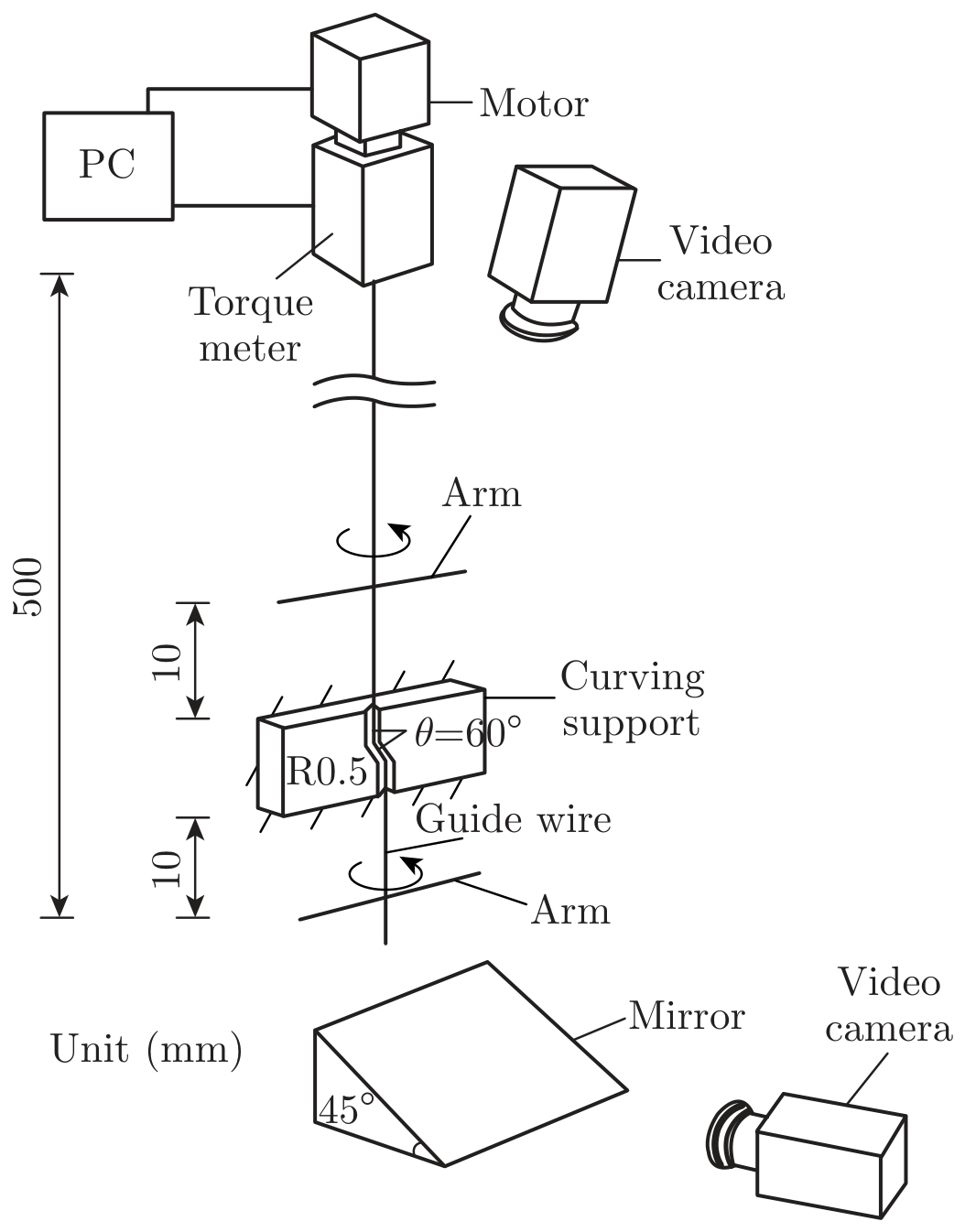
The device for the measurement of rotational response is shown in Figure 4. A torque transducer for the distal side was not used. Two arms made of polyvinyl chloride (length: 23 mm, diameter 0.4 mm) were attached to the guide-wire at 10 mm upper and lower to the curving support. Rotations of the guide-wire of proximal and distal sides were measured as the rotation of the two arms. Movies of rotating arms were recorded by high speed video cameras in synchronization with the rotation of the motor on the proximal side. A mirror was used for recording the movie of the rotation of distal side arm. We used image measurement software to digitize the rotation angle from the movie of the rotation of arms. Torque of proximal side was measured by torque transducer for proximal side.
Figure 4.
Device for the measurement of rotational rotational response.
3. Results
3.1. Torque Transmission
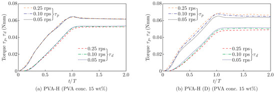
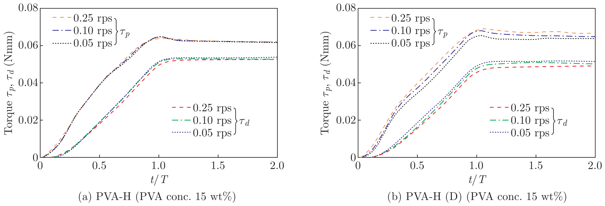
Distal torque τd and proximal torque τp for PVA-H and PVA-H (D) are shown in Figure 5. τd is smaller than τp, and the increase of τd with time is delayed than that of τp. Therefore, loss of torque transmission exists in the blood vessel model. With the increase in rotation speed of proximal side, τd is decreased while τp is increased.
Figure 5.
Distal torque τd and proximal torque τp (do=4 mm).
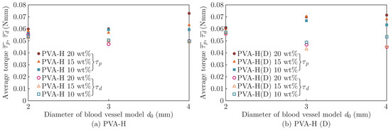
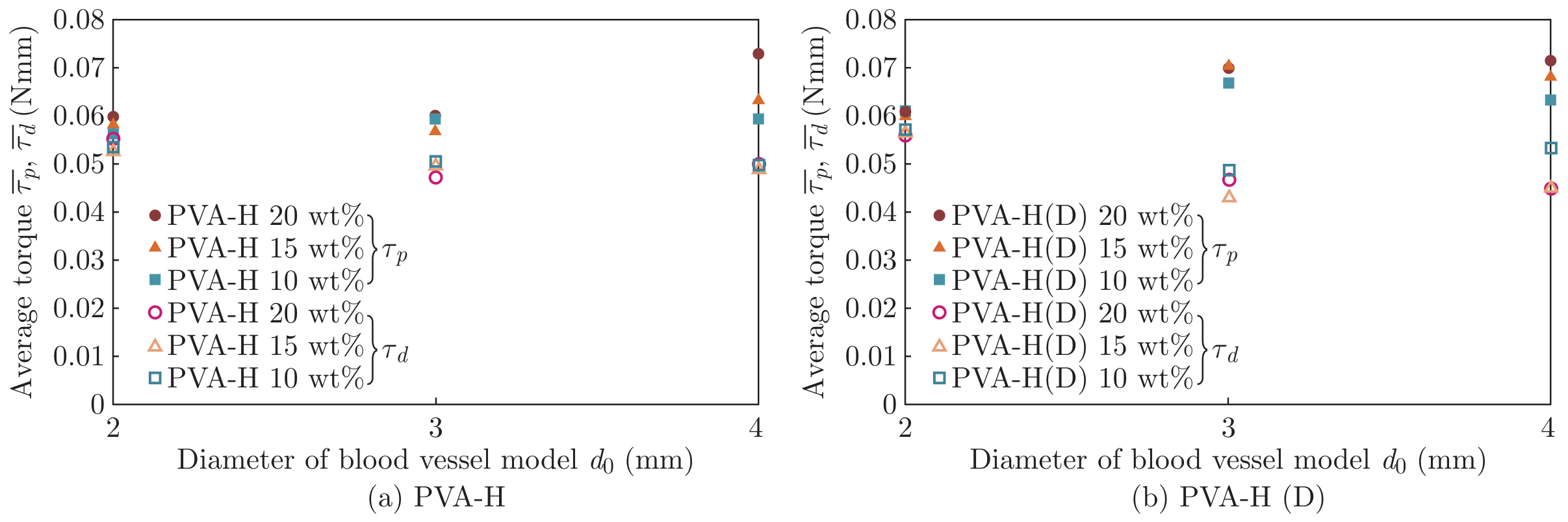
Figure 6 shows the average distal torque and average proximal torque during t/T=0.0 to 2.0 (Rotational speed of proximal side ω=0.25 rps). The following results for the torque loss (τloss = − ) were found: (1) τloss is the minimum when do=2 mm. (2) τloss for the PVA-H (D) iss greater than that for PVA-H. (3) τloss with change in PVA concentration is greater in greater thickness of blood vessel model.
Figure 6.
Average distal torque and average proximal torque during t/T=0.0 to 2.0. (Rotational speed of proximal side ω=0.25 rps).
3.2. Rotational Response
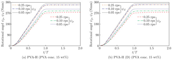
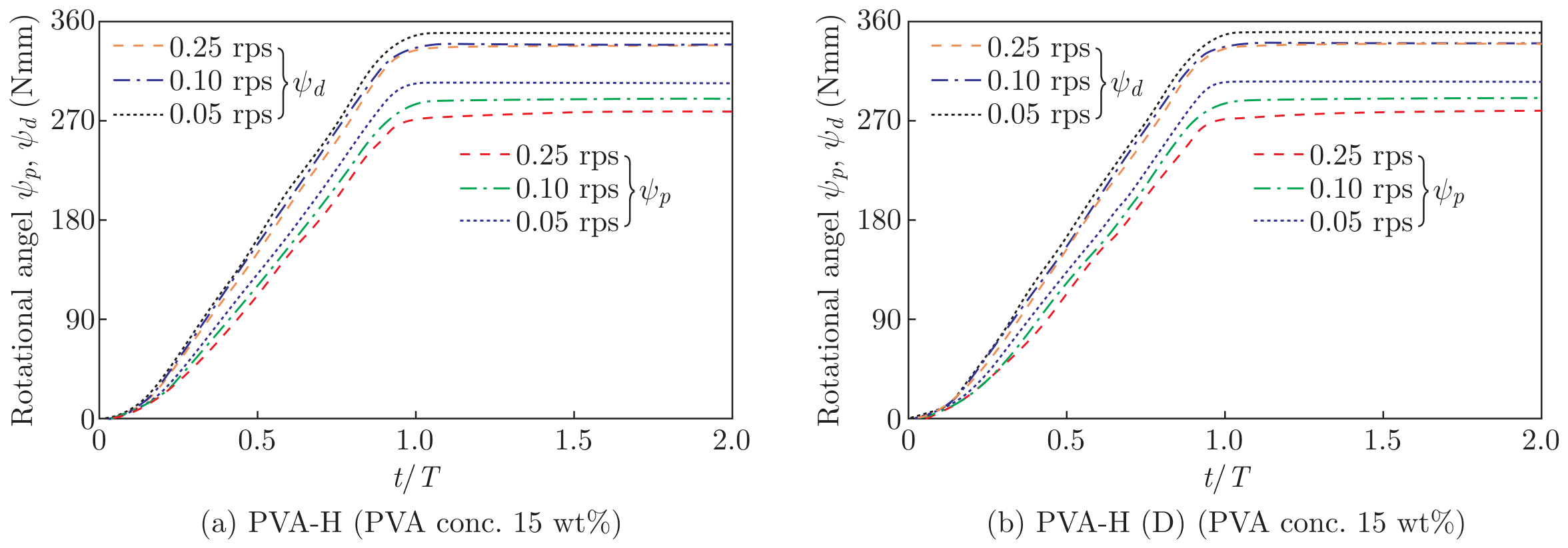
Distal rotational angle ψd and proximal rotational angle ψp for PVA-H and PVA-H (D) are shown in Figure 7. ψd is greater than ψp. ψd and ψp are increased with decreasing rotational speed of proximal side.
Figure 7.
Distal rotational angle ψd proximal rotational angle ψp (do=4 mm).
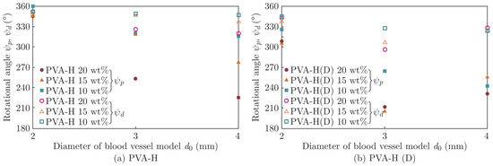
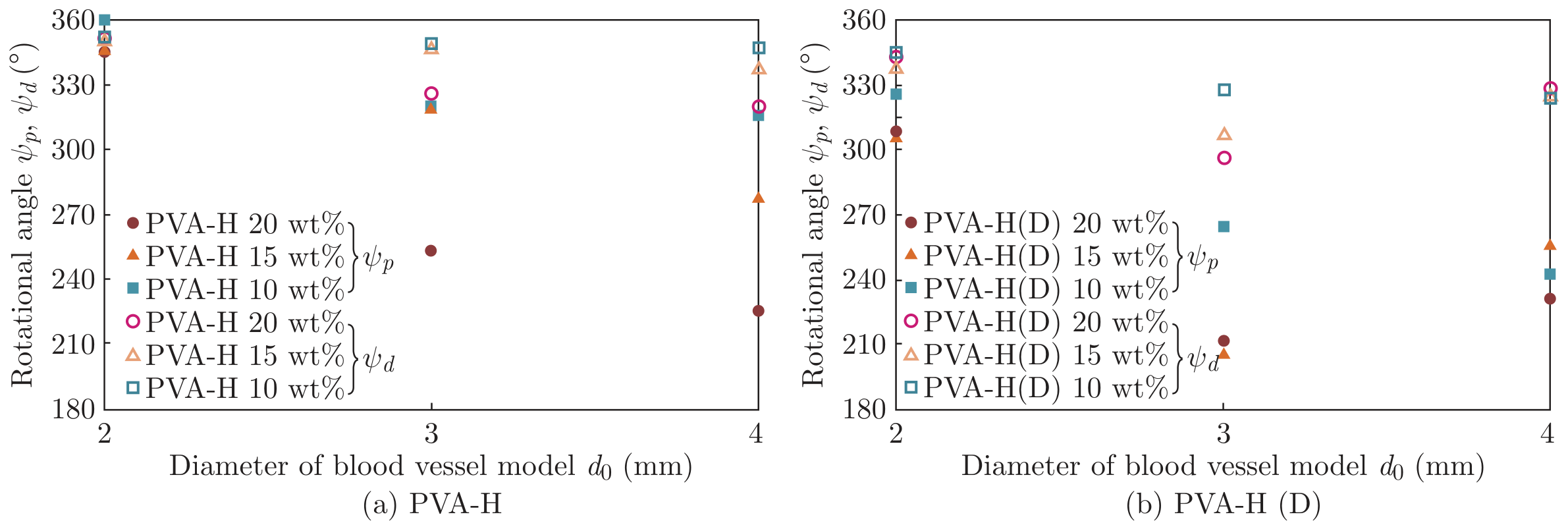
Figure 8 shows rotational angle ψp and ψd when t/T=2.0. The minimum value of ψp and ψd appeared when d0=4.0 mm for PVA-H (PVA conc. 10, 15, 2 0wt%) and PVA-H (D) (PVA conc. 10 wt%), and d0=3.0 mm for PVA-H (D) (PVA conc. 15 and 20 wt%).
Figure 8.
Rotational angle ψp and ψd when t/T=2.0. (Rotational speed of proximal side ω=0.25 rps).
4. Discussion
PVA-H and PVA-H(D) are elastmer material. Their frictional force depends on the contact force and area [8]. Contact portion between the blood vessel model and guide-wire is deformed locally during the experiment. When the blood vessel model is thicker, local deformation of the contact portion is greater, thus the frictional force is greater. The torque loss of PVA-H(D) is greater than that of PVA-H. This is explained bythe force to deform the contact portion of PVA-H(D) is greater, because the elasticity of PVA-H(D) is greater.
PVA-H and PVA-H(D) have viscoelastic properties. We have measured the dynamic viscoelastic properties of PVA-H and PVA-H(D) [9]. Their shear storage modulus increases with increasing frequency. During the torque transmission measurement, the guide-wire is twisted, and therefore it results to a shear deformation in the contact portion between the blood vessel model and guidewire. The greater rotational speed of guide-wire the greater the force of shear deformation. This is the reason why ψp was greater and ψd was lower at a higher proximal rotational speed.
There were no significant differences of ψp and ψd with change in the rotation speed for PVA-H. If we increase the rotation speed, the difference would be greater, but rotation speed over 0.5 rps could not be applied in the procedure of PTCA.
The rotational response corresponds to the tendency of the torque transmission; greater thickness, higher elasticity and higher rotational speed cause lower rotational response. The distal rotational angle is greater than the proximal angle. This is explained by the structure of the guide-wire; whatever outer spring coil stopped by the curved blood vessel model, the center core attached to the distal tip of guide-wire rotates. In this experiment, arm is attached to the distal tip.
We have measured the torque for porcine coronary artery [10]. The overall tendency was the same but the distal torque was smaller and the effect of rotational speed was greater.
5. Conclusion
We have measured torque and rotational angle of the guide-wire in curved blood vessel model, and discussed the effect of the PVA concentration of the hydrogel and thickness of blood vessel model on torque transmission and rotational response of the guide-wire. The following results were obtained:
- (1)
- Torque transmission and rotational response were reduced with increase in thickness, elasticity, and rotational speed.
- (2)
- PVA-H is greater than PVA-H(D) for the torque transmission and rotational response.
Problems to be solved in future are the following:
- (1)
- Fabrication of more realistic blood vessel models for stiffness parameter and surface condition.
- (2)
- The measurement in blocked blood vessel model of chronic total occlusion lesion.
References
- Schröder, J. The Mechanical Properties of Guidewires, Part I: Stiffness and Torsional Strength. Cardiovasc Interv. Radiol. 1993, 16, 43–46. [Google Scholar] [CrossRef] [Scilit] [PubMed]
- Schr¨oder, J. The Mechanical Properties of Guidewires, Part III: Sliding Friction. Cardiovasc Interv. Radiol. 1993, 16, 93–97. [Google Scholar]
- Zako, M.; et al. Study on Evaluation Method of Mechanical Properties of Medical Cathter. Proc. Conference of the Society of Material Science Japan, (in Japanese). 2000; pp. 213–214. [Google Scholar]
- Ceschinski, H.; et al. Torquability of Microcatheter Guidewires: The resulting Torsional Moment. Bio-Med. Mater. Eng. 2000, 10, 31–42. [Google Scholar]
- Nanto, S.; et al. New Coronary Model to Train Interventionalists and Test Devices of Angioplasty. Jpn.-Ger. Med. Rep. (in Japanese). 2000, 45(3), 92–96. [Google Scholar]
- Kobayashi, S.; et al. Collapse in High-grade Stenosis During Pulsatile Flow Experiments. JSME Int. J. Ser. C 2004, 47(4), 1010–1018. [Google Scholar] [CrossRef] [Scilit]
- Hayashi, K.; et al. Biomechanical Study of the Constitutive Laws of Vascular Walls. Exp. Mech. 1974, 14(11), 440–444. [Google Scholar] [CrossRef] [Scilit]
- Uchiyama, Y. Wear and Friction of Rubber. J. Soc. Rubber Ind. Jpn. (in Japanese). 1988, 61(5), 315–316. [Google Scholar] [CrossRef] [Scilit]
- Kubo, Y.; Kobayashi, S.; Morikawa, H. Torque Transmission Characteristics of Medical Guidewire in Viscoelastic Blood Vessel Model. Proc. 8th Nagano District Symposium, Division of Koushinetsu, (in Japanese). 2010; Japanese Society for Medical and Biological Engineering; pp. 1–2. [Google Scholar]
- Kubo, Y.; Kobayashi, S.; Morikawa, H. Torque Transmission Characteristics of Medical Guidewire in Porcine Coronary Artery and Blood Vessel Model. Proc. 30th Koshinetsu branch Conference, Japanese Society for Medical and Biological Engineering, (in Japanese). 2010; pp. 35–36. [Google Scholar]

© 2012 Textile Bioengineering and Informatics Society. This article is an open access article distributed under the terms and conditions of the Creative Commons Attribution (CC BY) license (https://creativecommons.org/licenses/by/4.0/).