


Abstract
The degradation behavior of silk fibroin biomaterials in human body is definitely vital for the growth of tissues. Therefore, an investigation to regulate the degradation behaviors of silk fibroin films by changing the degree of cross-linking is presented in this paper. The in-vitro experiments in the simulated human body environment showed that the degradation rate of cross-linked silk fibroin films was inversely proportional to the degree of cross-linking. After degradation, the ratio of crystalline part in the SF films increased. This approach would provide a new direction in controlling the degradation time by cross-linking the silk fibroin for specific tissue engineering application.
1. Introduction
Silk Fibroin (SF) is a natural protein produced by the domestic silkworm, Bombyx mori. The amino acid composition of silk fibroin from Bombyx mori consists primarily of glycine, alanine and serine [1]. The three simple amino acids form the crystalline regions of silk fibroin, while the amino acids with bulky and polar side chains form the amorphous regions [2]. The silk polymorphs include silk I, silk II and an air/water assembled interfacial silk III [1,3].
Due to the unique chemical and mechanical properties and biocompatibility, Bombyx mori silk fibroin materials have been invested as biomaterials for years. An ideal biomaterial is one that is non-immunogenic, has non-toxicity, and biocompatible, which can be functionalized with bioactive proteins and chemicals [4]. Another key factor is that the material should be biodegradable, and the degradation products should be non-toxic, easily metabolized and cleared from the body [5].
The silk has been used for manufacturing surgical sutures for one century [6]. In recent years, regenerated silk fibroin have been used extensively in biomedical applications, such as burnwound dressings [7], drug delivery matrices [8], vascular prostheses and structural implants [9,10], ligaments [11], bone [12,13], cartilage [13], nets [14], and so on. These results show that silk fibroin is one of the most precious raw materials for being used as biomaterials.
Recently, based on the importance of degradation for biomaterial applications in tissue engineering, the degradation of SF materials has been well studied [2,6,15,16,17,19]. Silk is defined by United States Pharmacopeia as non-degradable for its negligible tensile strength loss in vivo [4]. However, according to the literature, as a protein, silk can be gradually digested by proteolytic enzymes and absorbed in vivo over a long time [16]. The rate of degradation depends on the properties of the material itself, as well as the chemical and biochemical environment of the site of implantation [17]. These results highlight that silk fibroin biomaterials are biodegradable, and the degradation behaviors could be regulated. However, the relationship between structure, processing, and degradability is not clear.
The goal of the present study is to make a preliminary research of the relationship between the degree of cross-linking of the SF films and the degradation behavior in vitro, controlling over the rate of degradation. Chemical, physical, and morphological features of the biodegraded SF films were examined by means of XRD, SEM.
2. Materials and Methods
2.1. Preparation of Silk Fibroin Films
The Bombyx mori silk samples were treated three times with Na2CO3 solution respectively to remove sericin. Then they were rinsed and air dried. The pure silk fibroin fibers were dissolved with triad solvent CaCl2/CH3CH2OH/H2O (mole ratio = 1:2:8) through stirring. The prepared solution was purified by dialyzed against distilled water for 4 days, to obtain silk fibroin solution. The genipin (GP) powder was added to the silk fibroin solution with constant stirring for 20 minutes, according to the percentages (0%, 5%, 10%, 20%, 30%) of solute mass. The mixed solution was incubated at 37 °C for 12 hrs. Then 40 ml solution was cast on a polyethylene plate at 60 °C for 2 hrs.
2.2. Evaluation of Cross-linking Index
The degree of cross-linking was measured using ninhydrin assay [18]. Ninhydrin (2, 2-dihydroxy-1, 3-indanedione) is widely used as a chemical reagent for the colorimetric determination of amino acids. It reacts with free α-amino groups to produce an aldehyde, carbon dioxide, and reduced ninhydrin through reaction. Ninhydrin assay was used to determine the amount of free amino groups of each test sample. The silk fibroin films with a constant mass (0.05 g) were incubated in 1.5 ml distilled water for 1 hour, and then 450 µL 0.1% ninhydrin solution was added to the sample. The test sample with a ninhydrin solution was heated for 20 min at 100 °C. Then the optical absorbance at 570 nm (the wavelength of the blue-purple color) of the solution was recorded. Use 200 µg/ml glycine solutions as standard. The cross-linking index was defined as (1).
Cross-linking index (%) = [(free amino groups before cross-linking) − (free amino groups after cross-linking)]/[free amino groups before cross-linking]
2.3. In Vitro Degradation System
Enzymatic degradation was performed as previously described [19]. Briefly, the silk fibroin films with a constant mass (0.05 g) were incubated in 2.5 ml solution of 1.0 U/ml collagenase IA (Sigma USA) at 37 °C. The silk fibroin films in Phosphate Buffer Saline (PBS) at pH 7.4 were used as controlled group, without enzyme. At 1, 3, 6, 12, 18, 24 and 30 days, 3-6 films were collected from each sample, and then the solution containing soluble peptides was replaced with newly prepared enzyme solution.
2.4. X-ray Diffraction
X-ray diffraction was performed by a Rigaku D/Max-3C diffractometer with Cu-Kα radiation (λ = 0.15418 nm). The voltage of the X-ray source was 40 kV at a current of 40 mA. The diffraction intensity curves were obtained at a scanning rate of 2°/min and within the scanning region of 2θ = 5–40°.
2.5. Scanning Electron Microscopy
The morphologic changes in the silk fibroin films after degradation were examined by using scanning electron microscopy (HITACHI.S-570, Hitachi, Japan).
3. Results and Discussion
3.1. Evaluation of Cross-linking Index
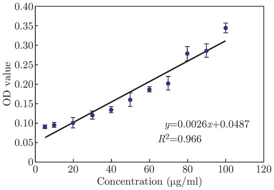
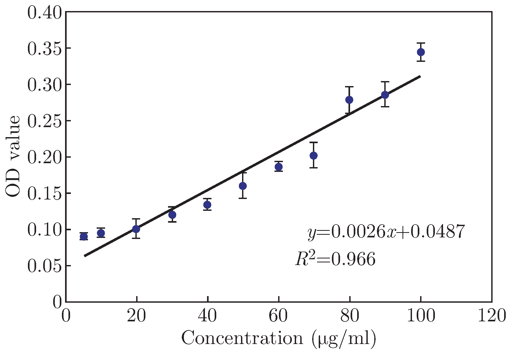
Figure 1 shows the standard curve of glycine solution. Table 1 shows the 5% genipin was sufficient to crosslink about 69.16% of the amino groups. The extent of cross-linking increased with the concentration of genipin up to a maximum value of about 91.38%. It did not exhibit significant variations at genipin concentration higher than 20%. The number of free amino groups in SF solution was constant, when the concentration of genipin was lower, and the extent of cross-linking increased with the increasing of genipin concentration. But, as the GP concentration was high enough, all the free amino groups were nearly reacted, and the extent of cross-linking could not obviously be changed.
Figure 1.
Standard curve of glycine solution.
Table 1.
The degree of cross-linking of silk fibroin films.
3.2. Morphologic Changes in SF Films During Enzymatic Degradation
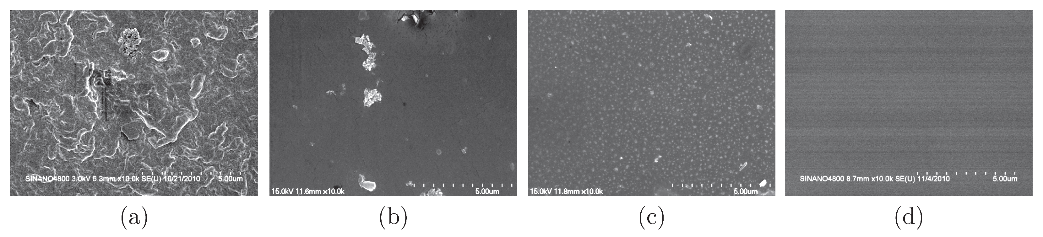
Morphological changes induced by enzymatic treatment were studied by Scanning Electron Microscopy (SEM). The surface of silk films before treatment was smooth (Figure 2A(a), images not all shown), with no differences among the films. After degradation for 30 days, and the morphology of silk films was drastically changed. When SF films incubated with collagenase IA for 6 days, the 5% GP/SF SF films demonstrated a higher degree of surface roughness and there appeared some cracks in the 10% GP/SF films. But the surface of 20% and 30% GP/SF films did not change. After 30 days, the 5% GP/SF Films showed the presence of extensive degradation, which appeared in the form of surface stripping. In the surface of 10% GP/SF SF films, there appeared holes and cracks. 20% and 30% GP/SF SF films were attacked at a low extent, which showed a few cracks. This phenomenon could be explained by XRD (described later). The results indicated that the extent of degradation depended on the percentage of GP added into silk fibroin films. With the increasing of the cross-linking degree of silk fibroin films, the degradation of silk fibroin films decreased. Figure 2C shows the morphology of cross-linked SF films immersed in PBS. The 5% GP/SF films exhibited a rough surface, indicating that some soluble components released in PBS, while other SF films only showed a small amount of changes.
Figure 2A.
SEM of SF films incubated with collagenase IA for 6 days, (a) 20% GP/SF before degradation; (b) 5% GP/SF; (c) 10% GP/SF; (d) 20% GP/SF; (e) 30% GP/SF.

Figure 2B.
SEM of SF films incubated with collagenase IA for 30 days, (a) 5% GP/SF; (b) 10% GP/SF; (c) 20% GP/SF; (d) 30% GP/SF.
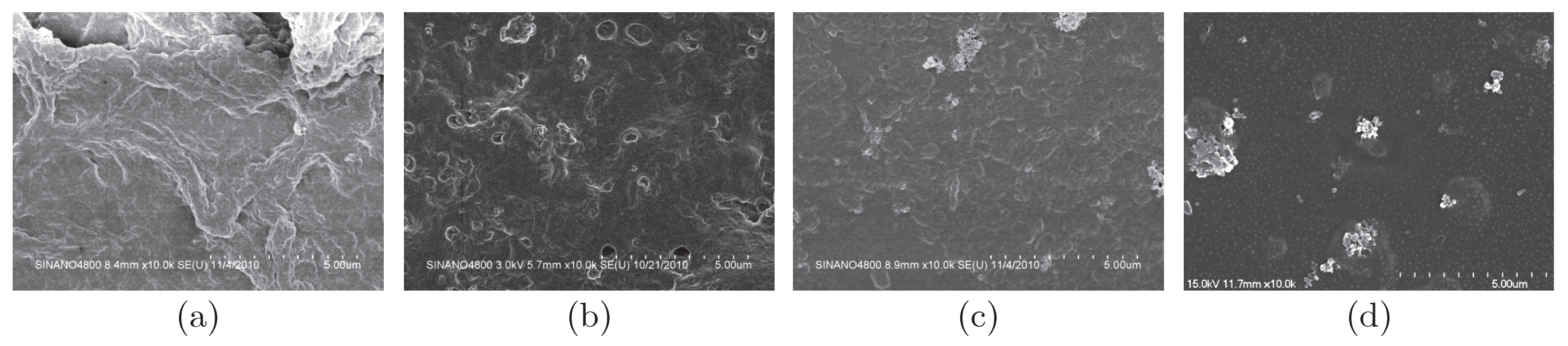
Figure 2C.
SEM of SF films immersed in PBS for 30 days, (a) 5% GP/SF; (b) 10% GP/SF; (c) 20% GP/SF; (d) 30% GP/SF.
The results demonstrate the GP cross-linked SF films could be biodegradable. The significant differences of enzymatic degradation among the cross-linked SF films reflected that the degradation behavior could be regulated by changing the degree of cross-linking.
3.3. Structural Analysis
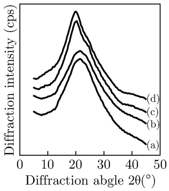
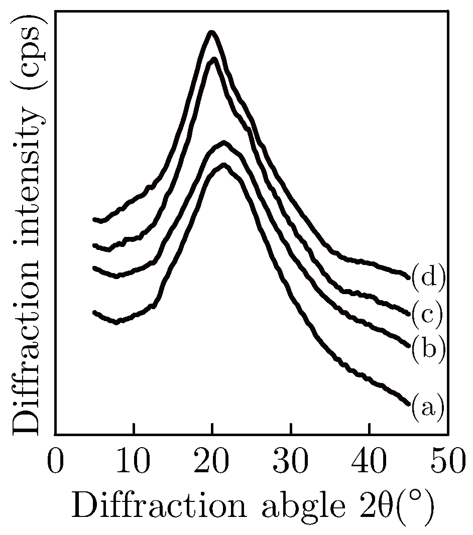
The principal diffraction peaks of the Silk I crystal structure are 7.25 Å (12.2°, ms), 4.50 Å (19.7°, s), 3.60 Å (24.7°, m), 3.16 Å (28.2°, m); the diffraction peaks of Silk II are 9.7 Å (9.1°, ms), 4.69 Å (18.9°, ms), 4.3 Å (20.7°, vs) [19]. As shown in Figure 3, the structure of the 5% and 10% GP/SF SF films primarily showed amorphous. There were some diffraction peaks around 19.7° (s) and 39.7° (w) in the XRD curves of 20% and 30% GP/SF. According to the XRD principal diffraction peaks, it revealed that as the GP concentration increased, the ratio of Silk II crystal structure increased.
Figure 3.
X-ray diffraction curves of cross-linked silk fibroin films before degradation. (a) 5% GP/SF; (b) 10% GP/SF; (c) 20% GP/SF; (d) 30% GP/SF.
The enzyme can penetrate into the amorphous regions more easily, cleave the sensitive peptide bonds and release free soluble peptides or free amino acids [2]. Thus the cross-linked SF films consisted of more random structures could be degraded quickly. These results closely corresponded to the surface changes of the SF films.
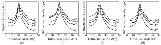
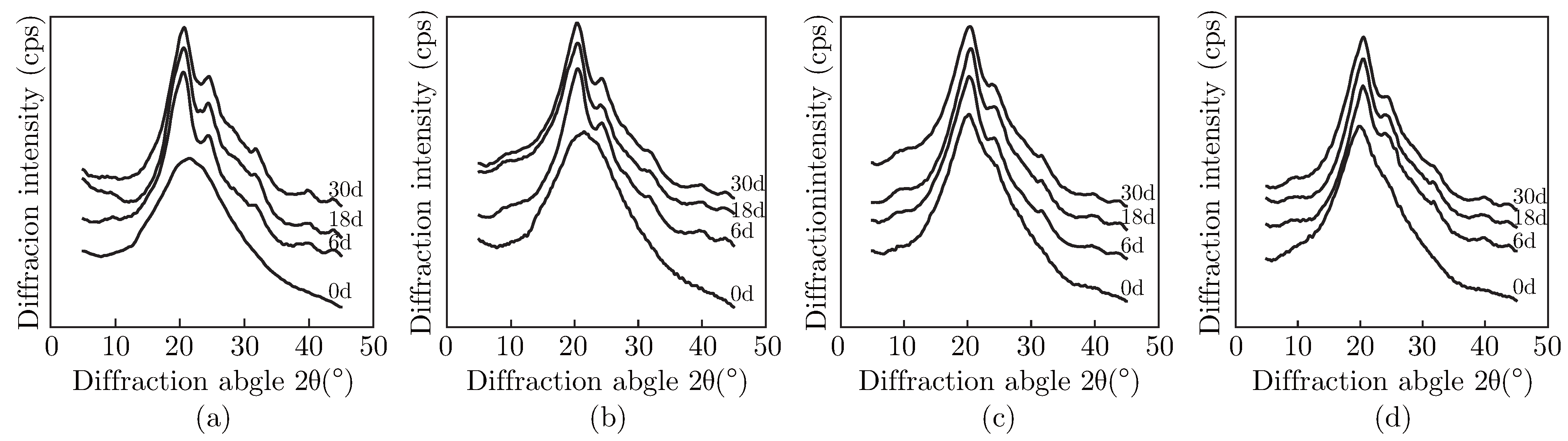
The diffraction peaks in the XRD curves of the residual SF films degraded for different days showed significant differences with the SF films before degradation (Figure 4). After treatment with collagenase IA, there appeared typical diffraction peaks around 20.7°, 24.3° and 39.7° in the XRD curves of all the cross-linked SF films, indicating that the condensed structure of the SF films after degradation mainly consisted of Silk II structure.
Figure 4.
X-ray diffraction curves of cross-linked silk fibroin films after degradation. (a) 5% GP/SF; (b) 10% GP/SF; (c) 20% GP/SF; (d) 30% GP/SF.
After degradation, the changes of diffraction peaks revealed that the ratio of crystalline part in the SF films increased upon biodegradation. The XRD curves indicated that all the crosslinked SF films could be degraded by collagenase IA. The degree of crystallinity of biodegraded films increased to an extent directly related to the degradation, that is, the more the films were degraded, the higher the degree of crystallinity. The results confirmed that the enzyme attack preferentially occurred in amorphous regions.
4. Conclusion
The results here reported allow to conclude that GP cross-linked SF films were biodegradable, but the extent of degradation depended on the degree of cross-linking of silk fibroin films. The condensed structure of cross-linked SF films primarily were amorphous, as the genipin concentrations varied from 5 to 30%, the content of Silk I and Silk II structure increased, at the same time, the degradation of cross-linked SF films decreased. The in vitro approach used in the present study may represent a useful tool for regulating the degradation rate of silk fibroin biomaterials.
Acknowledgments
This work was supported by the National Natural Science Foundation of China (30970714), Nature Science Foundation of Jiangsu Province (BK2010252), and Priority Academic Program Development of Jiangsu Higher Education Institutions.
References
- Vepari, C.; Kaplan, D.L. Silk as a biomaterial. Prog Polym Sci 2007, 32, 991–1007. [Google Scholar] [CrossRef] [Scilit] [PubMed]
- Arai, T.; Freddi, G.; et al. Biodegradation of Bombyx mori Silk Fibroin Fibers and Film. J Appl Polym Sci 2004, 91, 2383–2390. [Google Scholar] [CrossRef] [Scilit]
- Regina, V.; Samuel, P.G.; Wayne, M.; et al. Orientation of silk III at the air-water interface. Bio Macro 1999, 24, 237–245. [Google Scholar] [CrossRef] [Scilit] [PubMed]
- Cao, Y.; Wang, B. Biodegradation of Silk Biomaterials. Int. J. Mol. Sci. 2009, 10, 1514–1524. [Google Scholar] [CrossRef] [Scilit] [PubMed]
- Lloyd, A.W. Interfacial bioengineering to enhance surface biocompatibility. Med Device Technol 2002, 13, 18–21. [Google Scholar] [PubMed]
- Wang, Y.Z.; Darya, D.R.; et al. In vivo degradation of three-dimensional silk fibroin scaffolds. Biomaterials 2008, 29, 3415–3428. [Google Scholar] [CrossRef] [Scilit] [PubMed]
- Santin, M.; Motta, A.; Freddi, G.; Cannas, M. In vitro evaluation of the inflammatory potential of the silk fibroin. J Biomed Mater Res 1999, 46, 382–389. [Google Scholar] [CrossRef] [Scilit]
- Murphy, A.R.; Kaplan, D.L. Biomedical applications of chemically-modified silk fibroin. J Mater Chem 2009, 19, 6443–6450. [Google Scholar] [CrossRef] [Scilit] [PubMed]
- Dalpra, I.; Freddi, G.; et al. De novo engineering of reticular connective tissue in vivo by silk fibroin nonwoven materials. Biomaterials 2005, 26, 1987–1999. [Google Scholar] [CrossRef] [Scilit] [PubMed]
- Meinel, L.; Fajardo, R.; Kaplan, D.L.; et al. Silk implants for the healing of critical size bone defects. Bone 2005, 37, 688–698. [Google Scholar] [CrossRef] [Scilit] [PubMed]
- Altman, G.H.; Horan, R.L.; Lu, H.H.; et al. Silk matrix for tissue engineered antior ruciate ligaments. Biomaterials 2002, 23, 4131–4141. [Google Scholar] [CrossRef] [Scilit] [PubMed]
- Kim, H.J.; Kim, U.J.; et al. Bone regeneration on macroporous aqueous-derived silk 3-D scaffolds. Macromol Biosci 2007, 7, 634–655. [Google Scholar] [CrossRef] [Scilit] [PubMed]
- Hutmacher, D.W. Scaffolds in tissue engineering bone and cartilage. Biomaterials 2000, 21, 2529–2543. [Google Scholar] [CrossRef] [Scilit] [PubMed]
- Unger, R.E.; Peters, K.; Wolf, M.; et al. Endothelialization of a non-woven silk fibroin net for use in tissue engineering: growth and gene regulation of human endothelial cells. Biomaterials 2004, 25, 5137–5146. [Google Scholar] [CrossRef] [Scilit] [PubMed]
- Yang, Y.M.; Zhao, Y.H.; et al. Degradation behaviors of nerve guidance conduits made up of silk fibroin in vitro and in vivo. Polym Degrad and Stab 2009, 94, 2213–2220. [Google Scholar] [CrossRef] [Scilit]
- Kojthung, A.; Meesilpa, P.; et al. Effects of gamma radiation on biodegradation of Bombyx mori silk fibroin. Int Biodeterior Biodegrad 2008, 62, 487–490. [Google Scholar] [CrossRef] [Scilit]
- Taddei, P.; Arai, T.; et al. In Vitro Study of the Proteolytic Degradation of Antheraea pernyi Silk Fibroin. Biomacromolecules 2006, 7, 259–267. [Google Scholar] [CrossRef] [Scilit] [PubMed]
- Yao, C.H.; Liu, B.S.; et al. Preparation of networks of gelatin and genipin as degradable biomaterials. Mater Chem and Phy 2004, 83, 204–208. [Google Scholar] [CrossRef] [Scilit]
- Li, M.Z.; Masayo, O.; et al. Enzymatic degradation behavior of porous silk fibroin sheets. Biomaterials 2003, 24, 357–365. [Google Scholar] [CrossRef] [Scilit] [PubMed]

© 2011 Textile Bioengineering and Informatics Society. This article is an open access article distributed under the terms and conditions of the Creative Commons Attribution (CC BY) license (https://creativecommons.org/licenses/by/4.0/).