


Abstract
Ornithoctonus huwenna spiders can be bred in mass production, and potential applications of the spider silk in medical materials should be paid attentions. Regenerated nano-scale spider silk nonwovens were prepared by electrospinning from tanglesome Ornithoctonus huwenna spider silk. The morphologies of the electrospun spider silk fibers were investigated, and the cell compatibilities were explored. The results showed that the electric field strength was an important factor for the diameter, degree of crystal and molecular conformation of electrospun Ornithoctonus huwenna spider silk. With the increase of electric field strength, fibers became fine and even, and the crystallinity and β-sheet structure were improved. The electrospun nanofiber nonwoven had good compatibility with rat bone marrow stromal cells (rBMSCs). The cell survival rates were above 98% on the fiber surfaces.
1. Introduction
Orb-web spiders secret unique silk fibers named as dragline silks from the major amplullate glands, which are used as radial threads that support a spider’s web and dragline to make spiders rapid vertical descents [1]. Spider dragline silk is a flexible, lightweight fiber with extraordinary strength and toughness [2], so that researchers have being focused on the fiber since last decade, especially recent 10~15 years. The dragline silk, a protein fiber, has important applications in impact-proof textiles or other structural fabrics. Moreover the inherent biocompatibility makes it to be used in the fields of controlled release biomaterials and scaffolds for tissue engineering. Therefore spider silk has been studied extensively as a kind of proteinbased biomaterial and achieved great fruits [3,4,5,6,7,8]. Biomimetic production of dragline silks has had great progress [9,10,11,12,13,14]. However, if we want to make commercial use of the biosynthetic fibers, firstly, we must develop a method of spidroin solution mass production, then, an appropriate spinning technology is necessary to make spidroin solution into fine and even fibers. Electrospinning is a simple spinning method widely used in fabrication of various nanoscale polymer fibers, such as PVA, PLA, and PAN etc [15,16,17,18]. Electrospun SF fibers have been obtained as well for the following: vascular grafts, wound dressings or tissue engineering scaffolds due to the very high surface area to volume ratio and its biocompatibility and biodegradability [19,20,21]. However, except that Zarkoob et al first reported the electrospinning of Nephila clavipes dragline silks, there are few of research on electrospinning spider silk [22]. The author Pan has developed an electrospinning method to obtain nano-scale spider silk fiber nonwoven from dragline and egg-case silk of Araneus ventricosus spiders [23].
Ornithoctonus huwenna, also named as cutworm, is one of the bigger spiders with the length and weight 6~9 cm, 14~16g respectively. Some special farms breed Ornithoctonus huwenna spiders for extracting toxin as new medicine materials [24,25,26]. At the same time, a lot of spider silk can be obtained. In our research, Tanglesome Ornithoctonus huwenna spider silk was regenerated, and nano-scale fiber nonwovens were prepared by electrospinning. SEM, FTIR and WAXD were used to investigate the morphologies of the regenerated spider silk fibers. Meanwhile, the cell compatibility of the nanofiber nonwoven was explored.
2. Experimental
2.1. Materials
Ornithoctonus huwenna spider silk was kindly provided by North spider breeding institute, Nanning city, Guangxi province. 99% Hexaflorisopropanol (HFIP) was purchased from Dupont Chemical Solution Enterprise.
Ornithoctonus huwenna spider silk was dissolved in 99% HFIP for 10h at room temperature. Regenerated nano-scale spider silk nonwovens were prepared from the 0.9wt% spidroin solution by electrospinning.
2.2. Measurements
2.2.1. Longitudinal Shape and Diameter
The electrospun nano-scale fiber nonwoven was fixed on a sample holder by double side adhesive tape and coated with gold. Then, the longitudinal images of fibers were observed and recorded by Hitach S-4700 SEM. The fiber diameters were calculated with HJ2000 software. 100 fibers were processed for every sample and average diameter was obtained.
2.2.2. Molecular Conformation
About 2 mg powdered sample was mixed with 200mg spectrum degree KBr, and pressed in slice on standard procedure. Then the FT-IR spectrum was measured with NICOLE 5700 FT-IR instrument (USA). Every sample was scanned 32 times with wave numbers range 4000-500cm-1 and spectrum resolution 4cm-1.
2.2.3. Crystalline Structure
X′Pert Pro MPD X-ray diffraction system (CuKα target, acceleration voltage 40 KV, electric current 40mA, λ= 0.154 nm) of Holand PAN alytical company was used to measure X-ray diffraction intensity curves of powdered samples, and diffraction angle 2θ ranged from 5° to 45°.
The resulting plots of X-ray diffraction intensity versus 2θ were analyzed using the profile fitting program Peak Fit 4.1(AISN Software Inc.). Each peak was modeled using a Gaussian-Lorentzian peak shape. Areas of the peaks obtained from the analysis were used to estimate the degree of crystallinity, i.e. the ratio of the areas of the crystalline reflections to that of total area of the scattering curve (amorphous+crystalline). In general, agreement between model predictions and the data were exceptional, yielding coefficients of determination, i.e.R2 above 0.98.
2.2.4. Cell Compatibility
rBMSCs were inoculated on the surface of regenerated spider silk nonwoven electrospun at voltage 12KV and distance from capillary tip to collecting screen 8cm from 0.9wt% Ornithoctonus huwenna spider silk / HFIP solution . After being inoculated for 24 h, 48 h and 72 h, the sample was leached two times by L-DMEM without blood serum, then, incubated in 1 mM Calcein and 1 mM ethidium homodimer at 37°C for 30 min. Cell survival rate was obtained by using an Olympus inverted fluorescence microscope.
3. Results and Discussion
3.1. Effects of Electrospinning Parameters on the Fiber Microstructures
3.1.1. Longitudinal Shape and Diameter
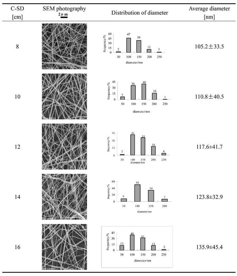
Table 1 shows the SEM images, distribution of diameter and average sizes of electrospun regenerated Ornithoctonus huwenna spider silk (EROHS) fibers at different distance from capillary tip to collecting screen (C-SD) at Voltage 12kv. Ornithoctonus huwenna spidroin could be electrospun into smooth and continuous nano-scale fibers. Nanometer scale diameter of the protein fibers would be a useful feature in some biomaterial and tissue engineering applications to enhance surface area while maintaining high porosity.
Table 1.
Longitudinal shapes and sizes of EROHS fibers at different C-SD (Voltage 12 kv).
Their diameters mainly distributed 50~150nm, and average diameters were 105.2±33.5nm, 110.8±40.5nm, 117.6±41.7nm, 123.8±32.9nm and 135.9±45.4nm respectively when C-SD were 8, 10, 12, 14 and 16cm.
With the increase of C-SD, the electric field strength decreases, thus, the electric force loaded on the spinning jet weakens and fiber diameters augment. On the other hand, large distance makes the spinning jet has large space and enough time to split, thus fine fibers can be obtained. The experimental results revealed that the former influence was dominant for 0.9wt% regenerated Ornithoctonus huwenna spidroin / HFIP solution. The fiber diameter was positive correlation with C-SD.
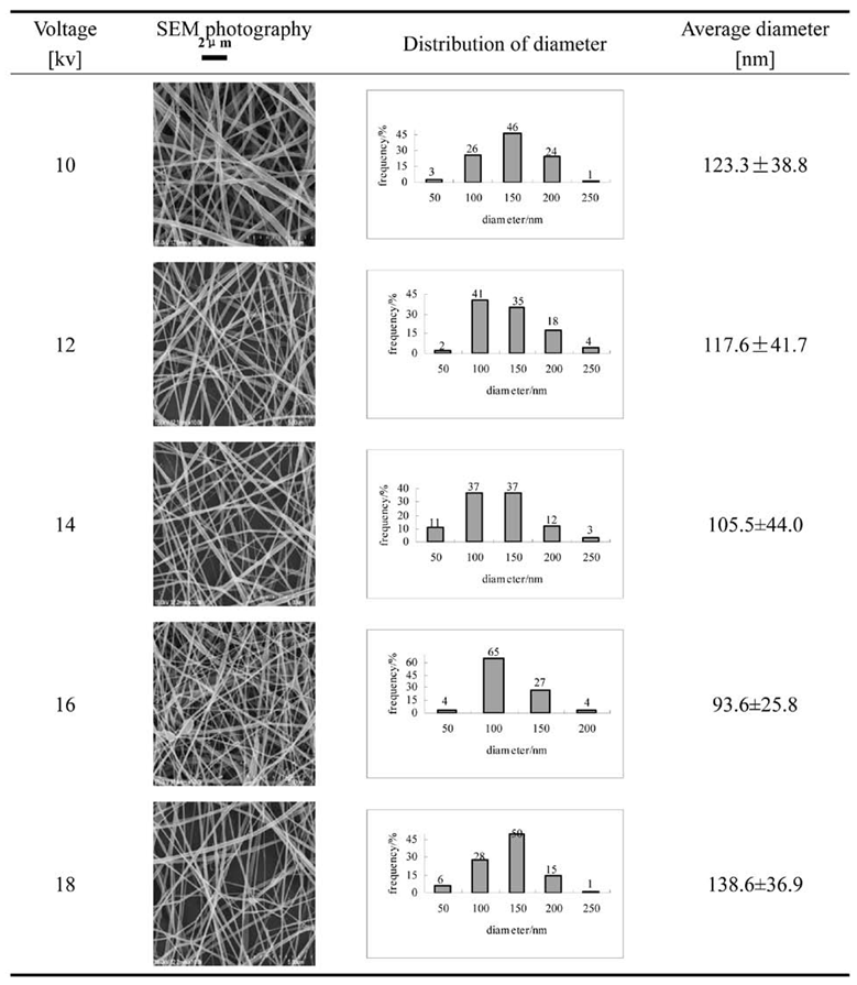
Table 2 shows the SEM photographs, distribution of diameter and average sizes of EROHS fibers at different Voltage (C-SD 12cm). Their diameters mainly distributed 50~150nm, and average diameters were 123.3±38.8nm, 117.6±41.7nm, 105.5±44.0nm, 93.6 ±25.8nm, 138.6±36.9nm respectively when voltages were 10, 12, 14, 16 and 18kv.
Table 2.
Longitudinal shapes and sizes of EROHS fibers at different Voltage (C-SD 12 cm).
Voltage has positive and negative effects on electrospinning process. With the increase of voltage, electric force is enhanced and the fiber diameter decreases. On the other hand, when a voltage exceeds a critical value, more solution is drawn from the spinneret [27], resultantly, the fiber diameter increases. Our results are consistent with the theoretic analysis. When voltage was between 10~16KV, the fiber diameter was negative correlation with voltage and reached the minimum at 16KV.
3.1.2. Molecular Conformation
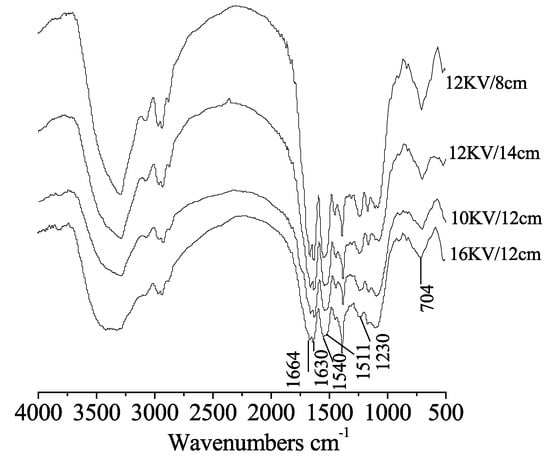
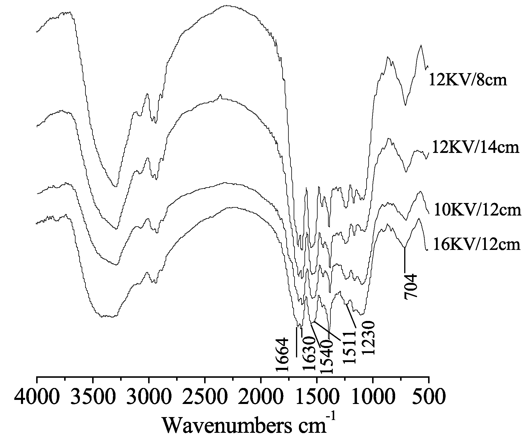
As shown in Figure 1, the FT-IR spectra of EROHS fibers at different C-SD were very similar, in which peaks at about 1664.6 cm-1 and 1630.0cm-1 were α-heilx/random coil and β-sheet structure respectively, 1540.6 cm-1 in amide II region was corresponding to α-heilx/random coil, and absorbing bands at about 1230.0 cm-1 and 704.5 cm-1 were β-sheet characters. The results indicated that enlarging distance from capillary tip to receiving screen almost didn’t change the molecular conformations of EROHS fibers. However, when the electrospinning voltage was upped from 10 to 16KV, there was a new peak band 1511.1 cm-1 in amide II region, which was attribute to β-sheet structure. Otherwise, the regenerated spider silk electrospun at 16KV/12cm had the same FTIR spectrum as that of nature Ornithoctonus huwenna spider silk [28].
Figure 1.
FTIR spectra of EROHS fibers at different voltage /C-SD.
3.1.3. Crystalline Structure
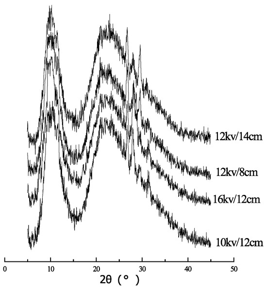
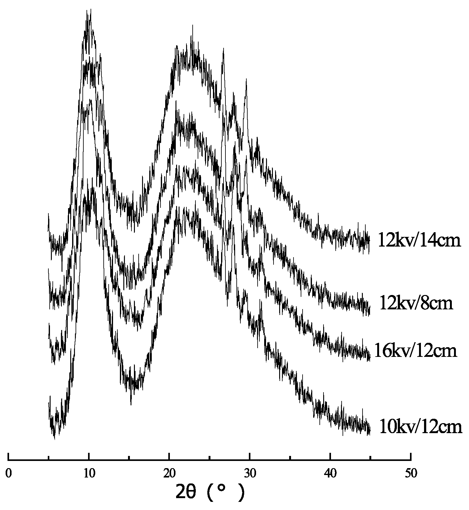
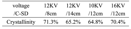
Figure 2 shows the X-ray diffraction intensity curves of EROHS fibers at different voltage /C-SD, and the crystallinities are listed in Table 3. Our research revealed that increasing of electric field strength, shortening the distance between a capillary tip and a collecting screen (C-SD) or enlarging the voltage, is an effective way to improve the crystal structures of electrospun regenerated spider silk fibers. High crystallinity is beneficial to improve the fiber’ s stability in solvent and enhance its mechanical property.
Figure 2.
X-ray diffraction intensity curves of EROHS fibers at different voltage /C-SD.
Table 3.
Crystallinity of EROHS fibers at different voltage/C-SD.
3.2. Compatibility with rBMSCs
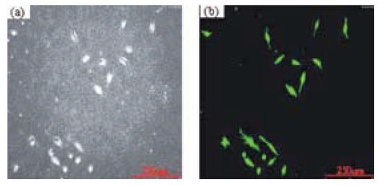
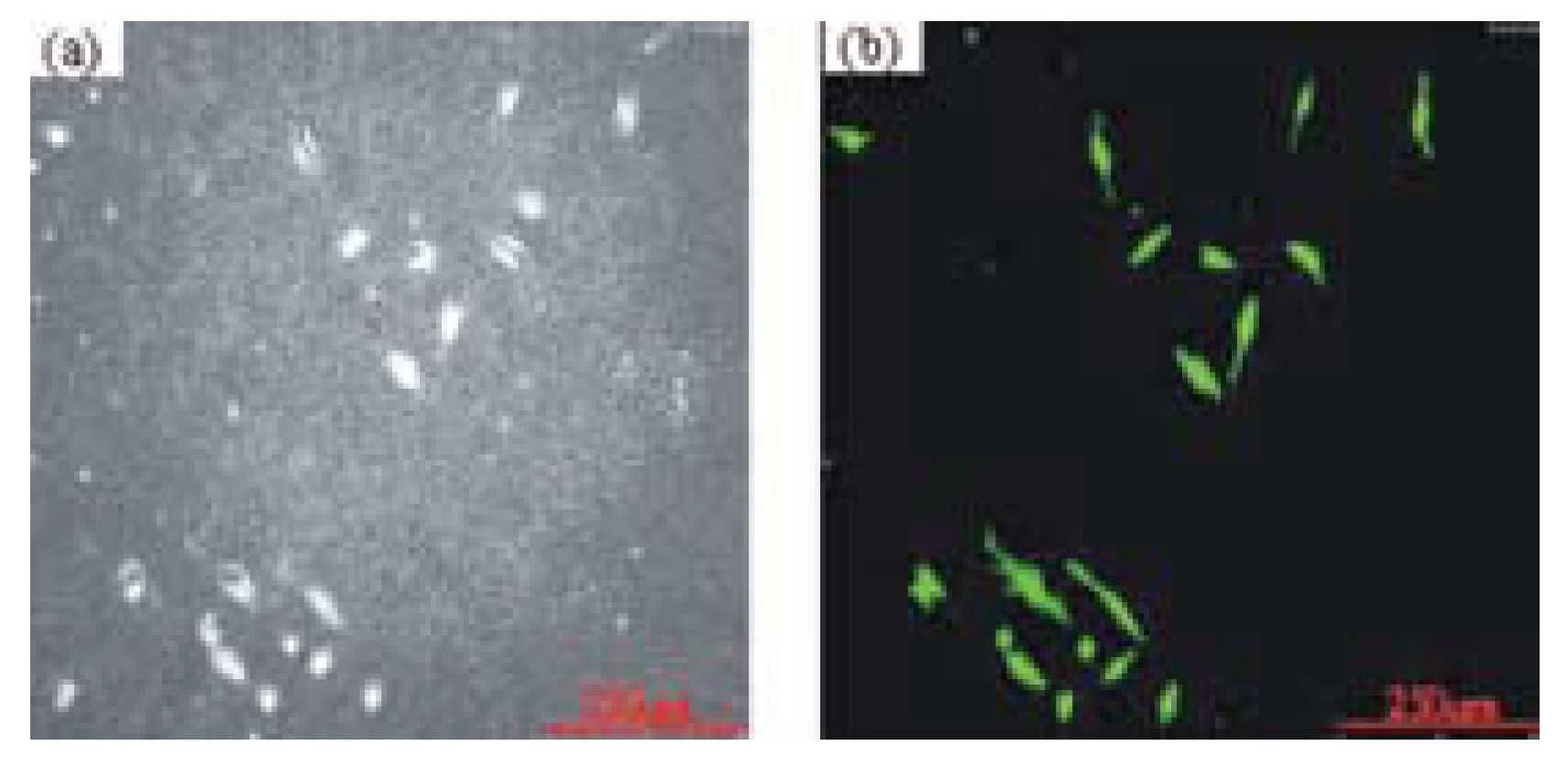
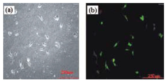
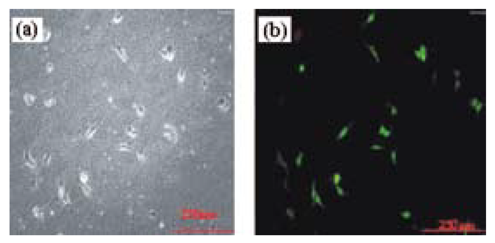
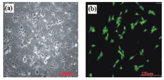
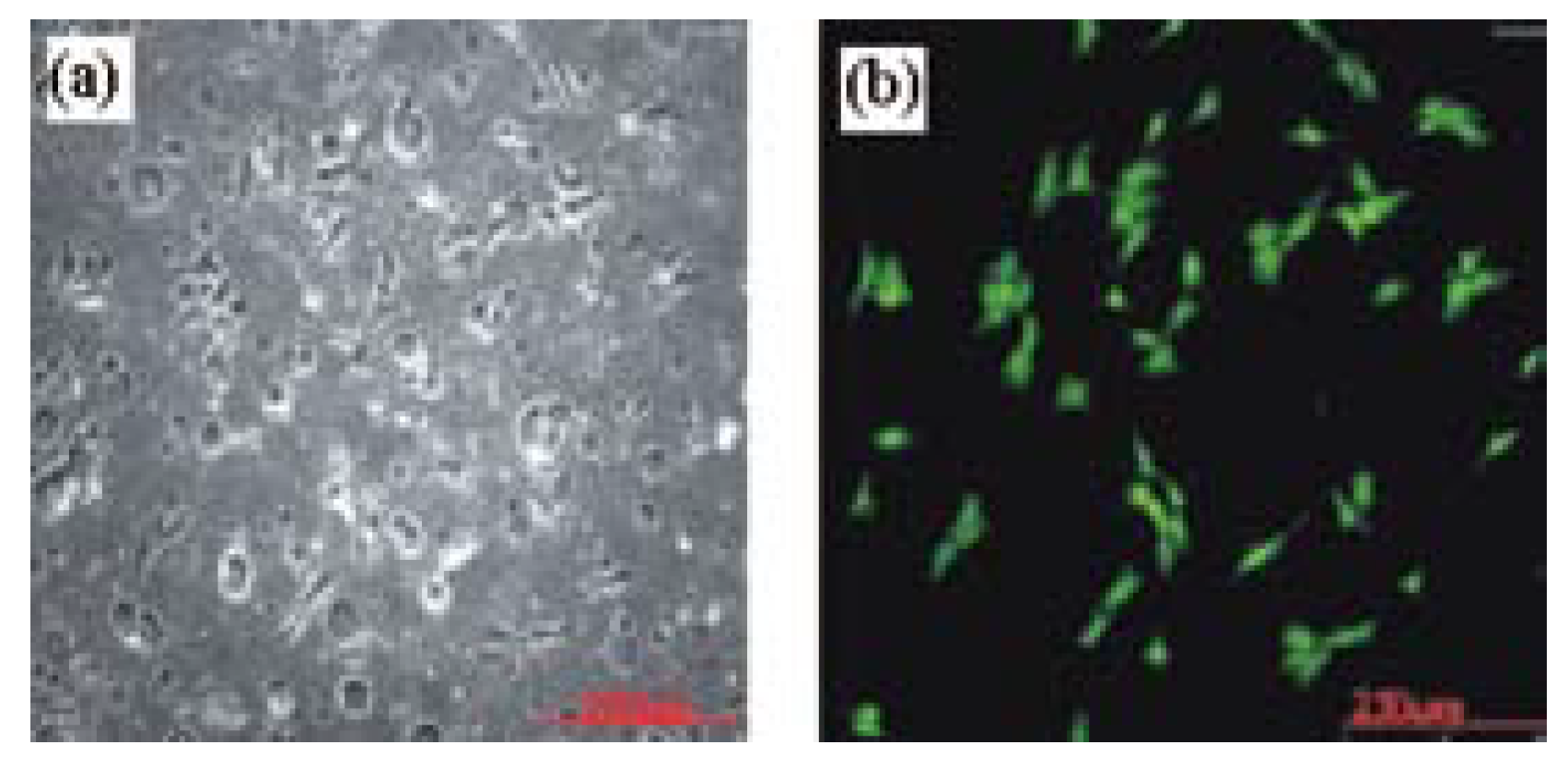
Figure 3, Figure 4 and Figure 5 are images of cells survival states respectively after being inoculated for 24h, 48h and 72h on the surfaces of electrospun spider silk mats, in which (a) is rBMSCs on the surface of the EROHS fiber nonwoven, (b) reflects live rBMSCs with green plots. The cell survival rates were 98.08±2.99%, 98.90%±1.71% and 99.46±0.84%. Therefore, we can conclude that electrospun regenerated Ornithoctonus huwenna spider silk (EROHS) fibers have good compatibilities with rBMSCs.
Figure 3.
Images of cell survival state (inoculated 24h). (a) rBMSCs on the surface of the EROHS fiber nonwoven, (b)green plots are live rBMSCs.
Figure 4.
Images of cell survival state (inoculated 48h). (a) rBMSCs on the surface of the EROHS fiber nonwoven, (b)green plots are live rBMSCs.
Figure 5.
Images of cell survival state (inoculated 72h). (a) rBMSCs on the surface of the EROHS fiber nonwoven, (b)green plots are live rBMSCs.
4. Conclusions
Ornithoctonus huwenna spider silk was effectively solved in 99%HFIP, and the regenerated spidroin solution was electrospun. A nonwoven fabric constructed of continuous and smooth nano-scale fibers was obtained. The fiber diameter primarily ranged from 50 to 150 nm, and the larger an electric field strength, the more fine an EROHS fiber with higher degree of crystal could be obtained. However, the distance between a capillary tip and a collecting screen (C-SD) had no obvious effect on the fiber molecular conformations. Electrospun regenerated Ornithoctonus huwenna spider silk (EROHS) fibers can be used as a candidate biomaterial due to its good cell compatibility.
Acknowledgments
Financial support for this work was provided by the Key Laboratory of Silk Engineering of Jiangsu Province.
References
- Vollrath, F. Strength and structure of spiders’ silks. Rev. Mol. Biotechnol. 2000, 74(2), 67–83. [Google Scholar] [CrossRef] [Scilit] [PubMed]
- Vollrath, F.; David, P.K. Liquid crystalline spinning of spider silk. Nature 2001, 410(6828), 541–548. [Google Scholar] [CrossRef] [Scilit] [PubMed]
- Gregory, H.A.; Frank, D.; Caroline, J. Silk based Biomaterials. Biomaterials 2003, 24, 401–406. [Google Scholar] [CrossRef] [Scilit] [PubMed]
- Vadlamudi S, Hussain MD, Lewis R V. Spider silk: potential for biomedical application. PDD:7171.
- Vollrath, F.; Barth, P.; Basedow, A. Local tolerance to spider silks and protein polymers in vivo. In Vivo 2002, 18, 229–234. [Google Scholar]
- Allmeling, C.; Jokuszies, A.; Reimers, K. Use of spider silk fibres as an innovative material in a biocompatible artificial nerve conduit. J. Cell Mol. Med. 2006, 10, 770–777. [Google Scholar] [CrossRef] [Scilit] [PubMed]
- Cao, B.; Mao, C. Oriented Nucleation of hydroxylapatite crystals on spider dragline silks. Langmuir 2007, 23(21), 10701–10705. [Google Scholar] [CrossRef] [Scilit] [PubMed]
- Stark, M.; Grip, S.; Rising, A. Macroscopic fibers selfassembled from recombinant miniature spider silk proteins. Biomacromolecules 2007, 8, 1695–1701. [Google Scholar] [CrossRef] [Scilit] [PubMed]
- Service, R.F. Mammalian cells spin a spidery new Yarn. Science 2002, 295(5554), 419–421. [Google Scholar] [CrossRef] [Scilit] [PubMed]
- Lazaris, A.; Arcidiacono, S.; Huang, Y.; Zhou, J.F.; Duguay, F.; Chretien, N.; Welsh, E.A.; Soares, J.W.; Karatzas, C.N. Spider silk fibers spun from recombinant silk produced in mammalian cells. Science 2002, 295(5554), 472–476. [Google Scholar] [CrossRef] [Scilit] [PubMed]
- Arcidiacono, S.; Mello, C.M.; Butler, M.; Welsh, E.A.; Soares, J.W.; Allen, A.; Ziegler, D.; Laue, T.; Chase, S. Aqueous processing and fiber spinning of recombinant spider silks. Macromolecules 2002, 35, 1262–1266. [Google Scholar] [CrossRef] [Scilit]
- Roth, D.A.; Lewis, R.V. Expression of spider silk proteins in higher plants. U.S. Patent No. 60296184, 2007. [Google Scholar]
- Charlotte, V.; Thomas, S. Biotechnological production of spider-silk proteins enables new applications. Macromol. Bioscie 2007, 7, 401–409. [Google Scholar] [CrossRef] [Scilit] [PubMed]
- Zhou, C.; Leng, B.; Yao, J. Synthesis and characterization of multiblock copolymers based on spider dragline silk proteins. Biomacromolecules 2006, 7, 2415–2419. [Google Scholar] [CrossRef] [Scilit] [PubMed]
- Koski, A.; Yim, K.; Shivkumar, S. Effect of molecular weight on fibrous PVA produced by electrospinning. Mater. Lett. 2004, 58, 493–497. [Google Scholar] [CrossRef] [Scilit]
- Son, W.K.; Youk, J.H.; Lee, T.S. Electrospinning of ultrafine cellulose acetate fibers: Studies of a new solvent system and deacetylation of ultrafine cellulose acetate fibers. J. Polym. Sci. Polym. Phys. 2004, 42, 5–11. [Google Scholar] [CrossRef] [Scilit]
- Yordem, O.S.; Papila, M.; Menceloglu, Y.Z. Effects of electrospinning parameters on polyacrylonitrile nanofiber diameter: An investigation by response surface methodology. Mater. Des. 2008, 29, 34–44. [Google Scholar] [CrossRef] [Scilit]
- Min, B.M.; Lee, G.; Kim, S.H. Electrospining of silk fibroin nanofibers and its effect on the adhesion and spreading of normal human keratinocytes and fibroblasts in vitro. Biomaterial 2004, 25, 1289–1297. [Google Scholar] [CrossRef] [Scilit] [PubMed]
- Frenot, A.; Chronakis, I.S. Polymer nanofibers assembled by electrospinning. Curr. Opin. Colloid 2003, 8, 64–75. [Google Scholar] [CrossRef] [Scilit]
- Yoshimoto, H.; Shinand, Y.M.; Terai, H. A biodegradable nanofiber scaffold by electrospinning and its potential for bone tissue engineering. Biomaterials 2003, 24, 2077–2082. [Google Scholar] [CrossRef] [Scilit] [PubMed]
- Huang, Z.M.; Zhang, Y.Z.; Kotaki, M. A review on polymer nanofibers by electrospinning and their applications in nanocomposites. Compos Sci. Technol. 2003, 63, 2223–2253. [Google Scholar] [CrossRef] [Scilit]
- Zarkoob, S.; Eby, R.K.; Reneker, D.H. Structure and morphology of electrospun silk nanofibers. Polymer 2004, 45, 3973–3977. [Google Scholar] [CrossRef] [Scilit]
- Pan ZJ, Xu Q. Fabricating method of nanoscale regenerated spider silk. CN Patent No.ZL200510038319.4, 2007.
- Liu MS. Spider breeding and technique utilizing. China Forestry Publication, 2005.
- Wang, Z. A primary study on population structure and food habit of ornithoctonus huwena in ningming county of guangxi. J. Econ. Anim. 2006, 10(2), 101–103. [Google Scholar]
- Chen, D.H.; Liu, Y.; Lin, S.L. Preliminary observation on biological characteristics of ornithoctonus huwena. Acta Arachnol. Sin. 2001, 10(2), 34–38. [Google Scholar]
- Theron SC, Zussman E, Yarin AL. Experimental investigation of the governing parameters in the electrospinning of polymer solutions. Polymer 2004;45:2017-2030.
- Diao JY, Pan ZJ. Chemical compositions and morphologies of ornithoctonus huwenna spider dragline silk. The 6th China international conference and the 2nd international textile forum, 2007. p. 82-86.

© 2008 Textile Bioengineering and Informatics Society. This article is an open access article distributed under the terms and conditions of the Creative Commons Attribution (CC BY) license (https://creativecommons.org/licenses/by/4.0/).