


Abstract
Background/Objectives: Primary thyroid lymphoma accounts for approximately 0.2–2% of thyroid malignancies. Plasmablastic lymphoma (PBL), an aggressive large B-cell neoplasm with plasma-cell differentiation and frequent loss of conventional B-cell markers, is a rare thyroid presentation. We report a thyroid PBL presenting as an acute surgical airway emergency in an immunocompetent patient and highlight the diagnostic and management pitfalls that distinguish this from anaplastic thyroid carcinoma. Case Presentation: A 75-year-old man without any significant past medical history presented with rapidly progressive right-sided neck swelling, dysphagia, inspiratory stridor, and respiratory failure. Imaging demonstrated a large, thyroid-centered mass with tracheal involvement, initially raising concern for anaplastic thyroid carcinoma. Histopathology revealed a diffuse infiltrate of large, atypical cells with immunoblastic and plasmablastic morphology. The neoplastic cells were CD20- and CD138-negative but strongly MUM1-positive, with lambda light-chain restriction, bright CD38 by flow cytometry, a Ki-67 proliferation index exceeding 95%, aberrant cytoplasmic CD3 expression, and a MYC::IGH rearrangement, supporting a diagnosis of PBL. Staging identified extranodal perinephric disease and mesenteric lymphadenopathy, consistent with disseminated extranodal Ann Arbor stage IV disease. The patient underwent systemic treatment and initially had an excellent response; however, one month after the last treatment cycle they presented to the hospital with a mass consistent with recurrence. Discussion: Rapid growth, fixation, and tracheal invasion strongly suggest anaplastic thyroid carcinoma in routine clinical practice. However, plasmablastic lymphomas can present similarly and require fundamentally different treatment. In this case, loss of conventional B-cell markers, CD138 negativity, and aberrant cytoplasmic CD3 expression created substantial diagnostic challenges. Light-chain restriction, plasma-cell-associated markers, flow cytometry, and MYC cytogenetics were vitally important. Conclusions: Thyroid-presenting PBL is exceptionally rare and may closely mimic anaplastic thyroid carcinoma, including presentation with life-threatening airway compromise and tracheal involvement. This case highlights several diagnostic pitfalls: CD138 negativity despite plasma-cell differentiation, and aberrant cytoplasmic CD3 expression. Prompt airway stabilization, adequate tissue acquisition, broad immunophenotyping, light-chain assessment, flow cytometry, EBV/HHV8/ALK testing, and MYC cytogenetics are essential for accurate diagnosis and lymphoma-directed treatment.
1. Introduction
A rapidly enlarging, firm thyroid mass in an older adult raises immediate concern for anaplastic thyroid carcinoma (ATC), the most aggressive and lethal thyroid malignancy. Several aggressive hematologic neoplasms can mimic this presentation, and distinguishing among them has major therapeutic implications. ATC is associated with a grave prognosis and limited durable systemic options, whereas thyroid lymphomas are often chemosensitive and, in select cases, potentially curable [1,2,3]. Primary thyroid lymphoma (PTL) is uncommon, representing approximately 0.4–2% of thyroid malignancies and roughly 2% of extranodal lymphomas, with an estimated incidence near 0.5 per 100,000 individuals [4]. It predominantly affects women in the sixth and seventh decades and is strongly associated with Hashimoto thyroiditis, which increases risk by an estimated 40- to 80-fold [5]. Most cases are B-cell non-Hodgkin lymphomas, specifically diffuse large B-cell lymphoma (DLBCL), followed by extranodal marginal-zone lymphoma of mucosa-associated lymphoid tissue. Regarding presentation, between 70% and 90% of patients present with a rapidly enlarging goiter, and approximately 10–25% develop clinically significant airway compromise from compression or, less commonly, direct tracheal invasion [5,6,7].
Plasmablastic lymphoma (PBL) is a distinct and highly aggressive large B-cell lymphoma characterized by immunoblastic-to-plasmablastic morphology and a terminal B-cell, plasma-cell differentiation program. Tumor cells typically lose mature B-cell markers such as CD20, CD79a, and PAX5 while expressing plasma-cell–associated antigens, including MUM1/IRF4 and often CD138. Light-chain restriction and a very high proliferative index are common [8,9,10]. PBL is classically associated with immunodeficiency, especially human immunodeficiency virus (HIV) infection, and with Epstein–Barr virus (EBV), which is detectable in many cases. However, a minority of cases arise in immunocompetent individuals, who tend to be older and more likely to have extra-oral, clinically aggressive disease [8,9,10].
PBL involving the thyroid is remarkably rare, with only a small number of reported cases [11,12,13]. This case is notable for manifesting as an acute surgical airway emergency with tracheal invasion in an immunocompetent, EBV-negative patient, although plasma EBV DNA PCR showed low-level positivity. It also demonstrated an unusual CD138-negative, aberrantly cytoplasmic CD3-positive immunophenotype. We describe the clinical presentation, airway management, histopathologic and cytogenetic findings, and diagnostic pitfalls of this rare pathology.
2. Case Presentation
This report was prepared in accordance with the CARE guidelines for case reports; details are available in Supplementary Materials [14,15].
2.1. Clinical Presentation and Airway Management
A 75-year-old man presented with a multi-week history of progressive right-sided neck swelling, dysphagia, and inspiratory stridor. He had no known history of HIV infection, organ transplantation, immunosuppressive therapy, autoimmune disease, or prior lymphoma. Upon evaluation, he exhibited signs of impending respiratory failure. Flexible fiberoptic laryngoscopy revealed a massive thyroid mass causing severe tracheal narrowing, right vocal-fold immobility, and restricted left vocal-fold lateralization, together producing a markedly narrowed laryngeal inlet.
Given the severity of the upper-airway obstruction, the patient underwent emergent awake fiberoptic intubation. Contrast-enhanced computed tomography (CT) of the neck demonstrated a large, heterogeneous, thyroid-centered mass infiltrating the trachea and displacing midline structures. After stabilization in the intensive care unit, he was transferred to the operating room for definitive airway management. Intraoperative evaluation confirmed a massive, firm tumor invading the tracheal wall. Tumor debulking and sampling were performed, followed by tracheostomy to secure the airway.
2.2. Laboratory and Staging Evaluation
Laboratory evaluation demonstrated overt biochemical hypothyroidism, with thyroid-stimulating hormone of 22.06 mIU/L and free thyroxine of 0.47 ng/dL, consistent with extensive replacement of normal thyroid parenchyma by tumor. Notably, the patient was HIV negative and negative for any hepatitis serologies. Plasma EBV DNA was detected at a level below the assay’s lower limit of quantification (<300 IU/mL). Additionally, tumor EBER in situ hybridization was negative and baseline LDH was 380 U/L. Chemistry panel demonstrated a BUN of 17 mg/dL, creatinine of 0.89 mg/dL, AST of 32 U/L, ALT of 23 U/L, and total bilirubin of 0.6 mg/dL, without evidence of renal or hepatic dysfunction. Staging evaluation identified extranodal disease involving the left perinephric space and mesenteric lymphadenopathy, which was classified as Ann Arbor stage IV.
2.3. Histopathology and Immunophenotyping
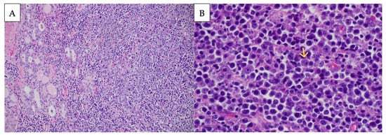
Based on frozen section findings demonstrating sheets of atypical blue cells, a lymphoma workup was initiated. Hematoxylin and eosin (H&E)–stained sections from formalin-fixed, paraffin-embedded (FFPE) tissue revealed sheets of large, atypical cells with immunoblastic and plasmablastic morphology diffusely infiltrating the thyroid and adjacent soft tissue (Figure 1). The discohesive growth pattern, cytologic features, and dim membranous CD45 expression supported a hematolymphoid process.
Figure 1.
Histologic features of the thyroid mass. (A) Hematoxylin and eosin (H&E)-stained section showing a diffuse infiltrate (right) invading thyroid follicles (left) (original magnification 200×). (B) Higher magnification (600×) demonstrating discohesive large neoplastic cells with immunoblastic and plasmablastic morphology. The yellow arrow highlights a cell with plasmablastic features, including a prominent nucleolus, eccentric nucleus, and moderate amount of cytoplasm.
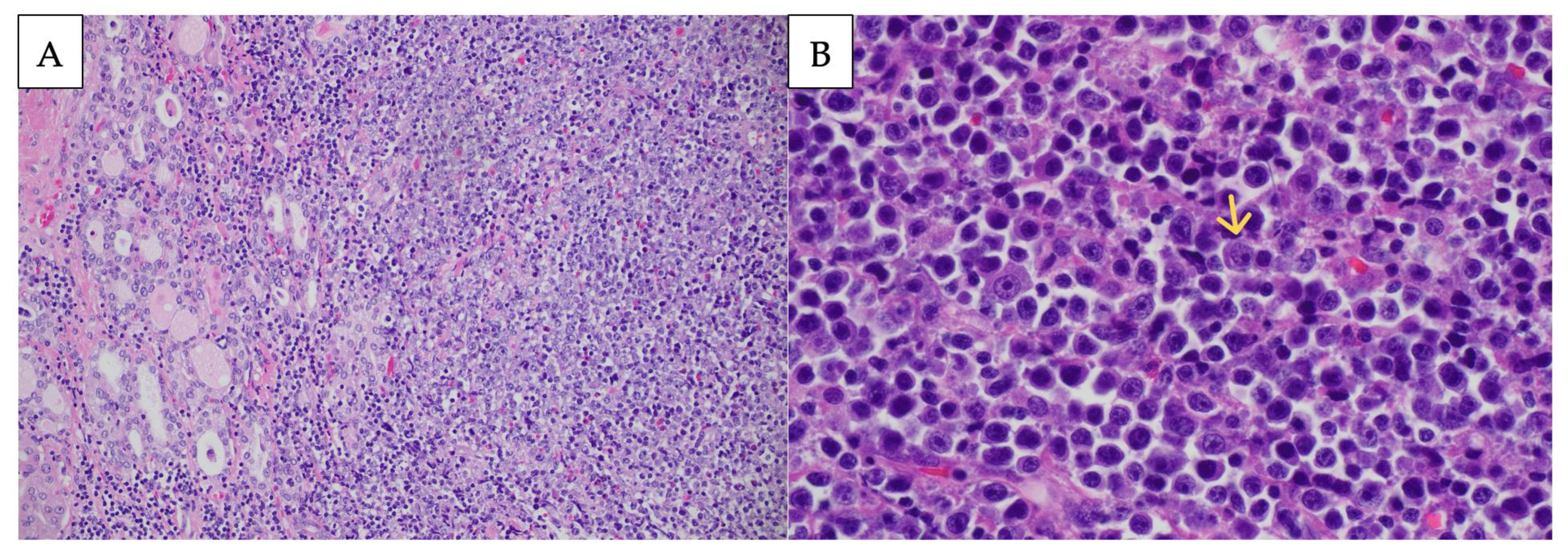
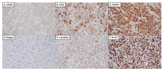
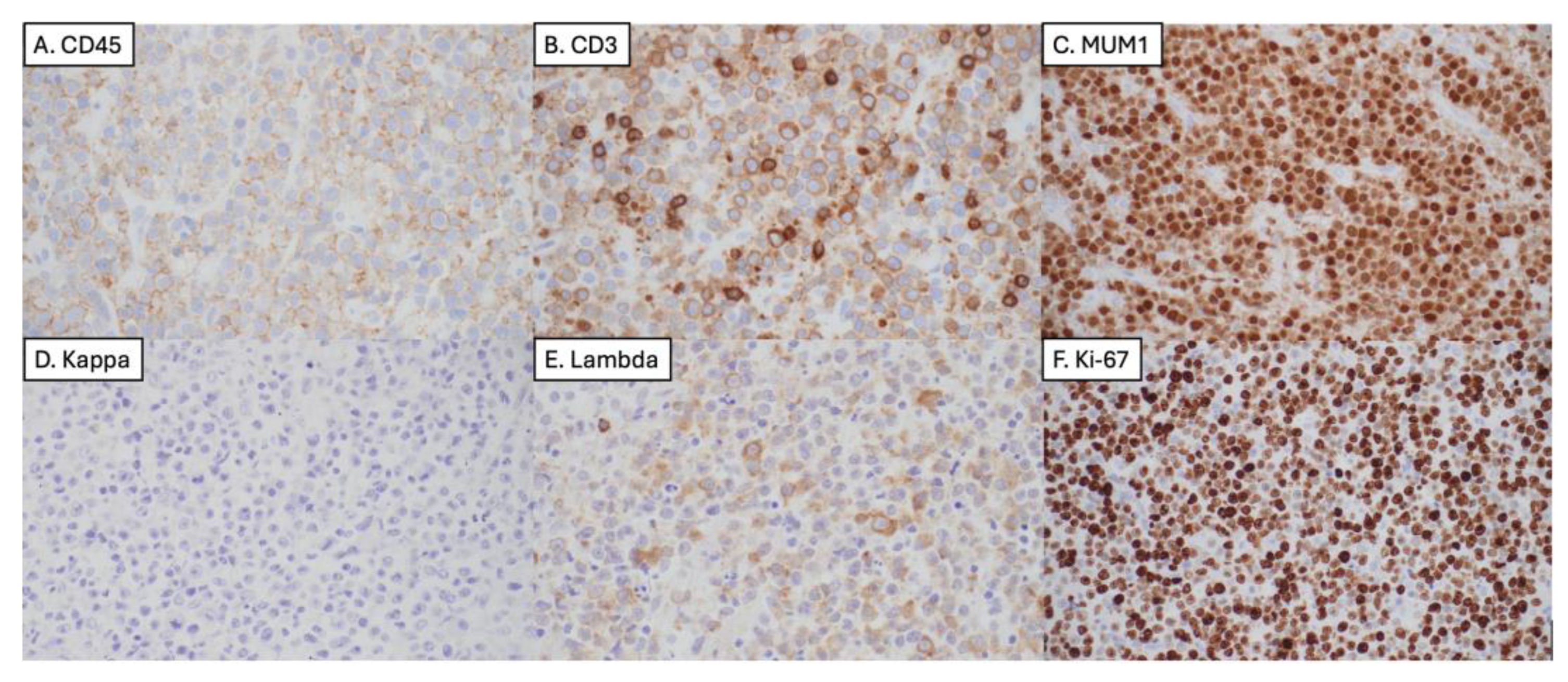
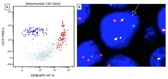
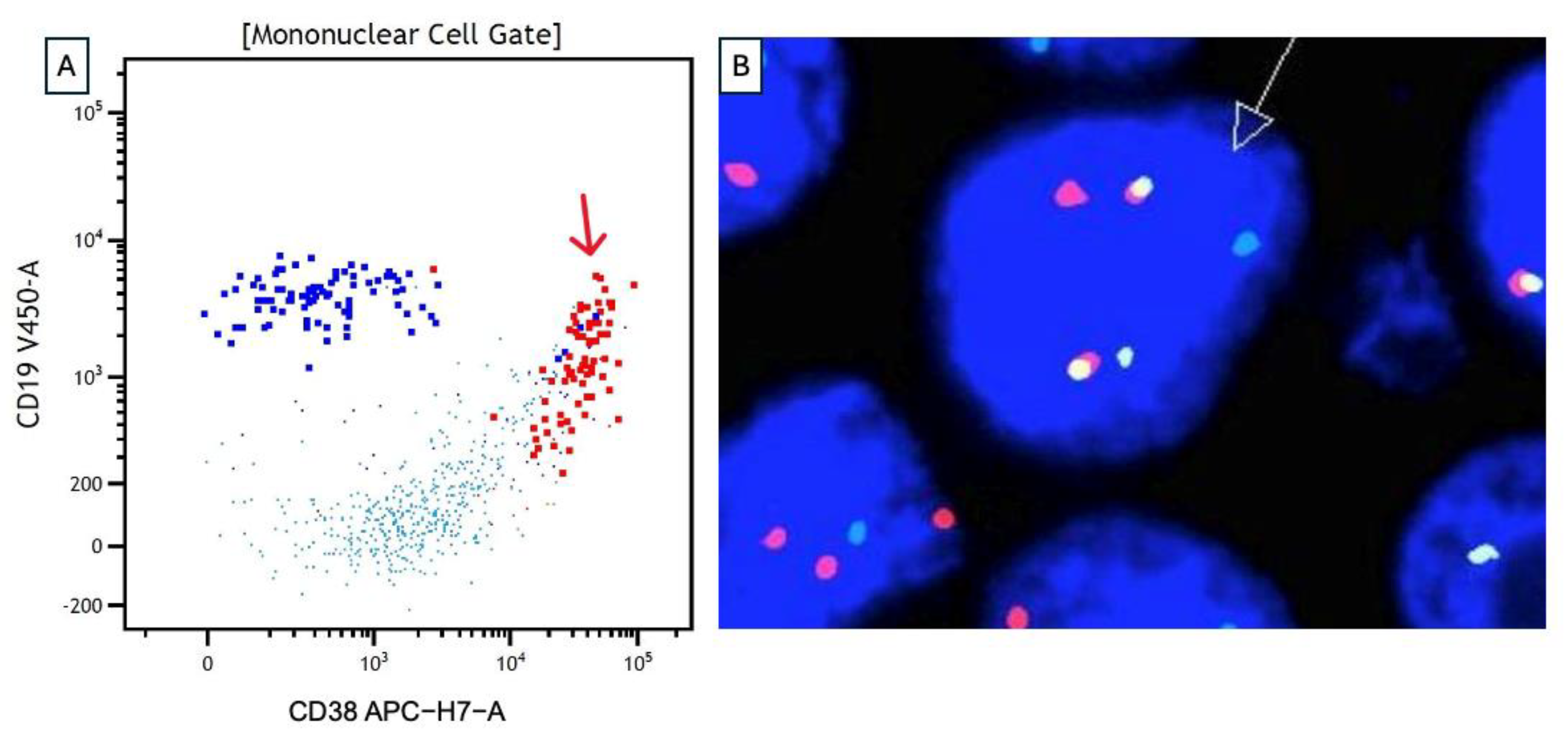
Further evaluation with immunohistochemical and in situ hybridization studies showed that the tumor cells expressed MUM1, CD30, MYC, BCL2, and demonstrated lambda light chain restriction, with dim cytoplasmic positivity for CD3, and weak expression of CD19. The tumor cells were negative for other markers tested (Table 1; selected immunohistochemical and in situ hybridization studies are shown in Figure 2). Flow cytometry identified an atypical population with plasma cell phenotype. Fluorescence in situ hybridization (FISH) was performed using 2 DNA probes, which were simultaneously hybridized to formalin-fixed paraffin-embedded tissue. Approximately 200 interphase cells in each probe set underwent pathologic evaluation. FISH on FFPE tissue using MYC and IGH probes demonstrated t(8;14) rearrangement (Figure 3). The above findings supported the diagnosis of plasmablastic lymphoma (PBL).
Table 1.
Immunophenotypic summary.
Figure 2.
Selected immunohistochemical and in situ hybridization studies. The neoplastic cells demonstrate dim membranous staining for CD45 (A), dim cytoplasmic staining for CD3 (B), and strong, diffuse expression of MUM1 (C). In situ hybridization for kappa (D) and lambda (E) demonstrates lambda light-chain restriction. (F) Ki-67 staining demonstrates a proliferation index exceeding 95%. Original magnification 600×.
Figure 3.
Ancillary studies. (A) Flow cytometric immunophenotyping of the thyroid sample demonstrated an atypical population (red arrow) with plasma cell phenotype characterized by bright CD38 with variable CD19 expression. (B) Fluorescence in situ hybridization using dual-color, dual-fusion probes for MYC and IGH demonstrated fusion signals in 57% of the cells. The representative image shows one red (MYC), one green (IGH), and two fusion (yellow) signals, consistent with a MYC::IGH rearrangement.
Serum protein electrophoresis demonstrated several faint bands in the gamma region interpreted as oligoclonal, a pattern associated with chronic inflammation versus autoimmune disease rather than with a clonal plasma cell disorder. Urine protein electrophoresis demonstrated a small amount of albumin with traces of other proteins and urine immunofixation electrophoresis demonstrated polyclonal immunoglobulins. In both measurements, no paraproteins were identified. No paraproteins were identified in serum and urine analysis. The serum free light-chain assay showed a free kappa of 35.3 mg/L and a free lambda of 39.1 mg/L, giving a kappa to lambda ratio of 0.90 (ref: 0.26–1.65). The concordant, mildly elevated absolute values with a preserved ratio indicate a polyclonal free light-chain elevation, which has been observed in systemic inflammation, aggressive lymphoma, and reduced renal clearance, and does not suggest a clonal light-chain process. Further, the tumor did not display detectable lambda excess or ratio inversion in serum, strongly arguing against a secretory plasma cell neoplasm diagnosis.
During initial evaluation, the patient met no CRAB criteria. The serum calcium was 8.7 mg/dL, creatinine 0.89 mg/dL with an eGFR of 89 mL/min, calculated using the 2021 CKD-EPI creatinine equation [16], and hemoglobin of 14.9 g/dL. Whole-body FDG-PET/CT and contrast-enhanced CT demonstrated no osteolytic lesions. Bone marrow aspirate, clot section, touch preparation, and core biopsy demonstrated a mildly hypercellular marrow (40%) with trilinear hematopoiesis and unremarkable to mildly increased megakaryocytes. Infiltrates of large, atypical cells were inconspicuous, and there was no plasmacytosis; clonal plasma cells were not identified by morphology, immunohistochemistry, or flow cytometry.
2.4. Treatment and Course
The patient was diagnosed on 28 January 2026. He received six cycles of dose-adjusted etoposide, prednisone, vincristine, cyclophosphamide, and doxorubicin (EPOCH) with the addition of the anti-CD38 monoclonal antibody daratumumab in each cycle, together with intrathecal methotrexate for central nervous system prophylaxis. This regimen was biologically rational for the present tumor, which demonstrated bright CD38 expression by flow cytometry. PBL characteristically lacks CD20 and typically expresses plasma-cell–associated antigens, including CD38. Therefore, rituximab is not an effective systemic therapy agent, and CD38-directed therapy provides a targetable surface antigen. The combination of daratumumab with EPOCH-based chemotherapy was developed on this rationale and has previously shown feasibility and encouraging early activity in newly diagnosed PBL, including complete responses across HIV-positive and HIV-negative patients.
The patient completed all six planned cycles. Cycle 2 was complicated by grade 4 febrile neutropenia with influenza B and Pseudomonas pneumonia, resulting in septic shock requiring vasopressor support and ICU admission from 11–17 March. He also developed bilateral upper-extremity deep vein thromboses and a left internal jugular vein thrombosis. Treatment response was assessed clinically and by an interim PET/CT performed on 24 April, after the fourth cycle of treatment. Compared with the pretreatment CT of the neck on 26 January, the PET/CT demonstrated a marked interval decrease in the thyroid mass involving both lobes. The patient continued to demonstrate a favorable clinical response through treatment completion on 2 June. The peak serum LDH level was 380 U/L on 5 February, before treatment initiation; it decreased to 349 U/L after the first cycle on 25 February and to 130 U/L after the sixth cycle on 9 June. A Deauville score and standardized RECIL target-lesion measurements were not documented, and no baseline pretreatment PET/CT was available. Therefore, a formal complete response could not be confirmed, and the outcome was characterized as a marked radiographic and clinical response.
This initial favorable outcome is notable given that intensive EPOCH-based regimens alone have historically yielded complete-response in approximately 50% of cases and limited median survival in PBL. This outcome may also support continued investigation of CD38-directed strategies in CD38-bright disease. Unfortunately, one month after completing treatment he presented with a rapidly growing neck mass that was consistent with persistent/relapsed disease. He was subsequently admitted to the hospital for airway management and palliative radiation therapy. At the last follow-up on 3 August, the follow-up duration was 6.2 months from the initial diagnosis.
3. Discussion
This case highlights an unusual presentation of plasmablastic lymphoma (PBL) involving the thyroid. PBL is an aggressive large B-cell neoplasm characterized by immunoblastic to plasmablastic morphology and a terminal B-cell differentiation phenotype. Aligning with the WHO 5th edition description of PBL, the diagnosis in this case was supported by plasmablastic morphology, strong MUM1 expression, bright CD38, and demonstration of lambda light chain restriction, absent conventional B-cell markers, and high Ki-67. The presence of a MYC rearrangement further supported the PBL diagnosis, as MYC alterations have been identified in a substantial proportion of cases.
Although PBL most commonly arises in the setting of immunodeficiency, particularly HIV infection, it can also occur in immunocompetent individuals. Additionally, EBV is detected in approximately 60% of cases, but neither HIV-positive nor EBV-positive statuses are required for diagnosis. Aberrant expression of T-cell markers, including cytoplasmic CD3, has been described and may lead to diagnostic confusion with T-cell lymphoma. A comprehensive immunophenotypic evaluation is essential for accurate classification [11,12,17].
Rapid growth, fixation, vocal-fold immobility, and tracheal invasion strongly suggest ATC in routine clinical practice, and the initial clinical and radiographic impression in this case favored locally aggressive ATC. However, PTL is a recognized mimic of ATC, and this distinction is therapeutically pivotal [18]. ATC is associated with poor survival and often limited durable systemic options, whereas many thyroid lymphomas are chemosensitive and, in limited-stage disease, potentially curable [4,19,20]. Based on CD20, CD138, and CD3 expression, the differential diagnosis was broad and included ATC, T-cell lymphoma, ALK-positive large B-cell lymphoma, and additional neoplasms summarized in Table 2.
Table 2.
Differential diagnoses and basis for exclusion.
PBL and plasmablastic myeloma share many similar pathological characteristics, such as positivity for CD38 and MUM1 as well as negativity for CD20 [21]. Therefore, the final diagnosis depended on integrating morphology, immunophenotype, light-chain restriction, flow cytometry, and cytogenetics. PBL was favored, rather than plasmablastic myeloma, given the absence of a prior history of myeloma and lack of clonal plasma cells in the bone marrow. Additionally, the serum-free light chain assay free-kappa to free-lambda ratio of 0.90 argued against a secretory plasma cell neoplasm. This diagnosis was also supported by the absence of CRAB criteria.
According to the WHO 5th edition, PBL is primarily composed of immunoblastic or plasmablastic cells [22]. Tumor cells characteristically lose CD20 and PAX5 while expressing markers of plasma-cell differentiation, particularly MUM1/IRF4 and frequently CD138 [8,9,11,12,23,24]. Despite negative CD138 expression, other plasma cell features, such as strong MUM1 expression, lambda light-chain restriction, bright CD38 by flow cytometry, high Ki-67, and MYC::IGH rearrangement collectively supported the PBL diagnosis [11,12,23].
Negative CD138 expression posed a diagnostic challenge in this case of PBL as CD138 negativity has been associated with difficulty establishing a plasma cell phenotype [25]. A prior study reported that approximately 10% of PBL cases are CD138-negative and reported that additional diagnostic characteristics observed in our case, such as CD20 negativity, high Ki-67, and MYC rearrangement, support PBL diagnosis [26].
Aberrant cytoplasmic CD3 expression also created an additional challenge during diagnosis, as CD3-positive plasmablastic B-cell neoplasms are a documented diagnostic trap and may be misclassified as T-cell lymphoma if light-chain restriction and plasma-cell markers are not evaluated [17]. Here, the absence of other T-cell markers and the presence of lambda light-chain restriction established a clonal B-/plasma-cell process despite dim cytoplasmic CD3 expression. This case therefore underscores the need for broad, lineage-spanning panels whenever morphology suggests a plasmablastic neoplasm, especially when conventional B-cell markers are absent.
MYC rearrangement was the first recurrent cytogenetic abnormality identified in PBL and is detected in a substantial proportion of cases, most often with an immunoglobulin gene partner such as IGH [27,28]. The MYC::IGH fusion in this case is therefore consistent with PBL biology. MYC rearrangement is more frequent in EBV-positive tumors, although this case was EBER-negative, illustrating the heterogeneity of the entity [24,28].
Additionally, we observed low-level plasma EBV DNA positivity and EBER-negative PBL. Since the EBV DNA concentration was below quantification, we classify this finding as incidental rather than associated with the PBL.
The prognostic impact of EBV and MYC status remains unsettled. Modern research suggests that neither MYC translocation nor EBV status is a consistent independent predictor of overall survival. Furthermore, EBV positivity has been associated with improved progression-free survival in some analyses, while MYC data remains limited by incomplete testing [23,24]. Regardless, immunocompetent PBL is well documented and tends to occur in older patients with extra-oral, aggressive presentations [9,11,12,29], consistent with the clinical profile of the present patient.
PBL outcomes have historically been poor, although contemporary multicenter data suggest heterogeneity in survival, with some patients achieving longer-term disease control [24]. The CHOP combination chemotherapy regimen (cyclophosphamide, doxorubicin, vincristine, and prednisone) has generally been viewed as suboptimal for PBL, and expert practice has often favored more intensive regimens such as dose-adjusted EPOCH or CODOX-M/IVAC. Given the plasma-cell biology of PBL, proteasome inhibitor–containing regimens such as bortezomib-EPOCH have also been explored [23,24,29].
However, no single standard frontline regimen has been definitively established, and retrospective data have not consistently demonstrated a survival advantage for higher-intensity chemotherapy or frontline proteasome-inhibitor use [3,27]. Management should therefore be individualized in close coordination with hematology-oncology, considering performance status, disease extent, actionable marker profiling, central-nervous-system risk, eligibility for clinical trials, and potential consolidation strategies. For instance, accurate immunophenotyping of CD20 directly determines systemic therapy and treatment selection.
The marked radiographic and clinical response observed in this patient is encouraging given the limited efficacy of conventional chemotherapy in plasmablastic lymphoma. In a multicenter retrospective study of 344 patients, 220 received frontline EPOCH-based regimens, with an overall response rate of 64% and a complete response rate of 52% [24]. However, detailed demographic and clinical characteristics were not reported specifically for the EPOCH-treated subgroup, limiting direct comparison with the present case. Historically, median overall survival for plasmablastic lymphoma was estimated at approximately 8–15 months, although more recent studies have reported improved and heterogeneous outcomes [24,28,29].
Several features of this case may have favored this outcome. The tumor’s bright CD38 expression provided a rational target for daratumumab. The addition of a CD38-directed antibody to dose-adjusted EPOCH, an approach that has shown feasibility and complete responses in newly diagnosed PBL irrespective of HIV status, may have contributed to disease control beyond what chemotherapy alone might have achieved [30]. Equally important, the early surgical decompression converted a life-threatening airway obstruction into a stable platform from which full-intensity systemic therapy could be delivered without interruption, allowing the patient to complete all six planned cycles.
The tracheostomy, necessary at presentation, for managing tumor-related tracheal invasion and obstruction became unnecessary as the mass regressed with systemic therapy, and the patient was successfully decannulated. This sequence illustrates that tracheostomy in thyroid-presenting PBL should be viewed as a temporary measure rather than a permanent outcome. This initial complete response and decannulation underscores that aggressive, coordinated management can yield favorable results even in an elderly patient presenting with a catastrophic airway emergency.
Limitations
Although MYC rearrangement testing was performed, broader molecular profiling by next-generation sequencing (NGS) was not performed, limiting assessment for additional genetic alterations that may have contributed to this rare presentation.
4. Conclusions
Thyroid-presenting PBL is a rare but important mimic of anaplastic thyroid carcinoma and can present with life-threatening airway compromise. In this case, the absence of conventional B-cell markers, CD138 negativity, aberrant cytoplasmic CD3 expression, and MYC::IGH rearrangement created substantial diagnostic risk. Early airway stabilization and adequate tissue acquisition were essential, but definitive diagnosis depended on broad immunophenotyping, light-chain studies, flow cytometry, EBV/HHV8/ALK assessment, and MYC cytogenetics. The patient’s complete response and decannulation after CD38-directed EPOCH-based therapy are encouraging, but implications from a single case should remain hypothesis-generating. Nonetheless, the complete response observed may warrant further investigation of outcomes associated with CD-38-targeted agents.
Supplementary Materials
The following supporting information can be downloaded at https://www.mdpi.com/article/10.3390/ohbm7020032/s1, CARE Checklist of information to include when writing a case report.
Author Contributions
Conceptualization, D.M., D.Z.A. and M.S.; resources, D.Z.A., E.M., B.A. and M.S.; writing—original draft preparation, D.M. and D.Z.A.; writing—review and editing, D.M., D.Z.A., J.W.R., E.M., B.A., J.A.J., C.C.E. and M.S.; supervision, M.S. All authors have read and agreed to the published version of the manuscript.
Funding
This research received no external funding.
Institutional Review Board Statement
Emory does not require IRB review of studies that do not meet the definitions of “human subjects research” (DHHS) or “clinical investigation” (FDA).
Informed Consent Statement
Written informed consent has been obtained from the patient to publish this paper.
Data Availability Statement
The original contributions presented in this study are included in the article. Further inquiries can be directed to the corresponding author.
Conflicts of Interest
The authors declare no conflicts of interest.
References
- Bible, K.C.; Kebebew, E.; Brierley, J.; Brito, J.P.; Cabanillas, M.E.; Clark, T.J., Jr.; Di Cristofano, A.; Foote, R.; Giordano, T.; Kasperbauer, J.; et al. 2021 American Thyroid Association Guidelines for Management of Patients with Anaplastic Thyroid Cancer. Thyroid 2021, 31, 337–386. [Google Scholar] [CrossRef] [Scilit] [PubMed]
- Watanabe, N.; Noh, J.Y.; Narimatsu, H.; Takeuchi, K.; Yamaguchi, T.; Kameyama, K.; Kobayashi, K.; Kami, M.; Kubo, A.; Kunii, Y.; et al. Clinicopathological features of 171 cases of primary thyroid lymphoma: A long-term study involving 24,553 patients with Hashimoto’s disease. Br. J. Haematol. 2011, 153, 236–243. [Google Scholar] [CrossRef] [Scilit] [PubMed]
- Toloza, F.J.K.; Gubbi, S.; Klubo-Gwiezdzinska, J. Trends in Survival and Factors Associated with Survival in Primary Thyroid Lymphoma: A SEER-Based Analysis (1975–2021). Thyroid 2025, 35, 1074–1082. [Google Scholar] [CrossRef] [Scilit] [PubMed]
- Stein, S.A.; Wartofsky, L. Primary thyroid lymphoma: A clinical review. J. Clin. Endocrinol. Metab. 2013, 98, 3131–3138. [Google Scholar] [CrossRef] [Scilit] [PubMed]
- Pavlidis, E.T.; Pavlidis, T.E. A Review of Primary Thyroid Lymphoma: Molecular Factors, Diagnosis and Management. J. Investig. Surg. 2019, 32, 137–142. [Google Scholar] [CrossRef] [Scilit] [PubMed]
- Kesornpatumanant, K.; Numkiatsakul, N.; Sukswai, N.; Snabboon, T. Mantle Cell Lymphoma of the Thyroid: A Rare Cause of Life-Threatening Airway Compromise. Kaohsiung J. Med. Sci. 2025, 41, e70048. [Google Scholar] [CrossRef] [Scilit] [PubMed]
- Zhao, J.-L.; Zhao, J.-H.; Yang, X.-Y.; Guan, X.; Jia, Y.-C.; Cai, Y.-H.; Ge, R.-S.; Miao, K.; Wang, H.; Cao, S.-D.; et al. Comprehensive analysis of primary thyroid lymphoma: A case report and review of diagnostic and treatment protocols. AME Case Rep. 2026, 10, 26. [Google Scholar] [CrossRef] [Scilit] [PubMed]
- Alaggio, R.; Amador, C.; Anagnostopoulos, I.; Attygalle, A.D.; Araujo, I.B.d.O.; Berti, E.; Bhagat, G.; Borges, A.M.; Boyer, D.; Calaminici, M.; et al. The 5th edition of the World Health Organization Classification of Haematolymphoid Tumours: Lymphoid Neoplasms. Leukemia 2022, 36, 1720–1748. [Google Scholar] [CrossRef] [Scilit] [PubMed]
- Lopez, A.; Abrisqueta, P. Plasmablastic lymphoma: Current perspectives. Blood Lymphat. Cancer 2018, 8, 63–70. [Google Scholar] [CrossRef] [Scilit] [PubMed]
- Swerdlow, S.H.; Campo, E.; Pileri, S.A.; Harris, N.L.; Stein, H.; Siebert, R.; Advani, R.; Ghielmini, M.; Salles, G.A.; Zelenetz, A.D.; et al. The 2016 revision of the World Health Organization classification of lymphoid neoplasms. Blood 2016, 127, 2375–2390. [Google Scholar] [CrossRef] [Scilit] [PubMed]
- AlSheef, M.; Abuzied, Y.; Aleid, B.; Shafi, N.; Alshakweer, W.; Zaidi, A.R.Z. Plasmablastic Lymphoma in an Immunocompetent Patient: A Case Report and Literature Review. Cureus 2022, 14, e30732. [Google Scholar] [CrossRef] [Scilit] [PubMed]
- Bibas, M. Plasmablastic Lymphoma. A State-of-the-Art Review: Part 1-Epidemiology, Pathogenesis, Clinicopathologic Characteristics, Differential Diagnosis, Prognostic Factors, and Special Populations. Mediterr. J. Hematol. Infect. Dis. 2024, 16, e2024007. [Google Scholar] [CrossRef] [Scilit] [PubMed]
- Gallagher, T.M.; Narayana, S.; Malysz, J. Primary Plasmablastic Thyroid Lymphoma: A Case Report. AACE Clin. Case Rep. 2018, 4, e191–e194. [Google Scholar] [CrossRef] [Scilit]
- CARE Case Report Guidelines. What Are the CARE Guidelines? Available online: https://www.care-statement.org/ (accessed on 12 June 2026).
- Riley, D.S.; Barber, M.S.; Kienle, G.S.; Aronson, J.K.; von Schoen-Angerer, T.; Tugwell, P.; Kiene, H.; Helfand, M.; Altman, D.G.; Sox, H.; et al. CARE guidelines for case reports: Explanation and elaboration document. J. Clin. Epidemiol. 2017, 89, 218–235. [Google Scholar] [CrossRef] [Scilit] [PubMed]
- Inker Lesley, A.; Eneanya Nwamaka, D.; Coresh, J.; Tighiouart, H.; Wang, D.; Sang, Y.; Crews Deidra, C.; Doria, A.; Estrella Michelle, M.; Froissart, M.; et al. New Creatinine- and Cystatin C–Based Equations to Estimate GFR without Race. N. Engl. J. Med. 2021, 385, 1737–1749. [Google Scholar] [CrossRef] [Scilit] [PubMed]
- Pan, Z.; Chen, M.; Zhang, Q.; Wang, E.; Yin, L.; Xu, Y.; Huang, Q.; Yuan, Y.; Zhang, X.; Zheng, G.; et al. CD3-positive plasmablastic B-cell neoplasms: A diagnostic pitfall. Mod. Pathol. 2018, 31, 718–731. [Google Scholar] [CrossRef] [Scilit] [PubMed]
- Fang, S.; Gkiousias, V.; Hu, L.; Kapoor, K. Tracheal Invasion and Cardiopulmonary Compromise From Primary Thyroid Lymphoma. Cureus 2021, 13, e19302. [Google Scholar] [CrossRef] [Scilit] [PubMed]
- Alnahas, Z.; Horani, M.H. A Dramatic Response of a Thyroid Lymphoma to R-CHOP Chemotherapy Reversing Mechanical Airway Obstruction and Respiratory Failure. Case Rep. Endocrinol. 2022, 2022, 3719320. [Google Scholar] [CrossRef] [Scilit] [PubMed]
- Jaafari, A.; Mehaudens, S.; Gheysens, O.; Bailly, S.; Schobbens, N.; Mourad, M.; Jamar, F. Massive Primary Thyroid Lymphoma on [18F]-FDG-PET/CT: A Literature Review of a Rare Case of Rapidly Progressive Goitre. Diagnostics 2025, 15, 2180. [Google Scholar] [CrossRef] [Scilit] [PubMed]
- Ramadas, P.; Williams, M.; Duggan, D.B. Plasmablastic Lymphoma or Plasmablastic Myeloma: A Case of Post-Transplant Lymphoproliferative Disorder. Case Rep. Hematol. 2021, 2021, 4354941. [Google Scholar] [CrossRef] [Scilit] [PubMed]
- Kurz, K.S.; Ott, M.; Kalmbach, S.; Steinlein, S.; Kalla, C.; Horn, H.; Ott, G.; Staiger, A.M. Large B-Cell Lymphomas in the 5th Edition of the WHO-Classification of Haematolymphoid Neoplasms—Updated Classification and New Concepts. Cancers 2023, 15, 2285. [Google Scholar] [CrossRef] [Scilit] [PubMed]
- Castillo, J.J.; Guerrero-Garcia, T.; Baldini, F.; Tchernonog, E.; Cartron, G.; Ninkovic, S.; Cwynarski, K.; Dierickx, D.; Tousseyn, T.; Lansigan, F.; et al. Bortezomib plus EPOCH is effective as frontline treatment in patients with plasmablastic lymphoma. Br. J. Haematol. 2019, 184, 679–682. [Google Scholar] [CrossRef] [Scilit] [PubMed]
- Hamby, M.; Egleston, B.L.; Frosch, Z.A.K.; Steiner, R.E.; Noy, A.; Carvajal, V.; Chong, E.R.; Tolu, S.S.; Jean-Louis, G.; Amengual, J.E.; et al. Prognosis and treatment of plasmablastic lymphoma in the United States: A multicenter retrospective study. Blood Cancer J. 2026, 16, 43. [Google Scholar] [CrossRef] [Scilit] [PubMed]
- Yeung, C.C.S.; Mills, J.C.; Hassan, A.; Kreisel, F.H.; Nguyen, T.T.; Frater, J.L. MIST1-a novel marker of plasmacytic differentiation. Appl. Immunohistochem. Mol. Morphol. 2012, 20, 561–565. [Google Scholar] [CrossRef] [Scilit] [PubMed][Green Version]
- Choudhuri, J.; Pan, Z.; Yuan, J.; Chen, M.; Wu, X.; Zheng, G.; Zhao, C.; Yuan, Y.; Agarwal, B.; Liu, J.; et al. CD138− Plasmablastic Lymphoma: A Multi-institutional Study and Review of the Literature. Arch. Pathol. Lab. Med. 2023, 147, 643–654. [Google Scholar] [CrossRef] [Scilit] [PubMed]
- Barouqa, M.; Greipp, P.; King, R.; McPhail, E.D. Plasmablastic lymphoma with MYC::IGH fusion and BCL2 rearrangement. eJHaem 2022, 3, 1080–1081. [Google Scholar] [CrossRef] [Scilit] [PubMed]
- Li, J.W.; Peng, H.L.; Zhou, X.Y.; Wang, J.J. Plasmablastic lymphoma: Current knowledge and future directions. Front. Immunol. 2024, 15, 1354604. [Google Scholar] [CrossRef] [Scilit] [PubMed]
- Bibas, M. Plasmablastic Lymphoma. A State-of-the-Art Review: Part 2-Focus on Therapy. Mediterr. J. Hematol. Infect. Dis. 2024, 16, e2024015. [Google Scholar] [CrossRef] [Scilit] [PubMed]
- Ryu, Y.K.; Ricker, E.C.; Soderquist, C.R.; Francescone, M.A.; Lipsky, A.H.; Amengual, J.E. Targeting CD38 with Daratumumab Plus Chemotherapy for Patients with Advanced-Stage Plasmablastoid Large B-Cell Lymphoma. J. Clin. Med. 2022, 11, 4928. [Google Scholar] [CrossRef] [Scilit] [PubMed]

Disclaimer/Publisher’s Note: The statements, opinions and data contained in all publications are solely those of the individual author(s) and contributor(s) and not of MDPI and/or the editor(s). MDPI and/or the editor(s) disclaim responsibility for any injury to people or property resulting from any ideas, methods, instructions or products referred to in the content. |
© 2026 by the authors. Licensee MDPI, Basel, Switzerland. This article is an open access article distributed under the terms and conditions of the Creative Commons Attribution (CC BY) license.