Abstract
A novel series of polypyridyl adducts, [Sm(ntfa)3(NN)] (2–4), with ntfa = 4,4,4-trifluoro-1-(naphthalen-2-yl)-1,3-butanedionate, NN = 2,2′-bipyridine (bipy), 4,4′-dimethyl-2,2′-bipyridine (4,4′-Me2bipy), and 5,5′-dimethyl-2,2′-bipyridine (5,5′-Me2bipy) were synthesized from the precursor complex [Sm(ntfa)3(MeOH)2] (1) and the corresponding pyridyl ligands. Single X-ray crystallography showed that the complexes displayed 8-coordinated geometry. The solid pyridyl adducts 2–4 exhibited emission of luminescence in the NIR and visible regions with close quantum yields (QY = 0.20–0.25%). The magnetic data of 1–4 showed larger values than those expected for magnetically noncoupled Sm(III) complexes in the 6H5/2 ground state, with no saturation on the applied high magnetic field static at a temperature of 2 K.
1. Introduction
Samarium-based compounds and metal–organic nanoparticles are interesting lanthanide emitters, especially in the NIR (700–1000 nm region), where the majority of the samarium(III) compounds exhibit orange/red light emission [1,2,3,4,5,6,7,8,9], but other lights are rare [9,10,11,12]. Many of these compounds have been widely used in biomedical fields in fluorescence imaging [13,14,15,16]. Other applications include Sm(III) complexes bound to β-diketones such as tris(4,4,4-trifluoro-1-phenyl-1,3-butanedionate) (btfa anion) and tris(4,4,4-trifluoro-1-(2-naphthyl)-1,3-butanedionate) (ntfa anion), which were further coordinated with auxiliary ligands such as 1,10-phenanthroline, and phosphine oxide derivatives were found to serve as luminescent probes for monitoring the progress of photopolymerization processes and the thickness of polymer coatings using the fluorescence probe technique (FPT) [17]. Recently, similar Sm(III)-β-diketonate complexes containing polypyridyl compounds were shown to exhibit white-light emission with reversible luminescence on/off switching properties, which increase their applications as sensors, display devices, biomarkers, and switches [18]. In addition, visible and near-infrared (NIR) emissions with high-luminescence quantum yields were observed in some related samarium(III)-β-diketonate complexes in ionic liquid mixtures [12,19].
According to the HSAB concept, lanthanide cations (Ln3+) are classified as hard Lewis acids, and as a result, they exhibit strong binding affinity for hard Lewis bases including those containing O-donor ligands such as β-diketone compounds, where tris(β-diketonate) species are most likely formed [20,21,22,23,24,25,26,27,28,29,30,31,32,33]. The resulting complexes show a high tendency to expand their coordination numbers (CNs) from 6 to 9, and in some cases up to 12, through further interaction with various solvent molecules and/or auxiliary ligands such as hetero-atomic molecules [26,27,28,29,30,31,32,33,34,35,36], where the resulting coordinated β-diketonates act as efficient “antenna ligands” for lanthanides emitting in the UV, visible, and NIR region [21,22,23,24,25,26,27,28,29,30,31,32,33,34,35,36,37,38,39,40,41,42]. It has been stated that luminescence efficiency of the β-diketonate complexes can be enhanced by the appropriate choice of the Ln3+ ion and a combination of aromatic and fluorinated alkyl groups into the β-diketone ligands. This may lead to tuning the position of the ligands’ triplet levels to result in good energy transfer between the diketonate ligands and the lanthanide ion [43,44,45] through reducing nonradiative quenching of lanthanide luminescence [43,44,45,46,47,48,49]. In addition, in some cases this approach may produce interesting double-functionality compounds of good luminescence emission in the visible and NIR regions and single-molecule magnet (SMM) behavior, as was observed in [Nd(ntfa)3(NN)] complexes, where NN = phen, 5,5′-Me2bipy and 4,4′-Mt2bipy (4,4′-Mt2bipy = 4,4′-dimethoxy-bipyridine) [32].
Following all the above, and in a continuous effort to explore the luminescence and magnetic properties of [Ln(β-diketonate)3(NN)] pyridyl adducts [29,30,31,32,33] with the hope of designing intense luminescent molecules [21,22,23,24,25,26,27,28,29,30,31,32,33,34,35,36,37,38,39,40,41,42] that may exhibit slow relaxation of magnetization behavior [23,24,25,32,34,39,50], the high tendency of samarium(III) fluorinated β-diketonate compounds as good luminescent emitters in the visible and NIR regions [12,51] allowed us to synthesize and structurally characterize a novel series of polypyridyl adducts of Sm(III)-based 4,4,4-trifluoro-1-(naphthalen-2-yl)-1,3-butanedionate and study the luminescence emission and magnetic properties of the synthesized complexes.
2. Experimental
2.1. Materials and Physical Measurements
4,4,4-Trifluoro-1-(naphthalen-2-yl)-butane-1,3-dione, 2,2′-bipyridine, 4,4′-dimethyl-2,2′-bipyridine, and 5,5′-dimethyl-2,2′-bipyridine were purchased from TCI, Sm(NO3)3.6H2O was purchased from Sigma Aldrich, and the other chemicals were of analytical-grade quality. Infrared spectra of solid complexes were recorded on a Bruker Alpha P (platinum-ATR-cap) spectrometer (Bruker AXS, Madison, WI, USA). PXRD patterns were recorded with a Bruker D8 Advance powder diffractometer (Cu-Kα radiation) (Bruker AXS, Madison, WI, USA) and full-pattern profile fits were performed (Highscore Plus, Panalytical, The Netherlands) [52]. Elemental microanalyses were carried out with an Elementar Vario EN3 analyzer (Langenselbold, Germany). The magnetic susceptibility and magnetization measurements were performed at the Magnetic Measurements Unit of the University of Barcelona using the Quantum Design MPMS-XL SQUID magnetometer. Diamagnetic corrections were estimated with the aid of Pascal’s constants [53], and were subtracted from the experimental susceptibilities to give the corrected molar magnetic susceptibilities.
2.2. X-ray Crystal Structure Analysis
A Bruker-AXS APEX CCD single-crystal X-ray diffractometer (Bruker-AXS; Madison, WI, USA) with Mo-Kα radiation (λ = 0.71073 Å) and equipped with an Oxford Cryostream 700 cooling system was used for data collection at 100(2) K. Crystallographic data of the four title complexes are summarized in Table 1. SADABS and APEX computer programs [54,55] for absorption corrections and data processing were applied. The SHELX program package [56,57] was used for structure solution and refinement (direct methods, F2 based full-matrix least-squares). The programs Mercury [58] and Platon [59] also were used. Partial disorder was observed in one -CF3 group of 2. CCDC 1,964,549–CCDC 1,964,553 contained the crystallographic data in CIF format for 1–4, respectively.

Table 1.
Crystallographic data and processing parameters for 1–4.
2.3. Fluorescence Measurements
Solid-state fluorescence spectra of the title compounds 1–4 were recorded on a Horiba Jobin Yvon SPEX Nanolog fluorescence spectrophotometer (Fluorolog-3 v3.2, HORIBA Jovin Yvon, Cedex, France), which was equipped with a three-slit double-grating excitation and emission monochromator with dispersions of 2.1 nm/mm (1200 grooves/mm) at room temperature. The steady-state luminescence of the solid samples was excited by unpolarized light from a 450 W xenon CW lamp and detected at an angle of 22.5° by a red-sensitive Hamamatsu R928 photomultiplier tube. Spectral corrections were made for both the emission spectral response (detector and grating) and the excitation source light-intensity variation (lamp and grating). NIR spectra (800–1400 nm) were recorded at an angle of 22.5° using a solid InGaAs detector and liquid nitrogen as a coolant. The lifetime excited state (τobs) of the 4G5/2 emissions was measured on the same instrument in the phosphorescence mode using a 450 W xenon pulsed lamp (1.5 ns pulse), and the measured decays were analyzed using the Origin software package. The decay curves of the compounds 1–4 were fitted monoexponentially with Equation (1):
The Pearson Chi2 method was used to determine the fit quality. Absolute quantum yield (ΦTOT) measurements were acquired in the G8 Quantum Integrating Sphere from GMP engaged with an interior reflective coating (Spectralon®). The ΦTOT was calculated using Equation (2):
where is the calculated area of the outgoing amount of light after interaction with the sample, and is the calculated area without interaction with the sample (blank) at the λexc and Ec referees to the calculated area from the emission spectrum of the sample and Ec (blank) from the corresponding spectrum of the blank.
2.4. Synthesis of the Complexes
2.4.1. [Sm(ntfa)3(MeOH)2] (1)
Sm(NO3)3·6H2O (444 mg, 1 mmol) dissolved in MeOH (10 mL) was added to a methanol solution (15 mL) containing NaOH (3 mmol, 0.120 g) and Hntfa (1 mmol, 799 mg). This solution was stirred for 2 h at room temperature, then 10 mL of deionized water was added to the reaction mixture and stirred at 50 °C for another 6 h. The resulting solution was filtered through celite and then allowed to crystallize at room temperature. On the following day, the well-shaped shiny yellow crystals were filtered and dried in air (overall yield: 886 mg, 88%). Anal. Calcd. for C44H32F9O8Sm (1010.066 g/mol): C, 52.32; H, 3.19%. Found: C, 52.33; H, 3.32%. Selected IR bands (ATR-IR, cm−1): 3410 (vw, br), 1607 (s), 1568 (m), 1530 (m), 1507 (m), 1457 (m), 1431 (m), 1353 (w), 1288 (m), 1251 (m), 1182 (m), 1124 (vs), 1076 (m), 984 (m), 956 (m), 863 (m), 795 (vs), 759 (m), 682 (s).
2.4.2. [Sm(ntfa)3(bipy)] (2)
[Sm(ntfa)3(MeOH)2] (211 mg, 0.21 mmol) was dissolved in 15 mL ethanol/acetone (4:1), and 2,2′-bipyridyl (36 mg, 0.23 mmol) was dissolved in 15 mL ethanol/acetone (4:1). The solutions were combined and stirred for 2 h. The reaction mixture was filtered and allowed to crystallize at room temperature. After seven days, the yellow crystals, which were separated, were collected by filtration and dried in air (yield: 149 mg, 64%). Anal. Calcd. for C52H32F9N2O6Sm (1102.16 g/mol): C, 56.67; H, 2.93; N, 2.54%. Found: C, 56.49; H, 2.84; N, 2.68%. Selected IR bands (ATR-IR, cm−1): 1609 (s), 1589 (m), 1567 (m), 1529 (m), 1508 (m), 1460 (m), 1435 (w), 1385 (w), 1355 (w), 1286 (s), 1182 (m), 1129 (s), 1072 (w), 1013 (m), 955 (m), 927 (w), 867 (w), 822 (w), 791 (s), 760 (s), 681 (s), 644 (w), 566 (m), 518 (w), 467 (m), 404 (w).
2.4.3. [Sm(ntfa)3(4,4′-Me2bipy)] (3)
[Sm(ntfa)3(MeOH)2] (109 mg, 0.11 mmol) and 4,4′-dimethyl-2,2′-dipyridyl (37 mg, 0.20 mmol) were dissolved in 30 mL ethanol/acetone (2:1). The solution was stirred for approximately for 2 h. The mixture was filtered and allowed to crystallize at room temperature. After seven days the yellow crystals, which were separated, were collected by filtration and dried in air (yield: 60 mg, 48%). Anal. Calcd. for C54H36F9N2O6Sm (1130.21 g/mol): C, 57.39; H, 3.21; N, 2.48%. Found: C, 57.61; H, 3.32; N, 2.39%. Selected IR bands (ATR-IR, cm−1): 1606 (s), 1590 (m), 1568 (m), 1526 (m), 1506 (m), 1474 (w), 1385 (w), 1353 (w), 1287 (s), 1219 (w), 1185 (m), 1132 (s), 1073 (w), 956 (m), 911 (w), 863 (w), 792 (s), 751 (m), 680 (m), 566 (m), 517 (w), 468 (m).
2.4.4. [Sm(ntfa)3(5,5′-Me2bipy)] (4)
[Sm(ntfa)3(MeOH)2] (238 mg, 0.24 mmol) was dissolved in 15 mL ethanol/acetone (4:1), and 5,5′-dimethyl-2,2′-dipyridyl (48 mg, 0.26 mmol) was dissolved in 15 mL ethanol/acetone (4:1). The solutions were combined and stirred approximately for 2 h. The reaction mixture was filtered and left to crystallize at room temperature. After 10 days the yellow crystals, which were separated, were collected by filtration and dried in air (yield: 157 mg, 58%). Anal. Calcd. for C54H36F9N2O6Sm (1130.21 g/mol): C, 57.39; H, 3.21; N, 2.48%. Found: C, 57.54; H, 3.14; N, 2.57%. Selected IR bands (ATR-IR, cm−1):1607 (s), 1589 (m), 1566 (m), 1526 (m), 1506 (m), 1475 (w), 1384 (w), 1353 (w), 1287 (s), 1218 (w), 1182 (m), 1132 (s), 1073 (w), 956 (m), 935 (w), 863 (w), 791 (s), 751 (m), 680 (m), 566 (m), 517 (w), 468 (m), 414 (w).
3. Results and Discussion
3.1. Synthetic Aspects and IR Spectra of the Complexes
The yellow crystalline complex [Sm(ntfa)3(MeOH)2] (1) was obtained at an 88% yield. The synthesis was straightforward, and conducted by stirring a methanolic solution containing Sm(NO3)3·6H2O, 4,4,4-trifluoro-1-(naphthalen-2-yl)-butane-1,3-dione (Hntfa), and NaOH in a stochiometric ratio of 1:3:3, followed by the addition of H2O and stirring for 6 h at 50 °C. The complex [Sm(ntfa)3(MeOH)2] (1) was used as the precursor for the synthesis of the yellow crystalline polypyridyl adducts [Sm(ntfa)3(NN)], where NN = bipy (2), 4,4′-Me2bipy (3) and 5,5′-Me2bipy (4) in EtOH-acetone mixtures in moderate yields (48–64%). This approach was successfully used for the synthesis of other similar monobipyridyl Ln(III) (Ln = La, Pr, Nd, and Ho) compounds containing β-diketonate derivatives [30,31,32,33,34]. The isolated complexes 1–4 were structurally characterized using elemental microanalyses, IR spectroscopy, and single-crystal X-ray crystallography. In addition, their purity was checked using PXRD powder diffraction (Supplementary Figures S1–S4 in the Supplementary Materials section).
The IR spectra of complexes 1–4 displayed general characteristic features, such as a strong vibrational band observed around 1608 ± 2 cm−1 that is typically assigned to the stretching frequency ν(C=O) of the coordinated carbonyl group of ntfa [47,48,49], as well as a series of medium intense bands over the frequency range of 1600–1506 cm−1. In addition, complex 1 exhibited a weak broad band centered at 3410 cm−1 assigned to the ν(O-H) stretching frequency of the coordinated methanol ligands.
3.2. Description of the Crystal Structures
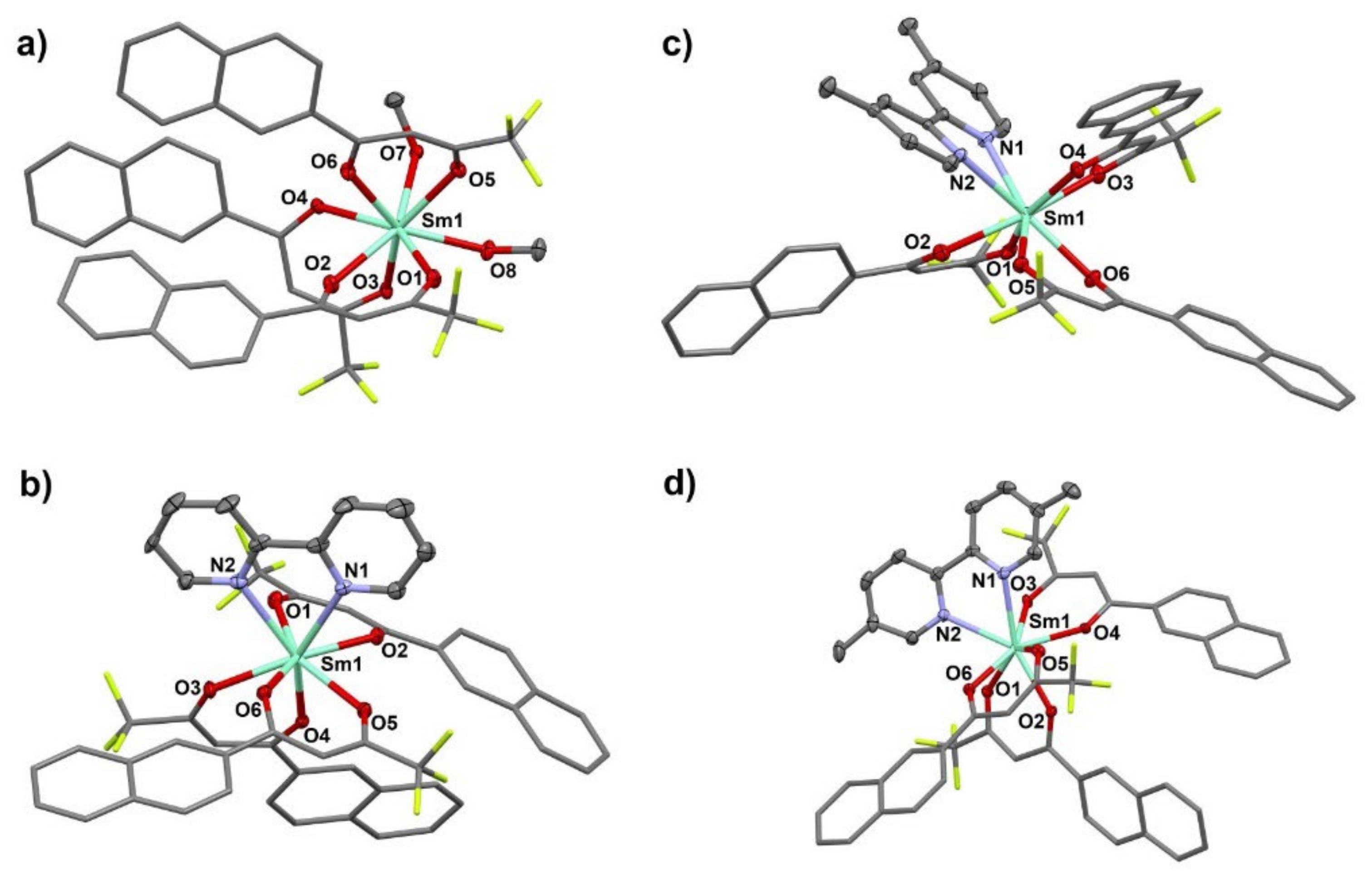
Partially labelled molecular plots of the title compounds 1–4 are presented in Figure 1, and coordination figures are depicted in Figure 2. In addition, the selected bond distances and bond angles of 1–4 are given in Table 2. Coordination number (CN) eight was observed around each Sm(III) center in the neutral monomeric complexes 1–4. All metal centers were ligated by six oxygen-donor atoms of three β-diketonate ligand anions with Sm1-O(keto) bond distances in the range of 2.3388(18) to 2.4296(17) Å, and O-Sm1-O keto-bite angles ranging from 69.86(6) to 71.68(8)°. In 1, the Sm1 center was further ligated by oxygen atoms of two terminal methanol molecules (Sm1-O7 = 2.5382(18), Sm1-O8 = 2.4487(18) Å). Coordination number 8 in compounds 2–4 was completed by two N atoms of chelating bipy or Me2-bipy ligands with Sm1-N bond distances varying from 2.562(3) to 2.597(3) Å and with N1-Sm1-N2 chelate bite angles varying from 62.25(9) to 63.12(16)°.

Figure 1.
Partially labelled molecular plots of (a) [Sm(ntfa)3(MeOH)2] (1), (b) [Sm(ntfa)3(bipy)] (2), (c) [Sm(ntfa)3(4,4′-Me2bipy)] (3), and (d) [Sm(ntfa)3(5,5′-Me2bipy)] (4). Color code: turquoise: Sm, red: oxygen, blue: nitrogen, black: carbon, and green: fluorine; H atoms omitted for clarity.

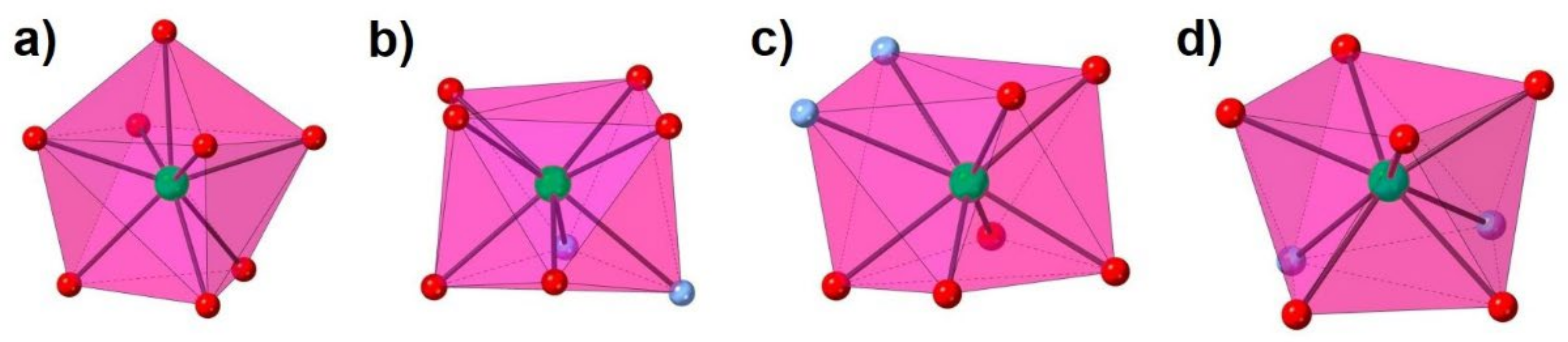
Figure 2.
Coordination figures (a–d) of 1–4 compounds, respectively. Color code: turquoise: Sm, red: oxygen, and blue: nitrogen.
Table 2.
Selected bond distances (Å) and bond angles (°) of 1–4.
The degree of distortion of the coordination polyhedra from their ideal polyhedron geometry were analyzed using the continuous shape measure theory with the SHAPE software [60,61]. Intermediate distortion from the ideal eight-vertex coordination polyhedra was observed for the LaO8 in 1 and LaO6N2 coordination polyhedron of 2–4. The lowest CShM values for compound 1 corresponded to a triangular dodecahedron (TDD-8), biaugmented trigonal prism (BTPR-8), square antiprism (SAPR-8), and biaugmented trigonal prism J50 (JBTPR-8), with values of 0.335, 1.652, 2.335, and 2.667, respectively. The following respective values were found: 1.239, 1.639, 1.262, and 2.197 in the case of 2; 1.050, 2.494, 1.498, and 3.044 for 3; and 2.258, 1.913, 0.696, and 2.453 for 4. All these values are summarized in Table 3 for the four compounds.
Table 3.
Lowest values of the calculated continuous shape measurements (CShMs) for compounds 1–4.
The packing plots of 1–4 are presented in Supplementary Figures S5–S8 in the Supplementary Materials section. The O-H groups of the two methanol molecules in 1 formed hydrogen bonds of type O-H···O (O7···O3(-x,-y,-z) = 2.713(2) Å; O7-H10···O3(-x,-y,-z) = 158(3)°; O8···O7(-x,-y,-z) = 2.740(3) Å; O8-H20···O7(-x,-y,-z) = 162(4)°) (Supplementary Figure S5). The aromatic naphthyl- (in 1–4) and pyridyl- (in 2–4) moieties were involved in numerous ring···ring and C-H/F···ring interactions, which further stabilized the packing of the mononuclear complexes (Supplementary Tables S1–S4).
3.3. Photoluminescent Properties
The UV–visible excitation spectra of solid samples of the complexes 1–4 represented in Supplementary Figure S9 show a slight redshift of the intense broad band over the 350–400 nm region, which may be taken as an indication of the complex formation. These bands located at 400, 394, 392, and 389 nm for 1–4, respectively, corresponded to π → π* electronic transition in the conjugated ligands coordinated to the central samarium(III) ion. Excitation of the solid sample at these wavelengths (λex) led to the Sm3+-centered emission transitions in the visible and NIR regions.
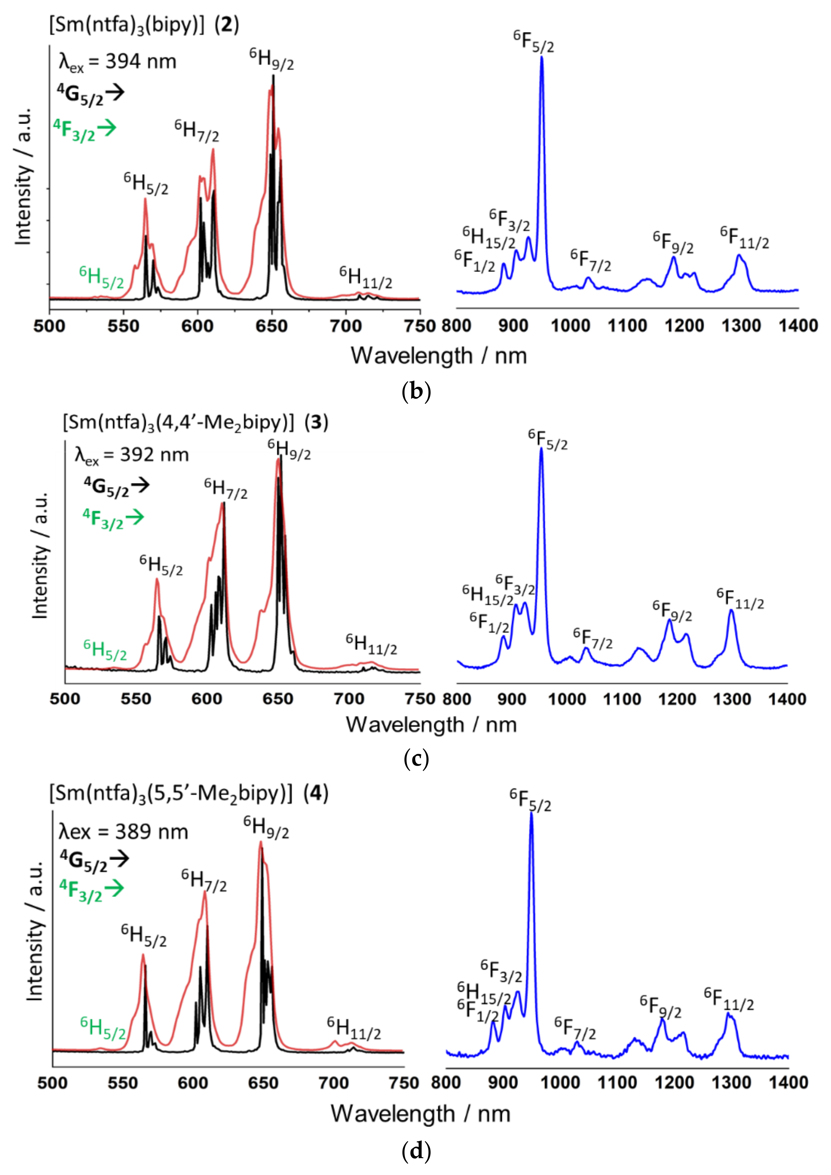
The excitation and emission spectra of the compounds were recorded at room temperature in the visible and NIR regions and at the liquid nitrogen temperature (77 K) in the visible range; these spectra are shown in Figure 3 for the four complexes. Inspection of these spectra indicated an almost similar emission trend for all compounds. The bands in the visible region were assigned to 4G5/2 → 6H5/2 at 565 ± 1 nm, 4G5/2 → 6H7/2 at 608 ± 2 nm, and 4G5/2 → 6H9/2 at 649 ± 2 nm, while a less intense band at 714 ± 3 nm corresponded to the 4G5/2→6H11/2 transition. Additionally, the very-low-intensity band located at 535 nm could be discerned as the f-f transition from an upper emissive level to the ground state: 4F3/2→6H5/2 [12,62].
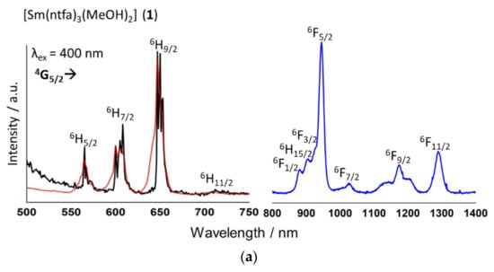
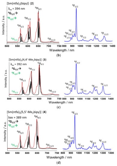
Figure 3.
(a–d) The solid-state emission spectra of compounds 1–4 in the visible (red color at room temperature and black at 77 K) and NIR (blue color at 77 K) regions.
For compound 1, the 4F3/2 → 6H5/2 transition at room temperature was not detected, and this probably could be attributed to the low-intensity residual emission from the ligand in the 450–500 nm range. For 2–4, no emission from the ligand could be perceived, indicating a rather good energy transfer efficiency from the ligand’s lowest triplet state to the resonant highest energy level of the Sm3+ ion. Moreover, the presence of a broad band from the absorption of the ligands in the excitation spectra suggested a good antenna effect [63]. The most intense band observed for the 1–4 compounds, which corresponded to the 4G5/2 → 6H9/2 transition, was a hypersensitive band (electric dipole allowed transition, ΔJ = 2) and due to its high intensity, it dominated in the final color that the compounds presented. Additionally, the 4G5/2 → 6H5/2 and 4G5/2 → 6H7/2 were magnetic dipole transitions (ΔJ = 0, 1 respectively), and the 4G5/2 → 6H9/2/4G5/2 → 6H5/2 ratio provided information about the polarizability of the Sm3+ chemical environment. Ratio values of 6.25, 4.52, 4.62, and 4.17 were determined for compounds 1–4, respectively. The similar ratio values observed in compounds 2–4 indicated that their coordination environment was not altered when the NN-donor ligand was changed. The higher ratio value detected in 1 indicated a more polarized environment around the Sm3+ ion, which obviously was attributed to the coordinated MeOH molecules [64]. The difference in the hypersensitive band intensity of 1–4 compounds was also perceived in the emission color, and could be seen by naked eye under a UV lamp and according to the CIE diagram represented in Figure 4.
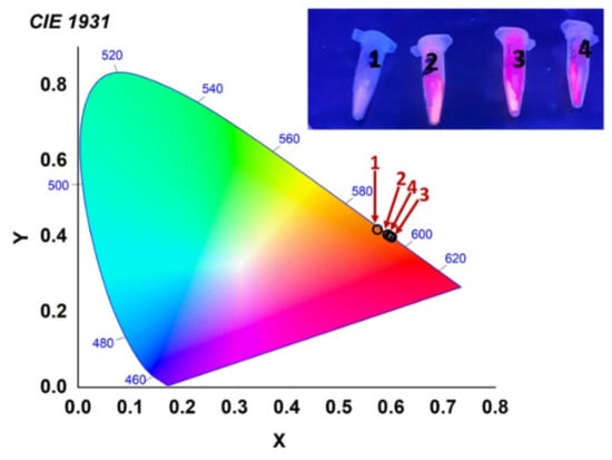
Figure 4.
CIE diagram of the emission color of compounds 1–4. Inset shows the emission of compounds under UV light.
The visible spectra of the compounds, which were also measured at the liquid nitrogen temperature (77 K), displayed the splitting of each band due to the crystal field perturbation, corresponding to the Stark sublevels of the Sm3+ Kramer ion. The 6HJ energy levels of the four complexes should split to a maximum of J + 1/2 Stark according to symmetries lower than cubic for J half-integer values. Higher asymmetry around the Sm3+ ion is related to an enhancement of the emission intensity [2,21]. Thus, based on the results obtained in the SHAPE measurements, compounds 1–4 presented symmetries lower than cubic (Oh, O, Td, Th and T). Thus, it was expected that each band corresponding to different 6HJ energy levels should split into three for 6H5/2, four for 6H7/2, five for 6H9/2, and six for 6H11/2. However, if the symmetry around the Sm3+ ion was cubic, then the splitting due to the crystal field would be two, three, three, and four for each energy level, respectively [65,66]. The magnetic-dipole-allowed transition 4G5/2 → 6H7/2 was taken as a reference. For compounds, deconvolution of the 4G5/2 → 6H7/2 band was conducted, and the best fittings for the four compounds were performed when four Gaussian functions were used (Supplementary Figure S10), corroborating that the symmetry of the coordination polyhedron was lower than cubic. Moreover, the low-intensity transition 4F3/2 → 6H5/2 seen at 535 nm at room temperature measurements was not discerned at 77 K. The 4F3/2 level, which was close in energy to the emitting 4G5/2 level, was thermally populated at room temperature [52]. Furthermore, the 4G5/2 → 6H9/2/4G5/2 → 6H5/2 intensity ratio was measured for the 77 K spectra. The obtained values were 6.29 for 1, 4.55 for 2, 4.66 for 3, and 4.24 for 4. These values were very similar to those obtained from the room temperature spectra, suggesting that there was no change in the coordination Sm3+ environment when the temperature changed.
The emission spectra of 1–4 enabled us to analyze the transition from the lowest emitting energy level to the ground state (4G5/2→6HJ) for each compound, and as can be seen in Figure 3, this emission presented five different bands that corresponded to the five expected levels for J = 5/2-13/2 centered at 565, 606, 647, 711, and 904 nm for 1; at 564, 606, 649, 712, and 905 nm for 2; at 565, 611, 651, 717, and 906 nm for 3; and at 566, 610, 649, 714, and 903 nm for 4. These transitions provided information about the crystal-field energy of the samarium ions in the complexes, and allowed us to estimate the energy between the ground (6H5/2) and first-excited (6H7/2) J states. The energy differences between these two transitions were about 1197, 1228, 1335, and 1275 cm−1 for the 1–4 complexes, respectively. The energy separation between the 6H5/2 ground state of the samarium(III) and the first-excited state 6H7/2 was evaluated as 1000 cm−1 [67], which was in good accordance with the calculated values for 1–4. From the energy separation between the first state 6H7/2 and the 6H5/2 ground state calculated previously from the emission spectra, we could evaluate the spin-orbit coupling parameter, λ, through the expression E(J) = λJ(J + 1)/2 [67]. These calculations led to λ values of 342, 351, 381, and 364 cm−1 for 1–4, respectively.
Compounds 1–4 also showed Sm3+-centered f-f emissions in the NIR region when exciting the samples at the corresponding λex of the ligands. The experimental emission bands were from the 4G5/2 emitting level to the 6FJ energy level. These bands were assigned to 4G5/2 → 6F1/2 at 882 ± 1 nm; 4G5/2 → 6F3/2 at 925 ± 2 nm (in 1, this band appeared as a shoulder); 4G5/2 → 6F5/2, which was the most intense band at 948 ± 3 nm; as well as the 4G5/2 → 6F7/2, 4G5/2 → 6F9/2, and 4G5/2 → 6F11/2 transitions at 1031 ± 5, 1180 ± 6, and 1294 ± 4 nm, respectively. In addition, the 4G5/2 → 6H15/2 transition was seen at 904 ± 1 nm [62,68,69,70]. All the bands arising from the f-f transitions of the Sm3+ ion (except for the 4F3/2 → 6H5/2 transition) came from the Sm3+ lowest emitting energy level, 4G5/2. However, the 4G5/2 was close in energy to the upper levels 4G7/2 and 4F3/2. These resulted in a fast nonradiative relaxation of the 4G7/2 and 4F3/2 to the 4G5/2. Then, further radiative relaxation to the 6FJ, in the NIR range, and to the ground state 6HJ, in the visible range took place [63].
The luminescence quantum yield (QYs) in the solid state were determined using an integrating sphere. Only the visible emission range was determined. For 1, the QY could not be calculated due to the low emission intensity that the sample presented, evidencing the effect of high-energy oscillators in the effectiveness of radiative deactivation. For 2, 3, and 4, the close QY values were evaluated as 0.23, 0.25, and 0.20%, respectively.
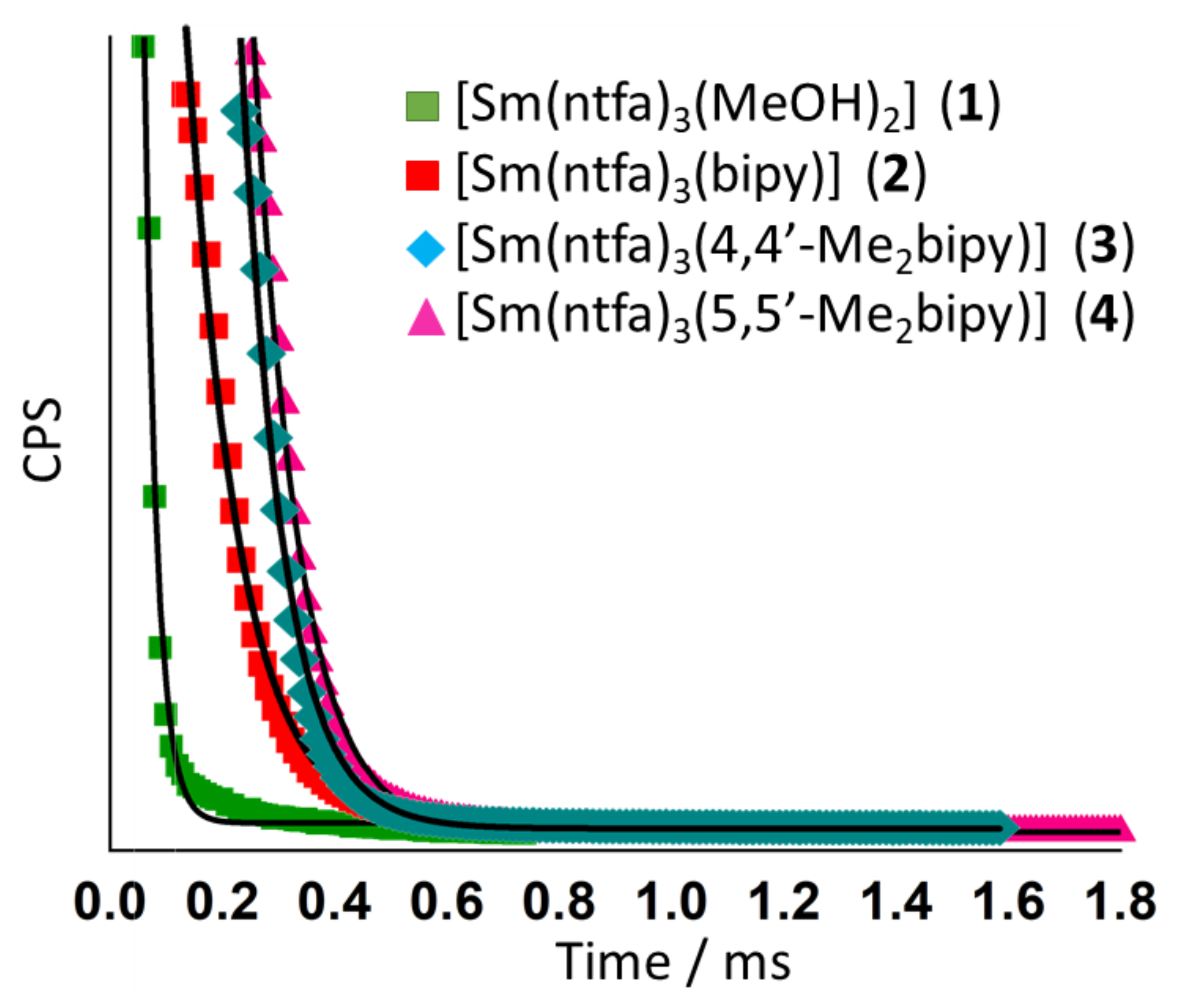
Excited-state lifetimes (τobs) of the 4G5/2 emitting state in the 1–4 complexes were collected at the maximum emission band in the visible range (4G5/2 → 6H9/2, 649 ± 2 nm), where the decay curves were fitted monoexponentially as (I) (Figure 5), according to single emitting species for all Sm3+ complexes. The measured τobs values were in the 30–74 μs range. The lowest value of τobs was obtained for the precursor compound. For compounds 2–4, τobs increased when the NN-donor ligand was used, in the order: 5,5′-Me2bipy < bipy < 4,4′-Me2bipy. The same tendency was followed for the measured QYs. Compound 3 showed more luminescence intensity, suggesting an enhancement of the electron density around the Sm3+ ion due to the electron-donating methyl groups in the 4,4′ positions of the bipyridine ligand [69]. The photoluminescent data for QYs and τobs are compiled in Table 4.

Figure 5.
Photoluminescence time decay of compounds 1–4 monitored at the maximum emission wavelength of 649 ± 2 nm (4G5/2 → 6H9/2). Solid lines represent the monoexponential fitting of the decay curves.
Table 4.
Compilation of the photoluminescent data of compounds 1–4. QY stands for the overall quantum yield, and τobs for the emission time delay.
In general, Sm3+ coordination compounds did not produce relatively high QYs because the energy gap between the lowest emitting sublevel 5G5/2 and the lower energy level 6F11/2 was 7500 cm−1. This value was low compared to the energy difference from Tb3+ or Eu3+ ions, which were 12,500 cm−1 {ΔE (5D0→7F6)} and 14,800 cm−1 {ΔE (5D4→7F6)}, respectively. The small energy gap in Sm3+ ion favored nonradiative relaxation processes that lowered the emission efficiency and luminescence lifetimes. However, samarium(III) coordination compounds are very interesting due to their capability of emission over a wide range within the electromagnetic spectrum, thus covering both the visible and NIR regions [70,71,72,73,74]. The results obtained here were similar to those for other previously published samarium(III)-β-diketonate systems [2,63,65,70]
3.4. Magnetic Properties
DC Magnetic Susceptibility Studies
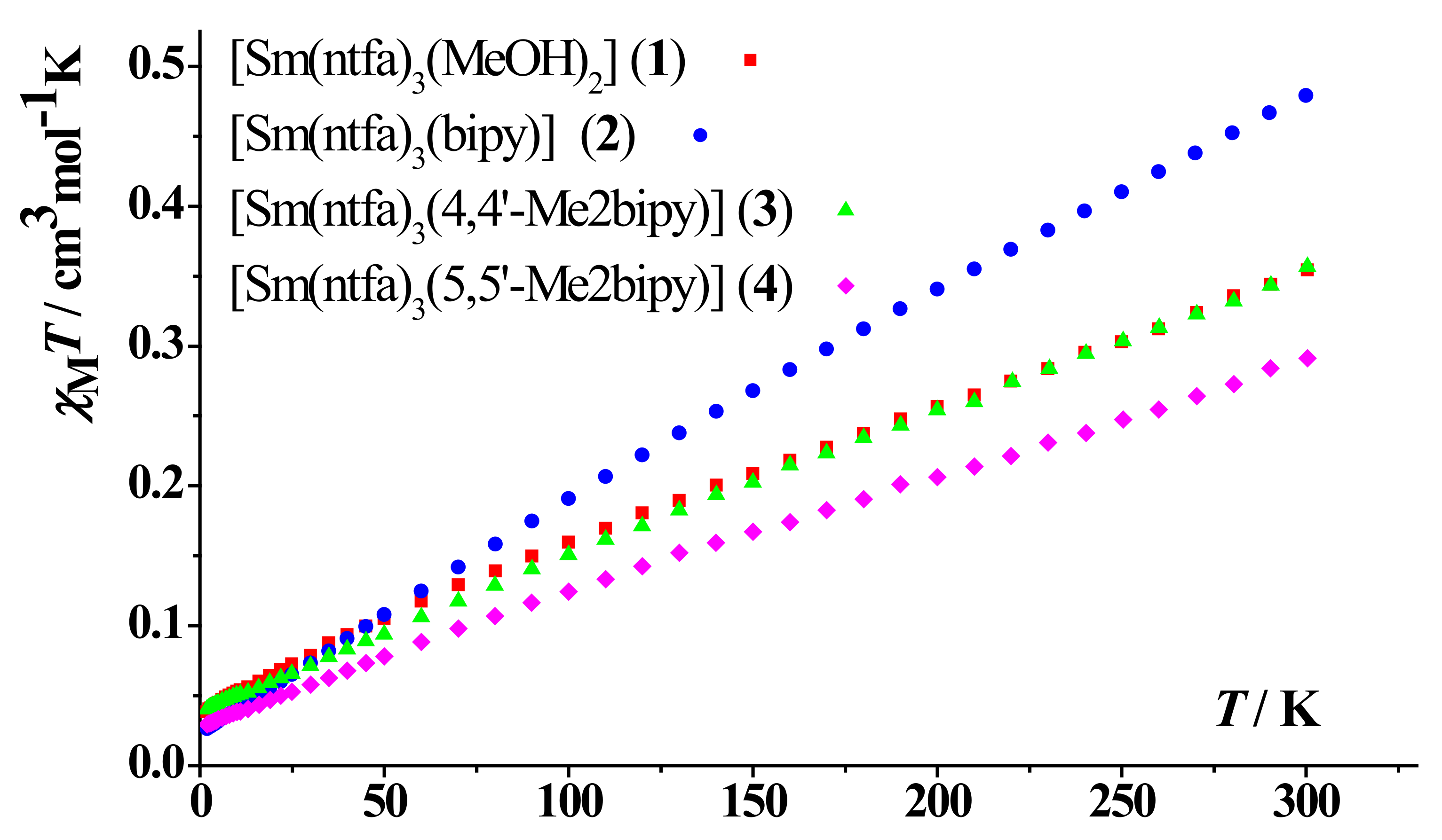
Powder samples of complexes 1–4 were measured under applied magnetic fields of 0.3 T (300–2 K). The data are plotted as χMT products versus T in Figure 6. Magnetization dependence of the applied field at 2 K for compounds were also recorded and are shown in Figure 7. The magnetic measurements of 1–4 revealed that the χMT values at 300 K were 0.35, 0.48, 0.35, and 0.29 cm3·mol−1·K, respectively, which were larger than the theoretical value for a free Sm(III) ion (0.09 cm3·mol−1·K) in the 6H5/2 ground state (gJ = 2/7) [75], but in accordance with the room temperature χMT values of previously reported Sm(III) complexes [76,77,78]. Upon cooling the samples, the χMT values decreased practically linearly, reaching values of 0.04, 0.03, 0.04, and 0.03 cm3·mol−1·K for compounds 1–4, respectively. These relatively small low temperature χMT values found in the compounds could be attributed to the significant crystal field (CF) splitting under an anisotropic coordination environment [78].
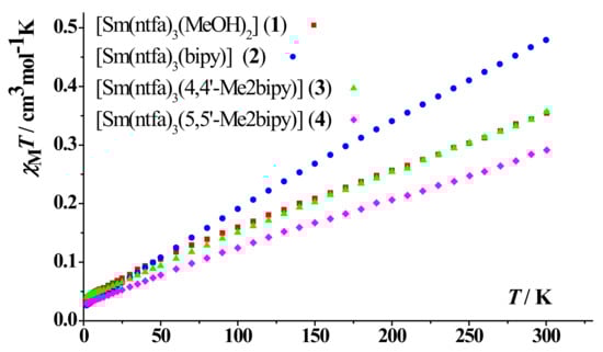
Figure 6.
χMT vs. T plots for compounds 1–4.
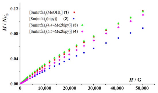
Figure 7.
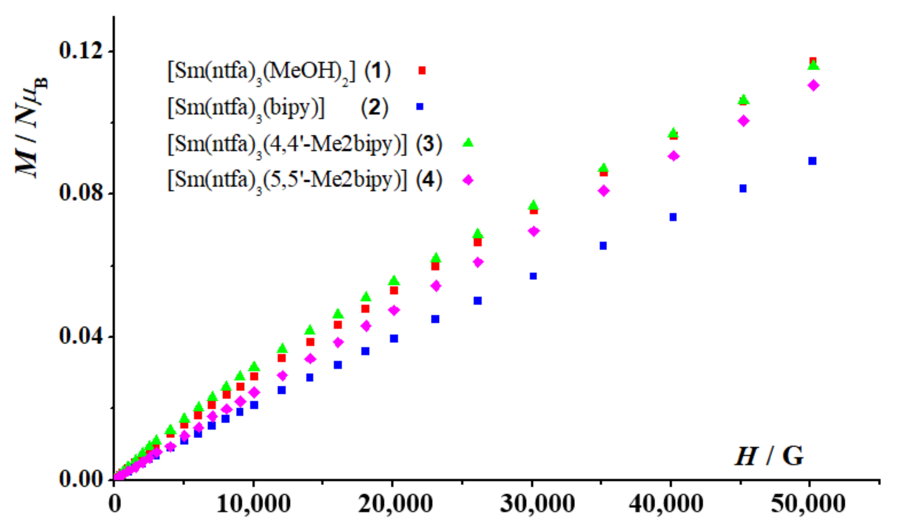
Magnetization plots measured at T = 2 K (field dependence of 1–4).
Field dependence of the magnetization on the magnetic static applied field at T = 2 K for complexes 1–4 (Figure 7) revealed no saturation in high fields, with similar values of 0.12, 0.09, 0.12, and 0.11 NµB at 5 T for 1–4, respectively. Taking into consideration the 4f5 ground configuration of the Sm(III) ion value, the saturated magnetization should have been 5/7 NµB (Msat = gJ·J·NµB; gJ = 2/7, J = 5/2).
4. Conclusions
Similar to most of the Ln3+ ions, the reaction of an aqueous or alcoholic Ln(III) salts with β-diketonate ligands (ntfa = 4,4,4-trifluoro-1-(naphthalen-2-yl)-1,3-butanedionate; btfa = 4,4,4-trifluoro-1-phenyl-1,3-butanedionate; acac = acetylacetonate) in the presence of three equivalents of NaOH affords the tris β-diketonate complexes [Ln(β-diketonate)3(Solv)2] (Solv = H2O, MeOH, EtOH) [29,30,31,32,33]. These compounds serve as precursors for the synthesis of many mononuclear polypyridyl adducts, with CNs varying between 8 in the cases of dipyridine derivatives such as phen, bipy, 4,4′-Me2bipy, and 5,5′-Me2bipy, and 9 in the case of terpy [26,27,29,30,31,32,33]. With no exception, four samarium(III) complexes [Sm(ntfa)3(MeOH)2] (1), [Sm(ntfa)3(bipy)2] (2), [Sm(ntfa)3(4,4′-Me2bipy)2] (3), and [Sm(ntfa)3(5,5′-Me2bipy)2] (4) were synthesized, and their single-crystal X-ray structures were determined. It has been reported that the use of auxiliary ligands with strong chelation properties enhanced the efficient transfer of energy in the visible and NIR regions, from the chelated pyridyl groups to the Ln3+ centers [29,30,31,32,38]. In addition, it was nearly established that combining aromatic rings and incorporation of fluorinated alkyl groups into the β-diketone skeletons significantly enhanced the emission luminescent of the resulting complexes [30,31,32,33,43,44,45,77]. Indeed, these two properties were included into the complexes 2–4, which revealed luminescence emissions in the visible and NIR regions, a property that makes these compounds potential candidates in the light-devices field [12,62,73]. The magnetic investigation of the complexes 1–4 did not reveal any unusual magnetic behavior.
Supplementary Materials
The following supporting information can be downloaded at: https://www.mdpi.com/article/10.3390/magnetochemistry8070072/s1. CCDC 1964549–CCDC 1964553 contain the crystallographic data in CIF format for 1–4, respectively. More Supplementary Materials corresponding to PXRD patterns are presented as Supplementary Figures S1–S4 and packing plots as Supplementary Figures S5–S8 for the four title complexes, respectively. Supplementary Tables S1–S4 contain C-H/F···ring and ring···ring noncovalent interactions for the four compounds, respectively. Absorption spectra (visible and UV region) of solid samples of 1–4 are represented in Supplementary Figure S9, and the deconvolution of the magnetic-dipole-allowed band 4G5/2 → 6H7/2 are shown in Supplementary Figure S10.
Author Contributions
Conceptualization, F.A.M., R.V. and S.S.M.; methodology, F.A.M., R.V., F.B., Á.T. and R.C.F.; software, F.A.M., R.C.F., F.B., S.S., Á.T. and R.V.; validation, F.A.M., R.V., R.C.F. and S.S.M.; investigation, F.B., Á.T., R.C.F. and S.S.; resources, F.A.M., R.C.F. and R.V.; data curation, F.A.M., F.B., R.C.F., S.S.; Á.T., R.V. and S.S.M.; writing and editing manuscript, F.A.M., S.S.M., Á.T. and R.V.; visualization, F.A.M., Á.T., R.V. and S.S.; administration of project and supervision, S.S.M., F.A.M. and R.V.; funding management, R.V.; The authors agree with the manuscript version presented. All authors have read and agreed to the published version of the manuscript.
Funding
Supported by TU Graz Open Access Publishing Fund. R.V. is thankful to MINECO for financial assistance (Project PGC2018-094031-B-I00).
Institutional Review Board Statement
Not applicable.
Informed Consent Statement
Not applicable.
Data Availability Statement
Data is contained within the article or Supplementary Materials.
Acknowledgments
R.V., S.S. and A.T. acknowledge the financial support from the Ministerio de Ciencia, Innovación y Universidades (Spain) under Project PGC2018-094031-B-100.
Conflicts of Interest
The authors declare no conflict of interest.
References
- Wang, H.-S.; Zhao, B.; Zhai, B.; Shi, W.; Cheng, P.; Liao, D.-Z.; Yan, D.-Z. Syntheses, structures, and photoluminescence of one-dimensional lanthanide coordination polymers with 2,4,6-pyridinetricarboxylic acid. Cryst. Growth Des. 2007, 7, 1851. [Google Scholar] [CrossRef]
- Melo, L.L.L.S.; Castro, G.P., Jr.; Gonçalves, S.M.C. Substantial intensification of the quantum Yield of samarium(III) complexes by mixing ligands: Microwave-assisted synthesis and luminescence Properties. Inorg. Chem. 2019, 58, 3265–3270. [Google Scholar] [CrossRef]
- Cui, Y.; Yue, Y.; Qian, G.; Chen, B. Luminescent functional Metal–Organic Frameworks. Chem. Rev. 2012, 112, 1126–1162. [Google Scholar] [CrossRef] [PubMed]
- Liu, W.; Li, Z.; Wang, N.; Li, X.; Wei, Z.; Yue, S.; Liu, Y. A new family of 3D heterometallic 3d–4f organodisulfonate complexes based on the linkages of 2D [Ln(nds)(H2O)]+ layers and [Cu(ina)2]− chains. Cryst. Eng. Comm. 2011, 13, 138–144. [Google Scholar] [CrossRef]
- Kelly, R.P.; Bell, T.D.M.; Cox, R.P.; Daniels, D.P.; Deacon, G.B.; Jaroschik, F.; Junk, P.C.; Le Goff, X.F.; Lemercier, G.; Martinez, A. Divalent tetra- penta-phenylcyclopentadienyl europium and samarium sandwich and half-sandwich complexes: Synthesis, characterization, and remarkable luminescence properties. Organometallics 2015, 34, 5624–5636. [Google Scholar] [CrossRef]
- Bo, Q.-B.; Sun, G.-X.; Geng, D.-L. Novel Three-Dimensional Pillared-Layer Ln(III)−Cu(I) Coordination polymers featuring spindle-shaped heterometallic building units. Inorg. Chem. 2010, 49, 561–571. [Google Scholar] [CrossRef]
- Feng, X.; Wang, L.-Y.; Zhao, J.-S.; Wang, J.-G.; Weng, N.S.; Liu, B.; Shi, X.-G. Series of anion-directed lanthanide-rigid-flexible frameworks: Syntheses, structures, luminescence, and magnetic properties. Cryst. Eng. Comm. 2010, 12, 774–783. [Google Scholar] [CrossRef]
- Chandler, B.D.; Cramb, D.T.; Shimizu, G.K.H. Microporous metal−organic frameworks formed in a stepwise manner from luminescent building blocks. J. Am. Chem. Soc. 2006, 128, 10403–10412. [Google Scholar] [CrossRef]
- Wu, Y.; Yang, J.; Lin, Y.; Xu, J. Synthesis of samarium-based metal organic compound nanoparticles with polychromatic-Photoluminescence for bio-tissue fluorescence Imaging. Molecules 2019, 24, 3657. [Google Scholar] [CrossRef] [Green Version]
- Bui, A.T.; Grichine, A.; Brassele, S.; Duperray, A.; Andraud, C.; Maury, O. Unexpected efficiency of a luminescent samarium(III) complex for combined visible and near-infrared biphotonic microscopy. Chem. Eur. J. 2015, 21, 17757–17761. [Google Scholar] [CrossRef]
- Knope, K.E.; de Lill, D.T.; Rowland, C.E.; Cantos, P.M.; de Bettencourt-Dias, A.; Cahill, C.L. Uranyl sensitization of samarium(III) luminescence in a two-dimensional coordination polymer. Inorg. Chem. 2012, 51, 201–206. [Google Scholar] [CrossRef] [PubMed]
- Lunstroot, K.; Nockemann, P.; Van Hecke, K.; Van Meervelt, L.; Gorller-Walrand, C.; Binnemans, K.; Driesen, K. Visible and near-infrared emission by samarium(III)-containing ionic liquid mixtures. Inorg. Chem. 2009, 48, 3018–3026. [Google Scholar] [CrossRef]
- Li, Z.; Yao, S.; Xu, J.; Wu, Y.; Li, C.; He, Z. Endoscopic near-infrared dental imaging with indocyanine green: A pilot study. Ann. N. Y. Acad. Sci. 2018, 1421, 88–96. [Google Scholar] [CrossRef] [PubMed]
- Schaafsma, B.E.; Mieog, J.S.D.; Hutteman, M.; Van Der Vorst, J.R.; Kuppen, P.J.K.; Löwik, C.W.G.M.; Frangioni, J.V.; Van De Velde, C.J.H.; Vahrmeijer, A.L. The clinical use of indocyanine green as a near-infrared fluorescent contrast agent for image-guided oncologic surgery. J. Surg. Oncol. 2011, 104, 323–332. [Google Scholar] [CrossRef] [PubMed] [Green Version]
- Troyan, S.L.; Kianzad, V.; Gibbs-Strauss, S.L.; Gioux, S.; Matsui, A.; Oketokoun, R.; Ngo, L.; Khamene, A.; Azar, F.; Frangioni, J.V. The FLARE intraoperative near-infrared fluorescence imaging system: A first-in-human clinical trial in breast cancer sentinel lymph node mapping. Ann. Surg. Oncol. 2009, 16, 2943–2952. [Google Scholar] [CrossRef] [Green Version]
- Monteiro, J.H.S.K. Recent advances in luminescence imaging of biological systems using lanthanide(III) luminescent complexes. Molecules 2020, 25, 2089. [Google Scholar] [CrossRef] [PubMed]
- Topa, M.; Ortyl, J.; Chachaj-Brekiesz, A.; Kamińska-Borek, I.; Maciej, P.; Popielarz, R. Applicability of samarium(III) complexes for the role of luminescent molecular sensors for monitoring progress of photopolymerization processes and control of the thickness of polymer coatings. Spectrochim. Acta Part A Mol. Biomol. Spectrosc. 2018, 199, 430–440. [Google Scholar] [CrossRef]
- Chen, J.; Xie, Z.; Meng, L.; Hu, Z.; Kuang, X.; Xie, Y.; Lu, C.-Z. Luminescence tunable europium and samarium complexes: Reversible On/Off switching and white-light emission. Inorg. Chem. 2020, 59, 6963–6977. [Google Scholar] [CrossRef]
- Philip, P.; Jose, T.; Jose, A.; Cherian, S.K. Studies on the structural and optical properties of samarium β-diketonate complex incorporated electrospun poly(methylmethacrylate) nanofibres with different architectures. Luminescence 2021, 36, 1032–1047. [Google Scholar] [CrossRef]
- Mara, D.; Artizzu, F.; Laforce, B.; Vincze, L.; Van Hecke, K.; Van Deun, R.; Kaczmarek, A.M. Novel tetrakis lanthanide β-diketonate complexes: Structural study, luminescence properties and temperature sensing. J. Lumin. 2019, 213, 343–355. [Google Scholar] [CrossRef]
- Bhat, S.A.; Iftikhar, K. Samarium complexes with fluorinated β-diketone and heterocyclic Lewis bases as UV light converters. Dye. Pigment. 2020, 179, 108383. [Google Scholar] [CrossRef]
- Dar, W.A.; Ahmed, Z.; Iftikhar, K. Cool white light emission from the yellow and blue emission bands of the Dy(III) complex under UV excitation. J. Photoch. Photobiol. A 2018, 356, 502–511. [Google Scholar] [CrossRef]
- Yao, X.; An, G.; Li, Y.; Yan, P.; Li, W.; Li, G. Effect of nuclearity and symmetry on the single-molecule magnets behaviour of seven-coordinated β-diketonate Dy(III) complexes. J. Solid State Chem. 2019, 274, 295–302. [Google Scholar] [CrossRef]
- Li, X.; Li, T.; Tian, L.; Liu, Z.Y.; Wang, X.G. Experimental and theoretical interpretation of the magnetic behaviour of two Dy(III) single-ion magnets constructed through β-diketonate ligands with different substituent groups (–Cl/OCH3). RSC Adv. 2015, 5, 74864–74873. [Google Scholar] [CrossRef]
- Liu, C.-M.; Zhang, D.Q.; Zhu, D.-B. Field-induced single-ion magnets based on enantiopure chiral β-diketonate ligands. Inorg. Chem. 2013, 52, 8933–8940. [Google Scholar] [CrossRef]
- Ansari, A.A.; Ganaie, A.B.; Iftikhar, K. Synthesis and 4f-4f absorption studies of tris(acetylacetonato) praseodymium(III) and holmium(III) complexes with imidazole and pyrazole in non-aqueous solvents. Structure elucidation by sparkle/PM7. J. Mol. Struct. 2019, 1198, 126826. [Google Scholar] [CrossRef]
- Ansari, A.A.; Ilmi, R.; Iftikhar, K. Hypersensitivity in the 4f–4f absorption spectra of tris(acetylacetonato)neodymium(III) complexes with imidazole and pyrazole in non-aqueous solutions. Effect of environment on hypersensitive transitions. J. Lumin. 2012, 132, 51–60. [Google Scholar] [CrossRef]
- Chen, G.-J.; Zhou, Y.; Jin, G.-X.; Dong, Y.-B. [Dy(acac)3(dppn)]∙C2H5OH: Construction of a single-ion magnet based on the square-antiprism dysprosium(III) ion. Dalton Trans. 2014, 43, 16659–16665. [Google Scholar] [CrossRef]
- Mautner, F.A.; Bierbaumer, F.; Gyurkac, M.; Fischer, R.C.; Torvisco, A.; Massoud, S.S.; Vicente, R. Synthesis and characterization of lanthanum(III) complexes containing 4,4,4-trifluoro-1-(2-naphthalen-yl)-butane-1,3-dionate. Polyhedron 2020, 179, 114384. [Google Scholar] [CrossRef]
- Mautner, F.A.; Bierbaumer, F.; Fischer, R.C.; Torvisco, A.; Vicente, R.; Font-Bardía, M.; Tubau, À.; Speed, S.; Massoud, S.S. Diverse coordination numbers and geometries in pyridyl adducts of lanthanide(III) complexes based on β-diketonate. Inorganics 2021, 9, 74. [Google Scholar] [CrossRef]
- Mautner, F.A.; Bierbaumer, F.; Fischer, R.C.; Vicente, R.; Tubau, À.; Ferran, A.; Massoud, S.S. Structural characterization, magnetic and luminescent properties of praseodymium(III)-4,4,4-trifluoro-1-(2-naphthyl)butane-1,3-dionato(1-) complexes. Crystals 2021, 11, 179. [Google Scholar] [CrossRef]
- Vicente, R.; Tubau, À.; Speed, S.; Mautner, F.A.; Bierbaumer, F.; Fischer, R.C.; Massoud, S.S. Slow magnetic relaxation and luminescence properties in neodymium(III)-4,4,4-trifluoro-1-(2-naphthyl)butane-1,3-dionato complexes incorporating bipyridyl ligands. New J. Chem. 2021, 45, 14713–14723. [Google Scholar] [CrossRef]
- Mautner, F.A.; Bierbaumer, F.; Vicente, R.; Speed, S.; Tubau, A.; Font-Bardía, M.; Fischer, R.C.; Massoud, S.S. The luminescence and magnetic properties of 8-coordinated holmium(III)-trifluoro-phenyl- and naphthalenyl-β-diketonate complexes. Molecules 2022, 27, 1129. [Google Scholar] [CrossRef] [PubMed]
- Casanovas, B.; Speed, S.; Maury, O.; Font-Bardía, M.; Vicente, R. Homodinuclear lanthanide 9-anthracenecarboxylate complexes: Field induced SMM and NIR-luminescence. Polyhedron 2019, 169, 187–194. [Google Scholar] [CrossRef]
- Ansari, A.A.; Hussain, H.A.; Iftikhar, K. Optical absorption spectroscopic studies on holmium(III) complexes with β-diketone and heterocyclic amines. The environment effect on 4f–4f hypersensitive transitions. Spectrochim. Acta Part A 2007, 68, 1305–1312. [Google Scholar] [CrossRef]
- Ansari, A.A.; Ahmed, Z.; Iftikhar, K. Nuclear magnetic resonance and optical absorption spectroscopic studies on paramagnetic praseodymium(III) complexes with β-diketone and heterocyclic amines. Spectrochim. Acta Part A 2007, 68, 176–183. [Google Scholar] [CrossRef]
- Chauhan, A.; Langyan, R. Photosensitization in highly luminescent nonmacrocyclic samarium(III) complexes for application in light-emitting systems. J. Photochem. Photobiol. A Chem. 2022, 424, 113627. [Google Scholar] [CrossRef]
- Bünzli, J.-C.G. On the design of highly luminescent lanthanide complexes. Coord. Chem. Rev. 2015, 293, 19–47. [Google Scholar] [CrossRef]
- Jia, J.-H.; Li, Q.-W.; Chen, Y.-C.; Liu, J.-L.; Tong, M.-L. Luminescent single-molecule magnets based on lanthanides: Design strategies, recent advances and magneto-luminescent studies. Coord. Chem. Rev. 2019, 378, 365–381. [Google Scholar] [CrossRef]
- Armelao, L.; Quici, S.; Barigelletti, F.; Accorsi, G.; Bottaro, G.; Cavazzini, M.; Tondello, E. Design of luminescent lanthanide complexes: From molecules to highly efficient photo-emitting materials. Coord. Chem. Rev. 2010, 254, 487–505. [Google Scholar] [CrossRef]
- Quici, S.; Cavazzini, M.; Marzanni, G.; Accorsi, G.; Armaroli, N.; Ventura, B.; Barigelletti, F. Visible and near-infrared intense luminescence from water-soluble lanthanide [Tb(III), Eu(III), Sm(III), Dy(III), Pr(III), Ho(III), Yb(III), Nd(III), Er(III)] complexes. Inorg. Chem. 2005, 44, 529–537. [Google Scholar] [CrossRef] [PubMed]
- Bünzli, J.-C.G.; Piguet, C. Taking advantage of luminescent lanthanide ions. Chem. Soc. Rev. 2005, 34, 1048–1077. [Google Scholar] [CrossRef] [PubMed]
- Tu, H.-R.; Sun, W.-B.; Li, H.-F.; Chen, P.; Tian, Y.-M.; Zhang, W.-Y.; Zhang, Y.-Q.; Yan, P.-F. Complementation and joint contribution of appropriate intramolecular coupling and local ion symmetry to improve magnetic relaxation in a series of dinuclear Dy2 single-molecule magnets. Inorg. Chem. Front. 2017, 4, 499–508. [Google Scholar] [CrossRef]
- Dasari, S.; Singh, S.; Sivakumar, S.; Patra, A.K. Dual-Sensitized luminescent europium(ΙΙΙ) and terbium(ΙΙΙ) complexes as bioimaging and light-responsive therapeutic agents. Chem.-Eur. J. 2016, 22, 17387–17396. [Google Scholar] [CrossRef] [PubMed]
- Bruno, S.M.; Ananias, D.; Paz, F.A.A.; Pillinger, M.; Valente, A.A.; Carlos, L.D.; Goncalves, I.S. Crystal structure and temperature-dependent luminescence of a heterotetranuclear sodium–europium (III) β-diketonate complex. Dalton Trans. 2015, 44, 488–492. [Google Scholar] [CrossRef]
- Zhang, S.; Ke, H.; Shi, Q.; Zhang, J.; Yang, Q.; Wei, Q.; Xie, G.; Wang, W.; Yang, D.; Chen, S. Dysprosium(iii) complexes with a square-antiprism configuration featuring mononuclear single-molecule magnetic behaviours based on different β-diketonate ligands and auxiliary ligands. Dalton Trans. 2016, 45, 5310–5320. [Google Scholar] [CrossRef]
- Trieu, T.-N.; Dinh, T.-H.; Nguyen, H.-H.; Abram, U.; Nguyen, M.-H. Novel lanthanide (III) ternary complexes with naphthoyltrifluoroacetone: A synthetic and spectroscopic study. Z. Anorg. Allg. Chem. 2015, 641, 1934–1940. [Google Scholar] [CrossRef]
- Hasegawa, Y.; Nakagawa, Y.; Kawai, T. Recent progress of luminescent metal complexes with photochromic units. Coord. Chem. Rev. 2010, 254, 2643–2651. [Google Scholar] [CrossRef]
- Maggini, I.; Traboulsi, H.; Yoosaf, K.; Mohanraj, J.; Wouters, J.; Pietraszkiewicz, O.; Pietraszkiewicz, M.; Armaroli, N.; Bonifazi, D. Electrostatically-driven assembly of MWCNTs with a europium complex. Chem. Commun. 2011, 47, 1625–1627. [Google Scholar] [CrossRef]
- Marin, R.; Brunet, G.; Murugesu, M. Shining new light on multifunctional lanthanide single-molecule magnets. Angew. Chem. 2019, 60, 1728–1746. [Google Scholar] [CrossRef]
- Vancaeyzeele, C.; Ornatsky, O.; Baranov, V.; Shen, L.; Abdelrahman, A.; Mitchell, A.; Winnik, M.A. Lanthanide-containing polymer nanoparticles for biological tagging applications: Nonspecific endocytosis and cell adhesion. J. Am. Chem. Soc. 2007, 129, 13653–13660. [Google Scholar] [CrossRef] [PubMed]
- Degen, T.; Sadki, M.; Bron, E.; König, U.; Nenert, G. The HighScore suite. Powder Diffr. 2014, 29, S13–S18. [Google Scholar] [CrossRef] [Green Version]
- Bain, G.A.; Berry, J.F. Diamagnetic corrections and Pascal’s constants. J. Chem. Educ. 2008, 85, 532–536. [Google Scholar] [CrossRef]
- Bruker. APEX, SAINT v. 8.37A; Bruker AXS Inc.: Madison, WI, USA, 2015. [Google Scholar]
- Sheldrick, G.M. SADABS v. 2; University of Goettingen: Goettingen, Germany, 2001. [Google Scholar]
- Sheldrick, G.M. Crystal structure refinement with SHELXL. Acta Crystallogr. C Struct. Chem. 2015, 71, 3–8. [Google Scholar] [CrossRef]
- Sheldrick, G.M. A Short history of SHELX. Acta Crystallogr. A 2008, 64, 112–122. [Google Scholar] [CrossRef] [Green Version]
- Macrae, C.F.; Edington, P.R.; McCabe, P.; Pidcock, E.; Shields, G.P.; Taylor, R.; Towler, T.; van de Streek, J.J. Mercury: Visualization and analysis of crystal structures. Appl. Cryst. 2006, 39, 453–457. [Google Scholar] [CrossRef] [Green Version]
- Spek, A.L. Platon, a Multipurpose Crystallographic Tool; Utrecht University: Utrecht, The Netherlands, 1999. [Google Scholar]
- Alvarez, S.; Alemany, P.; Casanova, D.; Cirera, J.; Llunell, M.; Avnir, D. Shape maps and polyhedral interconversion paths in transition metal chemistry. Coord. Chem. Rev. 2005, 249, 1693–1708. [Google Scholar] [CrossRef]
- Cirera, J.; Alvarez, S. Stereospinomers of pentacoordinate iron porphyrin complexes: The case of the [Fe(porphyrinato)(CN)]− anions. Dalton. Trans. 2013, 42, 7002–7008. [Google Scholar] [CrossRef]
- Foucault-Collet, A.; Shade, C.M.; Nazarenko, I.; Petoud, S.; Eliseeva, S.V. Polynuclear Sm(III) polyamidoamine-based dendrimer: A single probe for combined visible and near-infrared live-cell imaging. Angew. Chem. Int. Ed. Engl. 2014, 53, 2927–2930. [Google Scholar] [CrossRef]
- Bolton, J. New NIR emission from Sm3+ in Yb3+-Sm3+ co-doped tellurite glass. J. Lumin. 2021, 231, 117717. [Google Scholar] [CrossRef]
- Biju, S.; Eom, Y.K.; Bünzli, J.C.G.; Kim, H.K. A new tetrakis β-diketone ligand for NIR emitting Ln III ions: Luminescent doped PMMA films and flexible resins for advanced photonic applications. J. Mater. Chem. C 2013, 1, 6935–6944. [Google Scholar] [CrossRef] [Green Version]
- Brito, H.F.; Malta, O.L.; Felinto, M.C.F.C.; Teotonio, E.E.S.; Menezes, J.F.S.; Silva, C.F.B.; Tomiyama, C.S.; Carvalho, C.A.A. Luminescence investigation of the Sm(III)-β-diketonates with sulfoxides, phosphine oxides and amides ligands. J. Alloy. Compd. 2002, 344, 293–297. [Google Scholar] [CrossRef] [Green Version]
- De Bettencourt-Dias, A. (Ed.) Luminescence of Lanthanide Ions in Coordination Compounds and Nanomaterials; John Wiley & Sons Ltd.: Hoboken, NJ, USA, 2014. [Google Scholar]
- Kahn, O. Molecular Magnetism; VCH Publishers: Hoboken, NJ, USA, 1993. [Google Scholar]
- Chi, Y.-X.; Niu, S.-Y.; Jin, J. Syntheses, structures and photophysical properties of a series of Zn–Ln coordination polymers (Ln = Nd, Pr, Sm, Eu, Tb, Dy). Inorg. Chim. Acta 2009, 362, 3821–3828. [Google Scholar] [CrossRef]
- Yuan, B.; Wang, F.; Tao, J.; Li, M.; Yang, X. Self-assembly of one visible and NIR luminescent Sm(III) coordination polymer with flexible Schiff base ligand. Inorg. Chim. Acta 2019, 490, 24–28. [Google Scholar] [CrossRef]
- Sun, L.-N.; Yu, J.-B.; Zhang, H.-J.; Meng, Q.-G.; Ma, E.; Peng, C.-Y.; Yang, K.-Y. Near-infrared luminescent mesoporous materials covalently bonded with ternary lanthanide [Er(III), Nd(III), Yb(III), Sm(III), Pr(III)] complexes. Microporous Mesoporous Mater. 2007, 98, 156–165. [Google Scholar] [CrossRef]
- Chauhan, A.; Langyan, R. Preparation, characterization and luminescence behaviour of some samarium complexes. Rare Met. 2021, 40, 2618–2626. [Google Scholar] [CrossRef]
- Gonçalves e Silva, F.R.; Malta, O.L.; Reinhard, C.; Güdel, H.-U.; Piguet, C.; Jacques, E.; Moser, J.E.; Jean-Claude, G.; Bünzli, J.-C.G. Visible and near-infrared luminescence of lanthanide-containing dimetallic triple-stranded helicates: Energy transfer mechanisms in the SmIII and YbIII molecular edifices. J. Phys. Chem. A 2002, 106, 1670–1677. [Google Scholar] [CrossRef] [Green Version]
- Bassett, A.P.; Magennis, S.W.; Glover, P.B.; Lewis, D.J.; Spencer, N.; Parsons, S.; Williams, R.M.; De Cola, L.; Pikramenou, Z. Highly luminescent, triple- and quadruple-stranded, dinuclear Eu, Nd, and Sm(III) lanthanide complexes based on bis-diketonate ligands. J. Am. Chem. Soc. 2004, 126, 9413–9424. [Google Scholar] [CrossRef]
- Wang, S.; Xu, J.; Wang, J.; Wang, K.-Y.; Dang, S.; Song, S.; Liu, D.; Cheng, W. Luminescence of samarium(III) bis-dithiocarbamate frameworks: Codoped lanthanide emitters that cover visible and near-infrared domains. J. Mater. Chem. C 2017, 5, 6620–6628. [Google Scholar] [CrossRef]
- Atwood, D.A. (Ed.) The Rare Earth Elements: Fundamentals and Applications; John Wiley & Sons Ltd.: Hoboken, NJ, USA, 2005. [Google Scholar]
- Wu, A.Q.; Zheng, F.-K.; Liu, X.; Guo, G.-C.; Cai, L.-Z.; Dong, Z.-C.; Takano, Y.; Huang, J.-S. A novel bi-layered samarium complex with an unprecedented coordination mode of orotic acid [Sm2(HL)2(ox)(H2O)2]n·2.5nH2O (H3L = orotic acid, ox2− = oxalate2−): Synthesis, crystal structure and physical properties. Inorg. Chem. Commun. 2006, 9, 347–350. [Google Scholar] [CrossRef]
- Andruh, M.; Bakalbassis, E.; Kahn, O.; Trombe, J.C.; Porcher, P. Structure, spectroscopic and magnetic properties of rare earth metal (III) derivatives with the 2-formyl-4-methyl-6-(N-(2-pyridylethyl)(formimidoyl) phenol ligand. Inorg. Chem. 1993, 32, 1616–1622. [Google Scholar] [CrossRef]
- Pan, Y.-Z.; Hua, Q.-Y.; Lin, L.-S.; Qiu, Y.-B.; Liu, J.-L.; Zhou, A.-J.; Lin, W.-Q.; Leng, J.-D. A slowly magnetic relaxing SmIII monomer with a D5h equatorial compressed ligand field. Inorg. Chem. Front. 2020, 7, 2335–2342. [Google Scholar] [CrossRef]


Publisher’s Note: MDPI stays neutral with regard to jurisdictional claims in published maps and institutional affiliations. |
© 2022 by the authors. Licensee MDPI, Basel, Switzerland. This article is an open access article distributed under the terms and conditions of the Creative Commons Attribution (CC BY) license (https://creativecommons.org/licenses/by/4.0/).